Embed Size (px)
Citation preview
TitlePrevascularization-free Primary Subcutaneous Transplantationof Xenogeneic Islets Co-encapsulated with Hepatocyte GrowthFactor( Dissertation_全文 )
Author(s) Yang, Sin-Yu
Citation 京都大学
Issue Date 2021-05-24
URL https://doi.org/10.14989/doctor.k23368
Right
Type Thesis or Dissertation
Textversion ETD
Kyoto University
Dow
nloadedfrom
https://journals.lww.com
/transplantationdirectbyBhD
Mf5ePH
Kav1zEoum1tQ
fN4a+kJLhEZgbsIH
o4XMi0hC
ywCX1AW
nYQp/IlQ
rHD39sBibylnU
KvTC5D
DgTR
F3qR28G
d96V1M0InuR
S8DsyytJLjdXG
LL5Q==
on10/26/2020
Downloadedfromhttps://journals.lww.com/transplantationdirectbyBhDMf5ePHKav1zEoum1tQfN4a+kJLhEZgbsIHo4XMi0hCywCX1AWnYQp/IlQrHD39sBibylnUKvTC5DDgTRF3qR28Gd96V1M0InuRS8DsyytJLjdXGLL5Q==on10/26/2020
Transplantation DIRECT 2020 www.transplantationdirect.com 1
ISSN: 2373-8731
DOI: 10.1097/TXD.0000000000001078
Received 21 February 2020. Revision received 31 August 2020.Accepted 18 September 2020.1 Laboratory of Organ and Tissue Reconstruction, Institute for Frontier Life and Medical Sciences, Kyoto University, Kyoto, Japan.2 School of Dental Technology, College of Oral Medicine, Taipei Medical University, Taipei, Taiwan.The authors declare no conflicts of interest.S.-Y.Y. participated in the writing of the article, the performance of the research, and data analysis; K.-C.Y. participated in research design; and S.S. participated in research design and the writing of the article.Supplemental digital content (SDC) is available for this article. Direct URL citations appear in the printed text, and links to the digital files are provided in the HTML text of this article on the journal’s Web site (www.transplantationdirect.com).This study was supported by Japan Agency for Medical Research and Development (AMED; JP18lm0203006), JSPS KAKENHI (JP18K12057), and
Prevascularization-free Primary Subcutaneous Transplantation of Xenogeneic Islets Coencapsulated With Hepatocyte Growth FactorSin-Yu Yang, MS,1 Kai-Chiang Yang, PhD,1,2 and Shoichiro Sumi, MD, PhD1
Islet transplantation is a promising procedure for type 1 dia-betes (T1D) therapy, but plenty of drawbacks such as the
instant blood-mediated inflammatory reaction, which results in the death of transplanted islets immediately and thrombo-sis postinfusion, limit its application.1 Subcutaneous (SC) site draws a great attention as an extrahepatic islet transplanta-tion site, due to the easy accessibility, possible performance
by minimally invasive surgical procedure, and the conveni-ence in implant observation and retrieval.2 However, SC tis-sue has poor blood supply and the transplanted islets may be subjected to, in addition to mechanical stress, hypoxic injury. Many research groups including ours, have been using basic fibroblast growth factor (bFGF) to induce prevasculariza-tion to improve the engraftment and function of islet grafts in SC transplantation.3 In our recent experiment of rat to
Pancreas and Islet Transplantation
Background. Subcutaneous pouch is a potential site for islet transplantation. However, insufficient oxygen supply remains challenging. Pretreatment of neovascularization using basic fibroblast growth factor can solve this, but it needs 2× operations. We developed a device that contains rat islets in chitosan gel packed in a bag made of highly biocompatible ethylene vinyl alcohol copolymer porous membrane. This study investigated whether coencapsulation of hepatocyte growth factor (HGF) with islets in the device enables novel method of prevascularization-free primary subcutaneous transplanta-tion. Methods. In vitro experiments examined slow release of HGF from the chitosan gel and islet-protection effect of HGF against hypoxia. In the latter, rat islets with/without HGF (200 ng/mL) was cultured in 1% oxygen. In in vivo experiment, fabricated device with/without HGF (10 μg/device) containing rat islets was primarily transplanted to streptozotocin-induced diabetic mice subcutaneously. Results. In vitro experiments showed sustained release of HGF for 28 d and alleviating effect of HGF on cell death and glucose-responsive insulin release after hypoxic culture. Islet + HGF mice, but not islet-alone mice, showed decreased nonfasting blood glucose and regained body weight after transplantation. In intraperitoneal glu-cose tolerance test, islet + HGF mice exhibited decreased fasting blood glucose (200 ± 55 mg/dL) and good blood glucose disappearance rate (K value) (0.817 ± 0.101) comparing to normal mice (123 ± 28 mg/dL and 1.074 ± 0.374, respectively). However, in islet-alone mice, fasting blood glucose was high (365 ± 172 mg/dL) and K value was indeterminable. Serum insulin in islet + HGF mice (1.58 ± 0.94 μg/L) was close to normal mice (1.66 ± 0.55 μg/L), whereas those in islet-alone mice (0.279 ± 0.076 μg/L) and diabetic mice (0.165 ± 0.079 μg/L) were low. Immunohistochemical examination showed intact insulin- and glucagon-positive islets in retrieved devices with HGF, but no intact islet was found in the device without HGF. Conclusions. HGF could enhance islet survival in hypoxia and enhance in vivo function of encapsulated islets after primary subcutaneous transplantation.
(Transplantation Direct 2020;6: e620; doi: 10.1097/TXD.0000000000001078. Published online 23 October, 2020.)
Japan IDDM Network, and performed as a collaborative study between Kyoto University and Kuraray Co., Ltd.Correspondence: Shoichiro Sumi, MD, PhD, Laboratory of Organ and Tissue Reconstruction, Institute for Frontier Life and Medical Sciences, Kyoto University, 53 Shogoin-Kawahara-cho, Sakyo-ku, Kyoto 606-8507, Japan. ([email protected]).
Copyright © 2020 The Author(s). Transplantation Direct. Published by Wolters Kluwer Health, Inc.This is an open-access article distributed under the terms of the Creative Commons Attribution-Non Commercial-No Derivatives License 4.0 (CCBY-NC-ND), where it is permissible to download and share the work provided it is properly cited. The work cannot be changed in any way or used commercially without permission from the journal.
2 Transplantation DIRECT ■ 2020 www.transplantationdirect.com
mouse islet transplantation, transplantation of xenogeneic islets coencapsulated with bFGF in a semipermeable device was tested to investigate whether it enhances rat islet func-tion under hypoxic damage.4 This study showed that bFGF is effective only when used for pretreatment to prepare trans-plantation site but ineffective when used simultaneously with primary macroencapsulated islet transplantation. Anyway, encapsulation using an immune isolation barrier allowing the diffusion of nutrients and oxygen while blocking immune cells and immune-related larger molecules is a feasible way to enable xenotransplantation of islets.
To achieve successful primary SC islet transplantation, this study aimed to investigate the efficacy of islets coencapsulated with hepatocyte growth factor (HGF) in a semipermeable device. Several in vivo and in vitro studies have demonstrated the effects of HGF on pancreatic β-cells.5 For example, it has been proven that HGF could improve islet function and sur-vival after transplantation in humans,6 as well as increased β-cell proliferation and islet mass in vivo.7,8 As one of the diabetes-related growth factors, HGF is an insulinotropic agent that controls β-cell mass expansion and insulin secre-tion.9 Also, HGF induces vascularization in SC site.10,11 HGF is found to improve islet survival through the downstream target protein kinase B/Akt,12 and disruption of HGF/c-Met signaling can cause β-cell death.13 Likewise, HGF-stimulated activation of PI-3K/Akt suppresses hypoxia-induced intracel-lular oxidative stress and apoptosis.14,15 Garcia-Ocaña et al proved that HGF gene transfer increases β-cell number, islet number, islet size, and overall islet mass in rodents. Especially, both transgenic and viral overexpression of HGF in islets have been shown to improve islet transplant performance and reduces β-cell death.5,16 On the contrary, ablation of HGF receptor in pancreatic β-cell caused a decrease in islet size and impaired insulin secretion.17 HGF is also shown to decrease cell death of apoptotic islets and to improve revascularization of islets.18 On the whole, HGF could enhance the function and survival of transplanted islets. Therefore, we hypothesized that coencapsulation of HGF with islets in immune-isolation device may extend the graft survival and improve islet func-tion in primary SC transplantation.
MATERIALS AND METHODS
Islet IsolationIslets were isolated from Lewis rats at the age of 8 to 10 wk.
In brief, the abdomen of rat was opened under isoflurane anes-thesia. The pancreas was distended by infusing collagenase type XI (10 mg; Cat# C9407; Sigma-Aldrich, St. Louis, MO) solved in Hanks’ balanced salt solution (12 mL; Cat# 05906; Nissui Pharmaceutical Co., Ltd., Tokyo, Japan) with 0.1% BSA via the common bile duct. Distended pancreas was then excised and transferred to a 50-mL centrifuge tube followed by 37°C incubation for 17 min for digestion. After washing with Hanks’ balanced salt solution/0.1% BSA thrice, the pan-creatic islets were separated by discontinuous density gradient separation with the use of dextran solution (Dextran, Cat# 31390; Sigma-Aldrich). Islets were further purified by hand-picking and washed with CMRL-1066 medium (Cat# 11530-037; Gibco, Life Technologies) containing 10% fetal bovine serum (FBS; Cat# 10437-028; Gibco, Life Technologies) thrice with centrifuging at 1200 r/min for 3 min.
AnimalsThe animal experimental protocol followed the “Ethical
Guidelines for the Use of Animals in Research19” and was approved by the Institutional Animal Care and Use Committee of the Institute for Frontier Life and Medical Sciences, Kyoto University (Q-17-62). Mice of recipients (male C57BL/6JJmsSlc, 8 to 10 wk of age, 22 to 24 g in weight, Shimizu Lab. Supplies, Kyoto, Japan) were randomly sepa-rated to normal, diabetes, islet-alone, and islet + HGF groups, with number of 6 in each group. In the supplementary data of preliminary long-term experiment, the number was 3 in nor-mal, diabetes, and islet + HGF groups. Except for the normal group, mice were injected with streptozotocin (180 mg/kg in pH 4.5 citrate buffer, Cat# S0130; Sigma-Aldrich) intraperi-toneally to establish the diabetes model. Mice with nonfast-ing blood glucose (NFBG) levels higher than 400 mg/dL in 2 consecutive tests were defined as diabetics.
Device Fabrication and TransplantationThe fabrication of the islet–chitosan gel–ethylene vinyl
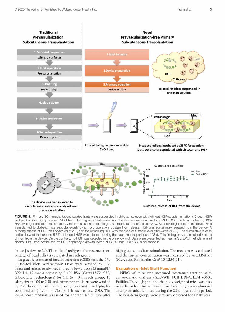
alcohol (EVOH) bag was performed as previously described.20 Briefly, isolated islets were suspended in chitosan solution (about 600 islets/100 μL; 800 islets equivalent with/without 10 μg HGF addition) and solution was sealed in an EVOH bag (Figure 1). Recombinant human HGF (hHGF, Lot#KSP01) was provided by Kringle Pharma, Inc., Osaka, Japan. The rec-tangular, 3 side heat-sealed with 1 side opened, EVOH bag (10 mm × 20 mm) was provided by Kuraray Co., Ltd. The opening side of EVOH bag was heat-sealed after infusing the islet-suspending chitosan solution. Chitosan solution was gelled by increasing temperature to 35°C. The devices were cultured in CMRL-1066 medium containing 10% FBS over-night before transplantation.
To examine the releasing profile of HGF, the device without islets was prepared and incubated in PBS (2 mL) in a 6-well plate (n = 3) at 37°C with the HGF-free device as a blank con-trol (n = 1). The PBS solution was collected periodically for 28 d. The HGF concentrations of collected PBS were meas-ured by an ELISA (R&D DHG00B Human HGF Quantikine ELISA kit, USA).
For device transplantation, the diabetic mice were anesthe-tized by isoflurane inhalation. After skin preparation, a mid-line skin incision (about 1.5 cm in length) was made at the back of mice and SC pocket was made by blunt dissection. The device was then implanted to the left side of the back subcutaneously (islet-alone and islet + HGF groups). The inci-sion was closed using a 4-0 nylon suture (Kyowa Precision Instrument Corp, Japan).
In Vitro Effect of HGF on Hypoxic IsletsIsolated rat islets were cultured in an oxygen-controlled
environment (1% O2 concentration controlled by BIONIX-1 nB-1 kit, Sugiyama-ken Corp, Japan) with and without 200 ng/mL hHGF (Lot#KSP01, Kringle Pharma, Inc.) in CMRL-1066 medium containing 10% FBS. On the contrary, the normally (20% O2) cultured islets with and without HGF were served for control groups (n = 3 in each group). After overnight cultured, the live/dead assay (Cellstain Double Staining Kit, 341‐07318; Dojindo, Kumamoto, Japan) was done. The treated islets were washed with PBS, then stained with fluorescent dye in 37°C for 15 min to identify surviving cells. To quantify live/dead staining, photos were analyzed by
© 2020 The Author(s). Published by Wolters Kluwer Health, Inc. Yang et al 3
Image J software 2.0. The ratio of red/green fluorescence (per-centage of dead cells) is calculated in each group.
In glucose-stimulated insulin secretion (GSIS) test, the 1% O2-treated islets with/without HGF were washed by PBS thrice and subsequently precultured in low glucose (3 mmol/L) RPMI‐1640 media containing 0.1% BSA (Cat#11879‐ 020; Gibco, Life Technologies) for 1 h (n = 3 in each group; 10 islets, size in 100 to 250 µm). After that, the islets were washed by PBS thrice and cultured in low glucose and then high-glu-cose medium (11.1 mmol/L) for 1 h each to test GSIS. The low-glucose medium was used for another 1-h culture after
high-glucose medium stimulation. The medium was collected and the insulin concentration was measured by an ELISA kit (Mercodia, Rat insulin Cat# 10-1250-01).
Evaluation of Islet Graft FunctionNFBG of mice was measured posttransplantation with
an automatic analyzer (GLU-WII; FUJI DRI-CHEM 4000i; Fujifilm, Tokyo, Japan) and the body weight of mice was also recorded at least twice a week. The clinical signs were observed and systematically noted during the 28-d observation period. The long-term groups were similarly observed for a half-year.
FIGURE 1. Primary SC transplantation: isolated islets were suspended in chitosan solution with/without HGF-supplementation (10 μg, hHGF) and packed in a highly porous EVOH bag. The bag was heat-sealed and the devices were cultured in CMRL-1066 medium containing 10% FBS overnight before transplantation. Chitosan solution becomes gel as temperature increases to 35°C. After overnight culture, the device was transplanted to diabetic mice subcutaneously by primary operation. Sustain HGF release: HGF was sustainingly released from the device. A bursting release of HGF was observed at d 1, and the remaining HGF was released at a stable level afterwards (n = 3). The cumulative release profile showed that around 5.5% of loaded HGF was released during the experimental periods of 28 d. This finding proved sustained release of HGF from the device. On the contrary, no HGF was detected in the blank control. Data were presented as mean ± SE. EVOH, ethylene vinyl alcohol; FBS, fetal bovine serum; HGF, hepatocyte growth factor; hHGF, human HGF; SC, subcutaneous.
4 Transplantation DIRECT ■ 2020 www.transplantationdirect.com
Glucose Tolerance TestAfter overnight fasting in clean cages without food and
feces, intraperitoneal glucose tolerance test (IPGTT) was per-formed to mice at 28 d after transplantation. The mice were injected with 20% glucose solution (2 g/kg body weight) intraperitoneally, and the blood glucose levels were deter-mined preadministration and postadministration. The blood glucose disappearance rate (K value) was determined using the equation K = 70/t1⁄2. The area under the curve (AUC) of blood glucose was also calculated and analyzed statistically. After IPGTT, the mice were euthanized, and the serum were collected and preserved for further analyses.
Rat Serum Insulin and Blood Urea Nitrogen LevelsThe serum of each mouse was collected immediately after
euthanizing on day 28 and stored at –80°C. The serum level of rat insulin was measured by an ELISA kit (Mercodia, Rat insulin Cat# 10-1250-01). In addition, as a renal function marker, blood urea nitrogen (BUN; BUN‐PIII; Fujifilm, Kyoto, Japan) was determined.
Implant Retrieval and Histological AssessmentThe implanted device with surrounding SC tissues were
excised from mice and fixed in 4% paraformaldehyde phos-phate buffer solution (Cat# 163-20145, FUJIFILM Wako Pure Chemical Co. Ltd., Tokyo, Japan) at 4°C for histological assessments. Histological sectioning, hematoxylin and eosin (H&E), and immunohistochemistry (IHC) staining were performed by the Center for Anatomical, Pathological and Forensic Medical Researches, Graduate School of Medicine, Kyoto University. Rat anti-insulin antibody (1:1250, Cat# ab8304; Abcam, Cambridge, United Kingdom), rat anti-glucagon antibody (1:1000, Cat# ab10988; Abcam), and anti-von-Willebrand Factor (vWF) antibody (1:50, Cat#AB7356; Millipore, Darmstadt, Germany,) were used as primary anti-bodies for IHC.
Statistical AnalysisStatistical analyses of GSIS, NFBG, body weight and blood
glucose of IPGTT were carried out by 2-way analysis of vari-ance (ANOVA) for repeated measures and Dunnett’s multiple comparison. One-way ANOVA was used to compare AUC, live/dead staining, serum insulin, and BUN. Statistical analysis was performed with GraphPad Prism 5 software. P < .05 was accepted as significant. All data were presented in mean ± SE.
RESULTS
In Vitro Releasing Profile of HGFA bursting release of HGF from the device was observed at
day 1, and the remaining HGF released in a stable level after-wards (n = 3, Figure 1). The cumulative release profile showed that around 5.5% of loaded HGF was released during the experimental periods of 28 d. This finding proved sustained release of HGF from the device. On the contrary, no HGF was detected in the blank control.
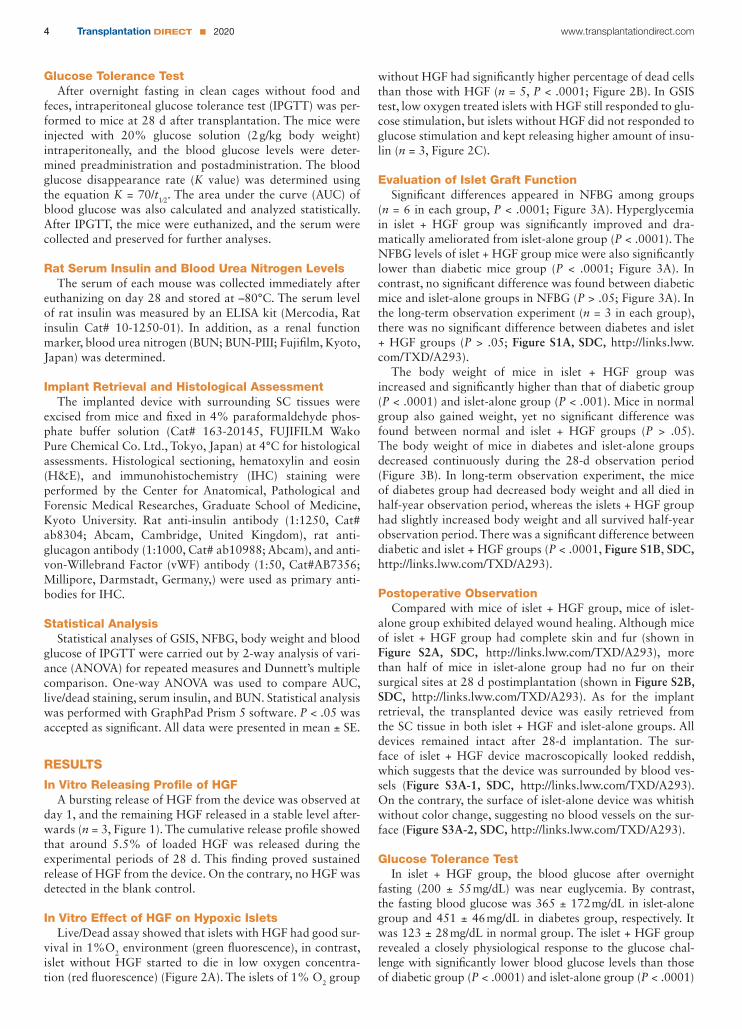
In Vitro Effect of HGF on Hypoxic IsletsLive/Dead assay showed that islets with HGF had good sur-
vival in 1%O2 environment (green fluorescence), in contrast, islet without HGF started to die in low oxygen concentra-tion (red fluorescence) (Figure 2A). The islets of 1% O2 group
without HGF had significantly higher percentage of dead cells than those with HGF (n = 5, P < .0001; Figure 2B). In GSIS test, low oxygen treated islets with HGF still responded to glu-cose stimulation, but islets without HGF did not responded to glucose stimulation and kept releasing higher amount of insu-lin (n = 3, Figure 2C).
Evaluation of Islet Graft FunctionSignificant differences appeared in NFBG among groups
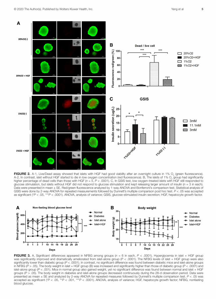
(n = 6 in each group, P < .0001; Figure 3A). Hyperglycemia in islet + HGF group was significantly improved and dra-matically ameliorated from islet-alone group (P < .0001). The NFBG levels of islet + HGF group mice were also significantly lower than diabetic mice group (P < .0001; Figure 3A). In contrast, no significant difference was found between diabetic mice and islet-alone groups in NFBG (P > .05; Figure 3A). In the long-term observation experiment (n = 3 in each group), there was no significant difference between diabetes and islet + HGF groups (P > .05; Figure S1A, SDC, http://links.lww.com/TXD/A293).
The body weight of mice in islet + HGF group was increased and significantly higher than that of diabetic group (P < .0001) and islet-alone group (P < .001). Mice in normal group also gained weight, yet no significant difference was found between normal and islet + HGF groups (P > .05). The body weight of mice in diabetes and islet-alone groups decreased continuously during the 28-d observation period (Figure 3B). In long-term observation experiment, the mice of diabetes group had decreased body weight and all died in half-year observation period, whereas the islets + HGF group had slightly increased body weight and all survived half-year observation period. There was a significant difference between diabetic and islet + HGF groups (P < .0001, Figure S1B, SDC, http://links.lww.com/TXD/A293).
Postoperative ObservationCompared with mice of islet + HGF group, mice of islet-
alone group exhibited delayed wound healing. Although mice of islet + HGF group had complete skin and fur (shown in Figure S2A, SDC, http://links.lww.com/TXD/A293), more than half of mice in islet-alone group had no fur on their surgical sites at 28 d postimplantation (shown in Figure S2B, SDC, http://links.lww.com/TXD/A293). As for the implant retrieval, the transplanted device was easily retrieved from the SC tissue in both islet + HGF and islet-alone groups. All devices remained intact after 28-d implantation. The sur-face of islet + HGF device macroscopically looked reddish, which suggests that the device was surrounded by blood ves-sels (Figure S3A-1, SDC, http://links.lww.com/TXD/A293). On the contrary, the surface of islet-alone device was whitish without color change, suggesting no blood vessels on the sur-face (Figure S3A-2, SDC, http://links.lww.com/TXD/A293).
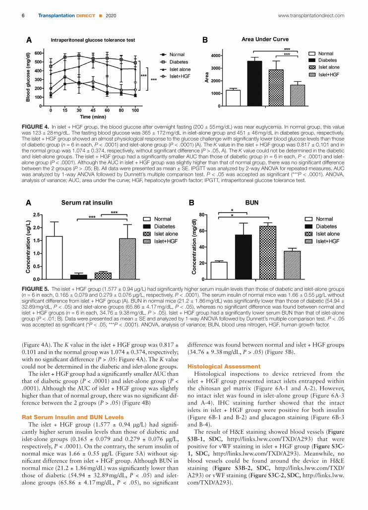
Glucose Tolerance TestIn islet + HGF group, the blood glucose after overnight
fasting (200 ± 55 mg/dL) was near euglycemia. By contrast, the fasting blood glucose was 365 ± 172 mg/dL in islet-alone group and 451 ± 46 mg/dL in diabetes group, respectively. It was 123 ± 28 mg/dL in normal group. The islet + HGF group revealed a closely physiological response to the glucose chal-lenge with significantly lower blood glucose levels than those of diabetic group (P < .0001) and islet-alone group (P < .0001)
© 2020 The Author(s). Published by Wolters Kluwer Health, Inc. Yang et al 5
FIGURE 2. A-1, Live/Dead assay showed that islets with HGF had good viability after an overnight culture in 1% O2 (green fluorescence). A-2, In contrast, islet without HGF started to die in low oxygen concentration (red fluorescence). B, The islets of 1% O2 group had significantly higher percentage of dead cells than those with HGF (n = 5, P < .0001). C, In GSIS test, low oxygen–treated islets with HGF still responded to glucose stimulation, but islets without HGF did not respond to glucose stimulation and kept releasing larger amount of insulin (n = 3 in each). Data were presented in mean ± SE. Red/green fluorescence analyzed by 1-way ANOVA and Bonferroni’s comparison test. Statistical analysis of GSIS were done by 2-way ANOVA for repeated measurements followed by Dunnett’s multiple comparison post hoc test. P < .05 was accepted as significant (*P < .05, ***P < .0001). ANOVA, analysis of variance; GSIS, glucose-stimulated insulin secretion; HGF, hepatocyte growth factor.
FIGURE 3. A, Significant differences appeared in NFBG among groups (n = 6 in each, P < .0001). Hyperglycemia in islet + HGF group was significantly improved and dramatically ameliorated from islet-alone group (P < .0001). The NFBG levels of islet + HGF group were also significantly lower than diabetic group (P < .0001). In contrast, no significant difference was found between diabetic mice and islet-alone groups in NFBG (P > .05). The body weight in islet + HGF group (B) was increased and significantly higher than those of diabetic group (P < .0001) and islet-alone group (P < .001). Mice in normal group also gained weight, yet no significant difference was found between normal and islet + HGF groups (P > .05). The body weight in diabetes and islet-alone groups decreased continuously during the 28-d observation period. Data were presented as mean ± SE and analyzed by 2-way ANOVA for repeated measures followed by Dunnett’s multiple comparison test. P < .05 was accepted as significant (*P < .05, **P < .001, ***P < .0001). ANOVA, analysis of variance; HGF, hepatocyte growth factor; NFBG, nonfasting blood glucose.
6 Transplantation DIRECT ■ 2020 www.transplantationdirect.com
(Figure 4A). The K value in the islet + HGF group was 0.817 ± 0.101 and in the normal group was 1.074 ± 0.374, respectively; with no significant difference (P > .05: Figure 4A). The K value could not be determined in the diabetic and islet-alone groups.
The islet + HGF group had a significantly smaller AUC than that of diabetic group (P < .0001) and islet-alone group (P < .0001). Although the AUC of islet + HGF group was slightly higher than that of normal group, there was no significant dif-ference between the 2 groups (P > .05) (Figure 4B)
Rat Serum Insulin and BUN LevelsThe islet + HGF group (1.577 ± 0.94 μg/L) had signifi-
cantly higher serum insulin levels than those of diabetic and islet-alone groups (0.165 ± 0.079 and 0.279 ± 0.076 μg/L, respectively, P < .0001). On the contrary, the serum insulin of normal mice was 1.66 ± 0.55 μg/L (Figure 5A) without sig-nificant difference from islet + HGF group. Although BUN in normal mice (21.2 ± 1.86 mg/dL) was significantly lower than those of diabetic (54.94 ± 32.89 mg/dL, P < .05) and islet-alone groups (65.86 ± 4.17 mg/dL, P < .05), no significant
difference was found between normal and islet + HGF groups (34.76 ± 9.38 mg/dL, P > .05) (Figure 5B).
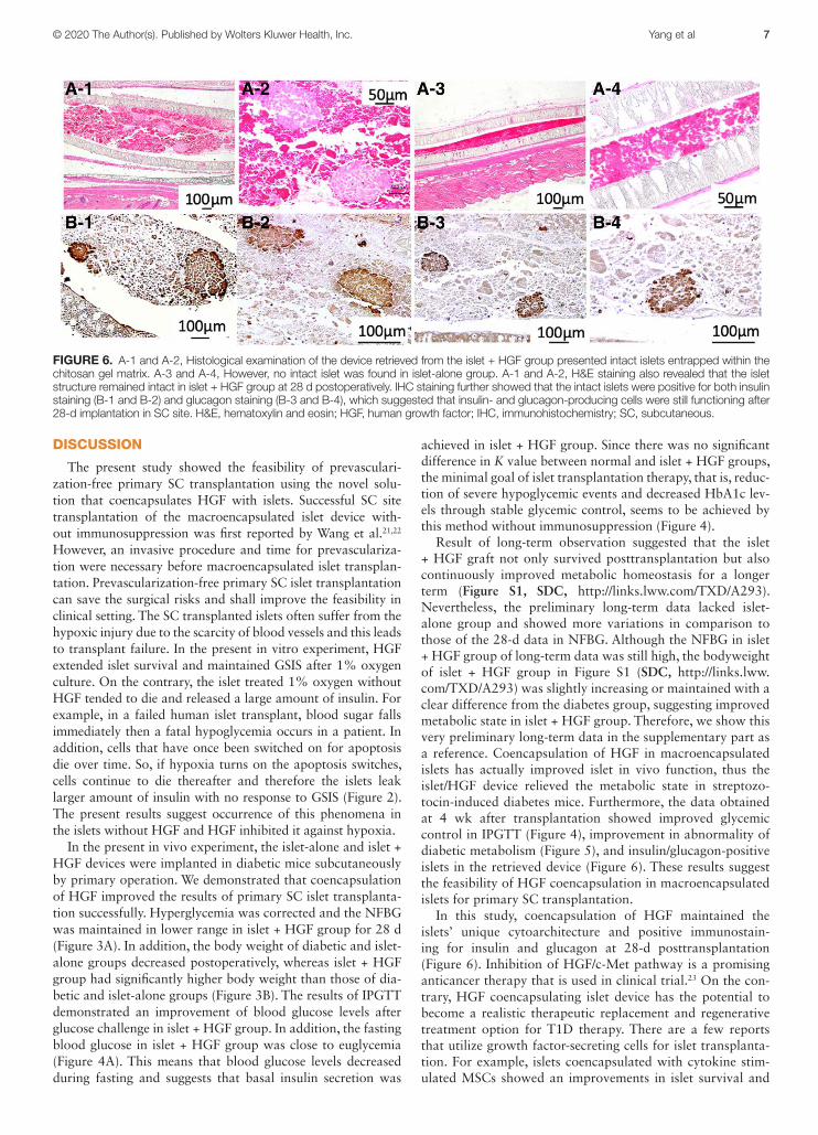
Histological AssessmentHistological inspections to device retrieved from the
islet + HGF group presented intact islets entrapped within the chitosan gel matrix (Figure 6A-1 and A-2). However, no intact islet was found in islet-alone group (Figure 6A-3 and A-4). IHC staining further showed that the intact islets in islet + HGF group were positive for both insulin (Figure 6B-1 and B-2) and glucagon staining (Figure 6B-3 and B-4).
The result of H&E staining showed blood vessels (Figure S3B-1, SDC, http://links.lww.com/TXD/A293) that were positive for vWF staining in islet + HGF group (Figure S3C-1, SDC, http://links.lww.com/TXD/A293). Meanwhile, no blood vessels could be found around the device in H&E staining (Figure S3B-2, SDC, http://links.lww.com/TXD/A293) or vWF staining (Figure S3C-2, SDC, http://links.lww.com/TXD/A293).
FIGURE 4. In islet + HGF group, the blood glucose after overnight fasting (200 ± 55 mg/dL) was near euglycemia. In normal group, this value was 123 ± 28 mg/dL. The fasting blood glucose was 365 ± 172 mg/dL in islet-alone group and 451 ± 46 mg/dL in diabetes group, respectively. The islet + HGF group showed an almost physiological response to the glucose challenge with significantly lower blood glucose levels than those of diabetic group (n = 6 in each, P < .0001) and islet-alone group (P < .0001) (A). The K value in the islet + HGF group was 0.817 ± 0.101 and in the normal group was 1.074 ± 0.374, respectively, without significant difference (P > .05, A). The K value could not be determined in the diabetic and islet-alone groups. The islet + HGF group had a significantly smaller AUC than those of diabetic group (n = 6 in each, P < .0001) and islet-alone group (P < .0001). Although the AUC in islet + HGF group was slightly higher than that of normal group, there was no significant difference between the 2 groups (P > .05; B). All data were presented as mean ± SE. IPGTT was analyzed by 2-way ANOVA for repeated measures. AUC was analyzed by 1-way ANOVA followed by Dunnett’s multiple comparison test. P < .05 was accepted as significant (***P < .0001). ANOVA, analysis of variance; AUC, area under the curve; HGF, hepatocyte growth factor; IPGTT, intraperitoneal glucose tolerance test.
FIGURE 5. The islet + HGF group (1.577 ± 0.94 μg/L) had significantly higher serum insulin levels than those of diabetic and islet-alone groups (n = 6 in each, 0.165 ± 0.079 and 0.279 ± 0.076 μg/L, respectively, P < .0001). The serum insulin of normal mice was 1.66 ± 0.55 μg/L without significant difference from islet + HGF group (A). BUN in normal mice (21.2 ± 1.86 mg/dL) was significantly lower than those of diabetic (54.94 ± 32.89 mg/dL, P < .05) and islet-alone groups (65.86 ± 4.17 mg/dL, P < .05), whereas no significant difference was found between normal and islet + HGF groups (n = 6 in each, 34.76 ± 9.38 mg/dL, P > .05). Islet + HGF group had a significantly lower serum BUN than that of islet-alone group (P < .01; B). Data were presented as mean ± SE and analyzed by 1-way ANOVA followed by Dunnett’s multiple comparison test. P < .05 was accepted as significant (*P < .05, ***P < .0001). ANOVA, analysis of variance; BUN, blood urea nitrogen, HGF, human growth factor.
© 2020 The Author(s). Published by Wolters Kluwer Health, Inc. Yang et al 7
DISCUSSION
The present study showed the feasibility of prevasculari-zation-free primary SC transplantation using the novel solu-tion that coencapsulates HGF with islets. Successful SC site transplantation of the macroencapsulated islet device with-out immunosuppression was first reported by Wang et al.21,22 However, an invasive procedure and time for prevasculariza-tion were necessary before macroencapsulated islet transplan-tation. Prevascularization-free primary SC islet transplantation can save the surgical risks and shall improve the feasibility in clinical setting. The SC transplanted islets often suffer from the hypoxic injury due to the scarcity of blood vessels and this leads to transplant failure. In the present in vitro experiment, HGF extended islet survival and maintained GSIS after 1% oxygen culture. On the contrary, the islet treated 1% oxygen without HGF tended to die and released a large amount of insulin. For example, in a failed human islet transplant, blood sugar falls immediately then a fatal hypoglycemia occurs in a patient. In addition, cells that have once been switched on for apoptosis die over time. So, if hypoxia turns on the apoptosis switches, cells continue to die thereafter and therefore the islets leak larger amount of insulin with no response to GSIS (Figure 2). The present results suggest occurrence of this phenomena in the islets without HGF and HGF inhibited it against hypoxia.
In the present in vivo experiment, the islet-alone and islet + HGF devices were implanted in diabetic mice subcutaneously by primary operation. We demonstrated that coencapsulation of HGF improved the results of primary SC islet transplanta-tion successfully. Hyperglycemia was corrected and the NFBG was maintained in lower range in islet + HGF group for 28 d (Figure 3A). In addition, the body weight of diabetic and islet-alone groups decreased postoperatively, whereas islet + HGF group had significantly higher body weight than those of dia-betic and islet-alone groups (Figure 3B). The results of IPGTT demonstrated an improvement of blood glucose levels after glucose challenge in islet + HGF group. In addition, the fasting blood glucose in islet + HGF group was close to euglycemia (Figure 4A). This means that blood glucose levels decreased during fasting and suggests that basal insulin secretion was
achieved in islet + HGF group. Since there was no significant difference in K value between normal and islet + HGF groups, the minimal goal of islet transplantation therapy, that is, reduc-tion of severe hypoglycemic events and decreased HbA1c lev-els through stable glycemic control, seems to be achieved by this method without immunosuppression (Figure 4).
Result of long-term observation suggested that the islet + HGF graft not only survived posttransplantation but also continuously improved metabolic homeostasis for a longer term (Figure S1, SDC, http://links.lww.com/TXD/A293). Nevertheless, the preliminary long-term data lacked islet-alone group and showed more variations in comparison to those of the 28-d data in NFBG. Although the NFBG in islet + HGF group of long-term data was still high, the bodyweight of islet + HGF group in Figure S1 (SDC, http://links.lww.com/TXD/A293) was slightly increasing or maintained with a clear difference from the diabetes group, suggesting improved metabolic state in islet + HGF group. Therefore, we show this very preliminary long-term data in the supplementary part as a reference. Coencapsulation of HGF in macroencapsulated islets has actually improved islet in vivo function, thus the islet/HGF device relieved the metabolic state in streptozo-tocin-induced diabetes mice. Furthermore, the data obtained at 4 wk after transplantation showed improved glycemic control in IPGTT (Figure 4), improvement in abnormality of diabetic metabolism (Figure 5), and insulin/glucagon-positive islets in the retrieved device (Figure 6). These results suggest the feasibility of HGF coencapsulation in macroencapsulated islets for primary SC transplantation.
In this study, coencapsulation of HGF maintained the islets’ unique cytoarchitecture and positive immunostain-ing for insulin and glucagon at 28-d posttransplantation (Figure 6). Inhibition of HGF/c-Met pathway is a promising anticancer therapy that is used in clinical trial.23 On the con-trary, HGF coencapsulating islet device has the potential to become a realistic therapeutic replacement and regenerative treatment option for T1D therapy. There are a few reports that utilize growth factor-secreting cells for islet transplanta-tion. For example, islets coencapsulated with cytokine stim-ulated MSCs showed an improvements in islet survival and
FIGURE 6. A-1 and A-2, Histological examination of the device retrieved from the islet + HGF group presented intact islets entrapped within the chitosan gel matrix. A-3 and A-4, However, no intact islet was found in islet-alone group. A-1 and A-2, H&E staining also revealed that the islet structure remained intact in islet + HGF group at 28 d postoperatively. IHC staining further showed that the intact islets were positive for both insulin staining (B-1 and B-2) and glucagon staining (B-3 and B-4), which suggested that insulin- and glucagon-producing cells were still functioning after 28-d implantation in SC site. H&E, hematoxylin and eosin; HGF, human growth factor; IHC, immunohistochemistry; SC, subcutaneous.
8 Transplantation DIRECT ■ 2020 www.transplantationdirect.com
insulin-secreting function posttransplantation, and reversed the diabetes in mice successfully.24 Jourdan et al25 also showed coencapsulation of insulin-like growth factor-II producing cells promoted pancreatic islet cell survival in diabetic mice. Perez-Basterrechea et al26 reported that adding fibroblast to plasma-based scaffold can enhance survival and function of subcutaneously transplanted islets. Yeung et al27 showed MSC can protect islets and improve graft survival by promoting cell survival and reduced inflammation via the secretion of HGF. Therefore, the above benefits could, at least partly, stem from the ability of HGF to protect islets.
Sustained release of HGF was achieved by coencapsulation of HGF in chitosan gel (Figure 1). This result suggests that not only the coencapsulated islets, but also the tissue surrounding the device (SC site) were influenced by HGF. In this study, the islet-alone group showed scarce fur growth and impaired wound healing in surgical site (Figure S2B, SDC, http://links.lww.com/TXD/A293). In contrast, the diabetic mice in islet + HGF group showed fur regrowth and closed wounds (Figure S2A, SDC, http://links.lww.com/TXD/A293). Diabetes is known to disturb interactions between endothelial cells and pericytes to influence angiogenesis,28 which can further impair blood supply and wound healing.29 Previous study also sug-gested that HGF enhances hair regeneration by stimulating the sheath fibroblasts surrounding hair follicles.30 Accordingly, our finding may suggest local action of coencapsulated HGF together with the favorable effect of improved diabetes. In addition, although angiogenesis was not found in islet-alone group and the surface of the devices remained whitish (Figure S3, SDC, http://links.lww.com/TXD/A293), the diabetic mice in the islet + HGF group had newly formations of the vascu-lar bed (reddish color) around the device (Figure S3, SDC, http://links.lww.com/TXD/A293). These results, that is, neo-vascularization, wound healing, and hair growth, seem to be induced by sustained release of HGF outside of the device.
As a promising T1D therapy, the device used in this study can not only apply to xenogeneic islet but also to islet-like tis-sue derived from pluripotent cells. Further application could encompass functioning tissues that are potentially suitable for SC site transplantation, such as endocrine tissues for hormone deficiencies and hepatocytes for metabolic anomalies.
In conclusion, this study showed that coencapsulation of HGF in macroencapsulated islets improved islet in vivo func-tion and may provide a potential method for prevasculariza-tion-free primary SC transplantation.
REFERENCES 1. Moberg L, Johansson H, Lukinius A, et al. Production of tissue factor
by pancreatic islet cells as a trigger of detrimental thrombotic reac-tions in clinical islet transplantation. Lancet. 2002;360:2039–2045.
2. Hwa AJ, Weir GC. Transplantation of macroencapsulated insulin-pro-ducing cells. Curr Diab Rep. 2018;18:50.
3. Kawakami Y, Iwata H, Gu YJ, et al. Successful subcutaneous pancre-atic islet transplantation using an angiogenic growth factor-releasing device. Pancreas. 2001;23:375–381.
4. Yang S-Y, Yang K-C, Sumi S. Effect of basic fibroblast growth fac-tor on xenogeneic islets in subcutaneous transplantation—A murine model. Transplant Proc. 2019;51:1458–1462.
5. García-Ocaña A, Vasavada RC, Cebrian A, et al. Transgenic over-expression of hepatocyte growth factor in the beta-cell mark-edly improves islet function and islet transplant outcomes in mice. Diabetes. 2001;50:2752–2762.
6. Lopez-Talavera JC, Garcia-Ocaña A, Sipula I, et al. Hepatocyte growth factor gene therapy for pancreatic islets in diabetes: reducing the minimal
islet transplant mass required in a glucocorticoid-free rat model of alloge-neic portal vein islet transplantation. Endocrinology. 2004;145:467–474.
7. Garcia-Ocaña A, Takane KK, Syed MA, et al. Hepatocyte growth fac-tor overexpression in the islet of transgenic mice increases beta cell proliferation, enhances islet mass, and induces mild hypoglycemia. J Biol Chem. 2000;275:1226–1232.
8. Hayek A, Beattie GM, Cirulli V, et al. Growth factor/matrix-induced pro-liferation of human adult beta-cells. Diabetes. 1995;44:1458–1460.
9. Demirci C, Ernst S, Alvarez-Perez JC, et al. Loss of HGF/c-Met signal-ing in pancreatic β-cells leads to incomplete maternal β-cell adapta-tion and gestational diabetes mellitus. Diabetes. 2012;61:1143–1152.
10. Oliveira AG, Araújo TG, Carvalho BM, et al. The role of hepato-cyte growth factor (HGF) in insulin resistance and diabetes. Front Endocrinol (Lausanne). 2018;9:503.
11. Trusolino L, Bertotti A, Comoglio PM. MET signalling: principles and functions in development, organ regeneration and cancer. Nat Rev Mol Cell Biol. 2010;11:834–848.
12. Fiaschi-Taesch N, Stewart AF, Garcia-Ocaña A. Improving islet trans-plantation by gene delivery of hepatocyte growth factor (HGF) and its downstream target, protein kinase B (PKB)/Akt. Cell Biochem Biophys. 2007;48(2-3):191–199.
13. Mellado-Gil J, Rosa TC, Demirci C, et al. Disruption of hepatocyte growth factor/c-Met signaling enhances pancreatic beta-cell death and accelerates the onset of diabetes. Diabetes. 2011;60:525–536.
14. Ozaki M, Haga S, Zhang HQ, et al. Inhibition of hypoxia/reoxygen-enation-induced oxidative stress in HGF-stimulated antiapoptotic signaling: role of PI3-K and Akt kinase upon rac1. Cell Death Differ. 2003;10:508–515.
15. He F, Wu LX, Shu KX, et al. HGF protects cultured cortical neurons against hypoxia/reoxygenation induced cell injury via ERK1/2 and PI-3K/Akt pathways. Colloids Surf B Biointerfaces. 2008;61:290–297.
16. Garcia-Ocana A, Takane KK, Reddy VT, et al. Adenovirus-mediated hepatocyte growth factor expression in mouse islets improves pancre-atic islet transplant performance and reduces beta cell death. J Biol Chem. 2003;278:343–351.
17. Dai C, Huh CG, Thorgeirsson SS, et al. Beta-cell-specific ablation of the hepatocyte growth factor receptor results in reduced islet size, impaired insulin secretion, and glucose intolerance. Am J Pathol. 2005;167:429–436.
18. Wu H, Yoon AR, Li F, et al. RGD peptide-modified adenovirus express-ing hepatocyte growth factor and X-linked inhibitor of apoptosis improves islet transplantation. J Gene Med. 2011;13:658–669.
19. Kilkenny C, Browne WJ, Cuthill IC, et al. Improving bioscience research reporting: The arrive guidelines for reporting animal research. Animals. 2013;4:35–44.
20. Yang KC, Yanai G, Yang SY, et al. Low-adhesive ethylene vinyl alcohol-based packaging to xenogeneic islet encapsulation for type 1 diabe-tes treatment. Biotechnol Bioeng. 2018;115:2341–2355.
21. Wang W, Gu Y, Tabata Y, et al. Reversal of diabetes in mice by xenotransplantation of a bioartificial pancreas in a prevascularized subcutaneous site. Transplantation. 2002;73:122–129.
22. Wang W, Gu Y, Hori H, et al. Subcutaneous transplantation of mac-roencapsulated porcine pancreatic endocrine cells normalizes hyper-glycemia in diabetic mice. Transplantation. 2003;76:290–296.
23. Kim KH, Kim H. Progress of antibody-based inhibitors of the HGF-cMET axis in cancer therapy. Exp Mol Med. 2017;49:e307.
24. Vaithilingam V, Evans MDM, Lewy DM, et al. Co-encapsulation and co-transplantation of mesenchymal stem cells reduces pericapsular fibrosis and improves encapsulated islet survival and function when allografted. Sci Rep. 2017;7:1–13.
25. Jourdan G, Dusseault J, Benhamou PY, et al. Co-encapsulation of bioengineered IGF-II-producing cells and pancreatic islets: effect on beta-cell survival. Gene Ther. 2011;18:539–545.
26. Perez-Basterrechea M, Esteban MM, Alvarez-Viejo M, et al. Fibroblasts accelerate islet revascularization and improve long-term graft survival in a mouse model of subcutaneous islet transplantation. PLoS One. 2017;12:e0180695.
27. Yeung TY, Seeberger KL, Kin T, et al. Human mesenchymal stem cells protect human islets from pro-inflammatory cytokines. PLoS One. 2012;7:e38189.
28. Wimmer RA, Leopoldi A, Aichinger M, et al. Human blood vessel orga-noids as a model of diabetic vasculopathy. Nature. 2019;565:505–510.
29. Brem H, Tomic-Canic M. Cellular and molecular basis of wound heal-ing in diabetes. J Clin Invest. 2007;117:1219–1222.
30. Qi Y, Li M, Xu L, et al. Therapeutic role of human hepatocyte growth factor (HGF) in treating hair loss. PeerJ. 2016;4:e2624.