Embed Size (px)
Citation preview
PRIMARY ARTERITIS OF AORTACAUSING RENAL ARTERY STENOSIS AND HYPERTENSION
BY
T. J. DANARAJ, H. 0. WONG, AND M. A. THOMAS
From the Departments of Medicine and Pathology, University of Singapore
Received January 30, 1962
Early reports of patients suffering from primary arteritis of the aorta described the effects ofinadequate blood supply to the brain, eyes, and upper limbs resulting from occlusion of the largebranches of the aortic arch (Savory, 1856; Takayashu, 1908; Giffin, 1939; Lewis and Stokes, 1942).These observations were subsequently confirmed, and the condition, which occurred mainly inyoung women, was differentiated from other diseases such as syphilis, atherosclerosis, dissectinganeurysm, and congenital anomalies that affected the aortic arch to produce a similar clinical picture(Shimizu and Sano, 1951; Caccamise and Whitman, 1952; Skipper and Flint, 1952; Ross andMcKusick, 1953; Ask-Upmark, 1954; Kalmansohn and Kalmansohn, 1957). Various names weregiven to the entity: pulseless disease (Shimizu and Sano, 1951), young female arteritis (Ross andMcKusick, 1953), Takayashu's syndrome (Trias de Bes, Lucas, and Barcons, 1955), primary arteritis.of the aortic arch (Barker and Edwards, 1955), obliterative brachiocephalic arteritis (Gibbons andKing, 1957), and aortic arch arteritis (Koszewski, 1958). In patients who came to necropsy, achronic non-specific inflammatory process was described affecting chiefly the arch of the aorta and.its main branches at or close to their origin and resulting in occlusion of their lumen (Frovig and-Loken, 1951). The peculiar anatomical site of the lesions explained the classical clinical picture.
In addition to the picture of brachiocephalic involvement, other associated features have beenrecorded. Loss of pulsation in one or both femoral arteries, in combination with signs of involve-ment of the aortic arch, has been reported in patients in whom the abdominal aorta was normallypulsatile: it was postulated that obstruction to the blood flow was present at the level of the iliac-arteries (Gibbons and King, 1957; Danaraj and Wong, 1960). Hypertension in the lower limbs incontrast to brachial hypotension was noted by Giffin (1939) who suggested the designation " reversedcoarctation." Similar observations were also made by Lewis and Stokes (1942), Skipper and Flint(1952), and Caldwell and Skipper (1961). The cause of hypertension was not known, but Lessof(1958) suggested that it could be of renal origin.
In an earlier paper (Danaraj and Wong, 1959), we reported the occurrence of hypertension in twochildren consequent on stenosis of the orifices of both renal arteries caused by localized arteritis ofthe abdominal aorta: the histological appearance of the aorta indicated that this was a variant ofTakayashu's syndrome. The purpose of this paper is to report on a further nine patients, all ofwhom presented with hypertension and in whom there was necropsy or radiological evidence ofdisease of the aorta, with involvement not only of the renal arteries but of other branches as well.A study of this series gives support to the thesis that primary arteritis of the aorta may affect not onlythe arch alone (to produce Takayashu's syndrome) but any part of the aorta, the clinical picturedepending on which segments and which branches are affected. Primary arteritis involving theabdominal aorta with renal artery obstruction produces the syndrome of Goldblatt hypertension,which may occur independently of or together with Takayashu's syndrome.
153
on February 9, 2022 by guest. P
rotected by copyright.http://heart.bm
j.com/
Br H
eart J: first published as 10.1136/hrt.25.2.153 on 1 March 1963. D
ownloaded from
DANARAJ, WONG, AND THOMAS
CASE REPORTSCase 1. A married Chinese woman, aged 21, was first seen on June 10, 1960, when she complained of
severe headache and vomiting. Five days earlier, she had been admitted to a surgical unit with localizedpain and tenderness in the right iliac fossa and an appendicectomy had been done: it was noted at the timethat her blood pressure was raised. Questioning elicited that she had not been well for a year, having hadattacks of breathlessness and palpitations especially on exertion, with swelling of the feet on one occasion.On examination, pulses were felt over the arteries of the limbs, being equal on both sides: pulsation of thecarotid arteries was visible and forceful. The blood pressure reading in the upper limbs was 120/110 mm.Hg, while in the lower limbs it was 155/130 on the left side and 140/100 mm. Hg on the right. The heartwas enlarged, the apex beat being forceful and felt in the sixth left intercostal space 10 cm. from the mid-sternal line. The second sound in the aortic area was accentuated and there was triple rhythm but nomurmurs. The fundi appeared normal.
The blood hemoglobin level was 13-3 g. per 100 ml., and the white cell count 14,000 per c. mm. with adifferential count of 90 per cent polymorphonuclear leucocytes, 6 per cent lymphocytes, and 4 per centmonocytes. The erythrocyte sedimentation rate (Westergren) was 33 mm. in one hour. The specific gravity ofthe urine was normal, but on a few occasions there were increased numbers of red blood cells and pus cellson microscopical examination. The blood urea level was 15 mg. per 100 ml. and blood culture and Kahntest were negative. Radiological examination showed an enlarged heart with a prominent left borderand normal lung fields. There was cardiographic evidence of left ventricular hypertrophy and strain.
Response to treatment with hypotensive drugs was not satisfactory. Fever of 100-101°F. (37 -8-38 -30C.)was present on four different days. On June 26, itwas not possible to feel the right femoral pulse, andfour days later she died suddenly.
Necropsy Findings. The heart (450 g.) was en-larged with hypertrophy of the left ventricle (1 7 cm.thick), but was otherwise normal. The pulmonaryarteries were not affected. On opening the aorta,three main areas of abnormality were seen. Thefirst involved the entire circumference of the ascend-ing aorta from its origin for a distance of 3 cm.
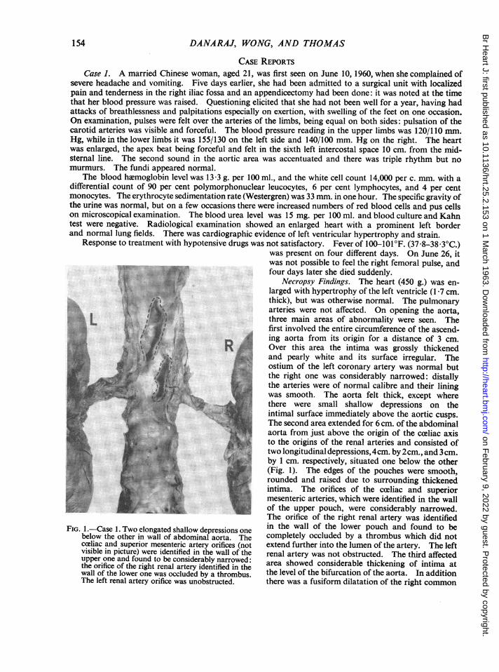
i0QQi: e--A Over this area the intima was grossly thickenedand pearly white and its surface irregular. Theostium of the left coronary artery was normal butthe right one was considerably narrowed: distallythe arteries were of normal calibre and their liningwas smooth. The aorta felt thick, except wherethere were small shallow depressions on theintimal surface immediately above the aortic cusps.The second area extended for 6 cm. of the abdominalaorta from just above the origin of the celiac axisto the origins of the renal arteries and consisted of
x_#̂1o two longitudinal depressions, 4cm. by 2cm.,and 3cm.| by 1 cm. respectively, situated one below the other
(Fig. 1). The edges of the pouches were smooth,rounded and raised due to surrounding thickenedintima. The orifices of the coeliac and superiormesenteric arteries, which were identified in the wallof the upper pouch, were considerably narrowed.The orifice of the right renal artery was identified
FIG- 1 Case 1. Two elongated shallow depressions one in the wall of the lower pouch and found to bebelow the other in wall of abdominal aorta. The completely occluded by a thrombus which did notcoeliac and superior mesenteric artery orifices (not extend further into the lumen of the artery. The leftvisible in picture) were identified in the wall of the renal artery was not obstructed. The third affectedupper one and found to be considerably narrowed: area showed considerable thickening of intima atthe orifice of the right renal artery identified in the area the cation ofthe o f itionwall of the lower one was occluded by a thrombus. the level of the bifurcation of the aorta. In additionThe left renal artery orifice was unobstructed. there was a fusiform dilatation of the right common
154
on February 9, 2022 by guest. P
rotected by copyright.http://heart.bm
j.com/
Br H
eart J: first published as 10.1136/hrt.25.2.153 on 1 March 1963. D
ownloaded from
PRIMARY ARTERITIS OF AORTA
.I
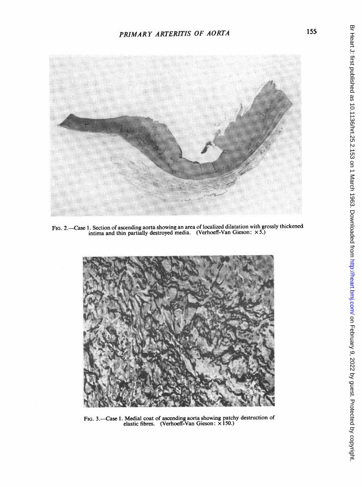
FIG. 2.-Case 1. Section of ascending aorta showing an area of localized dilatation with grossly thickenedintima and thin partially destroyed media. (Verhoeff-Van Gieson: x 5.)
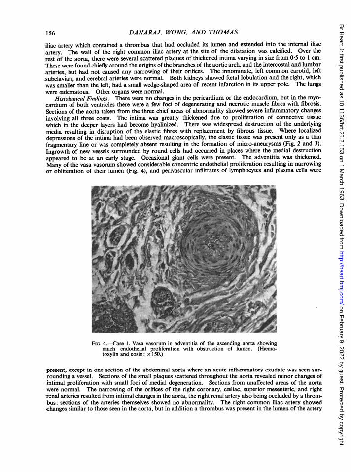
FiG. 3.-Case 1. Medial coat of ascending aorta showing patchy destruction ofelastic fibres. (Verhoeff-Van Gieson: x 150.)
155
on February 9, 2022 by guest. P
rotected by copyright.http://heart.bm
j.com/
Br H
eart J: first published as 10.1136/hrt.25.2.153 on 1 March 1963. D
ownloaded from
DANARAJ, WONG, AND THOMAS
-iliac artery which contained a thrombus that had occluded its lumen and extended into the internal iliacartery. The wall of the right common iliac artery at the site of the dilatation was calcified. Over therest of the aorta, there were several scattered plaques of thickened intima varying in size from 0-5 to 1 cm.These were found chiefly around the origins ofthe branches of the aortic arch, and the intercostal and lumbararteries, but had not caused any narrowing of their orifices. The innominate, left common carotid, left;subclavian, and cerebral arteries were normal. Both kidneys showed foetal lobulation and the right, whichwas smaller than the left, had a small wedge-shaped area of recent infarction in its upper pole. The lungswere cedematous. Other organs were normal.
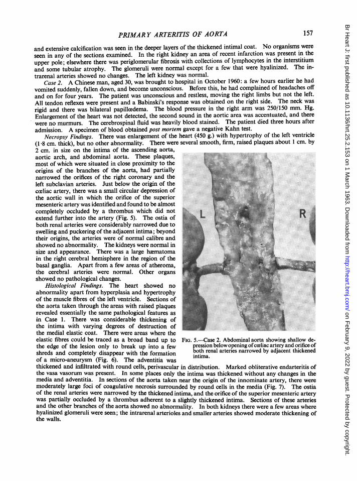
Histological Findings. There were no changes in the pericardium or the endocardium, but in the myo-cardium of both ventricles there were a few foci of degenerating and necrotic muscle fibres with fibrosis.Sections of the aorta taken from the three chief areas of abnormality showed severe inflammatory changesinvolving all three coats. The intima was greatly thickened due to proliferation of connective tissuewhich in the deeper layers had become hyalinized. There was widespread destruction of the underlyingmedia resulting in disruption of the elastic fibres with replacement by fibrous tissue. Where localizeddepressions of the intima had been observed macroscopically, the elastic tissue was present only as a thinfragmentary line or was completely absent resulting in the formation of micro-aneurysms (Fig. 2 and 3).Ingrowth of new vessels surrounded by round cells had occurred in places where the medial destructionappeared to be at an early stage. Occasional giant cells were present. The adventitia was thickened.Many of the vasa vasorum showed considerable concentric endothelial proliferation resulting in narrowing,or obliteration of their lumen (Fig. 4), and perivascular infiltrates of lymphocytes and plasma cells were
FIG. 4.-Case 1. Vasa vasorum in adventitia of the ascending aorta showingmuch endothelial proliferation with obstruction of lumen. (Hlma-toxylin and eosin: x 150.)
present, except in one section of the abdominal aorta where an acute inflammatory exudate was seen sur-rounding a vessel. Sections of the small plaques scattered throughout the aorta revealed minor changes ofintimal proliferation with small foci of medial degeneration. Sections from unaffected areas of the aortawere normal. The narrowing of the orifices of the right coronary, coeliac, superior mesenteric, and rightrenal arteries resulted from intimal changes in the aorta, the right renal artery also being occluded by a throm-bus: sections of the arteries themselves showed no abnormality. The right common iliac artery showedchanges similar to those seen in the aorta, but in addition a thrombus was present in the lumen of the artery
'I56
on February 9, 2022 by guest. P
rotected by copyright.http://heart.bm
j.com/
Br H
eart J: first published as 10.1136/hrt.25.2.153 on 1 March 1963. D
ownloaded from
PRIMARY ARTERITIS OF AORTA
and extensive calcification was seen in the deeper layers of the thickened intimal coat. No organisms wereseen in any of the sections examined. In the right kidney an area of recent infarction was present in theupper pole; elsewhere there was periglomerular fibrosis with collections of lymphocytes in the interstitiumand some tubular atrophy. The glomeruli were normal except for a few that were hyalinized. The in-trarenal arteries showed no changes. The left kidney was normal.
Case 2. A Chinese man, aged 30, was brought to hospital in October 1960: a few hours earlier he hadvomited suddenly, fallen down, and become unconscious. Before this, he had complained of headaches offand on for four years. The patient was unconscious and restless, moving the right limbs but not the left.All tendon reflexes were present and a Babinski's response was obtained on the right side. The neck wasrigid and there was bilateral papilloedema. The blood pressure in the right arm was 250/150 mm. Hg.Enlargement of the heart was not detected, the second sound in the aortic area was accentuated, and therewere no murmurs. The cerebrospinal fluid was heavily blood stained. The patient died three hours afteradmission. A specimen of blood obtained post mortem gave a negative Kahn test.
Necropsy Findings. There was enlargement of the heart (450 g.) with hypertrophy of the left ventricle(1 8 cm. thick), but no other abnormality. There were several smooth, firm, raised plaques about 1 cm. by2 cm. in size on the intima of the ascending aorta,aortic arch, and abdominal aorta. These plaques,aqmost of which were situated in close proximity to theorigins of the branches of the aorta, had partially ;: PR
narrowed the orifices of the right coronary and theleft subclavian arteries. Just below the origin of thecceliac artery, there was a small circular depression ofthe aortic wall in which the orifice of the superiormesenteric artery was identified and found to be almostcompletely occluded by a thrombus which did not >% Inextend further into the artery (Fig. 5). The ostia ofboth renal arteries were considerably narrowed due toswelling and puckering of the adjacent intima; beyondtheir origins, the arteries were of normal calibre andshowed no abnormality. The kidneys were normal insize and appearance. There was a large hematomain the right cerebral hemisphere in the region of thebasal ganglia. Apart from a few areas of atheroma,_the cerebral arteries were normal. Other organsshowed no pathological changes.
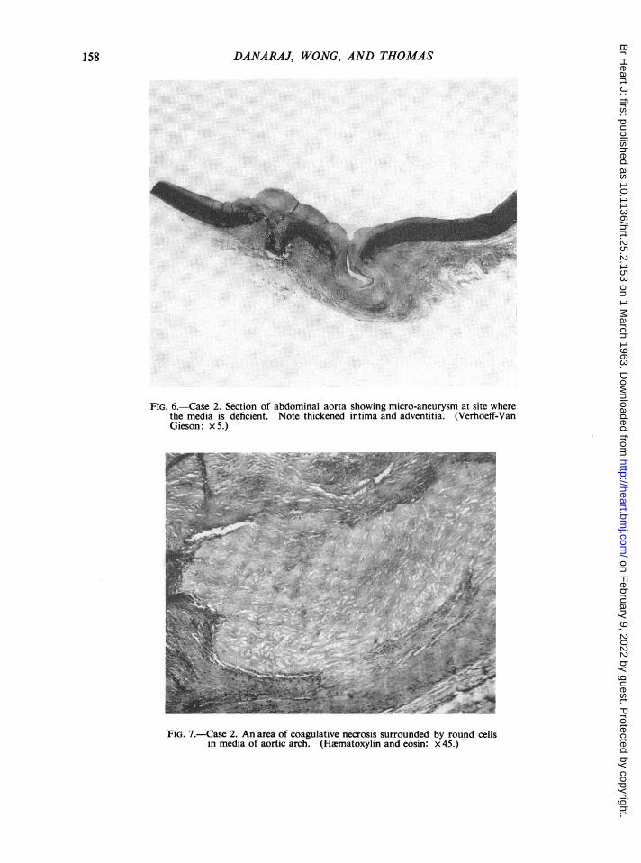
Histological Findings. The heart showed noabnormality apart from hyperplasia and hypertrophyof the muscle fibres of the left ventricle. Sections ofthe aorta taken through the areas with raised plaquesrevealed essentially the same pathological features asin Case 1. There was considerable thickening ofthe intima with varying degrees of destruction ofthe medial elastic coat. There were areas where theelastic fibres could be traced as a broad band up to FIG. 5.-Case 2. Abdominal aorta showing shallow de-the edge of the lesion only to break up into a few pression below opening of coeliac artery and orifice ofshreds and completely disappear with the formation both renal arteries narrowed by adjacent thickenedof a micro-aneurysm (Fig. 6). The adventitia was intima.thickened and infiltrated with round cells, perivascular in distribution. Marked obliterative endarteritis ofthe vasa vasorum was present. In some places only the intima was thickened without any changes in themedia and adventitia. In sections of the aorta taken near the origin of the innominate artery, there weremoderately large foci of coagulative necrosis surrounded by round cells in the media (Fig. 7). The ostiaof the renal arteries were narrowed by the thickened intima, and the orifice of the superior mesenteric arterywas partially occluded by a thrombus adherent to a slightly thickened intima. Sections of these arteriesand the other branches of the aorta showed no abnormality. In both kidneys there were a few areas wherehyalinized glomeruli were seen; the intrarenal arterioles and smaller arteries showed moderate thickening ofthe walls.
157
on February 9, 2022 by guest. P
rotected by copyright.http://heart.bm
j.com/
Br H
eart J: first published as 10.1136/hrt.25.2.153 on 1 March 1963. D
ownloaded from
DANARAJ, WONG, AND THOMAS
FIG. 6.-Case 2. Section of abdominal aorta showing micro-aneurysm at site wherethe media is deficient. Note thickened intima and adventitia. (Verhoeff-VanGieson: x 5.)
FIG. 7.-Case 2. An area of coagulative necrosis surrounded by round cellsin media of aortic arch. (Hamatoxylin and eosin: x 45.)
158
on February 9, 2022 by guest. P
rotected by copyright.http://heart.bm
j.com/
Br H
eart J: first published as 10.1136/hrt.25.2.153 on 1 March 1963. D
ownloaded from
PRIMARY ARTERITIS OF AORTA
Case 3. A Chinese youth, aged 17, had been ill for two months with progressive breathlessness on exertionand cough with sputum which was sometimes blood stained. When he was admitted on November 22,1961, he was in severe congestive cardiac failure. The heart was slightly enlarged and triple rhythm was
present, but there were no significant murmurs. Crepitations were heard over the bases of both lungs andthe liver was palpable three fingers' breadth below the costal margin. The blood pressure was 210/150mm. Hg and the retinal arteries were narrowed. Arterial pulsations in the limbs were present and equal on
both sides; the right carotid artery was strongly pulsatile, but the left was barely felt and over its course a
distinct systolic murmur was heard. The patient responded satisfactorily to treatment of the cardiac failureand with hypotensive drugs the blood pressure decreased to 160/110 mm. Hg. Laboratory investigationsrevealed a hemoglobin level of 10 1 g./100 ml., and normal total white cell and differential counts. Theerythrocyte sedimentation rate was 34 mm. in one hour. Lupus erythematosus cells were looked for in theperipheral blood, but none were found. The blood urea was 18 mg./100 ml.; blood culture and a Kahn testwere negative. The results of other examinations, namely, a platelet count, bleeding time, clotting time,serum cholesterol content, electrophoretic analysis of serum proteins and serum electrolytes, were withinnormal limits. Urine examination showed no abnormality. There was electrocardiographic evidence ofleft ventricular hypertrophy, and a radiograph of the chest revealed an enlarged heart with congested lungfields. He developed cardiac failure again and died on December 18, 1961.
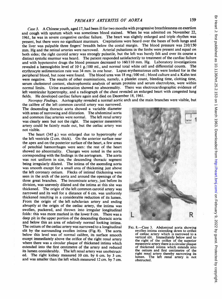
Necropsy Findings. Aortography revealed a normal aortic arch and the main branches were visible, butthe calibre of the left common carotid artery was narrowed.The descending thoracic aorta showed a variable diameterwith areas of narrowing and dilatation. The abdominal aortaand common iliac arteries were normal. The left renal artery !1iwas clearly seen but not the right. The superior mesentericartery could be faintly made out, but the cceliac artery wasnot visible.
The heart (345 g.) was enlarged due to hypertrophy ofthe left ventricle (2 cm. thick). On the anterior surface nearthe apex and on the posterior surface of the heart, a few areasof petechial hamorrhages were seen: the rest of the heartshowed no abnormality. Changes were noted in the aortacorresponding with the findings in the aortogram. The aortawas not uniform in size, the descending thoracic segmentbeing -irregularly dilated. The intima of the ascending aorta 7was smooth except for a small area of thickening just abovethe left coronary ostium. Flecks of intimal thickening wereseen in the arch of the aorta and around the openings of thethree great branches. The innominate artery, just before itsdivision, was unevenly dilated and the intima at this site wasthickened. The origin of the left common carotid artery wasnarrowed and its wall for a distance of 6 cm. was uniformlythickened resulting in a considerable reduction of its lumen.From the origin of the left subclavian artery and endingabruptly at the origin of the coeliac artery, the intima wasswollen, puckered, and thrown into irregular longitudinalfolds: this was more marked in the lower 8 cm. There was a Ldeep pit in the upper portion of the descending thoracic aortaand below this an area of relatively normal looking intima.The ostium of the coeliac artery was narrowed to a longitudinal FIG. 8. Case 3. Abdominal aorta showingslit by the surrounding swollen intima (Fig. 8). The aorta swollen intima extending down to orificebelow this level was of normal calibre, its surface smooth of cceliac artery which is narrowed to aexcept immediately above the orifice of the right renal artery vertical slith oridfiaceof the superiorwhere there was a circular plaque of thickened intima which mesenteric artery there is a circular plaqueextended into the first centimetre of the artery and reduced of thickened intima which extends intoits lumen considerably. The left renal artery was unobstruct- the ostium and first centimetre of theed. The right kidney measured 10 cm. by 6 cm. by 3 cm. right renal artery is notand was smaller than the left which measured 12 cm. by 7 cm. obstructed.
159
on February 9, 2022 by guest. P
rotected by copyright.http://heart.bm
j.com/
Br H
eart J: first published as 10.1136/hrt.25.2.153 on 1 March 1963. D
ownloaded from
DANARAJ, WONG, AND THOMAS
by 4 cm. The capsules of both kidneys stripped easily and the cut surfaces appeared normal. Other organswere congested but otherwise normal.
On microscopical examination, affected portions of the aorta and the innominate and left commoncarotid arteries revealed an extensive panarteritis similar to that described in the previous two cases. Sectionsof both kidneys showed no abnormality.
Case 4. A Chinese girl, aged 16, complained of breathlessness on exertion for three months. A few hoursbefore admission on December 8, 1956, she became very breathless and had cough with frothy blood-stained sputum. She was found to be in severe congestive cardiac failure with a blood pressure of 160/110mm. Hg. The heart was slightly enlarged, but there were no significant murmurs. All peripheral pulseswere palpable and equal. The fundi were normal. She responded satisfactorily to treatment of heartfailure. The hemoglobin was 12 6 g./100 ml. and total white cell and differential counts were normal.The erythrocyte sedimentation rate was 50 mm. in one hour. The blood Kahn test was negative. Otherblood examinations (as reported in the previous case) did not reveal any abnormality. Urine examinationwas normal. The electrocardiogram gave evidence of left ventricular hypertrophy, and on X-ray examinationthe heart was found to be slightly enlarged with a prominent left border. She was treated with hypotensivedrugs, but response was not satisfactory. After about a year of observation it was noted that the left radialpulse was not as forceful as the right and that there was a difference in blood pressures: left arm 140/110 andright arm 220/150 mm. Hg. A systolic bruit was now heard to the left of the umbilicus. Further investi-gations were then carried out.
Intravenous pyelography showed good excretion by both kidneys with the left kidney slightly smallerthan the right: the calyces were of normal size. There was no dilatation or stricture of either ureter, but thecalibre of the left was grossly irregular due to marginal indentations which were present almost the entirelength of its course. Aortography revealed a normal aortic arch with good visualization of the innominateand left common carotid branches, but the left subclavian did not fill beyond 1 cm. from its origin. Therest of the aorta was normal except that, about the level of the origin of the coeliac artery, the calibre of theabdominal aorta was considerably narrowed by a deep indentation on its left side; the aorta distal to thisconstriction was slightly dilated. The branches of the cceliac artery were clearly seen, but the superiormesenteric and left renal arteries were not. The right renal artery was slightly narrowed at its origin anddilated along the middle third of its course. The inferior mesenteric artery was hypertrophied with largeleft colic branches. In later films, small, tortuous, tightly coiled ureteric arteries were seen extending fromthe region of the left renal pedicle to below the level of the bifurcation of the aorta. Both kidneys were out-lined, the left being slightly smaller than the right.
The patient has refused operation and the blood pressure has remained raised in spite of treatment withvarious hypotensive drugs. The left radial pulse is not palpable now and the abdominal bruit has becomelouder. Radiological examination in November 1961 shows further cardiac enlargement, and a curvilinearcalcification of the abdominal aorta for a distance of 3 5 cm. at the level of the first and second lumbarvertebral bodies: the left kidney is much smaller than the right.
Case5. A Chinese girl, aged 15, was admitted to hospital in April 1954 for fits and was discovered to besuffering from hypertension. She was first seen by us in April 1957, when she complained of pain over theleft shoulder and arm of two months' duration. There was a mass of firm matted lymph nodes in the regionof the lower end of the left sternomastoid muscle and, along its length, several discrete nodes, one of which onbiopsy showed only lymphoid hyperplasia. Blood pressure taken in the right arm was 150/110 mm. Hg.Pulses were not felt in the left upper limb. An electrocardiogram and radiograph of the chest were normal.She was admitted to hospital in December 1959 because the cervical nodes had become larger. Tuberculouslymphadenitis was diagnosed after a second biopsy and specific treatment was started. By April 1960, thenodes were no longer palpable, and she felt well. On re-examination no pulses could be felt in the left upperlimb, while the blood pressure in the right arm was 180/115 mm. Hg. The heart was not enlarged and thefundi were normal.
The hmemoglobin level was 12-6 g./100 ml. and total white cell and differential counts were normal.The erythrocyte sedimentation rate was 14 mm. in one hour. Urine examination was normal. The bloodKahn test was negative. Other examinations of the blood did not disclose any abnormality. Aortographyshowed a normal aortic arch with good filling of the innominate and left common carotid arteries, but theleft subclavian artery was occluded one centimetre from its origin. The abdominal aortogram appearednormal and the right renal artery was faintly but clearly visible; the left one was not seen. The nephrogramshowed a large right kidney, but a small left one.
Two days after nephrectomy, the blood pressure in the right arm had fallen to 140/90 mm. Hg and it has
1 60
on February 9, 2022 by guest. P
rotected by copyright.http://heart.bm
j.com/
Br H
eart J: first published as 10.1136/hrt.25.2.153 on 1 March 1963. D
ownloaded from
PRIMARY ARTERITIS OF AORTA
remained at this level for the past year. In January 1961, the femoral arterial pulses felt weaker thanpreviously, and a soft systolic bruit was heard in the midline just above the umbilicus. Retrogradefemoral aortography was carried out. This revealed an abdominal aorta of normal calibre in its upper part,tapering to a narrowed segment at the level of the origin of the right renal artery, below which was a shortpost-stenotic dilatation with irregular walls: the (right) renal artery was unobstructed in its entire course.
The left kidney weighed 21 g. Its upper two-thirds appeared normal, but the lower third was shrunkenwith marked cortical atrophy: a clear line of demarcation separated the two portions. Microscopicalexamination of the lower portion ofthe kidney showed diffuse fibrosis with cellular infiltration by lymphocytesand plasma cells in the interstitium. There was tubular atrophy and hyalinization of the glomeruli whichwere crowded together. Some of the tubules contained eosinophilic hyaline material. Marked intimalthickening of the arterioles with narrowing of the lumen was present. Similar changes were noted in theupper part of the kidney, but to a much lesser degree. The renal artery and its main branches at the hilumshowed minimal intimal thickening, but there was no occlusion or narrowing of their lumen.
Case 6. A Chinese girl, aged 18, was seen in October 1958 for bancroftian filarial lymphangitis of bothlegs which responded to treatment with diethylcarbamazine. At that time it was noted that neither arterialpulsations nor blood pressure readings could be obtained in the upper limbs. She was admitted in January1959 with breathlessness on exertion of one month's duration and subsequently developed acute pulmonarycedema while in hospital. The blood pressure reading in the lower limbs was 230/130 mm. Hg. The patientwas treated for cardiac failure, responded satisfactorily, and was discharged. On March 15, 1959 shedeveloped headache, giddiness, blurring of vision, and fits, and was readmitted in a drowsy state. She wastreated with hypotensive drugs and recovered consciousness. In July 1959 she was seen again. There werestrong, visible pulsations of both carotid arteries. Radial, brachial, and axillary pulses were not felt andblood pressure readings were not obtained in either arm. Both femoral arteries were strongly pulsatile andthe blood pressure recorded over each thigh was 230/130mm. Hg. There was slight cardiac enlargement withan accentuated second sound in the aortic area, but no murmurs. The retinal arteries were narrowed.
The hemoglobin level was 12-4 g./100 ml. and the total white cell and differential counts were normal.The erythrocyte sedimentation rate was 40 mm. in one hour. The Kahn test was negative. A cardio-gram showed left ventricular hypertrophy. On radiological examination of the chest, the heart was foundto be slightly enlarged with a prominent left border; the lung fields were clear. Intravenous pyelographyshowed poor excretion on both sides with the right kidney smaller than the left. Aortography revealed anormal aortic arch with the innominate and left common carotid arteries arising from a common root.The right common carotid artery was well filled, but the right subclavian was not visible and its probable siteof origin was represented by a slight irregularity of the right wall of the innominate artery; the left subclavianwas visible only at its origin from the aortic arch. A large number of collateral vessels were seen in the neck.The lumen of the descending thoracic aorta was of uneven calibre with a definite constriction at the level ofthe 10th and 11th thoracic vertebre; below this there was a fusiform dilatation of the upper half of theabdominal aorta with an irregular outline. The lower half of the abdominal aorta was relatively narrow butof even calibre. The proximal part of the right renal artery was dilated; it then narrowed abruptly to a con-striction before dividing into its branches. There were two renal arteries on the left side, the upper one beingnarrowed at its origin. The nephrogram showed that the left kidney was much larger than the right. Aright nephrectomy was done in December 1959, but the blood pressure did not return to normal levels.Response to treatment with hypotensive drugs was poor. One year later retrograde aortography wasrepeated when it was found that stenosis of the left upper renal artery was greater than previously.
The right kidney weighed 50 g.: its capsule stripped easily revealing a faintly granular surface. Therewas a clear demarcation between medulla and cortex which was 0 5 cm. in width. On microscopicalexamination the glomeruli were seen to be crowded together due to atrophy of the convoluted tubules:the glomerular tufts were not abnormal although many showed periglomerular fibrosis. A chronic inflam-matory reaction, especially in the cortex, was a feature in all sections examined, lymphocytes and plasmacells being present either diffusely or in focal aggregates in the interstitial tissue. The intrarenal arteries andarterioles showed no changes. Except for some focal intimal thickening, the renal artery was normal.
Case 7. A Chinese girl, aged 15, was admitted in July 1959 complaining of breathlessness on exertion,palpitations, and cough with frothy, blood-tinged sputum. She was acutely ill and in congestive cardiacfailure. The heart was not enlarged but the apex beat was forceful, and there was gallop rhythm, but no mur-murs were heard. The blood pressure was 180/120 mm. Hg in both arms and all peripheral pulses were felt.The retinal arteries were narrowed. She responded satisfactorily to treatment of the cardiac failure. Exceptfor a hiemoglobin level of 11 -8 g./100 ml., a leucocytosis of 12,900 c.mm. with a normal differential count,M
161
on February 9, 2022 by guest. P
rotected by copyright.http://heart.bm
j.com/
Br H
eart J: first published as 10.1136/hrt.25.2.153 on 1 March 1963. D
ownloaded from
DANARAJ, WONG, AND THOMAS
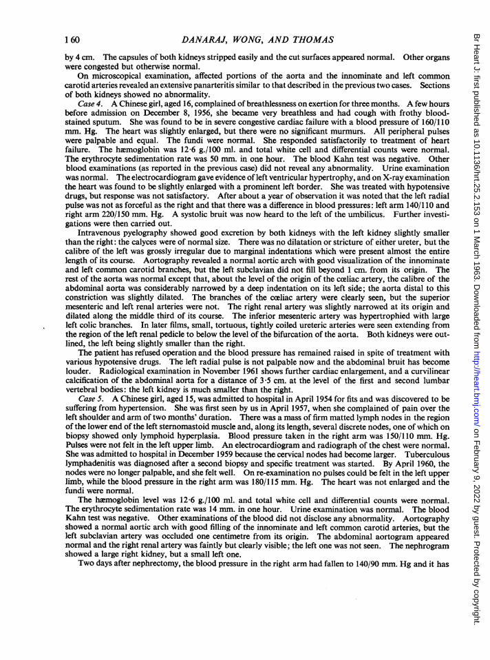
and an erythrocyte sedimentation rate of 23 mm. in one hour, examinations of blood and urine revealed noabnormality. The Kahn test was negative. There was electrocardiographic evidence of left ventricularhypertrophy. Radiological examination of the chest showed a slight increase in cardiac size. Intravenouspyelography was normal. Aortography revealed a marked constriction of the left renal artery at its originwith a post-stenotic dilatation: the abdominal aorta and iliac arteries were clearly visible and of normalcalibre. Twenty-four hours after left nephrectomy, the blood pressure became normal and has remained soduring the past two years. Routine examination of the pulses in all limbs during her periodic visits revealedno abnormality until towards the end of the first year after operation when the femoral arterial pulses werenoted to be weak, and on auscultation over the umbilicus a systolic murmur was heard. Aortography wasrepeated and revealed a narrowing of the abdominal aorta commencing immediately below the originof the right renal artery and extending for a distance of 6 cm. before a normal calibre was regained(Fig. 9).
The left kidney weighed 115 g.: the capsule.8 ....stripped easily leaving a smooth surface. There
. . . .. ..>wasclear demarcation of medulla and cortex
Au:g ~~which was 0-5 cm. wide. Microscopical exam-ination of the kidney showed no abnormality,except for a few lymphocytic aggregates in theinterstitium. The renal artery was normal.... ,.j,.,,.i'.8.' ' ' ,., ........Case8. A Chinese girl, aged 18, wasapparently well until 12 days before admission
A- : _ ] |iwhenshe suffered an attack of severe breathless-..............
ness which improved with treatment. She then. .: 'B*........becamebreathless on exertion and was admitted
to hospital on July 14, 1960 in heart failure.All peripheral arterial pulses were easily felt,but the carotids were forceful and the left radialpulse was weaker than the right. The bloodpressure reading in the right arm was 150/60, inthe left arm 120/100, and in the lower limbs180/70 mm. Hg. A forceful and displaced apexbeat was felt. On auscultation, triple rhythmwas noted, and along the left border of thesternum an early diastolic murmur of slightintensity was heard. A systolic bruit was heard
_p2!'5'8N"' over the epigastrium to the right of the midline.A few crepitations were heard over the bases ofboth lungs and the liver was enlarged to onefinger's breadth below the costal margin.
FIG. 9.-Case 7. Aortogram showing stenosis of segment of Examination of the fundi revealed a mildabdominal aorta below origin of right renal artery. degree of spasm of the arteries. She improved
on treatment of the cardiac failure.The hxemoglobin level was 10 4 g./100 ml. and both total white cell and differential counts were normal.
The erythrocyte sedimentation rate was 25 mm. in one hour. The Kahn test was negative. There wasevidence of left ventricular hypertrophy on electrocardiographic examination. A radiograph of the chestshowed a large heart with a prominent left ventricle. On intravenous pyelography, there was normalexcretion by both kidneys, but better visualization of the right kidney which was smaller than the left.Aortography revealed a slightly dilated ascending aorta with a moderate degree of regurgitation of dye intothe left ventricle. The innominate artery was dilated and irregular in outline and the left subclavian arterywas narrowed at its origin; the right subclavian was not visible. The calibre of the descending thoracicand upper part of the abdominal aorta was grossly irregular with areas of narrowing and dilatation. Therewas stenosis of the right renal artery with post-stenotic dilatation and the nephrograms confirmed thedifference in size of the kidneys. The patient was transferred to another hospital where she died inNovember 1960 in congestive heart failure.
Case 9. An Indian women, aged 32, was admitted on September 2, 1960 for breathlessness on effort often days' duration. She was in cardiac failure. All the peripheral pulses were felt, but those in the left
162
on February 9, 2022 by guest. P
rotected by copyright.http://heart.bm
j.com/
Br H
eart J: first published as 10.1136/hrt.25.2.153 on 1 March 1963. D
ownloaded from
PRIMARY ARTERITIS OF AORTA
upper limb were feeble. The blood pressure was 200/130 mm. Hg in the right arm, and 170/100 mm.Hg in the left. The heart was slightly enlarged, and there was triple rhythm but no murmurs. On ausculta-tion over the abdomen, a soft systolic bruit was heard above and to the left ofthe umbilicus. The hemoglobinlevel was 10-7 g./100 ml. and total white cell and differential counts were normal. The erythrocyte sedi-mentation rate was 13 mm. at the end of one hour, and the Kahn test was negative. The electrocardiograrnshowed no abnormality. Radiological examination confirmed the slight cardiac enlargement. No abnor-mality was noted following intravenous pyelography. Aortography showed a normal aortic arch andbranches, but there was obstruction to the flow of dye in the left and right subclavian arteries about 3 cm. and2 cm. respectively from their origins. The right renal artery was normal, but there were two arteries on theleft side, the upper and larger one being narrowed at its origin from the abdominal aorta. The segment ofaorta at the level of the renal arteries was of irregular calibre. The left kidney was slightly smaller than theright.
The patient responded satisfactorily to treatment for cardiac failure, but later refused operation. Shehas been observed over the past year and her response to hypotensive drugs has not been satisfactory.Pulses in both upper limbs are now feeble as are the carotids, but the femoral pulses remain forceful.
DISCUSSION
The clinical presentation in this series was one of cardiac failure, encephalopathy, or cerebralhiemorrhage resulting from hypertension. In three patients the cause of the hypertension wasestablished at necropsy and was due to renal ischemia consequent on stenosis of one or both renalartery orifices, producing in effect a clinical equivalent of the Goldblatt clamp. The primary lesion,however, was in the aorta and consisted of a panarteritis affecting various segments and the proximalportions of branches arising from such segments. The orifices of these arteries were narrowed bythickened intima or occluded by thrombi resulting in reduced blood supply to the dependent organor limb. In addition to the renal arteries, other visceral branches such as the right coronary,coeliac, and superior mesenteric, and all the large branches, including the common iliac, were affected.Localized dilatations of the abdominal aorta and right common iliac artery at the site of the lesionswere present in Case 1, and irregular dilatations of the descending thoracic aorta and innominateartery were noted in Case 3. The aortic wall between the affected areas appeared normal andatheroma did not complicate the pathological changes. However, it is possible that the arteritiscould have spread to involve the entire aorta were it not for the early death of these patients from theeffects of hypertension.
The histological changes, which were similar in the three cases, were confined to the aorta andproximal portions of affected branches. The adventitia was thickened, with formation of some newvessels and infiltration by lymphocytes and plasma cells, mainly perivascular in distribution: manyof the vasa vasorum showed marked endothelial proliferation with almost complete obliteration ofthe lumen. The characteristic change in the media was a gross destruction of elastic fibres resultingin their complete absence in some areas with localized dilatation of the wall: sharply demarcatedareas of coagulative necrosis surrounded by round cells were also present. In addition, there wasincreased vascularization, some lymphocytic infiltration, and a few giant cells. The intima wasgrossly thickened with thrombi at certain sites, and in one area calcification was present in the deeperparts. The segmental distribution of the lesions in the aorta, the sharply demarcated areas ofcoagulative necrosis in the media resembling small infarcts, and the endarteritis of the vasa vasorumresulting in occlusion of their lumen, all suggest that the necrotic changes in the media result from ob-struction to its blood supply. The intimal hyperplasia with thrombosis is probably secondary to thechanges in the media.
The naked eye appearance of the lesions and their histological picture closely conformed todescriptions of lesions in the aortic arch and its branches seen in classical Takayashu's syndrome.Although the histological appearance of the aortic lesions was somewhat similar to that seen insyphilis, and to what has been reported in some cases of ankylosing spondylitis (Clark, Kulka, andBauer, 1957) and giant-cell temporal arteritis (Cardell and Hanley, 1951), there was no other evidence
163
on February 9, 2022 by guest. P
rotected by copyright.http://heart.bm
j.com/
Br H
eart J: first published as 10.1136/hrt.25.2.153 on 1 March 1963. D
ownloaded from
DANARAJ, WONG, AND THOMAS
of these diseases, either clinically or at necropsy: in addition, naked eye appearance of the aorta didnot resemble syphilis, and the serological test was negative in these cases. No organisms were seenin the lesions to suggest an infective process.
Evidence obtained at necropsy from these and other reported cases indicates that any part of theaorta may be involved with resultant diversity of the clinical picture. When the ascending aorta isaffected, occlusion of the orifice of a coronary artery may occur with consequent myocardial in-farction (Barker and Edwards, 1955). Involvement of the aortic arch results in brachiocephalicmanifestations, the classical Takayashu syndrome. Narrowing of the lumen of the thoracic aortagives rise to absent pulses in the lower extremities and a clinical picture suggestive of coarctation ofthe aorta (Correa and Arauijo, 1958; Isaacson et al., 1959). The abdominal aorta may be affectedresulting in stenosis of one or both renal artery orifices and hypertension (Cases 1 to 3; Danaraj andWong, 1959; Isaacson, 1961). Mesenteric artery occlusion may cause abdominal pain. The lowerpart of the abdominal aorta or its bifurcation may be involved giving rise to absent femoral pulses onone (Case 1) or both sides. In all these patients the underlying lesion was a panarteritis, presenting asimilar microscopical appearance. Based on these clinico-pathological reports, the concept ofprimary arteritis of the aorta should be enlarged to include not only the early-recognized brachio-cephalic syndrome, but other modes of presentation depending on which segments of the aorta areaffected.
Renal artery involvement was demonstrated in aortograms in six patients (Cases 4 to 9). Fiveof these patients had weak or absent pulses in one or more limbs, and aortography revealed irre-gularity of the aortic calibre with narrowing or occlusion of one or more of its branches, in additionto stenosis of a renal artery. The only arterial involvement that could be detected in the remainingpatient (Case 7) when first seen was left renal artery stenosis. Following nephrectomy, her bloodpressure returned to normal, but a year later the femoral pulses became weak, and constriction of along segment of the aorta below the origin of the renal arteries was seen in an aortogram. It seemslikely that the primary disease process in these patients is in the aorta, and is similar to that found inthe patients who come to necropsy.
On the basis of clinical, radiological, and pathological evidence, this group of nine patients con-stitutes a single clinico-pathological entity resembling what has been described as Takayashu'ssyndrome except that other segments of the aorta besides the aortic arch were also affected.Although narrowing of the arterial lumen typifies the lesion, localized dilatation of the affectedarteries may occur as was noted in Cases 1 and 3 post mortem. Several areas of ectasia were des-cribed in the case reported by Cosma et al. (1959) and in the first case of Isaacson's series(1961). Furthermore, dilatation involving the aorta at its origin may result in aortic regurgitationas seen in Case 8 and as reported by Jervell (1954).
The disease runs a chronic and progressive course, increasing involvement of the aorta beingindicated by weakening or absence of pulses that were previously present (Cases 4, 5, 7, and 9), orthe development of murmurs over the aorta (Cases 5 and 7). Intravenous pyelography is animportant preliminary investigation in patients presenting with hypertension and may revealdifferences in kidney size (Hodson, 1957) or irregularity of the ureter caused by enlarged collateralureteric arteries to the ischimic kidney (Case 4; Thomas and Levin, 1961). However, aortographyis essential in establishing the extent of the aortic lesion, the diagnosis of renal artery stenosis, theinvolvement of other branches, and the existence of collateral circulation. When the large branchesto the head and limbs are occluded, the development of collateral circulation enables the patient tosurvive for several years. With involvement of renal arteries, however, prognosis immediatelybecomes serious because resultant hypertension is severe and leads to premature death.
Various workers (Poutasse, 1956; DeCamp and Birchall, 1958; Morris et al., 1960) have shownthat hypertension secondary to occlusive lesions of the renal artery may be successfully treated bynephrectomy or by vascular reconstructive procedures. In the three patients who were subjected tonephrectomy, the blood pressure returned to normal in two, but remained high in the third (Case 6)probably because of ischemia of the remaining kidney from concomitant stenosis of its artery.
164
on February 9, 2022 by guest. P
rotected by copyright.http://heart.bm
j.com/
Br H
eart J: first published as 10.1136/hrt.25.2.153 on 1 March 1963. D
ownloaded from
PRIMARY ARTERITIS OF AORTA
The removal of a kidney is a simpler and less hazardous operation than procedures to restore ade-quate renal blood flow, but in circumstances such as these, where the primary lesion is in the aortaand may progress to involve both renal arteries, some form of arterial reconstruction is to be pre-ferred.
The etiology of the condition remains obscure. Laboratory investigations did not show anyconsistent abnormality except for a raised erythrocyte sedimentation rate and a mild anemia. TheKahn test for syphilis was negative and no L.E. cells were found in the peripheral blood. Tuber-culous lymphadenitis was present in one patient (Case 5) in this series, and pulmonary tuberculosisin another patient reported earlier as a case of obliterative brachiocephalic arteritis (Danaraj andWong, 1960). One patient in the Japanese series (as reviewed by Caccamise and Whitman, 1952)had active pulmonary tuberculosis. In view of the high prevalence of tuberculosis both in Japanand Singapore this association is probably coincidental.
SUMMARYThe evidence presented by the nine cases described in this paper indicates that primary arteritis
of the aorta is a single clinico-pathological entity of which Takayashu's syndrome is a part. Differentsegments of the aorta may be affected resulting in a variety of symptom complexes. In this series,the mode of presentation was hypertension consequent on renal artery stenosis. Early diagnosis isimportant and surgical treatment is indicated to relieve the hypertension which carries an immedi-ately serious prognosis.
We are indebted to Professor Yeoh Ghim Seng and Mr. Yong Nen Khiong for the surgical treatment of Cases 5,6, and 7; to Dr. Tock Peng Chong for performing the necropsy on Case 3; and to Mr. V. Nalpon and Mr. Ho TatSeng for the photographs.
REFERENCESAsk-Upmark, E. (1954). Acta med. scand., 149, 161.Barker, N. W., and Edwards, J. E. (1955). Circulation, 11, 486.Caccamise, W. C., and Whitman, J. F. (1952). Amer. Heart J., 44, 629.Caldwell, R. A., and Skipper, E. W. (1961). Brit. Heart J., 23, 53.Cardell, B. S., and Hanley, T. (1951). J. Path. Bact., 63, 587.Clark, W. S., Kulka, J. P., and Bauer, W. (1957). Amer. J. Med., 22, 580.Correa, P., and Araujo, J. (1958). Amer. J. clin. Path., 29, 560.Cosma, J., Maruyama, Y., Pettet, J. R., and Cutshall, V. (1959). Circulation, 20, 267.Danaraj, T. J., and Wong, H. 0. (1959). Circulation, 20, 856.-~-~(1960). Amer. J. Cardiol., 5, 277.
DeCamp, P. T., and Birchall, R. (1958). Surgery, 43, 134.Frovig, A. G., and Loken, A. C. (1951). Acta psychiat. scand., 26, 313.Gibbons, T. B., and King, R. L. (1957). Circulation, 15, 845.Giffin, H. M. (1939). Proc. Mayo Clin., 14, 561.Hodson, C. J. (1957). Proc. roy. Soc. Med., 50, 539.Isaacson, C. (1961). J. Path. Bact., 81, 69.
, Klachko, D. M., Waybume, S., and Simson, I. W. (1959). Lancet, 2, 542.Jervell, A. (1954). Amer. Heart J., 47, 780.Kalmansohn, R. B., and Kalmansohn, R. W. (1957). Circulation, 15, 237.Koszewski, B. J. (1958). Angiology, 9, 180.Lessof, M. (1958). Guy's Hosp. Rep., 107, 53.Lewis, T., and Stokes, J. (1942). Brit. Heart J., 4, 57.Morris, G. C. Jr., De Bakey, M. E., Cooley, D. A., and Crawford, E. S. (1960). Ann. Surg., 151, 854.Poutasse, E. F. (1956). Circulation, 13, 37.Ross, R. S., and McKusick, V. A. (1953). Arch. intern. Med., 92, 701.Savory, W. S. (1856). Med.-chir. Trans., 39, 205.Shimizu, K., and Sano, K. (1951). J. Neuropath. clin. Neurol., 1, 37.Skipper, E., and Flint, F. J. (1952). Brit. med. J., 2, 9.Takayashu, M. (1908). Acta Soc. Ophthal. Jap., 12, 554.Thomas, R. G., and Levin, N. W. (1961). Brit. J. Radiol., 34, 438.Trias de Bes, L., Lucas, J. G. S., and Barcons, F. B. (1955). Brit. Heart J., 17, 484.
165
on February 9, 2022 by guest. P
rotected by copyright.http://heart.bm
j.com/
Br H
eart J: first published as 10.1136/hrt.25.2.153 on 1 March 1963. D
ownloaded from