Embed Size (px)
Citation preview
RESEARCH ARTICLE Open Access
Primary small cell carcinoma of the esophagus:clinicopathological study of 44 casesWei-Wei Chen1†, Feng Wang1†, Dong-Sheng Zhang1, Hui-Yan Luo1, Zhi-Qiang Wang1, Feng-Hua Wang1,Miao-Zhen Qiu1, Chao Ren1, Xiao-Li Wei1, Wen-Jing Wu1, Yu-Hong Li1 and Rui-Hua Xu2*
Abstract
Background: Primary small cell carcinoma of the esophagus (SCCE) is a highly aggressive disease characterized byearly dissemination and poor prognosis. Because of the rarity of this disease, few previous studies have investigatedthe biomarkers associated with its prognosis. Leucine-rich repeat-containing G-protein coupled receptor 5 (Lgr5) isa stem cell marker and a member of the canonical Wnt-signaling cascade. However, the clinical role of Lgr5 in SCCEremains unknown.
Methods: Tissue sections were obtained from 44 patients diagnosed with SCCE and expression of Lgr5 wasexamined by immunohistochemistry. The correlations between Lgr5 expression, and clinical parameters andprognostic significance were evaluated.
Results: Lgr5 was expressed in SCCE cancer tissues. High Lgr5 expression was significantly correlated with lymphnode metastasis (p = 0.003), late stage (p = 0.003) and unfavorable response to chemotherapy (p = 0.013) accordingto RECIST 1.0 criteria. Patients with higher Lgr5 expression levels had shorter overall survival times than those withlower expression levels.
Conclusions: These results demonstrated that overexpression of Lgr5 was significantly correlated with lymph nodemetastasis, tumor stage, and response to chemotherapy. Furthermore, high levels of Lgr5 expression appeared tobe associated with poorer survival in patients with SCCE.
Keywords: Small cell carcinoma, Esophagus, Prognosis, Lgr5
BackgroundEsophageal carcinoma is one of the most common ma-lignancies in China, but primary small cell carcinoma ofthe esophagus (SCCE) is a relatively rare histopatho-logical type, accounting for only 1-2.8% of esophagealcarcinomas [1,2]. SCCE is highly aggressive, and charac-terized by early dissemination and poor prognosis[3-10]. A total of 5,379 cases of esophageal cancer and2,061 cases of small cell carcinoma were recorded at SunYat-sen University Cancer Center from 1994 to 2012,including 93 cases of SCCE. Little is known about theclinicopathological features of SCCE, and it is necessaryto identify biomarkers for predicting prognosis and fordistinguishing individuals with unfavorable prognoses.
The Wnt signaling pathway is important for adult tis-sue maintenance. Perturbations in Wnt signaling causehuman cancers [11]. Leucine-rich repeat-containing G-protein coupled receptor 5 (Lgr5) is an orphan G-proteincoupled receptor (GPCR) and a stem cell marker firstdescribed as a Wnt target gene [12]. It is also a member ofthe canonical Wnt-signaling cascade, which forms a sig-naling gradient in the intestinal crypt, thereby regulatingcell proliferation and differentiation [13]. Lgr5 was alsoidentified as a marker of poor prognosis in colon, ovaryand liver cancers [14,15], and was considered to beinvolved in tumorigenesis in Barrett’s esophagus andesophageal adenocarcinoma [16].However, the expression and potential clinical signifi-
cance of Lgr5 in SCCE has not been determined. In thisstudy, we analyzed the expression levels of Lgr5 andtheir relationships with clinicopathological features in 44patients with SCCE.
* Correspondence: [email protected]†Equal contributors2Department of Medical Oncology, Sun Yat-sen University Cancer Center, 651Dong Feng Road East, Guangzhou 510060, ChinaFull list of author information is available at the end of the article
© 2014 Chen et al.; licensee BioMed Central Ltd. This is an Open Access article distributed under the terms of the CreativeCommons Attribution License (http://creativecommons.org/licenses/by/2.0), which permits unrestricted use, distribution, andreproduction in any medium, provided the original work is properly credited. The Creative Commons Public DomainDedication waiver (http://creativecommons.org/publicdomain/zero/1.0/) applies to the data made available in this article,unless otherwise stated.
Chen et al. BMC Cancer 2014, 14:222http://www.biomedcentral.com/1471-2407/14/222
MethodsTumor tissues and patient informationA total of 44 paraffin-embedded samples were obtainedfrom patients diagnosed with SCCE from January 1 1994to January 1 2012 at Sun Yat-sen University Cancer Cen-ter. The diagnosis of SCCE had been confirmed by thePathology Department of Sun Yat-sen University CancerCenter, based on the 2000 World Health Organizationhistological criteria for esophageal small cell carcinoma[17] and 2004 histological criteria for pulmonary smallcell carcinoma [18]. As described previously [10], tumorcells were characterized by small, spindle-like, round orovoid shape, scarce cytoplasm, indistinct cell borders,and an inconspicuous or absent nucleolus. Informationon the neuroendocrine markers neuron-specific enolase(NSE), synaptophysin (Syn), chromogranin A (CgA) andCD56 were obtained from patient pathology reports. Allpatients were positive for CgA and/or Syn expression;about 70.5% of patients were Syn-positive, 84.1% wereCgA-positive, 56.8% of cases were NSE-positive and34.1% were CD56-positive.Informed consent was obtained from all patients and
the study was approved by the Research Ethics Commit-tee of Sun Yat-Sen University. No patients had receivedany treatment prior to surgery or biopsy. Of the 44 sam-ples, 29 were obtained at surgery and 15 by biopsy. Theclinicopathological records of all patients were reviewed.The American Joint Committee on Cancer (AJCC) cli-nical and pathologic staging system was adopted for allpatients. Overall survival (OS) was defined as the timefrom the date of diagnosis to the point of death or thelast follow-up.
Immunohistochemistry (IHC)IHC was performed to study the expression of Lgr5 inSCCE tissues, as described previously [19]. Briefly, paraf-fin-embedded tissue sections were baked at 60°C for 2 h,deparaffinized, and rehydrated. After treatment with 3%hydrogen peroxide for 30 min, the sections were put in ahigh-pressure environment for antigen retrieval. Tissuesections were incubated with rabbit anti-Lgr5 (1:400;Abcam, United states) overnight at 4°C, then treatedwith anti-rabbit secondary antibody for 40 min, followedby treatment with diaminobenzidine tetrahydrochloride(DAB), and counterstaining with hematoxylin. Humancolon cancer tissues with strong Lgr5 staining were usedas positive controls, based on previous reports [20]. Lgr5immunostaining was evaluated by two independent ob-servers who were blinded to the clinicopathological cha-racteristics of the patients. Immunostaining scores wereawarded by two independent observers according to thepercentage and intensity of the stained cells. Positivityvalues were as follows: 0 (<10%), 1 (10–25%), 2 (25–50%),3 (50–75%), and 4 (>75%). Intensity values were as
follows: 0 (negative), 1 (weak staining), 2 (moderate stai-ning), and 3 (strong staining). The final score was calcu-lated by multiplying the above two values. For subsequent
Table 1 Clinicopathological characteristics and Lgr5expression in tumors from 44 patients with SCCE
Variable No.of patients %
Gender
Male 33 75.0
Female 11 25.0
Age
>60 15 34.1
<=60 29 65.9
PS score
>1 4 9.1
<=1 40 90.9
Tumor length(39 available)
> = 5 cm 21 53.8
<5 cm 18 46.2
Tumor location
Upper thoracic segment 7 15.9
Middle thoracic segment 22 50.0
Lower thoracic segment 15 34.1
AJCC stage(43 available)
I/II 19 44.2
III/IV T classification(40 available) 24 55.8
T < =2 20 45.5
T > 2 20 45.5
N classification(42 available)
N0 16 36.4
N+ 26 59.1
M classification(43 available)
M0 33 75.0
M1 10 22.7
Treatment approach
Surgery
No 11 25.0
Yes 33 75.0
Chemotherapy
No 16 36.4
Yes 28 63.6
Radiotherapy
No 16 36.4
Yes 28 63.6
Lgr5 expression
Low 23 52.3
High 21 47.7
Chen et al. BMC Cancer 2014, 14:222 Page 2 of 7http://www.biomedcentral.com/1471-2407/14/222
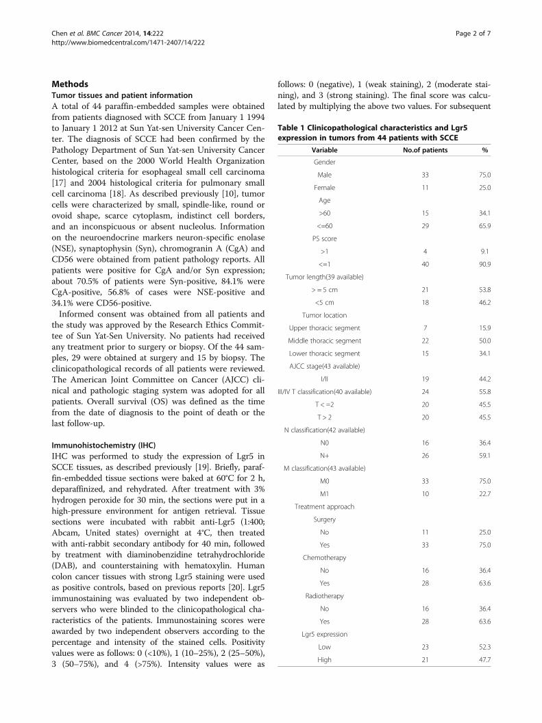
Figure 1 Immunohistochemical staining of Lgr5 in cancerous tissue and adjacent normal mucosa. A: Higher levels of Lgr5 expressionwere observed in tumor tissues compared with ANT (×100 magnification). Colon cancer tissue was used as a positive control (insert). B: Higherlevels of Lgr5 expression were observed in tumor tissues compared with ANT (×400 magnification). Abbreviations: T: tumor, ANT: adjacentnormal tissue.
Table 2 Correlation between clinicopathological characteristics and Lgr5 expression in patients with SCCE
Lgr5 expression
Characteristics Low or none No. cases High No. cases Chi-square test P-value
Gender .601
male 18 15
female 5 6
Age .241
>60 6 9
<=60 17 12
Tumor length(39 available) .256
> = 5 cm 9 12
<5 cm 11 7
Tumor location .721
Upper thoracic segment 3 4
Middle thoracic segment 11 11
Lower thoracic segment 9 6
AJCC stage(43 available) .003
I/II 15 4
III/IV 8 16
T classification(40 available) .206
T < =2 12 8
T > 2 8 12
N classification(42 available) .003
N0 13 3
N+ 9 17
M classification(43 available) .329
M0 19 14
M1 4 6
Chemotherapy response(19 available) .013
Reached CR/PR 5 1
Did not reach CR/PR 3 10
Chen et al. BMC Cancer 2014, 14:222 Page 3 of 7http://www.biomedcentral.com/1471-2407/14/222
analysis, high expression was defined as a final score >4and low expression was a score ≤4.
Statistical analysisStatistical analysis was carried out using SPSS 16.0. Thesignificance of correlations between biomarker expres-sion levels and clinical features were calculated using χ2
tests. Survival curves were displayed by Kaplan–Meieranalysis and differences in survival were assessed by log-rank tests. A two-sided α-error of less than 5% (p < 0.05)was considered as statistically significant.
ResultsExpression of Lgr5 in SCCE and correlation betweenexpression and clinicopathologic featuresDetailed information was collected for 44 patients(Table 1). The median follow-up time for all patientswas 11.1 months (3–84.9 months). Tissue sections weresubjected to IHC to investigate Lgr5 expression levelsand to correlate these with clinicopathological features.
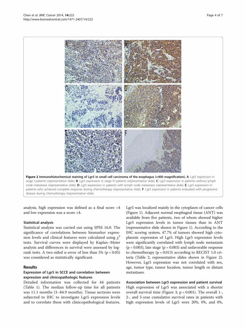
Lgr5 was localized mainly in the cytoplasm of cancer cells(Figure 1). Adjacent normal esophageal tissue (ANT) wasavailable from five patients, two of whom showed higherLgr5 expression levels in tumor tissues than in ANT(representative slide shown in Figure 1). According to theIHC scoring system, 47.7% of tumors showed high cyto-plasmic expression of Lgr5. High Lgr5 expression levelswere significantly correlated with lymph node metastasis(p = 0.003), late stage (p = 0.003) and unfavorable responseto chemotherapy (p = 0.013) according to RECIST 1.0 cri-teria (Table 2, representative slides shown in Figure 2).However, Lgr5 expression was not correlated with sex,age, tumor type, tumor location, tumor length or distantmetastases.
Association between Lgr5 expression and patient survivalHigh expression of Lgr5 was associated with a shorteroverall survival time (Figure 3, p = 0.001). The overall 1-,2-, and 5-year cumulative survival rates in patients withhigh expression levels of Lgr5 were 20%, 0%, and 0%,
Figure 2 Immunohistochemical staining of Lgr5 in small cell carcinoma of the esophagus (×400 magnification). A: Lgr5 expression instage I patients (representative slide). B: Lgr5 expression in stage IV patients (representative slide). C: Lgr5 expression in patients without lymphnode metastasis (representative slide). D: Lgr5 expression in patients with lymph node metastasis (representative slide). E: Lgr5 expression inpatients who achieved complete response during chemotherapy (representative slide). F: Lgr5 expression in patients evaluated with progressivedisease during chemotherapy (representative slide).
Chen et al. BMC Cancer 2014, 14:222 Page 4 of 7http://www.biomedcentral.com/1471-2407/14/222
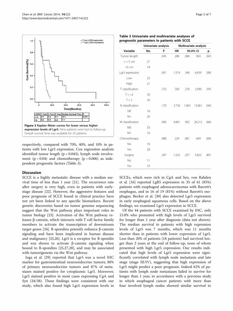
respectively, compared with 70%, 40%, and 10% in pa-tients with low Lgr5 expression. Cox regression analysisidentified tumor length (p = 0.043), lymph node involve-ment (p = 0.04) and chemotherapy (p = 0.006) as inde-pendent prognostic factors (Table 3).
DiscussionSCCE is a highly metastatic disease with a median sur-vival time of less than 1 year [21]. The recurrence rateafter surgery is very high, even in patients with early-stage disease [22]. However, the aggressive features andpoor prognosis of SCCE found in clinical practice havenot yet been linked to any specific biomarkers. Recentgenetic discoveries based on tumor genome sequencingsuggest that the Wnt pathway plays important roles intumor biology [23]. Activation of the Wnt pathway re-leases β-catenin, which interacts with T cell factor familymembers to activate the transcription of downstreamtarget genes [24]. R-spondins potently enhance β-cateninsignaling and have been implicated in human diseaseand malignancy [25,26]. Lgr5 is a receptor for R-spondinand was shown to activate β-catenin signaling whenbound to R-spondins [25,27,28], and may be associatedwith tumorigenesis via the Wnt pathway.Iuga et al. [29] reported that Lgr5 was a novel IHC
marker for gastrointestinal neuroendocrine tumors; 88%of primary neuroendocrine tumors and 87% of meta-stases stained positive for cytoplasmic Lgr5. Moreover,Lgr5 stained positive in most cases expressing CgA andSyn (34/38). These findings were consistent with ourstudy, which also found high Lgr5 expression levels in
SCCEs, which were rich in CgA and Syn. von Rahdenet al. [16] reported LgR5 expression in 35 of 41 (85%)patients with esophageal adenocarcinomas with Barrett’sesophagus, and in 16 of 19 (81%) without Barrett’s eso-phagus. Becker et al. [30] also detected Lgr5 expressionin early esophageal squamous cells. Based on the abovefindings, we examined Lgr5 expression in SCCE.Of the 44 patients with SCCE examined by IHC, only
15.8% who presented with high levels of Lgr5 survivedfor longer than 1 year after diagnosis (data not shown).The median survival in patients with high expressionlevels of Lgr5 was 7 months, which was 11 monthsshorter than in patients with lower expression of Lgr5.Less than 20% of patients (18 patients) had survived lon-ger than 2 years at the end of follow-up, none of whompresented with high Lgr5 expression. Our results indi-cated that high levels of Lgr5 expression were signi-ficantly correlated with lymph node metastasis and latestage (stage III/IV), suggesting that high expression ofLgr5 might predict a poor prognosis. Indeed 62% of pa-tients with lymph node metastases failed to survive forlonger than 1 year, in accordance with a previous studyin which esophageal cancer patients with more thanfour involved lymph nodes showed similar survival to
Figure 3 Kaplan–Meier curves for lower versus higherexpression levels of Lgr5. Nine patients were lost to follow-up.Overall survival time was available for 35 patients.
Table 3 Univariate and multivariate analyses ofprognostic parameters in patients with SCCE
Univariate analysis Multivariate analysis
Variable No. P HR 95.0% CI p
Tumor length .045 .288 .086 .963 .043
> = 5 cm 21
<5 cm 18
Lgr5 expression .001 1.519 .346 6.659 .580
Low 23
High 21
T classification .725 .760 .276 2.090 .595
T < =2 20
T > 2 20
N classification .170 3.726 1.063 13.061 .040
N0 16
N+ 26
M classification .096 4.861 .902 26.212 .066
M0 33
M1 10
Chemotherapy .088 .229 .081 .649 .006
No 16
Yes 28
Surgery .207 1.222 .257 5.822 .801
No 11
Yes 33
Chen et al. BMC Cancer 2014, 14:222 Page 5 of 7http://www.biomedcentral.com/1471-2407/14/222
patients with M1 disease [31]. In Cox regression analysis,lymph node involvement was an independent prognosticfactor, which might explain the association between highexpression of Lgr5 and poor prognosis.SCCE is a systemic disease [21]. Chemotherapy pro-
vides the backbone of SCCE therapy [21,32], which wasin accordance with the fact that chemotherapy wasan independent prognostic factor in the present study.Lgr5 expression may also predict response to chemo-therapy in SCCE. High Lgr5 expression was significantlycorrelated with unfavorable response in this study; only33% of patients with high expression achieved partial/complete responses during chemotherapy. Additionally,Lgr5 may also predict chemotherapy response in colo-rectal cancer [33].A recent study suggested that GPCRs and their signal
transduction pathways may provide promising new thera-peutic approaches [34]. They are involved in the controlof blood pressure, maintenance of kidney function, occur-rence of neurological diseases and the progression of can-cer [34]. Approximately 36% of currently-marketed drugstarget GPCRs [35]. Lgr5 has been suggested to be involvedin cancer progression through regulation of the Wntsignaling pathway [25,27,28]. Lgr5 knockdown was shownto induce cell death [36], and furthermore, a recently-developed monoclonal antibody (KM4056) was reportedto have potent complement-dependent cytotoxicity ac-tivity in vitro, and to show strong anti-tumor activityin vivo [37]. Lgr5 thus remains a potential for targetedtherapy.Overall, the results of this study suggest that Lgr5 pro-
tein expression may represent a possible prognosticmarker in SCCE patients. However, these results need tobe validated by further studies with larger sample sizesand in randomized patient cohorts before Lgr5 IHC canbe used in clinical applications.
ConclusionsIn summary, overexpression of Lgr5 was significantlycorrelated with lymph node metastasis, tumor stage andresponse to chemotherapy, while high levels of Lgr5expression were also associated with poor survival in pa-tients with SCCE.
AbbreviationsSCCE: Primary small cell carcinoma of esophagus; GPCR: Orphan G-proteincoupled receptor; AJCC: American Joint Committee on Cancer; OS: Overallsurvival; ANT: Adjacent normal tissue.
Competing interestsThe authors declare that they have no competing interests.
Authors’ contributionsWC, FW carried out the immunohistochemistry staining and drafted themanuscript. WC, FW, DZ, HL, ZW, FW, MQ, CR, XW and WW participated inthe design of the study and performed the statistical analysis. WC, FW, YLand RX conceived of the study, and participated in its design and
coordination and helped to draft the manuscript. All authors read andapproved the final manuscript.
AcknowledgementsWe thank Shu-Mei Yan and Mu-Yan Cai for diagnosis confirmation and YuChen for editorial assistance. This work was supported by the National HighTechnology Research and Development Program of China (863 Program),China (No.2012AA02A506); Science and Technology Department ofGuangdong Province, China (No. 2012B031800088); Medical ScientificResearch Foundation of Guangdong Province, China (No.C2011019).
Author details1Department of Medical Oncology and State Key Laboratory of Oncology inSouth China, Sun Yat-sen University Cancer Center, Guangzhou, China.2Department of Medical Oncology, Sun Yat-sen University Cancer Center, 651Dong Feng Road East, Guangzhou 510060, China.
Received: 4 September 2013 Accepted: 19 March 2014Published: 25 March 2014
References1. Kim JH, Lee SH, Park J, Kim HY, Lee SI, Nam EM, Park JO, Kim K, Jung CW,
Im YH, Kang WK, Lee MH, Park K: Extrapulmonary small-cell carcinoma:a single-institution experience. Jpn J Clin Oncol 2004, 34(5):250–254.
2. Ku GY, Minsky BD, Rusch VW, Bains M, Kelsen DP, Ilson DH: Small-cellcarcinoma of the esophagus and gastroesophageal junction: review ofthe Memorial Sloan-Kettering experience. Ann Oncol 2008, 19(3):533–537.
3. Nichols GL, Kelsen DP: Small cell carcinoma of the esophagus. The MemorialHospital experience 1970 to 1987. Cancer 1989, 64(7):1531–1533.
4. Huncharek M, Muscat J: Small cell carcinoma of the esophagus. TheMassachusetts General Hospital experience, 1978 to 1993. Chest 1995,107(1):179–181.
5. Nishimaki T, Suzuki T, Nakagawa S, Watanabe K, Aizawa K, Hatakeyama K:Tumor spread and outcome of treatment in primary esophageal smallcell carcinoma. J Surg Oncol 1997, 64(2):130–134.
6. Lam KY, Law S, Tung PH, Wong J: Esophageal small cell carcinomas:clinicopathologic parameters, p53 overexpression, proliferation marker,and their impact on pathogenesis. Arch Pathol Lab Med 2000,124(2):228–233.
7. Pantvaidya GH, Pramesh CS, Deshpande MS, Jambhekar NA, Sharma S,Deshpande RK: Small cell carcinoma of the esophagus: the TataMemorial Hospital experience. Ann Thorac Surg 2002, 74(6):1924–1927.
8. Hosokawa A, Shimada Y, Matsumura Y, Yamada Y, Muro K, Hamaguchi T,Igaki H, Tachimori Y, Kato H, Shirao K: Small cell carcinoma of theesophagus. Analysis of 14 cases and literature review.Hepatogastroenterology 2005, 52(66):1738–1741.
9. Yau KK, Siu WT, Wong DC, Chau CH, Li AC, Law BK, Li MK: Non-operativemanagement of small cell carcinoma of esophagus. Dis Esophagus 2007,20(6):487–490.
10. Yun JP, Zhang MF, Hou JH, Tian QH, Fu J, Liang XM, Wu QL, Rong TH:Primary small cell carcinoma of the esophagus: clinicopathological andimmunohistochemical features of 21 cases. BMC Cancer 2007, 7:38.
11. Nusse R: Wnt signaling in disease and in development. Cell Res 2005,15(1):28–32.
12. Van der Flier LG, Sabates-Bellver J, Oving I, Haegebarth A, De Palo M,Anti M, Van Gijn ME, Suijkerbuijk S, Van de Wetering M, Marra G, Clevers H:The Intestinal Wnt/TCF Signature. Gastroenterology 2007, 132(2):628–632.
13. Reya T, Clevers H: Wnt signalling in stem cells and cancer. Nature 2005,434(7035):843–850.
14. McClanahan T, Koseoglu S, Smith K, Grein J, Gustafson E, Black S,Kirschmeier P, Samatar AA: Identification of overexpression of orphan Gprotein-coupled receptor GPR49 in human colon and ovarian primary tu-mors. Cancer Biol Ther 2006, 5(4):419–426.
15. Yamamoto Y, Sakamoto M, Fujii G, Tsuiji H, Kenetaka K, Asaka M, Hirohashi S:Overexpression of orphan G-protein-coupled receptor, Gpr49, in humanhepatocellular carcinomas with beta-catenin mutations. Hepatology 2003,37(3):528–533.
16. von Rahden BH, Kircher S, Lazariotou M, Reiber C, Stuermer L, Otto C,Germer CT, Grimm M: LgR5 expression and cancer stem cell hypothesis:clue to define the true origin of esophageal adenocarcinomas with andwithout Barrett’s esophagus? J Exp Clin Cancer Res 2011, 30:23.
Chen et al. BMC Cancer 2014, 14:222 Page 6 of 7http://www.biomedcentral.com/1471-2407/14/222
17. Capella C, Solcia E, Sobin L, Arnold R: Endocrine Tumours of theOesophagus. In World Health Organization Classification of Tumors,Pathology & Genetics, Tumors of the Digestive System. Edited by Stanley R,Hamilton LAA. Lyon: IARC Press; 2000:26–27.
18. Travis W, Petersen I, Nicholson S, Meyerson M, Hirsch F, Hanash S, Pugatch B,Jen J, Geisinger K, Takahashi T, Brambilla E, Fernandez E, Gazdar A, Capron F:Small Cell Carcinoma. In World Health Organization Classification of Tumors,Pathology & Genetics, Tumors of the Lung, Pleura, Thymus and Heart. Edited byTravis WD, Brambilla E, Konrad Müller-Hermelink H, Harris CC. Lyon: IARC Press;2004:31–34.
19. Zeng ZL, Wu WJ, Yang J, Tang ZJ, Chen DL, Qiu MZ, Luo HY, Wang ZQ, Jin Y,Wang DS, Xu RH: Prognostic relevance of melanoma antigen D1 expressionin colorectal carcinoma. J Transl Med 2012, 10:181.
20. Ziskin JL, Dunlap D, Yaylaoglu M, Fodor IK, Forrest WF, Patel R, Ge N,Hutchins GG, Pine JK, Quirke P, Koeppen H, Jubb AM: In situ validation ofan intestinal stem cell signature in colorectal cancer. Gut 2013,62(7):1012–1023.
21. Lv J, Liang J, Wang J, Wang L, He J, Xiao Z, Yin W: Primary small cellcarcinoma of the esophagus. J Thorac Oncol 2008, 3(12):1460–1465.
22. Nemoto K, Zhao HJ, Goto T, Ogawa Y, Takai Y, Matsushita H, Takeda K,Takahashi C, Saito H, Yamada S: Radiation therapy for limited-stagesmall-cell esophageal cancer. Am J Clin Oncol 2002, 25(4):404–407.
23. Polakis P: Wnt signaling in cancer. Cold Spring Harb Perspect Biol 2012,4(5). doi: 10.1101/cshperspect.a008052.
24. Korinek V, Barker N, Morin PJ, van Wichen D, de Weger R, Kinzler KW,Vogelstein B, Clevers H: Constitutive transcriptional activation by abeta-catenin-Tcf complex in APC−/− colon carcinoma. Science 1997,275(5307):1784–1787.
25. Kazanskaya O, Glinka A, del Barco BI, Stannek P, Niehrs C, Wu W:R-Spondin2 is a secreted activator of Wnt/beta-catenin signaling and isrequired for Xenopus myogenesis. Dev Cell 2004, 7(4):525–534.
26. Parma P, Radi O, Vidal V, Chaboissier MC, Dellambra E, Valentini S, Guerra L,Schedl A, Camerino G: R-spondin1 is essential in sex determination, skindifferentiation and malignancy. Nat Genet 2006, 38(11):1304–1309.
27. Kim KA, Wagle M, Tran K, Zhan X, Dixon MA, Liu S, Gros D, Korver W,Yonkovich S, Tomasevic N, Binnerts M, Abo A: R-Spondin family membersregulate the Wnt pathway by a common mechanism. Mol Biol Cell 2008,19(6):2588–2596.
28. Nam JS, Turcotte TJ, Smith PF, Choi S, Yoon JK: Mouse cristin/R-spondinfamily proteins are novel ligands for the Frizzled 8 and LRP6 receptorsand activate beta-catenin-dependent gene expression. J Biol Chem 2006,281(19):13247–13257.
29. Iuga AC: Analysis of LGR5 Immunohistochemical Expression inGastrointestinal Neuroendocrine Tumors. 2012. Unpublished posterpresentation at United Stats and Canadian Academy of PathologyConference 2012, March 21, 2012.
30. Becker L, Huang Q, Mashimo H: Immunostaining of Lgr5, an intestinalstem cell marker, in normal and premalignant human gastrointestinaltissue. Sci World J 2008, 8:1168–1176.
31. Rizk N, Venkatraman E, Park B, Flores R, Bains MS, Rusch V: The prognosticimportance of the number of involved lymph nodes in esophagealcancer: implications for revisions of the American Joint Committee onCancer staging system. J Thorac Cardiovasc Surg 2006, 132(6):1374–1381.
32. Meng MB, Zaorsky NG, Jiang C, Tian LJ, Wang HH, Liu CL, Wang J, Tao Z,Sun Y, Pang QS, Zhao LJ, Yuan ZY, Ping W: Radiotherapy andchemotherapy are associated with improved outcomes over surgeryand chemotherapy in the management of limited-stage small cellesophageal carcinoma. Radiother Oncol 2013, 106(3):317–322.
33. Hsu HC, Liu YS, Tseng KC, Hsu CL, Liang Y, Yang TS, Chen JS, Tang RP,Chen SJ, Chen HC: Overexpression of Lgr5 correlates with resistance to5-FU-based chemotherapy in colorectal cancer. Int J Colorectal Dis 2013,28:1535–46.
34. Luo J, Zhou W, Zhou X, Li D, Weng J, Yi Z, Cho SG, Li C, Yi T, Wu X, Li XY,de Crombrugghe B, Hook M, Liu M: Regulation of bone formation andremodeling by G-protein-coupled receptor 48. Development 2009,136(16):2747–2756.
35. Rask-Andersen M, Almen MS, Schioth HB: Trends in the exploitation ofnovel drug targets. Nat Rev Drug Discov 2011, 10(8):579–590.
36. Al-Kharusi MR, Smartt HJ, Greenhough A, Collard TJ, Emery ED, Williams AC,Paraskeva C: LGR5 promotes survival in human colorectal adenoma cellsand is upregulated by PGE2: implications for targeting adenoma stemcells with NSAIDs. Carcinogenesis 2013, 34(5):1150–1157.
37. Sasaki Y, Kosaka H, Usami K, Toki H, Kawai H, Shiraishi N, Ota T, Nakamura K,Furuya A, Satoh M, Hasegawa K, Masuda K: Establishment of a novelmonoclonal antibody against LGR5. Biochem Biophys Res Commun 2010,394(3):498–502.
doi:10.1186/1471-2407-14-222Cite this article as: Chen et al.: Primary small cell carcinoma of theesophagus: clinicopathological study of 44 cases. BMC Cancer2014 14:222.
Submit your next manuscript to BioMed Centraland take full advantage of:
• Convenient online submission
• Thorough peer review
• No space constraints or color figure charges
• Immediate publication on acceptance
• Inclusion in PubMed, CAS, Scopus and Google Scholar
• Research which is freely available for redistribution
Submit your manuscript at www.biomedcentral.com/submit
Chen et al. BMC Cancer 2014, 14:222 Page 7 of 7http://www.biomedcentral.com/1471-2407/14/222
![Neuroendocrine carcinoma of the esophagus: Report of a ...[1,2]. Regarding to small cell (endocrine) carcinoma, small cell lung cancer is a more common disease, ac-counting for up](https://img.pdfslide.net/doc/110x75/5f251236fe328953f826e73a/neuroendocrine-carcinoma-of-the-esophagus-report-of-a-12-regarding-to-small.jpg)