Embed Size (px)
Citation preview
Principles of Human Anatomy and Physiology, 11e
1
Chapter 12
Nervous Tissue
Lecture Outline
2
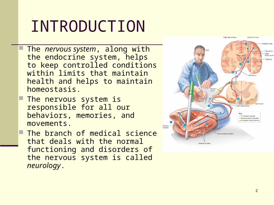
INTRODUCTION The nervous system, along with
the endocrine system, helps to keep controlled conditions within limits that maintain health and helps to maintain homeostasis.
The nervous system is responsible for all our behaviors, memories, and movements.
The branch of medical science that deals with the normal functioning and disorders of the nervous system is called neurology.
3
Chapter 12Nervous Tissue
Controls and integrates all body activities within limits that maintain life
Three basic functions sensing changes with sensory receptors
fullness of stomach or sun on your face interpreting and remembering those changes reacting to those changes with effectors
muscular contractions glandular secretions
4
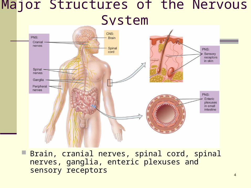
Major Structures of the Nervous System
Brain, cranial nerves, spinal cord, spinal nerves, ganglia, enteric plexuses and sensory receptors
5
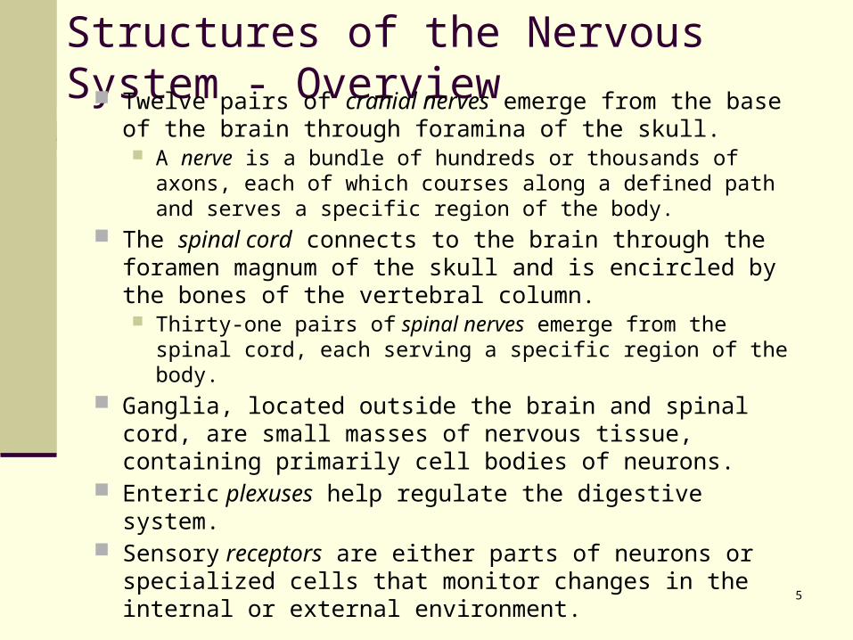
Structures of the Nervous System - Overview Twelve pairs of cranial nerves emerge from the base of
the brain through foramina of the skull. A nerve is a bundle of hundreds or thousands of axons,
each of which courses along a defined path and serves a specific region of the body.
The spinal cord connects to the brain through the foramen magnum of the skull and is encircled by the bones of the vertebral column. Thirty-one pairs of spinal nerves emerge from the spinal
cord, each serving a specific region of the body. Ganglia, located outside the brain and spinal cord, are
small masses of nervous tissue, containing primarily cell bodies of neurons.
Enteric plexuses help regulate the digestive system. Sensory receptors are either parts of neurons or
specialized cells that monitor changes in the internal or external environment.
6
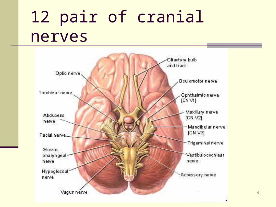
12 pair of cranial nerves
7
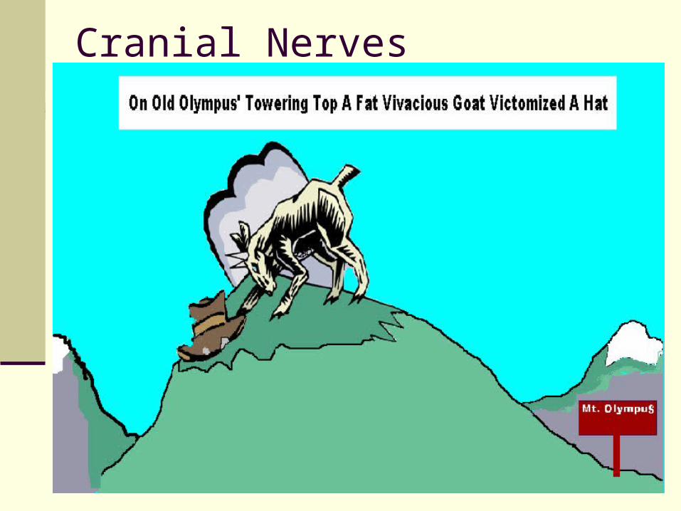
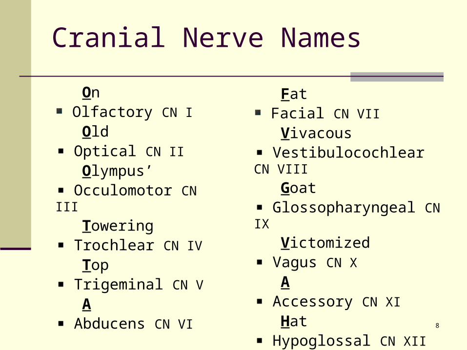
Cranial Nerves
8
Cranial Nerve Names
On Olfactory CN I
Old Optical CN II
Olympus’ Occulomotor CN III
Towering Trochlear CN IV
Top Trigeminal CN V
A Abducens CN VI
Fat Facial CN VII
Vivacous Vestibulocochlear CN VIII
Goat Glossopharyngeal CN IX
Victomized Vagus CN X
A Accessory CN XI
Hat Hypoglossal CN XII
9
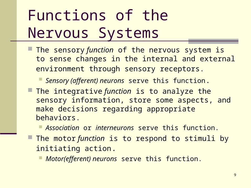
Functions of the Nervous Systems
The sensory function of the nervous system is to sense changes in the internal and external environment through sensory receptors. Sensory (afferent) neurons serve this function.
The integrative function is to analyze the sensory information, store some aspects, and make decisions regarding appropriate behaviors. Association or interneurons serve this function.
The motor function is to respond to stimuli by initiating action. Motor(efferent) neurons serve this function.
10
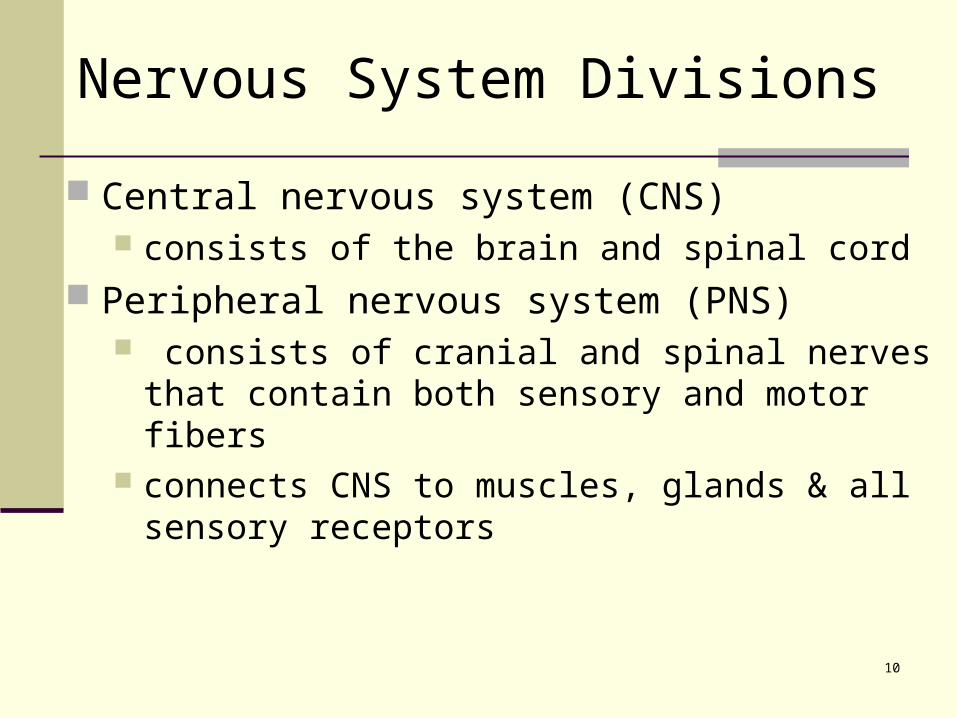
Nervous System Divisions
Central nervous system (CNS) consists of the brain and spinal cord
Peripheral nervous system (PNS) consists of cranial and spinal nerves that contain
both sensory and motor fibers connects CNS to muscles, glands & all sensory
receptors
11
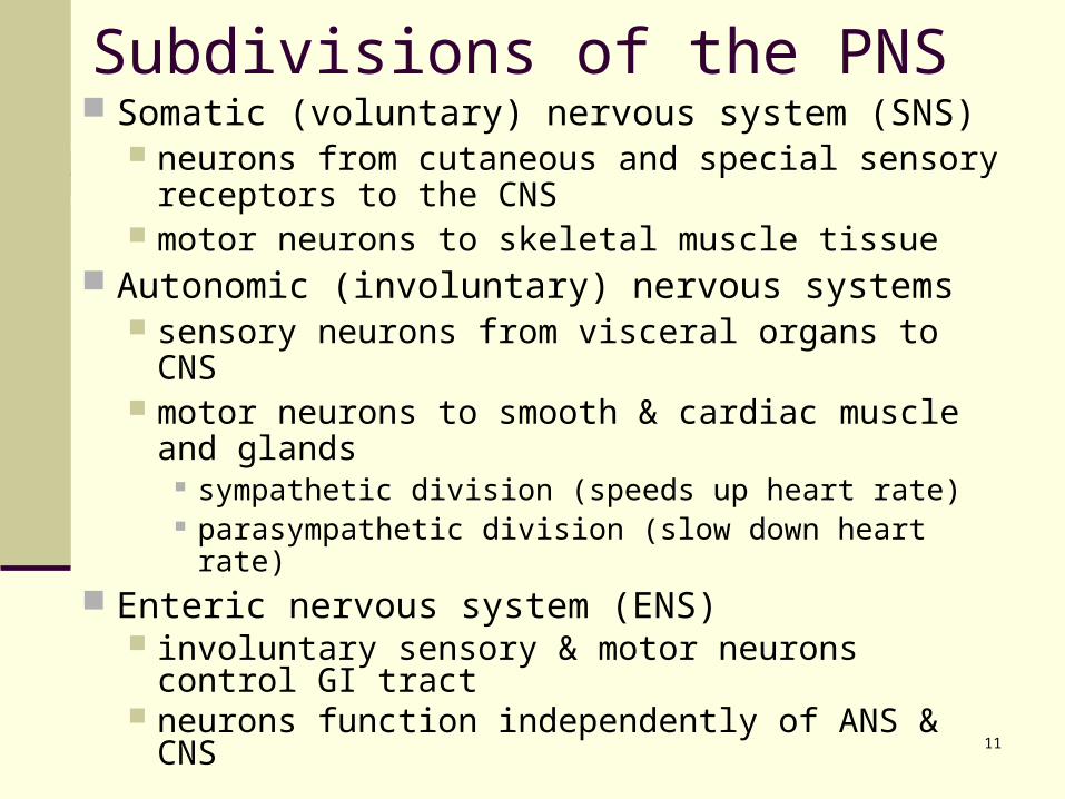
Subdivisions of the PNS Somatic (voluntary) nervous system (SNS)
neurons from cutaneous and special sensory receptors to the CNS
motor neurons to skeletal muscle tissue Autonomic (involuntary) nervous systems
sensory neurons from visceral organs to CNS motor neurons to smooth & cardiac muscle and
glands sympathetic division (speeds up heart rate) parasympathetic division (slow down heart rate)
Enteric nervous system (ENS) involuntary sensory & motor neurons control GI
tract neurons function independently of ANS & CNS
12
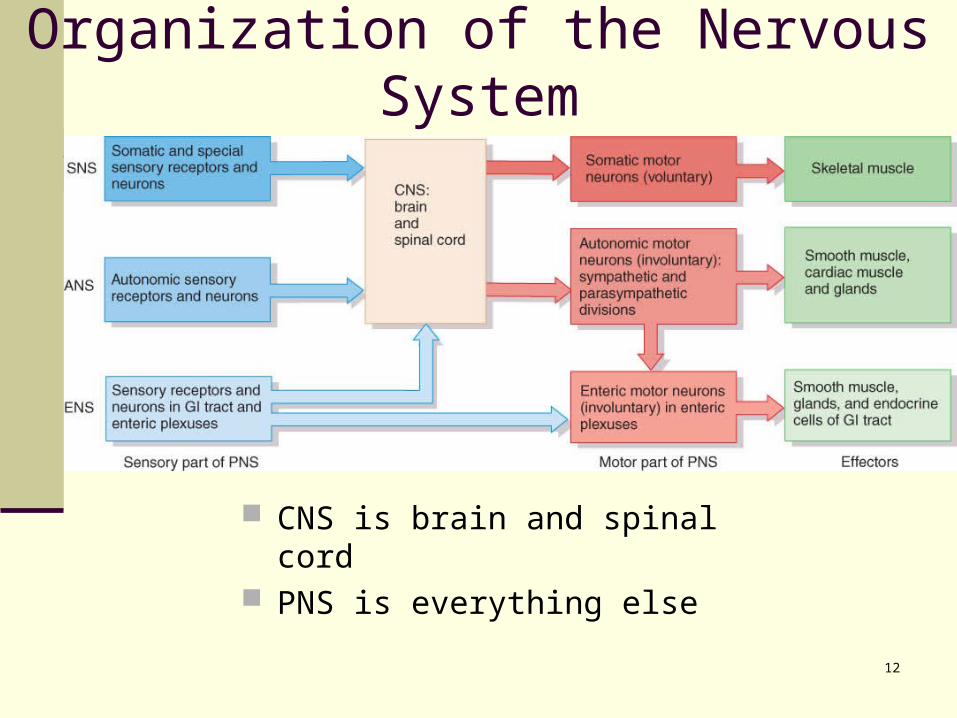
Organization of the Nervous System
CNS is brain and spinal cord PNS is everything else
13
Enteric NS
The enteric nervous system (ENS) consists of neurons in enteric plexuses that extend the length of the GI tract. Many neurons of the enteric plexuses function
independently of the ANS and CNS. Sensory neurons of the ENS monitor chemical
changes within the GI tract and stretching of its walls, whereas enteric motor neurons govern contraction of GI tract organs, and activity of the GI tract endocrine cells.
14
Neuronal Structure & Function
15
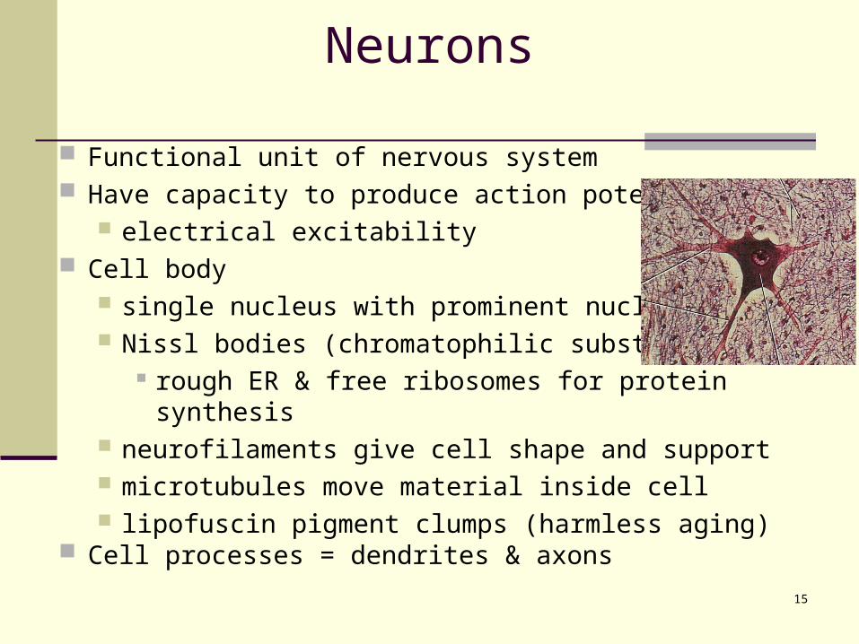
Neurons
Functional unit of nervous system Have capacity to produce action potentials
electrical excitability Cell body
single nucleus with prominent nucleolus Nissl bodies (chromatophilic substance)
rough ER & free ribosomes for protein synthesis
neurofilaments give cell shape and support microtubules move material inside cell lipofuscin pigment clumps (harmless aging)
Cell processes = dendrites & axons
16
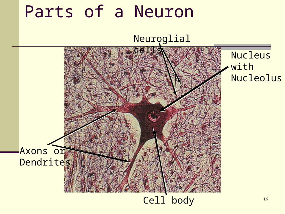
Nucleus with Nucleolus
Parts of a Neuron
Axons or Dendrites
Cell body
Neuroglial cells
17
Rat Neuron
18
19
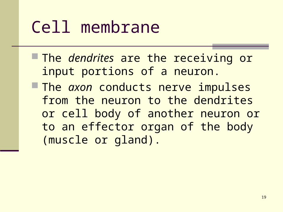
Cell membrane
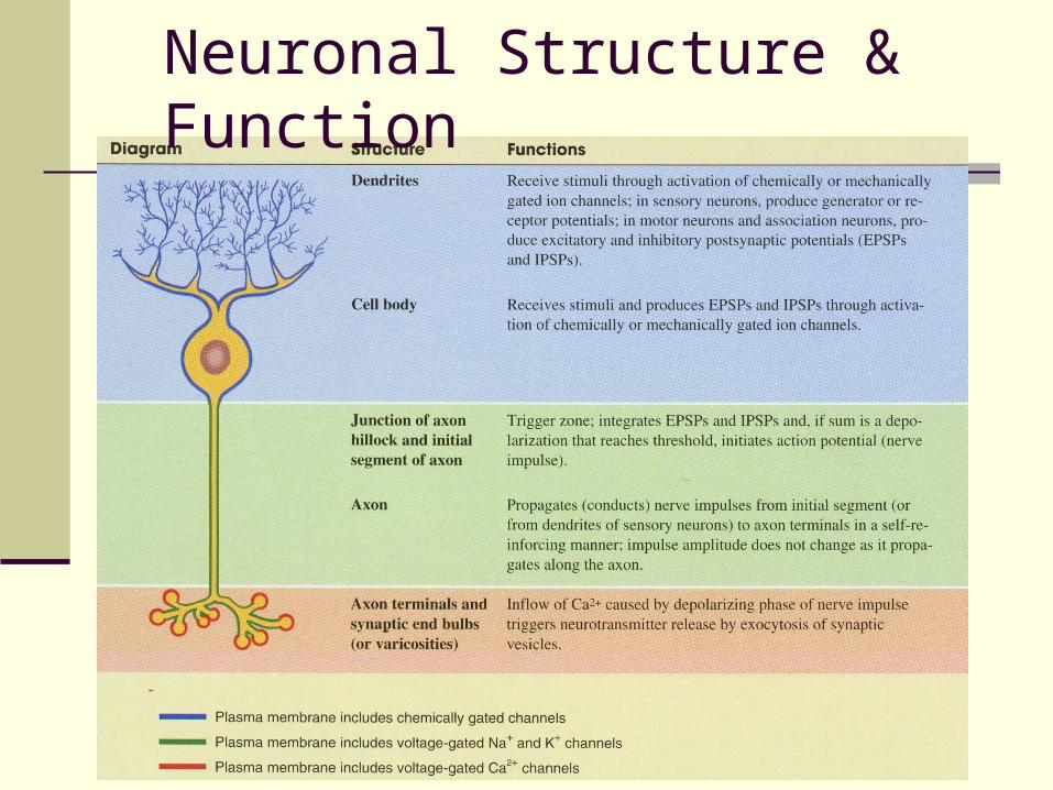
The dendrites are the receiving or input portions of a neuron.
The axon conducts nerve impulses from the neuron to the dendrites or cell body of another neuron or to an effector organ of the body (muscle or gland).
20
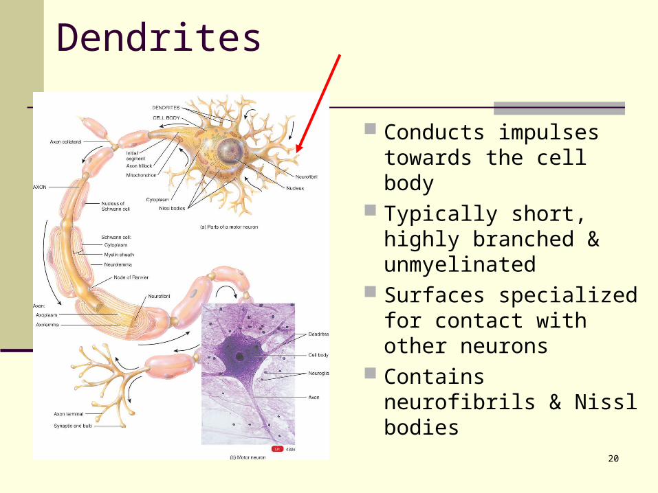
Dendrites
Conducts impulses towards the cell body
Typically short, highly branched & unmyelinated
Surfaces specialized for contact with other neurons
Contains neurofibrils & Nissl bodies
21
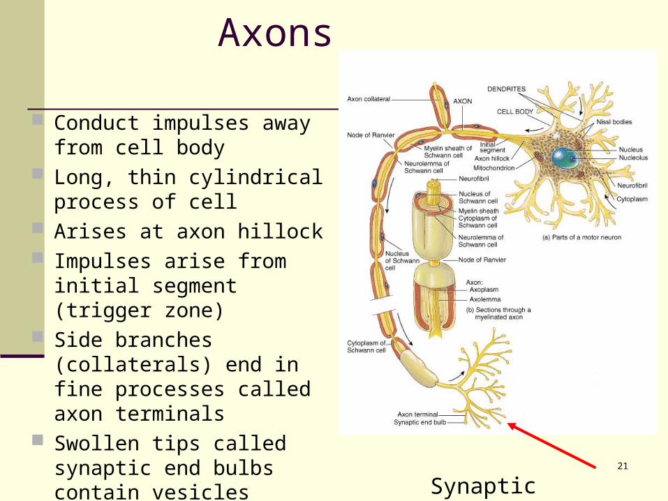
Axons
Conduct impulses away from cell body
Long, thin cylindrical process of cell
Arises at axon hillock Impulses arise from initial
segment (trigger zone) Side branches (collaterals)
end in fine processes called axon terminals
Swollen tips called synaptic end bulbs contain vesicles filled with neurotransmitters
Synaptic boutons
22
Axonal Transport
Cell body is location for most protein synthesis neurotransmitters & repair proteins
Axonal transport system moves substances slow axonal flow
movement in one direction only -- away from cell body movement at 1-5 mm per day
fast axonal flow moves organelles & materials along surface of
microtubules at 200-400 mm per day transports in either direction for use or for recycling in cell body
23
Axonal Transport & Disease
Fast axonal transport route by which toxins or pathogens reach neuron cell bodies tetanus (Clostridium tetani bacteria) disrupts motor neurons causing painful
muscle spasms Bacteria enter the body through a
laceration or puncture injury more serious if wound is in head or neck
because of shorter transit time
24
Diversity in Neurons
Both structural and functional features are used to classify the various neurons in the body.
On the basis of the number of processes extending from the cell body (structure), neurons are classified as multipolar, biopolar, and unipolar (Figure 12.4).
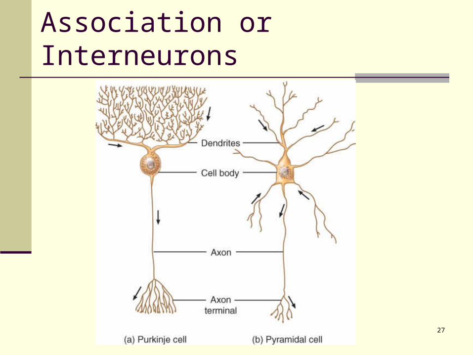
Most neurons in the body are interneurons and are often named for the histologist who first described them or for an aspect of their shape or appearance. Examples are Purkinje cells (Figure 12.5a) or Renshaw cells (Figure 12.5b).
25
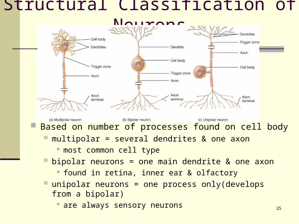
Structural Classification of Neurons
Based on number of processes found on cell body multipolar = several dendrites & one axon
most common cell type bipolar neurons = one main dendrite & one axon
found in retina, inner ear & olfactory unipolar neurons = one process only(develops from a
bipolar) are always sensory neurons
26
Functional Classification of Neurons
Sensory (afferent) neurons transport sensory information from skin,
muscles, joints, sense organs & viscera to CNS Motor (efferent) neurons
send motor nerve impulses to muscles & glands Interneurons (association) neurons
connect sensory to motor neurons 90% of neurons in the body
27
Association or Interneurons
28
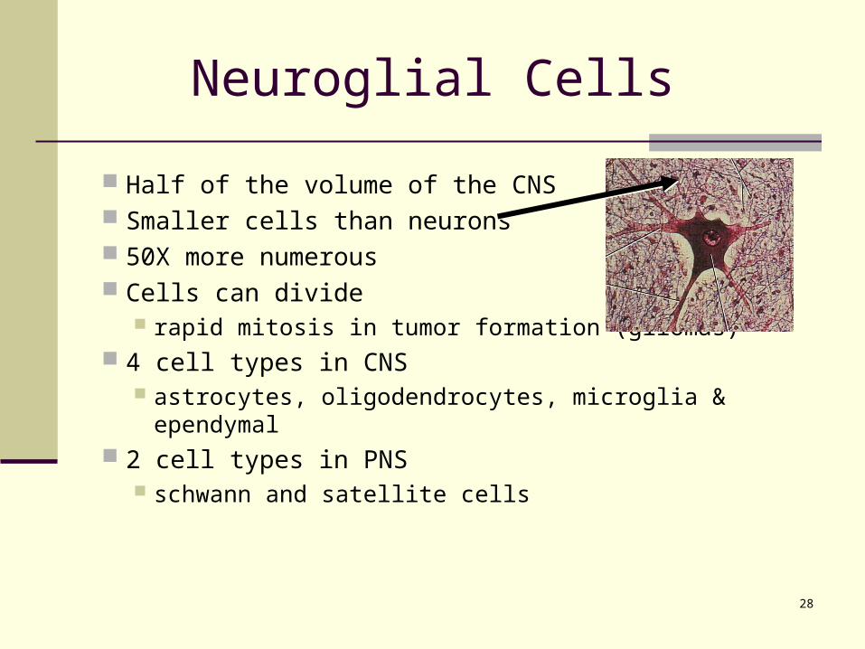
Half of the volume of the CNS Smaller cells than neurons 50X more numerous Cells can divide
rapid mitosis in tumor formation (gliomas) 4 cell types in CNS
astrocytes, oligodendrocytes, microglia & ependymal 2 cell types in PNS
schwann and satellite cells
Neuroglial Cells
29
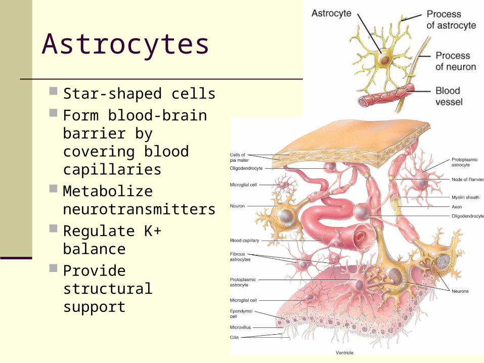
Astrocytes
Star-shaped cells Form blood-brain
barrier by covering blood capillaries
Metabolize neurotransmitters
Regulate K+ balance
Provide structural support
30
Microglia
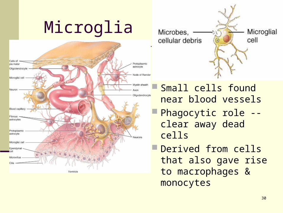
Small cells found near blood vessels
Phagocytic role -- clear away dead cells
Derived from cells that also gave rise to macrophages & monocytes
31
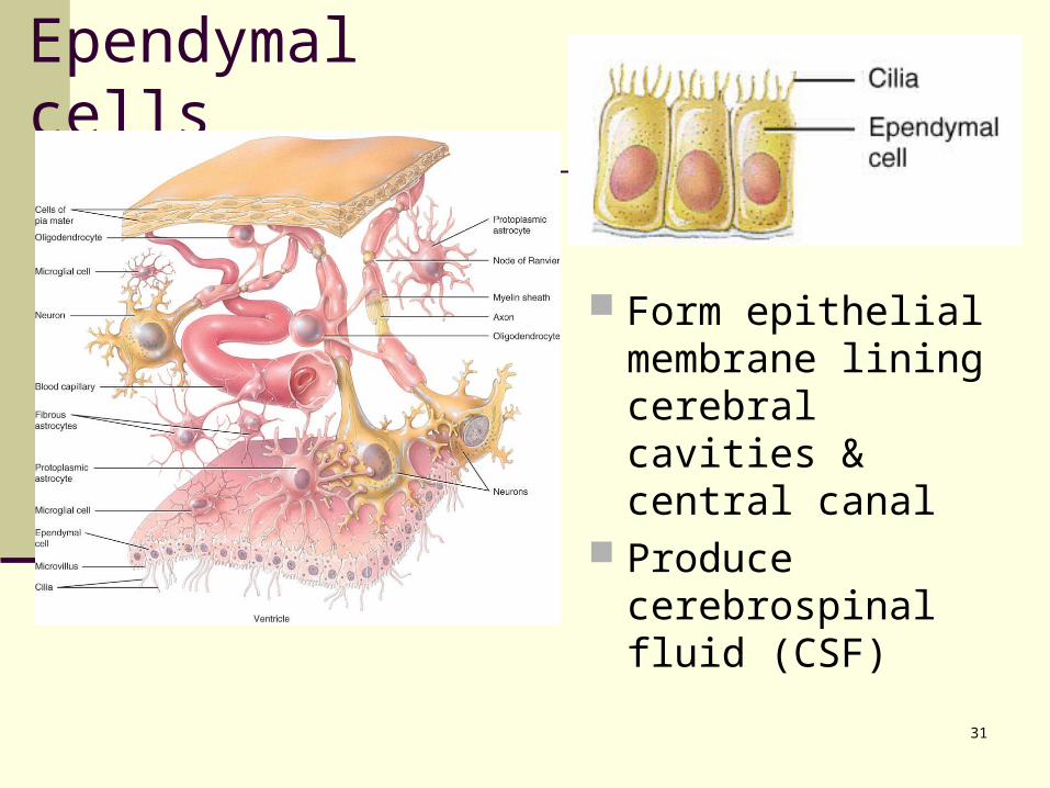
Ependymal cells
Form epithelial membrane lining cerebral cavities &
central canal Produce
cerebrospinal fluid (CSF)
32
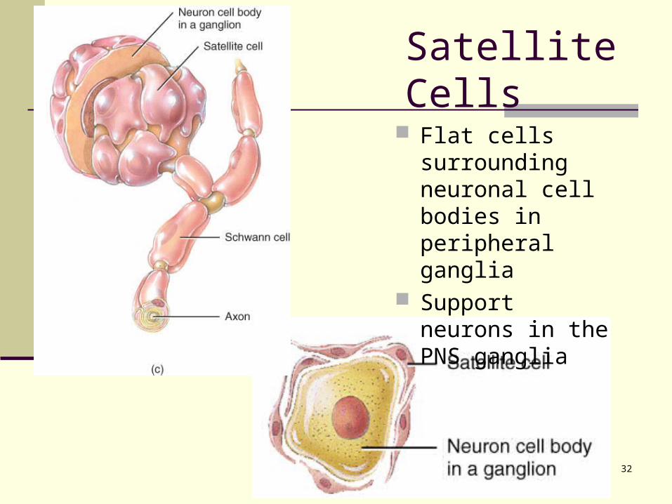
Satellite Cells
Flat cells surrounding neuronal cell bodies in peripheral ganglia
Support neurons in the PNS ganglia
33
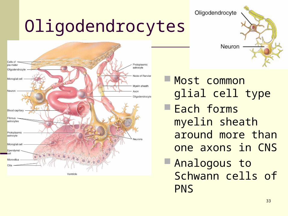
Oligodendrocytes
Most common glial cell type
Each forms myelin sheath around more than one axons in CNS
Analogous to Schwann cells of PNS
34
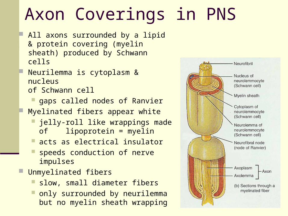
Myelination
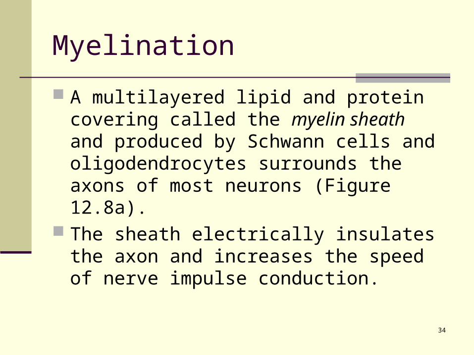
A multilayered lipid and protein covering called the myelin sheath and produced by Schwann cells and oligodendrocytes surrounds the axons of most neurons (Figure 12.8a).
The sheath electrically insulates the axon and increases the speed of nerve impulse conduction.
35
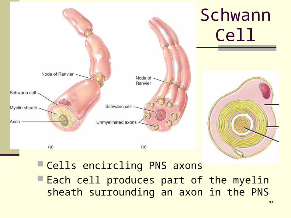
Schwann Cell
Cells encircling PNS axons Each cell produces part of the myelin sheath
surrounding an axon in the PNS
36
Axon Coverings in PNS All axons surrounded by a lipid &
protein covering (myelin sheath) produced by Schwann cells
Neurilemma is cytoplasm & nucleusof Schwann cell gaps called nodes of Ranvier
Myelinated fibers appear white jelly-roll like wrappings made of
lipoprotein = myelin acts as electrical insulator speeds conduction of nerve
impulses Unmyelinated fibers
slow, small diameter fibers only surrounded by neurilemma
but no myelin sheath wrapping
37
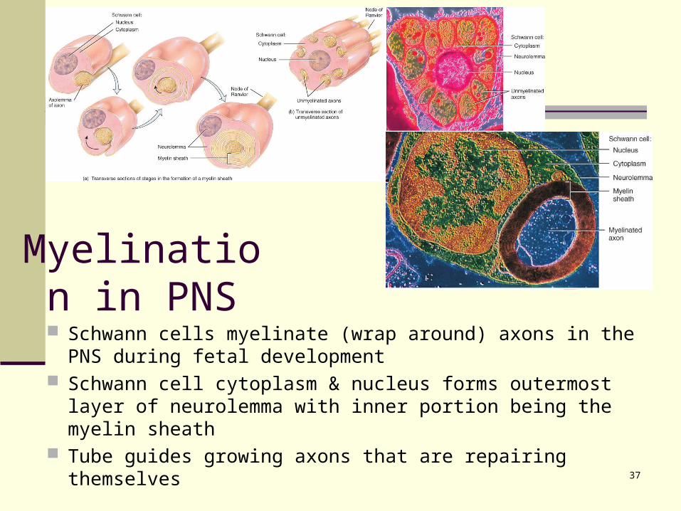
Myelination in PNS
Schwann cells myelinate (wrap around) axons in the PNS during fetal development
Schwann cell cytoplasm & nucleus forms outermost layer of neurolemma with inner portion being the myelin sheath
Tube guides growing axons that are repairing themselves
38
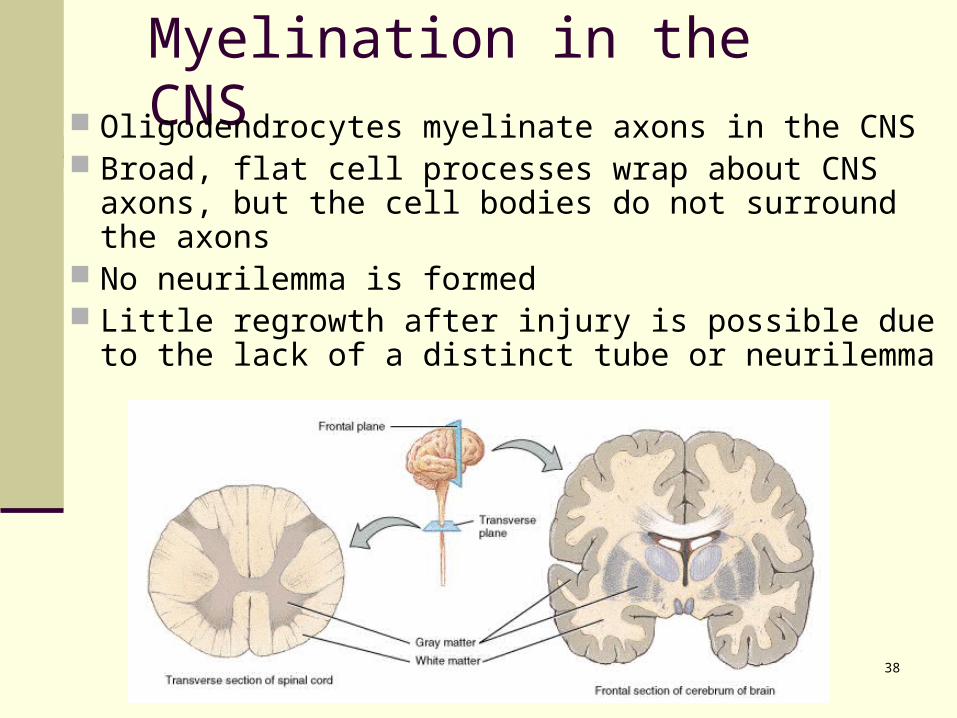
Myelination in the CNS Oligodendrocytes myelinate axons in the CNS Broad, flat cell processes wrap about CNS axons,
but the cell bodies do not surround the axons No neurilemma is formed Little regrowth after injury is possible due to the
lack of a distinct tube or neurilemma
39
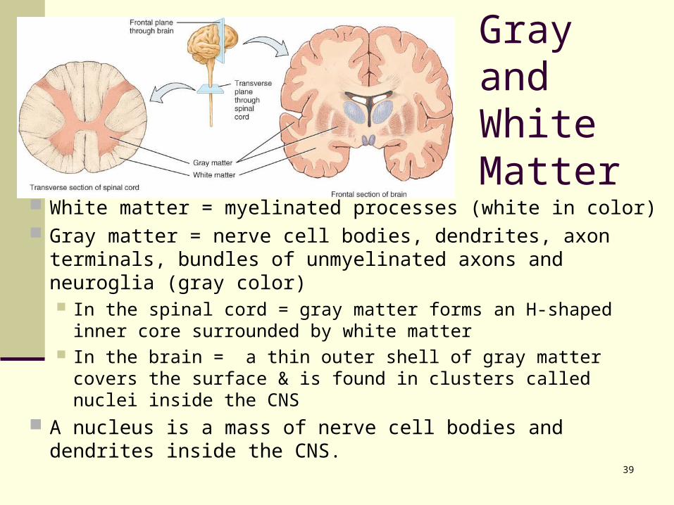
Gray and White Matter
White matter = myelinated processes (white in color) Gray matter = nerve cell bodies, dendrites, axon
terminals, bundles of unmyelinated axons and neuroglia (gray color) In the spinal cord = gray matter forms an H-shaped inner
core surrounded by white matter In the brain = a thin outer shell of gray matter covers the
surface & is found in clusters called nuclei inside the CNS A nucleus is a mass of nerve cell bodies and dendrites
inside the CNS.
40
Electrical Signals in Neurons Neurons are electrically excitable due to
the voltage difference across their membrane
Communicate with 2 types of electric signals action potentials that can travel long
distances graded potentials that are local
membrane changes only In living cells, a flow of ions occurs
through ion channels in the cell membrane
41
Two Types of Ion Channels
Leakage (nongated) channels are always open nerve cells have more K+ than Na+ leakage
channels as a result, membrane permeability to K+ is higher explains resting membrane potential of -70mV in
nerve tissue Gated channels open and close in response to a
stimulus results in neuron excitability
42
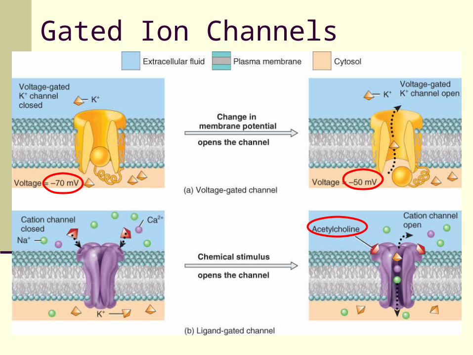
Ion Channels
Gated ion channels respond to voltage changes, ligands (chemicals), and mechanical pressure. Voltage-gated channels respond to a direct
change in the membrane potential (Figure 12.10a).
Ligand-gated channels respond to a specific chemical stimulus (Figure 12.10b).
Mechanically gated ion channels respond to mechanical vibration or pressure.
43
Gated Ion Channels
44
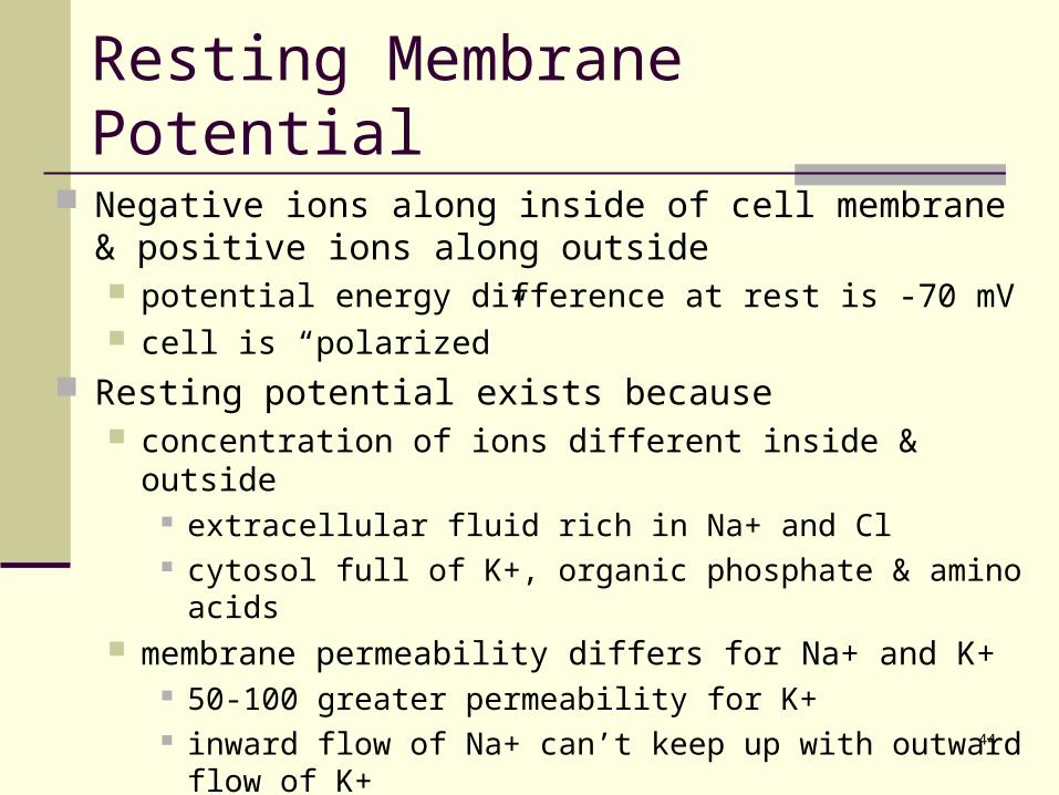
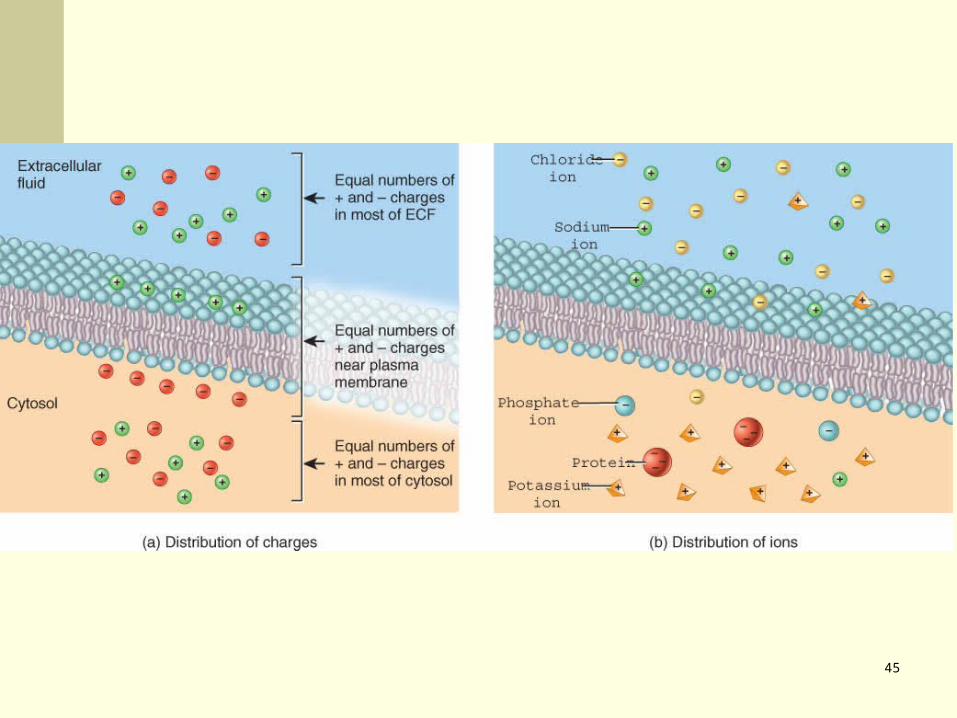
Resting Membrane Potential
Negative ions along inside of cell membrane & positive ions along outside potential energy difference at rest is -70 mV cell is “polarized”
Resting potential exists because concentration of ions different inside & outside
extracellular fluid rich in Na+ and Cl cytosol full of K+, organic phosphate & amino acids
membrane permeability differs for Na+ and K+ 50-100 greater permeability for K+ inward flow of Na+ can’t keep up with outward flow of K+ Na+/K+ pump removes Na+ as fast as it leaks in
45
46
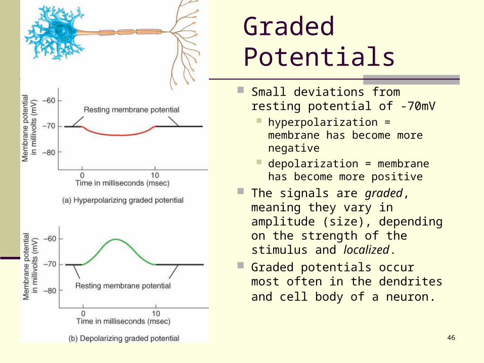
Graded Potentials
Small deviations from resting potential of -70mV hyperpolarization = membrane
has become more negative depolarization = membrane
has become more positive The signals are graded,
meaning they vary in amplitude (size), depending on the strength of the stimulus and localized.
Graded potentials occur most often in the dendrites and cell body of a neuron.
47
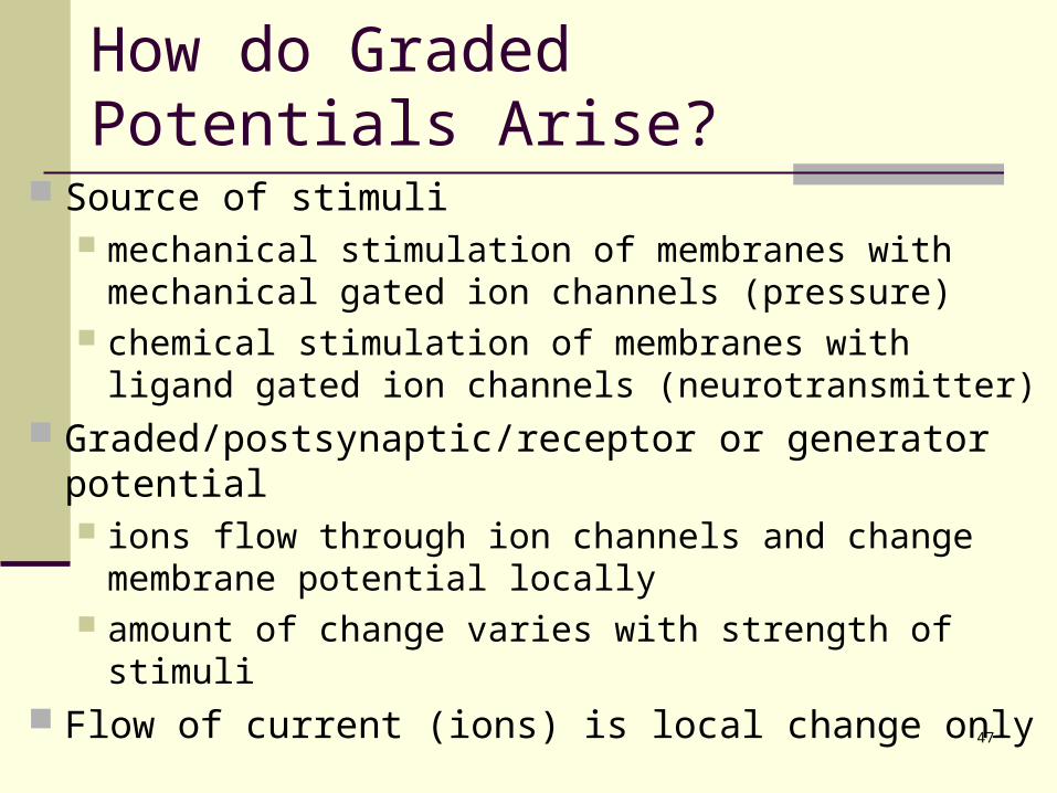
How do Graded Potentials Arise?
Source of stimuli mechanical stimulation of membranes with mechanical
gated ion channels (pressure) chemical stimulation of membranes with ligand gated
ion channels (neurotransmitter) Graded/postsynaptic/receptor or generator potential
ions flow through ion channels and change membrane potential locally
amount of change varies with strength of stimuli Flow of current (ions) is local change only
48
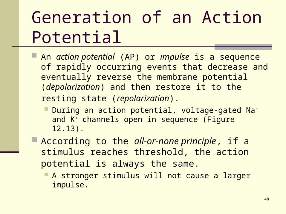
Generation of an Action Potential
An action potential (AP) or impulse is a sequence of rapidly occurring events that decrease and eventually reverse the membrane potential (depolarization) and then restore it to the resting state (repolarization). During an action potential, voltage-gated Na+ and K+
channels open in sequence (Figure 12.13).
According to the all-or-none principle, if a stimulus reaches threshold, the action potential is always the same. A stronger stimulus will not cause a larger impulse.
49
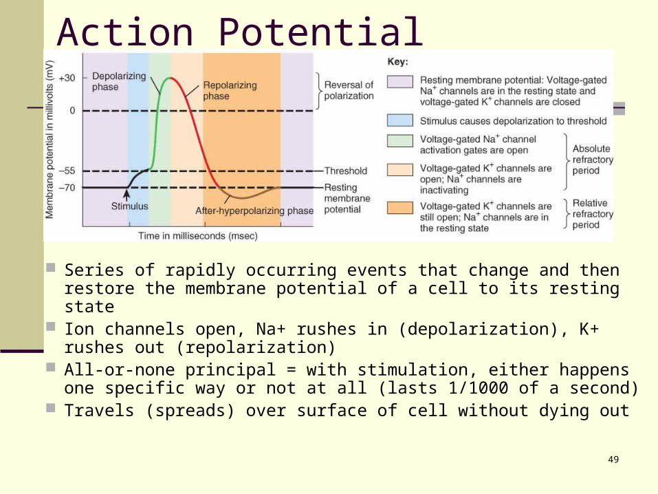
Action Potential
Series of rapidly occurring events that change and then restore the membrane potential of a cell to its resting state
Ion channels open, Na+ rushes in (depolarization), K+ rushes out (repolarization)
All-or-none principal = with stimulation, either happens one specific way or not at all (lasts 1/1000 of a second)
Travels (spreads) over surface of cell without dying out
50
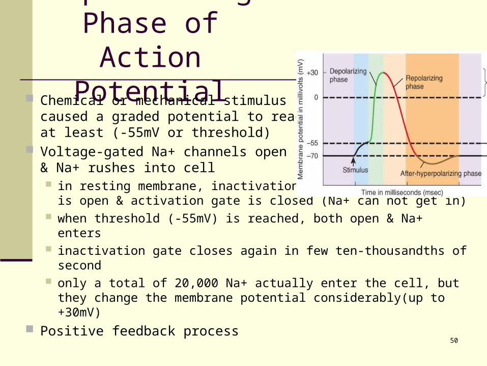
Depolarizing Phase of Action Potential
Chemical or mechanical stimuluscaused a graded potential to reachat least (-55mV or threshold)
Voltage-gated Na+ channels open& Na+ rushes into cell in resting membrane, inactivation gate of sodium channel is
open & activation gate is closed (Na+ can not get in) when threshold (-55mV) is reached, both open & Na+ enters inactivation gate closes again in few ten-thousandths of second only a total of 20,000 Na+ actually enter the cell, but they
change the membrane potential considerably(up to +30mV) Positive feedback process
51
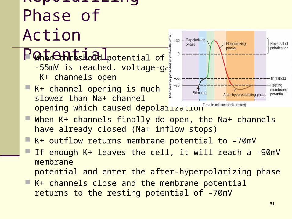
Repolarizing Phase of Action Potential When threshold potential of
-55mV is reached, voltage-gated K+ channels open
K+ channel opening is muchslower than Na+ channelopening which caused depolarization
When K+ channels finally do open, the Na+ channels have already closed (Na+ inflow stops)
K+ outflow returns membrane potential to -70mV If enough K+ leaves the cell, it will reach a -90mV membrane
potential and enter the after-hyperpolarizing phase K+ channels close and the membrane potential returns to the
resting potential of -70mV
52
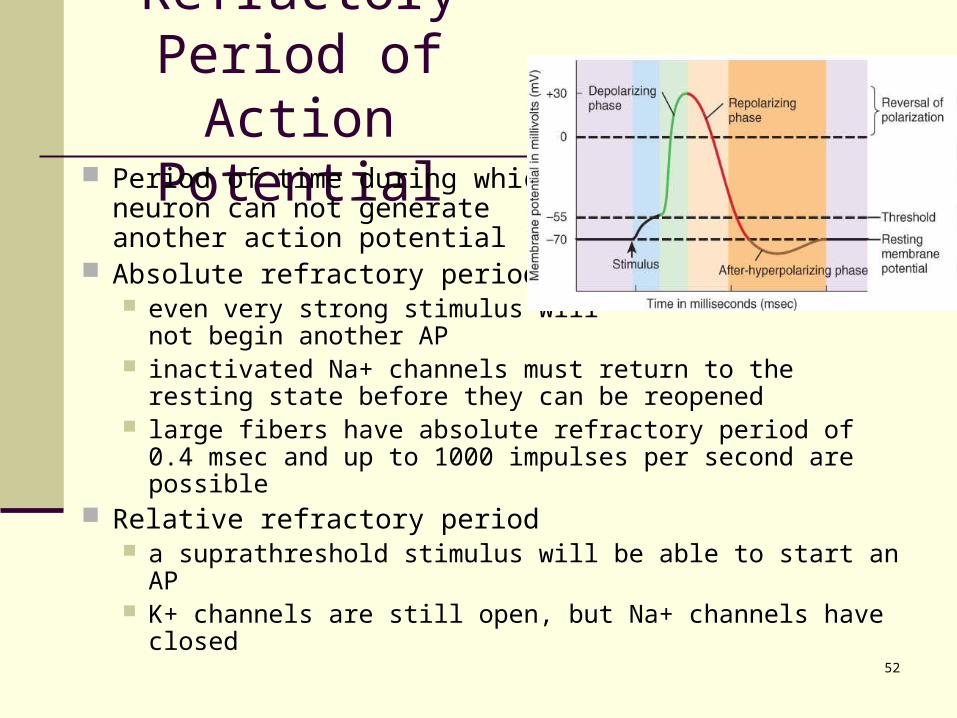
Refractory Period of Action Potential
Period of time during whichneuron can not generateanother action potential
Absolute refractory period even very strong stimulus will
not begin another AP inactivated Na+ channels must return to the resting state
before they can be reopened large fibers have absolute refractory period of 0.4 msec
and up to 1000 impulses per second are possible Relative refractory period
a suprathreshold stimulus will be able to start an AP K+ channels are still open, but Na+ channels have closed
53
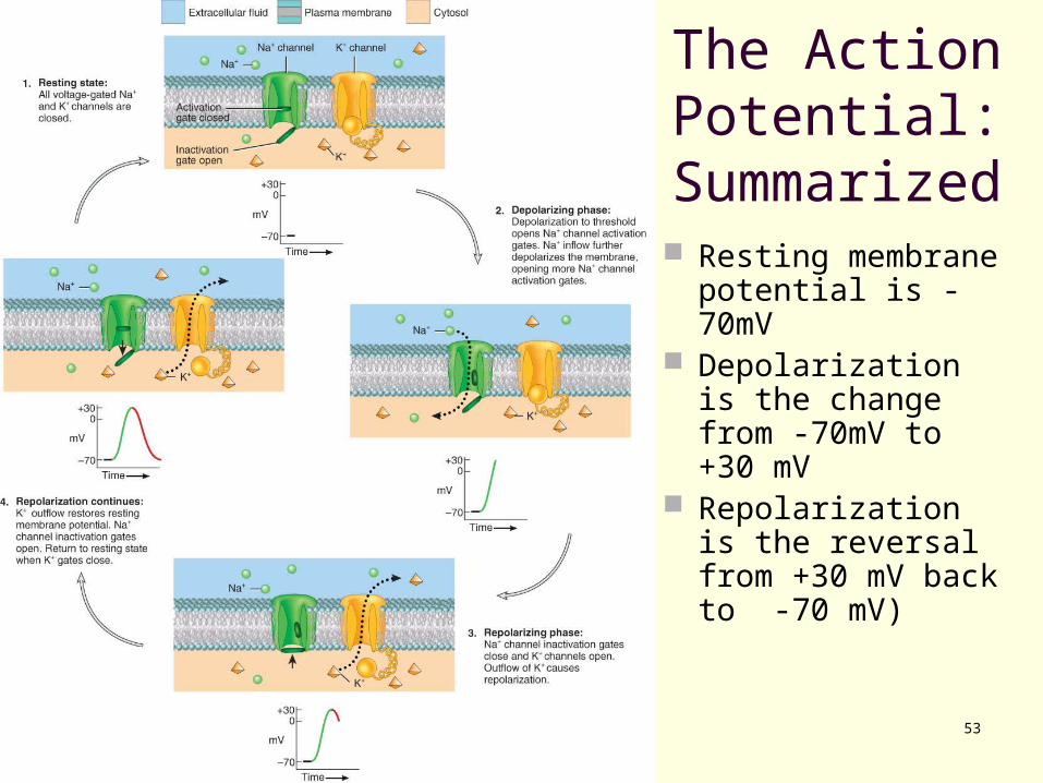
The Action Potential: Summarized Resting membrane
potential is -70mV Depolarization is
the change from -70mV to +30 mV
Repolarization is the reversal from +30 mV back to -70 mV)
54
Local Anesthetics
Local anesthetics and certain neurotoxins Prevent opening of voltage-gated Na+
channels Nerve impulses cannot pass the
anesthetized region
Examples: Novocaine and lidocaine
55
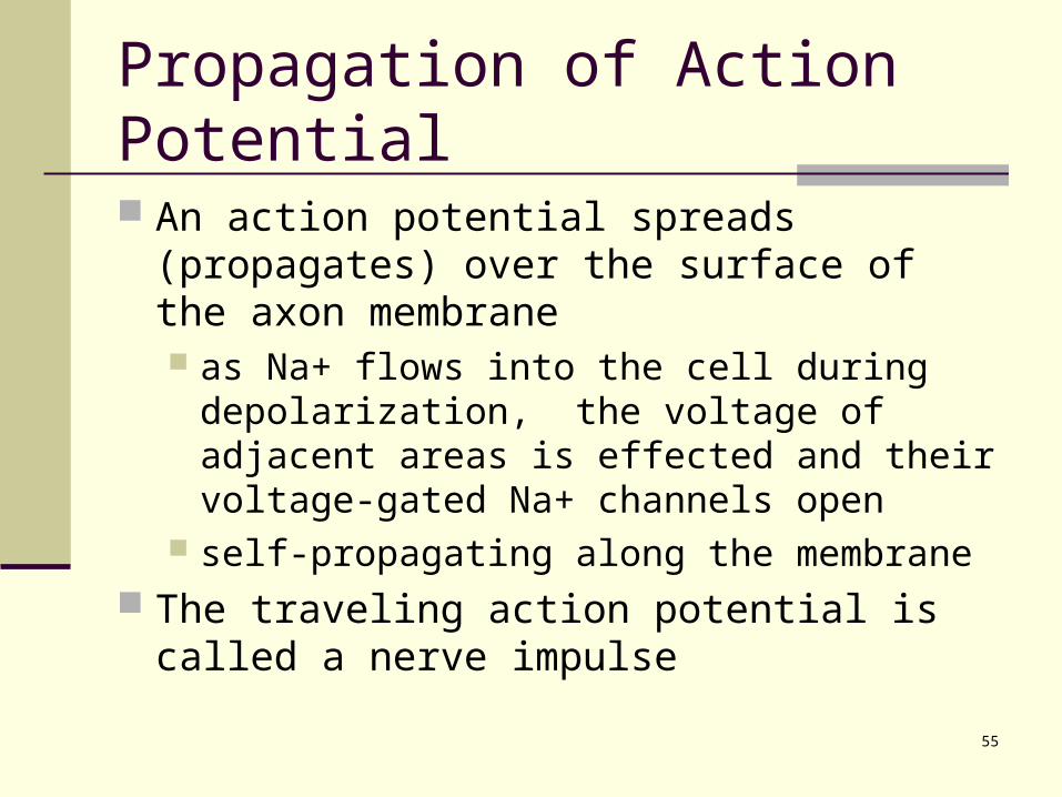
Propagation of Action Potential
An action potential spreads (propagates) over the surface of the axon membrane as Na+ flows into the cell during
depolarization, the voltage of adjacent areas is effected and their voltage-gated Na+ channels open
self-propagating along the membrane The traveling action potential is called a nerve
impulse
56
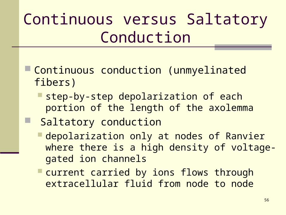
Continuous versus Saltatory Conduction
Continuous conduction (unmyelinated fibers) step-by-step depolarization of each portion of the
length of the axolemma Saltatory conduction
depolarization only at nodes of Ranvier where there is a high density of voltage-gated ion channels
current carried by ions flows through extracellular fluid from node to node
57
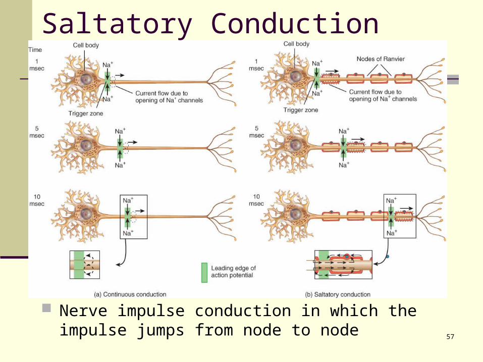
Saltatory Conduction
Nerve impulse conduction in which the impulse jumps from node to node
58
Speed of Impulse Propagation
The propagation speed of a nerve impulse is not related to stimulus strength. larger, myelinated fibers conduct impulses faster
due to size & saltatory conduction Fiber types
A fibers largest (5-20 microns & 130 m/sec) myelinated somatic sensory & motor to skeletal muscle
B fibers medium (2-3 microns & 15 m/sec) myelinated visceral sensory & autonomic preganglionic
C fibers smallest (.5-1.5 microns & 2 m/sec) unmyelinated sensory & autonomic motor
59
Encoding of Stimulus Intensity
How do we differentiate a light touch from a firmer touch? frequency of impulses
firm pressure generates impulses at a higher frequency
number of sensory neurons activated firm pressure stimulates more neurons than
does a light touch
60
Action Potentials in Nerve and Muscle
Entire muscle cell membrane versus only the axon of the neuron is involved
Resting membrane potential nerve is -70mV skeletal & cardiac muscle is closer to -90mV
Duration nerve impulse is 1/2 to 2 msec muscle action potential lasts 1-5 msec for skeletal
& 10-300msec for cardiac & smooth Fastest nerve conduction velocity is 18 times
faster than velocity over skeletal muscle fiber
61
SIGNAL TRANSMISSION AT SYNAPSES A synapse is the functional junction between
one neuron and another or between a neuron and an effector such as a muscle or gland.
62
Signal Transmission at Synapses
2 Types of synapses electrical
ionic current spreads to next cell through gap junctions faster, two-way transmission & capable of synchronizing
groups of neurons chemical
one-way information transfer from a presynaptic neuron to a postsynaptic neuron
axodendritic -- from axon to dendrite axosomatic -- from axon to cell body axoaxonic -- from axon to axon
63
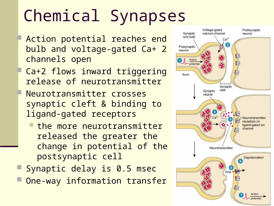
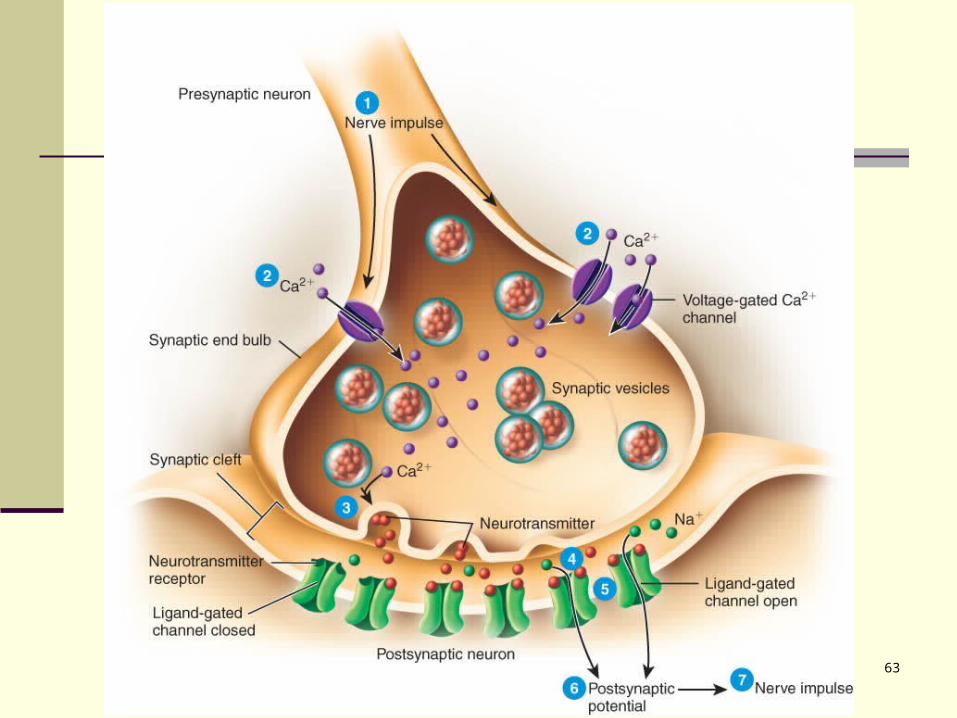
64
Chemical Synapses Action potential reaches end bulb
and voltage-gated Ca+ 2 channels open
Ca+2 flows inward triggering release of neurotransmitter
Neurotransmitter crosses synaptic cleft & binding to ligand-gated receptors the more neurotransmitter
released the greater the change in potential of the postsynaptic cell
Synaptic delay is 0.5 msec One-way information transfer

65
Excitatory & Inhibitory Potentials
The effect of a neurotransmitter can be either excitatory or inhibitory a depolarizing postsynaptic potential is called an
EPSP it results from the opening of ligand-gated Na+ channels the postsynaptic cell is more likely to reach threshold
an inhibitory postsynaptic potential is called an IPSP it results from the opening of ligand-gated Cl- or K+
channels it causes the postsynaptic cell to become more negative
or hyperpolarized the postsynaptic cell is less likely to reach threshold
66
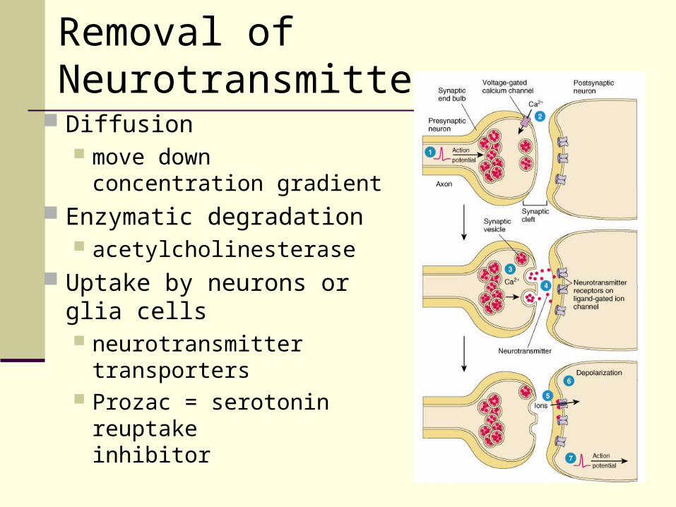
Removal of Neurotransmitter
Diffusion move down concentration
gradient Enzymatic degradation
acetylcholinesterase Uptake by neurons or glia
cells neurotransmitter
transporters Prozac = serotonin
reuptake inhibitor
67
Three Possible Responses
Small EPSP occurs potential reaches -56 mV only
An impulse is generated threshold was reached membrane potential of at least -55 mV
IPSP occurs membrane hyperpolarized potential drops below -70 mV
68
Origin GPs arise on dendrites and cell bodies APs arise only at trigger zone on axon hillock
Types of Channels AP is produced by voltage-gated ion channels GP is produced by ligand or mechanically-
gated channels Conduction
GPs are localized (not propagated) APs conduct over the surface of the axon
Comparison of Graded & Action Potentials
69
Comparison of Graded & Action Potentials
Amplitude amplitude of the AP is constant (all-or-none) graded potentials vary depending upon stimulus
Duration The duration of the GP is as long as the
stimulus lasts Refractory period
The AP has a refractory period due to the nature of the voltage-gated channels, and the GP has none.
70
Summation
If several presynaptic end bulbs release their neurotransmitter at about the same time, the combined effect may generate a nerve impulse due to summation
Summation may be spatial or temporal.
71
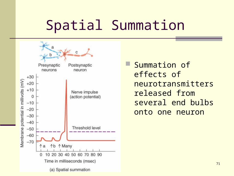
Summation of effects of neurotransmitters released from several end bulbs onto one neuron
Spatial Summation
72
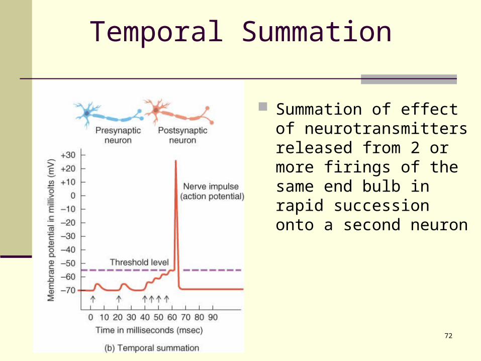
Temporal Summation
Summation of effect of neurotransmitters released from 2 or more firings of the same end bulb in rapid succession onto a second neuron
73
Summation
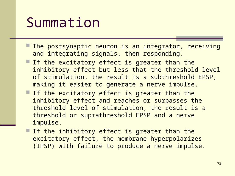
The postsynaptic neuron is an integrator, receiving and integrating signals, then responding.
If the excitatory effect is greater than the inhibitory effect but less that the threshold level of stimulation, the result is a subthreshold EPSP, making it easier to generate a nerve impulse.
If the excitatory effect is greater than the inhibitory effect and reaches or surpasses the threshold level of stimulation, the result is a threshold or suprathreshold EPSP and a nerve impulse.
If the inhibitory effect is greater than the excitatory effect, the membrane hyperpolarizes (IPSP) with failure to produce a nerve impulse.
74
Neurotransmitters
Both excitatory and inhibitory neurotransmitters are present in the CNS and PNS; the same neurotransmitter may be excitatory in some locations and inhibitory in others.
Important neurotransmitters include acetylcholine, glutamate, aspartate, gamma aminobutyric acid, glycine, norepinephrine, epinephrine, and dopamine.
75
Neurotransmitter Effects
Neurotransmitter effects can be modified synthesis can be stimulated or inhibited release can be blocked or enhanced removal can be stimulated or blocked receptor site can be blocked or activated
Agonist anything that enhances a transmitters effects
Antagonist anything that blocks the action of a neurotranmitter
76
Small-Molecule Neurotransmitters
Acetylcholine (ACh) released by many PNS neurons & some CNS excitatory on NMJ but inhibitory at others inactivated by acetylcholinesterase
Amino Acids glutamate released by nearly all excitatory neurons
in the brain ---- inactivated by glutamate specific transporters
GABA is inhibitory neurotransmitter for 1/3 of all brain synapses (Valium is a GABA agonist -- enhancing its inhibitory effect)
77
Biogenic Amines modified amino acids (tyrosine)
norepinephrine -- regulates mood, dreaming, awakening from deep sleep
dopamine -- regulating skeletal muscle tone serotonin -- control of mood, temperature
regulation, & induction of sleep removed from synapse & recycled or
destroyed by enzymes (monoamine oxidase or catechol-0-methyltransferase)
Small-Molecule Neurotransmitters

78
ATP and other purines (ADP, AMP & adenosine) excitatory in both CNS & PNS released with other neurotransmitters (ACh & NE)
Gases (nitric oxide or NO) formed from amino acid arginine by an enzyme formed on demand and acts immediately
diffuses out of cell that produced it to affect neighboring cells
may play a role in memory & learning first recognized as vasodilator that helps lower
blood pressure
Small-Molecule Neurotransmitters
79
Neuropeptides
3-40 amino acids linked by peptide bonds Substance P -- enhances our perception of
pain Pain relief
enkephalins -- pain-relieving effect by blocking the release of substance P (bind to morphine receptors in the central nervous system and have opioid properties)
acupuncture may produce loss of pain sensation because of release of opioids-like substances such as endorphins or dynorphins
80
Strychnine Poisoning
In spinal cord, Renshaw cells normally release an inhibitory neurotransmitter (glycine) onto motor neurons preventing excessive muscle contraction
Strychnine binds to and blocks glycine receptors in the spinal cord
Massive tetanic contractions of all skeletal muscles are produced when the diaphragm contracts & remains
contracted, breathing can not occur
81
Neuronal Circuits
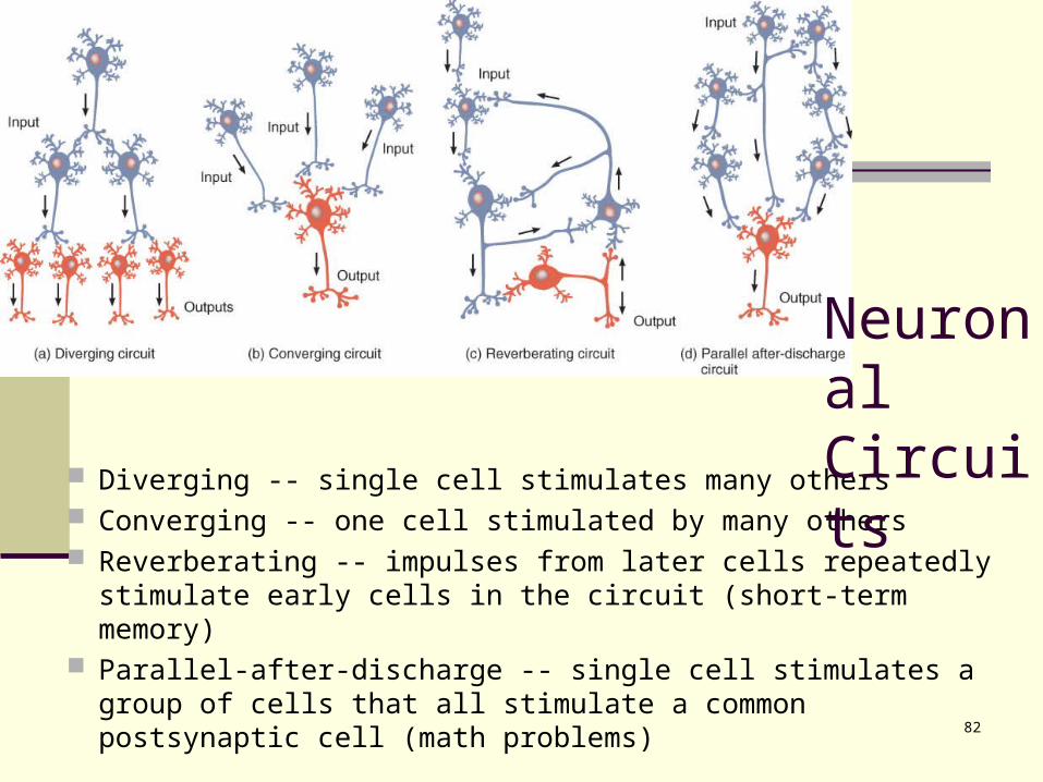
Neuronal pools are organized into circuits (neural networks.) These include simple series, diverging, converging, reverberating, and parallel after-discharge circuits (Figure 12.18 a-d).
A neuronal network may contain thousands or even millions of neurons.
Neuronal circuits are involved in many important activities breathing short-term memory waking up
82
Diverging -- single cell stimulates many others Converging -- one cell stimulated by many others Reverberating -- impulses from later cells repeatedly
stimulate early cells in the circuit (short-term memory) Parallel-after-discharge -- single cell stimulates a group of
cells that all stimulate a common postsynaptic cell (math problems)
Neuronal Circuits
83
Regeneration & Repair
Plasticity maintained throughout life sprouting of new dendrites synthesis of new proteins changes in synaptic contacts with other
neurons Limited ability for regeneration (repair)
PNS can repair damaged dendrites or axons CNS no repairs are possible
84
Damage and Repair in the Peripheral Nervous System
When there is damage to an axon, usually there are changes, called chromatolysis, which occur in the cell body of the affected cell; this causes swelling of the cell body and peaks between 10 and 20 days after injury.
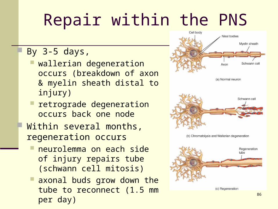
By the third to fifth day, degeneration of the distal portion of the neuronal process and myelin sheath (Wallerian degeneration) occurs; afterward, macrophages phagocytize the remains.
Retrograde degeneration of the proximal portion of the fiber extends only to the first neurofibral node.
Regeneration follows chromatolysis; synthesis of RNA and protein accelerates, favoring rebuilding of the axon and often taking several months.
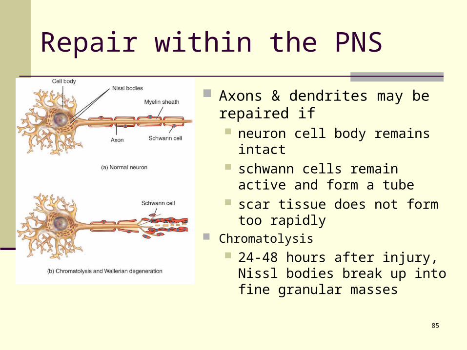
85
Axons & dendrites may be repaired if neuron cell body remains intact schwann cells remain active and
form a tube scar tissue does not form too
rapidly Chromatolysis
24-48 hours after injury, Nissl bodies break up into fine granular masses
Repair within the PNS
86
By 3-5 days, wallerian degeneration occurs
(breakdown of axon & myelin sheath distal to injury)
retrograde degeneration occurs back one node
Within several months, regeneration occurs neurolemma on each side of
injury repairs tube (schwann cell mitosis)
axonal buds grow down the tube to reconnect (1.5 mm per day)
Repair within the PNS
87
Neurogenesis in the CNS Formation of new neurons from stem cells was
not thought to occur in humans 1992 a growth factor was found that stimulates
adult mice brain cells to multiply 1998 new neurons found to form within adult
human hippocampus (area important for learning) There is a lack of neurogenesis in other regions
of the brain and spinal cord. Factors preventing neurogenesis in CNS
inhibition by neuroglial cells, absence of growth stimulating factors, lack of neurolemmas, and rapid formation of scar tissue
88
Multiple Sclerosis (MS)
Autoimmune disorder causing destruction of myelin sheaths in CNS sheaths becomes scars or plaques 1/2 million people in the United States appears between ages 20 and 40 females twice as often as males
Symptoms include muscular weakness, abnormal sensations or double vision
Remissions & relapses result in progressive, cumulative loss of function
89
The second most common neurological disorder affects 1% of population
Characterized by short, recurrent attacks initiated by electrical discharges in the brain lights, noise, or smells may be sensed skeletal muscles may contract involuntarily loss of consciousness
Epilepsy has many causes, including; brain damage at birth, metabolic disturbances,
infections, toxins, vascular disturbances, head injuries, and tumors
Epilepsy
90
end