Embed Size (px)
Citation preview

Helal et al. Bull Natl Res Cent (2021) 45:66 https://doi.org/10.1186/s42269-021-00522-0
RESEARCH
Probiotics role of Saccharomyces cerevisiae and Bacillus subtilis in improving the health status of rabbits’ gastrointestinal tractFarouk Helal1, Alaa El‑Badawi1, Soad El‑Naggar1, Mohamed Shourrap2, Osama Aboelazab1* and Salma Abu Hafsa3
Abstract
Background: Probiotics are direct‑fed microbial feed supplements which can modulate the gut microflora by competing intestinal pathogens through a competitive process. The present study was conducted to investigate the effect of feeding Saccharomyces cerevisiae, Bacillus subtilis or their mixture on blood biochemical constituents, intesti‑nal pathogenic load and intestinal histological changes of growing New Zealand White (NZW) rabbits.
Results: Serum total protein, albumin, and globulin were (P ≤ 0.05) increased for rabbits fed supplemented diets. Microbial pathogenic load of small intestinal and caecal contents (E. coli and C. perfringens) showed reduction (P ≤ 0.05) for rabbits fed supplemented diets, while, lactobacillus spp. recorded higher counts (P ≤ 0.05) in intestinal and caecal contents of rabbits fed probiotics supplemented diets than control group. Small intestine length, villus height and crypt depth were higher (P ≤ 0.05) with probiotic diets than control. Musculosa depth was depressed (P ≤ 0.05) with probiotic diets.
Conclusions: It could be concluded that the addition of Bacillus subtilis or Saccharomyces cerevisiae to diets of grow‑ing NZW rabbits by 0.1% is recommended to minimize the pathogenic intestinal load and increasing of beneficial lactobacillus strains as well as improving the intestinal barriers integrity.
Keywords: Blood constituents, Intestinal and caecal pathogens, Probiotics, Rabbits, Small intestine histomorphology
© The Author(s) 2021. Open Access This article is licensed under a Creative Commons Attribution 4.0 International License, which permits use, sharing, adaptation, distribution and reproduction in any medium or format, as long as you give appropriate credit to the original author(s) and the source, provide a link to the Creative Commons licence, and indicate if changes were made. The images or other third party material in this article are included in the article’s Creative Commons licence, unless indicated otherwise in a credit line to the material. If material is not included in the article’s Creative Commons licence and your intended use is not permitted by statutory regulation or exceeds the permitted use, you will need to obtain permission directly from the copyright holder. To view a copy of this licence, visit http://creat iveco mmons .org/licen ses/by/4.0/.
BackgroundRabbits have a unique digestive system; they are both mono-gastric and herbivore animals with special diges-tive and physiological characterization. It was stated that any imbalance of microflora in the digestive tract of rab-bits can result in alteration of pH, dysbiosis and prolif-eration of pathogens with serious effects on the animal’s health and productivity. Antibiotics have been widely used to resist exogenous pathogens and protect the health of gut (Becattini et al. 2016). But the long term and exten-sive use of antibiotics has led to the appearance of some
types of pathogenic bacteria resistant to drugs and the problem was found to be extended to human consume such products come from these drug-resistant bacteria. Therefore, the European Union Commission on year 2002 banned the use of antibiotics as a growth promoter in animal diets. This critical situation between the forbid-den use of antibiotics and the need to find alternative safe bio-additives forced researchers to explore the biological role of some non-antibiotic compounds with bacterio-static or bactericidal activity, i.e., probiotics, prebiotics, bacteriostatics, organic acids and herbal extracts, etc. (Oso et al. 2013; Olorunsola et al. 2016). Probiotics are direct-fed microbial feed supplements which can modu-late the gut microflora by competing intestinal pathogens through a competitive process (Chen et al. 2018). There
Open Access
Bulletin of the NationalResearch Centre
*Correspondence: [email protected]; [email protected] Animal Production Department, National Research Centre, Dokki, Giza 12622, EgyptFull list of author information is available at the end of the article

Page 2 of 9Helal et al. Bull Natl Res Cent (2021) 45:66
are numerous feed bio-additives which are live beneficial microorganisms i.e. Lactobacillus spp., Streptococcus spp., Bacillus spp. and Saccharomyces spp. These bio-active cultures were found to have specific dynamic effects on competing harmful gut flora and stimulating, immune system resistance against infectious agents, promote feed digestion and absorption and promote the development of intestinal tract (Chen et al. 2018). Furthermore, pro-biotics can be used as feed or water supplements either in the form of mono or mixed cultures of live microor-ganisms (Todorov et al. 2007). The ability of probiotics to resist the enteric diseases caused by enteric patho-gens such as Escherichia coli, Clostridium perfringens or other enteric pathogens in animals has been discussed in several studies (Alvarez et al. 2001; Kritas and Morrison 2005; Timmerman et al. 2005).
Many strains of bacteria including Bacillus subtilis and yeast as Saccharomyces cerevisiae were noted to have noticeable promising effects on host animals by improv-ing body weight gain, feed conversion efficiency and nutrients digestibility, as well as, preventing prolifera-tion of harmful microorganisms, maintaining intestinal comfort and stimulating the immune system (Kalima et al. 2016; Wang et al. 2017). The effect of bacterial or yeast on intestinal morphology and cell proliferation were histologically examined as length of villi, depth of crypts and glands and villi/crypt ratio. The villus height and the crypt depth are considered as indicators of good intestinal functions. On the contrary, the shorter villi and deeper crypts have been associated with the presence of toxins (Yason et al. 1987), and in such condition it has been resulted in decrease in the surface area for adequate nutrients absorption. Additionally, many researchers found a correlation between the crypt depth and the pro-liferation rate of epithelial cells, whereas Simon (1989) reported that the number of proliferations and the epi-thelial cell turnover has great impact on protein and energy requirements of the small intestine mucosa. Fan et al. (1997) reported that the increase in villus height-to-crypt depth ratio are directly correlated with the increase in epithelial turnover and they concluded that, probiotics are affecting the development of intestinal epithelia.
The aim of this study was to evaluate the effect of sup-plementing rabbit’s diet with two types of probiotics (Bacillus subtilis and live Saccharomyces cerevisiae) or their mixture on blood constituents, intestine and cae-cum microbial load and intestinal morphological changes of growing NZW rabbits.
MethodsThe experimental design and all the research protocols were approved by the Medical Research Ethics Commit-tee (MREC) of the National Research Center with ethical approval code 20/173.
Animals and feeding systemIn a feeding experiment lasted 10 weeks, sixty males growing New Zealand White rabbits (NZW) aged eight weeks with an average body weight of 837.0 ± 20.0 g were randomly distributed by weight into four equal groups (15 animals/group), each of five replicates. Growing NZW rabbits were obtained from a commer-cial farm (Agri-Feed farm, Buhaira governorate, Egypt). Experimental rabbits were housed in galvanized metal wire cages with separate feeding and water trough. The first group (control, R1) was fed on a basal diet con-sisted of: 32% alfalfa hay, 21% soybean meal (44%), 16% ground yellow corn, 16% barley, 9.2% wheat bran, 3% cane-molasses, 1% lime stone, 0.6% Di-calcium phos-phate, 0.5% sodium chloride, 0.5% vitamin & mineral premix and 0.2% DL-Methionine. The second, third and fourth groups (R2, R3 and R4) were fed on the basal diet supplemented with 0.1% live Saccharomyces cerevi-siae (dry yeast of 108 cfu/g Rumi Yeast—Saccharomyces cerevisiae Sc47, Neovia, France), 0.1% Bacillus subtilis (bacterial dry media of 3*107 cfu/g Enviva Pro—Bacil-lus subtilis—Dupont, USA), and a mixture of 0.05% Saccharomyces cerevisiae and 0.05% Bacillus subtilis, respectively. Experimental rations were fed in pellets of 0.3 cm diameter. The chemical composition of the basal diet shown in Table 1 indicates that the total crude protein was 17% and the crude fiber was 13.4% and the calculated digestible energy was around 2650 kcal/kg which seemed sufficient to provide growing rabbits with their nutritional requirements of energy and pro-tein (De Blas and Mateos 2010).
Experimental diets were offered ad libitum once daily at 8.30 a.m. Clean drinking water was freely available at
Table 1 Chemical composition of the basal experimental diet
Item Nutrients content
Moisture, % 10.00
Dry matter composition (DM), %
Organic matter (OM) 93.15
Crude protein (CP) 17.00
Crude fiber (CF) 13.44
Ether extract (EE) 4.56
Nitrogen‑free extract (NFE) 58.15
Ash 6.85
Page 3 of 9Helal et al. Bull Natl Res Cent (2021) 45:66
all times. During the whole experimental period, rab-bits were kept in a good ventilation brick made ben with daily cleaning by water and anti-septic reagents. At the end of the feeding experiment, five representa-tive rabbits from each group were fasted for 12 h, and then slaughtered to detect the impact of feeding bacte-rial or yeast probiotics on blood constituents, intestinal microbial load and intestinal morphological changes.
Blood samplingBlood samples were individually collected at slaughter time in plain centrifuge tubes, left to clot then centrifuged at 4000 rpm for 10 min for serum separation. Serum sam-ples were stored at − 20 °C until used for the biochemi-cal parameters. Serum total protein, creatinine and urea were determined according to the method described by Henry (1974). Determination of serum albumin was car-ried out according to Dumas et al. (1997). Globulin con-centration was calculated as the difference between total plasma protein and albumin. Total cholesterol was deter-mined according to Trinder (1969). Triglycerides con-tent was determined according to Fossati and Prencipe (1982). Liver enzymes alanine aminotransferase (ALT) and aspartate aminotransferase (AST) levels were meas-ured by using the method of Reitman and Frankel (1957).
Microbial load of small intestine and caecumSamples of small intestine and caecum contents from the slaughtered rabbits were individually collected for the microbiological examination. The samples were strained through fourfold of gauze. The microbial counts were studied using their selective media as described by Zim-bro and Power (2009) for E. coli and Clostridium perfrin-gens, while the method described by Kim and Goepfert (1971) was used for Lactobacillus spp.
Method of isolating bacteria from the small intestine and caecum in rabbitsCollected samples of small intestine and caecum were strained through four folds of gauze and directly dropped in sterilized tubes and kept in an ice bath for bacterial counts. The samples were then transferred into peptone water at ratio of 1:9 (w/v) and serial dilutions (tenfold) were prepared from each sample. One hundred micro-liter (100 μl) aliquot of these dilutions were plated on MacConkey agar medium for E. coli and incubated over-night at 37 °C; MRS agar for Lactobacillus spp. under anaerobic conditions overnight at 48 °C or Shahidi Fergu-son Perfringens (SFP) agar contains 5% egg yolk emulsion and a selective supplement for Clostridium perfringens and incubated under anaerobic conditions overnight at 37 °C, depending on the growth characteristics of the
bacterial species and the colonies were counted on cul-ture dishes expressed as cfu/g.
Length measurement of small intestineSmall intestine length of slaughtered rabbits was meas-ured by a scaled ribbon, where the intestine was sep-arated from the whole GIT at the posterior pyloric region as a beginning point and the junction of the ileum with caecum and colon as an end point. Each of the two ends was tied with a surgical thread before sep-aration. The small intestinal length was then measured and recorded in cm.
Intestinal histomorphometryThe small intestine of one randomly chosen ani-mal per replicate from the four groups was taken for histo-morphometric examination. The ilium samples approximately 4–5 cm from the pylorus were carefully dissected (2 cm of tissue samples) during the slaughter time and were first rinsed with saline (0.85% Nacl) and preserved in 10% formalin solution. The routine histo-logical methods were applied to the specimen and were trimmed and transverse sections of 4–5 micron were stained with hematoxylin and eosin. The slides were examined under ×10 magnification and micrographs were taken with trinocular light microscope (Labomed, LX 400. Labo America, Inc. USA) supplied with a com-puterized digital camera (IVU 3000). Images were ana-lyzed to measure the crypt depth and villi height using stereological image software (Wayne Rasband, National Institute of Health, USA), which its scale was calibrated to the micrometer unit (µm) using a micrometric ruler (PZO-WARS ZAWA-Made in Poland). The villus height was measured (3–5 villi per sample) from the villus tip to villus-crypt junction. Measurements for crypt depth were taken from the base of the villus to the submu-cosa. The villus: crypt ratio was also calculated for each segment. The muscular layer thickness (Musculosa depth) was the shortest vertical distance from the point between the epimysium to the submucosa layer.
After collecting all needed samples, all slaughtered rabbits terminated hygienically according to the Safety and Health Committee at National Research Centre, Egypt.
Statistical analysisData were subjected to one-way analysis of variance according to the following mathematical model:
Yij = µ + Tj + Eij
Page 4 of 9Helal et al. Bull Natl Res Cent (2021) 45:66
where µ = general mean, Tj = effect of probiotics source, Eij = experimental error.
The General Linear Model of SAS (1994) for PC was applied, and significant differences among treatment means were separated by using Duncan’s multiple range test (Duncan 1955) at 5% level of probability.
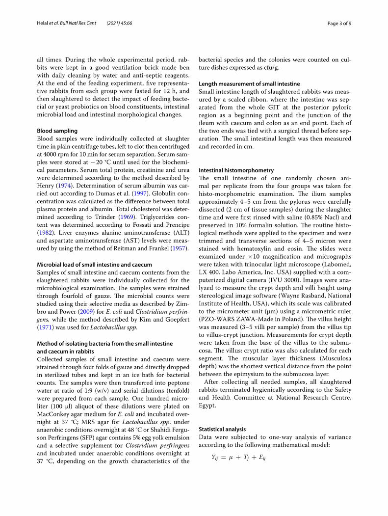
ResultsBlood biochemical constituentsData presented in Table 2 showed that, dietary supple-mentation of probiotics alone or in a mixture signifi-cantly (P ≤ 0.05) showed noticeable increases in serum total protein and albumin concentrations compared with un-supplemented control, while creatinine values showed no significant differences among experimental groups. The results of urea and triglyceride levels showed that, rabbits of the control diet (R1) recorded the highest values (P ≤ 0.05) compared with all supplemented groups
(R2, R3 and R4). Similar trend was almost observed for serum concentration of cholesterol.
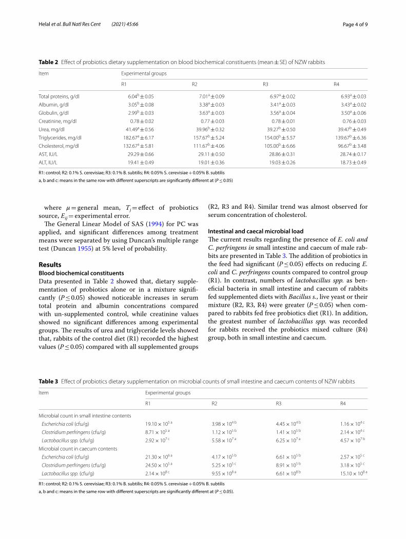
Intestinal and caecal microbial loadThe current results regarding the presence of E. coli and C. perfringens in small intestine and caecum of male rab-bits are presented in Table 3. The addition of probiotics in the feed had significant (P ≤ 0.05) effects on reducing E. coli and C. perfringens counts compared to control group (R1). In contrast, numbers of lactobacillus spp. as ben-eficial bacteria in small intestine and caecum of rabbits fed supplemented diets with Bacillus s., live yeast or their mixture (R2, R3, R4) were greater (P ≤ 0.05) when com-pared to rabbits fed free probiotics diet (R1). In addition, the greatest number of lactobacillus spp. was recorded for rabbits received the probiotics mixed culture (R4) group, both in small intestine and caecum.
Table 2 Effect of probiotics dietary supplementation on blood biochemical constituents (mean ± SE) of NZW rabbits
R1: control; R2: 0.1% S. cerevisiae; R3: 0.1% B. subtilis; R4: 0.05% S. cerevisiae + 0.05% B. subtilis
a, b and c: means in the same row with different superscripts are significantly different at (P ≤ 0.05)
Item Experimental groups
R1 R2 R3 R4
Total proteins, g/dl 6.04b ± 0.05 7.01a ± 0.09 6.97a ± 0.02 6.93a ± 0.03
Albumin, g/dl 3.05b ± 0.08 3.38a ± 0.03 3.41a ± 0.03 3.43a ± 0.02
Globulin, g/dl 2.99b ± 0.03 3.63a ± 0.03 3.56a ± 0.04 3.50a ± 0.06
Creatinine, mg/dl 0.78 ± 0.02 0.77 ± 0.03 0.78 ± 0.01 0.76 ± 0.03
Urea, mg/dl 41.49a ± 0.56 39.96b ± 0.32 39.27b ± 0.50 39.47b ± 0.49
Triglycerides, mg/dl 182.67a ± 6.17 157.67b ± 5.24 154.00b ± 5.57 139.67b ± 6.36
Cholesterol, mg/dl 132.67a ± 5.81 111.67b ± 4.06 105.00b ± 6.66 96.67b ± 3.48
AST, IU/L 29.29 ± 0.66 29.11 ± 0.50 28.86 ± 0.31 28.74 ± 0.17
ALT, IU/L 19.41 ± 0.49 19.01 ± 0.36 19.03 ± 0.26 18.73 ± 0.49
Table 3 Effect of probiotics dietary supplementation on microbial counts of small intestine and caecum contents of NZW rabbits
R1: control; R2: 0.1% S. cerevisiae; R3: 0.1% B. subtilis; R4: 0.05% S. cerevisiae + 0.05% B. subtilis
a, b and c: means in the same row with different superscripts are significantly different at (P ≤ 0.05).
Item Experimental groups
R1 R2 R3 R4
Microbial count in small intestine contents
Escherichia coli (cfu/g) 19.10 × 10⁵ a 3.98 × 10⁴ b 4.45 × 10⁴ b 1.16 × 10⁴ c
Clostridium perfringens (cfu/g) 8.71 × 10⁵ a 1.12 × 10⁵ b 1.41 × 10⁵ b 2.14 × 10⁴ c
Lactobacillus spp. (cfu/g) 2.92 × 10⁷ c 5.58 × 10⁷ a 6.25 × 10⁷ a 4.57 × 10⁷ b
Microbial count in caecum contents
Escherichia coli (cfu/g) 21.30 × 10⁶ a 4.17 × 10⁵ b 6.61 × 10⁵ b 2.57 × 10⁵ c
Clostridium perfringens (cfu/g) 24.50 × 10⁵ a 5.25 × 10⁵ c 8.91 × 10⁵ b 3.18 × 10⁵ c
Lactobacillus spp. (cfu/g) 2.14 × 10⁸ c 9.55 × 10⁸ a 6.61 × 10⁸ b 15.10 × 10⁸ a

Page 5 of 9Helal et al. Bull Natl Res Cent (2021) 45:66
Small intestinal length and its histo‑morphological changesThe supplementation of probiotics to the experimental diets significantly increased the length of small intestine compared to un-supplemented group, Table 4. The bac-terial probiotic has a superior significant effect on intes-tinal length where it was increased by 15%, while yeast
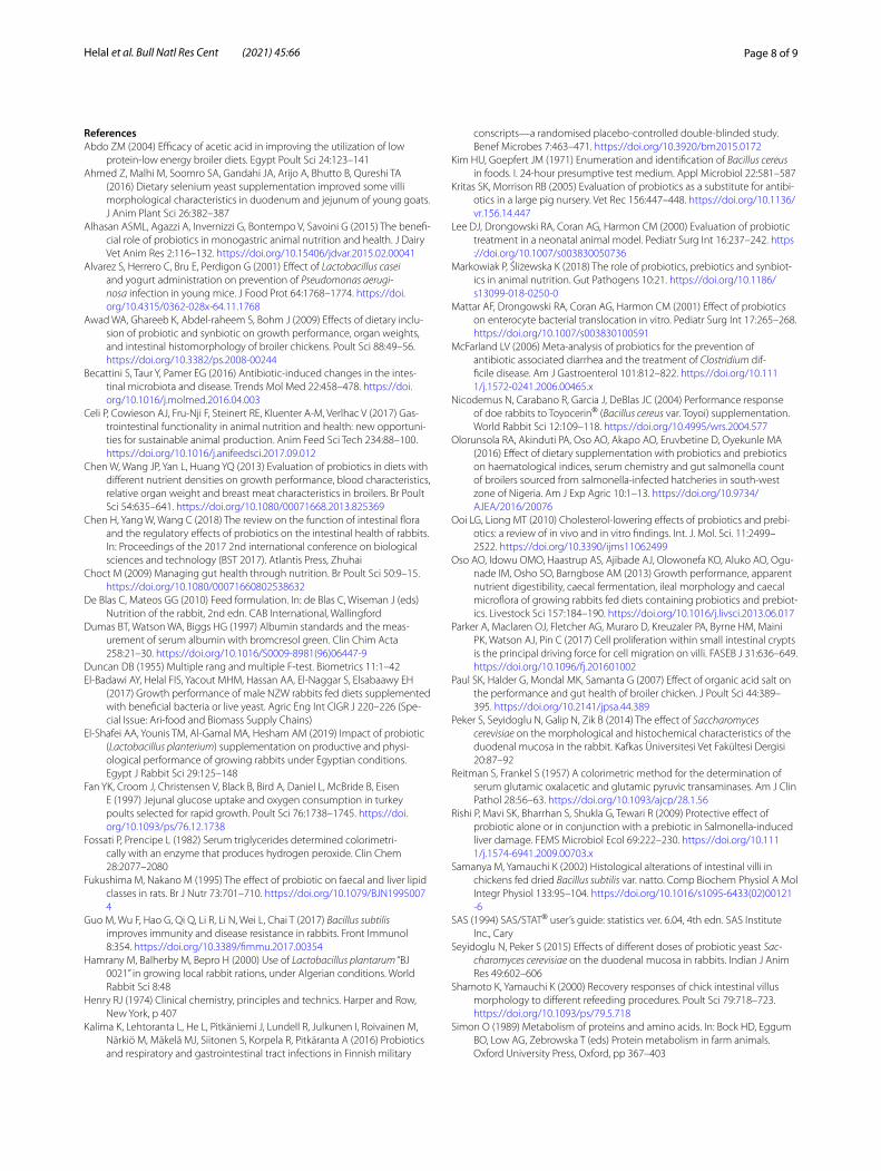
or mixed probiotic groups increased the length by 10% than that of the control group. The microarchitecture of ilium of the rabbits fed different probiotic diets is given in Fig. 1. Muscularis mucosa layer, submucosa, mucosa, crypts of Lieberkühn, and villi are clearly identified and measured.
Table 4 Effect of probiotics dietary supplementation on small intestine length and histo‑morphological parameters (mean ± SE) of NZW rabbits
* Each value is a mean of five replicates.
R1: control; R2: 0.1% S. cerevisiae; R3: 0.1% B. subtilis; R4: 0.05% S. cerevisiae + 0.05% B. subtilis
a, b and c: Means in the same row with different superscripts are significantly different at (P ≤ 0.05).
Item Experimental groups
R1 R2 R3 R4
Small intestine length, (cm)* 138.5c ± 0.87 152.1b ± 1.40 159.0a ± 1.70 152.5b ± 2.00
Villus height, (µm) 359.7b ± 18.04 446.1a ± 13.40 423.5ab ± 16.13 473.7a ± 12.36
Crypt depth, (µm) 68.7b ± 0.33 81.2a ± 2.29 80.2a ± 4.12 88.1a ± 3.02
Villus height: crypt depth 5.24 ± 0.34 5.59 ± 0.28 5.28 ± 0.22 5.60 ± 0.28
Musculosa depth, (µm) 112.8a ± 11.86 86.9b ± 3.44 94.1b ± 5.61 79.0b ± 2.90
2B
V
1A
3A
V
Cr
CrM
M
L L
V
Cr
L
M
SM
SM
L
V
CrM4B
SM
Fig. 1 Cross section through small intestine (ilium) from rabbits of different dietary treatments at 18 weeks of age. (M) muscularis mucosa; (SM) submucosa; (Cr) crypts of Lieberkühn; (V) Villi; (L) lumen. ((H & E × 100). 1a Control; 2b 0.1% S. cerevisiae; 3a 0.1% B. subtilis; 4b 0.05% S. cerevisiae + 0.05% B. subtilis

Page 6 of 9Helal et al. Bull Natl Res Cent (2021) 45:66
In the present study, the histo-morphological analysis of the ilium showed that the supplementation of growing rabbit diets with S. cerevisiae (R2), B. subtilis (R3) or their mixture (R4) has significant positive effects (P ≤ 0.05) on the length of villi (μm) as compared with the control group. It was noticeable that, rabbits of R2 and R4 groups had higher villi length compared to that of rabbits of R3 group, and numerically but not statistically significant the R4 had the highest villus. On the other side, crypt depth (μm) increased (P ≤ 0.05) in all experimental groups compared with the control and the superiority was for R4 group. However, villus height: crypt depth ratio was not significantly affected among experimental groups. Mus-culosa depth showed significant decreases (P ≤ 0.05) in muscular layer thickness of the ilium in all experimental groups compared with the control one.
DiscussionBlood biochemical constituentsThese findings are in agreement with many studies (Abdo 2004; Ooi and Liong 2010; El-Shafei et al. 2019), who reported that blood cholesterol decreased significantly by dietary probiotics. They hypothesized the effect of probiotics on lipid metabolism as: posing bile salt hydro-lase activity and precipitation of cholesterol by some microorganisms such as Lactobacillus and Bifidobac-terium, incorporation of cholesterol or binding to bac-teria and making of short-chain fatty acids by probiotic bacteria. Fukushima and Nakano (1995) had been stated another explanation. The authors stated that, probiotics hold hydroxymethyl-glutaryl-coenzyme A; an enzyme involved in the cholesterol synthesis pathway, and there-fore decrease the cholesterol synthesis.
It is clearly that the insignificant differences found among all experimental groups for both ALT and AST activity and creatinine concentration might point out to that rabbits could tolerate the addition of probiotics up to 0.1% without any deleterious effects on kidney or liver functions. The significantly increases in serum total pro-teins, and subsequently albumin and globulins is another indicator of healthy liver as most of blood proteins syn-thesized in liver. Moreover, previous study (Rishi et al. 2009) found that probiotics supplementation decreased bacterial translocation in the liver of mice challenged with Salmonella typhimurium and decreased levels of serum aminotransferases.
Intestinal and caecal microbial loadProbiotics might promote changes on enteric microbi-ota, so some pathogens cannot adhere effectively (Mat-tar et al. 2001). These results agreed with (Lee et al. 2000) who found that E. coli count was reduced by 25% in the small intestine of rabbits received probiotics. Also, some
probiotics caused reduction of Clostridium associated disease in humans (McFarland 2006; Yamano et al. 2006; Sivamaruthi et al. 2019). The sole addition of probiotic type in particular B. subtilis was (P ≤ 0.05) effective than live yeast or the mixed probiotics additives in reducing E. coli and C. perfringens counts in small intestine and caecum.
The positive effect of the sole addition of bacteria or yeast (R2 or R3), regarding intestinal Lactobacillus spp., was clearly greater than the mixture supplementation (R4). It could be explained as EL-Badawi et al. (2017) mentioned in our previous study that, there is an antag-onistic effect between bacteria and live yeast probiotics when fed in mixed culture.
These results are in agreement with Hamrany et al. (2000) who found a positive effect of a probiotic on E. coli occurrence in the caecum and small intestine of young rabbits. These may be regarded to that Bacillus spp. could stimulate biosynthetic capacities of Lactobacillus strains. As the result of increasing beneficial bacteria, mainly lactobacillus, and decreasing of Clostridium and E. coli populations in the small intestine and caecum the nor-mal intestinal microflora can competitively inhibit the survival and proliferation of harmful flora by competing for nutrients in the intestinal habitat, in turn better feed utilization is expected. Furthermore, probiotics improve intestinal balance of host animal and creating gut micro-organism conditions that inhibit pathogenic bacteria like E. coli and Clostridium and support beneficial bacte-ria like Lactobacillus which was reflected on better feed digestion and absorption (Chen et al. 2013; Alhasan et al. 2015; Markowiak and Śliżewska 2018).
Small intestinal length and its histo‑morphological changesIt seems logic to state that, bacterial or live yeast probiot-ics could promote the development of intestinal tract. In this concern, Slezak et al. (2014) found that the length of small intestine in sterilized mice was significantly smaller than that in normal ones. This description was also stated by William and Linda (2000). Villus height and the ratio of villus height to crypt depth are indicators of gastroin-testinal tract morphology (Shamoto and Yamauchi 2000) and intestinal histomorphology are one of the important indications of gut health in different animal species.
It is well-known that, intestine transfer nutrients required for maintenance and production of animals. The surface area of the intestinal villi plays an important role in the absorption of nutrients by small intestine. In addition, animal immunity affects by intestinal epi-thelium status, because of its action as a natural barrier against to pathogenic bacteria and toxic substances pre-sent in the intestinal lumen (Paul et al. 2007). Therefore,

Page 7 of 9Helal et al. Bull Natl Res Cent (2021) 45:66
the improvement of intestinal morphology prompts more available nutrients which leads to improvement of absorption process and intestinal health development (Fan et al. 1997; Choct 2009; Celi et al. 2017).
It was noticed that the increase in villus height and vil-lus height to crypt depth ratio are directly correlated with an increase in intestinal epithelium turnover (Fan et al. 1997), and longer villus is also associated with activated cell proliferation (Parker et al. 2017), whereas shorten-ing of villus and deeper crypts lead to poor nutrients absorption and increased gastrointestinal secretion, and consequently cause reduction of animal performance (Xu et al. 2003). In addition, intestinal villi elongation could enhance enzyme production and digestion by increasing the effective absorptive area and improving the nutrients transport system in intestinal tract (Awad et al. 2009).
Recently, many of researchers achieved beneficial effects of probiotic on gastrointestinal health in many animal species. Increasing the villi length and width led to increase in mucosal surface area by 36.2 and 62.48% in duodenum and jejunum of goats fed selenium yeast compared to control (Ahmed et al. 2016). Moreover, the improvement of available nutrients in intestine would, result in increasing weights of visceral organs and improving growth performance of animal. Also, Peker et al. (2014) reported that the addition of 3 g S. cerevi-siae/kg diet of rabbits affected the duodenum morphol-ogy by increasing the total mucosa, villus height, and the gland depth. In another study, Seyidoglu and Peker (2015) reported that the villus height, crypt depth, gland depth and total mucosa were increased significantly by yeast (S. cerevisiae) addition (2 g/kg and 4 g/kg) in rab-bits’ diet, although the villus crypt ratio was not changed. This enhancement in duodenum morphology was dose-dependently of S. cerevisiae. So, administration of S. cerevisiae in either low or high doses had positive effect on digestive and absorptive functions of the intestinal mucosa. In a recent research by Guo et al. (2017), rab-bits fed with B. subtilis showed good probiotic potential in rabbits, resulted in improving growth performance, serum immunoglobulin, immune organ index, intestinal homeostasis, and immune response of rabbits, as well as its antibacterial benefits.
Finally, although there are many improvements in blood metabolites, intestinal and caecum microbial load as a result to the mixture of B. subtilis and S. cerevisiae, however it was not extended to productive performance in comparison with the single addition of B. subti-lis or S. cerevisiae. The taller small intestine, for rabbits fed on the sole addition of B. subtilis or S. cerevisiae, and not for their mixture, may be having the key to explain why rabbits of groups R2 and R3 showed high final body weight with low feed conversion. So, wide surface area
for nutrients absorption, therefore high feed utilization, is closer related to good productive performance of rab-bits than improvements in blood metabolites, intestinal and caecum microbial load.
ConclusionsThis study demonstrated that the addition of Bacillus subtilis or Saccharomyces cerevisiae to diets of growing NZW rabbits by 0.1% is recommended to minimize the pathogenic intestinal load and increasing the beneficial lactobacillus strains as well as improving the intestinal barriers integrity.
Abbreviationsg: Gram; P: Probability; cfu: Colony‑forming unit; ALT: Alanine aminotrans‑ferase; AST: Aspartate aminotransferase; DM: Dry matter; OM: Organic matter; CP: Crude protein; CF: Crude fiber; EE: Ether extract; NFE: Nitrogen‑free extract; GIT: Gastrointestinal tract; SAS: Statistical Analysis System.
AcknowledgementsThe authors would like to thank the Commercial Division of MultiVita Com‑pany of Animal Nutrition, Egypt, for providing the feed additives needed in this study.
Authors’ contributionsConceptualization was carried out by F.H. and A.E.; data curation was per‑formed by A.E. and O.A.; investigation was done by S.E., M.S., and S.A.; S.A., M.S., O.A., and S.A. contributed to methodology; supervision was done by F.H. and A.E.; validation was carried out by F.H. and A.E.; visualization and writ‑ing—original draft were performed by F.H., A.E. and M.S.; writing—review and editing were done by A.E. and O.A. All authors read and approved the final manuscript..
FundingThere are currently no funding sources in the design of the study and collec‑tion, analysis and interpretation of data, and in writing of the manuscript.
Availability of data and materialsThe datasets used and/or analyzed during the current study are available from the corresponding author on request.
Declarations
Ethics approval and consent to participateThe experimental design and all the research protocols were approved by the Medical Research Ethics Committee (MREC) of the National Research Center with ethical approval code 20/173.
Consent for publicationNot applicable.
Competing interestsThe authors declare that they have no competing interests.
Author details1 Animal Production Department, National Research Centre, Dokki, Giza 12622, Egypt. 2 Poultry Production Department, Faculty of Agriculture, Ain Shams University, Shoubra El‑Kheima, Cairo 11241, Egypt. 3 Department of Livestock Research, Arid Lands Cultivation Research Institute, City of Scientific Research and Technology Applications, New Borg El‑Arab, Alexandria, Egypt.
Received: 31 December 2020 Accepted: 8 March 2021
Page 8 of 9Helal et al. Bull Natl Res Cent (2021) 45:66
ReferencesAbdo ZM (2004) Efficacy of acetic acid in improving the utilization of low
protein‑low energy broiler diets. Egypt Poult Sci 24:123–141Ahmed Z, Malhi M, Soomro SA, Gandahi JA, Arijo A, Bhutto B, Qureshi TA
(2016) Dietary selenium yeast supplementation improved some villi morphological characteristics in duodenum and jejunum of young goats. J Anim Plant Sci 26:382–387
Alhasan ASML, Agazzi A, Invernizzi G, Bontempo V, Savoini G (2015) The benefi‑cial role of probiotics in monogastric animal nutrition and health. J Dairy Vet Anim Res 2:116–132. https ://doi.org/10.15406 /jdvar .2015.02.00041
Alvarez S, Herrero C, Bru E, Perdigon G (2001) Effect of Lactobacillus casei and yogurt administration on prevention of Pseudomonas aerugi-nosa infection in young mice. J Food Prot 64:1768–1774. https ://doi.org/10.4315/0362‑028x‑64.11.1768
Awad WA, Ghareeb K, Abdel‑raheem S, Bohm J (2009) Effects of dietary inclu‑sion of probiotic and synbiotic on growth performance, organ weights, and intestinal histomorphology of broiler chickens. Poult Sci 88:49–56. https ://doi.org/10.3382/ps.2008‑00244
Becattini S, Taur Y, Pamer EG (2016) Antibiotic‑induced changes in the intes‑tinal microbiota and disease. Trends Mol Med 22:458–478. https ://doi.org/10.1016/j.molme d.2016.04.003
Celi P, Cowieson AJ, Fru‑Nji F, Steinert RE, Kluenter A‑M, Verlhac V (2017) Gas‑trointestinal functionality in animal nutrition and health: new opportuni‑ties for sustainable animal production. Anim Feed Sci Tech 234:88–100. https ://doi.org/10.1016/j.anife edsci .2017.09.012
Chen W, Wang JP, Yan L, Huang YQ (2013) Evaluation of probiotics in diets with different nutrient densities on growth performance, blood characteristics, relative organ weight and breast meat characteristics in broilers. Br Poult Sci 54:635–641. https ://doi.org/10.1080/00071 668.2013.82536 9
Chen H, Yang W, Wang C (2018) The review on the function of intestinal flora and the regulatory effects of probiotics on the intestinal health of rabbits. In: Proceedings of the 2017 2nd international conference on biological sciences and technology (BST 2017). Atlantis Press, Zhuhai
Choct M (2009) Managing gut health through nutrition. Br Poult Sci 50:9–15. https ://doi.org/10.1080/00071 66080 25386 32
De Blas C, Mateos GG (2010) Feed formulation. In: de Blas C, Wiseman J (eds) Nutrition of the rabbit, 2nd edn. CAB International, Wallingford
Dumas BT, Watson WA, Biggs HG (1997) Albumin standards and the meas‑urement of serum albumin with bromcresol green. Clin Chim Acta 258:21–30. https ://doi.org/10.1016/S0009 ‑8981(96)06447 ‑9
Duncan DB (1955) Multiple rang and multiple F‑test. Biometrics 11:1–42El‑Badawi AY, Helal FIS, Yacout MHM, Hassan AA, El‑Naggar S, Elsabaawy EH
(2017) Growth performance of male NZW rabbits fed diets supplemented with beneficial bacteria or live yeast. Agric Eng Int CIGR J 220–226 (Spe‑cial Issue: Ari‑food and Biomass Supply Chains)
El‑Shafei AA, Younis TM, Al‑Gamal MA, Hesham AM (2019) Impact of probiotic (Lactobacillus planterium) supplementation on productive and physi‑ological performance of growing rabbits under Egyptian conditions. Egypt J Rabbit Sci 29:125–148
Fan YK, Croom J, Christensen V, Black B, Bird A, Daniel L, McBride B, Eisen E (1997) Jejunal glucose uptake and oxygen consumption in turkey poults selected for rapid growth. Poult Sci 76:1738–1745. https ://doi.org/10.1093/ps/76.12.1738
Fossati P, Prencipe L (1982) Serum triglycerides determined colorimetri‑cally with an enzyme that produces hydrogen peroxide. Clin Chem 28:2077–2080
Fukushima M, Nakano M (1995) The effect of probiotic on faecal and liver lipid classes in rats. Br J Nutr 73:701–710. https ://doi.org/10.1079/BJN19 95007 4
Guo M, Wu F, Hao G, Qi Q, Li R, Li N, Wei L, Chai T (2017) Bacillus subtilis improves immunity and disease resistance in rabbits. Front Immunol 8:354. https ://doi.org/10.3389/fimmu .2017.00354
Hamrany M, Balherby M, Bepro H (2000) Use of Lactobacillus plantarum “BJ 0021” in growing local rabbit rations, under Algerian conditions. World Rabbit Sci 8:48
Henry RJ (1974) Clinical chemistry, principles and technics. Harper and Row, New York, p 407
Kalima K, Lehtoranta L, He L, Pitkäniemi J, Lundell R, Julkunen I, Roivainen M, Närkiö M, Mäkelä MJ, Siitonen S, Korpela R, Pitkäranta A (2016) Probiotics and respiratory and gastrointestinal tract infections in Finnish military
conscripts—a randomised placebo‑controlled double‑blinded study. Benef Microbes 7:463–471. https ://doi.org/10.3920/bm201 5.0172
Kim HU, Goepfert JM (1971) Enumeration and identification of Bacillus cereus in foods. I. 24‑hour presumptive test medium. Appl Microbiol 22:581–587
Kritas SK, Morrison RB (2005) Evaluation of probiotics as a substitute for antibi‑otics in a large pig nursery. Vet Rec 156:447–448. https ://doi.org/10.1136/vr.156.14.447
Lee DJ, Drongowski RA, Coran AG, Harmon CM (2000) Evaluation of probiotic treatment in a neonatal animal model. Pediatr Surg Int 16:237–242. https ://doi.org/10.1007/s0038 30050 736
Markowiak P, Śliżewska K (2018) The role of probiotics, prebiotics and synbiot‑ics in animal nutrition. Gut Pathogens 10:21. https ://doi.org/10.1186/s1309 9‑018‑0250‑0
Mattar AF, Drongowski RA, Coran AG, Harmon CM (2001) Effect of probiotics on enterocyte bacterial translocation in vitro. Pediatr Surg Int 17:265–268. https ://doi.org/10.1007/s0038 30100 591
McFarland LV (2006) Meta‑analysis of probiotics for the prevention of antibiotic associated diarrhea and the treatment of Clostridium dif‑ficile disease. Am J Gastroenterol 101:812–822. https ://doi.org/10.1111/j.1572‑0241.2006.00465 .x
Nicodemus N, Carabano R, Garcia J, DeBlas JC (2004) Performance response of doe rabbits to Toyocerin® (Bacillus cereus var. Toyoi) supplementation. World Rabbit Sci 12:109–118. https ://doi.org/10.4995/wrs.2004.577
Olorunsola RA, Akinduti PA, Oso AO, Akapo AO, Eruvbetine D, Oyekunle MA (2016) Effect of dietary supplementation with probiotics and prebiotics on haematological indices, serum chemistry and gut salmonella count of broilers sourced from salmonella‑infected hatcheries in south‑west zone of Nigeria. Am J Exp Agric 10:1–13. https ://doi.org/10.9734/AJEA/2016/20076
Ooi LG, Liong MT (2010) Cholesterol‑lowering effects of probiotics and prebi‑otics: a review of in vivo and in vitro findings. Int. J. Mol. Sci. 11:2499–2522. https ://doi.org/10.3390/ijms1 10624 99
Oso AO, Idowu OMO, Haastrup AS, Ajibade AJ, Olowonefa KO, Aluko AO, Ogu‑nade IM, Osho SO, Barngbose AM (2013) Growth performance, apparent nutrient digestibility, caecal fermentation, ileal morphology and caecal microflora of growing rabbits fed diets containing probiotics and prebiot‑ics. Livestock Sci 157:184–190. https ://doi.org/10.1016/j.livsc i.2013.06.017
Parker A, Maclaren OJ, Fletcher AG, Muraro D, Kreuzaler PA, Byrne HM, Maini PK, Watson AJ, Pin C (2017) Cell proliferation within small intestinal crypts is the principal driving force for cell migration on villi. FASEB J 31:636–649. https ://doi.org/10.1096/fj.20160 1002
Paul SK, Halder G, Mondal MK, Samanta G (2007) Effect of organic acid salt on the performance and gut health of broiler chicken. J Poult Sci 44:389–395. https ://doi.org/10.2141/jpsa.44.389
Peker S, Seyidoglu N, Galip N, Zik B (2014) The effect of Saccharomyces cerevisiae on the morphological and histochemical characteristics of the duodenal mucosa in the rabbit. Kafkas Üniversitesi Vet Fakültesi Dergisi 20:87–92
Reitman S, Frankel S (1957) A colorimetric method for the determination of serum glutamic oxalacetic and glutamic pyruvic transaminases. Am J Clin Pathol 28:56–63. https ://doi.org/10.1093/ajcp/28.1.56
Rishi P, Mavi SK, Bharrhan S, Shukla G, Tewari R (2009) Protective effect of probiotic alone or in conjunction with a prebiotic in Salmonella‑induced liver damage. FEMS Microbiol Ecol 69:222–230. https ://doi.org/10.1111/j.1574‑6941.2009.00703 .x
Samanya M, Yamauchi K (2002) Histological alterations of intestinal villi in chickens fed dried Bacillus subtilis var. natto. Comp Biochem Physiol A Mol Integr Physiol 133:95–104. https ://doi.org/10.1016/s1095 ‑6433(02)00121 ‑6
SAS (1994) SAS/STAT® user’s guide: statistics ver. 6.04, 4th edn. SAS Institute Inc., Cary
Seyidoglu N, Peker S (2015) Effects of different doses of probiotic yeast Sac-charomyces cerevisiae on the duodenal mucosa in rabbits. Indian J Anim Res 49:602–606
Shamoto K, Yamauchi K (2000) Recovery responses of chick intestinal villus morphology to different refeeding procedures. Poult Sci 79:718–723. https ://doi.org/10.1093/ps/79.5.718
Simon O (1989) Metabolism of proteins and amino acids. In: Bock HD, Eggum BO, Low AG, Zebrowska T (eds) Protein metabolism in farm animals. Oxford University Press, Oxford, pp 367–403

Page 9 of 9Helal et al. Bull Natl Res Cent (2021) 45:66
Sivamaruthi BS, Kesika P, Chaiyasut C (2019) A mini‑review of human studies on cholesterol‑lowering properties of probiotics. Sci Pharm 87:26. https ://doi.org/10.3390/sciph arm87 04002 6
Slezak K, Krupova Z, Rabot S, Loh G, Levenez F, Descamps A, Lepage P, Dore J, Bellier S, Blaut M (2014) Association of germfree mice with a simpli‑fied human intestinal microbiota results in a shortened intestine. Gut Microbes 5:176–182. https ://doi.org/10.4161/gmic.28203
Timmerman HM, Mulder L, Everts H, van Espen DC, van der Wal E, Klaassen G, Rouwers SMG, Hartemink R, Rombouts FM, Beynen AC (2005) Health and growth of veal calves fed milk replacer with or without probiotics. J Dairy Sci 88:2154–2165. https ://doi.org/10.3168/jds.S0022 ‑0302(05)72891 ‑5
Todorov N, Kraulov I, Dvuwinow D, Aleksandrov A (2007) Guide on animal nutrition. MATCOM, Sofia
Trinder P (1969) The determination of cholesterol in serum. Analyst 77:321–325
Wang Y, Wu Y, Wang Y, Xu H, Mei X, Yu D, Wang Y, Li W (2017) Antioxidant properties of probiotic bacteria. Nutrients 9:521. https ://doi.org/10.3390/nu905 0521
William JB, Linda MB (2000) Color atlas of veterinary histology, 2nd edn. Lip‑pincott Williams & Wilkins, Hoboken
Xu ZR, Hu CH, Xia MS, Zhan XA, Wang MQ (2003) Effects of dietary fructoo‑ligosaccharide on digestive enzyme activities, intestinal microflora and morphology of male broilers. Poult Sci 82:1030–1036. https ://doi.org/10.1093/ps/82.6.1030
Yamano T, Iino H, Takada M, Blum S, Rochat F, Fukushima Y (2006) Improve‑ment of the human intestinal flora by ingestion of the probiotic strain Lactobacillus johnsonii La1. Br J Nutr 95:303–312. https ://doi.org/10.1079/bjn20 05150 7
Yason CV, Summers BA, Schat KA (1987) Pathogenesis of rotavirus infection in various age groups of chickens and turkeys: pathology. Am J Vet Res 48:927–938
Zimbro MJ, Power DA (2009) Difco & BBL manual: manual of microbiological culture media, 2nd edn. Becton Dickinson and Co., Maryland, USA, Sparks
Publisher’s NoteSpringer Nature remains neutral with regard to jurisdictional claims in pub‑lished maps and institutional affiliations.