Embed Size (px)
Citation preview
Procedures in Scanning Probe Microscopy 2:2:4 1
Copyright © 1998 by John Wiley & Sons Ltd
Module Ultraflat Au surfaces2:2:4
Contributed by: Martin Hegner [1] and Peter Wagner [2]
In this module, a simple method to produce a well-characterized,‘atomically’ flat substrate is presented. Chemical and physical properties ofthe substrate are of primary importance. In addition to being chemicallyinert against O2 (but endowed with specific chemical reactivity) thesubstrate must be atomically flat over large areas; otherwise objects havingunfavourable length:width ratios will "sink" into an irregular topography.This is especially true for fibrillary structures of µm length, e.g., nucleicacids or organic polymers with diameters smaller than 3 nm, but also forindividual globular proteins. These macromolecules require substrates witha mean roughness as small as a few Å on areas of at least 5 µm2. Finally,the ideal substrates should also be reasonably priced and easy to prepare,to store, and to chemically modify.
Gold is a popular substrate. It is stable against O2, can be easilyprepared by vapor-deposition, and can bind with high affinity organic thiolsor bifunctional disulfides such as gold(I)alkanethiolates (RS-Au(I)) (seemodule 5.3.1 and module 7.12.2) also capable of forming self-assembledmonolayers. The epitaxial growth of f.c.c. metals on mica as a substrate iswell known, and polycrystalline gold films which form with (111)orientation on the (001) cleavage planes of mica have been extensivelystudied [3-5]
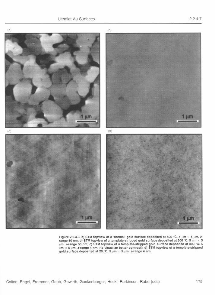
Au surfaces prepared by the above procedures suffer from a number ofimperfections (see, e.g. [5]). The terraces obtained by depositing Au onmica at optimal temperature, i.e., 300 °C - 500 °C, which is the mostwidespread technique, have varying sizes and are surrounded byunpredictable and rough topography (see Fig. 3a). Indeed with deposition atconstant temperature it is impossible to avoid the formation of deepdepressions. Hence these are useful only for macromolecules which aresmaller than the terraces themselves. Other Au surfaces (e.g., singlecrystals, annealed gold balls, etc.) suffer likewise from various limitations,such as high cost or lengthy preparation procedures [5]. Finally, all Ausurfaces described thus far have to be prepared immediately before use,which is time-consuming for routine use.
We have developed a simple procedure to reproducibly prepare ultraflattemplate-stripped gold surfaces (i.e.TSG)(see Fig. 3b-d). The procedure isdescribed in this module. An additional practical advantage of thistechnique is that the gold surfaces can be quickly obtained from theirimmediate precursors, which are stable and can be prepared and stored formonths in bulk.
Procedures in Scanning Probe Microscopy 2:2:4 2
Copyright © 1998 by John Wiley & Sons Ltd
Procedure Preparation of template-stripped gold (TSG)
A method has been devised to produce µm-sized gold films with‘atomically’ flat areas for use in scanning probe microscopy [3]. A meanroughness of ~ 2 Å for areas of 2.25 µm2, and about 3 Å for 25 µm2, can beeasily produced.
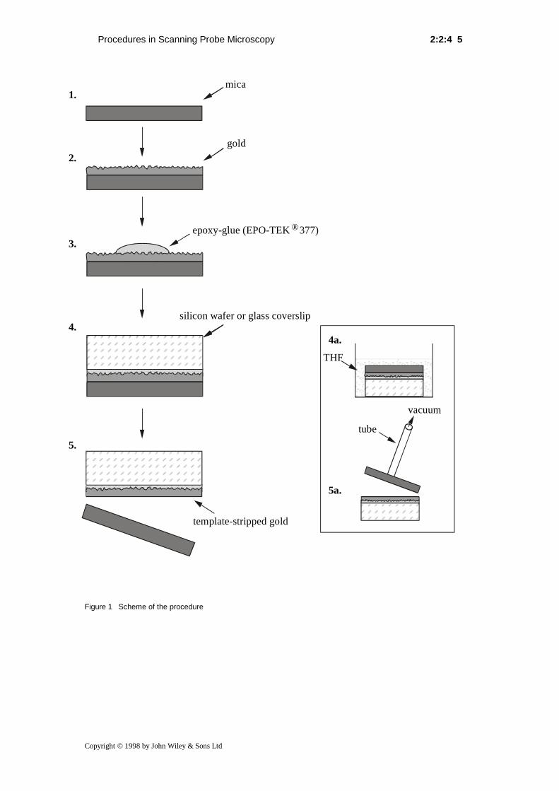
The method is based on (see Figure 1):
1. Epitaxial growth of gold on mica. Gold is deposited onto freshly cleavedmica sheets by thermal evaporation.
2. Glueing the gold surface onto a piece of Si wafer or glass coverslips.
3. Chemical stripping of the mica down to the appearing gold surface(TSG).
MATERIALS
• Gold (99,99%) or (99.999 %), e.g. from Cendres & Metaux SA (Biel,Switzerland) or Goodfellow Cambridge Limited (Cambridge, UK).
• Muscovite mica: Provac AG (Balzers, Liechtenstein) or GoodfellowCambridge Limited (Cambridge, UK).
• Small pieces of monocrystalline Si(100) wafers or glass coverslipsSuppliers include Wacker-Chemtronic GmbH (Stuttgart, Germany) andPlano, W. Plannet GmbH (Marburg, Germany).
• Low-viscosity epoxy glue epo-tek® 377. Suppliers: Polyscience (Cham,Switzerland) and Epoxy Technology Inc. (Billerica, MA, USA)
• Tetrahydrofuran (THF) commercial grade of highest purity.
EQUIPMENT REQUIREMENTS
High vacuum coating system for thermal evaporation with integrated quartzcrystal deposition controller (vacuum during deposition < 2,66 X 10⁻⁴ Pa).The mica sheet has to be radiatively heated in situ from the rear(temperatures 20 °C to 400 °C).
SAMPLE PREPARATION
Preparation of the gold films:
1. Cleave the muscovite mica sheets (~5 x 7 cm) with a glaucoma knife(without using water) and place them immediately cleaved side downinto the sample holder of the vacuum system.
2. Heat the mica sheet in situ from the rear through the sample holder,with temperature feedback control through a thermocouple. After
twenty hours at 300 °C at a pressure of less than 1.33 x 10⁻⁴ Pa,equilibrate 30 minutes at the substrate temperature chosen for the golddeposition , i.e. 20 - 25 °C or 300 °C.
Procedures in Scanning Probe Microscopy 2:2:4 3
Copyright © 1998 by John Wiley & Sons Ltd
Note: The temperature finally adopted for our routine work is 300 °C;see [3]. Carry out the evaporation of the gold at a pressure of less than2.66 x 10-4 Pa.
3. The deposition rate should be adjusted to 1 Å/s before opening of theshutter! Evaporation rate should be 1 Å/s and the film thickness ~200nm to increase the mechanical stability for stripping.
4. Close the shutter and anneal the samples for 2 - 6 hours at 300 °C.Cool slowly down to room temperature (10 °C / min). After removalfrom the evaporation chamber it is not necessary to store the gold filmsunder special protective conditions.
Note: In our system the resistively heated tungsten boat for thermalmetal evaporation is 25 cm directly below the mica substrate.
5. Cut the gold deposited mica sheets into small pieces (~1 cm2) andglue them face down onto Si(100) wafer pieces or glass coverslipsusing the epoxy glue epo-tek® 377. Heat the multilayer at 150 °C for 1-2 hour until the glue reaches an adequate hardness. This Si-Au-micamultilayer can be stored as a "stripping precursor" at least up to severalmonths without loss of quality.
Comment: Don’t use other epoxy glues! The glue should be exactly[±10 mg] weight out (w/w (!); 1:1) on a balance and thoroughly mixed. Asurplus of epoxy glue should be avoided. For a 1 cm2 piece use 10 - 15 µlof mixed glue.
Stripping:
We had seen in preliminary experiments that soaking the Si-Au-micamultilayer in various solvents lead to detachment of the complete mica-sheet from gold-Si ("chemically stripped gold surface") without undueeffect on the gold surface.
1. Soak the mica-gold-Si "sandwiches" in tetrahydrofuran (THF) atroom temperature. Check every two minutes with a vacuum suction(outside of the solvent, ~20-30 mm Hg) from the top whether the micasheet separates from the gold surface.
2. Rinse the freshly stripped gold surface with 5-10 ml THF.
3. Check the conductivity with a voltmeter.
Comment: Use fresh glassware for each stripping procedure. The template-stripped gold surfaces should be immediately used for biologicalexperiments or for the modification with a (bioreactive) self-assembledmonolayer reagent (see module 7.12.2). X-ray photoelectron spectroscopy(XPS) analyses showed that only trace amounts of Si and Al from theoriginal mica were left on the Au(111) surface (data not shown). The Si-gold multilayer (prepared with epo-tek® 377) is stable for days in aqueousbuffer, alcohols, 1,4-dioxane, and others for hours.
SPM measurements:
The measurements can be performed under ambient conditions on acommercially available scanning tunneling or atomic force microscope.
Procedures in Scanning Probe Microscopy 2:2:4 4
Copyright © 1998 by John Wiley & Sons Ltd
CONCLUSION:
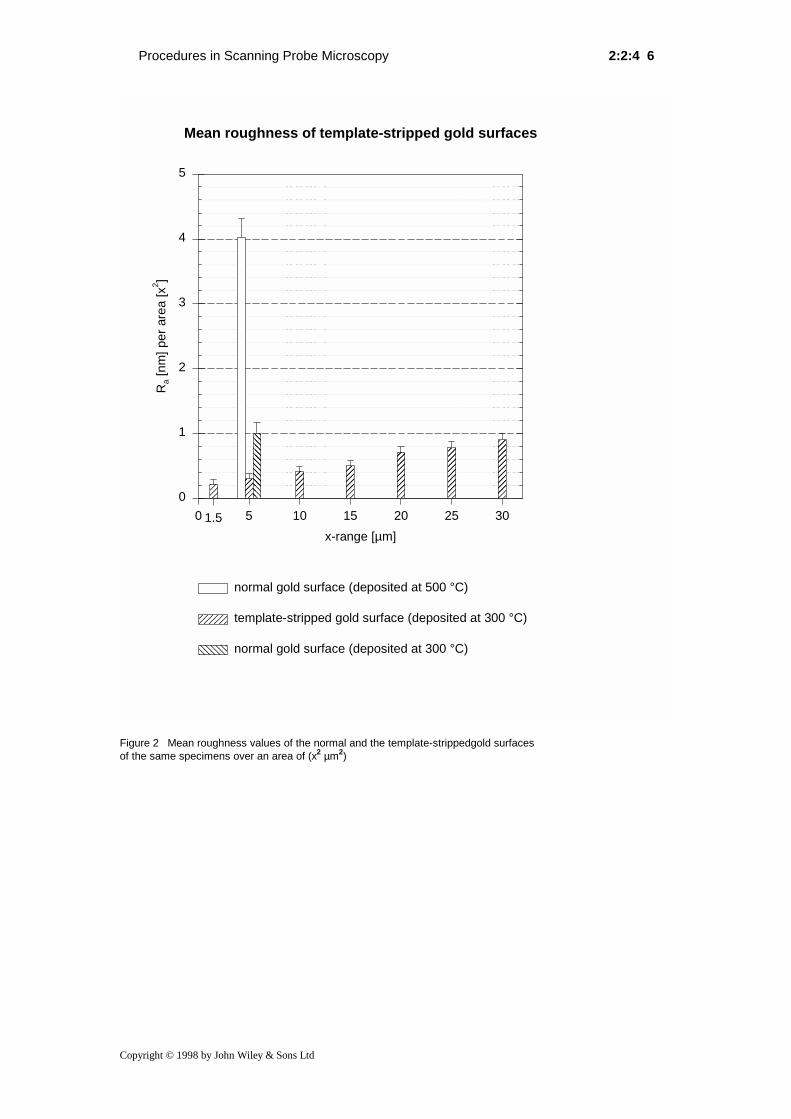
In summary, the template-stripped gold surfaces prepared by our strippingprocedures seem to be superior to other Au(111) surfaces in severalrespects: (i) they are very large, with very low roughness over extendedareas (see Fig. 2); (ii) they are easy to prepare, and (iii) can be madeimmediately before use from the gold-covered mica specimens; these arestable for months and can thus be prepared and stored in bulk. Images suchas that in Figure 3b)-d), which were obtained routinely, make our template-stripped surfaces highly suitable gold substrates for scanning probemicroscopy.
Finally, our template-stripped gold surfaces are well-characterized potentialalternative substrates for nanolithography by scanning tunnelingmicroscopy
References1. Dr. Martin Hegner,Howard Hughes Medical Res. Lab.,
Institute of Molecular Biology, University of Oregon, 1370Franklin Blvd., Eugene, OR 97403-1229, USA;e-mail: [email protected]
2. Dr. Peter Wagner,Beckman Center, B 405, Department ofBiochemistry, Stanford University, Stanford, CA 94305-5307, USA; email: [email protected].
3. M. Hegner, P. Wagner, and G. Semenza, Surf. Sci., 1993, 291,39 and P. Wagner, M. Hegner, H.-J. Güntherodt and G.Semenza, Langmuir, 1995, 11, 3867.
4. A detailed report of the effect of deposition temperature,deposition rate, and film thickness on the flatness of Au-films on mica:C.E.D. Chidsey, D.N. Loiacono, T. Sleator, and S. Nakahara,Surf. Sci., 1988, 200, 45
5. A review that includes various preparation methods forgold substrates:C.R. Clemmer and T.B. Beebe, Scanning Microscopy, 1992,6, 319.
6. related papers to TSG: M. Hegner, P. Wagner, and G.Semenza, FEBS Lett. 1993, 336, 452, P. Wagner, P. Kernen,M. Hegner, E. Ungewickell, and G. Semenza, FEBS Lett.1994, 356, 267 and P. Wagner, M. Hegner, P. Kernen, F.Zaugg and G. Semenza, 1995 Biophys. J. 70, 2052.
Procedures in Scanning Probe Microscopy 2:2:4 5
Copyright © 1998 by John Wiley & Sons Ltd
Figure 1 Scheme of the procedure
1.
2.
3.
4.
5.
4a.
5a.
epoxy-glue (EPO-TEK ® 377)
mica
gold
silicon wafer or glass coverslip
vacuum
THF
template-stripped gold
tube
Procedures in Scanning Probe Microscopy 2:2:4 6
Copyright © 1998 by John Wiley & Sons Ltd
Figure 2 Mean roughness values of the normal and the template-strippedgold surfacesof the same specimens over an area of (x2 µm2)
Mean roughness of template-stripped gold surfaces
x-range [µm]
0 5 10 15 20 25 30
Ra
[nm
] p
er
are
a [
x2]
0
1
2
3
4
5
�����
�����
���
���
������
normal gold surface (deposited at 500 °C)
template-stripped gold surface (deposited at 300 °C)��normal gold surface (deposited at 300 °C)��
1.5