Embed Size (px)
Citation preview

Close this window to return to IVIS www.ivis.org
Proceedings of the European Veterinary Conference
Voorjaarsdagen
Amsterdam, the Netherlands Apr. 18 - 20, 2013
Next Meeting:
Apr. 17 – 19, 2014 - Amsterdam, the Netherlands
Reprinted in IVIS with the permission of the Conference Organizers http://www.ivis.org
AbstrActs | EuropEan VEtErinary ConfErEnCE VoorjAArsdAgen 2013 www.VoorjAArsdAgen.eu
PrintseArcH bAcK HoMe
coMPAnion AniMAl1
coMPAnion AniMAl Posters
Dariush Vosough1, Nima Ghalekhani2*1Assistant Professor, Department of Clinical Sciences, Veterinary Faculty, Shahid Bahonar University of Kerman2School of veterinary medicine, Shahid Bahonar University of KermanCorresponding E-mail: [email protected]
MeAsuring oPticAl nerVe diAMeter in PersiAn cAt using Mri And tHree diMensionAl ultrAsonogrAPHy
the ocular structures are detectable using ultrasonography and magnetic resonance inaging(Mri) due to the presence of a considerable amount of fluid around vitreous and anterior chambers. using modern techniques may be helpful in assessing accurately normal structures of the retro bulbar space.
a total of ten normal persian cats,(5 males, 5 females), weighting 3±1.1 kg, underwent ultrasonographic examination. a GE Voloson ultrasonography machine and three-di-mensional (3D) linear array transducers (5-10 MHz) were used. Mri was performed with a 1.5 t GE scanner and a phased array human eye coil. the field of view ranged from 4 to 8 cm. transverse, sagittal and dorsal spin-echo t1 and t2 weighted images were acquired in all samples.
the mean optical nerve diameter in males by 3D-ultrasonography and Mri were 1.85mm (SD=0.002) and in females it was 1.86mm (SD=0.001). no significant difference was observable between ocular nerve measurements in male and female cats and left and right eyes using 3D-ultrasonography or Mri (p-value<0.05).
the 3D-ultrasonography provides useful images for diagnostic purposes and lesions of the caudal part of the orbit. the results of the eye 3D-ultrasonography in cats showed marked advantages in image acquisition for interpretation of all aspects of the ocular structures. this study suggests that using 3D-ultrasonography in veterinary ophthal-mology may be better than Mri because the images of 3D-ultrasonography are more flexible and can provide all the voluntary views. on top of that, the scanning period is shorter and anaesthesia is not required when using 3D-ultrasonography.
references:1. Downey DB, nicolle Da, Levin Mf and fenster a. three dimensional ultrasound imaging of the eye. Eye. 1996;
10: 75-81.
2. finger pt, Khoobehi a, ponce-Contreras Mr, Dellarocca D and Garcia JpS. three dimensional ultrasound of
retinoblastoma. initial experience. British Journal of ophthalmology. 2002; 86: 1136-1138.
oPMerKingenredactie:- opmerking
Reprinted in IVIS with the permission of the Organizers Close window to return to IVIS
AbstrActs | EuropEan VEtErinary ConfErEnCE VoorjAArsdAgen 2013 www.VoorjAArsdAgen.eu
PrintseArcH bAcK HoMe
coMPAnion AniMAl1
coMPAnion AniMAl Posters
Sanam Darabi, Nazanin Jafari, DVM, Mohamad Hejazi., DVM, DVScStudent of NODET (National Organization for Development of Exceptional Talents)No.12, 16th Alley, Khajeh abdollah Ave., Shariati Ave., Tehran, Zip code: 1661683581IranEmail: [email protected]
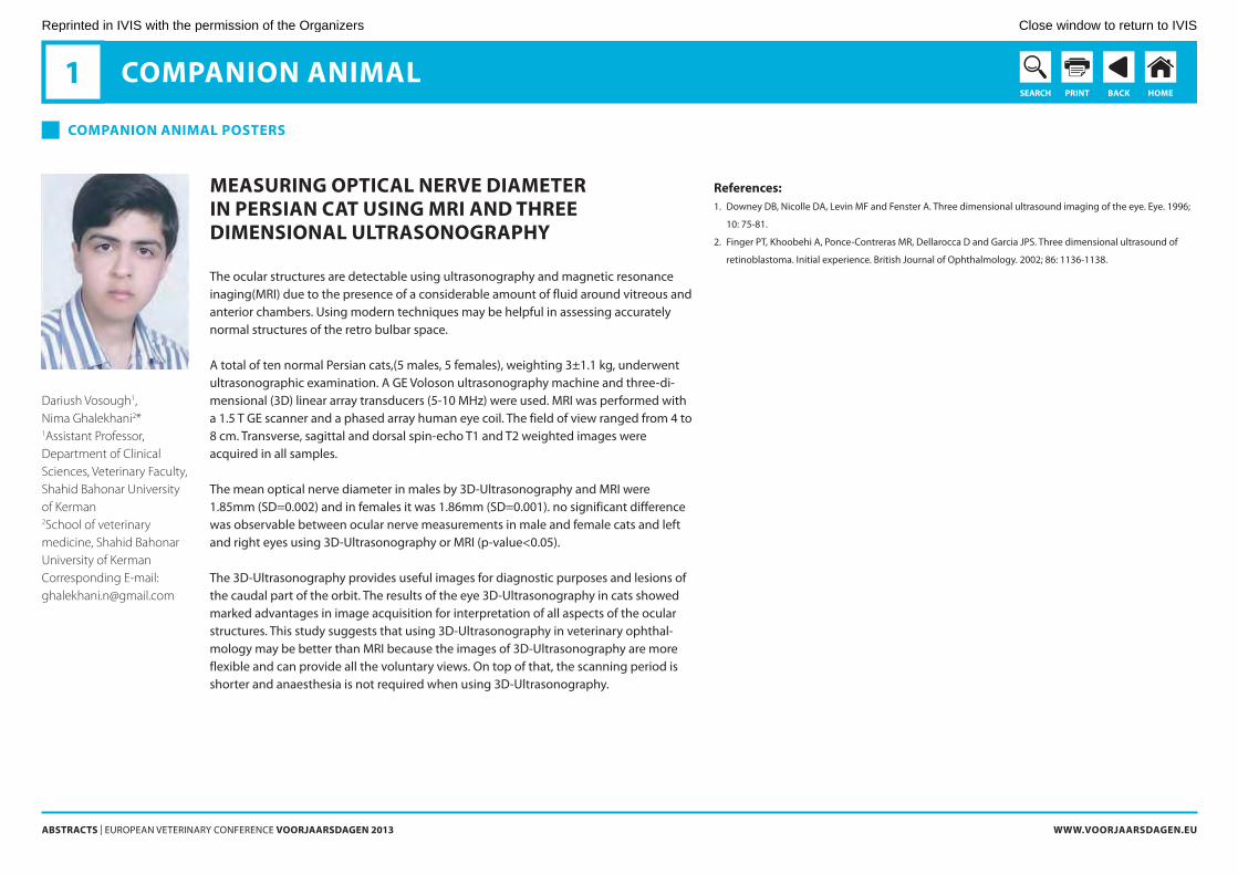
surgicAl rePAir of An AxillAry sKin defect by usAge of trAnsPosition flAP in rAbbit
transposition flaps are rectangular, local flaps that bring additional skin when rotated into defects. forelimb fold flap is a transposition flap harvested to close axillary or ster-nal wounds.in forelimb fold flaps, the size and length of the skin flap vary depending on the body conformation of the animal (1).
in spring 2011, a 10 months old new Zealandwhite rabbit was found with a deep wound in rightaxillary area. after lavage with normal saline and removal of all necrotic tissues, the wound was treated with routine open wound management for 11 days, including daily lavage with saline and usage of topical ointments. after this period of time, to repair the big skin defect, the decision was made on use of a forelimb skin flap. then, two lateral and medial skin incisions were created to define the width of the flap and these incisions were connected together with a crescent-shaped incision proximal to the elbow. the flap was elevated, transposed, and sutured into the wound and the donor site was closed easily. then the forelimb was bandaged to the body for 13 days. the surgical site was monitored for complications every 2-3 days. antibiotic therapy was done. after 13 days, the bandage and the sutures were removed. the rabbit can use the forelimb very well and the appearance of the surgical site after about 20 days was per-fectly acceptable. Dog ears occurred but were flatten with time.
the goal of using transposition flaps is rapid coverage of wound bed, prevention of delayed healing and contraction results from secondary healing(1).
references:1. Hedlund Ch. S., Surgery of the integumentary System.in: fossum th. W. eds. Small animal Surgery, Mosby:
Elsevier,2007; 15:205-216.
oPMerKingenredactie:- opmerking
Reprinted in IVIS with the permission of the Organizers Close window to return to IVIS
AbstrActs | EuropEan VEtErinary ConfErEnCE VoorjAArsdAgen 2013 www.VoorjAArsdAgen.eu
PrintseArcH bAcK HoMe
coMPAnion AniMAl1
coMPAnion AniMAl Posters
Francisco Sánchez-Margallo (DVM, PhD); Angelo Tapia Araya (DVM, GPCert SAS, MSc); Idoia Díaz-Güemes (DVM, PhD); Jesús Usón Gargallo (DVM, PhD).Angelo Tapia Araya, Laparoscopic Unit, “Jesús Usón” Minimally Invasive Surgery Centre. N. 521, 41,800. 10071. Cáceres. Spain. [email protected]
cAse rePort: A single-Port lAPAroscoPic oVArioHysterectoMy in A dog
introductionMinimally invasive surgery (MiS) has awakened great interest amongst veterinarians due to laparoscopic advantages presented to the patients: less surgical trauma, thera-peutic safety and faster recovery (1). regarding MiS approaches, single-port surgery constitutes an evolution of laparoscopic surgery, reducing even more the surgical trauma associated with every procedure (2). there are just a few bibliographic refer-ences about ovariectomy in dogs through a single-port laparoscopic approach (3, 4).
objectivethe main goal of this study was to report and assess the feasibility of single-port laparo-scopicfor ovariohysterectomy in a dog.
Methodsa 7-year-old, 13 kg, female Beagle dog was used to perform a single-port laparoscopic ovariohisterectomy with a commercially available device SiLStM port. first, a 3 cm skin incision was performed on the periumbilical region to introduce the SiLS™ port device. then, insufflation of Co2 was initiated to establish pneumoperitoneum at an intra-ab-dominal pressure of 10 mm Hg. next, specially-designed laparoscopic instruments were used to facilitate the procedure: the 5 mm LigasuretM sealing device and a roticulated 5 mm grasper were used for dissection and vascular sealing of the ovarian pedicle, sus-pensory ligament, broad ligament and body of the uterus. all the mentioned instru-ments were introduced through the same access port. the ovaries and uterus were extracted by directly removing the SiLS™ port.
resultsthe total surgical time was 58 minutes and the only skin incision was 3.2 cm. Both ova-ries and uterus were extracted directly with the SiLS™ port without any tissue rupture. at postoperative follow-up no lesions, hemorrhages, signs of pain or other surgical complications were observed.
conclusionsafter adequate acquisition of laparoscopic skills, the ovariohysterectomy by single-port laparoscopy is a feasible, safe and effective approach in dogs. the procedure needs sim-ilar surgical time when compared to the traditional laparoscopic ovariohysterectomy.
1. Mayhew p. Developing minimally invasive surgery in companion animals. Vet rec 2012; 169:177-178.
2. rao pp, Bhagwat S. Single-incision laparoscopic surgery - current status and controversies. J Minim access
Surg 2011;7:6-16.
3. Manassero M, Leperlier D, Vallefuoco r, Viateau V. Laparoscopic ovariectomy in dogs using a single-port multi-
ple-access device. Vet rec 2012;171(3):69.
4. runge JJ, Curcillo pG 2nd, King Sa, podolsky Er, Holt DE, Davidson J, agnello Ka. initial application of reduced
port Surgery using a Single port access technique for Laparoscopic ovariectomy. Vet Surg 2012;41(7):803-6.
oPMerKingenredactie:- opmerking
Reprinted in IVIS with the permission of the Organizers Close window to return to IVIS
AbstrActs | EuropEan VEtErinary ConfErEnCE VoorjAArsdAgen 2013 www.VoorjAArsdAgen.eu
PrintseArcH bAcK HoMe
coMPAnion AniMAl1
coMPAnion AniMAl Posters
Kirsten L. van Bokhorst, Yvonne R.A. van Zeeland, DVM, MVR, Johannes T. Lumeij, DVM, PhD, Nico J. Schoemaker, DVM, PhDUtrecht University, Faculty of Veterinary Medicine, Department of Clinical Sciences of Companion Animals, Division of Zoological Medicine, Yalelaan 108, 3584CM UtrechtThe [email protected]
resPonses to stress in grey PArrots (PsittAcus eritHAcus) witH And witHout feAtHer dAMAging beHAVior
in various animal species (e.g. laying hens, pigs) a high correlation exists between a proactive coping style (i.e. response to stress) and the occurrence of behavioral prob-lems (e.g. feather pecking, tail biting) (1-5). Based on analogies with feather pecking in laying hens, it is hypothesized that a similar correlation between feather damaging behavior (fDB) and proactive coping styles is present in parrots (6). findings of a pilot study, conducted under standardized conditions in a parrot refuge, support this hypothesis (unpublished observations). the current study was designed to determine whether the previously found correlation between fDB and type of coping style could also be demonstrated in a larger cohort of parrots.
ninety-five privately owned grey parrots (with and without fDB) were subjected to three behavioral tests (i.e. open field test, novel object test and Manual restraint test, performed at the university clinic). in these tests both behavioral and neurophysiologi-cal parameters were analyzed to determine the birds’ coping styles.
in contrast to the previous pilot study, no significant differences were found between parrots with and without fDB. results may, however, have been influenced by various external factors (e.g. stress due to traveling or separation from the owner) that could have influenced the parrots’ responses in the tests performed at the clinic. it may be concluded that the current study design has limited applications in future research and adjustments have to be made to enable analysis of coping styles in privately owned parrots with and without fDB.
1. Koolhaas JM, Korte SM, de Boer Sf, van der Vegt BJ, van reenen CG, Hopster H, de Jong iC, ruis MaW, Blokhuis
HJ. Coping styles in animals: current status in behavior and stresspsychology. neurosc Biobeh rev
1999;23:925-35
2. Korte SM, Beuving G, ruesink W, Blokhuis HJ. plasma catecholamine and corticosterone levels during manual
restraint in chicks from a high and low feather pecking line of laying hens. physiol Behav 1997;62:437-41
3. Korte SM, prins J, Vinkers CH, olivier B. on the origin of allostasis and stress-induced pathology in farm ani-
mals: Celebrating Darwin’s legacy. Vet J 2009;182:378-83
4. rodenburg tB, Buitenhuis aJ, ask B, uitdehaag Ka, Koene p, van der poel JJ, Bovenhuis H. Heritability of
feather pecking and open-field response in laying hens at two different ages. poult Sci 2003;82:861-7
5. uitdehaag Ka, Komen H, rodenburg tB, Kemp B, van arendonk J. the novel object test as predictor of feather
damage in cage-housed rhode island red and White Leghorn laying hens. appl anim Behav Sci 2008;109:292-
305
6. Van Zeeland yra, Spruit BM, rodenburg tB, riedstra B, van Hierden yM, Buitenhuis B, Korte SM, Lumeij Jt.
feather damaging behaviour in parrots: a review with consideration of comparative aspects. appl anim Behav
Sci 2009;121:75-95
oPMerKingenredactie:- opmerking
Reprinted in IVIS with the permission of the Organizers Close window to return to IVIS
AbstrActs | EuropEan VEtErinary ConfErEnCE VoorjAArsdAgen 2013 www.VoorjAArsdAgen.eu
PrintseArcH bAcK HoMe
coMPAnion AniMAl1
coMPAnion AniMAl Posters
Yasemin Salgirli Demirbas, DVM, PhD, Mehmet Bahri Emre, DVM, PhD, Mustafa Kockaya, DVMDepartment of Physiology, Faculty of Veterinary Medicine, Ankara University, 06110 Diskapi, Ankara, Turkeyyaseminsalgirli@ veterinary.ankara.edu.tr
integrAtion Ability of strAy dogs into AdoPtiVe fAMilies’ enVironMent: PreliMinAry results
introductionSeveral researches investigated post-adoption period of shelter dogs in terms of inte-gration into the new home and behavioral problems encountered by new owners (1,2,3). the most common behavior problems displayed by shelter dogs are suggested as fearfulness, escaping, sexual problems, excessive activity and barking (1,3). However, up till now no detailed investigation has been conducted on post adoption period of stray dogs.
the aim of this study was to identify behavioral characteristics of stray dogs in adoptive families’ environment during post adoption period.
Materials and Methodsthe data were obtained from 25 item questionnaires completed by volunteer owners of stray dogs, i.e. the dog of an unknown breed was found loose on the street. fifty five people responded to the questionnaire up until the present.
resultsthe vast majority of respondents (75 %) reported that their dogs showed timidity when they first came home. Large majority of the owners (80 %), however, stated that temper-aments of their dogs have changed throughout post adoption period, mostly in the first 6 months (73 %). Seventy five percent of the dogs were reported as becoming more self-confident. the most common behavior problems currently present in the dogs were indicated as hyper attachment to the owner (40 %), escaping (36 %), barking (22 %) and fearfulness (18 %). Majority of the owners reported that they did not have a trouble either in house training (75 %) or in leash training (69 %).
discussionpreliminary findings indicate that although majority of stray dogs tend to display behavior problems at the beginning of post adoption period, they show considerable improvements in their behaviors in a timely manner. Considering the preliminary results of the study, it can be suggested that stray dogs are highly adaptable to live in adoptive families’ environment.
references1. Wells DL, Hepper pG. prevalence of behaviour problems reported by owners of dogs purchased from an ani-
mal rescue shelter. appl. anim. Behav. Sci. 2000; 69:, 55–65.
2. Marston LC, Bennett pC, Coleman GJ. What happens to shelter dogs? an analysis of data for 1 year from three
australian shelters. J. appl. anim. Welfare Sci. 2004; 7(1): 27-47.
3. Stephen J, Ledger r. relinquishing dogs owners’ ability to predict behavioural problems in shelter dogs post
adoption. appl. anim. Behav. Sci. 2007;107:88-99.
oPMerKingenredactie:- opmerking
Reprinted in IVIS with the permission of the Organizers Close window to return to IVIS
AbstrActs | EuropEan VEtErinary ConfErEnCE VoorjAArsdAgen 2013 www.VoorjAArsdAgen.eu
PrintseArcH bAcK HoMe
coMPAnion AniMAl1
coMPAnion AniMAl Posters
Nihat Yumusak, DVM, PhD, Murat Caliskan, DVM, PhD, Osman Kutsal, DVM, PhD.Department of Veterinary Pathology, Faculty of Veterinary Medicine, Harran University, [email protected]
cytoPAtHologicAl diAgnosis of tHe cAnine HePAtoid glAnd tuMors
introductiona hepatoid (perianal) gland tumor is a type of tumor found near the anus in dogs that arises from specialized glandular tissue (1,2). it is also known as a hepatoid tumor because of the similarity in cell shape to hepatocytes (liver cells) (1,2,3). in this study, 23 tumors of dogs were evaluated and compered between cytopathologically and his-topathologically.
Material and Methods23 tumors of dogs were evaluated and compered between cytopathological and cis-topathological. for cytopathology examination fnaB technique were performed and slides were stained with May-Grünwald Giemsa. for histopathological examination tis-sue samples were fixed formaldehyde solution, after processed by paraffin technique sections were stained by Hematoxylin-Eosin.
resultstotal 23 tumors were diagnosed by cytology and confirmed by histology. Macroscopi-cally, the masses covered with hairless skin and often ulcerated. Cut sections were pale brown and multilobulated. Cytopathologically, large and cuboidal epithelial cells have low nuclear/cytoplasm ratios. nucleus were round to ovoid, pleomorphism were mild. also, abundant, granular bluish cytoplasm were seen. the distinctive histopathologic feature of this tumor is differentiation to large hepatocyte-like cells, which have an abundant eosinophilic cytoplasm. Within the tumor cells trabeculae and islands subdi-vided by a well vascularized stroma.
conclusionsCytopathological examination is a quick and easy procedure to perform which needs minimum of especial equipments and requires little training. it can be considered, in experienced hands and is reliable for the confirmation and differentiation of canine hepatoid gland tumors.
references1. Berrocal a, Vos JH, Van Den İngh tsgam, Molenbeek rf, Van Sluijs fJ. Canine perianal tumours. Zentralbl Veteri-
narmed a 1989;36:739–49.
2. Goldschmidt MH, Hendrick MJ. tumors of the skin and soft tissues. in: Meuton DJ. eds. tumors in domestic ani-
mals, Lowa: Lowa State press, 2002; 45-117.
3. Weiss E, frese K. tumours of the skin. Bull World Health organ 1974;50:79-100.
oPMerKingenredactie:- opmerking
Reprinted in IVIS with the permission of the Organizers Close window to return to IVIS
AbstrActs | EuropEan VEtErinary ConfErEnCE VoorjAArsdAgen 2013 www.VoorjAArsdAgen.eu
PrintseArcH bAcK HoMe
coMPAnion AniMAl1
coMPAnion AniMAl Posters
Nathalie Porters, Tim Bosmans, PhD, Hilde de Rooster, PhD, DipECVS, Kris Baert, PhD, Siska Croubels, PhD, Patrick De Backer, PhD, DipECVPT, Ingeborgh Polis, PhD Department of Medicine and Clinical Biology of Small Animals, Faculty of Veterinary Medicine, Ghent University, Salisburylaan 133, 9820 Merelbeke, Belgium [email protected]
feline PHArMAcoKinetics of orAl trAnsMucosAl And intrAMusculAr dexMedetoMidine And buPrenorPHine
introductionplasma concentrations and pharmacokinetics of dexmedetomidine and buprenorphine after oral transmucosal (otM) and intramuscular (iM) administration in cats were evalu-ated. the aim was to compare systemic absorption of this sedative-analgesic combina-tion following otM and iM administration.
Material and Methodsaccording to a cross-over protocol (1 month wash-out), a combination of dexmedeto-midine (40 µg/kg) and buprenorphine (20 µg/kg) was given otM (buccal cavity)(1) or iM (quadriceps muscle) in 6 female neutered cats. plasma samples were collected through a jugular catheter during a 24-hour period. plasma dexmedetomidine and buprenor-phine concentrations were determined by liquid chromatography-tandem mass spec-trometry. plasma concentration-time data were fitted to compartmental models (MW/pharm, Mediware). Wilcoxon signed rank tests were performed (p < 0.05).
resultsfor dexmedetomidine, the area under the plasma concentration-time curve from 0 to 12 h was significantly different between otM (9.59 ± 3.88 ng·h/ml) (mean ± SD) and iM administration (38.56 ± 8.66 ng·h/ml). Maximum plasma concentration of dexmedeto-midine was significantly lower after otM (3.59 ± 2.02 ng/ml) than after iM route (22.07 ± 8.91 ng/ml). for buprenorphine, the area under the plasma concentration-time curve from 0 to 6 h was significantly different between otM (16.91 ± 7.68 ng·h/ml) and iM (178.63 ± 71.50 ng·h/ml) administration. Buprenorphine achieved maximum plasma concentrations of 5.19 ± 3.09 ng/ml at 1.18 ± 0.34 h after otM administration and of 104.31 ± 0.52 ng/ml at 0.15 ± 0.15 h after iM injection with a significant difference between both treatments. Dexmedetomidine and buprenorphine relative bioavailabili-ties after otM compared to iM administration were 25.9 ± 11.2 % and 11 ± 6.4 %, respectively.
conclusions these data suggest that absorption of combined dexmedetomidine and buprenor-phine is less after otM administration compared to iM injection in cats. Clinical efficacy of this sedative-analgesic regimen after otM administration is questionable.
1. robertson Sa, taylor pM, Sear JW. Systemic uptake of buprenorphine by cats after oral mucosal administra-
tion. Vet rec. 2003 ;152:675-8.
oPMerKingenredactie:- opmerking
Reprinted in IVIS with the permission of the Organizers Close window to return to IVIS
AbstrActs | EuropEan VEtErinary ConfErEnCE VoorjAArsdAgen 2013 www.VoorjAArsdAgen.eu
PrintseArcH bAcK HoMe
coMPAnion AniMAl1
coMPAnion AniMAl Posters
Amrut Bhogle MVSc. PhD, David Sutton BVetMed, (on photo) Norman Spibey BSc. PhD, Leo van der Waart BSc., Michael Francis EurProBiol CBiol FSB PhDDavid Sutton, MSD Animal Health, Walton Manor, Walton, Milton Keynes, MK7 [email protected]
inVestigAting tHe coMbined use of A MultiVAlent liVe VirAl dHPPi VAccine And An inActiVAted rAbies VAccine witH A noVel tetrAVAlent cAnine lePtosPirosis VAccine
introductionrecently, a novel liquid tetravalent canine leptospirosis vaccine developed in line with current independent vaccination recommendations (1) has been authorised in Europe. this new vaccine (nobivac® L4 – MSD animal Health) will commonly be used to recon-stitute the respective routine injectable lyophilized viral multicomponent vaccines, and studies to support this simultaneous use have been reported (2). in this study we have investigated the possibility of any adverse effect or serological interference in dogs administered nobivac L4, nobivac DHppi and nobivac rabies at the same time.
Materials and Methodstwenty-one 9 week old Spf dogs were divided into three equal groups. Group 1 was vaccinated with DHppi reconstituted with L4, group 2 with DHppi reconstituted with L4 and also a separate inoculation of rabies vaccine, group 3 with just rabies vaccine. all dogs were observed for any adverse local or systemic signs post-inoculation. Serum samples were collected post-vaccination in order to compare the antibody responses to all the components between the different groups.
results and conclusionsno systemic reactions were observed in any of the vaccinated dogs. Some of the dogs vaccinated with all three vaccines showed small, painless local swellings at the sites of both inoculations which were considered to be minor and acceptable. the dogs in groups 1 and 2 showed similar antibody responses to the DHppi and L4 antigens and thus there was no evidence of any serological interference arising from the rabies vaccination. Like-wise the dogs in groups 2 and 3 showed a similar antibody response to the rabies compo-nent thus showing no evidence of any interference from the DHppi and L4 vaccination. it can be concluded that nobivac rabies, nobivac DHppi and nobivac L4 all appear to be well tolerated and serologically compatible when administered at the same time.
references1. Ellis Wa. Control of canine leptospirosis in Europe: time for a change? Veterinary record 2010;167:602-5
2. Klaasen HLBM, van der Veen M, Molkenboer MJCH, Sutton D. a novel tetravalent Leptospira bacterin protects
against infection and shedding following challenge in dogs. Veterinary record published online november 23,
2012; doi:10.1136/vr.101100
oPMerKingenredactie:- opmerking
Reprinted in IVIS with the permission of the Organizers Close window to return to IVIS
AbstrActs | EuropEan VEtErinary ConfErEnCE VoorjAArsdAgen 2013 www.VoorjAArsdAgen.eu
PrintseArcH bAcK HoMe
coMPAnion AniMAl1
coMPAnion AniMAl Posters
Henricus L.B.M. Klaasen PhD DVM, Henriëtte M.A. Adriaanse BSc, Mark van der Veen BSc, David Sutton BVetMed (on photo)Henricus L.B.M. Klaasen, Dept Microbiological R&D, MSD Animal Health, 5830 AA BoxmeerThe [email protected]
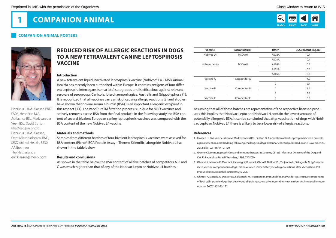
reduced risK of Allergic reActions in dogs to A new tetrAVAlent cAnine lePtosPirosis VAccine
introductiona new tetravalent liquid inactivated leptospirosis vaccine (nobivac® L4 – MSD animal Health) has recently been authorized within Europe. it contains antigens of four differ-ent Leptospira interrogans (sensu lato) serogroups and is efficacious against relevant serovars of serogroups Canicola, icterohaemorrhagiae, australis and Grippotyphosa (1). it is recognized that all vaccines carry a risk of causing allergic reactions (2) and studies have shown that bovine serum albumin (BSa), is an important allergenic excipient in this respect (3,4). the VaccipuretM filtration process is unique for MSD vaccines and actively removes excess BSa from the final product. in the following study the BSa con-tent of several bivalent European canine leptospirosis vaccines was compared with the BSa content of the new nobivac L4 vaccine.
Materials and methodsSamples from different batches of four bivalent leptospirosis vaccines were assayed for BSa content (pierce® BCa protein assay – thermo Scientific) alongside nobivac L4 as shown in the table below.
results and conclusionsas shown in the table below, the BSa content of all five batches of competitors a, B and C was much higher than that of any of the nobivac Lepto or nobivac L4 batches.
Vaccine Manufacturer batch bsA content (mg/ml)
nobivac L4
MSD aH
a002a 0.4
a003a 0.4
nobivac Lepto
MSD aH
a103B 0.3
a101a 0.5
a105B 0.3
Vaccine a
Competitor a
1 4.0
2 2.4
Vaccine B
Competitor B 1 3.6
2 3.8
Vaccine C Competitor C 1 6.3
assuming that all of these batches are representative of the respective licensed prod-ucts this implies that nobivac Lepto and nobivac L4 contain the lowest amount of potentially allergenic BSa. it can be concluded that after vaccination of dogs with nobi-vac Lepto or nobivac L4 there is a likely to be a lower risk of allergic reactions.
references1. Klaasen HLBM, van der Veen M, Molkenboer MJCH, Sutton D. a novel tetravalent Leptospira bacterin protects
against infection and shedding following challenge in dogs. Veterinary record published online november 23,
2012; doi:10.1136/vr.101100.
2. Greene CE. immunoprophylaxis and immunotherapy. in: Greene, CE. ed. infectious Diseases of the Dog and
Cat. philadelphia, pa: WB Saunders, 1998; 717-750.
3. ohmori K, Masuda K, Maeda S, Kaburagi y, Kurata K, ohno K, DeBoer DJ, tsujimoto H, Sakaguchi M. igE reactiv-
ity to vaccine components in dogs that developed immediate-type allergic reactions after vaccination. Vet
immunol immunopathol 2005;104:249-256.
4. ohmori K, Masuda K, DeBoer DJ, Sakaguchi M, tsujimoto H. immunoblot analysis for igE-reactive components
of fetal calf serum in dogs that developed allergic reactions after non-rabies vaccination. Vet immunol immun-
opathol 2007;115:166-171.
oPMerKingenredactie:- opmerking
Reprinted in IVIS with the permission of the Organizers Close window to return to IVIS
AbstrActs | EuropEan VEtErinary ConfErEnCE VoorjAArsdAgen 2013 www.VoorjAArsdAgen.eu
PrintseArcH bAcK HoMe
coMPAnion AniMAl1
coMPAnion AniMAl Posters
Maurizio Dondi, Pier Luigi Dodi, Fausto Quintavalla
Dept. of Veterinary Science, University of Parma, Parma, Italy
interActions between cArbAMAzePine And troxerutine in A dog Affected by idioPAtHic ePilePsy
Carbamazepine (CBZ) represents a first line drug for partial epilepsy in people. its use in veterinary medicine is limited by the short elimination half-life in plasma (1-2 h), although in the past has been used with good results in the dog. trying to increase its plasma half-life we have associated to the therapy troxerutin. our study describes a case of psychomotor epilepsy in a dog treated with the association of these two drugs, and investigates the variations in plasma total CBZ with and without troxerutin associa-tion.
idiopathic epilepsy with complex partial seizures was diagnosed in a 4 years old, male, Labrador retriever dog, based on history, clinical findings, laboratory results, EEG and Mri.
the first week, the dog received 3,3 mg/kg of CBZ (tegretol) tiD oS. at the end of the week 12 blood samples, one every 30 minutes were analyzed to measure the plasma total CBZ. During the second week, 16,6 mg/kg of troxerutine (Venoruton) BiD oS were added. at the end of the second week plasma total CBZ concentration was re-evaluated in the same way.
the correlation between the two CBZ curves suggested an interaction of the drugs. in particular, an effect of troxerutin on the longer elimination half-life in plasma of CBZ, on the precocity of his absorption and on the shape of the peak of the drug in plasma can be hypothesized from the results of the study. the absence of a plasmatic peak after 3h and the relatively constant concentration of CBZ in the 7h after administration may have important practical implications. in facts, based on these findings we have chosen to continue the treatment with both drugs. today, after three years of this therapy the dog experienced only three minor crisis without motor signs and any adverse effects.
oPMerKingenredactie:- opmerking
Reprinted in IVIS with the permission of the Organizers Close window to return to IVIS
AbstrActs | EuropEan VEtErinary ConfErEnCE VoorjAArsdAgen 2013 www.VoorjAArsdAgen.eu
PrintseArcH bAcK HoMe
coMPAnion AniMAl1
coMPAnion AniMAl Posters
*Irem ERGIN DVM PhD, Ali Evren HAYDARDEDEOGLU DVM PhD, Ekrem Cagatay COLAKOGLU DVM, Hadi ALIHOSSEINI DVM PhD, Oytun Okan SENEL DVM PhD*Ankara University, Faculty of Veterinary Medicine, Department of Surgery, 06110, Ankara, [email protected]
clinicAl, HeMAtologicAl findings And treAtMent of cAnine leisHMAniosis: A cAse rePort
Canine leishmaniosis is a chronic, severe systemic protozoal disease caused by Leishma-nia spp. Leishmaniosis, transmitted by the bite of an infected phlebotomus or Lutzo-myia sandfly, is a zoonotic disease and dogs are considered the main reservoir for sys-temic leishmaniosis in humans. Leishmaniosis due to Leishmania infantum is one of the most common diseases of dogs in all countries around the Mediterranean Sea.
the aim of this study was to evaluate clinical and hematological alterations in Leishma-nia infantum in an infected dog and to diagnose it by polymerase chain reaction (pCr) and immunofluorescent-antibody test (ifat) in blood serum samples.
a 3 year old mixed breed female dog was referred to the clinic with complaints of weak-ness, weight loss, ocular discharge and common skin lesions. in clinical examination, severe dry exfoliative dermatitis, ulcerations on the pinnae, nasal depigmentation and ulceration, onychogryposis, bilateral periocular alopecia and pale mucous membranes were observed. Bilateral blepharitis and severe keratoconjunctivitis sicca (KCS) were detected as ocular manifestations. there was generalized lymphadenopathy. Hemato-logical findings were thrombocytopenia, anemia and eosinophilia. needle biopsy was performed from popliteal lymph node and amastigote forms of the parasite were seen with Diff-Quick stain in samples. the diagnosis was confirmed by pCr and ifat per-formed on blood serum samples.
for medical treatment of the disease, domperidone (2 mg/kg, SiD, po) was used for one month and allopurinol (30 mg/kg, SiD, po) was applied for 6 months. in addition to sys-temic treatment, lomefloxacin, acetylcystein, cyclosporin and sodium hyaluronate oph-thalmic solutions were used topically in each eye for treatment of KCS.
Canine leishmaniosis may be difficult to diagnose, hard to treat and its recurrence rates are very high. in this case report, treatment of a dog with leishmaniosis has resulted in disappearance of the clinical signs and there was no recurrence for two years.
references1. figueredo La, paiva-Cavalcanti M, almeida EL, Brandao-filho Sp, Dantas-torres f. Clinical and hematological
findings in Leishmania braziliensis-infected dogs from pernambuco, Brazil. rev Bras parasitol Vet 2012; 21(4):
418-420.
2. naranjo C, fondevila D, Leiva M, roura X, pena t. Characterization of lacrimal gland lesions and possible patho-
genic mechanisms of keratoconjunctivitis sicca in dogs with leishmaniosis. Vet parasitol 2005; 133: 37-47.
3. naranjo C, fondevila D, atlet L, francino o, rios J, roura X, pena t. Evaluation of the presence of Leishmania
spp. by real-time pCr in the lacrimal glands of dogs with leishmaniosis. Veterinary Journal 2012; 193: 168-173.
4. pena Mt, naranjo C, Klauss G, fondevila D, Leiva M, roura X, Davidson MG, Dubielzig rr. Histopathological fea-
tures of ocular leishmaniosis in the dog. J Comp path 2008; 138: 32-39.
5. Scott DW, Miller WH, Griffin CE. Viral, rickettsial, and protozoal Skin Diseases. in: Muller&Kirk’s small animal der-
matology. philadelphia: Saunders, 2001; 517-542
oPMerKingenredactie:- opmerking
Reprinted in IVIS with the permission of the Organizers Close window to return to IVIS
AbstrActs | EuropEan VEtErinary ConfErEnCE VoorjAArsdAgen 2013 www.VoorjAArsdAgen.eu
PrintseArcH bAcK HoMe
coMPAnion AniMAl1
coMPAnion AniMAl Posters
Jesús Usón Gargallo (DVM, PhD); Angelo Tapia Araya (DVM, GPCert SAS, MSc); Miguel Sánchez-Hurtado (DVM, MSc); Idoia Díaz-Güemes (DVM, PhD); Francisco Sánchez-Margallo (DVM, PhD)Angelo Tapia Araya, Laparoscopic unit, “Jesús Usón” Minimally Invasive Surgery Centre. N. 521, 41,800. 10071. Cáceres. Spain. [email protected]
deVeloPMent of A reAlistic siMulAtor for MiniMAlly inVAsiVe VeterinAry surgery trAining
introductionthe interest of veterinary in minimally invasive surgery (MiS) has increased gradually for veterinarians and owners in the past few years (1). nevertheless, the learning curve to overcome laparo-endoscopic surgery training challenges requires time and funds (2).
objectives1) to develop a high-realistic MiS simulator for veterinary 2) to use a Ct-Scan to reliably measure the average spatial dimension of the canine species 3) to assess the subjective validity of the physical simulator with veterinary surgeons.
Material and Methodthree Beagle dogs were used for the Ct-Scan study (philips Brilliance 6 Ct Scanner). to proceed with it a pneumoperitoneum was created at 11-12 mmHg on each dog. Exact data of abdominal-thoracic perimeter; height and volume of the cavities and of their internal organs were obtained. the data in DiCoM format was processed by aMira® to generate 3D models and a design to create the simulator.Subjective validity: 30 veterinarians (expert group, n=7; basic group, n=23) practiced exercises of abdominal laparoscopy. these tasks were qualified by surveys of 7 ques-tions with 1-5 Likert scales. in addition, the simulator was rated globally in a 1-10 scale.
resultsSiMuLVEt®, a multifunctional simulator for practicing laparoscopy, thoracoscopy, flexi-ble endoscopy, notES and single port surgery, was created.More than 60 % of the veterinarians expressed not to have previous experience with simulation. SiMuLVEt® impressions for the 7 questions in both groups were positive or very posi-tive. there weren’t significant differences between the different groups’ opinions. Global average qualification of the SiMuLVEt® was 8,7±1,1 points on a 10 point scale.
conclusionsimages from DiCoM obtained by tC and processed by specific software were useful to develop a high-realistic MiS simulator for veterinary SiMuLVEt®. the subjective valida-tion was acceptable. further studies are needed on training programs (3), training and simulation methods for veterinary laparoscopy (4).
1. Mayhew p. Developing minimally invasive surgery in companion animals. Vet rec 2011;169:177-178.
2. Schout BM, Hendrikx aJ, Scheele f, Bemelmans BL, Scherpbier aJ. Validation and implementation of surgical
simulators: a critical review of present, past, and future. Surg Endosc 2010;24:536-546.
3. usón J, Sánchez fM, Sánches Ma, pérez fJ Hashizume M. Basisc principles. in: usón J, Sánchez fM, pascual S,
Climent S. 4tH Ed. Step by Step training in Laparoscopic Surgery. “Jesús usón” Minimally invasive Surgery Cen-
tre. Cáceres, Spain, 2010; 23-89.
4. fransson Ba, ragle Ca. assessment of laparoscopic skills before and after simulation training with a canine
abdominal model. J am Vet Med assoc 2010; 236:1079-1084.
oPMerKingenredactie:- opmerking
Reprinted in IVIS with the permission of the Organizers Close window to return to IVIS
AbstrActs | EuropEan VEtErinary ConfErEnCE VoorjAArsdAgen 2013 www.VoorjAArsdAgen.eu
PrintseArcH bAcK HoMe
coMPAnion AniMAl1
coMPAnion AniMAl Posters
Baharak Akhtardanesh1, Nima Ghalekhani1*, Hadi Nematollahi21Department of Clinical Sciences, Faculty of Veterinary Medicine, Shahid Bahonar University of Kerman , Iran.2Herbal and Traditional Medicines Research Center, Kerman University of Medical Sciences, Iran.Corresponding E-mail: [email protected]
diAgnostic APPlicAtion of seruM Adenosine deAMinAse leVel in cAnine infectious diseAse
adenosine deaminazse is a cytoplasmic enzyme, which is essential for the proliferation and differentiation of lymphocytes. increased aDa is derived mainly from increased immune cells numbers, whereas inhibition or genetic deficiency of aDa resulted in immunodeficiency.
in this study, three canine chronic infectious diseases (visceral leishmaniosis, ehrlichiosis and brucellosis) were selected for evaluation of serum aDa alteration in compare with clinically healthy dogs.
Different diagnostic methods were used for detection ehrlichiosis, brucellosis and leish-maniosis among 378 serum sample that obtained from sick owned dogs. on the other hand, 50 healthy dogs which, referred for health checks up and vaccination and their hematological and biochemical evaluation were in normal limit were randomly selected as control group. total aDa level, were measured by a commercial kit via autoanalyzer in all selected samples.
total aDa level were measured as 7.5±.0.8 iu/l, 5.7±0.48 iu/l, 5.2±0.63 iu/l and 2.26 ±0.25 iu/l in visceral leishmaniosis, ehrlichiosis, brucellosis and healthy dogs respec-tively. there was no significant difference in aDa level between diseased groups but aDa level in leishmaniosis, ehrlichiosis and brucellosis groups showed a significant increase in compare with healthy animals.
Based to the results of the present study, total serum aDa level may be used as an important biomarker for the diagnosis of canine chronic infection disease and can be added to the other routine biochemical tests that are used in the veterinary clinical pathology.
references:1. agrawal S. Study of adenosine Deaminase activity as a biochemical marker of cell mediated immunity in
tuberculous meningitis, tuberculous pleural effusion and tuberculous ascites. J MEDiCinE. 2012; 13:32-38.
2. altuğ n, yüksek n, ağaoğlu Zt, Özkan C. Decreased serum adenosine deaminase activities in Van cats with
feline retrovirus infections. Med Weter. 2007; 63:184-186.
3. Bremer H, Bauer i, Brock J. adenosine deaminase activity and immune dysfunction. allerg immunol. 1981;
27:3-13.
oPMerKingenredactie:- opmerking
Reprinted in IVIS with the permission of the Organizers Close window to return to IVIS
AbstrActs | EuropEan VEtErinary ConfErEnCE VoorjAArsdAgen 2013 www.VoorjAArsdAgen.eu
PrintseArcH bAcK HoMe
coMPAnion AniMAl1
coMPAnion AniMAl Posters
Els Wouters, BSc, Dr. Susanne AEB Boroffka, Prof. Dr. George Voorhout
Department of Companion Animals, Faculty of Veterinary Medicine, Utrecht University
Mr iMAges for cHArActerizing cAnine PriMAry brAin tuMors: A coMPArison witH HistoPAtHology
the Mr images and histopathological slides of twenty primary brain tumors (ten menin-giomas and ten gliomas) were compared, using defined Mr characteristics and the WHo classification. to find a relationship between histological features and Mr images the histological slides were compared one-on-one with the Mr images for twelve of the tumors.
the aim of this study was to improve the value of Mri for the characterization of pri-mary brain tumors in dogs, using the pathologic findings as feedback. this was done by comparing histopathologic findings and Mr images.
this study concluded that neither for meningiomas, nor for gliomas, a distinction between different histological subtypes can be made based on the Mr characteristics. Comparing the Mr characteristics of meningiomas and gliomas, it can be concluded that it is hard to distinguish them based on these Mr characteristics, because both types of tumors show a variety of Mr characteristics. for a definite diagnosis, other methods, such as tissue biopsies, are needed.
the clinical case of Mickey, an eight years old male West Highland White terrier, is an example of a patient with an intracranial mass. Mickey suffered from epileptic seizures and his Mr images showed an intracranial mass. He is one of the twenty patients used in this study. the intracranial mass was an anaplastic meningioma according to the his-tological classification. after a successful surgery Mickey clinically improved. However, the Mr images revealed a possible remission of the tumor 20 month post-surgery. up untill today, Mickey is closely monitored.
oPMerKingenredactie:- opmerking
Reprinted in IVIS with the permission of the Organizers Close window to return to IVIS
AbstrActs | EuropEan VEtErinary ConfErEnCE VoorjAArsdAgen 2013 www.VoorjAArsdAgen.eu
PrintseArcH bAcK HoMe
coMPAnion AniMAl1
coMPAnion AniMAl Posters
Drs. Ties T Koolen; Drs. Roelof J Maarschalkerweerd; Dr. Lars FH TheyseDepartment of Clinical Science of Companion Animals, Faculty of Veterinary Medicine, UtrechtUniversityThe [email protected] Royal Canin Student Award
doxy tHougHt sHe could fly
Doxy, a 7 year old female neutered domestic shorthair was referred to the orthopaedic department of the MCD after a fall from considerable height. She was presented in lat-eral recumbency, unable to stand and walk and she reacted painful to palpation of the pelvic region. Based on the physical exam and radiographs of the pelvis, a fracture of the ilial body was diagnosed.
Lateral plate fixation is the most frequently used surgical technique to stabilise feline ilial fractures, however it is associated with high percentages of implant failure. in our study we investigated whether the application of a second lateral plate dorsally or ven-trally of the first lateral plate creates a more rigid construction, leading to a lower per-centage of implant failure. retrospective research of surgical records and radiographs of 80 cats treated at the orthopaedic department of the MCD (35 cats with a single lateral plate, 48 with double lateral plates) showed that implant failure occurred significantly less in cats treated with double lateral plates. these results indicate that with little extra costs and surgery time, the complication rates of lateral plate fixation of feline ilial frac-tures can be lowered.
Doxy’s ilial fracture was surgically stabilised using two laterally applied bone plates. She recovered without complications of the fixation. She thought she could fly but now knows she really can’t.
oPMerKingenredactie:- opmerking
Reprinted in IVIS with the permission of the Organizers Close window to return to IVIS
AbstrActs | EuropEan VEtErinary ConfErEnCE VoorjAArsdAgen 2013 www.VoorjAArsdAgen.eu
PrintseArcH bAcK HoMe
coMPAnion AniMAl1
coMPAnion AniMAl Posters
Imke Hennink, BVSc. Supervisors: Marjolein L. den Toom, DVM and Christine J. Piek, DVM, Diplomate ECVIM-CADepartment of Clinical Science of Companion Animals, Faculty of Veterinary Medicine, Utrecht University, Yalelaan 108, 3584 CM UtrechtThe [email protected]
systeMAtic reView of tHe AntiPlAtelet effects of AcetylsAlicylic Acid And cloPidogrel in cAts witH HyPertroPHic cArdioMyoPAtHy
Cats with underlying heart disease have a significant increased risk of developing an arterial thromboembolism (atE). Especially cats with hypertrophic cardiomyopathy (HCM) are at risk to develop an atE. acetylsalicylic acid (aspirin ®) therapy has been advised as thromboprophylaxis for many years. More recently however, the prophylac-tic use of clopidogrel has gained more scientific interest.
the aim of this literature study was to evaluate the epidemiology of atE and the in vitro and in vivo effects of thromboprophylactic therapy with acetylsalicylic acid and clopi-dogrel in feline HCM patients.
the true incidence of atE in cats with HCM is unknown. over the past decades, acetylsali-cylic acid has been given as a prophylactic therapy in high (up to 25 mg/kg/q72h) and low dosages (as low as 5 mg/kg/q72h). Studies showed recurrence rates between 25 and 75 % and no significant difference in recurrence rate between low and high dose aspirin therapy. in high dosages (≥25mg/kg/q72h), aspirin can give toxic side effects in cats and therefore high dose aspirin therapy is no longer advised. platelet aggregation studies, reveal that a dose of at least 25 mg/kg is needed to inhibit in vitro arachidonic acid dependent and col-lagen induced platelet aggregation. aDp and thrombin induced platelet aggregation were not inhibited in this high dose. the recurrence rate in cats treated with clopidogrel is still unknown. recently, a small platelet aggregation study showed good in vitro antiplatelet effects of clopidogrel in healthy cats. in a dose of 18,75 mg/kg/day clopidogrel showed sig-nificant reduction of platelet aggregation in response to aDp and collagen.
the results of this literature study support the conclusion that prophylactic low dose acetylsalicylic acid therapy (5 mg/kg/q72h) will not prevent intra-cardiac thrombus for-mation or atE in feline HCM patients. Clopidogrel® seems a promising prophylactic antiplatelet drug, but further research is warranted.
oPMerKingenredactie:- opmerking
Reprinted in IVIS with the permission of the Organizers Close window to return to IVIS
AbstrActs | EuropEan VEtErinary ConfErEnCE VoorjAArsdAgen 2013 www.VoorjAArsdAgen.eu
PrintseArcH bAcK HoMe
coMPAnion AniMAl1
coMPAnion AniMAl Posters
Drs. Floor Driessen, Dr. Anne Kummeling, Drs. Ronald J. CorbeeDepartment of Clinical Sciences of Companion Animals, Faculty of Veterinary Medicine, Utrecht University, The [email protected]
ureterAl obstruction in A cAt: diAgnosis And treAtMent
a 14 year old female neutered cat, with problems of pollakiuria, stranguria, haematuria, urinary incontinence, and pain on the left abdominal wall since 1.5 year was presented at our clinic. in the past, both struvite and calciumoxalate crystals were found during urinary examination. During clinical examination at our clinic, abdominal palpation was painful, the right kidney was very good palpable and the bladder was not palpable.
ultrasound of the abdomen showed a small left kidney with multiple mineralisations, and some ditches were seen on the cortex. in the left ureter echogenic material was seen with some mineralisations. the right kidney was 4,8 cm in diameter and showed no mineralisations. to determine the functionality of the kidney, a renogram was made. the left kidney excreted 11% of the urinary volume compared to 89% in the right kid-ney. these numbers show a hardly functional left kidney. Blood work was done to deter-mine kidney values, which were within the reference ranges, so the right kidney func-tioned well enough to compensate.
following the diagnostic work, a ureteronefrectomy was performed, followed by a cystotomy to remove any concrements out of the bladder. the cat was sent home the day after, with oxybutinin and meloxicam as treatment. pathology showed no underly-ing cause for the urolithiasis.
urolithiasis is a common problem in first line practice. unfortunately, not all veterinari-ans are familiar with the treatment options for this problem, in particular not with ure-teral stone problems. a simple stream diagram demonstrates that even first line practi-tioners can do more with these patients.
oPMerKingenredactie:- opmerking
Reprinted in IVIS with the permission of the Organizers Close window to return to IVIS