Embed Size (px)
Citation preview

Programme of next practicals
• April 17th
Revision practical + Microscopic structure of the heart and blood vessels.
• April 24th
Blood cells: Cytology of formed elements of blood. Hematopoiesis – demonstration of developmental stages.
Repetition test II (Epithelial tissue, muscle tissue and nervous tissue.)
• May 1st, May 8th - Holiday
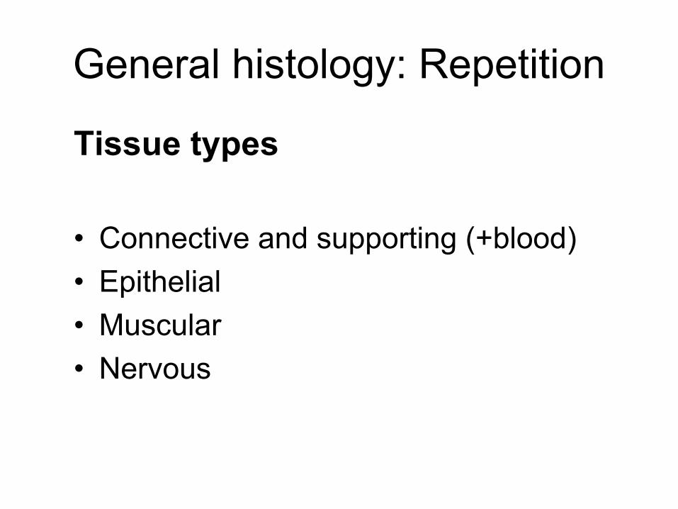
General histology: Repetition
Tissue types
• Connective and supporting (+blood)
• Epithelial
• Muscular
• Nervous

Connective tissue
developed from mesenchymedeveloped from mesenchyme
consists of:consists of:
•• cells cells
•• intercellular matrix: intercellular matrix:
a)a) amorphous ground substanceamorphous ground substance
b)b) fibersfibers

Classification
•• connective tissue properconnective tissue proper
•• specialized connective (supporting) tissue: specialized connective (supporting) tissue:
cartilage cartilage
bonebone
FunctionsFunctions
•• mechanical (cartilage, bone)mechanical (cartilage, bone)
•• nutritional (intercellular substance)nutritional (intercellular substance)
•• defensive (cells: histiocytes, plasma cells, defensive (cells: histiocytes, plasma cells, leukocytes leukocytes –– immunocompetence, production of immunocompetence, production of antibodies)antibodies)

Connective tissue proper
• mucous (jelly-like)
• loose collagenous (areolar)
• dense collagenous (regular, irregular)
• reticular
• elastic
• adipose tissue (white, brown)

Connective tissue proper
Mucous (jelly-like) connective tissue

Loose (areolar) connective tissue

Dense connective tissue (regular,
irregular)

Elastic tissue

Reticular tissue

Adipose tissue (white)
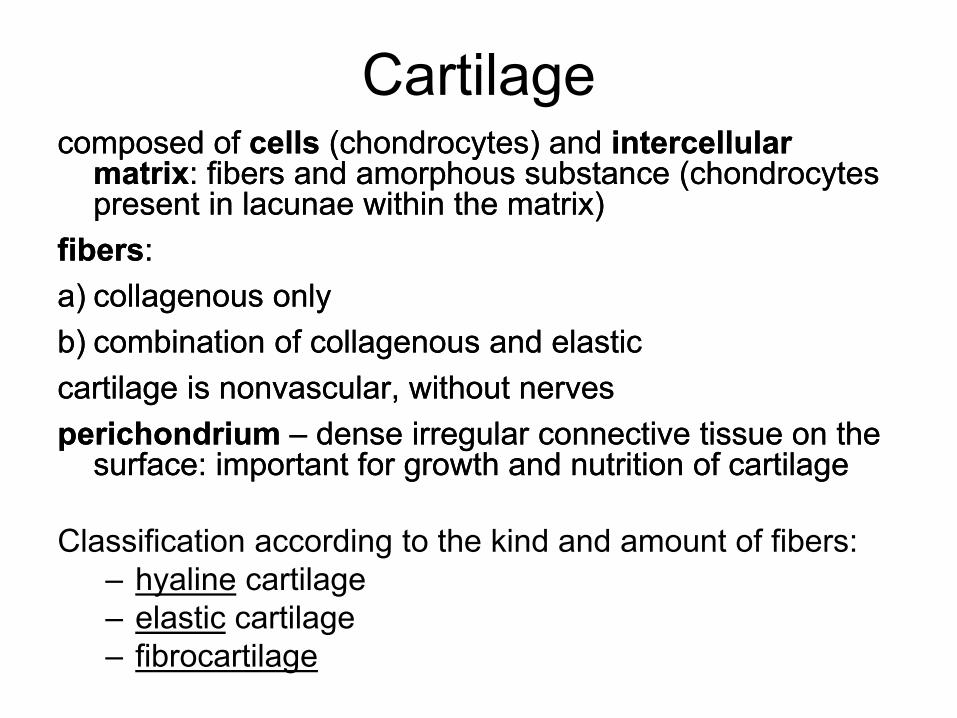
Cartilage composed of composed of cellscells (chondrocytes) and (chondrocytes) and intercellular intercellular
matrixmatrix: fibers and amorphous substance (chondrocytes : fibers and amorphous substance (chondrocytes present in lacunae within the matrix) present in lacunae within the matrix)
fibersfibers: :
a)a) collagenous onlycollagenous only
b)b) combination of collagenous and elasticcombination of collagenous and elastic
cartilage is nonvascular, without nervescartilage is nonvascular, without nerves
perichondrium perichondrium –– dense irregular connective tissue on the dense irregular connective tissue on the surface: important for growth and nutrition of cartilagesurface: important for growth and nutrition of cartilage
Classification according to the kind and amount of fibers:
– hyaline cartilage
– elastic cartilage
– fibrocartilage

Cartilage (hyaline)

Cartilage (elastic)

Fibrocartilage

Bone
• Cells: osteocytes, osteoblasts, osteoclasts, osteoprogenitor cells
• Intercellular matrix: collagenous fibers (type I), amorphous substance, inorganic salts
• Macroscopically - 2 types: compact (dense) and spongy (cancelous)
• Microscopically – 2 types according to the organisation of intercellular substance: woven (nonlamellar) and Haversian (lamellar)

Woven bone

Lamellar (Haversian) bone

Epithelial tissue Classification on the structural basis (arrangement of
cells):
• membranes – cells form sheets – the most common type, including most exocrine glands
• trabecular – cells are arranged into anastomosing trabeculae – liver, endocrine glands
• reticular – stellate cells form a network – thymus
Classification on the basis of function :
• Covering (lining) epithelia – epithelial membranes
• Glandular epithelium
• Absorptive epithelium – enterocytes (intestine)
• Respiratory epithelium – pneumocytes (lung)
• Sensory epithelium – olfactory ep., taste buds
• Myoepithelial cells (exocrine glands, m. dilatator pupillae)

Covering epithelia (epithelial
membranes) • Simple squamous

Covering epithelia (epithelial
membranes)
• Simple cuboidal

Covering epithelia (epithelial
membranes) • Simple columnar

Covering epithelia (epithelial
membranes)
• pseudostratified

Covering epithelia (epithelial
membranes)
• Stratified squamous nonkeratinized

Covering epithelia (epithelial
membranes)
• Stratified squamous keratinized

Covering epithelia (epithelial
membranes)
• Stratified columnar

Covering epithelia (epithelial
membranes)
• transitional

Glandular epithelium
Unicellular glands
Goblet cells Paneth cells

Glandular epithelium multicellular glands – ducts and secretory
portions (acini, tubules)

Glandular epithelium
• Multicellular glands – serous acini and
mucous tubules

Muscle tissue Morphological unit of:
• Skeletal muscle
cell is called muscle fiber – rhabdomyocyte (multinucleated, nuclei at periphery)
myofibrils are structures inside the cell, consist of myofilaments (actin, myosin)
• Cardiac muscle
cell – cardiomyocyte (uninucleated, nucleus centrally)
myofibrils, intercallated discs
• Smooth muscle
cell – leiomyocyte (uninucleated, nucleus centrally)
no myofibrils, only myofilaments

Skeletal muscle

Cardiac muscle

Smooth muscle

Nervous tissue
Anatomically:
• CNS (central nervous system): brain, spinal cord
• PNS (peripheral nervous system): nerves,
ganglia
Histologically it consists of 2 principal cell types:
• nerve cells (neurons) – excitability (irritability)
and conductivity
• supporting cells (neuroglia)
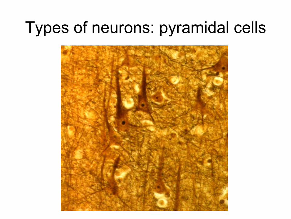
Types of neurons: pyramidal cells

Types of neurons: Purkinje cell

Types of neurons: motor neurons
of spinal cord

Types of neurons: pseudounipolar
neurons

Peripheral nerve

Peripheral nerve