Embed Size (px)
Citation preview
lable at ScienceDirect
Progress in Biophysics and Molecular Biology 105 (2011) 208e222
Contents lists avai
Progress in Biophysics and Molecular Biology
journal homepage: www.elsevier .com/locate/pbiomolbio
Review
Condensed DNA: Condensing the concepts
Vladimir B. Teif a,b,*, Klemen Bohinc c,d
aBioQuant and German Cancer Research Center, Im Neuenheimer Feld 267, 69120 Heidelberg, Germanyb Institute of Bioorganic Chemistry, Belarus National Academy of Sciences, Kuprevich 5/2, 220141, Minsk, Belarusc Faculty of Health Sciences, Zdravstvena pot 5, 1000 Ljubljana, Sloveniad Faculty of Electrical Engineering, University of Ljubljana, Trzaska 25, 1000 Ljubljana, Slovenia
a r t i c l e i n f o
Article history:Available online 16 July 2010
Keywords:DNA condensationLigand bindingCounterion correlationsMacromolecular crowdingChromatinGene regulation
* Corresponding author. Group Genome RegulationGermany. Tel.: +49 178 8063165; fax.: +49 6221 5451
E-mail addresses: [email protected]
0079-6107/$ e see front matter � 2010 Elsevier Ltd.doi:10.1016/j.pbiomolbio.2010.07.002
a b s t r a c t
DNA is stored in vivo in a highly compact, so-called condensed phase, where gene regulatory processesare governed by the intricate interplay between different states of DNA compaction. These systems oftenhave surprising properties, which one would not predict from classical concepts of dilute solutions. Themechanistic details of DNA packing are essential for its functioning, as revealed by the recent devel-opments coming from biochemistry, electrostatics, statistical mechanics, and molecular and cell biology.Different aspects of condensed DNA behavior are linked to each other, but the links are often hidden inthe bulk of experimental and theoretical details. Here we try to condense some of these concepts andprovide interconnections between the different fields. After a brief description of main experimentalfeatures of DNA condensation inside viruses, bacteria, eukaryotes and the test tube, main theoreticalapproaches for the description of these systems are presented. We end up with an extended discussion ofthe role of DNA condensation in the context of gene regulation and mention potential applications ofDNA condensation in gene therapy and biotechnology.
� 2010 Elsevier Ltd. All rights reserved.
Contents
1. Introduction . . . . . . . . . . . . . . . . . . . . . . . . . . . . . . . . . . . . . . . . . . . . . . . . . . . . . . . . . . . . . . . . . . . . . . . . . . . . . . . . . . . . . . . . . . . . . . . . . . . . . . . . . . . . . . . . . . . . . . . . .22. The concept of DNA condensation . . . . . . . . . . . . . . . . . . . . . . . . . . . . . . . . . . . . . . . . . . . . . . . . . . . . . . . . . . . . . . . . . . . . . . . . . . . . . . . . . . . . . . . . . . . . . . . . . . . . .23. DNA condensation in viruses . . . . . . . . . . . . . . . . . . . . . . . . . . . . . . . . . . . . . . . . . . . . . . . . . . . . . . . . . . . . . . . . . . . . . . . . . . . . . . . . . . . . . . . . . . . . . . . . . . . . . . . . . .24. DNA condensation in bacteria . . . . . . . . . . . . . . . . . . . . . . . . . . . . . . . . . . . . . . . . . . . . . . . . . . . . . . . . . . . . . . . . . . . . . . . . . . . . . . . . . . . . . . . . . . . . . . . . . . . . . . . . .35. DNA condensation in eukaryotes . . . . . . . . . . . . . . . . . . . . . . . . . . . . . . . . . . . . . . . . . . . . . . . . . . . . . . . . . . . . . . . . . . . . . . . . . . . . . . . . . . . . . . . . . . . . . . . . . . . . . . .36. DNA condensation in vitro . . . . . . . . . . . . . . . . . . . . . . . . . . . . . . . . . . . . . . . . . . . . . . . . . . . . . . . . . . . . . . . . . . . . . . . . . . . . . . . . . . . . . . . . . . . . . . . . . . . . . . . . . . . .4
6.1. Experimental methods . . . . . . . . . . . . . . . . . . . . . . . . . . . . . . . . . . . . . . . . . . . . . . . . . . . . . . . . . . . . . . . . . . . . . . . . . . . . . . . . . . . . . . . . . . . . . . . . . . . . . . . . . . 46.2. Condensing agents . . . . . . . . . . . . . . . . . . . . . . . . . . . . . . . . . . . . . . . . . . . . . . . . . . . . . . . . . . . . . . . . . . . . . . . . . . . . . . . . . . . . . . . . . . . . . . . . . . . . . . . . . . . . 46.3. Morphologies of condensed DNA . . . . . . . . . . . . . . . . . . . . . . . . . . . . . . . . . . . . . . . . . . . . . . . . . . . . . . . . . . . . . . . . . . . . . . . . . . . . . . . . . . . . . . . . . . . . . . . . 46.4. Reentrant effects . . . . . . . . . . . . . . . . . . . . . . . . . . . . . . . . . . . . . . . . . . . . . . . . . . . . . . . . . . . . . . . . . . . . . . . . . . . . . . . . . . . . . . . . . . . . . . . . . . . . . . . . . . . . . . 4
7. Insights from physics . . . . . . . . . . . . . . . . . . . . . . . . . . . . . . . . . . . . . . . . . . . . . . . . . . . . . . . . . . . . . . . . . . . . . . . . . . . . . . . . . . . . . . . . . . . . . . . . . . . . . . . . . . . . . . . . .57.1. The coileglobule transition . . . . . . . . . . . . . . . . . . . . . . . . . . . . . . . . . . . . . . . . . . . . . . . . . . . . . . . . . . . . . . . . . . . . . . . . . . . . . . . . . . . . . . . . . . . . . . . . . . . . . . 57.2. Hydration forces . . . . . . . . . . . . . . . . . . . . . . . . . . . . . . . . . . . . . . . . . . . . . . . . . . . . . . . . . . . . . . . . . . . . . . . . . . . . . . . . . . . . . . . . . . . . . . . . . . . . . . . . . . . . . . 57.3. Counterion condensation . . . . . . . . . . . . . . . . . . . . . . . . . . . . . . . . . . . . . . . . . . . . . . . . . . . . . . . . . . . . . . . . . . . . . . . . . . . . . . . . . . . . . . . . . . . . . . . . . . . . . . . 57.4. Counterion correlations . . . . . . . . . . . . . . . . . . . . . . . . . . . . . . . . . . . . . . . . . . . . . . . . . . . . . . . . . . . . . . . . . . . . . . . . . . . . . . . . . . . . . . . . . . . . . . . . . . . . . . . . 67.5. Counterion bridging . . . . . . . . . . . . . . . . . . . . . . . . . . . . . . . . . . . . . . . . . . . . . . . . . . . . . . . . . . . . . . . . . . . . . . . . . . . . . . . . . . . . . . . . . . . . . . . . . . . . . . . . . . . . 67.6. DNA charge reversal . . . . . . . . . . . . . . . . . . . . . . . . . . . . . . . . . . . . . . . . . . . . . . . . . . . . . . . . . . . . . . . . . . . . . . . . . . . . . . . . . . . . . . . . . . . . . . . . . . . . . . . . . . . . 77.7. DNAeDNA recognition . . . . . . . . . . . . . . . . . . . . . . . . . . . . . . . . . . . . . . . . . . . . . . . . . . . . . . . . . . . . . . . . . . . . . . . . . . . . . . . . . . . . . . . . . . . . . . . . . . . . . . . . . . 77.8. Computer simulations . . . . . . . . . . . . . . . . . . . . . . . . . . . . . . . . . . . . . . . . . . . . . . . . . . . . . . . . . . . . . . . . . . . . . . . . . . . . . . . . . . . . . . . . . . . . . . . . . . . . . . . . . . 77.9. Lattice models for chromatin . . . . . . . . . . . . . . . . . . . . . . . . . . . . . . . . . . . . . . . . . . . . . . . . . . . . . . . . . . . . . . . . . . . . . . . . . . . . . . . . . . . . . . . . . . . . . . . . . . . . 7
8. DNA condensation in the context of gene regulation . . . . . . . . . . . . . . . . . . . . . . . . . . . . . . . . . . . . . . . . . . . . . . . . . . . . . . . . . . . . . . . . . . . . . . . . . . . . . . . . . . . . .9
and Function (B066), BioQuant and German Cancer Research Center, Im Neuenheimer Feld 267, 69120 Heidelberg,487.elberg.de (V.B. Teif), [email protected] (K. Bohinc).
All rights reserved.
V.B. Teif, K. Bohinc / Progress in Biophysics and Molecular Biology 105 (2011) 208e222 209
8.1. Non-enzymatic reaction rates . . . . . . . . . . . . . . . . . . . . . . . . . . . . . . . . . . . . . . . . . . . . . . . . . . . . . . . . . . . . . . . . . . . . . . . . . . . . . . . . . . . . . . . . . . . . . . . . . . . 98.2. Protein binding site search . . . . . . . . . . . . . . . . . . . . . . . . . . . . . . . . . . . . . . . . . . . . . . . . . . . . . . . . . . . . . . . . . . . . . . . . . . . . . . . . . . . . . . . . . . . . . . . . . . . . . 98.3. Enzymatic reaction rates . . . . . . . . . . . . . . . . . . . . . . . . . . . . . . . . . . . . . . . . . . . . . . . . . . . . . . . . . . . . . . . . . . . . . . . . . . . . . . . . . . . . . . . . . . . . . . . . . . . . . . . 98.4. Condensation and origin of life . . . . . . . . . . . . . . . . . . . . . . . . . . . . . . . . . . . . . . . . . . . . . . . . . . . . . . . . . . . . . . . . . . . . . . . . . . . . . . . . . . . . . . . . . . . . . . . . . 98.5. Epigenetic gene regulation . . . . . . . . . . . . . . . . . . . . . . . . . . . . . . . . . . . . . . . . . . . . . . . . . . . . . . . . . . . . . . . . . . . . . . . . . . . . . . . . . . . . . . . . . . . . . . . . . . . . . . 9
9. Potential applications in medicine and biotechnology . . . . . . . . . . . . . . . . . . . . . . . . . . . . . . . . . . . . . . . . . . . . . . . . . . . . . . . . . . . . . . . . . . . . . . . . . . . . . . . . . . .1010. Summary . . . . . . . . . . . . . . . . . . . . . . . . . . . . . . . . . . . . . . . . . . . . . . . . . . . . . . . . . . . . . . . . . . . . . . . . . . . . . . . . . . . . . . . . . . . . . . . . . . . . . . . . . . . . . . . . . . . . . . . . . . .10
Acknowledgments . . . . . . . . . . . . . . . . . . . . . . . . . . . . . . . . . . . . . . . . . . . . . . . . . . . . . . . . . . . . . . . . . . . . . . . . . . . . . . . . . . . . . . . . . . . . . . . . . . . . . . . . . . . . . . . . . . 11References . . . . . . . . . . . . . . . . . . . . . . . . . . . . . . . . . . . . . . . . . . . . . . . . . . . . . . . . . . . . . . . . . . . . . . . . . . . . . . . . . . . . . . . . . . . . . . . . . . . . . . . . . . . . . . . . . . . . . . . . . 11
1. Introduction
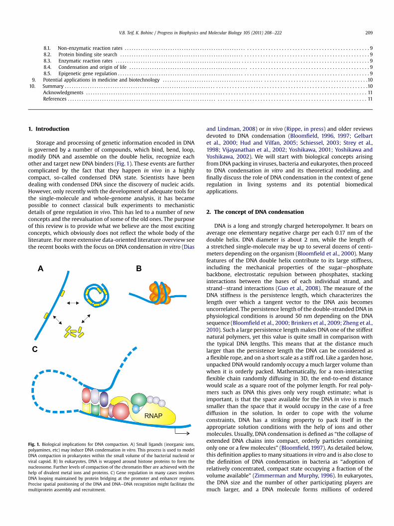
Storage and processing of genetic information encoded in DNAis governed by a number of compounds, which bind, bend, loop,modify DNA and assemble on the double helix, recognize eachother and target new DNA binders (Fig. 1). These events are furthercomplicated by the fact that they happen in vivo in a highlycompact, so-called condensed DNA state. Scientists have beendealing with condensed DNA since the discovery of nucleic acids.However, only recently with the development of adequate tools forthe single-molecule and whole-genome analysis, it has becamepossible to connect classical bulk experiments to mechanisticdetails of gene regulation in vivo. This has led to a number of newconcepts and the reevaluation of some of the old ones. The purposeof this review is to provide what we believe are the most excitingconcepts, which obviously does not reflect the whole body of theliterature. For more extensive data-oriented literature overview seethe recent books with the focus on DNA condensation in vitro (Dias
Fig. 1. Biological implications for DNA compaction. A) Small ligands (inorganic ions,polyamines, etc) may induce DNA condensation in vitro. This process is used to modelDNA compaction in prokaryotes within the small volume of the bacterial nucleoid orviral capsid. B) In eukaryotes, DNA is wrapped around histone proteins to form thenucleosome. Further levels of compaction of the chromatin fiber are achieved with thehelp of divalent metal ions and proteins. C) Gene regulation in many cases involvesDNA looping maintained by protein bridging at the promoter and enhancer regions.Precise spatial positioning of the DNA and DNAeDNA recognition might facilitate themultiprotein assembly and recruitment.
and Lindman, 2008) or in vivo (Rippe, in press) and older reviewsdevoted to DNA condensation (Bloomfield, 1996, 1997; Gelbartet al., 2000; Hud and Vilfan, 2005; Schiessel, 2003; Strey et al.,1998; Vijayanathan et al., 2002; Yoshikawa, 2001; Yoshikawa andYoshikawa, 2002). We will start with biological concepts arisingfromDNA packing in viruses, bacteria and eukaryotes, then proceedto DNA condensation in vitro and its theoretical modeling, andfinally discuss the role of DNA condensation in the context of generegulation in living systems and its potential biomedicalapplications.
2. The concept of DNA condensation
DNA is a long and strongly charged heteropolymer. It bears onaverage one elementary negative charge per each 0.17 nm of thedouble helix. DNA diameter is about 2 nm, while the length ofa stretched single-molecule may be up to several dozens of centi-meters depending on the organism (Bloomfield et al., 2000). Manyfeatures of the DNA double helix contribute to its large stiffness,including the mechanical properties of the sugarephosphatebackbone, electrostatic repulsion between phosphates, stackinginteractions between the bases of each individual strand, andstrandestrand interactions (Guo et al., 2008). The measure of theDNA stiffness is the persistence length, which characterizes thelength over which a tangent vector to the DNA axis becomesuncorrelated. The persistence length of the double-stranded DNA inphysiological conditions is around 50 nm depending on the DNAsequence (Bloomfield et al., 2000; Brinkers et al., 2009; Zheng et al.,2010). Such a large persistence lengthmakes DNA one of the stiffestnatural polymers, yet this value is quite small in comparison withthe typical DNA lengths. This means that at the distance muchlarger than the persistence length the DNA can be considered asa flexible rope, and on a short scale as a stiff rod. Like a garden hose,unpacked DNAwould randomly occupy a much larger volume thanwhen it is orderly packed. Mathematically, for a non-interactingflexible chain randomly diffusing in 3D, the end-to-end distancewould scale as a square root of the polymer length. For real poly-mers such as DNA this gives only very rough estimate; what isimportant, is that the space available for the DNA in vivo is muchsmaller than the space that it would occupy in the case of a freediffusion in the solution. In order to cope with the volumeconstraints, DNA has a striking property to pack itself in theappropriate solution conditions with the help of ions and othermolecules. Usually, DNA condensation is defined as “the collapse ofextended DNA chains into compact, orderly particles containingonly one or a fewmolecules” (Bloomfield, 1997). As detailed below,this definition applies to many situations in vitro and is also close tothe definition of DNA condensation in bacteria as “adoption ofrelatively concentrated, compact state occupying a fraction of thevolume available” (Zimmerman and Murphy, 1996). In eukaryotes,the DNA size and the number of other participating players aremuch larger, and a DNA molecule forms millions of ordered
V.B. Teif, K. Bohinc / Progress in Biophysics and Molecular Biology 105 (2011) 208e222210
nucleoprotein particles, the nucleosomes, which are just the firstlevel of DNA packing, as detailed below. Thus, one can hardlydevelop a single definition for DNA condensation valid for allsystems. Conceptually, it should be understood that DNA conden-sation refers to a concentrated macromolecular phase whereneighboring DNA segments may be separated by just a few layers ofsolvent molecules. The local alignment of these segments is thusnot the requirement, but rather a consequence of the condensedstate.
3. DNA condensation in viruses
In viruses and bacteriophages, the DNA or RNA is surrounded bya protein capsid, sometimes further enveloped by a lipidmembrane. Double-stranded DNA is stored inside the capsid in theform of a spool, which can have different types of coiling (Hud,1995) leading to different types of liquid-crystalline packing(Earnshaw and Harrison, 1977; Hud and Downing, 2001; Knoblerand Gelbart, 2009; Leforestier and Livolant, 2009). This packingcan change from hexagonal to cholesteric to isotropic at differentstages of the phage functioning (Leforestier and Livolant, 2010).Although the double helices are always locally aligned, the DNAinside viruses does not represent real liquid crystals, because itlacks fluidity. On the other hand, DNA condensed in vitro, e.g. withthe help of polyamines which are also present in viruses, is bothlocally ordered and fluid (Sikorav et al., 1994). Highly symmetricDNA packing inside viruses is probably the only way to fill the smallspace of the symmetric viral capsid (Johnson and Chiu, 2007).When a virus is being assembled, motor proteins push the DNAinside the capsid, where it is stored under large pressures w6 MPa(Evilevitch et al., 2008; Rickgauer et al., 2008; São-José et al., 2007).In order to infect the host cell, the virus needs to open up a smallhole by a conformational transition in the portal gatekeeperproteins (Lhuillier et al., 2009). The DNA injection is then triggeredby the differences in the osmotic pressure and ionic conditions,with the DNA ejecting as fast as 60 kbp/s (Grayson et al., 2007). Theenergy stored in the form of DNA compaction is enough to inject asmuch as 1/5 of the viral DNA before motor proteins start pushingthe DNA (São-José et al., 2007). RNA packing in RNA-containingviruses is even more intricate: a recent analysis shows that the sizeof the optimally compacted RNA matches the size of the corre-sponding viral capsid, suggesting that there might be evolutionarypressure for the genome to have an appropriate size (Yoffe et al.,2008). Notably, organisms which do not have such strict packingconstraints are easily increasing the genome size at the evolu-tionary timescale. Viral packing provides the most condensed stateof genomic DNA. Scientists are trying to follow the Nature’s way ofdelivering and releasing specifically synthesized nucleic acids forthe purpose of gene therapy. Many effective DNA-packing strate-gies have been developed, but none as elegant as viruses.
4. DNA condensation in bacteria
Bacterial DNA is packed with the help of polyamines andproteins. Protein-associated DNA occupies about 1/4 of the intra-cellular volume forming a concentrated viscous phase with liquid-crystalline properties, called the nucleoid (Cunha et al., 2001;Wiggins et al., 2010). Similar DNA packaging exists also in chloro-plasts (Sekine et al., 2002) and mitochondria (Friddle et al., 2004).Bacterial DNA is sometimes referred to as the bacterial chromo-some (Saier, 2008). In fact, the bacterial nucleoid evolutionaryrepresents an intermediate engineering solution between theprotein-free DNA packing in viruses and protein-determinedpacking in eukaryotes (Luijsterburg et al., 2008). There are severalmain nucleoid-associated proteins, such as HeNS, HU, Fis, IHF and
Dps, all of which contribute to DNA packing and also regulate geneexpression (Dame, 2005; Pettijohn, 1988; Travers andMuskhelishvili, 2005). For example, HeNS has functions analo-gous to eukaryotic histones, while HU resembles eukaryotic high-mobility-group (HMG) proteins (Luijsterburg et al., 2008). Indeed,yeast HMGB proteins have been shown to be to some extentinterchangeable with Escherichia coli HU proteins (Becker et al.,2008). In analogy to the eukaryotic chromatin, most of bacterialDNA is covered by HeNS and HU proteins, which bind coopera-tively and form short protected DNA stretches (functionally anal-ogous to eukaryotic nucleosomes) and larger DNA loops ofe10 kblength. The DNA in bacterial loops is confined to a small volume.The latter is achieved by coiling the loop and fixing the obtainedsuperhelical structures by protein bridges. In such structures, theDNA is said to be in the supercoiled state. Supercoiling is anotherNature’s trick allowing energy storage combined with tight packing(Cozzarelli et al., 2006; Luijsterburg et al., 2008). The level of DNApacking in the bacterial nucleoid is also regulated by the macro-molecular crowding, which is defined as excluded volume effectsfavoring compact molecular conformations (Zimmerman andMinton, 1993; Zimmerman and Murphy, 1996). However, nucleoidstructure is determined not only by non-specific interactions. Forexample, HeNS proteins have a slight sequence-specificity, whichadds even more to the analogy with eukaryotic histones (Fang andRimsky, 2008; Navarre et al., 2006). Furthermore, a recent study hasshown that specific bacterial genes are nonrandomly positioned in3D within the nucleoid (Wiggins et al., 2010) in analogy with theconcept of spatial positioning of genes in the eukaryotic genome(Lieberman-Aiden et al., 2009).
5. DNA condensation in eukaryotes
In comparison with bacteria or viruses, eukaryotic chromatin isthe “state of the art” of DNA condensation, and also a large field ofscientific efforts, which we will only briefly mention here.Eukaryotic DNA with a typical length of dozens of centimetersshould be orderly packed to be readily accessible inside themicrometer-size nucleus. Thus DNA is always “condensed” inchromatin, but there are different states of DNA condensation. Inprimitive unicellular eukaryotes such as dinoflagellates, it ispossible to distinguish liquid-crystalline chromosomal orderingsimilar to bacterial chromosomes, just with higher DNA density(Chow et al., 2010). However, this is the only exception in theeukaryotic world. In other eukaryotes, DNA is arranged in the cellnucleus with the help of histones (Van Holde,1989). In this case, thebasic level of DNA compaction is the nucleosome, where the doublehelix is wrapped around the histone octamer containing two copiesof each histone H2A, H2B, H3 and H4. Linker histone H1 binds theDNA between nucleosomes and facilitates packaging of the 10 nm“beads on the string” nucleosomal chain into a more condensed30 nm fiber (Rippe et al., 2008). Most of the time, between celldivisions, chromatin is optimized to allow easy access of tran-scription factors to active genes, which are characterized by a lesscompact structure called euchromatin, and to alleviate proteinaccess in more tightly packed regions called heterochromatin.During the cell division, chromatin compaction increases evenmore to form the classical chromosomes, which can copewith largemechanical forces dragging them into each of the two daughtercells (Van Holde, 1989). The transitions between different states ofchromatin compaction are regulated by the dynamic exchange ofhistones, as well as other proteins such as HMG-proteins competingfor DNA binding with linker histones (Gerlitz et al., 2009), HP1proteins recruited by the nucleosomal histone tails (Müller et al.,2009) and larger players such as CTCF proteins defining theboundaries between the regions with different nucleosome
V.B. Teif, K. Bohinc / Progress in Biophysics and Molecular Biology 105 (2011) 208e222 211
arrangement (Ohlsson et al., 2010) and cohesines linking sisterchromatids in meiosis (Suja and Barbero, 2009), to name just a few.Furthermore, specific energy-dependent molecular motors, so-called chromatin remodelers, reorder chromatin following the cellcycle, cell differentiation and external signals (Corpet andAlmouzni, 2009). All transitions between the states of DNAcompaction are precisely controlled. Damaging the integrity of DNApacking is lethal to the cell. Although morphological changes inchromatin during apoptosis (programmed cell death) are alsodescribed by the word “condensation” (Widlak et al., 2002), thistransition is accompanied by DNA fragmentation as opposed to thereversible DNA condensation occurring in the normal cell. Severalanticancer drugs also act through crosslinking the DNA, looping itand establishing condensed untranscribed structures (Hou et al.,2009; Kida et al., 2010). Transitions between different states ofDNA compaction in vivo regulate a number of processes, from viralinvasion to the cell cycle, differentiation, and apoptosis. Before weproceed to these biological processes, we have to make a step backand learn the basic physical properties of DNA condensation invitro.
6. DNA condensation in vitro
6.1. Experimental methods
Most of our knowledge about condensed DNA states comes fromcomparatively simple in vitro experiments started in the 1970s(Gosule and Shellman, 1976; Lerman, 1971). In such experiments,DNA is compacted by adding different condensing agents, fromsimple inorganic ions to large macromolecules, which representimportant model systems to understand DNA functioning in vivoand also to achieve controlled drug delivery in gene therapy. Duringfour decades of such experiments, DNA condensation has beenstudied using a variety of methods including sedimentation (Jaryand Sikorav, 1999), light scattering (Vijayanathan et al., 2001;Wilson and Bloomfield, 1979), viscometry (Slita et al., 2007;Slonitskii and Kuptsov, 1989), osmotic equilibrium (Parsegianet al., 2000; Strey et al., 1998), IR, UV and Raman spectroscopy(Marty et al., 2009), as well as electron microscopy (Hud and Vilfan,2005) and atomic force microscopy (Hansma et al., 2004). Morerecently, capillary electrophoresis techniques have been added tothe bulk experiments (Keyser et al., 2010). Today one can also usesingle-molecule optical and magnetic tweezers (Baumann et al.,2000; Besteman et al., 2007; Chien et al., 2007; Dias andLindman, 2008; Todd et al., 2008).
6.2. Condensing agents
DNA condensation can be induced in vitro either by applyingexternal force to bring the double helices together, or by inducingattractive interactions between the DNA segments. The former canbe achieved e.g. with the help of the osmotic pressure exerted bycrowding polymers, as is done in the case of the j-DNA compactedby adding neutral polymers in the presence of monovalent salts(Greek letter “psi” stands for “polymer-and salt-induced”)(Evdokimov et al., 1972; Lerman, 1971). In this case, the forcespushing the double helices together are coming from entropicrandom collisions with the crowding polymers surrounding DNAcondensates, and salt is required to neutralize DNA charges anddecrease DNAeDNA repulsion. The second possibility can be real-ized by inducing attractive interactions between the DNA segmentsby multivalent cationic charged ligands (Gosule and Shellman,1976), as detailed in the following sections. More exotic ways toinduce attraction between DNA molecules also exist, including e.g.an external electrical field alternating at a certain frequency
(Bruinsma and Riehn, 2009). However, typical biologically moti-vated DNA condensing agents are either neutral polymers such aspolyethylene glycol plus Naþ, or multivalent counterions (ligands).
In water solutions, DNA condensation usually requires coun-terions with charge 3 þ or higher (Bloomfield, 1996, 1997). Amongtypical ligands used in experiments are trivalent metal ions andinorganic cations such as Co(NH3)63þ (Kankia et al., 2001), naturallyoccurring polyamines and their analogs (Nayvelt et al., 2010),protamines (Balhorn, 2007), natural and synthetic peptides(Korolev et al., 2009; Saccardo et al., 2009), lipids and liposomes(Rao, 2010), bacterial nucleoid-associated proteins and eukaryoticchromatin proteins. Monovalent counterions such as Naþ cannotinduce DNA condensation unless under additional osmotic pres-sure exerted e.g. by neutral polymers such as PEG (Lerman, 1971;Parsegian et al., 2000; Zimmerman and Minton, 1993). Divalentmetal ions provide a boundary case. They cannot induce conden-sation of linear DNA molecules in water solutions, but they can doso in the presence of lipids that partition DNAmolecules in lamellarsandwich-like structures (Harries et al., 1998; Koltover et al., 2000;Mengistu et al., 2009), or under special conditions if the DNA iscircular. In the latter case, DNA supercoiling is a factor favoring DNAcondensation as suggested by experimental observations thatMn2þ is able to induce condensation of the circular but not linearDNA (Ma and Bloomfield, 1994), the conclusion also supported bythe Brownian Dynamics simulations (Sottas et al., 1999). Not allmetal ions of the same charge are equally effective: transitionmetals such asMn2þ, which can interact bothwith DNA phosphates(electrostatically) and bases (e.g. forming chelate complexes) arestronger condensing agents in comparison with alkali metals,which can interact only with DNA phosphates. Similar consider-ations apply also to more complex ions such as divalent amines:their condensing efficiency strongly depends on the structure andthe ability to form DNAeDNA bridges (Saminathan et al., 1999).Alcohols and condensing ligands may act synergistically to locallydestabilize the double helix, permitting DNA foldbacks that canlead to a higher proportion of rod-like condensates (Bloomfield,1996). Temperature elevation can help aggregate DNA in thepresence of divalent ions (Andrushchenko et al., 2003; Bloomfield,1996, 1997). A recent study has shown a reversible condensation ofT4 genomic DNA in solutions of poly(N-isopropylacrylamide) upona temperature increase from 30 to 35 �C (Chen et al., 2010).
6.3. Morphologies of condensed DNA
Upon addition of critical concentrations of condensing ligands,double-stranded DNA molecules condense from a random coil intotoroids, rods, or more sophisticated structures (Bloomfield, 1997).The morphology of the condensates depends on the solutionproperties and condensing agent structure (Bloomfield, 1996,1997). Depending on the DNA length, DNA condensation mayhappen either as a monomolecular collapse (Post and Zimm, 1979;Widom and Baldwin, 1983) or as a multimolecular aggregation(Post and Zimm, 1982). On the other hand, DNA molecules shorterthan the persistence length form ordered liquid-crystalline phases(Rill, 1986; Schellman and Parthasarathy, 1984; Sikorav et al., 1994).With the help of specifically designed metallo-supramolecularcylinders, DNA coiling around the nucleosome core can bemimicked (Meistermann et al., 2002). Lipids or liposomes condenseDNA in well-defined small particles, which can be used for thepurpose of gene delivery (Rao, 2010; Vijayanathan et al., 2002). Itshould be noted, that DNA condensation is inmany cases associatedwith precipitation of the condensed particles or aggregates. On theother hand, DNA precipitation by itself, e.g. the one achieved byadding large quantities of ethanol or a similar solvent as in classicalDNA extraction techniques, is usually not called condensation.
V.B. Teif, K. Bohinc / Progress in Biophysics and Molecular Biology 105 (2011) 208e222212
6.4. Reentrant effects
Experimentally, DNA condensates are stable within a largeinterval of condensing ligand concentrations. If one continuesincreasing the ligand concentration, DNA condensation is followedby a reverse transitionwhen the aggregates resolubilize (Pelta et al.,1996; Raspaud et al., 1998; Saminathan et al., 1999). In the case ofa long DNA, resolubilization is associated with decondensation ofindividual molecules (Chien et al., 2007; Jary and Sikorav, 1999;Murayama et al., 2003). The effect of reentrant condensation hasbeen observed for short or long DNA molecules, single- or double-stranded DNA, small or high DNA concentrations. Reentrantcondensation may also be essential for the functioning of theeukaryotic chromatin (Poirier et al., 2002) and is conceptuallyrelated to the reentrant effect of DNAeproteineDNA bridgeassembly and disassembly in gene regulation (Vilar and Saiz, 2006).The corresponding bell-shaped condensationedecondensationcurves coupled to different biochemical processes have attractedmuch attention of the biophysical community (Chien et al., 2007;Murayama et al., 2003; Raspaud et al., 1998; Saminathan et al.,1999; Sikorav and Church, 1991; Sikorav et al., 1994; Teif, 2005;Todd and Rau, 2008; Yang and Rau, 2005).
7. Insights from physics
7.1. The coileglobule transition
Condensation of long double-helical DNAs is a sharp phasetransition, which takes place within a narrow interval of ligandconcentrations (Bloomfield, 1996, 1997; Yoshikawa et al., 1996).Unlike protein folding, the general features of the DNA coileglobuletransition such as the topology of the condensate are mainlydetermined by the average polymer and solution properties (DNAlength, concentration, solution content and temperature) and notby the DNA sequence. On the other hand, the DNA sequencedetermines local interactions and recognition between the doublehelices in the condensed DNA phase (Kornyshev and Leikin, 1998;Sitko et al., 2003). The thermodynamic basis for the coileglobuletransitions was set by the classical polymer theories (Birshtein andPtitsyn, 1964; Flory, 1969) and adopted later for DNA condensation(Bloomfield, 1997; Grosberg and Zhestkov, 1985, 1986; Grosbergand Khokhlov, 1994; Post and Zimm, 1979, 1982; Yoshikawa et al.,1996). For stiff long polymers, theory predicts that DNA conden-sation is a first-order transition. High stiffness of the DNA doublehelix is due to both its internal properties (sugarephosphatebackbone with stacked base pairs) and its charge (like-chargedphosphates repel each other and resist DNA bending). Single-molecule microscopy has confirmed that DNA condensationappears either to be an all-or-none transition for a single long DNAmolecule, or a continuous transition when an ensemble of DNAmolecules is considered (Yoshikawa et al., 1996).
7.2. Hydration forces
Since the double helices come very closely to each other in thecondensed phase, this leads to the restructuring of water mole-cules, which gives rise to the so-called hydration forces (Gelbartet al., 2000; Parsegian et al., 2000; Strey et al., 1998). Each watermolecule (as well as many other molecules dissolved in the water)represents a dipole, which can be statistically oriented in theelectrical field perturbed by the neighboring surface of the DNA.Such ordering would decrease exponentially with the distancefrom the DNA surface. Dipoles would predominantly orient in thesolution perpendicular to the charged surface (Maset and Bohinc,2007). Several approached allow one to study surface-induced
structural perturbation of the solvent. For example, Mengistu et al.have incorporated a solvent of interacting Langevin dipoles into thePB theory (Mengistu et al., 2009). Ruckenstein et al. have showedthat the solvent polarization spatially decays with increasingdistance from the charged surface (Ruckenstein andManciu, 2002).The influence of a finite volume of ions and water molecules wasalso studied. In this case the dielectric permittivity profile close tothe charged surface is mainly determined by two mechanisms, i.e.the depletion of water dipoles at the charged surface due to accu-mulation and of counterions and decreased orientational orderingof water dipoles as a function of increased distance from thecharged surface (Gavryushov, 2009; Iglic et al., 2010). In addition towater restructuring, the counterions dissolved in the water alsorearrange in the vicinity of the charged DNA molecule, which givesrise to the counterion-correlated attraction discussed in the nextsections. Despite many experimental efforts and theoretical studiesthe relative contributions of these constituents of attractive inter-actions still require a clarification (Todd et al., 2008).
7.3. Counterion condensation
DNA is a highly chargedmolecule, which cannot exist in solutionwithout other ions. Although the abbreviation “DNA” meansdeoxyribonucleic acid, usually DNA comes as a salt of Naþ or otheralkali metals. Physiological solutions also contain large amounts ofdivalent metal ions, which are essential for functioning of enzymesand play structural roles in DNA, RNA and proteins. Finally, the DNAis surrounded by a variety of other charged molecules, from smallpolyamines to proteins. Most electrostatic approaches treat DNAcounterions as point-like, sphere-like or rod-like, but real multi-valent ions and molecules possess an internal structure withspatially distributed charges. The simplest examples of such ionsare spermidine3þ and spermine4þ e naturally occurring poly-amines, which are flexible linear molecules widely used as DNAcondensing agents (Bloomfield, 1997; Gelbart et al., 2000). Thedistribution of multivalent ions in aqueous solutions close to themacroion is mainly determined by the competition between elec-trostatic interactions within the system and the entropy of theconstituents in the solution. At a thermodynamic equilibrium thecounterions are attracted to the charged surface, while co-ions aredepleted from this region, and a diffuse electric double layer iscreated (McLaughlin, 1989).
In 1960s Gerald Manning described some of the properties ofthe ionic cloud around a charged cylindrical surface representingthe polyelectrolyte in solution (Manning, 1969). Above a criticalsurface charge density, the counterions condense on the cylinderand reduce its charge density down to the critical value (Grosberget al., 2002). Counterion condensation is the consequence of thevictory of Coulomb potential energy over the entropy. In theManning picture the condensed layer of counterions is limited toa macroscopically small region near the charged cylinder. TheManning condensation theory holds only for an infinitely longcylinder and low concentrations. Surprisingly, it has a number ofsuccessful applications even when these conditions are violated(Manning and Ray, 1998).
As follows from the counterion condensation concept, a ligandbinding to the DNA has to replace several monovalent ions from theManning layer. Therefore proteineDNA interactions depend on theconcentration of the monovalent salt. The equation of Record andcoauthors predicts the following logarithmic dependence (Recordet al., 1978): v(ln(K))/v(ln[Naþ]) ¼ � 4$Z, where K is the bindingconstant of a protein or multivalent ligand of charge Z, [Naþ] is theNaþ concentration in solution, 4 equals to the number of thermo-dynamically released Naþ ions per phosphate upon ligand binding(4 z 0.88 for linear helical DNA). More advanced solutions to this
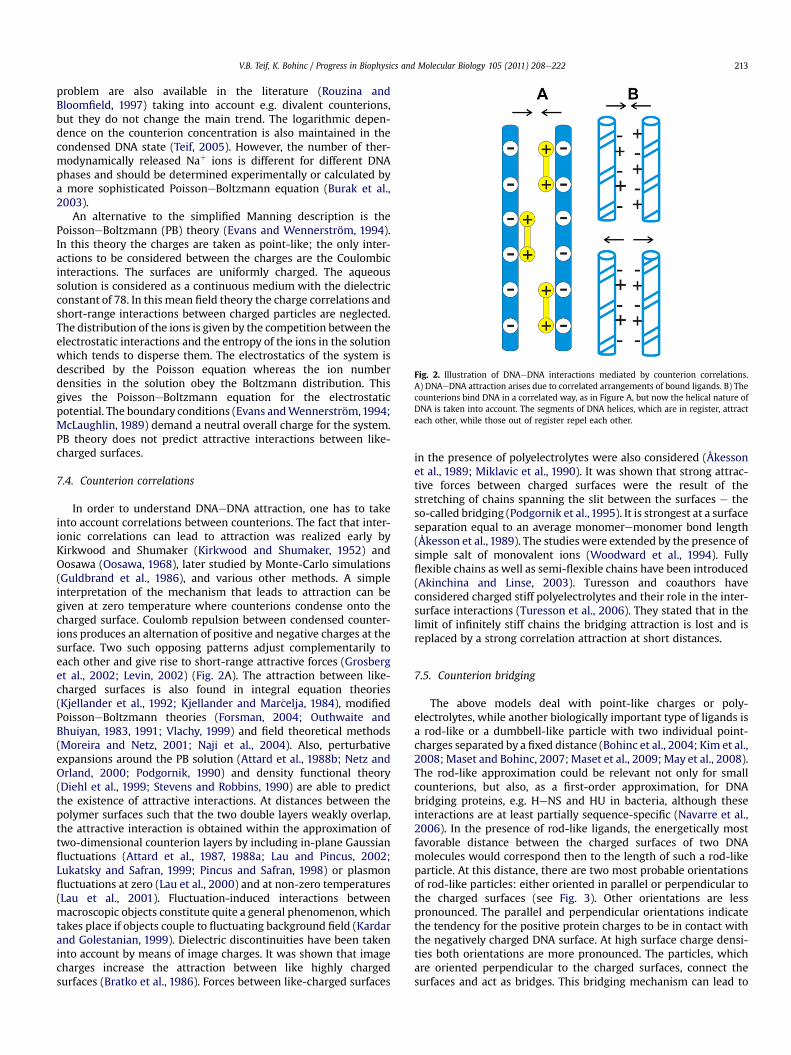
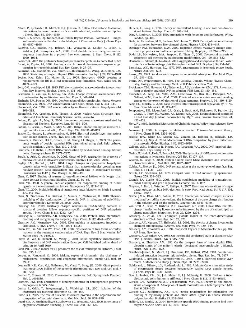
Fig. 2. Illustration of DNAeDNA interactions mediated by counterion correlations.A) DNAeDNA attraction arises due to correlated arrangements of bound ligands. B) Thecounterions bind DNA in a correlated way, as in Figure A, but now the helical nature ofDNA is taken into account. The segments of DNA helices, which are in register, attracteach other, while those out of register repel each other.
V.B. Teif, K. Bohinc / Progress in Biophysics and Molecular Biology 105 (2011) 208e222 213
problem are also available in the literature (Rouzina andBloomfield, 1997) taking into account e.g. divalent counterions,but they do not change the main trend. The logarithmic depen-dence on the counterion concentration is also maintained in thecondensed DNA state (Teif, 2005). However, the number of ther-modynamically released Naþ ions is different for different DNAphases and should be determined experimentally or calculated bya more sophisticated PoissoneBoltzmann equation (Burak et al.,2003).
An alternative to the simplified Manning description is thePoissoneBoltzmann (PB) theory (Evans and Wennerström, 1994).In this theory the charges are taken as point-like; the only inter-actions to be considered between the charges are the Coulombicinteractions. The surfaces are uniformly charged. The aqueoussolution is considered as a continuous medium with the dielectricconstant of 78. In this mean field theory the charge correlations andshort-range interactions between charged particles are neglected.The distribution of the ions is given by the competition between theelectrostatic interactions and the entropy of the ions in the solutionwhich tends to disperse them. The electrostatics of the system isdescribed by the Poisson equation whereas the ion numberdensities in the solution obey the Boltzmann distribution. Thisgives the PoissoneBoltzmann equation for the electrostaticpotential. The boundary conditions (Evans andWennerström,1994;McLaughlin, 1989) demand a neutral overall charge for the system.PB theory does not predict attractive interactions between like-charged surfaces.
7.4. Counterion correlations
In order to understand DNAeDNA attraction, one has to takeinto account correlations between counterions. The fact that inter-ionic correlations can lead to attraction was realized early byKirkwood and Shumaker (Kirkwood and Shumaker, 1952) andOosawa (Oosawa, 1968), later studied by Monte-Carlo simulations(Guldbrand et al., 1986), and various other methods. A simpleinterpretation of the mechanism that leads to attraction can begiven at zero temperature where counterions condense onto thecharged surface. Coulomb repulsion between condensed counter-ions produces an alternation of positive and negative charges at thesurface. Two such opposing patterns adjust complementarily toeach other and give rise to short-range attractive forces (Grosberget al., 2002; Levin, 2002) (Fig. 2A). The attraction between like-charged surfaces is also found in integral equation theories(Kjellander et al., 1992; Kjellander and Mar�celja, 1984), modifiedPoissoneBoltzmann theories (Forsman, 2004; Outhwaite andBhuiyan, 1983, 1991; Vlachy, 1999) and field theoretical methods(Moreira and Netz, 2001; Naji et al., 2004). Also, perturbativeexpansions around the PB solution (Attard et al., 1988b; Netz andOrland, 2000; Podgornik, 1990) and density functional theory(Diehl et al., 1999; Stevens and Robbins, 1990) are able to predictthe existence of attractive interactions. At distances between thepolymer surfaces such that the two double layers weakly overlap,the attractive interaction is obtained within the approximation oftwo-dimensional counterion layers by including in-plane Gaussianfluctuations (Attard et al., 1987, 1988a; Lau and Pincus, 2002;Lukatsky and Safran, 1999; Pincus and Safran, 1998) or plasmonfluctuations at zero (Lau et al., 2000) and at non-zero temperatures(Lau et al., 2001). Fluctuation-induced interactions betweenmacroscopic objects constitute quite a general phenomenon, whichtakes place if objects couple to fluctuating background field (Kardarand Golestanian, 1999). Dielectric discontinuities have been takeninto account by means of image charges. It was shown that imagecharges increase the attraction between like highly chargedsurfaces (Bratko et al., 1986). Forces between like-charged surfaces
in the presence of polyelectrolytes were also considered (Åkessonet al., 1989; Miklavic et al., 1990). It was shown that strong attrac-tive forces between charged surfaces were the result of thestretching of chains spanning the slit between the surfaces e theso-called bridging (Podgornik et al.,1995). It is strongest at a surfaceseparation equal to an average monomeremonomer bond length(Åkesson et al., 1989). The studies were extended by the presence ofsimple salt of monovalent ions (Woodward et al., 1994). Fullyflexible chains as well as semi-flexible chains have been introduced(Akinchina and Linse, 2003). Turesson and coauthors haveconsidered charged stiff polyelectrolytes and their role in the inter-surface interactions (Turesson et al., 2006). They stated that in thelimit of infinitely stiff chains the bridging attraction is lost and isreplaced by a strong correlation attraction at short distances.
7.5. Counterion bridging
The above models deal with point-like charges or poly-electrolytes, while another biologically important type of ligands isa rod-like or a dumbbell-like particle with two individual point-charges separated by a fixed distance (Bohinc et al., 2004; Kim et al.,2008; Maset and Bohinc, 2007; Maset et al., 2009; May et al., 2008).The rod-like approximation could be relevant not only for smallcounterions, but also, as a first-order approximation, for DNAbridging proteins, e.g. HeNS and HU in bacteria, although theseinteractions are at least partially sequence-specific (Navarre et al.,2006). In the presence of rod-like ligands, the energetically mostfavorable distance between the charged surfaces of two DNAmolecules would correspond then to the length of such a rod-likeparticle. At this distance, there are two most probable orientationsof rod-like particles: either oriented in parallel or perpendicular tothe charged surfaces (see Fig. 3). Other orientations are lesspronounced. The parallel and perpendicular orientations indicatethe tendency for the positive protein charges to be in contact withthe negatively charged DNA surface. At high surface charge densi-ties both orientations are more pronounced. The particles, whichare oriented perpendicular to the charged surfaces, connect thesurfaces and act as bridges. This bridging mechanism can lead to
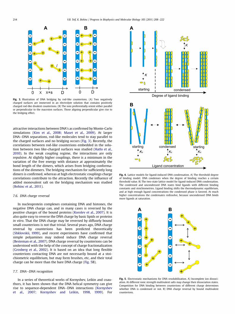
Fig. 3. Illustration of DNA bridging by rod-like counterions. (A) Two negativelycharged surfaces are immersed in an electrolyte solution that contains positivelycharged rod-like divalent counterions. (B) The ions preferentially orient either parallelor perpendicular to the macroion surfaces. Those aligning perpendicular give rise tothe bridging effect.
Fig. 4. Lattice models for ligand-induced DNA condensation. A) The threshold degreeof binding model. DNA condenses when the degree of binding reaches a certainthreshold value. B) The two-state lattice model for ligand-induced DNA condensation.The condensed and uncondensed DNA states bind ligands with different bindingconstants and stoichiometries. Ligand binding shifts the thermodynamic equilibrium,and at high enough ligand concentrations the condensed phase is favored. At muchhigher concentrations the condensates redissolve, because uncondensed DNA bindsmore ligands at saturation.
Fig. 5. Electrostatic mechanisms for DNA resolubilization. A) Incomplete ion dissoci-ation. At different ionic strength multivalent salts may change their dissociation states.Competition for DNA binding between counterions of different charge determineswhether DNA is condensed or not. B) DNA charge reversal by bound multivalentcounterions.
V.B. Teif, K. Bohinc / Progress in Biophysics and Molecular Biology 105 (2011) 208e222214
attractive interactions between DNA’s as confirmed byMonte-Carlosimulations (Kim et al., 2008; Maset et al., 2009). At largerDNAeDNA separations, rod-like molecules tend to stay parallel tothe charged surfaces and no bridging occurs (Fig. 3). Recently, thecorrelations between rod-like counterions embedded in the solu-tion between two like-charged surfaces was studied (Hatlo et al.,2010). In the weak coupling regime, the interactions are onlyrepulsive. At slightly higher couplings, there is a minimum in thevariation of the free energy with distance at approximately thebond length of the dimers, which arises from bridging conforma-tions of the dimmers. The bridging mechanism for sufficiently longdimers is confirmed, whereas at high electrostatic couplings chargecorrelations contribute to the attraction. Recently the influence ofadded monovalent salt on the bridging mechanism was studied(Bohinc et al., 2011).
7.6. DNA charge reversal
In nucleoprotein complexes containing DNA and histones, thenegative DNA charge can, and in many cases is reversed by thepositive charges of the bound proteins (Korolev et al., 2007). It isalso quite easy to reverse the DNA charge by basic lipids or proteinsin vitro. That the DNA charge may be reversed by diffusely boundsmall counterions is not that trivial. Several years ago, DNA chargereversal by counterions has been predicted theoretically(Shklovskii, 1999), and recent experiments have confirmed thatsimple polyamines may indeed induce DNA charge reversal(Besteman et al., 2007). DNA charge reversal by counterions can beunderstood with the help of the concept of charge fractionalization(Grosberg et al., 2002). It is based on an idea that long flexiblecounterions contacting DNA are not necessarily bound at a stoi-chiometric equilibrium, but may form brushes, etc, and their totalcharge can be more than the bare DNA charge (Fig. 5B).
7.7. DNAeDNA recognition
In a series of theoretical works of Kornyshev, Leikin and coau-thors, it has been shown that the DNA helical symmetry can giverise to sequence-dependent DNAeDNA interactions (Kornyshevet al., 2007; Kornyshev and Leikin, 1998, 1999). For
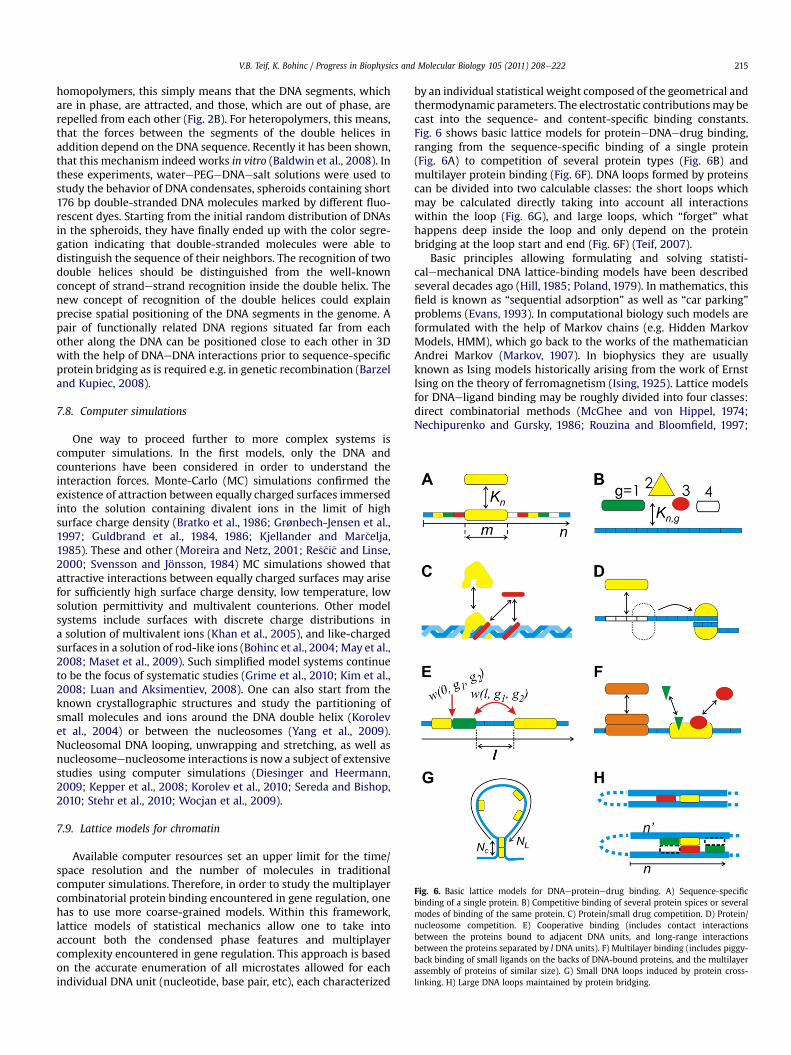
Fig. 6. Basic lattice models for DNAeproteinedrug binding. A) Sequence-specificbinding of a single protein. B) Competitive binding of several protein spices or severalmodes of binding of the same protein. C) Protein/small drug competition. D) Protein/nucleosome competition. E) Cooperative binding (includes contact interactionsbetween the proteins bound to adjacent DNA units, and long-range interactionsbetween the proteins separated by l DNA units). F) Multilayer binding (includes piggy-back binding of small ligands on the backs of DNA-bound proteins, and the multilayerassembly of proteins of similar size). G) Small DNA loops induced by protein cross-linking. H) Large DNA loops maintained by protein bridging.
V.B. Teif, K. Bohinc / Progress in Biophysics and Molecular Biology 105 (2011) 208e222 215
homopolymers, this simply means that the DNA segments, whichare in phase, are attracted, and those, which are out of phase, arerepelled from each other (Fig. 2B). For heteropolymers, this means,that the forces between the segments of the double helices inaddition depend on the DNA sequence. Recently it has been shown,that this mechanism indeed works in vitro (Baldwin et al., 2008). Inthese experiments, waterePEGeDNAesalt solutions were used tostudy the behavior of DNA condensates, spheroids containing short176 bp double-stranded DNA molecules marked by different fluo-rescent dyes. Starting from the initial random distribution of DNAsin the spheroids, they have finally ended up with the color segre-gation indicating that double-stranded molecules were able todistinguish the sequence of their neighbors. The recognition of twodouble helices should be distinguished from the well-knownconcept of strandestrand recognition inside the double helix. Thenew concept of recognition of the double helices could explainprecise spatial positioning of the DNA segments in the genome. Apair of functionally related DNA regions situated far from eachother along the DNA can be positioned close to each other in 3Dwith the help of DNAeDNA interactions prior to sequence-specificprotein bridging as is required e.g. in genetic recombination (Barzeland Kupiec, 2008).
7.8. Computer simulations
One way to proceed further to more complex systems iscomputer simulations. In the first models, only the DNA andcounterions have been considered in order to understand theinteraction forces. Monte-Carlo (MC) simulations confirmed theexistence of attraction between equally charged surfaces immersedinto the solution containing divalent ions in the limit of highsurface charge density (Bratko et al., 1986; Grønbech-Jensen et al.,1997; Guldbrand et al., 1984, 1986; Kjellander and Mar�celja,1985). These and other (Moreira and Netz, 2001; Re�s�ci�c and Linse,2000; Svensson and Jönsson, 1984) MC simulations showed thatattractive interactions between equally charged surfaces may arisefor sufficiently high surface charge density, low temperature, lowsolution permittivity and multivalent counterions. Other modelsystems include surfaces with discrete charge distributions ina solution of multivalent ions (Khan et al., 2005), and like-chargedsurfaces in a solution of rod-like ions (Bohinc et al., 2004; May et al.,2008; Maset et al., 2009). Such simplified model systems continueto be the focus of systematic studies (Grime et al., 2010; Kim et al.,2008; Luan and Aksimentiev, 2008). One can also start from theknown crystallographic structures and study the partitioning ofsmall molecules and ions around the DNA double helix (Korolevet al., 2004) or between the nucleosomes (Yang et al., 2009).Nucleosomal DNA looping, unwrapping and stretching, as well asnucleosomeenucleosome interactions is now a subject of extensivestudies using computer simulations (Diesinger and Heermann,2009; Kepper et al., 2008; Korolev et al., 2010; Sereda and Bishop,2010; Stehr et al., 2010; Wocjan et al., 2009).
7.9. Lattice models for chromatin
Available computer resources set an upper limit for the time/space resolution and the number of molecules in traditionalcomputer simulations. Therefore, in order to study the multiplayercombinatorial protein binding encountered in gene regulation, onehas to use more coarse-grained models. Within this framework,lattice models of statistical mechanics allow one to take intoaccount both the condensed phase features and multiplayercomplexity encountered in gene regulation. This approach is basedon the accurate enumeration of all microstates allowed for eachindividual DNA unit (nucleotide, base pair, etc), each characterized
by an individual statistical weight composed of the geometrical andthermodynamic parameters. The electrostatic contributionsmay becast into the sequence- and content-specific binding constants.Fig. 6 shows basic lattice models for proteineDNAedrug binding,ranging from the sequence-specific binding of a single protein(Fig. 6A) to competition of several protein types (Fig. 6B) andmultilayer protein binding (Fig. 6F). DNA loops formed by proteinscan be divided into two calculable classes: the short loops whichmay be calculated directly taking into account all interactionswithin the loop (Fig. 6G), and large loops, which “forget” whathappens deep inside the loop and only depend on the proteinbridging at the loop start and end (Fig. 6F) (Teif, 2007).
Basic principles allowing formulating and solving statisti-calemechanical DNA lattice-binding models have been describedseveral decades ago (Hill, 1985; Poland, 1979). In mathematics, thisfield is known as “sequential adsorption” as well as “car parking”problems (Evans, 1993). In computational biology such models areformulated with the help of Markov chains (e.g. Hidden MarkovModels, HMM), which go back to the works of the mathematicianAndrei Markov (Markov, 1907). In biophysics they are usuallyknown as Ising models historically arising from the work of ErnstIsing on the theory of ferromagnetism (Ising, 1925). Lattice modelsfor DNAeligand binding may be roughly divided into four classes:direct combinatorial methods (McGhee and von Hippel, 1974;Nechipurenko and Gursky, 1986; Rouzina and Bloomfield, 1997;
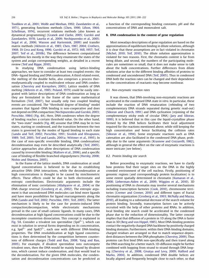
V.B. Teif, K. Bohinc / Progress in Biophysics and Molecular Biology 105 (2011) 208e222216
Tsodikov et al., 2001; Wolfe and Meehan, 1992; Zasedatelev et al.,1971), generating functions methods (Chen, 1990; Lifson, 1964;Schellman, 1974), recurrent relations methods (also known asdynamical programming) (Granek and Clarke, 2005; Gurskii andZasedatelev, 1978; Laurila et al., 2009; Nechipurenko et al., 2005;Segal et al., 2006; Wasson and Hartemink, 2009) and transfermatrix methods (Akhrem et al., 1985; Chen, 1987, 2004; Crothers,1968; Di Cera and Kong, 1996; Gurskii et al., 1972; Hill, 1957; Teif,2007; Teif et al., 2010b). The difference between computationalalgorithms lies mostly in the way they enumerate the states of thesystem and assign corresponding weights, as detailed in a recentreview (Teif and Rippe, 2010).
In studying DNA condensation using lattice-bindingapproaches, we have to consider at least two coupled events: theDNAeligand binding and DNA condensation. A third related event,the melting of the double helix, also comprises a process ther-modynamically coupled to multivalent release and DNA conden-sation (Cherstvy and Kornyshev, 2005). Lattice models of DNAmelting (Akhrem et al., 1985; Poland, 1979) could be easily inte-grated with lattice descriptions of DNA condensation as long asthey are formulated in the frame of the same mathematicalformalism (Teif, 2007), but usually only two coupled bindingevents are considered. The “threshold degree of binding” modelassumes that ligandeDNA binding is non-cooperative and doesnot depend on DNA compaction (Nechipurenko and Gurskii, 2003;Porschke, 1984) (Fig. 4A). Here, DNA condenses when the degreeof binding reaches a certain threshold value. On the other hand,the “two-state” models (Fig. 4B) assume that DNA may be in twostates, starting or condensed, and the transition between the twostates is governed by the modes of ligand binding to each state(Lando and Teif, 2002; Porschke, 1991; Sivolob and Khrapunov,1989; Teif, 2005; Teif and Lando, 2001a; Wittmer et al., 1995). Inthe case of non-specific reversible binding, DNA condensation/decondensation may even be described analytically (Teif, 2005).Lattice approaches also allow descriptions of DNA condensationcaused by irreversible binding (Maltsev et al., 2006), and a specificcase of binding of flexible branched oligopolymers (Horsky, 2008;Nishio and Shimizu, 2005).
In the frame of the lattice models, DNA condensation at smallligand concentrations is believed to be due to establishingattractive DNAeDNA interactions, while the decondensation athigh concentrations is thought to be caused by stoichiometriceffects. These effects could be due to both electrostatic andentropic contributions. Electrostatic arguments include theelimination of ionic correlations (Allahyarov et al., 2004) or theDNA charge reversal (Grosberg et al., 2002). The entropic argu-ment is that uncondensed DNA molecules can bind more ligandsand have more possibilities for ligand rearrangements along theDNA (Lando and Teif, 2002; Porschke, 1991; Teif, 2005). The lattermechanism is likely to be the case for protein-induced DNAcompaction/decompaction, while electrostatic mechanismsprevail in the case of small cationic ligands. In many cases, DNAdecondensation at high ligand concentrations could be due to theincomplete counterion dissociation. This concept is explained inFig. 5A. Consider a trivalent ion spermidine (Spd3þ), which canexist in solution in several forms depending on its concentration,e.g. Spd3þ and SpdCl2þ, each one with different DNA-bindingproperties. The DNA resolubilization at high ligand concentra-tions is then determined by the competition between counter-ions of different charges (Todd and Rau, 2008; Yang and Rau,2005). For example, if divalent spermidine ions outcompetetrivalent ones, then the DNA would be mainly bound by divalentions, which cannot induce condensation, and that would lead tothe decondensation. For the given DNA molecules, the conden-sation and decondensation concentrations can be predicted as
a function of the corresponding binding constants, pH and theconcentrations of DNA and monovalent salt.
8. DNA condensation in the context of gene regulation
Most nowadays descriptions of gene regulation are based on theapproximations of equilibrium binding in dilute solutions, althoughit is clear that these assumptions are in fact violated in chromatin(Michel, 2010; Teif, 2010). The dilute solution approximation isviolated for two reasons. First, the chromatin content is far frombeing dilute, and second, the numbers of the participating mole-cules are sometimes so small, that it does not make sense to talkabout the bulk concentrations. Further differences from dilutesolutions arise due to the different binding affinities of proteins tocondensed and uncondensed DNA (Teif, 2005). Thus in condensedDNA both the reaction rates can be changed and their dependenceon the concentrations of reactants may become nonlinear.
8.1. Non-enzymatic reaction rates
It was shown, that DNA-involving non-enzymatic reactions areaccelerated in the condensed DNA state in vitro. In particular, theseinclude the reaction of DNA renaturation (rebinding of twocomplementary DNA strands separated during genetic read-out)(Sikorav and Church, 1991) and DNA cyclization (rejoining of thecomplementary sticky ends of circular DNA) (Jary and Sikorav,1999). It is believed that in this case the liquid-crystalline phaseformed by the DNA helices facilitates their local adjustmentrequired for the reactions mentioned above, while still keeping thehigh concentration and hence facilitating the collision rates(Sikorav et al., 1994). Some enzymatic reactions such as DNAcatenation are also facilitated in the condensed DNA phase, prob-ably due to the same arguments (Krasnow and Cozzarelli, 1982),although in general the effect on the rate of enzymatic reactions ismore intricate (see below).
8.2. Protein binding site search
Before proceeding to enzymatic reactions, we have to clarifyhow proteins find their target sites on the DNA in the highlycrowded environment of the cell nucleus. Firstly, positioning ofgenomic regions (and correspondingly protein localization) is tosome extent spatially determined in chromatin (Kumaran et al.,2008; Lieberman-Aiden et al., 2009; Wiggins et al., 2010). 3Dpositioning of DNA in chromatin may involve several mechanismsincluding transcription factories (Cook, 2010), chromosome terri-tories (Cremer and Cremer, 2010) and different types of fractalchromatin organization (Grosberg et al., 1993; McNally and Mazza,2010), all leading to a substantial decrease of the search volume forprotein binding. Secondly, transcription factors can be activelyrecruited with the help of other proteins and RNAs. In addition,local binding site search could be accelerated in the condensedphase due to the reduction of dimensionality. The latter conceptimplies that that diffusion of a protein in 1D along the DNA is fasterthan in 3D (Berg and von Hippel, 1985). Most DNA-binding proteinscontact the negatively charged DNA backbone by positively chargedbinding domains. Furthermore, within their DNA-binding domains,charged residues are arranged so that to match sequence-depen-dent distances between the DNA phosphates (Cherstvy, 2009). Thusproteins can first bind DNA non-specifically and then travel alongthe DNA searching for a better match.1D-diffusionmight be furthercombined with hopping from strand to strand through DNA loop-ing (Cherstvy et al., 2008; Givatya and Levy, 2009; Halford andMarko, 2004). In addition, condensed DNA double helices arelocally aligned and frequently brought close to each other, so that
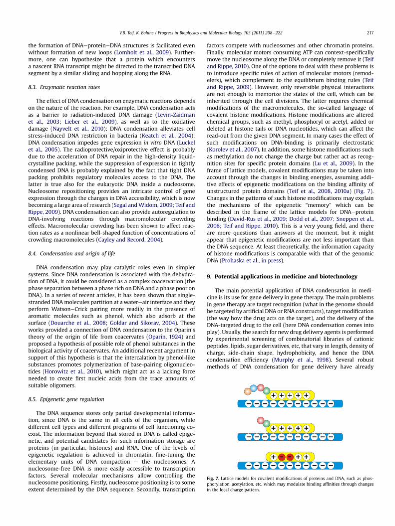
Fig. 7. Lattice models for covalent modifications of proteins and DNA, such as phos-phorylation, acetylation, etc, which may modulate binding affinities through changesin the local charge pattern.
V.B. Teif, K. Bohinc / Progress in Biophysics and Molecular Biology 105 (2011) 208e222 217
the formation of DNAeproteineDNA structures is facilitated evenwithout formation of new loops (Lomholt et al., 2009). Further-more, one can hypothesize that a protein which encountersa nascent RNA transcript might be directed to the transcribed DNAsegment by a similar sliding and hopping along the RNA.
8.3. Enzymatic reaction rates
The effect of DNA condensation on enzymatic reactions dependson the nature of the reaction. For example, DNA condensation actsas a barrier to radiation-induced DNA damage (Levin-Zaidmanet al., 2003; Lieber et al., 2009), as well as to the oxidativedamage (Nayvelt et al., 2010); DNA condensation alleviates cellstress-induced DNA restriction in bacteria (Keatch et al., 2004);DNA condensation impedes gene expression in vitro DNA (Luckelet al., 2005). The radioprotective/oxiprotective effect is probablydue to the acceleration of DNA repair in the high-density liquid-crystalline packing, while the suppression of expression in tightlycondensed DNA is probably explained by the fact that tight DNApacking prohibits regulatory molecules access to the DNA. Thelatter is true also for the eukaryotic DNA inside a nucleosome.Nucleosome repositioning provides an intricate control of geneexpression through the changes in DNA accessibility, which is nowbecoming a large area of research (Segal andWidom, 2009; Teif andRippe, 2009). DNA condensation can also provide autoregulation toDNA-involving reactions through macromolecular crowdingeffects. Macromolecular crowding has been shown to affect reac-tion rates as a nonlinear bell-shaped function of concentrations ofcrowding macromolecules (Cayley and Record, 2004).
8.4. Condensation and origin of life
DNA condensation may play catalytic roles even in simplersystems. Since DNA condensation is associated with the dehydra-tion of DNA, it could be considered as a complex coacervation (thephase separation between a phase rich on DNA and a phase poor onDNA). In a series of recent articles, it has been shown that single-stranded DNAmolecules partition at a watereair interface and theyperform WatsoneCrick pairing more readily in the presence ofaromatic molecules such as phenol, which also adsorb at thesurface (Douarche et al., 2008; Goldar and Sikorav, 2004). Theseworks provided a connection of DNA condensation to the Oparin’stheory of the origin of life from coacervates (Oparin, 1924) andproposed a hypothesis of possible role of phenol substances in thebiological activity of coacervates. An additional recent argument insupport of this hypothesis is that the intercalation by phenol-likesubstances promotes polymerization of base-pairing oligonucleo-tides (Horowitz et al., 2010), which might act as a lacking forceneeded to create first nucleic acids from the trace amounts ofsuitable oligomers.
8.5. Epigenetic gene regulation
The DNA sequence stores only partial developmental informa-tion, since DNA is the same in all cells of the organism, whiledifferent cell types and different programs of cell functioning co-exist. The information beyond that stored in DNA is called epige-netic, and potential candidates for such information storage areproteins (in particular, histones) and RNA. One of the levels ofepigenetic regulation is achieved in chromatin, fine-tuning theelementary units of DNA compaction e the nucleosomes. Anucleosome-free DNA is more easily accessible to transcriptionfactors. Several molecular mechanisms allow controlling thenucleosome positioning. Firstly, nucleosome positioning is to someextent determined by the DNA sequence. Secondly, transcription
factors compete with nucleosomes and other chromatin proteins.Finally, molecular motors consuming ATP can context-specificallymove the nucleosome along the DNA or completely remove it (Teifand Rippe, 2010). One of the options to deal with these problems isto introduce specific rules of action of molecular motors (remod-elers), which complement to the equilibrium binding rules (Teifand Rippe, 2009). However, only reversible physical interactionsare not enough to memorize the states of the cell, which can beinherited through the cell divisions. The latter requires chemicalmodifications of the macromolecules, the so-called language ofcovalent histone modifications. Histone modifications are alteredchemical groups, such as methyl, phosphoryl or acetyl, added ordeleted at histone tails or DNA nucleotides, which can affect theread-out from the given DNA segment. In many cases the effect ofsuch modifications on DNA-binding is primarily electrostatic(Korolev et al., 2007). In addition, some histone modifications suchas methylation do not change the charge but rather act as recog-nition sites for specific protein domains (Lu et al., 2009). In theframe of lattice models, covalent modifications may be taken intoaccount through the changes in binding energies, assuming addi-tive effects of epigenetic modifications on the binding affinity ofunstructured protein domains (Teif et al., 2008, 2010a) (Fig. 7).Changes in the patterns of such histone modifications may explainthe mechanisms of the epigenetic “memory” which can bedescribed in the frame of the lattice models for DNAeproteinbinding (David-Rus et al., 2009; Dodd et al., 2007; Sneppen et al.,2008; Teif and Rippe, 2010). This is a very young field, and thereare more questions than answers at the moment, but it mightappear that epigenetic modifications are not less important thanthe DNA sequence. At least theoretically, the information capacityof histone modifications is comparable with that of the genomicDNA (Prohaska et al., in press).
9. Potential applications in medicine and biotechnology
The main potential application of DNA condensation in medi-cine is its use for gene delivery in gene therapy. The main problemsin gene therapy are target recognition (what in the genome shouldbe targeted by artificial DNA or RNA constructs), targetmodification(the way how the drug acts on the target), and the delivery of theDNA-targeted drug to the cell (here DNA condensation comes intoplay). Usually, the search for new drug delivery agents is performedby experimental screening of combinatorial libraries of cationicpeptides, lipids, sugar derivatives, etc, that vary in length, density ofcharge, side-chain shape, hydrophobicity, and hence the DNAcondensation efficiency (Murphy et al., 1998). Several robustmethods of DNA condensation for gene delivery have already
Polymer properties
Polyelectrolyte properties
Solution properties
Collective properties
Looping, supercoiling
DNA liquid crystals
Coil-globule transition
Counterion condensation
Counterion correlations
DNA charge neutralization
DNA charge reversal
Positioning in 3D
DNA-DNA recognition
DNA-DNA attraction
1D binding lattice
Alleviated protein access
Changing reaction rates
Charge fractionalization
Macromolecular crowding
Incomplete ion dissociation
Facilitated site search Radio/oxidant protection
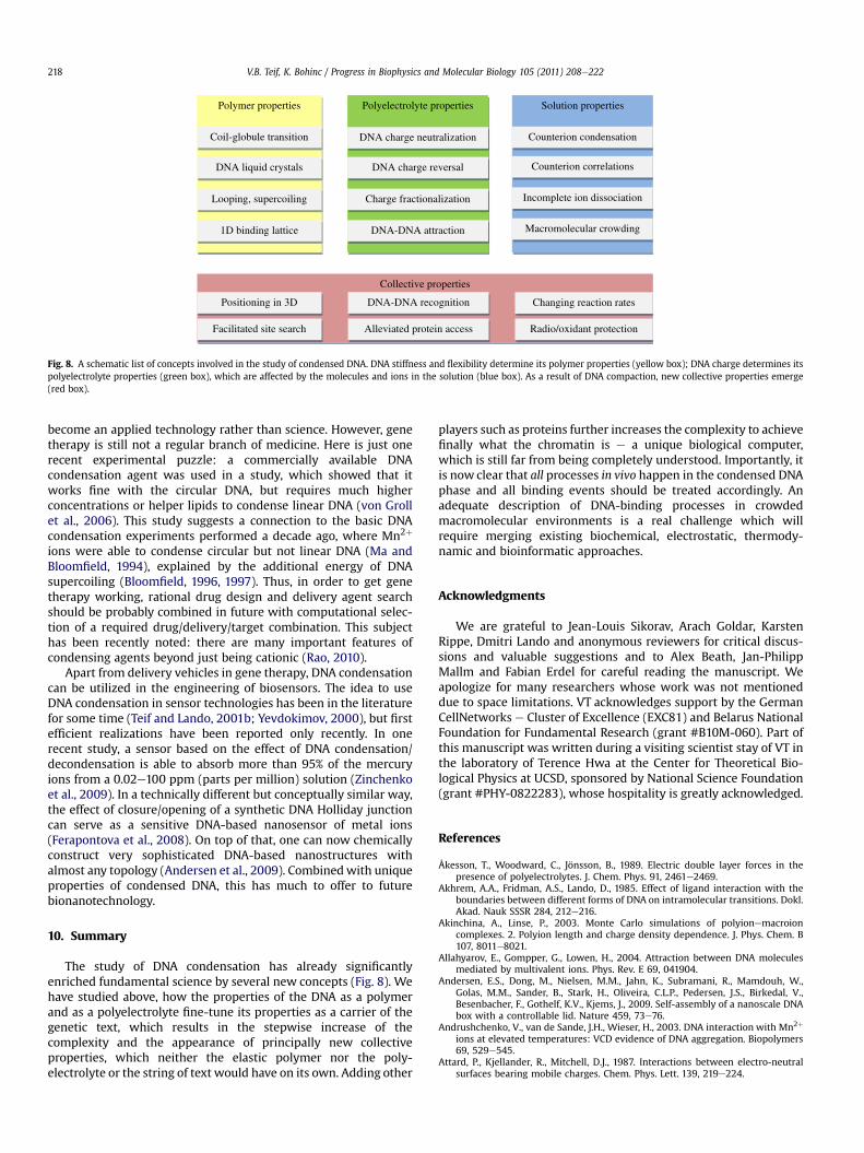
Fig. 8. A schematic list of concepts involved in the study of condensed DNA. DNA stiffness and flexibility determine its polymer properties (yellow box); DNA charge determines itspolyelectrolyte properties (green box), which are affected by the molecules and ions in the solution (blue box). As a result of DNA compaction, new collective properties emerge(red box).
V.B. Teif, K. Bohinc / Progress in Biophysics and Molecular Biology 105 (2011) 208e222218
become an applied technology rather than science. However, genetherapy is still not a regular branch of medicine. Here is just onerecent experimental puzzle: a commercially available DNAcondensation agent was used in a study, which showed that itworks fine with the circular DNA, but requires much higherconcentrations or helper lipids to condense linear DNA (von Grollet al., 2006). This study suggests a connection to the basic DNAcondensation experiments performed a decade ago, where Mn2þ
ions were able to condense circular but not linear DNA (Ma andBloomfield, 1994), explained by the additional energy of DNAsupercoiling (Bloomfield, 1996, 1997). Thus, in order to get genetherapy working, rational drug design and delivery agent searchshould be probably combined in future with computational selec-tion of a required drug/delivery/target combination. This subjecthas been recently noted: there are many important features ofcondensing agents beyond just being cationic (Rao, 2010).
Apart from delivery vehicles in gene therapy, DNA condensationcan be utilized in the engineering of biosensors. The idea to useDNA condensation in sensor technologies has been in the literaturefor some time (Teif and Lando, 2001b; Yevdokimov, 2000), but firstefficient realizations have been reported only recently. In onerecent study, a sensor based on the effect of DNA condensation/decondensation is able to absorb more than 95% of the mercuryions from a 0.02e100 ppm (parts per million) solution (Zinchenkoet al., 2009). In a technically different but conceptually similar way,the effect of closure/opening of a synthetic DNA Holliday junctioncan serve as a sensitive DNA-based nanosensor of metal ions(Ferapontova et al., 2008). On top of that, one can now chemicallyconstruct very sophisticated DNA-based nanostructures withalmost any topology (Andersen et al., 2009). Combinedwith uniqueproperties of condensed DNA, this has much to offer to futurebionanotechnology.
10. Summary
The study of DNA condensation has already significantlyenriched fundamental science by several new concepts (Fig. 8). Wehave studied above, how the properties of the DNA as a polymerand as a polyelectrolyte fine-tune its properties as a carrier of thegenetic text, which results in the stepwise increase of thecomplexity and the appearance of principally new collectiveproperties, which neither the elastic polymer nor the poly-electrolyte or the string of text would have on its own. Adding other
players such as proteins further increases the complexity to achievefinally what the chromatin is e a unique biological computer,which is still far from being completely understood. Importantly, itis now clear that all processes in vivo happen in the condensed DNAphase and all binding events should be treated accordingly. Anadequate description of DNA-binding processes in crowdedmacromolecular environments is a real challenge which willrequire merging existing biochemical, electrostatic, thermody-namic and bioinformatic approaches.
Acknowledgments
We are grateful to Jean-Louis Sikorav, Arach Goldar, KarstenRippe, Dmitri Lando and anonymous reviewers for critical discus-sions and valuable suggestions and to Alex Beath, Jan-PhilippMallm and Fabian Erdel for careful reading the manuscript. Weapologize for many researchers whose work was not mentioneddue to space limitations. VT acknowledges support by the GermanCellNetworks e Cluster of Excellence (EXC81) and Belarus NationalFoundation for Fundamental Research (grant #B10M-060). Part ofthis manuscript was written during a visiting scientist stay of VT inthe laboratory of Terence Hwa at the Center for Theoretical Bio-logical Physics at UCSD, sponsored by National Science Foundation(grant #PHY-0822283), whose hospitality is greatly acknowledged.
References
Åkesson, T., Woodward, C., Jönsson, B., 1989. Electric double layer forces in thepresence of polyelectrolytes. J. Chem. Phys. 91, 2461e2469.
Akhrem, A.A., Fridman, A.S., Lando, D., 1985. Effect of ligand interaction with theboundaries between different forms of DNA on intramolecular transitions. Dokl.Akad. Nauk SSSR 284, 212e216.
Akinchina, A., Linse, P., 2003. Monte Carlo simulations of polyionemacroioncomplexes. 2. Polyion length and charge density dependence. J. Phys. Chem. B107, 8011e8021.
Allahyarov, E., Gompper, G., Lowen, H., 2004. Attraction between DNA moleculesmediated by multivalent ions. Phys. Rev. E 69, 041904.
Andersen, E.S., Dong, M., Nielsen, M.M., Jahn, K., Subramani, R., Mamdouh, W.,Golas, M.M., Sander, B., Stark, H., Oliveira, C.L.P., Pedersen, J.S., Birkedal, V.,Besenbacher, F., Gothelf, K.V., Kjems, J., 2009. Self-assembly of a nanoscale DNAbox with a controllable lid. Nature 459, 73e76.
Andrushchenko, V., van de Sande, J.H., Wieser, H., 2003. DNA interaction with Mn2þ
ions at elevated temperatures: VCD evidence of DNA aggregation. Biopolymers69, 529e545.
Attard, P., Kjellander, R., Mitchell, D.J., 1987. Interactions between electro-neutralsurfaces bearing mobile charges. Chem. Phys. Lett. 139, 219e224.
V.B. Teif, K. Bohinc / Progress in Biophysics and Molecular Biology 105 (2011) 208e222 219
Attard, P., Kjellander, R., Mitchell, D.J., Jonsson, B., 1988a. Electrostatic fluctuationinteractions between neutral surfaces with adsorbed, mobile ions or dipoles.J. Chem. Phys. 89, 1664e1680.
Attard, P., Mitchell, D.J., Ninham, B.W., 1988b. Beyond PoissoneBoltzmann e imagesand correlations in the electric double-layer. 1. Counterions Only. J. Chem. Phys.88, 4987e4996.
Baldwin, G.S., Brooks, N.J., Robson, R.E., Wynveen, A., Goldar, A., Leikin, S.,Seddon, J.M., Kornyshev, A.A., 2008. DNA double helices recognize mutualsequence homology in a protein free environment. J. Phys. Chem. B 112,1060e1064.
Balhorn, R., 2007. The protamine family of spermnuclear proteins. GenomeBiol. 8, 227.Barzel, A., Kupiec, M., 2008. Finding a match: how do homologous sequences get
together for recombination? Nat. Rev. Genet. 9, 27e37.Baumann, C.G., Bloomfield, V.A., Smith, S.B., Bustamante, C., Wang, M.D., Block, S.M.,
2000. Stretching of single collapsed DNA molecules. Biophys. J. 78, 1965e1978.Becker, N.A., Kahn, J.D., Maher III, L.J., 2008. Eukaryotic HMGB proteins as
replacements for HU in E. coli repression loop formation. Nucl. Acids Res. 36,4009e4021.
Berg, O.G., von Hippel, P.H., 1985. Diffusion-controlled macromolecular interactions.Ann. Rev. Biophys. Biophys. Chem. 14, 131e160.
Besteman, K., Van Eijk, K., Lemay, S.G., 2007. Charge inversion accompanies DNAcondensation by multivalent ions. Nat. Phys. 3, 641e644.
Birshtein, T.M., Ptitsyn, O.B., 1964. Conformations ofMacromolecules. Nauka, Moscow.Bloomfield, V.A., 1996. DNA condensation. Curr. Opin. Struct. Biol. 6, 334e341.Bloomfield, V.A., 1997. DNA condensation by multivalent cations. Biopolymers 44,
269e282.Bloomfield, V.A., Crothers, D.M., Tinoco, I.J., 2000. Nucleic Acids. Structure, Prop-
erties and Functions. University Science Books, Sausalito.Bohinc, K., Iglic, A., May, S., 2004. Interaction between macroions mediated by
divalent rod-like ions. Europhys. Lett. 68, 494e500.Bohinc, K., Re�s�ci�c, J., Maset, S., May, S., 2011. DebyeeHückel theory for mixtures of
rigid rodlike ions and salt. J. Chem. Phys. 134, 074111e074119.Bratko, D., Jönsson, B., Wennerström, H., 1986. Electrical double layer interactions
with image charges. Chem. Phys. Lett. 128, 449e454.Brinkers, S., Dietrich, H.R.C., Groote, F.H.d., Young, I.T., Rieger, B., 2009. The persis-
tence length of double stranded DNA determined using dark field tetheredparticle motion. J. Chem. Phys. 130, 215105.
Bruinsma, R.F., Riehn, R., 2009. DNA condensation by field-induced non-equilibriumnoise. Chemphyschem 10, 2871e2875.
Burak, Y., Ariel, G., Andelman, D., 2003. Onset of DNA aggregation in presence ofmonovalent and multivalent counterions. Biophys. J. 85, 2100e2110.
Cayley, S.M., Record Jr., M.T., 2004. Large changes in cytoplasmic biopolymerconcentration with osmolality indicate that macromolecular crowding mayregulate proteineDNA interactions and growth rate in osmotically stressedEscherichia coli K-12. J. Mol. Recogn. 17, 488e496.
Chen, Y., 1987. Binding of n-mers to one-dimensional lattices with longer thanclose-contact interactions. Biophys. Chem. 27, 59e65.
Chen, Y.D., 1990. A general secular equation for cooperative binding of n-merligands to a one-dimensional lattice. Biopolymers 30, 1113e1121.
Chen, Y.D., 2004. Multiple binding of ligands to a linear biopolymer. Meth. Enzymol.379, 145e152.
Chen, N., Zinchenko, A.A., Kidoaki, S., Murata, S., Yoshikawa, K., 2010. Thermo-switching of the conformation of genomic DNA in solutions of poly(N-iso-propylacrylamide). Langmuir 26, 2995e2998.
Cherstvy, A.G., 2009. Positively charged residues in DNA-binding domains ofstructural proteins follow sequence-specific positions of DNA phosphategroups. J. Phys. Chem. B 113, 4242e4247.
Cherstvy, A.G., Kolomeisky, A.B., Kornyshev, A.A., 2008. ProteineDNA interactions:reaching and recognizing the targets. J. Phys. Chem. B 112, 4741e4750.
Cherstvy, A.G., Kornyshev, A.A., 2005. DNA melting in aggregates: impeded orfacilitated? J. Phys. Chem. B 109, 13024e13029.
Chien, F.T., Lin, S.G., Lai, P.Y., Chan, C.K., 2007. Observation of two forms of confor-mations in the reentrant condensation of DNA. Phys. Rev. E Stat. Nonlin. SoftMatter Phys. 75, 041922.
Chow, M., Yan, K., Bennett, M., Wong, J., 2010. Liquid crystalline chromosomes:birefringence and DNA condensation. Eukaryot. Cell Published online ahead ofprint on 16 April 2010.
Cook, P.R., 2010. A model for all genomes: the role of transcription factories. J. Mol.Biol. 395, 1e10.
Corpet, A., Almouzni, G., 2009. Making copies of chromatin: the challenge ofnucleosomal organization and epigenetic information. Trends Cell. Biol. 19,29e41.
Cozzarelli, N.R., Cost, G.J., Nollmann, M., Viard, T., Stray, J.E., 2006. Giant proteinsthat move DNA: bullies of the genomic playground. Nat. Rev. Mol. Cell Biol. 7,580e588.
Cremer, T., Cremer, M., 2010. Chromosome territories. Cold Spring Harb. Perspect.Biol. 2, a003889.
Crothers, D.M., 1968. Calculation of binding isotherms for heterogeneous polymers.Biopolymers 6, 575e584.
Cunha, S., Odijk, T., Suleymanoglu, E., Woldringh, C.L., 2001. Isolation of theEscherichia coli nucleoid. Biochimie 83, 149e154.
Dame, R.T., 2005. The role of nucleoid-associated proteins in the organization andcompaction of bacterial chromatin. Mol. Microbiol. 56, 858e870.
David-Rus, D., Mukhopadhyay, S., Lebowitz, J.L., Sengupta, A.M., 2009. Inheritance ofepigenetic chromatin silencing. J. Theor. Biol. 258, 112e120.
Di Cera, E., Kong, Y., 1996. Theory of multivalent binding in one and two-dimen-sional lattices. Biophys. Chem. 61, 107e124.
Dias, R., Lindman, B., 2008. DNA Interactions with Polymers and Surfactants. Wiley,London, pp. 408.
Diehl, A., Tamashiro, M.N., Barbosa, M.C., Levin, Y., 1999. Density-functional theoryfor attraction between like-charged plates. Physica A 274, 433e445.
Diesinger, P.M., Heermann, D.W., 2009. Depletion effects massively change chro-matin properties and influence genome folding. Biophys. J. 97, 2146e2153.
Dodd, I.B., Micheelsen, M.A., Sneppen, K., Thon, G., 2007. Theoretical analysis ofepigenetic cell memory by nucleosome modification. Cell 129, 813e822.
Douarche, C., Sikorav, J.L., Goldar, A., 2008. Aggregation and adsorption at the airewaterinterface of bacteriophage phiX174 single-stranded DNA. Biophys. J. 94, 134e146.
Earnshaw, W.C., Harrison, S.C., 1977. DNA arrangement in isometric phage heads.Nature 268, 598e602.
Evans, J.W., 1993. Random and cooperative sequential adsorption. Rev. Mod. Phys.65, 1281e1329.
Evans, D.F., Wennerström, H., 1994. The Colloidal Domain, Where Physics, Chem-istry, Biology and Technology Meet. VCH Publishers, New York.
Evdokimov, Y.M., Platonov, A.L., Tikhonenko, A.S., Varshavsky, Y.M., 1972. A compactform of double-stranded DNA in solution. FEBS Lett. 23, 180e184.
Evilevitch, A., Fang, L.T., Yoffe, A.M., Castelnovo, M., Rau, D.C., Parsegian, V.A.,Gelbart, W.M., Knobler, C.M., 2008. Effects of salt concentrations and bendingenergy on the extent of ejection of phage genomes. Biophys. J. 94, 1110e1120.
Fang, F.C., Rimsky, S., 2008. New insights into transcriptional regulation by HeNS.Curr. Opin. Microbiol. 11, 113e120.
Ferapontova, E.E., Mountford, C.P., Crain, J., Buck, A.H., Dickinson, P., Beattie, J.S.,Ghazal, P., Terry, J.G., Walton, A.J., Mount, A.R., 2008. Electrochemical control ofa DNA Holliday Junction nanoswitch by Mg2þ ions. Biosens. Bioelectron. 24,422e428.
Flory, P.J., 1969. Statistical Mechanics of Chain Molecules. Wiley (Interscience), NewYork.
Forsman, J., 2004. A simple correlation-corrected PoissoneBoltzmann theory.J. Phys. Chem. B 108, 9236e9245.
Friddle, R.W., Klare, J.E., Martin, S.S., Corzett, M., Balhorn, R., Baldwin, E.P.,Baskin, R.J., Noy, A., 2004. Mechanism of DNA compaction by yeast mitochon-drial protein Abf2p. Biophys. J. 86, 1632e1639.
Gelbart, W.M., Bruinsma, R., Pincus, P.A., Parsegian, V.A., 2000. DNA-inspired elec-trostatics. Phys. Today 53, 38.
Gerlitz, G., Hock, R., Ueda, T., Bustin,M., 2009. Thedynamics ofHMGproteinechromatininteractions in living cells. Biochem. Cell Biol. 87, 127e137.
Givatya, O., Levy, Y., 2009. Protein sliding along DNA: dynamics and structuralcharacterization. J. Mol. Biol. 385, 1087e1097.
Goldar, A., Sikorav, J.L., 2004. DNA renaturation at the waterephenol interface. Eur.Phys. J. E 14, 211e239.
Gosule, L.C., Shellman, J.A., 1976. Compact form of DNA induced by spermidine.Nature 259, 333e335.
Granek, J.A., Clarke, N.D., 2005. Explicit equilibrium modeling of transcription-factor binding and gene regulation. Genome Biol. 6, R87.
Grayson, P., Han, L., Winther, T., Phillips, R., 2007. Real-time observations of singlebacteriophage lambda DNA ejections in vitro. Proc. Natl. Acad. Sci. U S A 104,14652e14657.
Grime, J.M.A., Khan, M.O., Bohinc, K., 2010. Interaction between charged surfacesmediated by rodlike counterions: the influence of discrete charge distributionin the solution and on the surfaces. Langmuir 26, 6343e6349.
von Groll, A., Levin, Y., Barbosa, M.C., Ravazzolo, A.P., 2006. Linear DNA low effi-ciency transfection by liposome can be improved by the use of cationic lipid ascharge neutralizer. Biotechnol. Prog. 22, 1220e1224.
Grosberg, A., et al., 1993. Crumpled globule model of the three-dimensionalstructure of DNA. Europhys. Lett. 23, 373.
Grosberg, A.Y., Nguyen, T.T., Shklovskii, B.I., 2002. The physics of charge inversion inchemical and biological systems. Rev. Mod. Phys. 74, 329e345.
Grosberg, A.Y., Khokhlov, A.R., 1994. Statistical Physics of Macromolecules, pp. 187.AIP Press, New York.
Grosberg, A., Zhestkov, A.V., 1985. On the toroidal condensed state of closed circularDNA. J. Biomol. Struct. Dyn. 3, 515e520.
Grosberg, A., Zhestkov, A.V., 1986. On the compact form of linear duplex DNA:globular states of the uniform elastic (persistent) macromolecule. J. Biomol.Struct. Dyn. 3, 859e872.
Grønbech-Jensen, N., Mashl, R.J., Bruinsma, R.F., Gelbart, W.M., 1997. Counterion-induced attraction between rigid polyelectrolytes. Phys. Rev. Lett. 78, 2477.
Guldbrand, L., Jonsson, B., Wennerstrom, H., Linse, P., 1984. Electrical double layerforces. A Monte Carlo study. J. Chem. Phys. 80, 2221e2228.
Guldbrand, L., Nilsson, L.G., Nordenskiold, L., 1986. A Monte Carlo simulation studyof electrostatic forces between hexagonally packed DNA double helices.J. Chem. Phys. 85, 6686e6698.
Guo, Z., Taubes, C.H., Oh, J.-E., Maher III, L.J., Mohanty, U., 2008. DNA on a tube:electrostatic contribution to stiffness. J. Phys. Chem. B 112, 16163e16169.
Gurskii, G.V., Zasedatelev, A.S., Vol’kenshtein, M.V., 1972. Theory of one-dimen-sional absorption. II. Adsorption of small molecules on a heteropolymer. Mol.Biol. 6, 385e393.
Gurskii, G.V., Zasedatelev, A.S., 1978. Precise relationships for calculating thebinding of regulatory proteins and other lattice ligands in double-strandedpolynucleotides. Biofizika 23, 932e946.
Halford, S.E., Marko, J.F., 2004. How do site-specific DNA-binding proteins find theirtargets? Nucleic Acids Res. 32, 3040e3052.
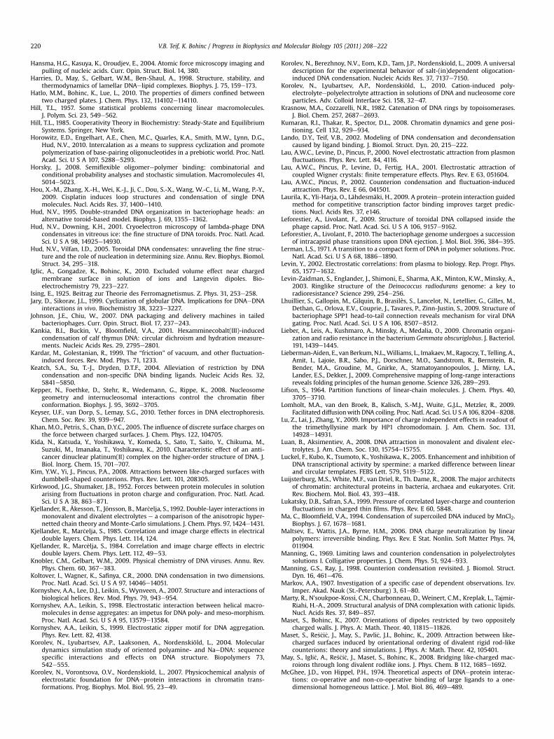
V.B. Teif, K. Bohinc / Progress in Biophysics and Molecular Biology 105 (2011) 208e222220
Hansma, H.G., Kasuya, K., Oroudjev, E., 2004. Atomic force microscopy imaging andpulling of nucleic acids. Curr. Opin. Struct. Biol. 14, 380.
Harries, D., May, S., Gelbart, W.M., Ben-Shaul, A., 1998. Structure, stability, andthermodynamics of lamellar DNAelipid complexes. Biophys. J. 75, 159e173.
Hatlo, M.M., Bohinc, K., Lue, L., 2010. The properties of dimers confined betweentwo charged plates. J. Chem. Phys. 132, 114102e114110.
Hill, T.L., 1957. Some statistical problems concerning linear macromolecules.J. Polym. Sci. 23, 549e562.
Hill, T.L., 1985. Cooperativity Theory in Biochemistry: Steady-State and EquilibriumSystems. Springer, New York.
Horowitz, E.D., Engelhart, A.E., Chen, M.C., Quarles, K.A., Smith, M.W., Lynn, D.G.,Hud, N.V., 2010. Intercalation as a means to suppress cyclization and promotepolymerization of base-pairing oligonucleotides in a prebiotic world. Proc. Natl.Acad. Sci. U S A 107, 5288e5293.
Horsky, J., 2008. Semiflexible oligomerepolymer binding: combinatorial andconditional probability analyses and stochastic simulation. Macromolecules 41,5014e5023.
Hou, X.-M., Zhang, X.-H., Wei, K.-J., Ji, C., Dou, S.-X., Wang, W.-C., Li, M., Wang, P.-Y.,2009. Cisplatin induces loop structures and condensation of single DNAmolecules. Nucl. Acids Res. 37, 1400e1410.
Hud, N.V., 1995. Double-stranded DNA organization in bacteriophage heads: analternative toroid-based model. Biophys. J. 69, 1355e1362.
Hud, N.V., Downing, K.H., 2001. Cryoelectron microscopy of lambda-phage DNAcondensates in vitreous ice: the fine structure of DNA toroids. Proc. Natl. Acad.Sci. U S A 98, 14925e14930.
Hud, N.V., Vilfan, I.D., 2005. Toroidal DNA condensates: unraveling the fine struc-ture and the role of nucleation in determining size. Annu. Rev. Biophys. Biomol.Struct. 34, 295e318.
Iglic, A., Gongadze, K., Bohinc, K., 2010. Excluded volume effect near chargedmembrane surface in solution of ions and Langevin dipoles. Bio-electrochemistry 79, 223e227.
Ising, E., 1925. Beitrag zur Theorie des Ferromagnetismus. Z. Phys. 31, 253e258.Jary, D., Sikorav, J.L., 1999. Cyclization of globular DNA. Implications for DNAeDNA
interactions in vivo. Biochemistry 38, 3223e3227.Johnson, J.E., Chiu, W., 2007. DNA packaging and delivery machines in tailed
bacteriophages. Curr. Opin. Struct. Biol. 17, 237e243.Kankia, B.I., Buckin, V., Bloomfield, V.A., 2001. Hexamminecobalt(III)-induced
condensation of calf thymus DNA: circular dichroism and hydration measure-ments. Nucleic Acids Res. 29, 2795e2801.
Kardar, M., Golestanian, R., 1999. The “friction” of vacuum, and other fluctuation-induced forces. Rev. Mod. Phys. 71, 1233.
Keatch, S.A., Su, T.-J., Dryden, D.T.F., 2004. Alleviation of restriction by DNAcondensation and non-specific DNA binding ligands. Nucleic Acids Res. 32,5841e5850.
Kepper, N., Foethke, D., Stehr, R., Wedemann, G., Rippe, K., 2008. Nucleosomegeometry and internucleosomal interactions control the chromatin fiberconformation. Biophys. J. 95, 3692e3705.
Keyser, U.F., van Dorp, S., Lemay, S.G., 2010. Tether forces in DNA electrophoresis.Chem. Soc. Rev. 39, 939e947.
Khan, M.O., Petris, S., Chan, D.Y.C., 2005. The influence of discrete surface charges onthe force between charged surfaces. J. Chem. Phys. 122, 104705.
Kida, N., Katsuda, Y., Yoshikawa, Y., Komeda, S., Sato, T., Saito, Y., Chikuma, M.,Suzuki, M., Imanaka, T., Yoshikawa, K., 2010. Characteristic effect of an anti-cancer dinuclear platinum(II) complex on the higher-order structure of DNA. J.Biol. Inorg. Chem. 15, 701e707.
Kim, Y.W., Yi, J., Pincus, P.A., 2008. Attractions between like-charged surfaces withdumbbell-shaped counterions. Phys. Rev. Lett. 101, 208305.
Kirkwood, J.G., Shumaker, J.B., 1952. Forces between protein molecules in solutionarising from fluctuations in proton charge and configuration. Proc. Natl. Acad.Sci. U S A 38, 863e871.
Kjellander, R., Åkesson, T., Jönsson, B., Mar�celja, S., 1992. Double-layer interactions inmonovalent and divalent electrolytes e a comparison of the anisotropic hyper-netted chain theory and Monte-Carlo simulations. J. Chem. Phys. 97, 1424e1431.
Kjellander, R., Mar�celja, S., 1985. Correlation and image charge effects in electricaldouble layers. Chem. Phys. Lett. 114, 124.
Kjellander, R., Marc�elja, S., 1984. Correlation and image charge effects in electricdouble layers. Chem. Phys. Lett. 112, 49e53.
Knobler, C.M., Gelbart, W.M., 2009. Physical chemistry of DNA viruses. Annu. Rev.Phys. Chem. 60, 367e383.
Koltover, I., Wagner, K., Safinya, C.R., 2000. DNA condensation in two dimensions.Proc. Natl. Acad. Sci. U S A 97, 14046e14051.
Kornyshev, A.A., Lee, D.J., Leikin, S., Wynveen, A., 2007. Structure and interactions ofbiological helices. Rev. Mod. Phys. 79, 943e954.
Kornyshev, A.A., Leikin, S., 1998. Electrostatic interaction between helical macro-molecules in dense aggregates: an impetus for DNA poly- and meso-morphism.Proc. Natl. Acad. Sci. U S A 95, 13579e13584.
Kornyshev, A.A., Leikin, S., 1999. Electrostatic zipper motif for DNA aggregation.Phys. Rev. Lett. 82, 4138.
Korolev, N., Lyubartsev, A.P., Laaksonen, A., Nordenskiöld, L., 2004. Moleculardynamics simulation study of oriented polyamine- and NaeDNA: sequencespecific interactions and effects on DNA structure. Biopolymers 73,542e555.
Korolev, N., Vorontsova, O.V., Nordenskiold, L., 2007. Physicochemical analysis ofelectrostatic foundation for DNAeprotein interactions in chromatin trans-formations. Prog. Biophys. Mol. Biol. 95, 23e49.
Korolev, N., Berezhnoy, N.V., Eom, K.D., Tam, J.P., Nordenskiold, L., 2009. A universaldescription for the experimental behavior of salt-(in)dependent oligocation-induced DNA condensation. Nucleic Acids Res. 37, 7137e7150.
Korolev, N., Lyubartsev, A.P., Nordenskiöld, L., 2010. Cation-induced poly-electrolyteepolyelectrolyte attraction in solutions of DNA and nucleosome coreparticles. Adv. Colloid Interface Sci. 158, 32e47.
Krasnow, M.A., Cozzarelli, N.R., 1982. Catenation of DNA rings by topoisomerases.J. Biol. Chem. 257, 2687e2693.
Kumaran, R.I., Thakar, R., Spector, D.L., 2008. Chromatin dynamics and gene posi-tioning. Cell 132, 929e934.
Lando, D.Y., Teif, V.B., 2002. Modeling of DNA condensation and decondensationcaused by ligand binding. J. Biomol. Struct. Dyn. 20, 215e222.
Lau, A.W.C., Levine, D., Pincus, P., 2000. Novel electrostatic attraction from plasmonfluctuations. Phys. Rev. Lett. 84, 4116.
Lau, A.W.C., Pincus, P., Levine, D., Fertig, H.A., 2001. Electrostatic attraction ofcoupled Wigner crystals: finite temperature effects. Phys. Rev. E 63, 051604.
Lau, A.W.C., Pincus, P., 2002. Counterion condensation and fluctuation-inducedattraction. Phys. Rev. E 66, 041501.
Laurila, K., Yli-Harja, O., Lähdesmäki, H., 2009. A proteineprotein interaction guidedmethod for competitive transcription factor binding improves target predic-tions. Nucl. Acids Res. 37, e146.
Leforestier, A., Livolant, F., 2009. Structure of toroidal DNA collapsed inside thephage capsid. Proc. Natl. Acad. Sci. U S A 106, 9157e9162.
Leforestier, A., Livolant, F., 2010. The bacteriophage genome undergoes a successionof intracapsid phase transitions upon DNA ejection. J. Mol. Biol. 396, 384e395.
Lerman, L.S., 1971. A transition to a compact form of DNA in polymer solutions. Proc.Natl. Acad. Sci. U S A 68, 1886e1890.
Levin, Y., 2002. Electrostatic correlations: from plasma to biology. Rep. Progr. Phys.65, 1577e1632.
Levin-Zaidman, S., Englander, J., Shimoni, E., Sharma, A.K., Minton, K.W., Minsky, A.,2003. Ringlike structure of the Deinococcus radiodurans genome: a key toradioresistance? Science 299, 254e256.
Lhuillier, S., Gallopin, M., Gilquin, B., Brasilès, S., Lancelot, N., Letellier, G., Gilles, M.,Dethan, G., Orlova, E.V., Couprie, J., Tavares, P., Zinn-Justin, S., 2009. Structure ofbacteriophage SPP1 head-to-tail connection reveals mechanism for viral DNAgating. Proc. Natl. Acad. Sci. U S A 106, 8507e8512.
Lieber, A., Leis, A., Kushmaro, A., Minsky, A., Medalia, O., 2009. Chromatin organi-zation and radio resistance in the bacterium Gemmata obscuriglobus. J. Bacteriol.191, 1439e1445.
Lieberman-Aiden, E., vanBerkum,N.L.,Williams, L., Imakaev,M., Ragoczy, T., Telling, A.,Amit, I., Lajoie, B.R., Sabo, P.J., Dorschner, M.O., Sandstrom, R., Bernstein, B.,Bender, M.A., Groudine, M., Gnirke, A., Stamatoyannopoulos, J., Mirny, L.A.,Lander, E.S., Dekker, J., 2009. Comprehensive mapping of long-range interactionsreveals folding principles of the human genome. Science 326, 289e293.
Lifson, S., 1964. Partition functions of linear-chain molecules. J. Chem. Phys. 40,3705e3710.
Lomholt, M.A., van den Broek, B., Kalisch, S.-M.J., Wuite, G.J.L., Metzler, R., 2009.Facilitated diffusionwith DNA coiling. Proc. Natl. Acad. Sci. U S A 106, 8204e8208.
Lu, Z., Lai, J., Zhang, Y., 2009. Importance of charge independent effects in readout ofthe trimethyllysine mark by HP1 chromodomain. J. Am. Chem. Soc. 131,14928e14931.
Luan, B., Aksimentiev, A., 2008. DNA attraction in monovalent and divalent elec-trolytes. J. Am. Chem. Soc. 130, 15754e15755.
Luckel, F., Kubo, K., Tsumoto, K., Yoshikawa, K., 2005. Enhancement and inhibition ofDNA transcriptional activity by spermine: a marked difference between linearand circular templates. FEBS Lett. 579, 5119e5122.
Luijsterburg, M.S., White, M.F., van Driel, R., Th. Dame, R., 2008. The major architectsof chromatin: architectural proteins in bacteria, archaea and eukaryotes. Crit.Rev. Biochem. Mol. Biol. 43, 393e418.
Lukatsky, D.B., Safran, S.A., 1999. Pressure of correlated layer-charge and counterionfluctuations in charged thin films. Phys. Rev. E 60, 5848.
Ma, C., Bloomfield, V.A., 1994. Condensation of supercoiled DNA induced by MnCl2.Biophys. J. 67, 1678e1681.
Maltsev, E., Wattis, J.A., Byrne, H.M., 2006. DNA charge neutralization by linearpolymers: irreversible binding. Phys. Rev. E Stat. Nonlin. Soft Matter Phys. 74,011904.
Manning, G., 1969. Limiting laws and counterion condensation in polyelectrolytessolutions I. Colligative properties. J. Chem. Phys. 51, 924e933.
Manning, G.S., Ray, J., 1998. Counterion condensation revisited. J. Biomol. Struct.Dyn. 16, 461e476.
Markov, A.A., 1907. Investigation of a specific case of dependent observations. Izv.Imper. Akad. Nauk (St.-Petersburg) 3, 61e80.
Marty, R., N’soukpoe-Kossi, C.N., Charbonneau, D., Weinert, C.M., Kreplak, L., Tajmir-Riahi, H.-A., 2009. Structural analysis of DNA complexation with cationic lipids.Nucl. Acids Res. 37, 849e857.
Maset, S., Bohinc, K., 2007. Orientations of dipoles restricted by two oppositelycharged walls. J. Phys. A: Math. Theor. 40, 11815e11826.
Maset, S., Re�s�ci�c, J., May, S., Pavli�c, J.I., Bohinc, K., 2009. Attraction between like-charged surfaces induced by orientational ordering of divalent rigid rod-likecounterions: theory and simulations. J. Phys. A: Math. Theor. 42, 105401.
May, S., Igli�c, A., Re�s�ci�c, J., Maset, S., Bohinc, K., 2008. Bridging like-charged mac-roions through long divalent rodlike ions. J. Phys. Chem. B 112, 1685e1692.
McGhee, J.D., von Hippel, P.H., 1974. Theoretical aspects of DNAeprotein interac-tions: co-operative and non-co-operative binding of large ligands to a one-dimensional homogeneous lattice. J. Mol. Biol. 86, 469e489.
V.B. Teif, K. Bohinc / Progress in Biophysics and Molecular Biology 105 (2011) 208e222 221
McLaughlin, S., 1989. The electrostatic properties of membranes. Annu. Rev. Bio-phys. Biophys. Chem. 18, 113e136.
McNally, J.G., Mazza, D., 2010. Fractal geometry in the nucleus. EMBO J. 29, 2e3.Meistermann, I., Moreno, V., Prieto, M.J., Moldrheim, E., Sletten, E., Khalid, S.,
Rodger, P.M., Peberdy, J.C., Isaac, C.J., Rodger, A., Hannon, M.J., 2002. Intra-molecular DNA coiling mediated by metallo-supramolecular cylinders: differ-ential binding of P and M helical enantiomers. Proc. Natl. Acad. Sci. U S A 99,5069e5074.
Mengistu, D.H., Bohinc, K., May, S., 2009. Binding of DNA to zwitterionic lipid layersmediated by divalent cations. J. Phys. Chem. B 113, 12277e12282.
Michel, D., 2010. How transcription factors can adjust the gene expression flood-gates. Prog. Biophys. Mol. Biol. 102, 16e37.
Miklavic, S.J., Woodward, C.E., Jösson, B., Åkesson, T., 1990. The interaction ofcharged surfaces with grafted polyelectrolytes: a PoissoneBoltzmann andMonte Carlo study. Macromolecules 23, 4149e4157.
Moreira, A.G., Netz, R.R., 2001. Binding of similarly charged plates with counterionsonly. Phys. Rev. Lett. 87, 078301.
Müller, K.P., Erdel, F., Caudron-Herger, M., Marth, C., Fodor, B.D., Richter, M.,Scaranaro, M., Beaudouin, J., Wachsmuth, M., Rippe, K., 2009. Multiscale anal-ysis of dynamics and interactions of heterochromatin protein 1 by fluorescencefluctuation microscopy. Biophys. J. 97, 2876e2885.
Murayama, Y., Sakamaki, Y., Sano, M., 2003. Elastic response of single DNA mole-cules exhibits a reentrant collapsing transition. Phys. Rev. Lett. 90, 018102.
Murphy, J.E., Uno, T., Hamer, J.D., Cohen, F.E., Dwarki, V., Zuckermann, R.N., 1998. Acombinatorial approach to the discovery of efficient cationic peptoid reagentsfor gene delivery. Proc. Natl. Acad. Sci. U S A 95, 1517e1522.
Naji, A., Arnold, A., Holm, C., Netz, R.R., 2004. Attraction and unbinding of like-charged rods. Europhys. Lett. 67, 130e136.
Navarre, W.W., Porwollik, S., Wang, Y., McClelland, M., Rosen, H., Libby, S.J.,Fang, F.C., 2006. Selective silencing of foreign DNA with low GC content by theHeNS protein in salmonella. Science 313, 236e238.
Nayvelt, I., Hyvönen, M.T., Alhonen, L., Pandya, I., Thomas, T., Khomutov, A.R.,Vepsäläinen, J., Patel, R., Keinänen, T.A., Thomas, T.J., 2010. DNA condensation bychiral a-methylated polyamine analogues and protection of cellular DNA fromoxidative damage. Biomacromolecules 11, 97e105.
Nechipurenko, Y.D., Jovanovic, B., Riabokon, V.F., Gursky, G.V., 2005. Quantitativemethods of analysis of footprinting diagrams for the complexes formed by a ligandwith a DNA fragment of known sequence. Ann. N Y Acad. Sci. 1048, 206e214.
Nechipurenko, Y.D., Gurskii, G.V., 2003. Thermodynamic models of binding ligandsto nucleic acids. Biofizika 48, 773e796.
Nechipurenko, Y.D., Gursky, G.V., 1986. Cooperative effects on binding of proteins toDNA. Biophys. Chem. 24, 195e209.
Netz, R.R., Orland, H., 2000. One and two-component hard-core plasmas. Eur. Phys.J. E 1, 67e73.
Nishio, T., Shimizu, T., 2005. Model analysis of surfactantepolymer interaction ascooperative ligand binding to linear lattice. Biophys. Chem. 117, 19e25.
Ohlsson, R., Lobanenkov, V., Klenova, E., 2010. Does CTCF mediate between nuclearorganization and gene expression? BioEssays 32, 37e50.
Oosawa, F., 1968. Interaction between parallel rodlike macroions. Biopolymers 6,1633e1647.
Oparin, A.I., 1924. The origin of life, Mosk. Rabochij., Moskow. In: Carrington, R.(Ed.), The Origin of Life. The Widenfeld and Nicolson Natural History. ReadersUnion, London, pp. 199e234. Engl. Transl.: Bernal, J.D. (1967).
Outhwaite, C.W., Bhuiyan, L.B., 1983. An improved modified PoissoneBoltzmannequation in electric-double-layer theory. J. Chem. Soc. Faraday Trans. 79,707e718.
Outhwaite, C.W., Bhuiyan, L.B., 1991. A modified PoissoneBoltzmann analysis of theelectric double-layer around an isolated spherical macroion. Mol. Phys. 74,367e381.
Parsegian, V.A., Rand, R.P., Rau, D.C., 2000. Osmotic stress, crowding, preferentialhydration, and binding: a comparison of perspectives. Proc. Natl. Acad. Sci. U S A97, 3987e3992.
Pelta, J., Livolant, F., Sikorav, J.L., 1996. DNA aggregation induced by polyamines andcobalthexamine. J. Biol. Chem. 271, 5656e5662.
Pettijohn, D.E., 1988. Histone-like proteins and bacterial chromosome structure.J. Biol. Chem. 263, 12793e12796.
Pincus, P.A., Safran, S.A., 1998. Charge fluctuations and membrane attractions.Europhys. Lett. 42, 103e108.
Podgornik, R., 1990. An analytic treatment of the first-order correction to thePoissoneBoltzmann interaction free energy in the case of counterion-onlyCoulomb fluid. J. Phys. A: Math. Gen. 23, 275.
Podgornik, R., Åkesson, T., Jönsson, B., 1995. Colloidal interactions mediated viapolyelectrolytes. J. Chem. Phys. 102, 9423e9434.
Poirier, M.G., Monhait, T., Marko, J.F., 2002. Reversible hypercondensation anddecondensation of mitotic chromosomes studied using combined chem-icalemicromechanical techniques. J. Cell. Biochem. 85, 422e434.
Poland, D., 1979. Cooperative Equilibrium in Physical Biochemistry. Clarendon,Oxford.
Porschke, D., 1984. Dynamics of DNA condensation. Biochemistry 23, 4821e4828.Porschke, D., 1991. Nature of protamineeDNA complexes. A special type of ligand
binding co-operativity. J. Mol. Biol. 222, 423e433.Post, C.B., Zimm, B.H., 1979. Internal condensation of a single DNA molecule.
Biopolymers 18, 1487e1501.Post, C.B., Zimm, B.H., 1982. Theory of DNA condensation: collapse versus aggre-
gation. Biopolymers 21, 2123e2137.
Prohaska, S.J., Stadler, P.F., Krakauer, D.C., 2010. Innovation in gene regulation: thecase of chromatin computation. J. Theor. Biol. 265, 27e44.
Rao, N.M., 2010. Cationic lipid-mediated nucleic acid delivery: beyond beingcationic. Chem. Phys. Lipids 163, 245e252.
Raspaud, E., Olvera de la Cruz, M., Sikorav, J.L., Livolant, F., 1998. Precipitation of DNAby polyamines: a polyelectrolyte behavior. Biophys. J. 74, 381e393.
Record Jr., M.T., Anderson, C.F., Lohman, T.M., 1978. Thermodynamic analysis of ioneffects on the binding and conformational equilibria of proteins and nucleicacids: the roles of ion association or release, screening, and ion effects on wateractivity. Q. Rev. Biophys. 11, 103e178.
Re�s�ci�c, J., Linse, P., 2000. Charged colloidal solutions with short flexible counterions.J. Phys. Chem. B 104, 7852e7857.
Rickgauer, J.P., Fuller, D.N., Grimes, S., Jardine, P.J., Anderson, D.L., Smith, D.E., 2008.Portal motor velocity and internal force resisting viral DNA packaging inbacteriophage phi 29. Biophys. J. 94, 159e167.
Rill, R.L., 1986. Liquid crystalline phases in concentrated aqueous solutions of Naþ
DNA. Proc. Natl. Acad. Sci. U S A 83, 342e346.Rippe, K., Mazurkiewicz, J., Kepper, N., 2008. Interactions of histones with DNA:
nucleosome assembly, stability and dynamics. In: Dias, R.S., Lindman, B.(Eds.), DNA Interactions with Polymers and Surfactants. Wiley, London, pp.135e172.
Rippe, K. Genome Organization and Function in the Cell Nucleus, Wiley-VCH,Weinheim, in press.
Rouzina, I., Bloomfield, V.A., 1997. Competitive electrostatic binding of chargedligands to polyelectrolytes: practical approach using the non-linear Pois-soneBoltzmann equation. Biophys. Chem. 64, 139e155.
Ruckenstein, E., Manciu, M., 2002. The coupling between the hydration and doublelayer interactions. Langmuir 18, 7584e7593.
Saccardo, P., Villaverde, A., González-Montalbán, N., 2009. Peptide-mediated DNAcondensation for non-viral gene therapy. Biotechnol. Adv. 27, 432e438.
Saier, M.H., 2008. The bacterial chromosome. Crit. Rev. Biochem. Mol. Biol. 43,89e134.
Saminathan, M., Antony, T., Shirahata, A., Sigal, L.H., Thomas, T., Thomas, T.J., 1999.Ionic and structural specificity effects of natural and synthetic polyamines onthe aggregation and resolubilization of single-, double-, and triple-strandedDNA. Biochemistry 38, 3821e3830.
São-José, C., de Frutos, M., Raspaud, E., Santos, M.A., Tavares, P., 2007. Pressurebuilt by DNA packing inside virions: enough to drive DNA ejection in vitro,largely insufficient for delivery into the bacterial cytoplasm. J. Mol. Biol. 374,346e355.
Schellman, J.A., 1974. Cooperative multisite binding to DNA. Isr. J. Chem. 12,219e238.
Schellman, J.A., Parthasarathy, N., 1984. X-ray diffraction studies on cation-collapsedDNA. J. Mol. Biol. 175, 313e329.
Schiessel, H., 2003. The physics of chromatin. J. Physics: Cond. Mat. 15, R699eR774.Segal, E., Fondufe-Mittendorf, Y., Chen, L., Thåström, A., Field, Y., Moore, I.K.,
Wang, J.P., Widom, J., 2006. A genomic code for nucleosome positioning. Nature442, 772e778.
Segal, E., Widom, J., 2009. From DNA sequence to transcriptional behaviour:a quantitative approach. Nat. Rev. Genet. 10, 443e456.
Sekine, K., Hase, T., Sato, N., 2002. Reversible DNA compaction by sulfite reductaseregulates transcriptional activity of chloroplast nucleoids. J. Biol. Chem. 277,24399e24404.
Sereda, Y.V., Bishop, T.C., 2010. Evaluation of elastic rod models with long rangeinteractions for predicting nucleosome stability. J. Biomol. Struct. Dyn. 27,867e887.
Shklovskii, B.I., 1999. Screening of a macroion by multivalent ions: correlation-induced inversion of charge. Phys. Rev. E 60, 5802e5811.
Sikorav, J.L., Church, G.M., 1991. Complementary recognition in condensed DNA:accelerated DNA renaturation. J. Mol. Biol. 222, 1085e1108.
Sikorav, J.L., Pelta, J., Livolant, F., 1994. A liquid crystalline phase in spermidine-condensed DNA. Biophys. J. 67, 1387e1392.
Sitko, J.C., Mateescu, E.M., Hansma, H.G., 2003. Sequence-dependent DNAcondensation and the electrostatic zipper. Biophys. J. 84, 419e431.
Sivolob, A.V., Khrapunov, S.N., 1989. A theoretical model of the mechanism of DNAcompactization by polycations. Biofizika 34, 28e33.
Slita, A.V., Kasyanenko, N.A., Nazarova, O.V., Gavrilova, I.I., Eropkina, E.M.,Sirotkin, A.K., Smirnova, T.D., Kiselev, O.I., Panarin, E.F., 2007. DNA-polycationcomplexes: effect of polycation structure on physico-chemical and biologicalproperties. J. Biotechnol. 127, 679e693.
Slonitskii, S.V., Kuptsov, V., 1989. Binding of polyamines by the double-helical DNAmolecule in unfolded and compact forms. Mol. Biol. (Mosk) 23, 507e517.
Sneppen, K., Micheelsen, M.A., Dodd, I.B., 2008. Ultrasensitive gene regulation bypositive feedback loops in nucleosome modification. Mol. Syst. Biol. 4, 182.
Sottas, P.-E., Larquet, E., Stasiak, A., Dubochet, J., 1999. Brownian dynamics simu-lation of DNA condensation. Biophys. J. 77, 1858e1870.
Stehr, R., Schopflin, R., Ettig, R., Kepper, N., Rippe, K., Wedemann, G., 2010. Exploringthe conformational space of chromatin fibers and their stability by numericaldynamic phase diagrams. Biophys. J. 98, 1028e1037.
Stevens, M.J., Robbins, M.O., 1990. Density functional theory of ionic screening:when do like charges attract? Europhys. Lett. 12, 81.
Strey, H.H., Podgornik, R., Rau, D.C., Parsegian, V.A., 1998. DNAeDNA interactions.Curr. Opin. Struct. Biol. 8, 309e313.
Suja, J.A., Barbero, J.L., 2009. Cohesin complexes and sister chromatid cohesion inmammalian meiosis. Genome Dyn. 5, 94e116.
V.B. Teif, K. Bohinc / Progress in Biophysics and Molecular Biology 105 (2011) 208e222222
Svensson, B., Jönsson, B., 1984. The interaction between charged aggregates in elec-trolyte solution. AMonte Carlo simulation study. Phys. Chem. Lett.108, 580e584.
Teif, V.B., 2005. Ligand-induced DNA condensation: choosing the model. Biophys. J.89, 2574e2587.
Teif, V.B., 2007. General transfer matrix formalism to calculate DNAeproteinedrugbinding in gene regulation: application to OR operator of phage lambda. NucleicAcids Res. 35, e80.
Teif, V.B., 2010. Predicting gene-regulation functions: lessons from temperatebacteriophages. Biophys. J. 98, 1247e1256.
Teif, V.B., Harries, D., Lando, D.Y., Ben-Shaul, A., 2008. Matrix formalism for site-specific binding of unstructured proteins to multicomponent lipid membranes.J. Pept. Sci. 14, 368e373.
Teif, V.B., Lando, D., 2001a. Calculation of DNA condensation, caused by adsorptionof ligands. Mol. Biol. 35, 117e119.
Teif, V.B., Lando, D.Y., 2001b. DNA condensation caused by ligand binding may serveas a sensor. In: Elwenspoek, M. (Ed.), Sensor Technology 2001. Kluver, Dor-drecht, pp. 155e160.
Teif, V.B., Rippe, K., 2009. Predicting nucleosome positions on the DNA: combiningintrinsic sequence preferences and remodeler activities. Nucl. Acids Res. 37,5641e5655.
Teif, V.B., Rippe, K., 2010. Statistical-mechanical lattice models for protein-DNAbinding in chromatin. J. Phys. Condens. Matter 22, 414105.
Teif, V.B., Harries, D., Lando, D.Y., Ben-Shaul, A., 2010a. Matrix formalism forsequence-specific polymer binding to multicomponent lipid membranes. In:Castanho, M. (Ed.), Membrane-Active Peptides: Methods and Results onStructure and Function. International University Line, La Jolla, pp. 29e52.
Teif, V.B., Ettig, R., Rippe, K., 2010b. A lattice model for transcription factor access tonucleosomal DNA. Biophys. J. 99, 2597e2607.
Todd, B.A., Parsegian, V.A., Shirahata, A., Thomas, T.J., Rau, D.C., 2008. Attractive forcesbetween cation condensed DNA double helices. Biophys. J. 94, 4775e4782.
Todd, B.A., Rau, D.C., 2008. Interplay of ion binding and attraction in DNAcondensed by multivalent cations. Nucleic Acids Res. 36, 501e510.
Travers, A., Muskhelishvili, G., 2005. Bacterial chromatin. Curr. Opin. Gen. Dev. 15,507e514.
Tsodikov, O.V., Holbrook, J.A., Shkel, I.A., Record Jr., M.T., 2001. Analytic bindingisotherms describing competitive interactions of a protein ligand with specificand nonspecific sites on the same DNA oligomer. Biophys. J. 81, 1960e1969.
Turesson, M., Forsman, J., Åkesson, T., 2006. Surface forces mediated by chargedpolymers: effects of intrinsic chain stiffness. Langmuir 22, 5734e5741.
Van Holde, K., 1989. Chromatin. Springer-Verlag, New York, pp. 497.Vijayanathan, V., Thomas, T., Shirahata, A., Thomas, T.J., 2001. DNA condensation by
polyamines: a laser light scattering study of structural effects. Biochemistry 40,13644e13651.
Vijayanathan, V., Thomas, T., Thomas, T.J., 2002. DNA nanoparticles and develop-ment of DNA delivery vehicles for gene therapy. Biochemistry 41, 14085e14094.
Vilar, J.M., Saiz, L., 2006. Multiprotein DNA looping. Phys. Rev. Lett. 96, 238103.Vlachy, V., 1999. Ionic effects beyond PoissoneBoltzmann theory. Annu. Rev. Phys.
Chem. 50, 145e165.Wasson, T., Hartemink, A.J., 2009. An ensemble model of competitive multi-factor
binding of the genome. Genome Res. 19, 2101e2112.
Widlak, P., Palyvoda, O., Kumala, S., Garrard, W.T., 2002. Modeling apoptotic chro-matin condensation in normal cell nuclei. J. Biol. Chem. 277, 21683e21690.
Widom, J., Baldwin, R.L., 1983. Monomolecular condensation of lambda-DNAinduced by cobalt hexamine. Biopolymers 22, 1595e1620.
Wiggins, P.A., Cheveralls, K.C., Martin, J.S., Lintner, R., Kondev, J., 2010. Strongintranucleoid interactions organize the Escherichia coli chromosome intoa nucleoid filament. Proc. Natl. Acad. Sci. U S A 107, 4991e4995.
Wilson, R.W., Bloomfield, V.A., 1979. Counterion-induced condensation of deoxy-ribonucleic acid. A light-scattering study. Biochemistry 18, 2192e2196.
Wittmer, J.A., Johner, A., Joanny, J.F., 1995. Precipitation of polyelectrolytes in thepresence of multivalent salts. J. Phys. (Paris) 5, 635e654.
Wocjan, T., Klenin, K., Langowski, J., 2009. Brownian dynamics simulation of DNAunrolling from the nucleosome. J. Phys. Chem. B 113, 2639e2646.
Wolfe, A.R., Meehan, T., 1992. Use of binding site neighbor-effect parameters toevaluate the interactions between adjacent ligands on a linear lattice: effects onligand-lattice association. J. Mol. Biol. 223, 1063e1087.
Woodward, C.E., Åkesson, T., Jönsson, B., 1994. Forces between polyelectrolytecoated surfaces in the presence of electrolyte. J. Chem. Phys. 101, 2569e2576.
Yang, Y., Lyubartsev, A.P., Korolev, N., Nordenskiöld, L., 2009. Computer modelingreveals that modifications of the histone tail charges define salt-dependentinteraction of the nucleosome core particles. Biophys. J. 96, 2082e2094.
Yang, J., Rau, D.C., 2005. Incomplete ion dissociation underlies the weakenedattraction between DNA helices at high spermidine concentrations. Biophys. J.89, 1932e1940.
Yevdokimov, Y.M., 2000. Double-stranded DNA liquid-crystalline dispersions asbiosensing units. Biochem. Soc. Trans. 28, 77e81.
Yoffe, A.M., Prinsen, P., Gopal, A., Knobler, C.M., Gelbart, W.M., Ben-Shaul, A., 2008.Predicting the sizes of large RNA molecules. Proc. Natl. Acad. Sci. U S A 105,16153e16158.
Yoshikawa, K., 2001. Controlling the higher-order structure of giant DNA molecules.Adv. Drug Deliv. Rev. 52, 235e244.
Yoshikawa, K., Takahashi, M., Vasilevskaya, V.V., Khokhlov, A.R., 1996. Large discretetransition in a single DNA molecule appears continuous in the ensemble. Phys.Rev. Lett. 76, 3029e3031.
Yoshikawa, K., Yoshikawa, Y., 2002. Compaction and condensation of DNA. In:Mahato, R.I., Kim, S.W. (Eds.), Pharmaceutical Perspectives of Nucleic Acid-Based Therapeutics. Taylor & Francis, pp. 137e163.
Zasedatelev, A.S., Gurskii, G.V., Vol’kenshtein, M.V., 1971. Theory of one-dimensionaladsorption. I. Adsorption of small molecules on a homopolymer. Mol. Biol. 5,194e198.
Zheng, G., Czapla, L., Srinivasan, A.R., Olson, W.K., 2010. How stiff is DNA? Phys.Chem. Chem. Phys. 12, 1399e1406.
Zimmerman, S.B., Minton, A.P., 1993. Macromolecular crowding: biochemical,biophysical, and physiological consequences. Annu. Rev. Biophys. Biomol.Struct. 22, 27e65.
Zimmerman, S.B., Murphy, L.D., 1996. Macromolecular crowding and the mandatorycondensation of DNA in bacteria. FEBS Lett. 390, 245e248.
Zinchenko, A.A., Sakai, H., Matsuoka, S., Murata, S., 2009. Application of DNAcondensation for removal of mercury ions from aqueous solutions. J. Hazard.Mater. 168, 38e43.