Embed Size (px)
Citation preview

PCWLa
ATfeaealtamclimlbeecfcova
KAc
smCl
mta0
Ed
Review Article
9
roliferation of Epithelial Cell Rests, Formation of Apicalysts, and Regression of Apical Cysts after Periapicalound Healing
ouis M. Lin, BDS, DMD, PhD,* George T.-J. Huang, DDS, MDS, DSc,†
nd Paul A. Rosenberg, DDS*
Ipstappp
eptaRadsope(pdp
fderranitomtasftc
bstracthere is continuing controversy regarding the potentialor inflammatory apical cysts to heal after nonsurgicalndodontic therapy. Molecular cell biology may providenswers to a series of related questions. How are thepithelial cell rests of Malassez stimulated to prolifer-te? How are the apical cysts formed? How does the
ining epithelium of apical cysts regress after endodon-ic therapy? Epithelial cell rests are induced to dividend proliferate by inflammatory mediators, proinflam-atory cytokines, and growth factors released from host
ells during periradicular inflammation. Quiescent epithe-ial cell rests can behave like restricted-potential stem cellsf stimulated to proliferate. Formation of apical cysts is
ost likely caused by the merging of proliferating epithe-ial strands from all directions to form a three-dimensionalall mass. After endodontic therapy, epithelial cells inpithelial strands of periapical granulomas and the liningpithelium of apical cysts may stop proliferating be-ause of a reduction in inflammatory mediators, proin-lammatory cytokines, and growth factors. Epithelialells will also regress because of activation of apoptosisr programmed cell death through deprivation of sur-ival factors or by receiving death signals during peri-pical wound healing. (J Endod 2007;33:908–916)
ey Wordspical cyst, apical periodontitis, apoptosis, epithelialell rests, regression of apical cyst
From the *Department of Endodontics, New York Univer-ity College of Dentistry, New York, New York; and †Depart-ent of Endodontics, Prosthodontics, and Operative Dentistry,ollege of Dental Surgery, Dental School, University of Mary-
and, Baltimore, Maryland.Address requests for reprints to Dr. Louis M. Lin, Depart-
ent of Endodontics, New York University College of Den-istry, 345 East 24th Street, New York, NY 10010. E-mailddress: [email protected]/$0 - see front matter
Copyright © 2007 by the American Association ofndodontists.oi:10.1016/j.joen.2007.02.006
t
08 Lin et al.
nflammatory apical cysts are associated with endodontically involved teeth. They arebelieved to form by proliferation of the epithelial cell rests of Malassez in inflamederiradicular tissues (1–5). Two types of inflammatory apical cysts have been de-cribed histologically (6, 7). The pocket cyst has its cavity open to the root canal. Therue cyst is completely enclosed by lining epithelium and may be attached to the rootpex by a cord of epithelium (6, 7). The reported incidence of apical cysts amongeriapical lesions varies considerably from 6% to 55% (7). Nair et al. (7) examined 256eriapical lesions and found that 9% of them were apical true cysts and 6% were apicalocket cysts.
There is no direct evidence that inflammatory apical cysts can or cannot heal afterndodontic therapy. No accepted preoperative method can be applied to differentiateeriapical granulomas from apical cysts (8 –11). Biopsy is routinely used to differen-
iate between periapical granulomas and apical cysts. Therefore, all inflammatory peri-pical lesions should be initially treated with conservative nonsurgical procedures.ecently, it has been shown that ultrasound real-time imaging (12), ultrasound (13),nd cone-beam computed tomography scans (14) are capable of diagnosing perira-icular granulomas and apical cysts. Indirect clinical evidence appears to indicate thatome inflammatory apical cysts, such as pocket cysts, may heal after nonsurgical end-dontic therapy (5, 6, 15). Nevertheless, how an epithelial cell rest is stimulated toroliferate, how an apical cyst is formed in apical periodontitis, and how the liningpithelium of an apical cyst regresses after endodontic therapy are not fully understood16). The purpose of this article is to present the best available evidence concerningroliferation of epithelial cell rests and formation of apical cysts in inflammatory perira-icular lesions as well as regression of apical cysts after endodontic therapy from theerspective of molecular cell biology.
Epithelial Cell Rests of MalassezDuring tooth development, the external and internal dental epithelium fuses to
orm the cervical loop, which invaginates into the underlying connective tissue of theental organ. The fused external and internal dental epithelium is known as Hertwig’spithelial root sheath, which controls root formation (17, 18). After the deposition ofoot dentin, Hertwig’s epithelial root sheath disintegrates, and nests of epithelial cellsemain in the periodontal ligament as the epithelial cell rests of Malassez (ERM), whichre present around all teeth (19 –21). ERM are surrounded by the basal lamina and areot an isolated group of cells but are connected as a network. McHugh and Zander (22)
njected rhesus monkeys with tritiated thymidine and examined autoradiographs of theeeth and surrounding structures after sacrifice of the animals. They found no labelingf epithelial cell rests in the periodontal ligament and concluded that epithelial cell restsust be completely quiescent. Ten Cate (23, 24), using histochemical, light, and elec-
ron microscope investigations, showed that epithelial cell rests were not metabolicallyctive and contained minimal amounts of rough endoplasmic reticulum and free ribo-omes and a high nuclear-cytoplasmic ratio. Therefore, he discounted a functional roleor epithelial cell rests. Valderhaug and Nylen (25) also showed ultrastructurally thathe epithelial cell rests were in a resting state based on the morphology of their mito-hondria and the scarcity of Golgi complexes. In a healthy tooth, both ERM and odon-
oblasts are in the G0 phase of the cell cycle. However, odontoblasts are postmitotic cellsJOE — Volume 33, Number 8, August 2007
odEdp
ta((
fewdeopptrasmtopetiabIbgi
imtb
dsctpaicplsaf6gcepmaptrpfita
casatucimcaacpasc2odf
Gt
FiiL
Review Article
J
r terminally differentiated cells and do not have the potential to un-ergo mitosis even in the presence of mitogens (growth factors) (26).RM are stable cells and have regenerative potential to undergo cellivision if appropriate extracellular signals, such as mitogens, areresent to stimulate their entry into the cell cycle.
The Exact Function of ERM in the Normal PeriodontalLigament Is Still Unknown
Waerhaung (27), and Loe and Waerhaug (28) believed that epi-helial cell rests might prevent resorption of the root surface. ERM maylso serve to maintain the periodontal space and to prevent ankylosis29, 30). In addition, ERM may be the source of apical cyst formation2–5).
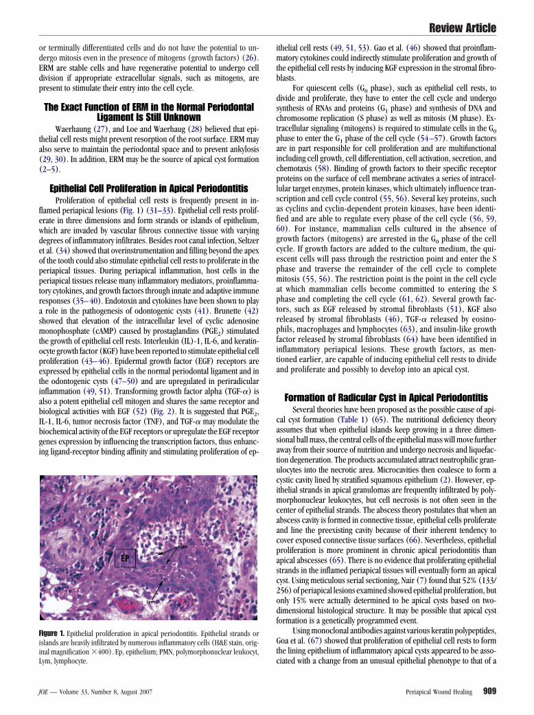
Epithelial Cell Proliferation in Apical PeriodontitisProliferation of epithelial cell rests is frequently present in in-
lamed periapical lesions (Fig. 1) (31–33). Epithelial cell rests prolif-rate in three dimensions and form strands or islands of epithelium,hich are invaded by vascular fibrous connective tissue with varyingegrees of inflammatory infiltrates. Besides root canal infection, Seltzert al. (34) showed that overinstrumentation and filling beyond the apexf the tooth could also stimulate epithelial cell rests to proliferate in theeriapical tissues. During periapical inflammation, host cells in theeriapical tissues release many inflammatory mediators, proinflamma-ory cytokines, and growth factors through innate and adaptive immuneesponses (35– 40). Endotoxin and cytokines have been shown to playrole in the pathogenesis of odontogenic cysts (41). Brunette (42)
howed that elevation of the intracellular level of cyclic adenosineonophosphate (cAMP) caused by prostaglandins (PGE2) stimulated
he growth of epithelial cell rests. Interleukin (IL)-1, IL-6, and keratin-cyte growth factor (KGF) have been reported to stimulate epithelial cellroliferation (43– 46). Epidermal growth factor (EGF) receptors arexpressed by epithelial cells in the normal periodontal ligament and inhe odontogenic cysts (47–50) and are upregulated in periradicularnflammation (49, 51). Transforming growth factor alpha (TGF-�) islso a potent epithelial cell mitogen and shares the same receptor andiological activities with EGF (52) (Fig. 2). It is suggested that PGE2,L-1, IL-6, tumor necrosis factor (TNF), and TGF-� may modulate theiochemical activity of the EGF receptors or upregulate the EGF receptorenes expression by influencing the transcription factors, thus enhanc-ng ligand-receptor binding affinity and stimulating proliferation of ep-
igure 1. Epithelial proliferation in apical periodontitis. Epithelial strands orslands are heavily infiltrated by numerous inflammatory cells (H&E stain, orig-nal magnification �400). Ep, epithelium; PMN, polymorphonuclear leukocyt,
cym, lymphocyte.
OE — Volume 33, Number 8, August 2007
thelial cell rests (49, 51, 53). Gao et al. (46) showed that proinflam-atory cytokines could indirectly stimulate proliferation and growth of
he epithelial cell rests by inducing KGF expression in the stromal fibro-lasts.
For quiescent cells (G0 phase), such as epithelial cell rests, toivide and proliferate, they have to enter the cell cycle and undergoynthesis of RNAs and proteins (G1 phase) and synthesis of DNA andhromosome replication (S phase) as well as mitosis (M phase). Ex-racellular signaling (mitogens) is required to stimulate cells in the G0hase to enter the G1 phase of the cell cycle (54 –57). Growth factorsre in part responsible for cell proliferation and are multifunctionalncluding cell growth, cell differentiation, cell activation, secretion, andhemotaxis (58). Binding of growth factors to their specific receptorroteins on the surface of cell membrane activates a series of intracel-
ular target enzymes, protein kinases, which ultimately influence tran-cription and cell cycle control (55, 56). Several key proteins, suchs cyclins and cyclin-dependent protein kinases, have been identi-ied and are able to regulate every phase of the cell cycle (56, 59,0). For instance, mammalian cells cultured in the absence ofrowth factors (mitogens) are arrested in the G0 phase of the cellycle. If growth factors are added to the culture medium, the qui-scent cells will pass through the restriction point and enter the Shase and traverse the remainder of the cell cycle to completeitosis (55, 56). The restriction point is the point in the cell cycle
t which mammalian cells become committed to entering the Shase and completing the cell cycle (61, 62). Several growth fac-
ors, such as EGF released by stromal fibroblasts (51), KGF alsoeleased by stromal fibroblasts (46), TGF-� released by eosino-hils, macrophages and lymphocytes (63), and insulin-like growthactor released by stromal fibroblasts (64) have been identified innflammatory periapical lesions. These growth factors, as men-ioned earlier, are capable of inducing epithelial cell rests to dividend proliferate and possibly to develop into an apical cyst.
Formation of Radicular Cyst in Apical PeriodontitisSeveral theories have been proposed as the possible cause of api-
al cyst formation (Table 1) (65). The nutritional deficiency theoryssumes that when epithelial islands keep growing in a three dimen-ional ball mass, the central cells of the epithelial mass will move furtherway from their source of nutrition and undergo necrosis and liquefac-ion degeneration. The products accumulated attract neutrophilic gran-locytes into the necrotic area. Microcavities then coalesce to form aystic cavity lined by stratified squamous epithelium (2). However, ep-thelial strands in apical granulomas are frequentlty infiltrated by poly-
orphonuclear leukocytes, but cell necrosis is not often seen in theenter of epithelial strands. The abscess theory postulates that when anbscess cavity is formed in connective tissue, epithelial cells proliferatend line the preexisting cavity because of their inherent tendency toover exposed connective tissue surfaces (66). Nevertheless, epithelialroliferation is more prominent in chronic apical periodontitis thanpical abscesses (65). There is no evidence that proliferating epithelialtrands in the inflamed periapical tissues will eventually form an apicalyst. Using meticulous serial sectioning, Nair (7) found that 52% (133/56) of periapical lesions examined showed epithelial proliferation, butnly 15% were actually determined to be apical cysts based on two-imensional histological structure. It may be possible that apical cyst
ormation is a genetically programmed event.Using monoclonal antibodies against various keratin polypeptides,
oa et al. (67) showed that proliferation of epithelial cell rests to formhe lining epithelium of inflammatory apical cysts appeared to be asso-
iated with a change from an unusual epithelial phenotype to that of aPeriapical Wound Healing 909

sccmtliimlnw
iamtd
fffcdmnittm
tpoirt
FEp F-a, tr
T
Review Article
9
tratified nonkeratinized squamous epithelium. The quiescent epithelialell rests can be considered as restricted-potential or unipotent stemells. When they are stimulated to proliferate, they will undergo asym-etric division to yield two daughter cells (68). One will be similar to
he parental restricted-potential stem cell, which rests on the basalamina as a basal cell. Another one will be a progenitor cell, which isncapable of undergoing mitosis and will eventually mature and developnto a terminally differentiated, suprabasal squamous cell. This process
aintains the population of restricted-potential basal stem cells in pro-iferating epithelial strands. Upon receiving appropriate stimulating sig-als, the restricted-potential basal stem cells in the epithelial strandsill continue to divide and proliferate.
It can be hypothesized that formation of inflammatory apical cystss most likely caused by the merging of proliferating epithelial strands inll directions to form a three-dimensional ball mass. The tissues,ainly fibrous connective tissue with varying degrees of inflamma-
ory cells, trapped inside the epithelial ball mass will graduallyegenerate because of a loss of blood supply, and a cyst cavity will be
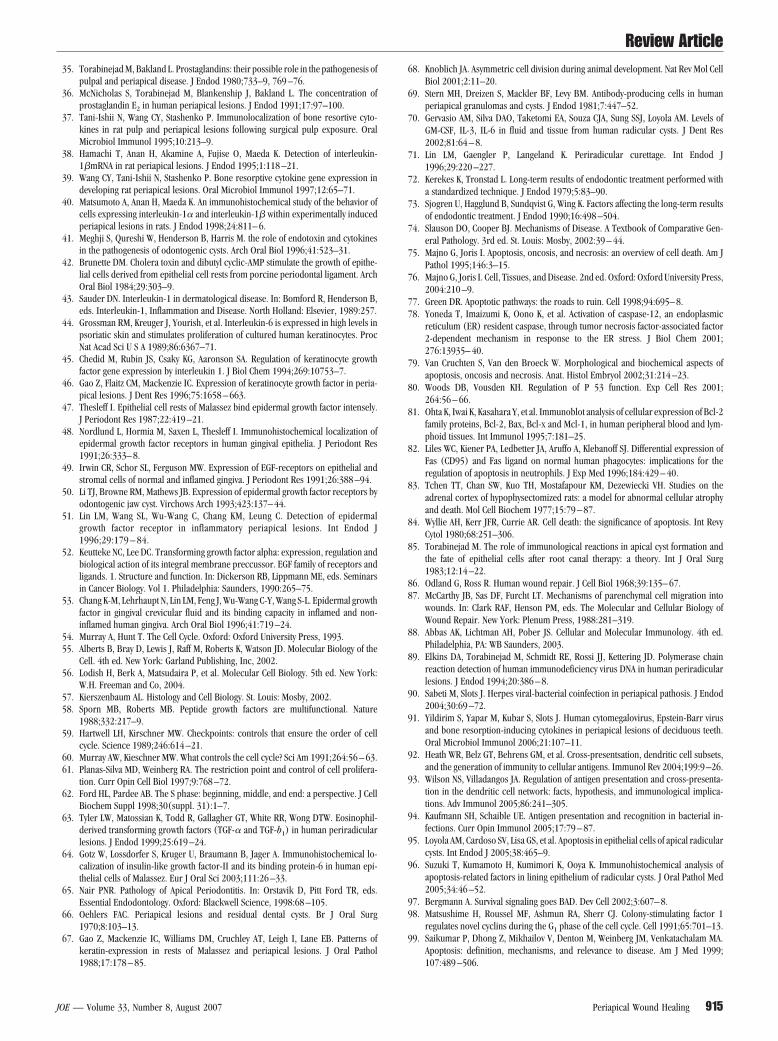
igure 2. Schematic illustration of the major mechanisms that activate proliferatRM, epithelial cell rests of Malassez; OC, osteoclast; EO, eosinophil; PMN,rostaglandins; EGF, epidermal grow factor; IGF, insulin-like growth factor; TG
ABLE 1. Theory of Apical Cyst Formation
Nutritional deficiency theory As islands of epitheliumnutritional supply anmass as liquefaction
Abscess theory An abscess cavity is foris completely surrounsquamous epithelium
Merging of epithelial strands theory As epithelial strands co
When the connective tissu10 Lin et al.
ormed (Table 1). Consequently, inflammatory mediators, proin-lammatory cytokines, growth factors, and immunoglobulins can beound in a cyst cavity (69, 70). It has been speculated that apicalysts may expand after periapical osteoclastic bone resorption me-iated by inflammatory mediators (prostagladins) and proinflam-atory cytokines (IL-1, IL-6, and TNF) (35– 40). However, there is
o time-lapse study showing that the size of apical cysts graduallyncreases as periapical bone destruction increases. It is interestingo note that most inflammatory mediators and proinflammatory cy-okine, which mediate proliferation of epithelial cell rests, also
ediate bone resorption in inflammatory periapical lesions.An inflammatory apical cyst may resist conventional root canal
herapy. However, the lining epithelium of apical cysts may act as ahysical barrier to confine irritants inside the root canal or in the lumenf a cyst and prevents spread of intracanal infection into the surround-
ng alveolar bone. Therefore, epithelial cell proliferation in apical pe-iodontitis may be considered as a defense measure of the periapicalissues in response to irritants inside the root canal (71).
epithelial cell rests in apical periodontitis. M�, macrophage; TH, helper T cell;orphonuclear leukocyte; IL, interleukin; TNF, tumor necrosis factor; PGs,
ansforming growth factor-alpha.
and, more central epithelial cells are distanced from theirdergo necrosis. A cystic cavity results in the center of the cellsis occurs.
in the periapical connective tissues. Subsequently, the abscessby epithelium because of the natural inclination of stratifiedine exposed connective tissue surfaces.e to grow, they merge to form a three-dimensional ball mass.
ion ofpolym
expd unnecromeddedto l
ntinu
e trapped inside the ball mass degenerates, a cyst is formed.JOE — Volume 33, Number 8, August 2007

lhlbipitactcmobtbbAlisavwfa
bsi(c(tTab
iogdpad7pcbvaTa
pss
ts(raf7rapcSa
lrteeiwlkebc(eeeagtgodTcmcctTcmcirmgcpbgcwHIcgi
Review Article
J
Regression of Proliferated Epithelium AfterNonsurgical Endodontic Therapy
After proper nonsurgical endodontic therapy, most periradicularesions except apical true cysts heal (72, 73). The processes of woundealing of periapical lesions after nonsurgical endodontic therapy fol-ow the same principles as that of connective tissues elsewhere in theody. Once irritants in the canals are removed by chemomechanicalnstrumentation and the canal is completely sealed, all cell componentsarticipating in inflammatory reaction will gradually resolve (18). Dur-ng wound healing, the majority of inflammatory cells and some endo-helial cells and fibroblasts are no longer needed and are deleted bypoptosis or programmed cell death (74 –76) rather than by cell ne-rosis. If apoptosis fails to occur, the inflammatory reaction will con-inue to persist because of the release of proinflammatory intracellularontents, such as lysosomal proteolytic enzymes and arachidonic acidetabolites from lysed necrotic cells into the surrounding tissues. Ap-
ptosis will not cause inflammation because apoptotic cells do notecome lysed and release proinflammatory intracellular contents intohe tissue (75). Apoptotic cells will break off to become small mem-rane-bound apoptotic bodies, which will be phagocytosed by neigh-oring cells or macrophages without any trace of inflammation (75).poptotic cells do not die massively but one by one similar to individualeaves falling off a tree when winter is approaching. Apoptosis can benduced by physiological processes (e.g., normal development of tis-ues and organs) and pathological processes (e.g., deletion of autore-ctive T lymphocytes, regression of hyperplasia, atrophy, bacterial andiral infection, radiation, toxins, cell-mediated immune responses, andound healing). Apoptosis occurs when cells fail to receive survival
actors, such as trophic factors, or when they receive death signals, suchs tumor necrosis factor or Fas ligand (Fas-L) (56).
Mechanisms of apoptosis are complex. Several pathways haveeen proposed (76, 77): (A) the surface receptor pathway (extrin-ic) in which the binding of specific surface receptors leads tontracellular procaspase activation; (B) the mitochondrial pathwayintrinsic) in which the mitochondria are induced to release cyto-hrome c, a procaspase activator; (C) the endoplasmic reticulumER) pathway, recently discovered in which a procaspase residing inhe ER is activated by ER stress (78, 79); and (D) the p53 pathway.he superfamily of p53 transcription factors can sense cell stressnd DNA damage and activate transcription of proapoptotic mem-ers of the Bcl-2 gene family (56, 80).
Extrinsic and intrinsic apoptotic pathways have been extensivelynvestigated (Fig. 3). In extrinsic pathway, binding of Fas-L or TNF to Fasr TNF-R1 receptor on the membrane of the cell membrane pro-rammed to die initiates a cell-signaling cascade. The intracellulareath domain of the Fas or TNF receptor enables the coupling of adaptorrotein (Fas or TNF receptor-associated death domain) that in turnctivates caspases-8. Caspases-8 then activates effector caspases to in-uce chromatin fragmentation and inhibition of DNA repair (56, 57,6). In the mitochondrial pathway, caspase-8 activates Bax protein, aroapoptotic member of Bcl-2 family, which is inserted in the mito-hondrial membrane to facilitate the leakage of cytochrome c, whichecomes attached to a protein called Apaf-1 (apoptotic protease acti-ating factor) and the two self-assemble into a seven-spoke wheel calledpoptosome. The apoptosome recruits procaspase-9 and activates it.he activated caspase-9 in turn activates effector caspases, leading topoptosis (56, 57, 76).
Signals to activate apoptosis may be either extrinsic (receptorathway) or intrinsic (mitochondrial pathway) (57, 76). Extrinsicignals may be positive or negative. Positive signals activate apopto-
is, such as binding of death signals, Fas-L, or TNF with their recep- tOE — Volume 33, Number 8, August 2007
ors, Fas or TNF receptor (76). Negative signals suppress apoptosis,uch as binding of trophic or growth factors with their receptors76). Two important apoptotic pathways mediated by cell-surfaceeceptors have been identified. There are the Fas receptor (CD95),transmembrane protein that is a member of the tumor necrosis
actor receptor super-family, and the TNF receptor (TNF-R1) (56,4). Fas receptor is expressed on the cells programmed to die. TNFeceptor is present on almost all cell types. Fas-L is expressed byctivated polymorphonuclear leukocytes, natural killer cells, T lym-hocytes, and also by the cell programmed to die (16, 56, 57). Fas-Lan be either free floating or bound to the surface of another cell.ignals for apoptosis in one cell type may induce cell proliferationnd differentiation in other cell types.
The intriguing question is how do many epithelial strands or is-ands in periapical granulomas and the lining epithelium of apical cystsegress after nonsurgical endodontic therapy? Bhaskar (9) suggestedhat if the root canal was instrumented beyond the apex and the liningpithelium of cysts was intentionally broken with an instrument duringndodontic therapy, an acute inflammatory reaction would be inducedn the lining epithelium, and activated polymorphonuclear leukocytesould destroy the lining epithelium of cyst. However, the lining epithe-
ium of apical cysts is frequently infiltrated by polymorphonuclear leu-ocytes (PMN) (31). In addition, PMNs are not capable of digestingpithelium (18). Nevertheless, it has been shown that PMNs expressoth Fas and Fas-L and can release soluble Fas-L (81). The soluble Fas-Lould induce apoptosis of epithelial cells that express Fas antigens16, 82). Seltzer et al. (34) showed histologically that proliferatedpithelium in the periapical tissues was trapped by the collagen that islaborated by fibroblasts during the repair process, and most of thepithelium degenerated 1 year after endodontic therapy. Oehlers (66)lso observed that many inflammatory residual cysts of the jaws re-ressed after extraction of the involved teeth. The residual lining epi-helium underwent atrophy because the factors responsible for cysticrowth had been removed. Absence of growth factors can cause atrophyf tissues or organs through cell deletion by means of programmed celleath, such as hypophysectomy leading to adrenal atrophy (74, 83, 84).orabinejad (85) proposed a theory that activated epithelial cell restsould acquire antigenic properties through ingestion of the antigenicaterials, which continuously egress from an infected necrotic root
anal system into the periapical tissues because epithelial cells areapable of phagocytosing foreign materials (86, 87). The ingested ma-erial(s) and epithelial cells could be recognized as an antigenic unit.he activated epithelium could be destroyed by antibody-dependentomplement-mediated immune mechanism and cell-mediated immuneechanism through macrophages, cytotoxic T cells, or natural killer
ells after root canal therapy. Apoptosis is commonly seen in situationsn which target cell destruction is induced by cell-mediated immuneesponse (88). Cytotoxic T cells, one of the effector cells of a cell-ediated immune response, can induce target cells to undergo pro-
rammed cell death by two strategies (55). In one strategy, cytotoxic Tells bind to a target cell and are stimulated to release a pore-formingrotein, perforin, which forms a large aqueous channel in the lipidilayer of the target cell plasma membrane and thus induces pro-rammed cell death of the target cell (55). The second strategy involvesytotoxic T cell activating a receptor (Fas) on the target cell membrane,hich signals the target cell to undergo programmed cell death (55).owever, cytotoxic T cells usually engage in killing virus-infected cells.
f viruses infect the epithelial cells in periradicular lesions (89 –91),ytotoxic T cells will recognize these infected cells and execute pro-rammed cell death. Alternatively, bacterial antigens may be presentedn association with a MHC class I molecule via cross-presentation be-
ween MHC class I and class II pathways in antigen-ingested epithelialPeriapical Wound Healing 911

cstopcsab
scce
bcaaerdsamldf
F
Review Article
9
ells, thereby recognized by cytotoxic T cells (92–94). Loyola et al. (95)howed immunohistochemically using antibody against Bcl-2 antigenhat apoptosis was present in both hyperplastic and atrophic epitheliumf apical cysts but was present more frequently in atrophic than hyper-lastic epithepium. Suzuki et al. (96) also showed immunohistochemi-ally using monoclonal antibodies or polyclonal antisera against single-tranded DNA and p53, Bax, Bcl-2, caspases-3, Fas, Fas-L, or Ki-67ntigen that apoptotic cell death occurred in the lining epithelium ofoth apical cysts and residual apical cyst.
Based on the previously described studies, regression of epithelialtrands in periapical granulomas and the lining epithelium of apicalysts are most likely caused by programmed cell death. After nonsurgi-al endodontic therapy, the restricted-potential basal stem cells in the
igure 3. An overview of extrinsic and intrinsic apoptotic pathways.
pithelial strands or lining epithelium of cyst will stop proliferating c
12 Lin et al.
ecause of a reduction in inflammatory mediators, proinflammatoryytokines, and growth factors. Furthermore, the terminally differenti-ted squamous cells in the epithelial strands and the lining epithelium ofcyst will die of programmed cell death similar to that of surface
pithelial cells of the oral epithelium. Eventually, most basal cells willegress or become atrophic through programmed cell death because ofeprivation of survival factors or receiving death signals during progres-ive wound healing of periradicular lesions (Figs. 4, 5, 6) (97, 98). It islso possible that epithelial cells in epithelial strands and in apical cystsay be biologically similar to autoreactive T lymphocytes and are de-
eted by programmed cell death. Therapeutically, any clinically availablerugs that can prevent release of inflammatory mediators and/or proin-
lammatory cytokines might be able to inhibit proliferation of epithelial
ell rests in apical periodontitis.JOE — Volume 33, Number 8, August 2007

icfccsrcabpgswahdot
tat
ggalapttle(pm(
Fc
Ftb
FF
Review Article
J
Tissues have the ability to restore their original structure afternjury by means of a repair process that is tightly regulated throughell-cell and cell-matrix signaling, a variety of cytokines and growthactors, as well as by cell mitosis and apoptosis so that the injured tissuesan be orderly restored (76). Although the damaged periapical tissuesan be repaired, they will never completely return to their originaltructure after nonsurgical endodontic therapy because of uncouplingeaction (18). Proliferation of epithelial cell rests in apical periodontitisan be viewed as an inflammatory hyperplasia, which can regress in thebsence of inflammatory stimulation (99). For instance, in chronicronchitis caused by infection, the bronchial epithelium becomes hy-erplastic, but the hyperplastic epithelium will regress through pro-rammed cell death in the absence of causative stimulus (74). Apopto-is was also observed in pulp cells during wound healing after pulpsere capped with capping agents (100). Programmed cell death playsn important role in tissue remodeling and homeostasis during woundealing (74). Apoptosis is also a regular feature of normal animalevelopment in order to control size, shape, and function of tissues andrgans (101, 102). Although increased inflammation has been shown
igure 4. Mechanisms of regression of inflammatory apical cysts after periapi-al wound healing.
o be associated with increased epithelial cell apoptosis in the periodon- h
OE — Volume 33, Number 8, August 2007
ium of patients with periodontal disease (103), epithelial cell mitosisppears to be more active than apoptosis in inflamed hyperplastic epi-helium of apical cysts (104, 105).
Recently, Brozovic et al. (106) showed that Porphyromonas gin-ivalis could cause upregulation of Fas-Fas-L expression on humaningival epithelial cells and induce apoptotic cell death. P gingivalis islso present in infected root canals (107, 108) and may cause upregu-ation of Fas-Fas-L expression on epithelial cells in inflammatory peri-pical lesions similar to that on gingival epithelial cells. TNF is mainlyroduced by activated macrophages, which are abundant in inflamma-
ory periapical lesions (109, 110). Therefore, natural killer cells, cyto-oxic T cells, or macrophages can kill epithelial cells in periapicalesions expressing Fas or TNF receptor. Apoptosis in normal and dis-ased oral tissues has been recently reviewed in detail by Loro et al.111). It appears that antiapoptotic proteins are preferentially ex-ressed in basal cells, whereas proapoptotic protein is expressed inore differentiated supra-basal squamous cells of the oral epithelium
111).
igure 5. Immediately after completion of nonsurgical root canal therapy ofooth #30. A well-defined radiolucent lesion surrounded by a thin radiopaqueorder suggestive as a cyst is associated with the apex of the mesial root.
igure 6. Approximately 1-year postoperative follow-up of tooth #30 inigure 5. The periapical lesion associated with the apex of the mesial root
as regressed.Periapical Wound Healing 913

acetpwTdianteotio
bafsnpodcasppoebpdadoawckadb
gutetnw
Review Article
9
It is apparent that epithelial cell proliferation in the inflamed peri-pical tissues is sustained by inflammatory stimulation. In apical pocketysts, the irritants are in the canal and can be eliminated by nonsurgicalndodontic procedures. Epithelial cell proliferation in the periradicularissues will be inhibited by a reduction of inflammatory mediators,roinflammatory cytokines, or growth factors. Epithelial cell apoptosisill be induced by activation of extracellular positive signals, Fas-L, orNF or by deprivation of survival factors (76, 97, 98) during perira-icular wound healing. However, in apical true cysts, besides intracanalrritants, other irritants, such as cholesterol or possibly unidentifiedntigens (4, 112–114), are within the cyst, which cannot be removed byonsurgical root canal therapy and will continuously sustain inflamma-ory stimulation to the restricted-potential basal stem cells of the cysticpithelium. Therefore, most basal stem cells will remain in the G1 phasef the cell cycle. Of course, terminally differentiated squamous cells ofhe lining epithelium will undergo programmed cell death. Becauserritants in apical true cysts cannot be eliminated by nonsurgical end-dontic procedure, an apical true cyst must be treated surgically (15).
Regression of Proliferated Epithelium After SurgicalEndodontic Therapy
An apical surgical procedure represents a form of artificial de-ridement of wounded tissue and can effectively eliminate the irritantsnd encourage satisfactory wound healing (76). However, one of mostrequently asked questions is that what would happen if epithelialtrands in apical granulomas or the lining epithelium of apical cysts isot completely removed during surgical procedure? It has been pro-osed that the reactionary tissues including epithelium in apical peri-dontitis do not have to be completely removed during surgical proce-ures if hemostasis is under control and irritants are eliminated orompletely sealed inside the root canal by a root-end filling (71). Peri-pical tssue reactions, such as inflammatory cell infiltration, bone re-orption and epithelial proliferation in apical periodontitis are theroducts of root canal infection. After surgical endodontic therapy,eriapical wound healing should follow exactly the same course as thatf nonsurgical endodontic therapy. The only difference is that surgicalndodontic therapy will heal faster than nonsurgical endodontic therapyecause of more effective artificial debridement of infected or woundederiapical tissues by surgical procedures as compared with biologicalebridement by phagocytes in nonsurgical procedures (76). Grupe etl. (115) suggested that the epithelial cells of apical cysts are capable ofivision and proliferation by virtue of their ability to undertake anaer-bic glycolysis. There is no evidence that epithelial cells in inflammatorypical cysts behave like malignant neoplastic cells, which are encodedith oncogenes and can self divide in the absence of appropriate extra-ellular signals, such as mitogens, cyclins, or cyclin-dependent proteininases (55). The remnants of epithelium left in the periapical tissuesfter surgical endodontic procedures will regress by programmed celleath similar to that of nonsurgical endodontic therapy if irritants haveeen removed.
Although apoptosis is most likely the mechanism of epithelial re-ression in inflammatory apical cysts after endodontic therapy, the reg-lation of apoptosis is complex (55, 102). Cell biologists are still trying
o define all of the signals that regulate the survival and proliferation ofach cell type, to determine how the levels of the signals are controlledo balance cell proliferation and cell death according to the varyingeeds of the organs, and to understand how the individual cell decideshether to live or die and whether to divide or remain quiescent (55).
References1. Valdehaung J. A histologic study of experimentally induced radicular cysts. Int J Oral
Surg 1972;1:137– 47.
14 Lin et al.
2. Ten Cate AR. The epithelial cell rests of Malassez and the genesis of the dental cyst.Oral Surg Oral Med Oral Pathol 1972;34:56 – 64.
3. Harris M, Toller P. The pathogenesis of dental cysts. Br Med Bull 1975;31:159 – 63.4. Shear M. Cysts of the jaw: recent advances. J Oral Pathol 1985;14:43–59.5. Seltzer S, Bender IB, Smith J, Freedman I, Nazimo H. Endodontic failures: an
analysis based on clinical, roentgenographic and histologic findings. Oral Surg OralMed Oral Pathol 1967;23:500 –30.
6. Simon JHS. Incidence of periapical cysts in relation to the root canal. J Endod1980;6:845– 8.
7. Nair PNR, Pajarola G, Schroeder HE. Types and incidence of human periapicallesions obtained with extracted teeth. Oral Surg Oral Med Oral Pathol Oral RadiolEndod 1996;81:93–102.
8. Linenberg WB, Waldron CA, DeLaune GF. A clinical, roentgenographic, and his-topathologic evaluation of periapical lesions. Oral Surg Oral Med Oral Pathol1964;17:467–72.
9. Bhaskar SN. Periapical lesions—types, incidence and clinical feature. Oral SurgOral Med Oral Pathol 1966;21:657–71.
10. Lalonde ER. A new rationale for the management of periapical granulomas andcysts. An evaluation of histopathological and radiographic findings. J Am Dent Assoc1970;80:1056 –9.
11. Mortensen H, Winther JE, Birin H. Periapical granulomas and cysts. An investigationof 1.600 cases. Scand J Dent Res 1970;78:241–50.
12. Cotti F, Campisi G, Ambu R, Dettori C. Ultrasound real-time imaging in the differ-ential diagnosis of periapical lesions. Int Endod J 2003;36:556 – 63.
13. Gundappa M, Ng SY, Whaites EJ. Comparison of ultrasound, digital and conventionalradiography in differentiating periapical lesions. Dentomaxillofac Radiol 2006;35:326 –33.
14. Simon JHS, Enciso R, Malfaz JM, Roges R, Bailey-Perry M, Patel A. Differentialdiagnosis of large periapical lesions using cone-beam computed tomography mea-surements and biopsy. J Endod 2006;32:833–7.
15. Nair PNR. New perspectives on radicular cysts: do they heal? Int Endod J1998.31:155– 60.
16. Takahashi K. Microbiological, pathological, inflammatory, immunological and mo-lecular biological aspects of periradicular disease. Int Endod J 1998;31:311–25.
17. Ten Cate AR Oral Histology: Development, Structure and Function. St. Louis: CVMosby, 1998.
18. Seltzer S. Endodontology. Biologic Considerations in Endodontic Procedures. 2nded. Philadephia, PA: Lea & Febiger, 1988.
19. Wentz FM, Weinmann JP, Schour I. The prevalence, distribution, and morphologicchanges of the epithelial remnants in the molar region of the rat molar. J Dent Res1950;29:637– 46.
20. Reeve CM, Wentz FM. The prevalence, morphology, and distribution of epithelialrests in human periodontal ligament. Oral Surg 1962;15:785–93.
21. Valderhaung J, Zander HA. Relationship of epithelial cell rests of Malassez to otherperiodontal structure. Periodontics 1967;5:254 – 8.
22. McHugh WD, Zander HA. Cell division in the periodontium of developing anderupted teeth. Dent Pract 1965;15:451–7.
23. Ten Cate AR. The histochemical demonstration of specific oxidative enzymes andglycogen in the epithelial cell rests of Malassez. Arch Oral Biol 1965;10:207–13.
24. Ten Cate AR. The formation and function of the epithelial cell rests of Malassez. In:Anderson DJ, Eastoe JE, Melcher AH, Picton DCA, eds. The Mechanisms of ToothSupport. A Symposium. Bristol, UK: Wright, 1967.
25. Valderhaung JP, Nylen MU. Function of epithelial rests as suggested by their ultra-structure. J Periodontol Res 1966;1:69 –78.
26. D’Sousa R. Development of the Pulpodentin Complex. In: Hargreaves KM, GoodiesHE, eds. Dental Pulp. Chicago, IL: Quintessence Publishing Co, Inc, 2002.
27. Waerhaung J. Effect of C-avitaminosis on the supporting structures of the teeth. JPeriodontol Res 1958;29:87–97.
28. Loe H, Waerhaung J. Experimental replantation of teeth in dogs and monkeys. ArchOral Biol 1961;3:76 – 84.
29. Lindskog S, Blomlof L, Hammarstrom L. Evidence for a role of odontogenic epithe-lium in maintaining the periodontal space. J Clin Periodontol 1988;15:371–3.
30. Spouge JD. A new look at the rests of Malassez. A review of their embryologicalorigin, anatomy, and possible role in periodontal health and disease. J Periodontol1980;51:437– 44.
31. Langeland K, Block RM, Grossman LI. A histopathologic study of 35 periapicalendodontic surgical specimens. J Endod 1977;3:8 –23.
32. Bergenholtz G, Lekholm U, Liljenberg B, Lindhe J. Morphometric analysis ofchronic inflammatory periapical lesions in root filled teeth. Oral Surg Oral Med OralPathol 1983;55:295–301.
33. Nair PNR, Schmid-Meier E. An apical granuloma with epithelial integument. OralSurg Oral Med Oral Pathol 1986;62:698 –703.
34. Seltzer S, Soltanoff W, Bender IB. Epithelial proliferation in periapical lesions. Oral
Surg Oral Med Oral Pathol 1969;27:111–21.JOE — Volume 33, Number 8, August 2007
Review Article
J
35. Torabinejad M, Bakland L. Prostaglandins: their possible role in the pathogenesis ofpulpal and periapical disease. J Endod 1980;733–9, 769 –76.
36. McNicholas S, Torabinejad M, Blankenship J, Bakland L. The concentration ofprostaglandin E2 in human periapical lesions. J Endod 1991;17:97–100.
37. Tani-Ishii N, Wang CY, Stashenko P. Immunolocalization of bone resortive cyto-kines in rat pulp and periapical lesions following surgical pulp exposure. OralMicrobiol Immunol 1995;10:213–9.
38. Hamachi T, Anan H, Akamine A, Fujise O, Maeda K. Detection of interleukin-1�mRNA in rat periapical lesions. J Endod 1995;1:118 –21.
39. Wang CY, Tani-Ishii N, Stashenko P. Bone resorptive cytokine gene expression indeveloping rat periapical lesions. Oral Microbiol Immunol 1997;12:65–71.
40. Matsumoto A, Anan H, Maeda K. An immunohistochemical study of the behavior ofcells expressing interleukin-1� and interleukin-1� within experimentally inducedperiapical lesions in rats. J Endod 1998;24:811– 6.
41. Meghji S, Qureshi W, Henderson B, Harris M. the role of endotoxin and cytokinesin the pathogenesis of odontogenic cysts. Arch Oral Biol 1996;41:523–31.
42. Brunette DM. Cholera toxin and dibutyl cyclic-AMP stimulate the growth of epithe-lial cells derived from epithelial cell rests from porcine periodontal ligament. ArchOral Biol 1984;29:303–9.
43. Sauder DN. Interleukin-1 in dermatological disease. In: Bomford R, Henderson B,eds. Interleukin-1, Inflammation and Disease. North Holland: Elsevier, 1989:257.
44. Grossman RM, Kreuger J, Yourish, et al. Interleukin-6 is expressed in high levels inpsoriatic skin and stimulates proliferation of cultured human keratinocytes. ProcNat Acad Sci U S A 1989;86:6367–71.
45. Chedid M, Rubin JS, Csaky KG, Aaronson SA. Regulation of keratinocyte growthfactor gene expression by interleukin 1. J Biol Chem 1994;269:10753–7.
46. Gao Z, Flaitz CM, Mackenzie IC. Expression of keratinocyte growth factor in peria-pical lesions. J Dent Res 1996;75:1658 – 663.
47. Thesleff I. Epithelial cell rests of Malassez bind epidermal growth factor intensely.J Periodont Res 1987;22:419 –21.
48. Nordlund L, Hormia M, Saxen L, Thesleff I. Immunohistochemical localization ofepidermal growth factor receptors in human gingival epithelia. J Periodont Res1991;26:333– 8.
49. Irwin CR, Schor SL, Ferguson MW. Expression of EGF-receptors on epithelial andstromal cells of normal and inflamed gingiva. J Periodont Res 1991;26:388 –94.
50. Li TJ, Browne RM, Mathews JB. Expression of epidermal growth factor receptors byodontogenic jaw cyst. Virchows Arch 1993;423:137– 44.
51. Lin LM, Wang SL, Wu-Wang C, Chang KM, Leung C. Detection of epidermalgrowth factor receptor in inflammatory periapical lesions. Int Endod J1996;29:179 – 84.
52. Keutteke NC, Lee DC. Transforming growth factor alpha: expression, regulation andbiological action of its integral membrane preccussor. EGF family of receptors andligands. 1. Structure and function. In: Dickerson RB, Lippmann ME, eds. Seminarsin Cancer Biology. Vol 1. Philadelphia: Saunders, 1990:265–75.
53. Chang K-M, Lehrhaupt N, Lin LM, Feng J, Wu-Wang C-Y, Wang S-L. Epidermal growthfactor in gingival crevicular fluid and its binding capacity in inflamed and non-inflamed human gingiva. Arch Oral Biol 1996;41:719 –24.
54. Murray A, Hunt T. The Cell Cycle. Oxford: Oxford University Press, 1993.55. Alberts B, Bray D, Lewis J, Raff M, Roberts K, Watson JD. Molecular Biology of the
Cell. 4th ed. New York: Garland Publishing, Inc, 2002.56. Lodish H, Berk A, Matsudaira P, et al. Molecular Cell Biology. 5th ed. New York:
W.H. Freeman and Co, 2004.57. Kierszenbaum AL. Histology and Cell Biology. St. Louis: Mosby, 2002.58. Sporn MB, Roberts MB. Peptide growth factors are multifunctional. Nature
1988;332:217–9.59. Hartwell LH, Kirschner MW. Checkpoints: controls that ensure the order of cell
cycle. Science 1989;246:614 –21.60. Murray AW, Kieschner MW. What controls the cell cycle? Sci Am 1991;264:56 – 63.61. Planas-Silva MD, Weinberg RA. The restriction point and control of cell prolifera-
tion. Curr Opin Cell Biol 1997;9:768 –72.62. Ford HL, Pardee AB. The S phase: beginning, middle, and end: a perspective. J Cell
Biochem Suppl 1998;30(suppl. 31):1–7.63. Tyler LW, Matossian K, Todd R, Gallagher GT, White RR, Wong DTW. Eosinophil-
derived transforming growth factors (TGF-� and TGF-b1) in human periradicularlesions. J Endod 1999;25:619 –24.
64. Gotz W, Lossdorfer S, Kruger U, Braumann B, Jager A. Immunohistochemical lo-calization of insulin-like growth factor-II and its binding protein-6 in human epi-thelial cells of Malassez. Eur J Oral Sci 2003;111:26 –33.
65. Nair PNR. Pathology of Apical Periodontitis. In: Orstavik D, Pitt Ford TR, eds.Essential Endodontology. Oxford: Blackwell Science, 1998:68 –105.
66. Oehlers FAC. Periapical lesions and residual dental cysts. Br J Oral Surg1970;8:103–13.
67. Gao Z, Mackenzie IC, Williams DM, Cruchley AT, Leigh I, Lane EB. Patterns ofkeratin-expression in rests of Malassez and periapical lesions. J Oral Pathol
1988;17:178 – 85.OE — Volume 33, Number 8, August 2007
68. Knoblich JA. Asymmetric cell division during animal development. Nat Rev Mol CellBiol 2001;2:11–20.
69. Stern MH, Dreizen S, Mackler BF, Levy BM. Antibody-producing cells in humanperiapical granulomas and cysts. J Endod 1981;7:447–52.
70. Gervasio AM, Silva DAO, Taketomi EA, Souza CJA, Sung SSJ, Loyola AM. Levels ofGM-CSF, IL-3, IL-6 in fluid and tissue from human radicular cysts. J Dent Res2002;81:64 – 8.
71. Lin LM, Gaengler P, Langeland K. Periradicular curettage. Int Endod J1996;29:220 –227.
72. Kerekes K, Tronstad L. Long-term results of endodontic treatment performed witha standardized technique. J Endod 1979;5:83–90.
73. Sjogren U, Hagglund B, Sundqvist G, Wing K. Factors affecting the long-term resultsof endodontic treatment. J Endod 1990;16:498 –504.
74. Slauson DO, Cooper BJ. Mechanisms of Disease. A Textbook of Comparative Gen-eral Pathology. 3rd ed. St. Louis: Mosby, 2002:39 – 44.
75. Majno G, Joris I. Apoptosis, oncosis, and necrosis: an overview of cell death. Am JPathol 1995;146:3–15.
76. Majno G, Joris I. Cell, Tissues, and Disease. 2nd ed. Oxford: Oxford University Press,2004:210 –9.
77. Green DR. Apoptotic pathways: the roads to ruin. Cell 1998;94:695– 8.78. Yoneda T, Imaizumi K, Oono K, et al. Activation of caspase-12, an endoplasmic
reticulum (ER) resident caspase, through tumor necrosis factor-associated factor2-dependent mechanism in response to the ER stress. J Biol Chem 2001;276:13935– 40.
79. Van Cruchten S, Van den Broeck W. Morphological and biochemical aspects ofapoptosis, oncosis and necrosis. Anat. Histol Embryol 2002;31:214 –23.
80. Woods DB, Vousden KH. Regulation of P 53 function. Exp Cell Res 2001;264:56 – 66.
81. Ohta K, Iwai K, Kasahara Y, et al. Immunoblot analysis of cellular expression of Bcl-2family proteins, Bcl-2, Bax, Bcl-x and Mcl-1, in human peripheral blood and lym-phoid tissues. Int Immunol 1995;7:181–25.
82. Liles WC, Kiener PA, Ledbetter JA, Aruffo A, Klebanoff SJ. Differential expression ofFas (CD95) and Fas ligand on normal human phagocytes: implications for theregulation of apoptosis in neutrophils. J Exp Med 1996;184:429 – 40.
83. Tchen TT, Chan SW, Kuo TH, Mostafapour KM, Dezewiecki VH. Studies on theadrenal cortex of hypophysectomized rats: a model for abnormal cellular atrophyand death. Mol Cell Biochem 1977;15:79 – 87.
84. Wyllie AH, Kerr JFR, Currie AR. Cell death: the significance of apoptosis. Int RevyCytol 1980;68:251–306.
85. Torabinejad M. The role of immunological reactions in apical cyst formation andthe fate of epithelial cells after root canal therapy: a theory. Int J Oral Surg1983;12:14 –22.
86. Odland G, Ross R. Human wound repair. J Cell Biol 1968;39:135– 67.87. McCarthy JB, Sas DF, Furcht LT. Mechanisms of parenchymal cell migration into
wounds. In: Clark RAF, Henson PM, eds. The Molecular and Cellular Biology ofWound Repair. New York: Plenum Press, 1988:281–319.
88. Abbas AK, Lichtman AH, Pober JS. Cellular and Molecular Immunology. 4th ed.Philadelphia, PA: WB Saunders, 2003.
89. Elkins DA, Torabinejad M, Schmidt RE, Rossi JJ, Kettering JD. Polymerase chainreaction detection of human immunodeficiency virus DNA in human periradicularlesions. J Endod 1994;20:386 – 8.
90. Sabeti M, Slots J. Herpes viral-bacterial coinfection in periapical pathosis. J Endod2004;30:69 –72.
91. Yildirim S, Yapar M, Kubar S, Slots J. Human cytomegalovirus, Epstein-Barr virusand bone resorption-inducing cytokines in periapical lesions of deciduous teeth.Oral Microbiol Immunol 2006;21:107–11.
92. Heath WR, Belz GT, Behrens GM, et al. Cross-presentsation, dendritic cell subsets,and the generation of immunity to cellular antigens. Immunol Rev 2004;199:9 –26.
93. Wilson NS, Villadangos JA. Regulation of antigen presentation and cross-presenta-tion in the dendritic cell network: facts, hypothesis, and immunological implica-tions. Adv Immunol 2005;86:241–305.
94. Kaufmann SH, Schaible UE. Antigen presentation and recognition in bacterial in-fections. Curr Opin Immunol 2005;17:79 – 87.
95. Loyola AM, Cardoso SV, Lisa GS, et al. Apoptosis in epithelial cells of apical radicularcysts. Int Endod J 2005;38:465–9.
96. Suzuki T, Kumamoto H, Kumimori K, Ooya K. Immunohistochemical analysis ofapoptosis-related factors in lining epithelium of radicular cysts. J Oral Pathol Med2005;34:46 –52.
97. Bergmann A. Survival signaling goes BAD. Dev Cell 2002;3:607– 8.98. Matsushime H, Roussel MF, Ashmun RA, Sherr CJ. Colony-stimulating factor 1
regulates novel cyclins during the G1 phase of the cell cycle. Cell 1991;65:701–13.99. Saikumar P, Dhong Z, Mikhailov V, Denton M, Weinberg JM, Venkatachalam MA.
Apoptosis: definition, mechanisms, and relevance to disease. Am J Med 1999;
107:489 –506.Periapical Wound Healing 915

1
11
1
1
1
1
1
1
1
1
1
1
1
1
1
Review Article
9
00. Kitamura C, Ogawa Y, Morotomi T, Terashita M. Differential induction of apoptosisby capping agents during pulp wound healing. J Endod 2003;29:41–3.
01. Vaux DL, Korsmeyer SJ. Cell death in development. Cell 1999;96:245–54.02. Zuzarte-Luis V, Hurle JM. Programmed cell death in the developing limb. Int J Dev
Biol 2002;46:871– 6.03. Carro OM, Evans SA, Leone CW. Effect of inflammation on the proliferation of human
gingival epithelial cells in vivo. J Periodontol 1997;68:1070 –5.04. Takahashi K, MacDonald DG, Murayama Y, Kinane DF. Cell synthesis, proliferation
and apoptosis in human dental periapical lesions analyzed by in situ hybridizationand immunohistochemistry. Oral Dis 1999;5:313–20.
05. Nickolaychuk B, McNicol A, Gilchrist J, Birek C. Evidence for a role of mitogen-activated protein kinases in proliferating and differentiating odontogenic epitheliaof inflammatory and developmental cysts. Oral Surg Oral Med Oral Pathol OralRadiol Endod 2002;93:720 –9.
06. Brozovic S, Sahoo R, Barve S, et al. Porphyromonas gingivalis enhaces Fas-L ex-pression via up-regulation of NFKappa B-mediated gene transcription and inducescell death in human gingival epithelial cells. Microbiology 2006;152:797– 806.
07. Sundqvist G. Bacteriological studies of necrotic dental pulps (dissertation). Umea,
Sweden: Umea University Odontological Dissertation, 1976.16 Lin et al.
08. Sundqvist G. Taxonomy, ecology, and pathogenecity of the root canal flora. OralSurg Oral Med Oral Pathol Oral Radiol Endod 1994;78:522–30.
09. Stern MH, Dreizen S, Mackler BF, Selbst AG, Levy BM. Quantitative analysis ofcellular composition of human periapical granulomas. J Endod 1981;7:117–22.
10. Weiner S, McKinney RV, Walton RE. Characterization of the periapical surgicalspecimen. A morphologic and histochemical study of the inflammatory patterns.Oral Surg 1982;53:293–302.
11. Loro LL, Vintermyr OK, Johannessen AC. Apoptosis in normal and diseased oraltissues. Oral Dis 2005;11:274 – 87.
12. Trott JR. Chebib F, Galindo Y. Factors related to cholesterol formation in cysts andgranulomas. J Can Dent Assoc 1973;39:550 –55.
13. Browne RM. The origin of cholesterol in odontogenic cysts in man. Arch Oral Biol1971;16:107–13.
14. Nair PNR, Sjogren U, Schumacher E, Sundqvist G. Radicular cyst affecting a root-filled human tooth: a long-term posttreatment follow-up. Int Endod J1993;26:225–33.
15. Grupe HE Jr, Ten Cate AR, Zander HA. A histological and radiobiological study of invitro and in vivo human epithelial cell rest proliferation. Arch Oral Biol
1967;12:1321–9.JOE — Volume 33, Number 8, August 2007