Embed Size (px)
Citation preview
Promoting Type-1 CD4+ T Cell Immune Responses Against Tumor-Associated Antigen MAGE-A6
by
Lazar N. Vujanović
B.S. Biology, Pennsylvania State University, 1998
Submitted to the Graduate Faculty of
School of Medicine in partial fulfillment
of the requirements for the degree of
Doctor of Philosophy
University of Pittsburgh
2006
UNIVERSITY OF PITTSBURGH
FACULTY OF SCHOOL OF MEDICINE
This dissertation was presented
by
Lazar Vujanović
It was defended on
May 26, 2006
and approved by
William Chambers, Ph.D.
Russell Salter, Ph.D.
Angus Thomson, Ph.D.
Louis Falo, M.D.
Ora Weisz, Ph.D.
Hannah Rabinowich, Ph.D.
Walter J. Storkus, Ph.D. Dissertation Director
ii
Promoting Type-1 CD4+ T Cell Immune Responses Against Tumor-Associated Antigen MAGE-A6
Lazar Vujanović, PhD
University of Pittsburgh, 2006
Abstract
One of the main challenges facing tumor immunologists is to develop strategies that
would effectively stimulate Type-1 anti-tumor T cell responses, which have been correlated with
better clinical outcome and prolonged survival of cancer patients. As CD4+ T cells were shown
to play a critical role in mediating these responses, it was of interest to examine novel ways of
effectively stimulating and enhancing Type-1 CD4+ T cell responses. For these studies I used
MAGE-A6, a tumor associated antigen (TAA) expressed by a broad range of human cancer
types. Two novel MAGE-A6 T-helper epitopes were identified and were shown to be
recognized by CD4+ T cells isolated from the majority of normal donors or patients with
melanoma, regardless of their HLA genotype (i.e. poly-DR presented epitopes). Furthermore,
peptide-specific T cells also recognized autologous monocytes pulsed with recombinant MAGE-
A6 protein, supporting the natural processing and MHC presentation of these epitopes.
Interestingly, one of the novel MAGE-A6 epitopes possesses a high-degree of homology with a
microbial peptide. CD4+ T cells stimulated in vitro with this microbial peptide cross-reacted
against the MAGE-A6 homologue peptide, and could recognize naturally-processed MAGE-A6
epitopes more effectively than T cells stimulated with MAGE-A6 peptides. This study showed
that it is possible to stimulate, and even enhance tumor-specific T cell responses using microbial
epitopes that are homologous to TAA-derived peptides. In the final study, human dendritic cells
(DC) were engineered to secrete high levels of IFN-γ-inducing cytokines IL-12p70 and IL-18 via
iii
recombinant adenoviral infection to generate an in vitro stimulus capable of promoting
previously deficient patient Th1-type responses. DC engineered to secrete both of these
cytokines simultaneously (DC.IL-12/18) were highly effective at stimulating MAGE-A6-specific
Th1-type CD4+ T cell responses from patients with melanoma, particularly when loaded with
MAGE-A6 protein. Poly-DR presented epitopes and MAGE-A6 protein defined in this thesis, if
loaded onto DC.IL-12/18, could prove clinically useful as a vaccine modality capable of
promoting the recovery and/or enhancement of tumor antigen-specific, Th1-type CD4+ T cell
responses in the majority of patients harboring MAGE-A6+ cancers.
iv
TABLE OF CONTENTS
PREFACE...................................................................................................................................... ix 1. INTRODUCTION .................................................................................................................. 1
1.1. Tumor Associated Antigen Classification ...................................................................... 2 1.2. Melanoma Antigen Gene (MAGE) Family .................................................................... 3
1.2.1. MAGE Function...................................................................................................... 4 1.2.2. MAGE-A Subfamily............................................................................................... 5 1.2.3. MAGE-A6............................................................................................................... 9
1.3. Tumor Antigen Processing ........................................................................................... 10 1.3.1. Classical MHC Class I/Peptide Presentation ........................................................ 11 1.3.2. Cross-Presentation ................................................................................................ 12 1.3.3. MHC Class II/Peptide Presentation ...................................................................... 14
1.4. General Overview of T Cell Selection.......................................................................... 15 1.5. CD4+ T Cell-Mediated Immunity ................................................................................. 16 1.6. Immunoregulatory Function of IFN-γ........................................................................... 18 1.7. CD8+ T Cell-Mediated Immunity ................................................................................. 20 1.8. Regulatory T Cells ........................................................................................................ 21 1.9. Dendritic Cells (DCs) ................................................................................................... 24
1.9.1. Lymphocyte Polarization Depends on the Subtype of Stimulating DC ............... 25 1.9.1.1. Role of Interleukin-12 in Promoting Therapeutic Immunity........................ 27 1.9.1.2. Role of Interleukin-18 in Promoting Therapeutic Immunity........................ 28
1.10. Cancer Vaccines and Therapies ................................................................................ 30 1.10.1. Pre-clinical experience of DC-based cancer vaccines and therapies .................... 31 1.10.2. IL-12-based therapy of cancer: recombinant protein vs. engineered DC ............. 33 1.10.3. Enhancement of TAA-Specific T Cell Responses Using Epitope Analogues...... 35 1.10.4. Poor clinical results for DC-based vaccines: limited by lack of Type-1 Th responses? ............................................................................................................................. 36
1.11. Basis for This Project................................................................................................ 39 1.12. Summary ................................................................................................................... 40
Scope of This Thesis..................................................................................................................... 43 Preface Chapter 2.......................................................................................................................... 45 2. MAGE-A6 Encodes Multiple Naturally-Processed, Promiscuous Th Epitopes, One of Which is Immunologically-Related to a Mycoplasma Penetrans HF-2 Permease-Derived Peptide 46
2.1. ABSTRACT.................................................................................................................. 47 2.2. INTRODUCTION ........................................................................................................ 48 2.3. MATERIALS AND METHODS.................................................................................. 51
2.3.1. Cell lines ............................................................................................................... 51 2.3.2. Isolation of Patient and Normal Donor PBMC..................................................... 51 2.3.3. HLA-DR Typing................................................................................................... 51 2.3.4. DC1 Preparations .................................................................................................. 52
v
2.3.5. CD4+ T cell isolation from PBMC and in vitro stimulation (IVS) ....................... 52 2.3.6. ELISPOT............................................................................................................... 52 2.3.7. Peptides ................................................................................................................. 53 2.3.8. PCR....................................................................................................................... 55 2.3.9. rMAGE-A6 generation and Western Blot analysis .............................................. 55 2.3.10. Statistical Analysis................................................................................................ 56
2.4. RESULTS ..................................................................................................................... 57 2.4.1. Selection and testing of poly-DR binding peptides derived from MAGE-A6...... 57 2.4.2. Recognition of naturally-processed MAGE-A6 epitopes by peptide-stimulated CD4+ T cells.......................................................................................................................... 61 2.4.3. Recognition of poly-DR presented MAGE-A6 epitopes by normal donors and potential cross-reactivity against environmental pathogens. ................................................ 62 2.4.4. CD4+ T cell responses to the MAGE-A6172-187 and the MPHF2 homologue peptide are immunologically-related. ................................................................................................ 64 2.4.5. MPHF2-stimulated CD4+ T cells recognize HLA-DR matched, MAGE-A6+ melanoma cell lines in vitro.................................................................................................. 66 2.4.6. MPHF2-stimulated CD4+ T cells exhibit a higher functional avidity for MAGE-A6172-187 loaded target cells than T cells primed against the MAGE-A6 peptide itself........ 68
2.5. DISCUSSION............................................................................................................... 70 Preface Chapter 3.......................................................................................................................... 74 3. IL-12p70 and IL-18 Gene-Modified Dendritic Cells Loaded with Tumor Antigen-Derived Peptides or Recombinant Protein Effectively Stimulate Specific Type-1 CD4+ T cell Responses From Normal Donors and Melanoma Patients In Vitro................................................................ 75
3.1. Abstract ......................................................................................................................... 76 3.2. Introduction................................................................................................................... 77 3.3. Materials and Methods.................................................................................................. 79
3.3.1. Recombinant Adenoviral Vectors......................................................................... 79 3.3.2. Cytokine ELISAs .................................................................................................. 80 3.3.3. MAGE-A6 Protein and Peptides........................................................................... 80 3.3.4. Isolation of Patient and Normal Donor PBMC..................................................... 80 3.3.5. DC Generation ...................................................................................................... 81 3.3.6. CD4+ T cell isolation ............................................................................................ 81 3.3.7. In Vitro Stimulation (IVS) .................................................................................... 81 3.3.8. ELISPOT............................................................................................................... 82 3.3.9. Statistical Analyses ............................................................................................... 82
3.4. Results........................................................................................................................... 83 3.4.1. Recombinant adenoviral (Ad) vectors encoding IL-12p70 and mature IL-18 efficiently transduce DC resulting in the secretion of bioactive cytokines .......................... 83 3.4.2. DC co-infected with Ad.IL-12 and Ad.IL-18 exhibit enhanced Th1-type CD4+ T cell immunostimulatory capacity when compared to control DC. ....................................... 85 3.4.3. DC.IL-12/18 loaded with MAGE-A6 peptides/protein effectively stimulate epitope-specific Th1-type responses in melanoma patients following IVS.......................... 87
3.5. Discussion..................................................................................................................... 89 GENERAL DISCUSSION ........................................................................................................... 92 BIBLIOGRAPHY....................................................................................................................... 107
vi
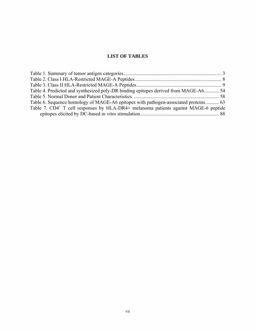
LIST OF TABLES Table 1. Summary of tumor antigen categories .............................................................................. 3 Table 2. Class I HLA-Restricted MAGE-A Peptides ..................................................................... 8 Table 3. Class II HLA-Restricted MAGE-A Peptides.................................................................... 9 Table 4. Predicted and synthesized poly-DR binding epitopes derived from MAGE-A6............ 54 Table 5. Normal Donor and Patient Characteristics. .................................................................... 58 Table 6. Sequence homology of MAGE-A6 epitopes with pathogen-associated proteins........... 63 Table 7. CD4+ T cell responses by HLA-DR4+ melanoma patients against MAGE-6 peptide
epitopes elicited by DC-based in vitro stimulation............................................................... 88
vii
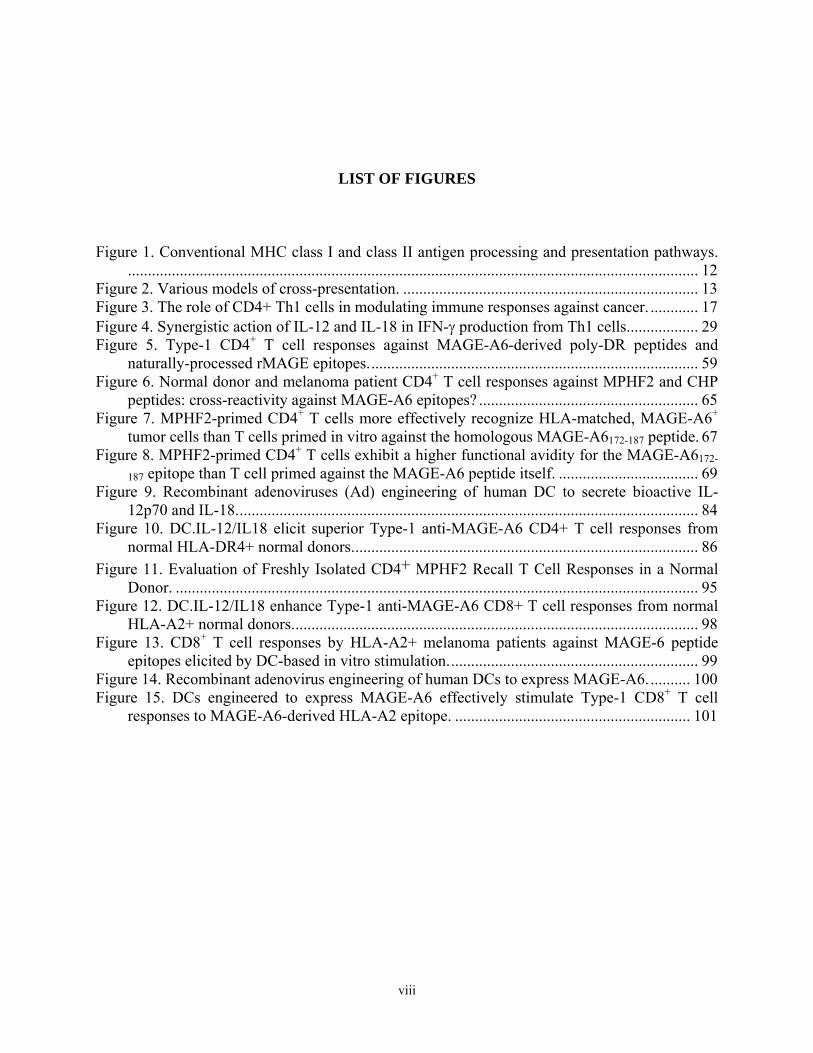
LIST OF FIGURES Figure 1. Conventional MHC class I and class II antigen processing and presentation pathways.
............................................................................................................................................... 12 Figure 2. Various models of cross-presentation. .......................................................................... 13 Figure 3. The role of CD4+ Th1 cells in modulating immune responses against cancer. ............ 17 Figure 4. Synergistic action of IL-12 and IL-18 in IFN-γ production from Th1 cells.................. 29 Figure 5. Type-1 CD4+ T cell responses against MAGE-A6-derived poly-DR peptides and
naturally-processed rMAGE epitopes................................................................................... 59 Figure 6. Normal donor and melanoma patient CD4+ T cell responses against MPHF2 and CHP
peptides: cross-reactivity against MAGE-A6 epitopes? ....................................................... 65 Figure 7. MPHF2-primed CD4+ T cells more effectively recognize HLA-matched, MAGE-A6+
tumor cells than T cells primed in vitro against the homologous MAGE-A6172-187 peptide. 67 Figure 8. MPHF2-primed CD4+ T cells exhibit a higher functional avidity for the MAGE-A6172-
187 epitope than T cell primed against the MAGE-A6 peptide itself. ................................... 69 Figure 9. Recombinant adenoviruses (Ad) engineering of human DC to secrete bioactive IL-
12p70 and IL-18.................................................................................................................... 84 Figure 10. DC.IL-12/IL18 elicit superior Type-1 anti-MAGE-A6 CD4+ T cell responses from
normal HLA-DR4+ normal donors....................................................................................... 86 Figure 11. Evaluation of Freshly Isolated CD4+ MPHF2 Recall T Cell Responses in a Normal
Donor. ................................................................................................................................... 95 Figure 12. DC.IL-12/IL18 enhance Type-1 anti-MAGE-A6 CD8+ T cell responses from normal
HLA-A2+ normal donors...................................................................................................... 98 Figure 13. CD8+ T cell responses by HLA-A2+ melanoma patients against MAGE-6 peptide
epitopes elicited by DC-based in vitro stimulation............................................................... 99 Figure 14. Recombinant adenovirus engineering of human DCs to express MAGE-A6........... 100 Figure 15. DCs engineered to express MAGE-A6 effectively stimulate Type-1 CD8+ T cell
responses to MAGE-A6-derived HLA-A2 epitope. ........................................................... 101
viii

PREFACE
A Japanese proverb states, “Fall down seven times. Get up eight.” Getting up is
particularly easy when you are surrounded by caring and supportive people that will stand by
you regardless of adversities. I have been blessed to be surrounded by a number of such
individuals. The first of these people I would like to thank is my mentor, Dr. Walter J. Storkus. I
have met Dr. Storkus years before I joined his lab, and once I came to the University of
Pittsburgh he was the clear choice for my advisor. I feel extremely fortunate and honored to have
spent my graduate school years under the tutelage of such an exceptional scientist and human
being. I would also like to thank all the people who have been a part of the Storkus Lab during
my years here, and who have always provided support, insight, help, and friendship that made
my tenure more gratifying. I would next like to thank my Thesis Committee for their guidance
and patience throughout my time here. Furthermore, I would like to thank all of my friends who
gave me regular reality checks.
Finally, I would like to extend a special thank you to my wonderful family, without
whom I would have not made it this far. Throughout my life, my mother Svetlana, sister Dušica,
grandmother Nadežda, and father Nikola have provided me with love, wisdom and
encouragement that gave me strength in moments of doubt. Your guidance and support has never
been more valuable than during my time in graduate school, and for that I dedicate this thesis to
you. A Serbian proverb states, “Be humble for you are made of earth. Be noble for you are made
of stars.” One always needs to remember his roots.
ix
1. INTRODUCTION
Previously it has been observed that some human tumors, especially melanoma and renal
cell carcinoma (RCC), can occasionally undergo spontaneous regression (1, 2). These findings
have inspired the imagination of clinicians and scientists, and have created hope that the immune
system can specifically recognize and eliminate cancers. Since the first description of a
molecularly-defined human tumor-associated antigen (TAA) recognized by cytotoxic T cells 15
years ago (3), advances in understanding the nature of tumor-specific immune responses and
mechanisms of tolerance induction have encouraged researchers and clinicians alike to develop a
more refined approach to immune-mediated therapies. Studies utilizing expression cloning of
TAA cDNAs have been integrated with novel strategies such as reverse immunology,
biochemical methods, genetic approaches, and serological analysis of recombination expression
libraries (SEREX) to identify a number of TAAs. Reverse immunology refers to a strategy where
epitopes are predicted on the basis of known HLA-binding motifs from an already identified
TAA. Biochemical methods involve eluting and fractionating TAA peptides naturally expressed
on tumor cells in the context of HLA molecules by reverse-phase high-performance liquid
chromatography (HPLC) and mass spectrometry. Genetic approaches are used to identify tumor
genes coding for the epitopes recognized by isolated patient cytotoxic T cell clones reactive
against autologous tumors. SEREX is based on the recognition of tumor antigens by cancer
patient’s autologous sera. All of these strategies have successfully been utilized to identify a
number of TAA that can be presented by tumor cells or by antigen presenting cells (APCs) in the
context of major histocompatibility complex (MHC) molecules on their cell surfaces (4-7).
1
1.1. Tumor Associated Antigen Classification
According to the pattern of expression in neoplastic and normal tissues, TAAs can be
classified into four major categories (Table 1). The first category is cancer-testis antigens. These
are proteins encoded by genes expressed in various tumors but not in normal tissues, except for
testis and placenta. Antigens that belong to this group are MAGE, GAGE, and BAGE families,
as well as NY-ESO-1 and its alternative ORF products LAGE and CAMEL. The second group
represents differentiation antigens that are shared between tumors and the normal tissue from
which the tumor arose. Of the ones discovered so far, most are expressed in melanoma and
normal melanocytes, such as tyrosinase, Melan-A/MART-1, gp100, TRP-1, and TRP-2. The
third category is tumor-specific antigens. These antigens are generated by point mutations (e.g.
p53, Ras, CDK4, β-catenin) (5, 6) or tumor-specific splicing aberrations in genes that are
ubiquitously expressed (e.g. TRP-2/INT2) (8), and are expressed only in tumors where they were
identified (unlike cancer-testis antigens). These molecular changes are associated with
neoplastic transformation and/or progression. The fourth group of antigens is widely occurring,
over-expressed TAA. These are proteins that have been detected in histologically different types
of tumors (often with no preferential expression on a certain type of cancer) as well as in many
normal tissues, generally with lower expression levels. Some of the antigens belonging to this
group include survivin, MUC1/2 and EphA2, among others.
2
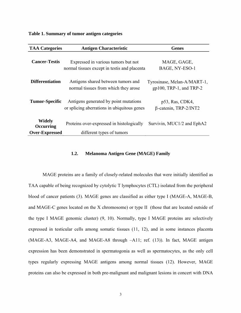
Table 1. Summary of tumor antigen categories
TAA Categories Antigen Characteristic Genes
Cancer-Testis Expressed in various tumors but not MAGE, GAGE,
normal tissues except in testis and placenta BAGE, NY-ESO-1
Differentiation Antigens shared between tumors and Tyrosinase, Melan-A/MART-1, normal tissues from which they arose gp100, TRP-1, and TRP-2
Tumor-Specific Antigens generated by point mutations p53, Ras, CDK4, or splicing aberrations in ubiquitous genes β-catenin, TRP-2/INT2
Widely Occurring Proteins over-expressed in histologically Survivin, MUC1/2 and EphA2
Over-Expressed different types of tumors
1.2. Melanoma Antigen Gene (MAGE) Family
MAGE proteins are a family of closely-related molecules that were initially identified as
TAA capable of being recognized by cytolytic T lymphocytes (CTL) isolated from the peripheral
blood of cancer patients (3). MAGE genes are classified as either type I (MAGE-A, MAGE-B,
and MAGE-C genes located on the X chromosome) or type II (those that are located outside of
the type I MAGE genomic cluster) (9, 10). Normally, type I MAGE proteins are selectively
expressed in testicular cells among somatic tissues (11, 12), and in some instances placenta
(MAGE-A3, MAGE-A4, and MAGE-A8 through –A11; ref. (13)). In fact, MAGE antigen
expression has been demonstrated in spermatogonia as well as spermatocytes, as the only cell
types regularly expressing MAGE antigens among normal tissues (12). However, MAGE
proteins can also be expressed in both pre-malignant and malignant lesions in concert with DNA
3
hypomethylation (14, 15). As both testis and placenta are considered to represent
immunologically privileged regions due to their lack of/deficiency in MHC class I expression,
any potential vaccination strategies using these antigens would be expected to have only limited
pathologic effects in patients, making these antigens acceptable targets for cancer vaccines.
1.2.1. MAGE Function
While most of the members of the MAGE family have been molecularly characterized,
their cellular function remains a major mystery. This is particularly true for type I MAGE
proteins. Most of the functional analyses reported have thus far been performed on necdin and
MAGE-D1 (also known as NRAGE), growth suppressors expressed predominantly in post-
mitotic neurons that have been implicated in their terminal differentiation (reviewed in ref. (16)).
Necdin is a cell cycle regulator necessary for the terminal differentiation and survival of primary
dorsal root ganglion neurons. It serves as a growth suppressor that is functionally similar to the
retinoblastoma (RB) tumor suppressor protein. Necdin is involved in the terminal differentiation
and survival of nerve growth factor (NGF)-dependent dorsal root ganglion neurons. Suppression
of necdin expression in neurons leads to caspase-3-dependent apoptosis (17). Necdin appears to
interact with cell cycle promoting proteins such as simian virus 40 large T antigen, adenovirus
E1A, and transcription factor E2F1. It represents a growth suppressor that targets and modulates
the biological functions of p53 in post-mitotic neurons (18). Necdin markedly suppresses p53-
dependent activation of the p21/WAF promoter, and in doing so, inhibits p53-induced apoptosis
of tumor cells. Furthermore, necdin and p53 inhibit cell growth in an additive manner. MAGE-
D1 was identified as a binding partner for the p75 neurotrophin receptor, the apoptosis inhibitory
4
protein XIAP, and the Dlx/MSX homeodomain proteins. It appears to block cell cycle
progression, and unlike necdin, is involved in cellular pro-apoptotic pathways (16, 19).
Limited data has been accumulated regarding the function of type I MAGE genes,
particularly MAGE-A1 and MAGE-A4. Stable (enforced) expression of MAGE-A1 reduces the
susceptibility of tumor cell lines to TNF-α-mediated cytotoxicity (20), suggesting that MAGE-
A1 is cyto-protective. Contrary to this paradigm, MAGE-A4 has been reported to bind to, and
suppress, the oncoprotein gankyrin in hepatocellular carcinoma (21). MAGE-A4 also partially
suppresses both anchorage-independent growth in vitro and tumor formation in athymic mice
(21). MAGE-A4 appears to promote cellular apoptosis in both p53-dependent and p53-
independent manners. It stabilizes p53 protein levels, but decreases cellular expression of p21 by
binding to Miz-1, in concert with down-regulating Bcl-xL expression during the process of
apoptosis (22). These conflicting reports suggest that, while highly-homologous, MAGE family
proteins may mediate disparate functions associated with cell cycling and death.
1.2.2. MAGE-A Subfamily
MAGE-A1 was one of the first TAA reported based on modern molecular cloning
approaches (3). Subsequently, new members of this family have been isolated, largely based on
homology searches predicated on a MAGE-A1 template. The MAGE-A gene family is currently
composed of 12 members (i.e. MAGE-A1 through -A12), that are in aggregate expressed by
more than half of all human cancers. The MAGE-A gene cluster is located on chromosome
Xq28, and all open reading frames are contained within a single exon (23). These genes encode
intracellular proteins that have most commonly been observed in the cytoplasm (24, 25), but in
5
some cases they have also been observed in the nuclei of well-differentiated tumors (25).
MAGE-A expression is frequently observed in melanoma specimens (26, 27), but not in naevi
(including Spitz, dysplastic naevi, junctional and compound naevi) (28). In fact, greater
immunohistochemical staining of tumor cells with anti-MAGE antibody has been associated
with an invasive phenotype and a decreased in the overall survival rate of cancer patients (29,
30).
MAGE-A antigens have been evaluated as targets for immunoreactivity in a number of
published studies. A summary of MAGE-A-derived MHC class I- and class II-restricted epitopes
that have been previously reported is provided in Tables 2 and 3 (7). What is unique about these
proteins is that they are highly homologous, and immunogenic peptides identified within one
MAGE-A protein are often shared or highly-homologous with epitopes encoded by other
members of the MAGE-A family. In vivo vaccination studies utilizing MAGE-A
peptides/cDNAs showed that epitope-specific CD4+ (31) and CD8+ (32) T cell responses can be
primed in immunized patients. In one particular study, vaccination of metastatic melanoma
patients with cutaneous injections of a recombinant canary pox virus carrying a mini-gene
coding for two HLA-A1-restricted peptides encoded by MAGE-A1 and MAGE-A3, resulted in
the enhancement of anti-tumor CTL responses. Anti-tumor CTLs (i.e. specific for TAA other
than MAGE-A1 and MAGE-A3 epitopes) were 10,000 times more frequent among tumor
infiltrating lymphocytes (TILs) than vaccine-specific T cells, suggesting that treatment-induced
CTLs were not likely to represent the effectors associated with therapeutic benefit in these
patients. It suggests instead that through the process of epitope spreading, vaccine-associated T
cells may enable large numbers of anti-tumor (although not necessarily MAGE-specific) CTLs to
be effectively cross-primed in vivo, yielding a clinically-effective, tumoricidal T cell repertoire
6

(33, 34). These studies suggest that MAGE-A antigens likely possess potential therapeutic value
as targets of vaccine intervention strategies.
7
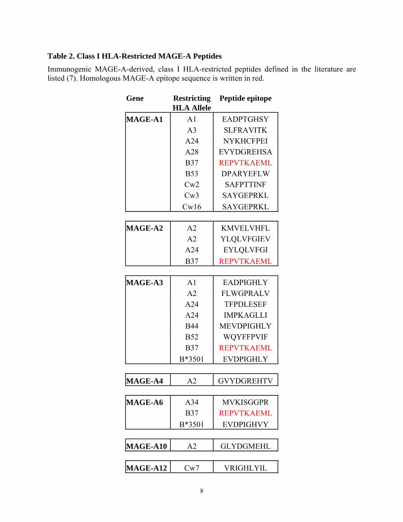
Table 2. Class I HLA-Restricted MAGE-A Peptides
Immunogenic MAGE-A-derived, class I HLA-restricted peptides defined in the literature are listed (7). Homologous MAGE-A epitope sequence is written in red.
Gene Restricting Peptide epitope HLA Allele MAGE-A1 A1 EADPTGHSY A3 SLFRAVITK A24 NYKHCFPEI A28 EVYDGREHSA B37 REPVTKAEML B53 DPARYEFLW Cw2 SAFPTTINF Cw3 SAYGEPRKL Cw16 SAYGEPRKL MAGE-A2 A2 KMVELVHFL A2 YLQLVFGIEV A24 EYLQLVFGI B37 REPVTKAEML MAGE-A3 A1 EADPIGHLY A2 FLWGPRALV A24 TFPDLESEF A24 IMPKAGLLI B44 MEVDPIGHLY B52 WQYFFPVIF B37 REPVTKAEML B*3501 EVDPIGHLY MAGE-A4 A2 GVYDGREHTV MAGE-A6 A34 MVKISGGPR B37 REPVTKAEML B*3501 EVDPIGHVY MAGE-A10 A2 GLYDGMEHL MAGE-A12 Cw7 VRIGHLYIL
8
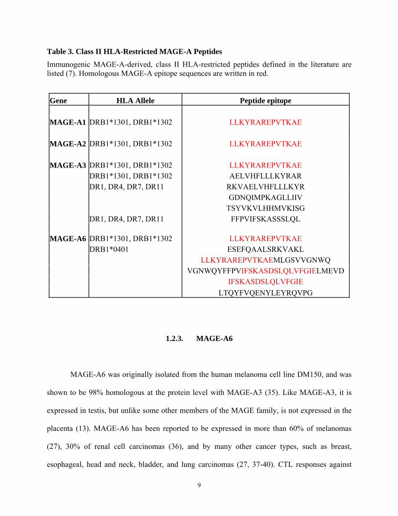
Table 3. Class II HLA-Restricted MAGE-A Peptides
Immunogenic MAGE-A-derived, class II HLA-restricted peptides defined in the literature are listed (7). Homologous MAGE-A epitope sequences are written in red.
Gene HLA Allele Peptide epitope MAGE-A1 DRB1*1301, DRB1*1302 LLKYRAREPVTKAE MAGE-A2 DRB1*1301, DRB1*1302 LLKYRAREPVTKAE MAGE-A3 DRB1*1301, DRB1*1302 LLKYRAREPVTKAE DRB1*1301, DRB1*1302 AELVHFLLLKYRAR DR1, DR4, DR7, DR11 RKVAELVHFLLLKYR GDNQIMPKAGLLIIV TSYVKVLHHMVKISG DR1, DR4, DR7, DR11 FFPVIFSKASSSLQL MAGE-A6 DRB1*1301, DRB1*1302 LLKYRAREPVTKAE DRB1*0401 ESEFQAALSRKVAKL LLKYRAREPVTKAEMLGSVVGNWQ VGNWQYFFPVIFSKASDSLQLVFGIELMEVD IFSKASDSLQLVFGIE LTQYFVQENYLEYRQVPG
1.2.3. MAGE-A6
MAGE-A6 was originally isolated from the human melanoma cell line DM150, and was
shown to be 98% homologous at the protein level with MAGE-A3 (35). Like MAGE-A3, it is
expressed in testis, but unlike some other members of the MAGE family, is not expressed in the
placenta (13). MAGE-A6 has been reported to be expressed in more than 60% of melanomas
(27), 30% of renal cell carcinomas (36), and by many other cancer types, such as breast,
esophageal, head and neck, bladder, and lung carcinomas (27, 37-40). CTL responses against
9
MAGE-A6 have been reported to occur naturally in the setting of spontaneously regressing
human melanoma (41), suggesting that the immune targeting of this antigen may be linked with
tumor regression in situ, bolstering its potential therapeutic value. It has been shown to react with
sera extracted from breast cancer patients but not normal donors (42). The clinical relevance of
the MAGE-A6 antigen has been further substantiated in a clinical study where MAGE-A6-
specific CD8+ T cell clones were detected in a metastatic melanoma patient that had complete
tumor regression following adoptive transfer of autologous tumor-specific tumor-infiltrating
lymphocytes (TILs) (43). This is compounded by its wide range of expression among cancer
types, and its lack of expression by normal tissues, which theoretically limits concerns over
autoimmune pathology resulting from MAGE-A6-based cancer vaccines and immunotherapies.
1.3. Tumor Antigen Processing
Endogenous and exogenous (usually internalized by APCs) TAAs are processed
principally via the cytosolic and endo/lysosomal pathways, respectively. They are presented as
short protein fragments by MHC class I (endogenous peptides 8-9 amino acids long) and class II
(exogenous peptides up to 35 amino acids in length) (44, 45). Tumor peptides associate with
MHC molecules in intracellular compartments [endoplasmic reticulum (ER) for MHC class I,
and endolysosome for MHC class II], and once forming stable complexes, they are transported to
the cell surface where they become accessible to T cell scrutiny (45, 46). Most TAAs contain a
number of sequences that have been predicted and/or documented to bind to MHC molecules
(47). Typically, only a few of the potential epitopes elicit a strong (immunodominant epitopes)
10
cytotoxic T cell responses, while the majority elicit weak or no responses (sub-dominant or
cryptic epitopes) (48, 49).
1.3.1. Classical MHC Class I/Peptide Presentation
Classical MHC class I antigen presentation (Figure 1) starts with the degradation of
endogenous (intracellularly synthesized) proteins by the proteasome. Peptides of the correct
length and sequence (possessing the correct anchor residues) bind to class I with the slowest off-
rate (50, 51). Peptides that are longer, or do not have appropriate anchor residues bind with faster
off-rates (52, 53). A small fraction of the peptide fragments that result from this degradation
survive complete destruction and are transported into the ER and loaded onto the MHC by the
peptide loading complex composed of one TAP1/TAP2 (transporter associated with antigen
presentation) heterodimer associated with 4 tapasin, 4 calreticulin and 4 MHC class I heavy
chain/beta-2 microglobulin (β2m) dimers (54). In the ER, the peptides are loaded onto newly
synthesized MHC class I molecules, forming ternary complexes, each composed of MHC class I
heavy chain, β2m and peptide. These stable complexes are then transported to the cell surface
(55) where they are exposed to CD8+ T cell surveillance.
11
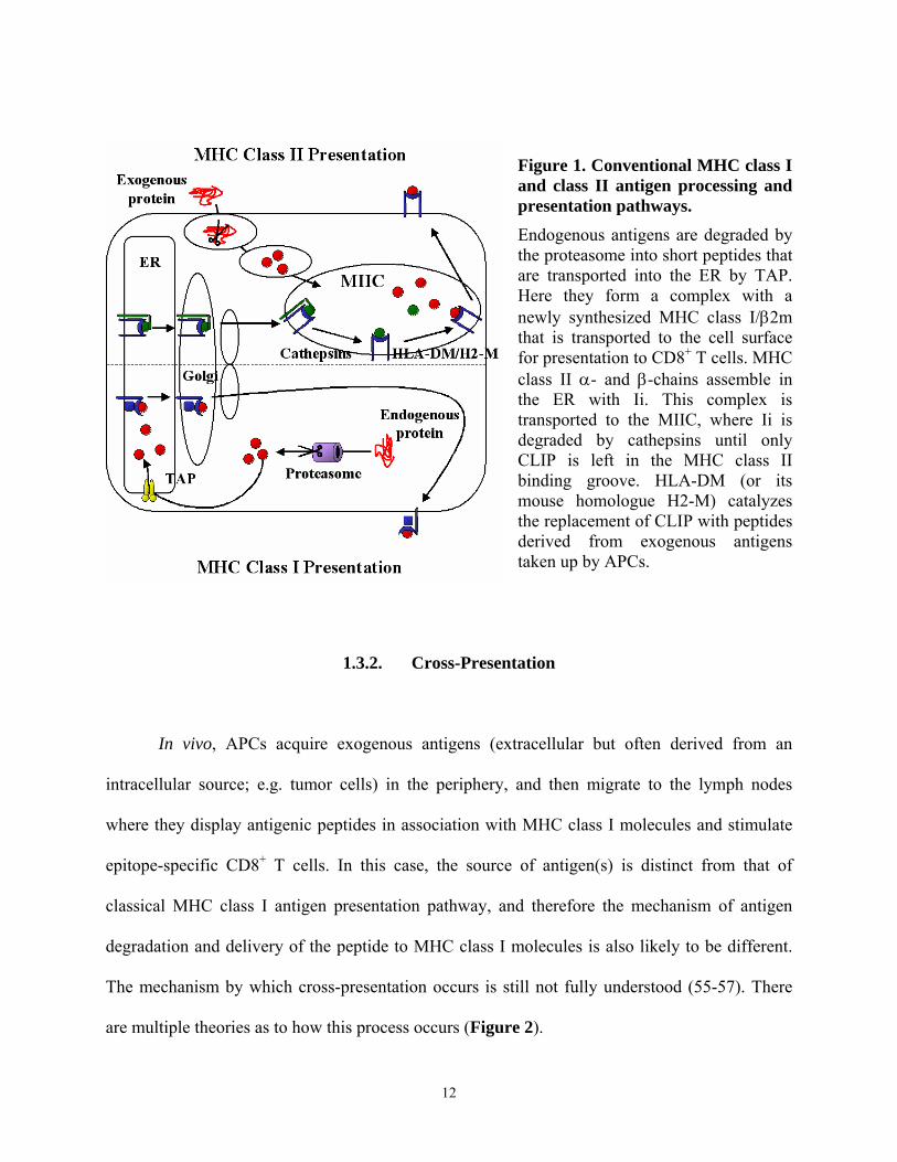
Figure 1. Conventional MHC class I and class II antigen processing and presentation pathways. Endogenous antigens are degraded by the proteasome into short peptides that are transported into the ER by TAP. Here they form a complex with a newly synthesized MHC class I/β2m that is transported to the cell surface for presentation to CD8+ T cells. MHC class II α- and β-chains assemble in the ER with Ii. This complex is transported to the MIIC, where Ii is degraded by cathepsins until only CLIP is left in the MHC class II binding groove. HLA-DM (or its mouse homologue H2-M) catalyzes the replacement of CLIP with peptides derived from exogenous antigens taken up by APCs.
1.3.2. Cross-Presentation
In vivo, APCs acquire exogenous antigens (extracellular but often derived from an
intracellular source; e.g. tumor cells) in the periphery, and then migrate to the lymph nodes
where they display antigenic peptides in association with MHC class I molecules and stimulate
epitope-specific CD8+ T cells. In this case, the source of antigen(s) is distinct from that of
classical MHC class I antigen presentation pathway, and therefore the mechanism of antigen
degradation and delivery of the peptide to MHC class I molecules is also likely to be different.
The mechanism by which cross-presentation occurs is still not fully understood (55-57). There
are multiple theories as to how this process occurs (Figure 2).
12
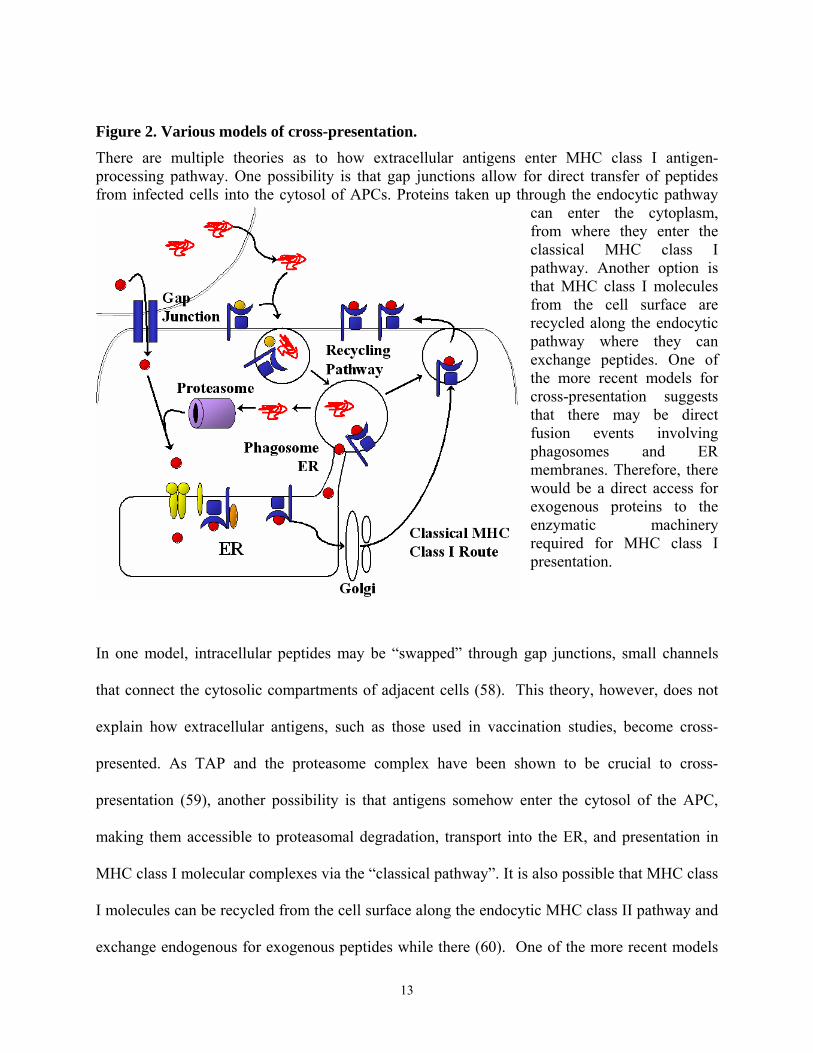
Figure 2. Various models of cross-presentation. There are multiple theories as to how extracellular antigens enter MHC class I antigen-processing pathway. One possibility is that gap junctions allow for direct transfer of peptides from infected cells into the cytosol of APCs. Proteins taken up through the endocytic pathway
can enter the cytoplasm, from where they enter the classical MHC class I pathway. Another option is that MHC class I molecules from the cell surface are recycled along the endocytic pathway where they can exchange peptides. One of the more recent models for cross-presentation suggests that there may be direct fusion events involving phagosomes and ER membranes. Therefore, there would be a direct access for exogenous proteins to the enzymatic machinery required for MHC class I presentation.
In one model, intracellular peptides may be “swapped” through gap junctions, small channels
that connect the cytosolic compartments of adjacent cells (58). This theory, however, does not
explain how extracellular antigens, such as those used in vaccination studies, become cross-
presented. As TAP and the proteasome complex have been shown to be crucial to cross-
presentation (59), another possibility is that antigens somehow enter the cytosol of the APC,
making them accessible to proteasomal degradation, transport into the ER, and presentation in
MHC class I molecular complexes via the “classical pathway”. It is also possible that MHC class
I molecules can be recycled from the cell surface along the endocytic MHC class II pathway and
exchange endogenous for exogenous peptides while there (60). One of the more recent models
13
for cross-presentation suggests that there may be direct fusion events involving phagosomes and
ER membranes. Therefore, there would be a direct access to the enzymatic machinery required
for MHC class I presentation (59). However, this model is rather controversial and has recently
been refuted (61). More studies are clearly needed to further dissect this phenomenon, for
without this knowledge, it will be difficult to completely rationalize optimal vaccine
development predicated on cross-presentation in vivo.
1.3.3. MHC Class II/Peptide Presentation
The MHC class II processing pathway processes and presents exogenous, as well as
self/intrinsic-antigens that are degraded in the endocytic pathway (Figure 1). MHC class II αβ
dimers assemble in the ER with the chaperone invariant chain (Ii) and its class II-associated Ii
peptide (CLIP) portion inserted within the MHC peptide-binding cleft, which stabilizes and
protects this site from interacting with other peptides in the ER microenvironment. MHC−Ii
complexes are transported to early endosomes, and then via late endosomes into lysosomal
compartments, during which time, they may encounter antigenic peptides resulting from the
degradation of endocytosed proteins (62). Endocytosed antigens may be unfolded by thiol-
reductases and then efficiently degraded by cathepsins, with peptides formed as intermediates
during late endosomal protein degradation loaded into MHC class II complexes in a reaction
catalyzed by the chaperone protein HLA-DM in the MHC class II compartment (MIIC), before
transport of mature class II/peptide complexes to the plasma membrane (63). In order for these
peptides to bind within the MHC class II groove, this pocket must be vacated by the Ii-derived
CLIP peptide. Displacement of CLIP is facilitated by acidic pH in endosomes which favors an
14
open conformation in the MHC class II molecule and hence peptide exchange, the activity of the
HLA-DM which stabilizes the open conformation, and by proteolytic elimination of the regions
of Ii that flank the CLIP peptide. The peptide-MHC class II complexes are then transported to
the cell surface, where they may be surveyed by CD4+ T cells (62). A study done by Lazarski et
al. has suggested that immunodominance of a given peptides is determined by the comparative
stability of MHC class II:peptide complexes (64). In other words, immunodominant peptides
typically possess long half-lives in class II complexes, while cryptic or poorly immunogenic
peptides display significantly shorter half-lives in these complexes.
1.4. General Overview of T Cell Selection
The T cell repertoire is provided via a broad array of clonotypic T cells exhibiting
heterogeneous usage of TCR Vα and Vβ chains. These T cells are capable of distinguishing
foreign from self-antigens, and are normally capable of responding uniquely and appropriately to
each of these stimuli. Thymic selection of T cells involves both positive (able to be “restricted”
by self MHC) and negative selective (not pathologically reactive against self MHC) mechanisms
based on the avidity of T cell interaction with antigen-MHC complexes. Apoptosis, or
programmed cell death, plays a critical role in selecting the thymocyte pool, deleting cells
expressing an unproductive T cell receptor (TCR), or exhibiting hyper-responsiveness upon
encountering self MHC/self-peptide complexes. Thymocytes progress through well-defined steps
during their maturation, exhibiting characteristic phenotypes at each stage. Immature thymocytes
will survive if signals generated by TCR-MHC/peptide engagement are interpreted as
appropriate (positive selection), but will be deleted by apoptosis if these generated signals are
interpreted as either inappropriately weak (death by neglect/glucocorticoid-induced cell death) or
15
inappropriately strong (negative selection, therefore posing an autoimmune risk) (65, 66). A
fraction of all T cells escapes thymic selection and ends up in the circulation, and these cells are
subjected to additional peripheral selection criteria during systemic immune responses to
antigenic challenge. It has been shown in mouse models that autoimmune T cells bearing high
affinity TCR that escape thymic selection may be deleted as a result of immunizing animals with
strongly immunogenic epitopes. However, if the animals were immunized with a weakly-
immunogenic analogue, high-affinity T cells expand (67). As TAAs are considered to be “self”
antigens, stimulating and sustaining an immune response to these antigens is a difficult
proposition. An antigen used for vaccination needs to be preferentially expressed on tumor cells,
therefore limiting any damage to healthy tissues, and must be capable of inducing as high avidity
T cell responders as possible, without consequently promoting their (apoptotic) deletion.
1.5. CD4+ T Cell-Mediated Immunity
Mature CD4+ are typically known as T-helper (Th) cells. CD4+ lymphocytes are believed
to polarize the adaptive immune response by secreting a dominant panel of cytokines in response
to specific antigen recognition. Based on these cytokine profiles, Th cells can be generally
segregated into three major subsets: Th1, Th2, and Th3/T-regulatory (Treg) subsets (68). Th1
cells provide help for cellular immunity and perform several major functions (Figure 3).
16
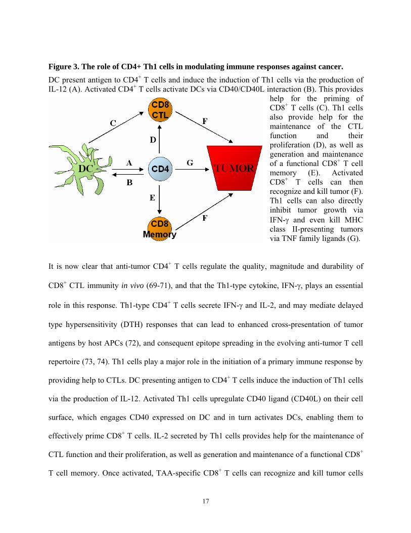
Figure 3. The role of CD4+ Th1 cells in modulating immune responses against cancer. DC present antigen to CD4+ T cells and induce the induction of Th1 cells via the production of IL-12 (A). Activated CD4+ T cells activate DCs via CD40/CD40L interaction (B). This provides
help for the priming of CD8+ T cells (C). Th1 cells also provide help for the maintenance of the CTL function and their proliferation (D), as well as generation and maintenance of a functional CD8+ T cell memory (E). Activated CD8+ T cells can then recognize and kill tumor (F). Th1 cells can also directly inhibit tumor growth via IFN-γ and even kill MHC class II-presenting tumors via TNF family ligands (G).
It is now clear that anti-tumor CD4+ T cells regulate the quality, magnitude and durability of
CD8 CTL immunity in vivo (69-71), and that the Th1-type cytokine, IFN-γ, plays an essential
role in this response. Th1-type CD4+ T cells secrete IFN-γ and IL-2, and may mediate delayed
type hypersensitivity (DTH) responses that can lead to enhanced cross-presentation of tumor
antigens by host APCs (72), and consequent epitope spreading in the evolving anti-tumor T cell
repertoire (73, 74). Th1 cells play a major role in the initiation of a primary immune response by
providing help to CTLs. DC presenting antigen to CD4 T cells induce the induction of Th1 cells
via the production of IL-12. Activated Th1 cells upregulate CD40 ligand (CD40L) on their cell
surface, which engages CD40 expressed on DC and in turn activates DCs, enabling them to
effectively prime CD8 T cells. IL-2 secreted by Th1 cells provides help for the maintenance of
CTL function and their proliferation, as well as generation and maintenance of a functional CD8
T cell memory. Once activated, TAA-specific CD8 T cells can recognize and kill tumor cells
+
+
+
+
+
17
(75). Furthermore, Th1-type CD4+ T cells may mediate direct tumoricidal activity via TNF
family ligand members and can inhibit tumor angiogenesis via locoregional production of IFN-γ
(76-79). Th2 cells and their associated cytokines are often linked to strong antibody (humoral)
responses, and they tend to inhibit Th1 responses.
The typical Th2-type cytokine profile includes the production of IL-4, IL-5 and IL-13
(68). T
1.6. Immunoregulatory Function of IFN-γ
Anti-tumor CD4+ T cells regulate the quality, magnitude and durability of CD8+ CTL
immun
hese cytokines are best known for supporting B cell growth and differentiation, leading to
the enhanced ability of B/plasma cells to secrete antibodies. Furthermore, they have been shown
to protect tumor cells in vivo by suppressing Th1-type anti-tumor immune responses, and their
presence in serum is usually correlated with poor prognosis and the reduced overall survival of
cancer patients (80). Th3/Treg cells generally produce IL-10 and/or TGF-β, with both cytokines
capable of strongly suppressing the proliferative and effector functions of Th1- and Th2-type
CD4+ T cells. As a consequence, these Th3/Treg cells are also known as T suppressor cells (68,
81, 82).
ity in vivo (69-71), and IFN-γ, a Type-1 cytokine, plays an essential role in this response.
The production and secretion of IFN-γ is promoted by IL-12 family members (i.e, IL-12p70, IL-
23 and IL-27), and by IL-18 (83-87). Type-1 CD4+ T cells (Th1), CD8+ T cells (Tc1) and natural
killer (NK) cells secrete IFN-γ and may mediate DTH responses, leading to cross-presentation of
tumor antigens by host APCs (72), and resulting in anti-tumor epitope spreading (73, 74).
18
Furthermore, CD4+ T cells were shown to inhibit tumor angiogenesis via locoregional
production of IFN-γ (76-79).
IFN-γ plays an important role in regulating key components involved in the MHC class I
and II processing and presentation machinery. Previous studies have shown that IFN-γ induces
MHC class I and II synthesis and expression (88-90), regulates peptide processing,
compartmentalization, MHC loading and MHC/peptide complex delivery to the cell surface (91-
93), and qualitatively influences presentation of cryptic MHC class I T cell epitopes (94). IFN-γ
induces the exchange of the three catalytic subunits (LMP2, LMP7, and MECL-1) of the
proteasome complex, thus forming the so-called “immunoproteasome” (95), which allows for
processing and presentation of otherwise cryptic epitopes. Furthermore, IFN-γ induces the
expression of PA-28, a proteasome activator, that is able to increase the proteolytic efficiency of
the 20S proteasome subunit (96).
Anti-tumor Th1-type CD4+ T cells, however, appear inhibited in many cancer patients
(71, 97, 98), as reflected by decreased proliferation and T cell receptor (TCR) signaling (99), as
well as, by increased frequencies and activity of regulatory T cells (100, 101). While Th1-type
responses have been associated with spontaneous or therapy-induced regression of tumor lesions
(98, 102), tumor infiltrating lymphocytes isolated from patients with progressive lesions have
been generally reported to exhibit dominant Th2-type (secreting IL-4, IL-5) or regulatory (Th3)-
type (secreting IL-10, TGF-β1) responses (97, 98, 102).
19
1.7. CD8+ T Cell-Mediated Immunity
Past studies have shown that in order for tumors to be rejected by the immune system, a
tumor-specific CD8+ T lymphocyte response must be stimulated and sustained in cancer patients.
Since tumor cells are considered to be poor APCs due to their inhibitory properties and lack of
co-stimulatory molecules such as B7.1 and B7.2 [27], naïve CD8+ T lymphocytes need to be
activated by mature DCs presenting TAA-derived epitopes. Upon recognition of their specific
peptide, anti-tumor CD8+ T cells undergo a proliferative burst and consequently differentiate into
effector/memory cells. Naïve CD8+ T cells are efficient producers of IFN-γ and TNF-α at early
time-points after their initial priming. Furthermore, they efficiently synergize with CD40L-
expressing naïve Th cells in the optimal activation of DCs in association with enhanced APC
secretion of IL-12p70, the key Th1-inducing cytokine (103). Following the interaction with
DCs, responding T cells undergo a developmental transformation to become effector cytotoxic T
lymphocytes (CTL) and acquire the ability to kill their target cells after specific antigen
recognition (104). Once activated, CTLs become “serial” killers (i.e. able to kill multiple targets;
ref. (105)). Perforin, granulysin, and granzymes stored in pre-formed lytic granules within CTLs
are secreted within the T cell/target cell interface, with perforin and granulysin forming pores in
the target cell membrane, resulting in the sensitization of target cells to granzymes (106-108).
Granzyme B, that is also secreted, induces apoptosis by directly activating target cell caspase-3
(109) and/or by destabilizing the mitochondrial membrane (110), while Granzyme A causes
single-strand DNA breaks and apoptosis via a slower lytic pathway (111).
Another way CTLs induce cell death is by engaging tumor necrosis factor receptors
(TNFR) on target cells. While TNFR family members vary in their primary sequence, all of them
20
contain a homologous intracellular death domain (112). The best-known receptor/ligand pair in
this family is Fas and FasL (CD95/CD178). When the CTL TCR are engaged and activated by
MHC class I complexes, T cells upregulate FasL expression (113). Just like the rest of the TNF
family members, FasL is a homotrimeric protein that binds to 3 Fas receptors on CTL target cells
(114). Once bound, the death domains of the 3 Fas receptors are clustered, allowing for the
recruitment of pro-apoptotic adaptor proteins (e.g. FADD) via interactions with the death
domains on the adaptor proteins. The secondary adaptor proteins then induce apoptosis in a
caspase-8 dependent manner (115).
1.8. Regulatory T Cells
In humans, Treg cells represent approximately 1-3% of circulating CD4+ T cells (116),
and are concentrated within the CD4+CD25hi (CD25: IL-2 receptor α-chain) subset of CD4+ T
lymphocytes (117) that express FoxP3, a gene that encodes a transcription factor required for
Treg development and function (118). These cells were initially described as a subpopulation of
suppressor T cells that mediate immune tolerance by suppressing autoreactive T cells (119).
Their physiological role in healthy individuals is to protect the host against the development of
autoimmunity by regulating immune responses against antigens expressed in normal tissues.
Indeed, Treg cells have been shown to recognize self-antigens more efficiently than other T cell
subsets (120). This has been further substantiated by observations that animals deficient in Treg
cells developed severe autoimmune diseases (116, 121). CD4+ Treg can be grouped into two
major subsets: 1) naturally-occurring Treg (nTreg) produced in the thymus and that exert
21
immunosuppressive effect by cell-to-cell contact, and 2) Th3 or Tr1 cells which are induced
peripherally and suppress immune responses via secretion of IL-10 and TGFβ (122).
nTreg cells serve the important role of maintaining peripheral immune tolerance, and are
largely composed of CD25+CD62L+ T cells and natural killer T (NKT) cells. Cell-to-cell
contacts mediated through membrane-associated receptors, such as CTLA4, appear critical for
their suppressive capacity. Expression of this receptor is increased on Treg, and CTLA4-specific
antibody was shown to inhibit the Treg-induced immunosuppression in autoimmunity animal
models (123). Another possible receptor involved in this process is glucocorticoid-induced tumor
necrosis factor receptor family-related protein (GITR; TNFRSF18). However, there is still
insufficient evidence to support a causal linkage of this receptor with nTreg function (123).
Previous studies have shown that elevated numbers of Treg cells can be found in the tissues
of advanced cancer patients (124) and that high Treg frequencies are associated with reduced
overall patient survival (125). Treg cells require TCR ligation and IL-2 to become activated, after
which, they can mediate immune suppression in an antigen-independent manner (126, 127).
Normally, Treg cells are anergic (i.e. incapable of proliferation and cytokine production in
response to conventional T cell stimulation) in vitro, however this anergy can be broken by the
addition of high doses of exogenous IL-2. Recombinant IL-2 (rIL-2), which is commonly used as
an immunotherapeutic agent in cancer patients, has been implicated as playing a major role in the
generation and maintenance of Treg cells. Patients with pediatric sarcoma that had been treated
with cyclophosphamide (CY)-based chemotherapy followed by a peptide-based tumor vaccine in
conjunction with systemically-administered rIL-2 had a marked increase in the number of Treg
in their circulation as compared to patients that had not been treated with rIL-2. These cells were
not regenerated in the thymus, but were enriched by amplification of circulating CD4+CD25hi T
22
lymphocytes that survived chemotherapy-induced lymphopenia (128). A number of murine
studies have also shown that depletion of Treg cells using anti-CD25 antibodies leads to a more
effective anti-tumor immune response, culminating in the prolonged survival of tumor-bearing
animals (129-131). Furthermore, deletion of CD4+CD25hi circulating T lymphocytes using a rIL-
2 diphtheria toxin conjugate DAB389IL-2 (also known as ONTAK) allowed for the significantly
improved stimulation of tumor-specific T cell responses in renal cell carcinoma (RCC) patients
following immunization with RNA-transfected DCs, when compared with vaccination alone
(132).
In human and mouse neoplasia, Treg cells accumulate in tumors, draining lymph nodes,
and the blood stream (133, 134). The mechanisms that lead to Treg cell accumulation in tumor-
bearing hosts are still largely unknown. Most current evidence suggests that during tumor
progression, DC exposed to the tumor microenvironment acquire the capacity to secrete TGF-β
and to stimulate the expansion of nTreg cells through signals mediated through the TGF-β-
receptor II (135). These DCs appear to be of an immature, myeloid phenotype that lack
expression of co-stimulatory molecules that are needed for promoting antigen-specific T cell
responses.
Antigen presentation by immature DCs in vivo is considered to be an important pathway
by which tolerance to “self” antigens is maintained. This occurs by inhibition of T cell
proliferation, the induction of anergy within a cohort of antigen-specific T cells, as well as, the
induction of immunosuppressive Treg cells (136). Immature DC have been shown to induce both
CD4+ and CD8+ IL-10-producing Treg (123, 137). Interestingly, CD40 expression by DC has
been implicated as a key factor in Treg induction, since antigen-loaded DCs which lack CD40
23
prevent T cell priming, suppress previously primed immune responses and induce IL-10-
secreting CD4+ Treg cells (138).
1.9. Dendritic Cells (DCs)
TAAs have been utilized as active immunogens in numerous anti-tumor vaccine studies
(6, 7). Various vaccine strategies have been developed to maximize the therapeutic effect of
these antigens. One of the most effective methods utilized so far involves the use of DCs pulsed
with tumor peptides or proteins, or transfected with TAA cDNA to induce anti-tumor immunity
(139-143). DCs are considered to be the most effective APCs for the priming and maintenance
of anti-tumor immunity (144-146) and are considered to be the only APCs capable of
productively activating naïve T cells (147). They take up antigens within their
microenvironment in the periphery and process them through the endogenous and/or exogenous
pathways (45, 49, 146). Soluble or particulate antigens are typically captured by “immature”
DCs through phagocytosis, pinocytosis and receptor-mediated endocytosis (e.g. Fc receptors,
integrins, C-type lectins, and “scavenger receptors” LOX-1 and CD91) (137). Immature DC also
express low surface levels of HLA molecules, CD80, CD86, and CD40 (137), and commonly
express the chemokine receptor CCR6 (148). Once they take up antigen (and receive
maturational or environmental “danger” signals), DCs migrate to draining lymph nodes where
they may efficiently prime and expand anti-tumor T cells. During this time, DCs decrease their
ability to uptake antigen and increase their capacity to (cross)present antigens to T cells via their
MHC class I and II complexes. These DCs are typically “mature” DCs. Such DCs express
increased levels of MHC class I and II complexes as well as co-stimulatory molecules such as
CD80, CD86, and CD40 (49, 137), upregulated levels of CD83 (149), downregulated levels of
24
CCR6 and upregulated levels of CCR7 (150). These phenotypic changes allow mature DC, as
compared to immature cells, to not only productively activate naïve T lymphocytes, but to form
long-lasting “synaptic” interactions with these responder T cells, even in the immediate absence
of antigens (151). This allows the interacting lymphocytes sufficient time to “scan” DC-
presented antigens, allowing for consequent cognate signaling into the specific T cell.
1.9.1. Lymphocyte Polarization Depends on the Subtype of Stimulating DC
The fate of naïve T cells upon exposure to Ag is determined by three signals that are
provided by DCs: 1) ligation of TCRs by DC-expressed MHC-peptide complexes, 2)
engagement of DC-expressed co-stimulatory molecules, without which lymphocytes may
become anergic, and 3) DC secretion of polarizing cytokines. The secreted cytokine profile of
the stimulating DC determines the type of responder T lymphocyte functional polarization. IL-
12, IL-18, IL-23 and IL-27 polarize toward Type-1 responses, while chemokine ligands CCL2,
CCL17, CCL22 or the absence of IL-12p70 skews the response towards a Type-2 result (137,
145). The DC cytokine secretion profile depends on many factors including: the DC subtype, the
local environment and anatomic location of the DC and the type of maturation stimulus received
by DCs (152).
Conditions under which DCs are primed are important for their cytokine profiles, and
therefore the resulting class of immune responses resulting from their stimulation. DC1
(myeloid) and DC2 (plasmacytoid) subtypes stimulate Type-1 and Type-2 cells, respectively
(153). DC1 are commonly associated with monocyte-derived DCs that typically promote Th1
differentiation, in part due to their secretion of IL-12p70 (153). DC2 are represented by
25
CD4+CD3-CD11c- plasmacytoid cells that induce Th2 differentiation of CD4+ T cells via
mechanisms that do not appear to involve IL-4 or IL-12 (153, 154). DC2 precursors are natural
IFN-producing (NIP) cells that are the primary producers of IFN-α and IFN-β in vivo (155).
Furthermore, a TGF-β-secreting subset of DCs (DC3) has been defined in the tumor
microenvironment. DCs that are exposed to tumor cells can acquire the capacity to secrete TGF-
β and to stimulate the expansion of nTreg cells through TGF-β-receptor II (135).
Toll-like receptors (TLRs) have been implicated as playing important roles in the process
of DC polarization. An original belief held that TLR triggering always resulted in the
development of DC1, however it has now been shown that ligation of TLRs may also promote
the development of non-Type-1 DCs. In particular, signals mediated through Toll-like receptor 2
(TLR2), which is expressed on most CD11c+ (myeloid) DCs, may induce DC secretion of either
IL-23 or IL-10 depending on the specific TLR ligands evaluated. TLR2 forms heterodimers with
TLR1 when triggered by bacterial lipoproteins, but when engaged by mycoplasma-derived
lipoproteins, they form heterodimers with TLR6 (145). Unlike activation via TLR4 (by LPS)
leading to DC production of IL-12, TLR2 ligation by bacterial lipoproteins induces DC
expression of messenger RNA encoding the p40 and p19 subunits of the Type-1-polarizing
cytokine IL-23, but not the p35 subunit of IL-12 (156). On the other hand, mycoplasma-derived
lipopeptide 2 induces DC production of IL-10, but not IL-12, and these resulting DCs, induce
unpolarized T-cell (i.e. Th0-type) responses (157). These observations suggest that TLR2
signaling may dictate distinct cytokine profiles secreted by DCs, resulting in differential
polarizing effects on T cells primed by these APCs.
26
1.9.1.1. Role of Interleukin-12 in Promoting Therapeutic Immunity
Interleukin-12 (IL-12) is one of five heterodimeric cytokines that belong to the IL-12
family (others include IL-23, IL-27, CLC-sCNTFR, and CLC-CLF-1). It was originally
identified as cytotoxic lymphocyte maturation factor, and is composed of two covalently linked
protein chains, p35 and p40, that form the p70 heterodimer that is produced in a restricted
manner by antigen presenting cells (DCs, monocytes, macrophages, neutrophils). It binds to the
IL-12 receptor (IL-12R) complex that is composed of two chains, IL-12Rβ1 and IL-12Rβ2. IL-
12Rβ1 binds IL-12 p40 and is associated with Tyk2, while IL-12Rβ2 recognizes the p70
heterodimer or the p35 and is associated with Jak2 (158). Most of the biological responses to IL-
12 are mediated through the STAT4 signaling pathway, and optimal Th1 polarization is only
achieved in the continuous presence of IL-12 (158, 159).
IL-12 p70 effectively stimulates IFN-γ production by T, NK, and other cell types, and is a
potent inducer of Th1 cell differentiation (158). It is also capable of irreversibly repolarizing Th2
CD4+ T cells towards the Th0/Th1 phenotype, and this change is accompanied by suppression of
GATA-3 (Th2-specific transcription factor) and induction of T-bet (Th1-specific transcription
factor) (160). While effective at inducing expansion and optimal activation of Th1 CD4+ T cells,
its role in CD8+ T cell generation is somewhat less studied. In vitro priming of T cells in the
presence of IL-12p70 increases the generation and improved survival of memory CD8+ T cells in
mice after adoptive transfer of activated cells (161). However, IL-12 p40- and IL-12Rβ1-
deficient mice showed similar levels of primary and memory CD8+ T cell responses, when
compared to wild-type mice, implying that endogenous IL-12p70 is not critical for the generation
27
of IFN-γ-secreting, CD8+ cytotoxic T lymphocytes in vivo (161, 162). Together, these results
suggest that IL-12p70 can serve as an important, but non-essential regulatory factor for the
development of CD8+ T cells.
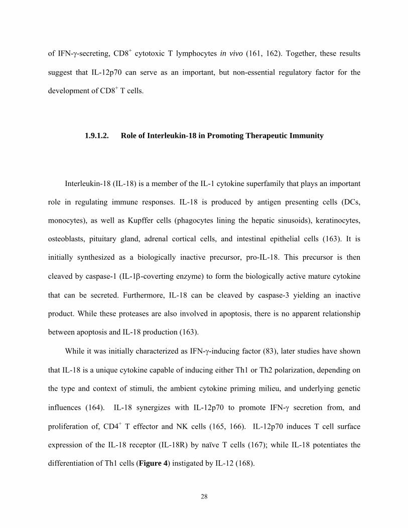
1.9.1.2. Role of Interleukin-18 in Promoting Therapeutic Immunity
Interleukin-18 (IL-18) is a member of the IL-1 cytokine superfamily that plays an important
role in regulating immune responses. IL-18 is produced by antigen presenting cells (DCs,
monocytes), as well as Kupffer cells (phagocytes lining the hepatic sinusoids), keratinocytes,
osteoblasts, pituitary gland, adrenal cortical cells, and intestinal epithelial cells (163). It is
initially synthesized as a biologically inactive precursor, pro-IL-18. This precursor is then
cleaved by caspase-1 (IL-1β-coverting enzyme) to form the biologically active mature cytokine
that can be secreted. Furthermore, IL-18 can be cleaved by caspase-3 yielding an inactive
product. While these proteases are also involved in apoptosis, there is no apparent relationship
between apoptosis and IL-18 production (163).
While it was initially characterized as IFN-γ-inducing factor (83), later studies have shown
that IL-18 is a unique cytokine capable of inducing either Th1 or Th2 polarization, depending on
the type and context of stimuli, the ambient cytokine priming milieu, and underlying genetic
influences (164). IL-18 synergizes with IL-12p70 to promote IFN-γ secretion from, and
proliferation of, CD4+ T effector and NK cells (165, 166). IL-12p70 induces T cell surface
expression of the IL-18 receptor (IL-18R) by naïve T cells (167); while IL-18 potentiates the
differentiation of Th1 cells (Figure 4) instigated by IL-12 (168).
28
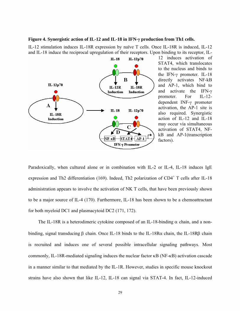
Figure 4. Synergistic action of IL-12 and IL-18 in IFN-γ production from Th1 cells. IL-12 stimulation induces IL-18R expression by naïve T cells. Once IL-18R is induced, IL-12 and IL-18 induce the reciprocal upregulation of their receptors. Upon binding to its receptor, IL-
12 induces activation of STAT4, which translocates to the nucleus and binds to the IFN-γ promoter. IL-18 directly activates NF-kB and AP-1, which bind to and activate the IFN-γ promoter. For IL-12-dependent INF-γ promoter activation, the AP-1 site is also required. Synergistic action of IL-12 and IL-18 may occur via simultaneous activation of STAT4, NF-kB and AP-1(transcription factors).
Paradoxically, when cultured alone or in combination with IL-2 or IL-4, IL-18 induces IgE
expression and Th2 differentiation (169). Indeed, Th2 polarization of CD4+ T cells after IL-18
administration appears to involve the activation of NK T cells, that have been previously shown
to be a major source of IL-4 (170). Furthermore, IL-18 has been shown to be a chemoattractant
for both myeloid DC1 and plasmacytoid DC2 (171, 172).
The IL-18R is a heterodimeric cytokine composed of an IL-18-binding α chain, and a non-
binding, signal transducing β chain. Once IL-18 binds to the IL-18Rα chain, the IL-18Rβ chain
is recruited and induces one of several possible intracellular signaling pathways. Most
commonly, IL-18R-mediated signaling induces the nuclear factor κB (NF-κB) activation cascade
in a manner similar to that mediated by the IL-1R. However, studies in specific mouse knockout
strains have also shown that like IL-12, IL-18 can signal via STAT-4. In fact, IL-12-induced
29

STAT-4 signaling is synergistically enhanced in combination with IL-18 (Figure 4) via NF-κB
and AP-1 transcription factors (164).
1.10. Cancer Vaccines and Therapies
The three traditional therapies for the clinical management of cancer are surgery, radiation,
and chemotherapy. Surgery, the process of physically removing the existing tumor from the
body, is usually the first step in treating the disease. If lesions are not easily accessible for
surgical resection, tumors are typically treated with locoregional radiation therapy. Radiotherapy
involves exposing cancerous tissues and its supportive vascular bed to various forms of radiation
in order to cause DNA damage, forcing these tissues to undergo differential apoptosis. If there is
a possibility that the disease has metastasized (spread to other tissues) or if the disease affects
leukocytes, chemotherapy is commonly applied as a systemic therapy. This generally involves
the administration of chemicals that inhibit the ability of cancer cells to survive and replicate.
While these three methods have showed some degree of clinical success, their long-term
benefits, particularly in the cases of radiotherapy and chemotherapy, are generally perceived as
limited. These treatments are often very destructive not only to tumor cells, but to normal tissues
as well. Furthermore, recurrence of disease is very common and is frequently found to be
resistant to the original treatment modality. For these reasons, it is necessary to establish novel
therapy methods for tumors that will provide more specific treatment, and long-term protection
from recurrence. Various immunotherapy strategies have the potential to provide these benefits.
Immunotherapeutic strategies utilize various components of the immune system to promote
immune responses against a specific disease, such as cancer. There are three lines of evidence
30
that suggest that cancer immunotherapy can be beneficial in humans: 1) immunosuppressed
transplant recipients display higher incidences of non-viral tumors, such as melanomas, colon,
lung, pancreas, bladder, kidney, and endocrine system cancers than immunocompetent control
populations (173); 2) the presence of lymphocytes within the tumor is often a positive prognostic
indicator of patient survival (174), and 3) a minority of cancer patients (< 5%) are able to
develop spontaneous innate and acquired immune responses to the tumors they bear (7, 175,
176). One of the first pieces of clinical evidence suggesting that the manipulation of the immune
system could be beneficial as a cancer therapy involved the administration of interleukin-2 (IL-2,
which is a lymphocyte proliferation-inducing cytokine produced by T cells that has the ability to
induce proliferation of T cells that have recognized their specific antigen) (177). In that study,
IL-2 treatment of patients with metastatic renal carcinoma or metastatic melanoma induced
tumor regression in 15-20% of treated patients. Since then, great progress has been made in
understanding the immune response to tumors, and based on this knowledge a number of
different immunization strategies designed to further augment the tumor-specific T cell immune
response in patients have been developed and tested.
1.10.1. Pre-clinical experience of DC-based cancer vaccines and therapies
Over the past several decades, tumor immunology has increasingly focused on approaches
to define, accentuate and sustain T cell-mediated immunity as a means to effectively prevent or
regulate tumor development and progression. With the discovery of TAAs and their derivative
MHC-presented epitopes, the molecular targets of immune reactivity have begun to be resolved.
Multiple active specific immunotherapy (i.e. immunization with specific TAA) strategies have
31
been employed, and utilizing the innate adjuvant properties (antigen uptaking, processing, and
presenting ability) of autologous (i.e. derived from the same donor) DCs emerged as the most
effective one for priming and maintenance of TAA-specific responses (144-146).
A number of DC-based approaches tested in vitro and in animal models have been
evaluated as the basis of understanding the potential clinical value of DCs. These strategies differ
in the type of tumor, source of DCs (directly sorted out of blood or solid tumor; monocyte-
derived, bone-marrow-derived, CD34+ hematopoietic precursor-derived) type of TAA used,
method of loading DCs with antigen (TAA-derived peptide, whole protein, TAA gene
expression, tumor cell lysate, tumor apoptotic bodies, DC-tumor cell fusion hybrid), method of
gene introduction (recombinant retroviral or adenoviral vectors, plasmid transfection, gene gun)
and/or DC maturation stimuli (cytokines, CpG motifs, microbial membrane motifs). These
strategies have shown promise for treating or preventing cancer, and several important
conclusions have been reached as a consequence of these studies.
Tumors are not homogenous tissues that can be treated with a single vaccination tactic.
They vary in TAA repertoire, as well as immuno-evasive properties. These variations are
observed between patients, tissues affected and at different time points in the malignant process
(178, 179). Such differences require strategies that are “tailor”-made for the specific tumor and a
specific patient. Since the expression of TAA is not uniform between tumors, it seems preferable
to co-administer several antigens rather than only one, to avoid the possibility that the single
TAA will prove non-immunogenic or that its epitopes may have been downregulated on the
tumor cell membrane in situ.
There are three criteria that are believed to be required in an effective anti-tumor therapy: 1)
the ability to promote a sufficient number of high-avidity effector T cells that are capable of
32
recognizing tumor cells; 2) the ability to support the effective trafficking and penetrance of
immune cells into tumor lesions; and 3) the ability to maintain anti-tumor effector cells in a
functional manner within the tumor lesion for an extended period of time. As Th1-type responses
have been associated with spontaneous or therapy-induced regression of tumor lesions (98, 102),
DC-based therapies should stimulate high-avidity, Th1-type T cell responders capable of
penetrating the tumor microenvironment and appropriately responding to the disease. To achieve
this, DCs need to be of a mature phenotype, and they should secrete a dominant balance of Th1-
polarizing cytokines in order to override the inhibitory effects of the tumor microenvironment
(137, 145).
Another important factor in inducing TAA-specific immunity is the format of antigen used.
The antigen format used in vaccination impacts which T cell subset may be preferentially
stimulated. Synthetic peptides can be used to stimulate either CD4+ or CD8+ T cell populations.
If DCs are loaded with whole TAA protein, the antigen is introduced to the endosomal, MHC
class II processing pathway and peptides derived from it will primarily stimulate CD4+T cell
responses. On the other hand, if DCs are infected/transfected with TAA cDNA, the protein may
be preferentially expressed in the cytosol where it enters the classical MHC class I antigen
processing pathway, and peptides derived from it may prompt primarily CD8+ T cell responses
(59, 62, 146).
1.10.2. IL-12-based therapy of cancer: recombinant protein vs. engineered DC
One strategy tested to enhance Th1-type responses in vitro and in vivo has been the
administration of various forms of IL-12p70. Furthermore, IL-12 combined with other
33
immunotherapy approaches, particularly co-administration of IL-18, has been shown to achieve
even better immuno-stimulatory results. While IL-12 has demonstrated significant efficacy in
inducing effective anti-tumor T cell responses in experimental models, clinical trials with
systemic recombinant IL-12 (rIL-12) have displayed unacceptable toxicities. Common toxicities
included fever/chills, fatigue, nausea, vomiting, headache, anemia, neutropenia, lymphopenia,
hyperglycemia, thrombocytopenia, hypoalbuminemia, and even death in humans (180-185).
Mouse studies have shown that these toxicities are largely mediated by IFN-γ overproduction by
NK cells (186). IL-12-mediated toxicity was particularly exacerbated with co-administration of
recombinant IL-18 (rIL-18). Mouse studies have also shown that simultaneous administration of
rIL-12 and rIL-18 causes, in a STAT4-dependent manner, severe systemic inflammation due to
NK cell-secreted IFN-γ and 100% mortality (187).
One possible way to eliminate these toxic effects is to utilize gene transfer methods to
confine IL-12 production within the tumor environment, thereby preventing systemic toxicity.
Tumor cells, dendritic cells, or autologous fibroblasts have been transfected with recombinant
adenoviruses or retroviruses encoding IL-12 cDNA, the injected intratumorally/perilesionally in
order to focus cytokine production. These approaches have demonstrated increased efficacy and
acceptable safety profiles (188-190). Indeed, my group has previously shown in murine models
that DC engineered to secrete both IL-12p70 and IL-18 ex vivo, and subsequently injected
intratumorally, promote acute tumor rejection in concert with enhanced Th1-type immunity and
determinant spreading in the curative anti-tumor CTL repertoire (191).
34
1.10.3. Enhancement of TAA-Specific T Cell Responses Using Epitope Analogues
In the past it was believed that individual T cell clones were capable of distinguishing and
responding to a unique epitope sequence presented in the context of an autologous MHC
complex. Recent studies have instead suggested that a fair degree of T cell cross-reactivity exists
and is in fact necessary to maintain an immune system with sufficient flexibility to adapt to a
continuously changing antigenic environment. Indeed, T cell clones thought to be specific for an
antigen have been shown to recognize peptides differing considerably in their amino acid
sequences (192-194). “Analogue” or “heteroclitic” peptides refer to those peptides that share a
high degree of homology with naturally-occurring, wild-type tumor epitopes, and induce cross-
reactive T cells to their homologues. Most of these studies examined CD8+ T cell responses
against 9-mer peptides. While the anchor residues at positions 2 and 9 of HLA class I-presented
epitopes have been shown to be highly restricted, the other amino acids of reactive peptides
differed at as many as six or seven of the remaining positions. Importantly, a large proportion
(one-third to one-half) of analogue peptides stimulated T cells to produce IFN-γ at concentrations
far lower than that of the naturally-occurring peptide, suggesting the higher functional avidity of
clonal TCR for analogue peptides presented by MHC class I molecules. In fact, analogue
epitopes have been shown to be more effective at breaking immunological tolerance than
cognate wild-type epitopes (195). In vivo studies further substantiated these observations. One
study showed that immunization with a gp100209-217 (210M) heteroclitic melanoma antigen
peptide promoted the development of circulating effector-memory T cells that were reactive
against the wild type gp100209-217 epitope (196). Individual amino acid substitutions have also
been associated with differential cytokine responses by MART-126-35-specific CD8+ T
35
lymphocytes. Substitution of the N-terminal amino acid of this 10-mer dictated whether the T
cell response would be Type-1 or Type-0 (both Type-1 and Type-2 cytokines secreted) (197).
Analogue peptides have also been observed in nature, and these cross-reactive epitopes
have been coined “epitope mimics”. Epitope mimicry has been described as a potential
mechanism underlying the induction of autoimmune diseases due to pathologic T cells primed
against infectious microorganisms that cross-react against host proteins in susceptible
individuals. Diseases such as viral myocarditis, lyme disease, rheumatoid arthritis (198), multiple
sclerosis (199), and virus-induced autoimmune diabetes (200, 201) have long been considered to
be initiated or exacerbated by microbial pathogens. From this observation came an idea that
immunotolerance to TAA could be broken by employing mimicking epitopes to stimulate TAA-
specific lymphocytes. A study performed by Loftus, et al. showed that the HLA-A2-presented
MART-127-35 epitope bears sufficient sequence or conformational homology to peptides derived
from microbial proteins to which many individuals may have become naturally primed against,
allowing for functional T cell-mediated cross-reactivity (202). Therefore, one may hypothesize
that there is a limited flexibility of TCR antigenic specificity that could potentially be exploited
in order to stimulate TAA-specific lymphocyte responses in patients who may have become
functionally tolerant to their progressive TAA.
1.10.4. Poor clinical results for DC-based vaccines: limited by lack of Type-1 Th
responses?
In 2004, Rosenberg and colleagues published a “watershed” article on the state of active
specific immunotherapy cancer trials (203). They analyzed 9 years worth of their own data, as
36
well as data from 35 reports of vaccine trials performed outside of NCI. Overall, they reviewed
1,306 solid tumor patients using the modified Response Evaluation Criteria in Solid Tumors
(RECIST; clinical response is defined as at least 50% reduction in the sum of the products of the
perpendicular diameters of all lesions without the 25% growth of any lesion or the appearance of
new lesions). The picture they portrayed was very grim, with an overall therapy-induced tumor
regression rate of only 3.3% (in patients vaccinated with either synthetic peptides, “naked” DNA,
peptide-pulsed dendritic cells, recombinant vaccinia viruses, recombinant fowlpox viruses and
recombinant adenoviruses expressing various TAAs). Of these immunization methods, peptide-
pulsed DCs seemed to be the most effective strategy, with 7.1% of treated patients exhibiting
tumor regression. While this frequency of response was higher than those frequencies found for
other vaccination strategies, the response was still very low. Furthermore, the overall vaccine
treatments of metastatic melanoma patients, when successful, were predominantly effective in
patients with disease at cutaneous or lymphatic sites, but not those with disseminated, visceral
disease.
Unlike chemotherapy, immunological vaccines do not follow linear dose-effect kinetics.
Instead, these strategies depend on the complex interplay of a number of variables, including the
administration method, minimum effective dose, vaccination schedule, type of immunological
adjuvant and the existing state of host immunological competence. The slightest discrepancy in
any of these variables can affect the patient outcome following therapeutic immunization. The
majority of patients treated in theses studies were late-stage metastatic patients that were heavily
pre-treated with conventional chemotherapeutic reagents prior to immunizations. Not only do
such tumors have potent immuno-inhibitory functions, but the implemented chemotherapies have
also been shown to non-specifically decrease the number of leukocytes in recipients, making
37
metastatic patients severely immuno-compromised. It was reported that pediatric sarcoma
patients treated with chemotherapy followed by a peptide-based tumor vaccine in conjunction
with recombinant IL-2 had a marked increase in the number of Treg in the circulation, as
compared to patients that were treated without IL-2 (128). These observations suggest that the
conditions under which patients were immunized was most likely sub-optimal for the priming of
clinically-meaningful tumor-specific Th1-type T cell responses.
There are several other possibilities to explain why the clinical response to these vaccines
was so poor. The immune system, while potentially effective, is limited by the frequency of
responders that can be stimulated by vaccinations. Even if TAA-specific responses were
stimulated by immunization, it is possible that the bulk tumor mass was too large at the time of
the treatment for the available effector T cell population to eliminate it efficiently. It is also
possible that while the vaccine-targeted antigens are expressed by the tumors, their derivative
peptides are not presenting on the cell surface in the context of MHC class I molecules, making
the tumor cells effectively invisible to CD8+ T cell recognition. Another possibility is that TAA
used for vaccinations were not expressed by targeted tumors and/or that TAA-derived peptides
used were not effective at eliciting high avidity T cell responders. Therefore, instead of tailoring
the vaccine to the individual patient’s TAA repertoire, these individuals may have been treated
with arguably irrelevant or weakly-immunogenic antigens that yield a clinically-meaningless
immune response. Due to the potential limitations under which these clinical trials have been
performed to date, novel vaccine strategies need to be developed that have the potential to
improve therapeutic outcome.
38
1.11. Basis for This Project
The breaking of immunological tolerance to TAA epitopes has been a central interest of
tumor immunologists in the last decade. As previously discussed, there are two main problems
when it comes to breaking immunological tolerance. Firstly, most TAA are considered to be
“self”-antigens that are commonly found in many healthy tissues, and the immune system
regards these as potential autoimmune targets. Most of the lymphocytes specific for these
antigens either undergo thymic and peripheral deletion or acquire a regulatory phenotype. That
means that in most cases only low-avidity and/or regulatory T lymphocytes specific for these
antigens remain in the circulation, limiting the sensitivity, magnitude, and appropriate functional
polarization of T cell immune responses to tumors. Secondly, tumors exhibit immunosuppressive
capacity that may protect them from immunosurveillance. Lymphocytes that are recruited into
the tumor site may be neutralized, ablated or acquire a regulatory phenotype. Multiple strategies
used to rescue and enhance tumor-specific T lymphocyte responses have been contemplated over
the past decade, with a number of these predicated on the antigen uptaking, processing, and
presenting capabilities of DCs. Two DC-based strategies that we evaluated were founded on
previous observations that antigen-specific responses can be enhanced by utilizing T cell epitope
analogues, and that the IFN-γ-inducing cytokines, IL-12 and IL-18, can synergize in the
enhancement of Type-1 epitope-specific responses.
39
1.12. Summary
Due to its wide range of expression among cancer types, its innate immunogenicity, and its
restricted expression in normal tissues, MAGE-A6 is an attractive target on which to base cancer
vaccines and immunotherapies. While MHC class II-restricted peptide epitopes have been
previously reported for MAGE-A6, their clinical monitoring and immunotherapeutic value is
limited due to the fact that the relevant HLA-DR alleles that present these epitopes to T cells are
expressed by only a minority of patients. Given these limitations, we sought to define MAGE-A6
T-helper epitopes that would be immunogenic to a high frequency of responders regardless of
their HLA-DR phenotype. I used MAGE-A6 peptide-pulsed DC as in vitro stimulators to
ascertain to whether these novel MAGE-A6 epitopes could elicit specific T cell responses. To
test for the natural-processing and MHC-presentation of these epitopes, I used autologous donor
monocytes loaded with newly constructed recombinant MAGE-A6 (rMAGE). Two of the
peptides investigated elicited epitope-specific, physiologically relevant CD4+ T cell responses in
a large cohort of randomly-selected melanoma patient and normal blood donors. Given
particularly strong primary in vitro sensitization of normal donor CD4+ T cells by these epitopes,
and theorizing that this could represent cross-reactivity against environmental stimuli, I
identified homologous peptides of microbial origin for each of these immunogenic peptides.
CD4+ T cells stimulated in vitro with one of these microbial peptides cross-reacted against
autologous monocytes pulsed with the MAGE-A6 homologue peptide or rMAGE, as well as,
HLA-matched MAGE-A6+ melanoma cell lines. These CD4+ T cells responses appeared
heteroclitic in nature and at a higher functional avidity than those primed with the MAGE-A6
40
peptide itself. This is a significant discovery because these results suggest that tolerance to tumor
self-antigens can potentially be broken using mimicking microbial epitopes.
The observation that anti-tumor Th1-type CD4+ T cells are inhibited in many cancer
patients strongly suggests that the future immune-based therapies must overcome existing
deficiencies in Type-1 anti-tumor CD4+ T cell responses in cancer patients with active disease in
order to be clinically effective. In my second study, I generated and applied novel recombinant
adenoviral vectors encoding Th1-polarizing human IL-12p70 (Ad.IL-12) and the mature form of
human IL-18 (Ad.IL-18) to engineer human DC in vitro, and subsequently evaluated the ability
of these gene-modified antigen presenting cells to promote Th1-type CD4+ T cell responses
against the MAGE-A6 tumor antigen in HLA-DR4+ normal donors and patients with melanoma.
My results indicate that DC co-infected with both Ad.IL-12 and Ad.IL-18 that are consequently
loaded with tumor peptides or recombinant tumor antigen effectively promoted in vitro epitope-
specific Type-1 CD4+ T cell immunity in patients with cancer who may display existing immune
dysfunction.
My studies are innovative for the following reasons: firstly, two novel Th epitopes
derived from a common tumor antigen have been defined that will dramatically expand the range
of cancer patients and types of cancer that may be treated with peptide-based vaccines; secondly,
this is the first study that shows that tumor-specific CD4+ T cells can be stimulated using
heteroclitic microbial peptides, that may circumvent limitations associated with tolerance
mechanisms linked to self tumor antigens. Thirdly, we are the first group to describe an
adenoviral vector encoding the mature form of human IL-18, which in conjunction with IL-12,
appears to define a combinational cytokine gene therapy that overcomes tumor-associated
inhibition of Type-1 T cell responses in patients. These results can serve as the basis for the
41

development of novel prospective immunotherapy protocols designed to elicit, enhance and
sustain the in vivo efficacy of therapeutic MAGE-A6-specific T cells.
42
Scope of This Thesis MAGE-A6 was previously characterized as a TAA in the setting of melanomas (Mel), as well as
renal cell carcinomas (RCC). I sought to define MAGE-A6 T-helper (Th) epitopes that would be
immunogenic to a high frequency of responders regardless of their HLA-DR phenotype. Based
on a computer algorithm analysis designed to identify peptides likely to have “promiscuous”
HLA-DR-binding tendencies (poly-DR), I defined two novel MHC class II-restricted MAGE-A6
epitopes and confirmed that a previously-defined peptide was immunogenic in a high frequency
of normal donors and patients with melanoma that were evaluated. Each of these epitopes was
naturally processed and cross-presented by monocytes after these APCs were pulsed with
recombinant, full-length MAGE-A6 protein. The most immunogenic peptides tested shared
significant homologies with multiple microbial sequences according to GenBank homology
search. CD4+ T cells stimulated in vitro with one of the microbial homologues exhibited a
higher functional avidity for target cells presenting the MAGE-A6 epitope than T cells evoked
against the MAGE-A6 peptide itself. Furthermore, they recognized MAGE-A6 protein-loaded,
autologous monocytes as well as MAGE-A6+, HLA-DR-matched melanoma cell lines. I then
sought to enhance Th1-stimulating ability of DCs by engineering them to secrete high levels of
the IFN-γ-inducing cytokines IL-12p70 and IL-18 via recombinant adenoviral infection to
generate an in vitro stimulus capable of promoting previously-deficient patient Th1-type
responses. DC co-infected with IL-12 and IL-18 vectors were more effective at stimulating
MAGE-A6-specific Th1-type CD4+ T cell responses than DC infected with either of the cytokine
vectors alone, control empty virus or uninfected DC. Furthermore, I show that IL-12 and IL-18
co-infected DCs loaded with recombinant MAGE-A6 protein (rMAGE) and used as in vitro
stimulators, promote Th1-type immunity that is frequently directed against multiple MAGE-A6-
43
derived epitopes. Based on these results, I believe that the MAGE-A6 and microbial Th epitopes
described here may prove useful in the development of cancer vaccines or immunomonitoring
strategies for patients harboring MAGE-A6+ tumor lesions, without limiting patient accrual due
to a requirement for expression of specific HLA haplotypes. Furthermore, I believe this Th1-
enhancing modality may prove clinically useful as a vaccine platform to promote the recovery of
tumor antigen-specific, Th1-type CD4+ T cell responses in patients with cancer.
44
Preface Chapter 2
Using DC1-based in vitro vaccinations as a model system, my goal was to define MAGE-
A6-derived epitopes recognized by CD4+ T-lymphocytes. I analyzed T cell responses to 4
putative epitopes that were hypothesized to be immunogenic to a high frequency of responders
regardless of their HLA-DR phenotype. Two novel (MAGE-A6172-187 and MAGE-A6280-302)
epitopes and a previously-defined (MAGE-6140-170) epitope were recognized by CD4+ T cells
isolated from most normal donors and patients with melanoma that were evaluated. Peptide-
specific CD4+ T cells also recognized autologous monocytes pulsed with full length MAGE-A6
protein, supporting the natural-processing and MHC-presentation of these epitopes. Interestingly,
I identified a peptide within the Mycoplasma penetrans HF-2 permease protein (MPHF2)
sequence that is highly-homologous to the MAGE-A6172-187 epitope. CD4+ T cells primed with
the MPHF2 peptide cross-reacted against autologous monocytes pulsed with the MAGE-A6172-
187 peptide or MAGE-A6 protein, and recognized HLA-matched MAGE-A6+ melanoma cell
lines. These responses appeared heteroclitic in nature since the functional avidity of MPHF2
peptide-primed CD4+ T cells for the MAGE-A6172-189 peptide was approximately 1000 times
greater than for CD4+ T cells primed with the MAGE-A6 peptide itself.
The studies in Chapter 2 support the immunogenicity of three poly-DR MAGE-A6
epitopes and that these may have broad clinical utility in cancer vaccines or immunomonitoring
strategies, without having to limit patient accrual due to a requirement for expression of specific
HLA haplotypes. Furthermore, this study shows that it could be possible to effectively stimulate
TAA-specific T cell responses using microbial peptide homologues.
45
2. MAGE-A6 Encodes Multiple Naturally-Processed, Promiscuous Th Epitopes, One of Which is Immunologically-Related to a Mycoplasma Penetrans HF-2 Permease-
Derived Peptide
Lazar Vujanovic, B.S.1, Maja Mandic, M.D.2, Walter C. Olson, Ph.D.3, John M. Kirkwood,
M.D.4,5 and Walter J. Storkus, Ph.D.1,2,5
From the Departments of 1Immunology, 2Dermatology, 3Surgery and 4Medicine, University of
Pittsburgh School of Medicine, and the 5University of Pittsburgh Cancer Institute, Pittsburgh, PA
15213.
All the results reported in this study were obtained by Lazar Vujanovic. Functional avidity
assays were repeated by Maja Mandic, M.D.. A portion of ELISPOT plate analyses were
performed by Walter C. Olson, Ph.D.
46
2.1. ABSTRACT
While T-helper (Th) epitopes have been previously reported for tumor antigen MAGE-A6, the
relevant HLA-DR alleles that present these peptides are expressed by only a minority of patients,
serving to limit their potential clinical utility. Given these limitations, I sought to define poly-DR
presented MAGE-A6 Th epitopes that would be immunogenic in a high frequency of responders.
I identified two novel (MAGE-A6172-187 and MAGE-A6280-302) epitopes and confirmed that a
previously-defined (MAGE-6140-170) epitope can be recognized by CD4+ T cells isolated from
most normal donors and patients with melanoma that were evaluated. Peptide-specific CD4+ T
cells also recognized autologous monocytes pulsed with recombinant MAGE-A6 (rMAGE)
protein, supporting the natural-processing and MHC-presentation of these epitopes. Given strong
primary in vitro sensitization of normal donor CD4+ T cells by the MAGE-A6172-187 epitope, and
theorizing that this could represent cross-reactivity against an environmental stimulus, I
identified a highly-homologous peptide within the Mycoplasma penetrans HF-2 permease
protein (MPHF2) sequence. MPHF2 peptide-primed CD4+ T cells cross-reacted against
autologous monocytes pulsed with the MAGE-A6172-187 peptide or rMAGE protein, and
recognized HLA-matched MAGE-A6+ melanoma cell lines. These responses appeared
heteroclitic in nature since the functional avidity of MPHF2 peptide-primed CD4+ T cells for the
MAGE-A6172-189 peptide was approximately 1000 times greater than for CD4+ T cells primed
with the MAGE-A6 peptide itself. I believe that these epitopes may have broad clinical utility in
cancer vaccines or immunomonitoring strategies, without having to limit patient accrual due to a
requirement for expression of specific HLA haplotypes. (246 words)
47
2.2. INTRODUCTION
Melanoma antigen gene (MAGE) proteins are a family of closely-related molecules that
were initially identified as tumor associated antigens (TAA) capable of being recognized by
cytolytic T lymphocytes (CTL) isolated from the peripheral blood of cancer patients (204).
MAGE genes are classified as either type I (MAGE-A, MAGE-B, and MAGE-C genes located
on the X chromosome) or type II (those that are located outside of the type I MAGE genomic
cluster) (9, 10). Normally, type I MAGE proteins are selectively expressed in testicular cells
among somatic tissues (11). However, they can also be expressed in both pre-malignant and
malignant lesions in concert with DNA hypomethylation (205). The MAGE-A proteins,
composed of 12 members (i.e. MAGE-A1 through -A12), are expressed by more than half of all
human cancers (26). For instance, MAGE-A6 is expressed in more than 60% of melanomas (27),
30% of renal cell carcinomas (36), and by many other cancer types, such as breast, esophageal,
head and neck, bladder, and lung carcinomas (27, 37-40). This wide range of expression among
cancer types, as well as the limited/lack of expression by normal tissues, has made MAGE
family members (including MAGE-A6) attractive targets on which to base cancer vaccines and
immunotherapies.
Previous studies have shown that melanoma is among the most responsive cancers to
immunotherapy (98, 206), making it a prototype for the development of anti-tumor vaccine
models. While most vaccine studies have focused on the effector CD8+ T cell compartment of
the anti-melanoma immune response as being most important for objective clinical responses, it
is clear that anti-tumor CD4+ T cell responses regulate the quality, magnitude and durability of
CD8+ CTL immunity in vivo (70, 207). CD4+ T cells have been shown to play a crucial role in
48
the induction of effective cellular anti-tumor immune responses (69, 207). They mediate IFN-γ-
mediated delayed type hypersensitivity (DTH)-like responses that can lead to enhanced cross-
presentation of tumor antigens by host APCs, and consequent epitope-spreading in the anti-
tumor T cell repertoire (74). Furthermore, CD4+ T cells may mediate direct tumoricidal activity
and inhibit tumor angiogenesis (76-79).
In the current study I analyzed three novel (MAGE-A6172-187, MAGE-A6192-214, and
MAGE-A6280-302), and one previously described (i.e. MAGE-A6140-170; ref. (36)), MAGE-A6-
derived peptide(s) as candidate poly-HLA-DR presented epitopes in vitro. I observed that the
MAGE-A6172-187 and MAGE-A6280-302 epitopes appeared to be the most effective at eliciting
Th1-type (i.e. IFN-γ) responses in both normal donors and patients with melanoma. Each of
these epitopes was naturally-processed and cross-presented by monocytes after these APCs were
pulsed with recombinant, full-length MAGE-A6 protein.
I also discerned that the MAGE-A6172-187 epitope was highly homologous to, and
immunologically cross-reactive with, a peptide derived from the Mycoplasma penetrans HF-2
permease protein (MPHF2). Exposure to this ubiquitous environmental pathogen could explain
the common CD4+ T cell responsiveness against the MAGE-A6 peptide I detected among
normal donors after primary in vitro sensitization. Notably, CD4+ T cells stimulated in vitro with
this microbial homologue recognized MAGE-A6 protein-loaded, autologous monocytes as well
as MAGE-A6+, HLA-DR-matched melanoma cell lines. Indeed, MPHF2 peptide-based
stimulation yielded CD4+ T cells exhibiting a higher functional avidity for target cells presenting
the MAGE-A6172-187 epitope than T cells evoked against the MAGE-A6172-187 itself. I believe
that the MAGE-A6 and MPHF2 Th epitopes described here may prove useful in the development
of cancer vaccines or immunomonitoring strategies for patients harboring MAGE-A6+ tumor
49

lesions, without limiting patient accrual due to a requirement for expression of specific HLA
haplotypes.
50
2.3. MATERIALS AND METHODS
2.3.1. Cell lines
Cell lines used included the melanoma cell lines Mel526 and SLM2, the SLR20 renal carcinoma
cell line (208), and T2.DR4, a human B x T cell hybrid cell line expressing HLA-DR4 class II
molecules (8). Cell lines were cultured in T75 culture flasks (COSTAR, Cambridge, MA), in
RPMI 1640 medium supplemented with 10% fetal bovine serum (FBS), 1% penicillin-
streptomycin, 1% HEPES, 1% L-glutamate and 1% non-essential amino acids (all reagents from
Invitrogen, Carlsbad, CA), in a humidified 37oC incubator under 5% CO2 tension.
2.3.2. Isolation of Patient and Normal Donor PBMC
Peripheral blood was obtained from normal donors or melanoma patients by venipuncture with
written consent, under an IRB-approved protocol. Blood was diluted 1:2 with PBS, applied to
ficoll-hypaque gradients (Cellgro; Mediatech, Inc., Herndon, VA), and centrifuged at 550 x g for
25 min at room temperature. Peripheral blood mononuclear cells (PBMC) were recovered from
the buoyant interface and washed three times with PBS in order to remove residual platelets and
ficoll-hypaque.
2.3.3. HLA-DR Typing
To test for donor HLA-DR alleles, genotyping was performed. DNA was extracted from PBMC
using the DNeasy Tissue Kit (Qiagen), according to the manufacturer’s protocol, with
consequent HLA-DR genotyping performed using the Dynal Allset+ SSP DR “low resolution”
Kit (Dynal Inc., Lake Success, NY) with extracted DNA samples. HLA-DR4+ phenotype of
51
PBMC or tumor cell lines was also corroborated by flow cytometric analysis using the 359-F10
mAb (36, 208).
2.3.4. DC1 Preparations
Type-1 polarized dendritic cells (DC1) were generated from CD14+ MACs (MACS; Miltenyi
Biotech, Auburn, CA)-isolated human monocyte precursors, as previously described (24).
Additional CD14+ monocytes were cryopreserved at -80oC and used as antigen presenting cells
in ELISPOT assays.
2.3.5. CD4+ T cell isolation from PBMC and in vitro stimulation (IVS)
Following monocyte separation, CD4+ T cells were isolated from CD14- PBMC by magnetic cell
sorting (MACS; Miltenyi Biotech), according to manufacturer’s protocol, and then
cryopreserved until needed. To establish DC-T cell cultures, CD4+ T cells were thawed at 37oC
and washed in AIM-V medium (GIBCO-Invitrogen, Carlsbad, CA), then resuspended in T cell
media [AIM-V supplemented with 5% human serum (GIBCO)]. DC1s were incubated for 1-3
hours in 1ml of T cell media with or without MAGE-A6 peptides (10 μg/ml) at 37oC.
Afterwards, DC1s were co-cultured with autologous CD4+ lymphocytes at a 1:10 DC1:T cell
ratio in T cell media for 11 days.
2.3.6. ELISPOT
On day 11 of IVS, the frequencies of peptide-specific CD4+ T cell responders were measured
using anti-human IFN-γ ELISPOT assays, as previously described (36, 208). Tumor cells used
in ELISPOT assays were pre-treated with IFN-γ (1000 U/ml) for 24 hours in order to upregulate
52
MHC class II expression and then irradiated (100 Gy) to prevent their proliferation. CD4+ T
cells, along with autologous CD14+ cells or HLA-DR-matched tumor cell lines were added to
ELISPOT wells at a 5:1 T cell:antigen presenting cell ratio. In antibody blocking tests, antigen-
presenting cells were pre-incubated with 20 μg/ml of L243 HLA-DR blocking antibody (ATCC,
Gaithersburg, MD) for 1h at 37oC prior to loading in ELISPOT wells. Peptides or rMAGE-A6
were added at 10 μg/ml, except in titration experiments where peptide concentrations were
varied between 0 and 30 μM. ELISPOT plates were incubated at 37oC for 24h (peptide and
tumor recognition), or 48h (protein responses), developed and evaluated using an ImmunoSpot
automatic plate reader (Cellular Technology Ltd., Cleveland, OH), as previously reported (36,
208). The number of peptide-specific CD4+ T cell responders was always statistically compared
to the background number of IFN-γ spots produced by T cells in response to APCs pulsed with
the malarial circumsporozooite CS326-345 peptide (for peptide-based assays) or with the TOP10
processed bacterial lysate (for protein-based assays). Positive control wells contained T cells and
10 μg/ml phytohemagglutinin (PHA; Sigma-Aldrich).
2.3.7. Peptides
The MAGE-A6 (GenBank accession no. AAA68875) and Mycoplasma penetrans HF-2
permease protein (GenBank accession no. NP_757962) proteins were analyzed using the ProPred
HLA-DR peptide-binding algorithm (http://www.imtech.res.in/raghava/propred/index.html).
MAGE-A6 peptide sequences were selected based on their predicted ability to bind the broadest
HLA-DR repertoire (Table 4). Subsequently, microbial peptides were selected based on a
homology search of GenBank sequences similar to that performed for the MAGE-A6 peptides
(Table 6). All peptides were synthesized using 9-fluorenylmethoxycarbonyl (FMOC) chemistry
53
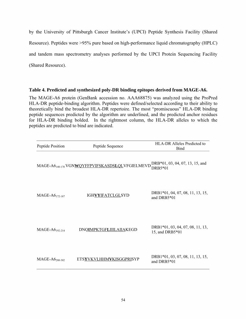
by the University of Pittsburgh Cancer Institute’s (UPCI) Peptide Synthesis Facility (Shared
Resource). Peptides were >95% pure based on high-performance liquid chromatography (HPLC)
and tandem mass spectrometry analyses performed by the UPCI Protein Sequencing Facility
(Shared Resource).
Table 4. Predicted and synthesized poly-DR binding epitopes derived from MAGE-A6.
The MAGE-A6 protein (GenBank accession no. AAA68875) was analyzed using the ProPred HLA-DR peptide-binding algorithm. Peptides were defined/selected according to their ability to theoretically bind the broadest HLA-DR repertoire. The most “promiscuous” HLA-DR binding peptide sequences predicted by the algorithm are underlined, and the predicted anchor residues for HLA-DR binding bolded. In the rightmost column, the HLA-DR alleles to which the peptides are predicted to bind are indicated.
Peptide Position Peptide Sequence HLA-DR Alleles Predicted to Bind
MAGE-A6140-170 VGNWQYFFPVIFSKASDSLQLVFGIELMEVD DRB*01, 03, 04, 07, 13, 15, and DRB5*01
MAGE-A6172-187 IGHVYIFATCLGLSYD DRB1*01, 04, 07, 08, 11, 13, 15, and DRB5*01
MAGE-A6192-214 DNQIMPKTGFLIIILAIIAKEGD DRB1*01, 03, 04, 07, 08, 11, 13, 15, and DRB5*01
MAGE-A6280-302 ETSYVKVLHHMVKISGGPRISYP DRB1*01, 03, 07, 08, 11, 13, 15, and DRB5*01
54
2.3.8. PCR
Cell lines were screened for MAGE-A6 expression by reverse transcription (RT)-PCR, while
Mycoplasma penetrans HF-2 contamination was tested by PCR. For MAGE-A6 analysis, RNA
was isolated from the cell lines using the RNeasy Tissue Kit (Qiagen, Valencia, CA) and cDNA
prepared using the GeneAmpR RNA PCR Kit (Applied Biosystems, Foster City, CA). MAGE-
A6 transcripts were analyzed, as previously described (36), using the following primer set;
forward: TGGAGGACCAGAGGCCCCC; reverse: CAGGATGATTATCAGGAAGCCTGT.
Mycoplasma penetrans HF-2 DNA contamination of cell lines was tested by PCR, as previously
described (209) using the primers; forward: CATGCAAGTCGGAC; reverse:
AGCATTTCCTCTTC. Mycoplasma penetrans HF-2 bacteria (ATCC# 55252; Gaithersberg,
MD) were used as positive DNA control, as was assessment for β-actin DNA using the primer
set; forward: GGCATCGTGATGGACTCCG; reverse: GCTGGAAGGTGGACAGCGA. The
PCR reaction parameters consisted of an initial 3 min denaturation step at 94°C followed by 32
amplification cycles that consisted of denaturation at 94°C for 45 sec, annealing at 68°C for 45
sec, and extension at 72°C for 1 min. The final cycle was followed by an additional extension
step at 72°C for 10 min.
2.3.9. rMAGE-A6 generation and Western Blot analysis
Full-length MAGE-A6 cDNA was generated by RT-PCR using the primer set; forward:
TGGAGGACCAGAGGCCCCC; reverse: AGGATGATTATCAGGAAGCCTGTC. cDNA was
isolated from the MAGE-A6+ SLR20 renal carcinoma cell line (208) and inserted into the pBAD
TOPO TA (Invitrogen) cloning vector, then amplified in TOP10 (Invitrogen) bacteria, according
55
to the manufacturer’s protocol. The sequence was confirmed using the sequencing primers
provided in the pBAD TOPO TA Cloning Kit. Bacterial extracted poly-His-tagged recombinant
MAGE-A6 (rMAGE) was purified using the BD Talon Purification Kit (BD Biosciences, San
Jose, CA), according to the manufacturer’s protocol. Non-transformed TOP10 bacteria were
grown and processed in an identical manner as for rMAGE purification, with the processed
elution fractions (TOP10) used as a negative control in ELISPOT readouts for immune response
to rMAGE. LPS levels for rMAGE and TOP10 control protein were tested using the QCL-1000
Kit (BioWhittaker, Walkersville, MD), and determined to be < 3 ng/ml (data not shown). For
Western Blotting, proteins were transferred onto Immobilon-P transfer membranes
(polyvinylidene fluoride microporous membrane; Millipore, Billerica, MA) and stained using the
anti-MAGE antibody, 57B (kindly provided by Dr. G. C. Spagnoli, University Hospital of Basel,
Basel, Switzerland; ref. 26) or isotype matched control Ab (Sigma-Aldrich, St. Louis, MO). Goat
anti-mouse HRP conjugated antibody (Sigma-Aldrich) was used as a detection antibody. After
extensive washing with PBS supplemented with 0.05% Tween, Western Lightning
Chemoluminescence Reagent Plus (PerkinElmer Life Sciences, Boston, MA) was added and the
blot developed by ECL chemiluminescence radiography (Kodak, Rochester, NY).
2.3.10. Statistical Analysis
Statistical comparisons were made using a two-tailed Student’s T-test, with a p value < 0.05
considered significant.
56
2.4. RESULTS
2.4.1. Selection and testing of poly-DR binding peptides derived from MAGE-A6.
I subjected the MAGE-A6 protein sequence to a computer algorithm screen designed to
identify peptides likely to have “promiscuous” HLA-DR-binding tendencies. Four such peptides
were identified (Table 4). While three of these peptides represent novel sequences (MAGE-
A6172-187, MAGE-A6192-214, and MAGE-A6280-302), I have previously defined the MAGE-A6140-
170 peptide as containing an HLA-DR4-restricted epitope (208).
These 4 peptides were evaluated for their immunogenicity in vitro using normal donor
and melanoma patient CD4+ T cells as responders and autologous Type-1 polarized DCs (i.e.
DC1; ref. (210, 211) as stimulator cells. A single round of in vitro stimulation was employed in
order to amplify recall Th responses to these epitopes and to limit the priming of naïve CD4+ T
cells, with IFN-γ ELISPOT assays performed to enumerate the resultant peptide-specific CD4+ T
cell responses in an initial assessment of 14 melanoma patients and 7 normal donors. A
summary of donor characteristics and their T cell responses to MAGE-A6 peptides are provided
in Table 5 and Fig. 5, respectively.
57
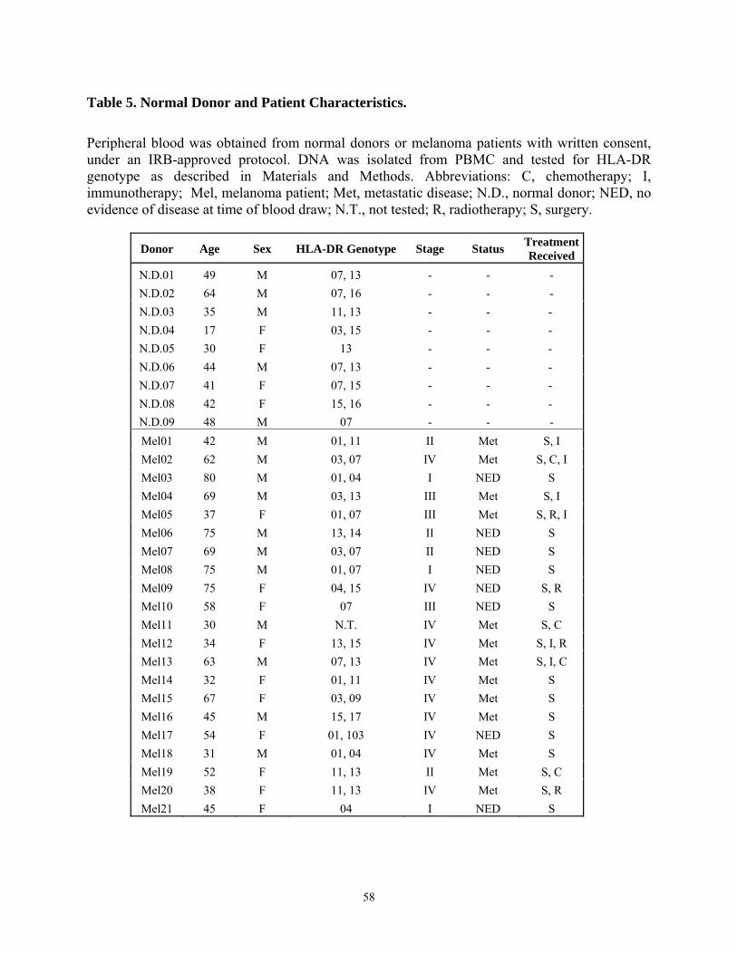
Table 5. Normal Donor and Patient Characteristics.
Peripheral blood was obtained from normal donors or melanoma patients with written consent, under an IRB-approved protocol. DNA was isolated from PBMC and tested for HLA-DR genotype as described in Materials and Methods. Abbreviations: C, chemotherapy; I, immunotherapy; Mel, melanoma patient; Met, metastatic disease; N.D., normal donor; NED, no evidence of disease at time of blood draw; N.T., not tested; R, radiotherapy; S, surgery. Donor Age Sex HLA-DR Genotype Stage Status Treatment
Received N.D.01 49 M 07, 13 - - - N.D.02 64 M 07, 16 - - - N.D.03 35 M 11, 13 - - - N.D.04 17 F 03, 15 - - - N.D.05 30 F 13 - - - N.D.06 44 M 07, 13 - - - N.D.07 41 F 07, 15 - - - N.D.08 42 F 15, 16 - - - N.D.09 48 M 07 - - - Mel01 42 M 01, 11 II Met S, I Mel02 62 M 03, 07 IV Met S, C, I Mel03 80 M 01, 04 I NED S Mel04 69 M 03, 13 III Met S, I Mel05 37 F 01, 07 III Met S, R, I Mel06 75 M 13, 14 II NED S Mel07 69 M 03, 07 II NED S Mel08 75 M 01, 07 I NED S Mel09 75 F 04, 15 IV NED S, R Mel10 58 F 07 III NED S Mel11 30 M N.T. IV Met S, C Mel12 34 F 13, 15 IV Met S, I, R Mel13 63 M 07, 13 IV Met S, I, C Mel14 32 F 01, 11 IV Met S Mel15 67 F 03, 09 IV Met S Mel16 45 M 15, 17 IV Met S Mel17 54 F 01, 103 IV NED S Mel18 31 M 01, 04 IV Met S Mel19 52 F 11, 13 II Met S, C Mel20 38 F 11, 13 IV Met S, R Mel21 45 F 04 I NED S
58
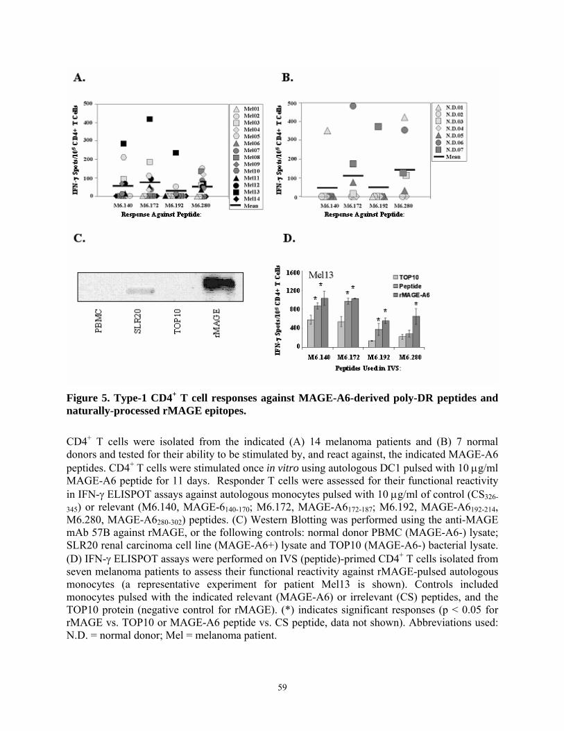
Figure 5. Type-1 CD4+ T cell responses against MAGE-A6-derived poly-DR peptides and naturally-processed rMAGE epitopes.
CD4+ T cells were isolated from the indicated (A) 14 melanoma patients and (B) 7 normal donors and tested for their ability to be stimulated by, and react against, the indicated MAGE-A6 peptides. CD4+ T cells were stimulated once in vitro using autologous DC1 pulsed with 10 μg/ml MAGE-A6 peptide for 11 days. Responder T cells were assessed for their functional reactivity in IFN-γ ELISPOT assays against autologous monocytes pulsed with 10 μg/ml of control (CS326-
345) or relevant (M6.140, MAGE-6140-170; M6.172, MAGE-A6172-187; M6.192, MAGE-A6192-214, M6.280, MAGE-A6280-302) peptides. (C) Western Blotting was performed using the anti-MAGE mAb 57B against rMAGE, or the following controls: normal donor PBMC (MAGE-A6-) lysate; SLR20 renal carcinoma cell line (MAGE-A6+) lysate and TOP10 (MAGE-A6-) bacterial lysate. (D) IFN-γ ELISPOT assays were performed on IVS (peptide)-primed CD4+ T cells isolated from seven melanoma patients to assess their functional reactivity against rMAGE-pulsed autologous monocytes (a representative experiment for patient Mel13 is shown). Controls included monocytes pulsed with the indicated relevant (MAGE-A6) or irrelevant (CS) peptides, and the TOP10 protein (negative control for rMAGE). (*) indicates significant responses (p < 0.05 for rMAGE vs. TOP10 or MAGE-A6 peptide vs. CS peptide, data not shown). Abbreviations used: N.D. = normal donor; Mel = melanoma patient.
59
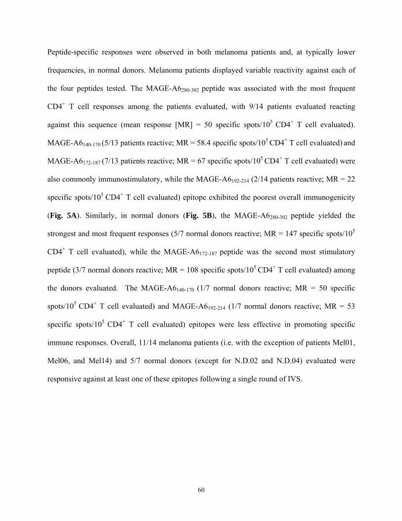
Peptide-specific responses were observed in both melanoma patients and, at typically lower
frequencies, in normal donors. Melanoma patients displayed variable reactivity against each of
the four peptides tested. The MAGE-A6280-302 peptide was associated with the most frequent
CD4+ T cell responses among the patients evaluated, with 9/14 patients evaluated reacting
against this sequence (mean response [MR] = 50 specific spots/105 CD4+ T cell evaluated).
MAGE-A6140-170 (5/13 patients reactive; MR = 58.4 specific spots/105 CD4+ T cell evaluated) and
MAGE-A6172-187 (7/13 patients reactive; MR = 67 specific spots/105 CD4+ T cell evaluated) were
also commonly immunostimulatory, while the MAGE-A6192-214 (2/14 patients reactive; MR = 22
specific spots/105 CD4+ T cell evaluated) epitope exhibited the poorest overall immunogenicity
(Fig. 5A). Similarly, in normal donors (Fig. 5B), the MAGE-A6280-302 peptide yielded the
strongest and most frequent responses (5/7 normal donors reactive; MR = 147 specific spots/105
CD4+ T cell evaluated), while the MAGE-A6172-187 peptide was the second most stimulatory
peptide (3/7 normal donors reactive; MR = 108 specific spots/105 CD4+ T cell evaluated) among
the donors evaluated. The MAGE-A6140-170 (1/7 normal donors reactive; MR = 50 specific
spots/105 CD4+ T cell evaluated) and MAGE-A6192-214 (1/7 normal donors reactive; MR = 53
specific spots/105 CD4+ T cell evaluated) epitopes were less effective in promoting specific
immune responses. Overall, 11/14 melanoma patients (i.e. with the exception of patients Mel01,
Mel06, and Mel14) and 5/7 normal donors (except for N.D.02 and N.D.04) evaluated were
responsive against at least one of these epitopes following a single round of IVS.
60
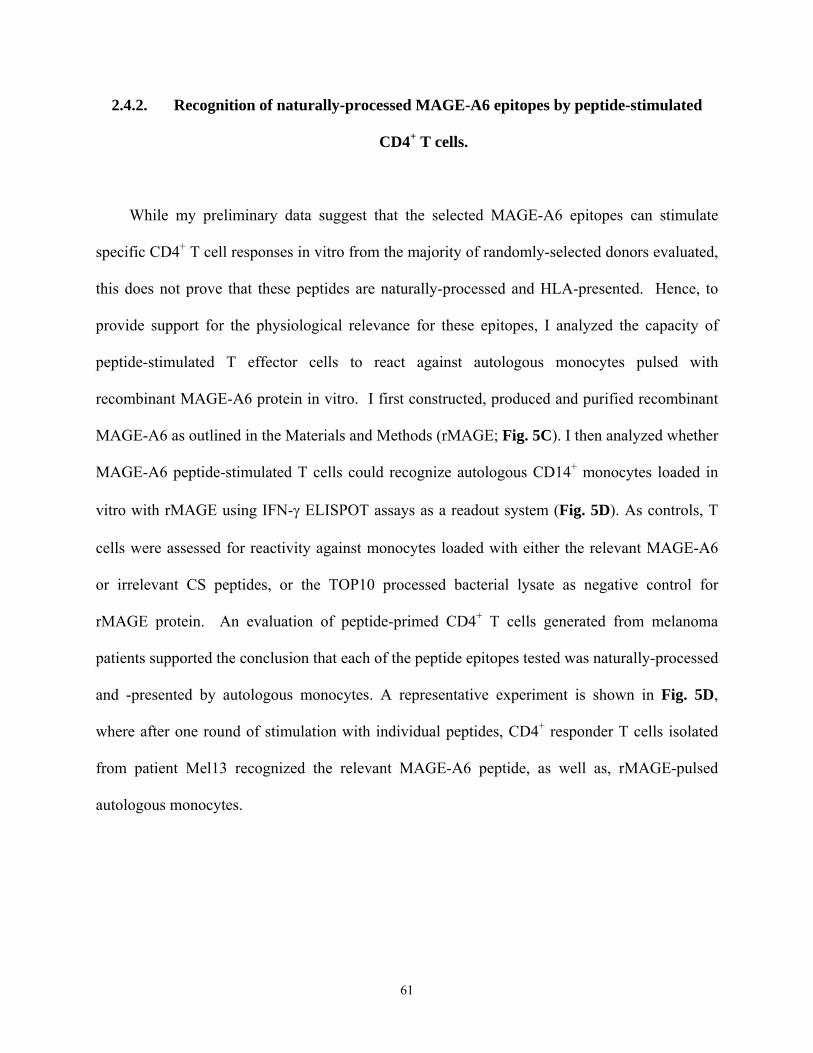
2.4.2. Recognition of naturally-processed MAGE-A6 epitopes by peptide-stimulated
CD4+ T cells.
While my preliminary data suggest that the selected MAGE-A6 epitopes can stimulate
specific CD4+ T cell responses in vitro from the majority of randomly-selected donors evaluated,
this does not prove that these peptides are naturally-processed and HLA-presented. Hence, to
provide support for the physiological relevance for these epitopes, I analyzed the capacity of
peptide-stimulated T effector cells to react against autologous monocytes pulsed with
recombinant MAGE-A6 protein in vitro. I first constructed, produced and purified recombinant
MAGE-A6 as outlined in the Materials and Methods (rMAGE; Fig. 5C). I then analyzed whether
MAGE-A6 peptide-stimulated T cells could recognize autologous CD14+ monocytes loaded in
vitro with rMAGE using IFN-γ ELISPOT assays as a readout system (Fig. 5D). As controls, T
cells were assessed for reactivity against monocytes loaded with either the relevant MAGE-A6
or irrelevant CS peptides, or the TOP10 processed bacterial lysate as negative control for
rMAGE protein. An evaluation of peptide-primed CD4+ T cells generated from melanoma
patients supported the conclusion that each of the peptide epitopes tested was naturally-processed
and -presented by autologous monocytes. A representative experiment is shown in Fig. 5D,
where after one round of stimulation with individual peptides, CD4+ responder T cells isolated
from patient Mel13 recognized the relevant MAGE-A6 peptide, as well as, rMAGE-pulsed
autologous monocytes.
61
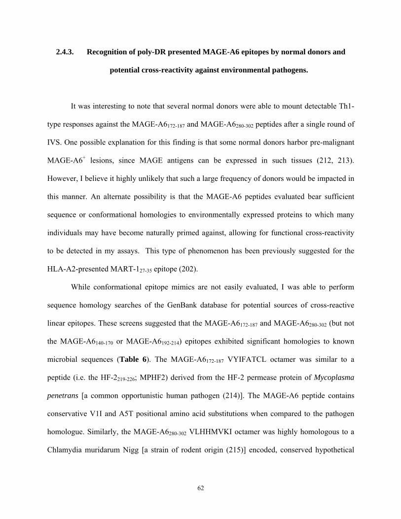
2.4.3. Recognition of poly-DR presented MAGE-A6 epitopes by normal donors and
potential cross-reactivity against environmental pathogens.
It was interesting to note that several normal donors were able to mount detectable Th1-
type responses against the MAGE-A6172-187 and MAGE-A6280-302 peptides after a single round of
IVS. One possible explanation for this finding is that some normal donors harbor pre-malignant
MAGE-A6+ lesions, since MAGE antigens can be expressed in such tissues (212, 213).
However, I believe it highly unlikely that such a large frequency of donors would be impacted in
this manner. An alternate possibility is that the MAGE-A6 peptides evaluated bear sufficient
sequence or conformational homologies to environmentally expressed proteins to which many
individuals may have become naturally primed against, allowing for functional cross-reactivity
to be detected in my assays. This type of phenomenon has been previously suggested for the
HLA-A2-presented MART-127-35 epitope (202).
While conformational epitope mimics are not easily evaluated, I was able to perform
sequence homology searches of the GenBank database for potential sources of cross-reactive
linear epitopes. These screens suggested that the MAGE-A6172-187 and MAGE-A6280-302 (but not
the MAGE-A6140-170 or MAGE-A6192-214) epitopes exhibited significant homologies to known
microbial sequences (Table 6). The MAGE-A6172-187 VYIFATCL octamer was similar to a
peptide (i.e. the HF-2219-226; MPHF2) derived from the HF-2 permease protein of Mycoplasma
penetrans [a common opportunistic human pathogen (214)]. The MAGE-A6 peptide contains
conservative V1I and A5T positional amino acid substitutions when compared to the pathogen
homologue. Similarly, the MAGE-A6280-302 VLHHMVKI octamer was highly homologous to a
Chlamydia muridarum Nigg [a strain of rodent origin (215)] encoded, conserved hypothetical
62
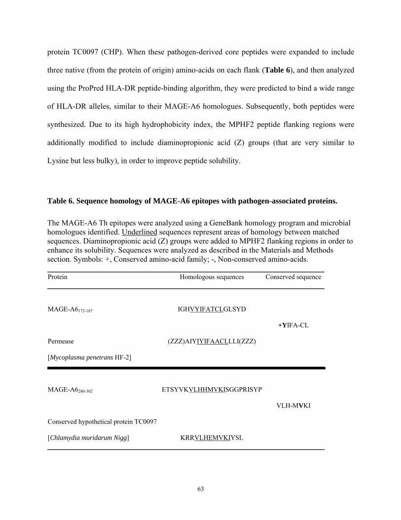
protein TC0097 (CHP). When these pathogen-derived core peptides were expanded to include
three native (from the protein of origin) amino-acids on each flank (Table 6), and then analyzed
using the ProPred HLA-DR peptide-binding algorithm, they were predicted to bind a wide range
of HLA-DR alleles, similar to their MAGE-A6 homologues. Subsequently, both peptides were
synthesized. Due to its high hydrophobicity index, the MPHF2 peptide flanking regions were
additionally modified to include diaminopropionic acid (Z) groups (that are very similar to
Lysine but less bulky), in order to improve peptide solubility.
Table 6. Sequence homology of MAGE-A6 epitopes with pathogen-associated proteins.
The MAGE-A6 Th epitopes were analyzed using a GeneBank homology program and microbial homologues identified. Underlined sequences represent areas of homology between matched sequences. Diaminopropionic acid (Z) groups were added to MPHF2 flanking regions in order to enhance its solubility. Sequences were analyzed as described in the Materials and Methods section. Symbols: +, Conserved amino-acid family; -, Non-conserved amino-acids. Protein Homologous sequences Conserved sequence
MAGE-A6172-187 IGHVYIFATCLGLSYD
+YIFA-CL
Permease (ZZZ)AIYIYIFAACLLLI(ZZZ)
[Mycoplasma penetrans HF-2]
MAGE-A6280-302 ETSYVKVLHHMVKISGGPRISYP
VLH-MVKI
Conserved hypothetical protein TC0097
[Chlamydia muridarum Nigg] KRRVLHEMVKIYSL
63
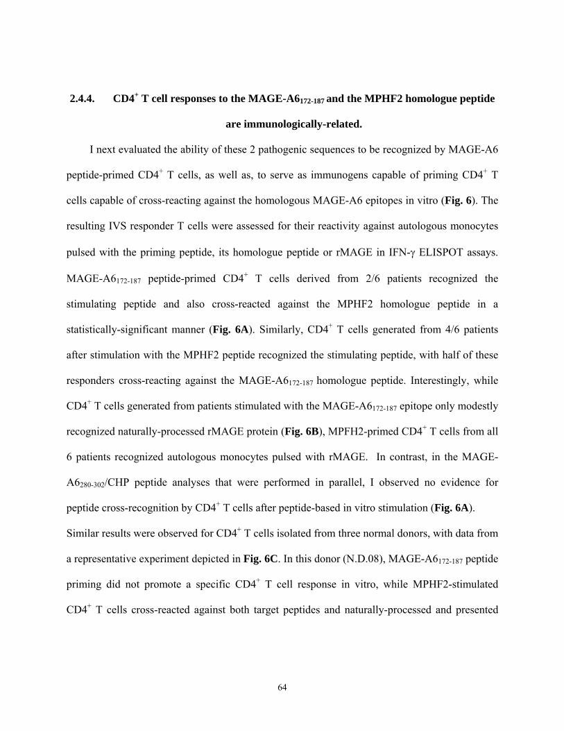
2.4.4. CD4+ T cell responses to the MAGE-A6172-187 and the MPHF2 homologue peptide
are immunologically-related.
I next evaluated the ability of these 2 pathogenic sequences to be recognized by MAGE-A6
peptide-primed CD4+ T cells, as well as, to serve as immunogens capable of priming CD4+ T
cells capable of cross-reacting against the homologous MAGE-A6 epitopes in vitro (Fig. 6). The
resulting IVS responder T cells were assessed for their reactivity against autologous monocytes
pulsed with the priming peptide, its homologue peptide or rMAGE in IFN-γ ELISPOT assays.
MAGE-A6172-187 peptide-primed CD4+ T cells derived from 2/6 patients recognized the
stimulating peptide and also cross-reacted against the MPHF2 homologue peptide in a
statistically-significant manner (Fig. 6A). Similarly, CD4+ T cells generated from 4/6 patients
after stimulation with the MPHF2 peptide recognized the stimulating peptide, with half of these
responders cross-reacting against the MAGE-A6172-187 homologue peptide. Interestingly, while
CD4+ T cells generated from patients stimulated with the MAGE-A6172-187 epitope only modestly
recognized naturally-processed rMAGE protein (Fig. 6B), MPFH2-primed CD4+ T cells from all
6 patients recognized autologous monocytes pulsed with rMAGE. In contrast, in the MAGE-
A6280-302/CHP peptide analyses that were performed in parallel, I observed no evidence for
peptide cross-recognition by CD4+ T cells after peptide-based in vitro stimulation (Fig. 6A).
Similar results were observed for CD4+ T cells isolated from three normal donors, with data from
a representative experiment depicted in Fig. 6C. In this donor (N.D.08), MAGE-A6172-187 peptide
priming did not promote a specific CD4+ T cell response in vitro, while MPHF2-stimulated
CD4+ T cells cross-reacted against both target peptides and naturally-processed and presented
64
rMAGE-derived epitope(s) in the ELISPOT readout assay. This donor also responded to the
MAGE-A6280-302, but not the CHP peptide (Fig. 6C).
Figure 6. Normal donor and melanoma patient CD4+ T cell responses against MPHF2 and CHP peptides: cross-reactivity against MAGE-A6 epitopes? (A) CD4+ T cells isolated from melanoma patients were tested for their ability to be stimulated by, and react against, MAGE-A6172-187 and MAGE-A6280-302, and their homologues MPHF2 and CHP, respectively. CD4+ T cells were stimulated as described in the Fig. 5 legend, with responder T cells analyzed in IFN-γ ELISPOT assays for reactivity against autologous monocytes pulsed with 10 μg/ml of stimulating peptide (A, top panel), the corresponding homologue peptide (A, bottom panel) or rMAGE (B). Negative controls included the CS326-345 peptide and TOP10 bacterial proteins. (C). CD4+ T cells isolated from normal donors were stimulated as above using the individual MAGE-A6, MPHF2 and CHP peptides, then analyzed for their reactivity against autologous monocytes pulsed with the indicated peptides or proteins (insert) in IFN-γ ELISPOT assays. The data displayed in panel C are representative of 3 normal donors analyzed in this manner.
65
2.4.5. MPHF2-stimulated CD4+ T cells recognize HLA-DR matched, MAGE-A6+
melanoma cell lines in vitro.
To further evaluate the potential physiological relevance of CD4+ T cell priming against
the MPH2 epitope, I compared MAGE-A6172-187 and MPHF2-stimulated CD4+ T cells for their
ability to recognize HLA-DR-matched, MAGE-A6+ melanoma cell lines in vitro. In these
experiments, CD4+ T cells isolated from an HLA-DR4+ melanoma patient blood were stimulated
twice at weekly intervals with autologous DC1 pulsed with either the MAGE-A6172-187 or
MPHF2 peptides. Two HLA-DR4+ (Fig. 7A)/MAGE-A6+ (Fig 7B) melanoma cell lines, SLM2
and Mel526.DR4, were then used as targets for responder T cells in IFN-γ ELISPOT assays (Fig.
7C). Pan-DR mAb (L243) was also added to replicate wells, as indicated, to demonstrate the
MHC class II-restricted nature of T cell responses. I observed that both populations of peptide-
primed CD4+ T cells recognized the two tumor cell lines in a manner that was partially blocked
by addition of mAb L243, with the MPHF2-stimulated CD4+ T cells exhibiting a greater
magnitude of response to tumor cell lines than T cells primed with the MAGE-A6172-187 epitope
(Fig. 7C). To rule out the possibility that T cell recognition was due to specific mycoplasma
infection of the target cell lines, both melanoma cell lines were shown to be negative for
Mycoplasma penetrans HF-2 contamination using a sensitive PCR method (Fig. 7D).
66
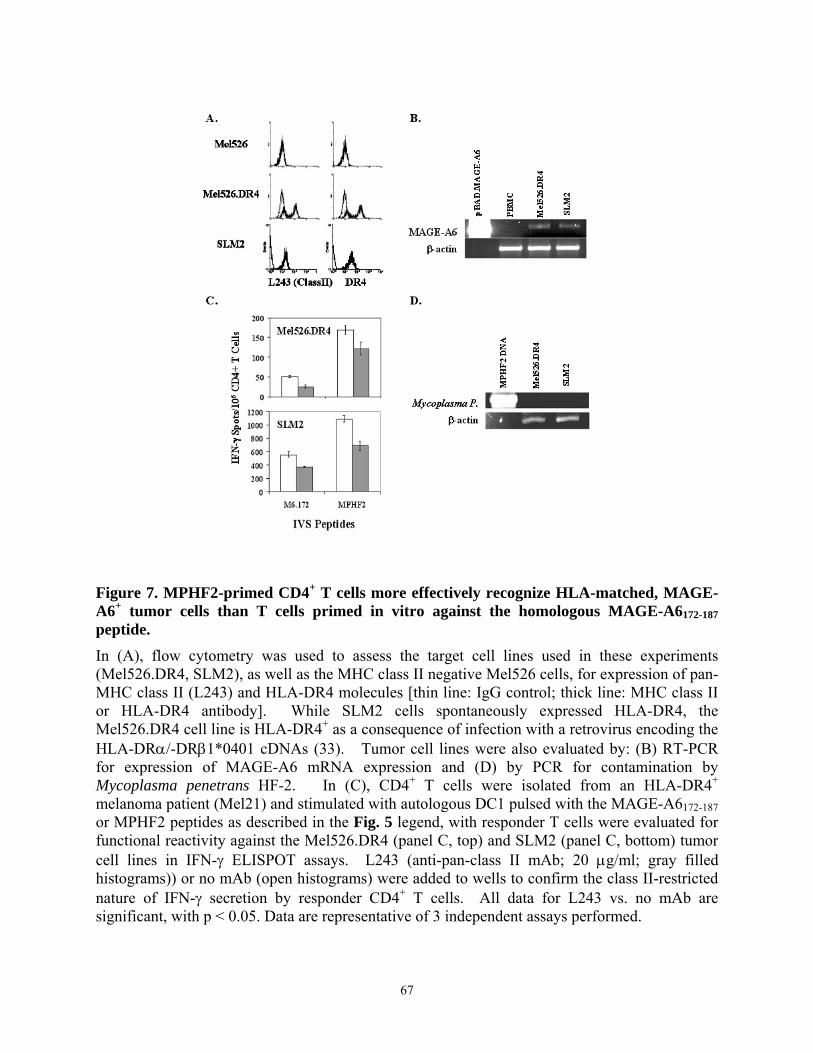
Figure 7. MPHF2-primed CD4+ T cells more effectively recognize HLA-matched, MAGE-A6+ tumor cells than T cells primed in vitro against the homologous MAGE-A6172-187 peptide.
In (A), flow cytometry was used to assess the target cell lines used in these experiments (Mel526.DR4, SLM2), as well as the MHC class II negative Mel526 cells, for expression of pan-MHC class II (L243) and HLA-DR4 molecules [thin line: IgG control; thick line: MHC class II or HLA-DR4 antibody]. While SLM2 cells spontaneously expressed HLA-DR4, the Mel526.DR4 cell line is HLA-DR4+ as a consequence of infection with a retrovirus encoding the HLA-DRα/-DRβ1*0401 cDNAs (33). Tumor cell lines were also evaluated by: (B) RT-PCR for expression of MAGE-A6 mRNA expression and (D) by PCR for contamination by Mycoplasma penetrans HF-2. In (C), CD4+ T cells were isolated from an HLA-DR4+ melanoma patient (Mel21) and stimulated with autologous DC1 pulsed with the MAGE-A6172-187 or MPHF2 peptides as described in the Fig. 5 legend, with responder T cells were evaluated for functional reactivity against the Mel526.DR4 (panel C, top) and SLM2 (panel C, bottom) tumor cell lines in IFN-γ ELISPOT assays. L243 (anti-pan-class II mAb; 20 μg/ml; gray filled histograms)) or no mAb (open histograms) were added to wells to confirm the class II-restricted nature of IFN-γ secretion by responder CD4+ T cells. All data for L243 vs. no mAb are significant, with p < 0.05. Data are representative of 3 independent assays performed.
67
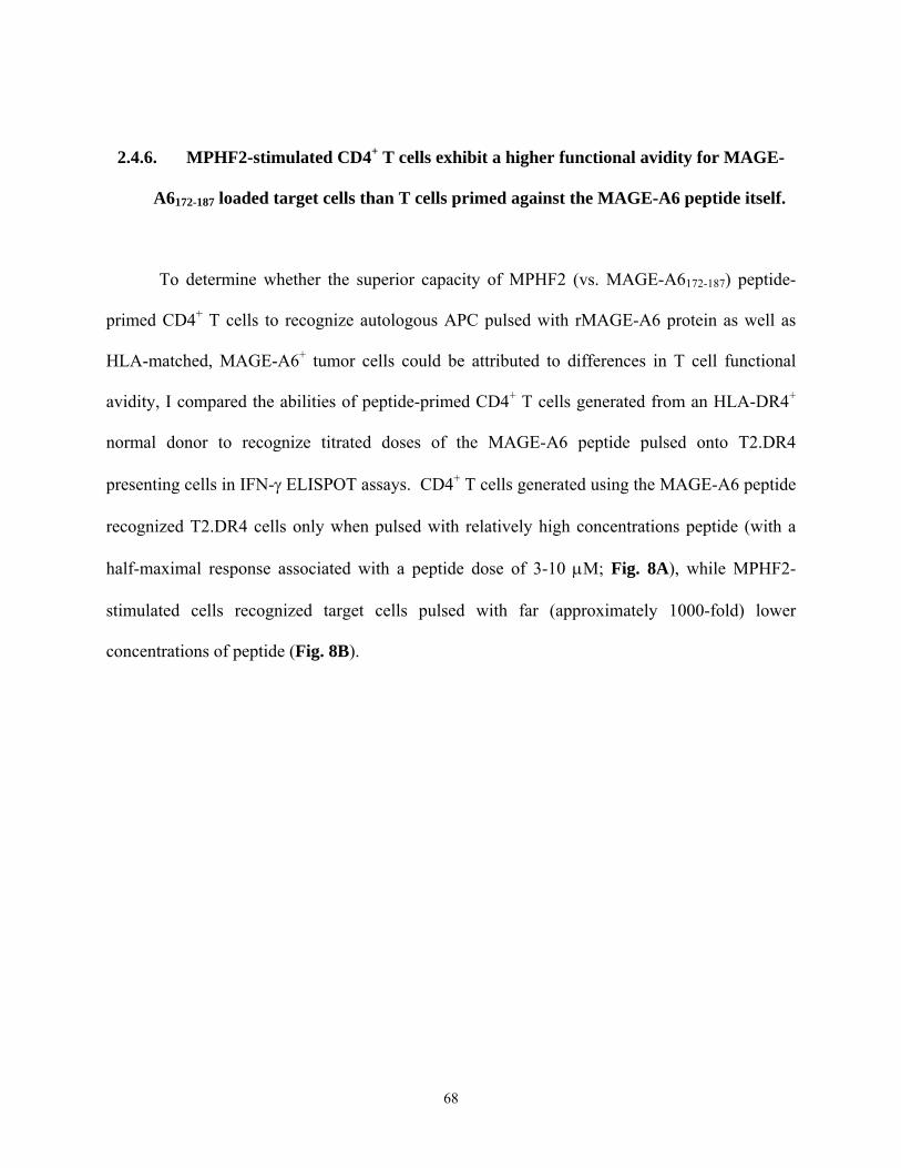
2.4.6. MPHF2-stimulated CD4+ T cells exhibit a higher functional avidity for MAGE-
A6172-187 loaded target cells than T cells primed against the MAGE-A6 peptide itself.
To determine whether the superior capacity of MPHF2 (vs. MAGE-A6172-187) peptide-
primed CD4+ T cells to recognize autologous APC pulsed with rMAGE-A6 protein as well as
HLA-matched, MAGE-A6+ tumor cells could be attributed to differences in T cell functional
avidity, I compared the abilities of peptide-primed CD4+ T cells generated from an HLA-DR4+
normal donor to recognize titrated doses of the MAGE-A6 peptide pulsed onto T2.DR4
presenting cells in IFN-γ ELISPOT assays. CD4+ T cells generated using the MAGE-A6 peptide
recognized T2.DR4 cells only when pulsed with relatively high concentrations peptide (with a
half-maximal response associated with a peptide dose of 3-10 μM; Fig. 8A), while MPHF2-
stimulated cells recognized target cells pulsed with far (approximately 1000-fold) lower
concentrations of peptide (Fig. 8B).
68
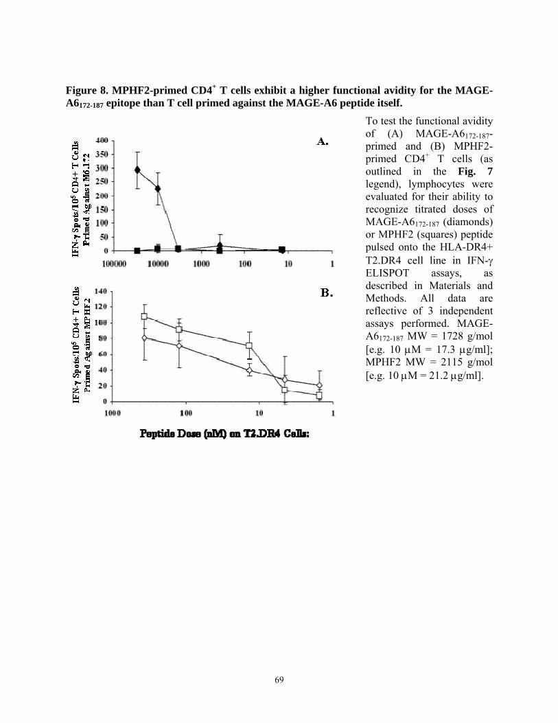
Figure 8. MPHF2-primed CD4+ T cells exhibit a higher functional avidity for the MAGE-A6172-187 epitope than T cell primed against the MAGE-A6 peptide itself.
To test the functional avidity of (A) MAGE-A6172-187-primed and (B) MPHF2-primed CD4+ T cells (as outlined in the Fig. 7 legend), lymphocytes were evaluated for their ability to recognize titrated doses of MAGE-A6172-187 (diamonds) or MPHF2 (squares) peptide pulsed onto the HLA-DR4+ T2.DR4 cell line in IFN-γ ELISPOT assays, as described in Materials and Methods. All data are reflective of 3 independent assays performed. MAGE-A6172-187 MW = 1728 g/mol [e.g. 10 μM = 17.3 μg/ml]; MPHF2 MW = 2115 g/mol [e.g. 10 μM = 21.2 μg/ml].
69
2.5. DISCUSSION
Previous studies of peptide-specific, CD4+ T cell responses against tumor antigens,
including MAGE-A6 have been traditionally skewed towards an analysis of a single (i.e. HLA-
DR4) or very limited set of HLA-DR restriction elements (208, 216, 217). Given the extreme
polymorphism among HLA-DR alleles, the translational utility of the epitopes defined to date
would be limited to a modest cohort of 20-30% of patients (23). In the current study I have
attempted to circumvent this limitation by identifying peptides that are likely to bind to, and be
presented by, as broad a range of HLA-DR alleles (i.e. poly-DR) as possible, thereby expanding
the range of patients to which MAGE-A6-based therapies might be applied clinically.
Peripheral blood CD4+ T cells were harvested from normal donors and patients with
melanoma, stimulated with a single round of peptide-based IVS, and evaluated for their ability to
recognize MAGE-A6 peptides (selected to be promiscuously presented by HLA class II) in IFN-
γ ELISPOT assays. My data support the identification of 3 novel MAGE-A6 peptide epitopes
recognized by Type-1 Th cells and substantiate the ability of these epitopes, along with the
previously-defined MAGE-A6140-170 peptide, to be recognized by a large proportion of
individuals (who overall exhibited a diverse array of HLA-DR haplotypes). Of the four peptides
analyzed, the MAGE-A6172-187 and MAGE-A6280-302 peptides were recognized at the highest
frequencies in peptide ELISPOT readouts by CD4+ T cells. In contrast, the MAGE-A6192-214
appeared comparatively non-immunogenic, and the MAGE-A6140-170 peptide was moderately
effective in this capacity. Based on the ability of peptide-primed CD4+ T cells to recognize
autologous monocytes pulsed with rMAGE protein, each of the peptides evaluated appear to
contain naturally-processed and -presented Th epitopes.
70
These MAGE-A6 poly-DR epitopes displayed a high degree of homology with sequences
contained in other MAGE-A family members, especially MAGE-A3. MAGE-A6140-170, MAGE-
A6172-187, and MAGE-A6280-302 differed from their MAGE-A3 counterparts by only a single
amino acid in each case. For MAGE-A6140-170 this difference is at position 156, where a D > S
substitution occurs in the MAGE-A3 protein. The MAGE-A6172-187 peptide differs from its
MAGE-A3 counterpart based on a conservative V175L substitution, and the MAGE-A6280-302 to
MAGE-A3 difference reflects an R298H substitution. Hence, while it remains to be formally
evaluated, I believe that in many cases, the selected MAGE-A6 epitopes will likely elicit CD4+ T
cell responses in a high frequency of patients that are capable of cross-reacting against their
MAGE-A3 homologues when presented by autologous APCs. As a result, these peptides could
represent promising candidates for inclusion in peptide-based vaccines designed to treat the
majority of patients harboring tumors that exhibit MAGE-A6+ and/or MAGE-A3+ phenotypes in
situ.
It was also noted in this study that CD4+ T cells isolated from several normal donors were
able to effectively recognize the MAGE-A6172-187 and MAGE-A6280-302 epitopes after a single
round of IVS. This observation could be attributed to epitope mimicry. Epitope mimicry has
been described as a potential mechanism underlying the induction of autoimmune diseases due to
pathologic T cells primed against infectious microorganisms that cross-react against host
proteins in susceptible individuals. Diseases such as viral myocarditis, lyme disease, rheumatoid
arthritis (198), multiple sclerosis (199), and virus-induced autoimmune diabetes (200, 201) have
long been considered to be initiated or exacerbated by microbial pathogens. As was previously
noted for the HLA-A2-presented, melanoma-associated MART-127-35 epitope (202), I
hypothesized that the high degree of normal donor response against the MAGE-A6 Th peptides
71
might be due to the cross-reactivity of T cells initially primed in vivo against highly-homologous
peptides within environmentally-encountered proteins. After performing a homology search of
the GenBank database, I selected the MPHF2 and CHP peptides as two likely candidate
homologues of the MAGE-A6172-187 and MAGE-A6280-302 peptides, respectively. The MPHF2
peptide derives from Mycoplasma penetrans HF-2, a ubiquitous species of Mycoplasmataceae,
which infects humans in the urogenital and respiratory tracts. A typical feature of this
microorganism is penetration into human cells and long-term intracellular replication and
persistence. In human disease, M. penetrans is clinically observed in cases of HIV-1 infection,
but has also been suggested to represent a primary cause of non-HIV-related urethritis and
respiratory disease (214). Importantly, while many strains of mycoplasma can commonly infect
laboratory cultures and affect derivative assay results, M. penetrans has not been reported to do
so, and I did not detected this agent in my cell lines. The CHP peptide derives from Chlamydia
muridarum strain Nigg, a mouse-tropic strain capable of causing respiratory disorders in mice.
This strain of Chlamydia diverges significantly from human-tropic strains (215). Given the low
degree of likelihood that humans would encounter this microbe, it was perhaps not surprising
that I did not observe any evidence for the cross-reactivity of these peptides by T cells in my
studies. This does not rule out a pathogenic homologue for the MAGE-A6280-302 peptide being
responsible for my common observation of IVS responses against this MAGE-A6 peptide among
normal donors, but suggests that additional studies will need to be performed to illuminate its
identity.
My results suggest that the MPHF2 peptide is immunogenic and capable of promoting
Type-1 effector CD4+ T cells in a majority of melanoma patients and normal donors. Responder
T cells were typically able to cross-react against the MPHF2 and MAGE-A6172-187 peptides and
72
to recognize the naturally-processed rMAGE epitope when presented by autologous monocytes.
Notably, anti-MPHF2 CD4+ T cells also appeared preferentially able (in contrast to MAGE-A6
peptide induced T cells) to recognize HLA-DR matched melanoma cell lines that constitutively
express the MAGE-A6 gene product. The ability of the MPHF2 peptide to promote “heteroclitic”
immunity against the MAGE-A6 (and presumably MAGE-A3) protein(s) may make this epitope
extremely attractive as a vaccine candidate in patients bearing tumor histotypes in which MAGE-
A3/MAGE-A6 expression is commonly observed (such as melanoma, renal cell carcinoma and
others).
73
Preface Chapter 3
Given that patients with cancer exhibit dysfunctional Th1-type responses against epitopes
derived from tumor antigens, such as MAGE-A6, I next investigated whether human DCs
engineered to secrete high levels of the IFN-γ-inducing cytokines IL-12p70 and IL-18 via
recombinant adenoviral infection could effectively generate Th1-type responses in vitro. I
showed that DC co-infected with Ad.IL-12 and Ad.IL-18 (DC.IL-12/18) and pulsed with HLA-
DR4-restricted MAGE-A6 peptides were more effective at stimulating MAGE-A6-specific Th1-
type CD4+ T cell responses than DC infected with either of the cytokine vectors alone, control
Ad.Ψ5 virus or uninfected DC. Furthermore, DC.IL-12/18 loaded with recombinant MAGE-A6
protein effectively promote Th1-type immunity that is frequently directed against multiple
MAGE-A6-derived epitopes.
The studies in Chapter 3 demonstrate the immuno-stimulatory properties of DC.IL-12/18,
particularly when loaded with the whole TAA. Based on these results, I believe this modality
may prove clinically useful as a vaccine platform to promote the recovery of tumor antigen-
specific, Th1-type CD4+ T cell responses in patients with cancer. This data has just been reported
in Cancer Gene Therapy in 2006.
74
3. IL-12p70 and IL-18 Gene-Modified Dendritic Cells Loaded with Tumor Antigen-Derived Peptides or Recombinant Protein Effectively Stimulate Specific Type-1 CD4+ T
cell Responses From Normal Donors and Melanoma Patients In Vitro
Lazar Vujanovic, B.S.*, Elena Ranieri, Ph.D.¶, Andrea Gambotto, M.D.†, Walter C. Olson,
Ph.D.†, John M. Kirkwood, M.D.‡,╫ and Walter J. Storkus, Ph.D.*,╪,╫
From the Departments of *Immunology, †Surgery, ‡Medicine and ╪Dermatology, University of
Pittsburgh School of Medicine, Pittsburgh, PA 15213; ╫University of Pittsburgh Cancer Institute,
Hillman Cancer Center, Pittsburgh, PA 15213; and the ¶Cattedra di Patologia Clinica,
Dipartimento di Scienze Biomediche, Universita' di Foggia, Ospedali Riuniti, Foggia-Italy.
This data has just been reported in Cancer Gene Therapy in 2006. All the results reported in this
study were obtained by Lazar Vujanovic. Evaluation of recombinant adenoviruses was repeated
by Elena Ranieri, Ph.D. ELISPOT plate analyses were performed by Walter C. Olson, Ph.D.
75
3.1. Abstract
While CD4+ Type-1 T helper (Th1) cells secreting interferon-γ (IFN-γ) appear to play an
essential role in promoting durable anti-tumor immunity, I have previously shown that patients
with cancer exhibit dysfunctional Th1-type responses against epitopes derived from tumor
antigens, such as MAGE-A6. Here, I engineered human dendritic cells (DC) to secrete high
levels of the IFN-γ-inducing cytokines IL-12p70 and IL-18 via recombinant adenoviral infection
to generate an in vitro stimulus capable of promoting previously-deficient patient Th1-type
responses. DC co-infected with Ad.IL-12 and Ad.IL-18 (DC.IL-12/18) were more effective at
stimulating MAGE-A6-specific Th1-type CD4+ T cell responses than DC infected with either of
the cytokine vectors alone, control Ad.Ψ5 virus or uninfected DC. Furthermore, I show that
DC.IL-12/18 loaded with recombinant MAGE-A6 protein (rMAGE) and used as in vitro
stimulators, promote Th1-type immunity that is frequently directed against multiple MAGE-A6-
derived epitopes. The superiority of DC.IL-12/18-based stimulations in melanoma patients was
independent of disease stage or current disease-status. Based on these results, I believe this
modality may prove clinically useful as a vaccine platform to promote the recovery of tumor
antigen-specific, Th1-type CD4+ T cell responses in patients with cancer.
76
3.2. Introduction
Previous studies have shown that melanoma represents a prototype for neoplasia responsive to
immunotherapy, and serves as a suitable model system for the development of anti-tumor
vaccines (206, 218). While most vaccine studies have focused solely on effector CD8+ T cell
responses as surrogates for clinical cancer responses, it is now clear that anti-tumor CD4+ T cells
regulate the quality, magnitude and durability of CD8+ CTL immunity in vivo (69-71), and that
the Th1-type cytokine, IFN-γ, plays an essential role in this response. Th1-type CD4+ T cells
secrete IFN-γ and may mediate delayed type hypersensitivity (DTH) responses that can lead to
enhanced cross-presentation of tumor antigens by host APCs (72), and consequent epitope
spreading in the evolving anti-tumor T cell repertoire (73, 74). Furthermore, CD4+ T cells may
mediate direct tumoricidal activity via TNF family ligand members and can inhibit tumor
angiogenesis via locoregional production of IFN-γ (76-79).
Anti-tumor Th1-type CD4+ T cells, however, appear inhibited in many cancer patients
(71, 97, 98), as reflected by decreased proliferation and T cell receptor (TCR) signaling (99), as
well as, by increased frequencies and activity of regulatory T cells (100, 101). While Th1-type
responses have been associated with spontaneous or therapy-induced regression of tumor lesions
(98, 102), tumor infiltrating lymphocytes isolated from patients with progressive lesions have
been generally reported to exhibit dominant Th2-type (secreting IL-4, IL-5) or regulatory (Th3)-
type (secreting IL-10, TGF-β1) responses (97, 98, 102). Our group has previously shown that
peripheral blood CD4+ T cells isolated from melanoma or renal cell carcinoma (RCC) patients
with active disease exhibit highly-skewed, non-Type-1 CD4+ T cell reactivity against epitopes
derived from tumor antigens, such as MAGE-A6 (36). These findings contrast noticeably with
77
normal donors and cancer patients who exhibit no current evidence of disease as a consequence
of therapeutic intervention, in whom either mixed Th1/Th2-type or strongly Th1-polarized
responses to MAGE-A6 peptides occur, respectively (208). These results strongly suggest that
the future immune-based therapies must overcome existing deficiencies in Type-1 anti-tumor
CD4+ T cell responses in cancer patients with active disease in order to be clinically effective.
IL-12p70 and IL-18 are crucial Th1-type cytokines that synergize in promoting IFN-γ
secretion from, and proliferation of, CD4+ T effector cells (165, 166). IL-12p70 induces T cell
surface expression of the IL-18 receptor (IL-18R) by naïve T cells (167); while IL-18, an IL-1
family member, potentiates the differentiation of Th1 cells instigated by IL-12p70 (168). I
hypothesized that dysfunctional anti-tumor Th1-type responses in cancer patients with active
disease might be recovered/enhanced by in vitro stimulation of patient CD4+ T cells using
vaccines containing autologous dendritic cells (DC) engineered to secrete IL-12p70 and/or IL-
18. This gene therapy approach could not only prove capable of supporting Type-1 immunity,
but would have the potential to obviate toxicities previously observed for systemic application
IL-12p70 alone (184, 186, 190) or IL-12p70 combined with IL-18 (187). Indeed, our group has
previously shown in murine models that DC engineered to secrete both IL-12p70 and IL-18 ex
vivo, and subsequently injected intratumorally, promote acute tumor rejection in concert with
enhanced Th1-type immunity and determinant spreading in the curative anti-tumor CTL
repertoire (191).
In the current study, I generated and applied novel recombinant adenoviral vectors
encoding human IL-12p70 (Ad.IL-12) and the mature form of human IL-18 (Ad.IL-18) to
engineer human DC in vitro, and subsequently evaluated the ability of these gene-modified
antigen presenting cells to promote Th1-type CD4+ T cell responses against the MAGE-A6
78
tumor antigen (36, 208) in HLA-DR4+ normal donors and patients with melanoma. To my
knowledge, this is the first study to describe a recombinant adenoviral vector encoding for a
mature, physiologically active form of human IL-18. Furthermore, I am the first to describe the
effects of Ad.IL-12 and Ad.IL-18 co-infection of DC on the induction of effective polarization
and enhancement of Th1-type CD4+ T cell responses in cancer patients. My results show that
DC.IL-12/18 loaded with tumor peptides or recombinant tumor antigens may represent an
effective vaccine capable of selectively promoting Type-1 specific immunity in patients with
cancer who may display existing immune dysfunction.
3.3. Materials and Methods
3.3.1. Recombinant Adenoviral Vectors
The Ad.ψ5, Ad.IL-12 and Ad.IL-18. E1- and E3-deleted adenoviral vector encoding the human
interleukin-12 (hIL-12) p70 and interleukin-18 (hIL-18) cDNAs were constructed through Cre-
lox recombination with reagents generously provided by Dr. S. Hardy (Somatix, Alameda, CA).
The prepro leader sequence of human GM-CSF was fused to the 5’ end of the mature human IL-
18 cDNA in order to facilitate secretion of bioactive IL-18 by infected cells. Sal I-Not I
fragments containing p40-IRES-p35 or modified mature form of hIL18 cDNAs were inserted
into the shuttle vector, pAdlox. Recombinant adenoviruses were generated by co-transfection of
Sfi I-digested pAdlox-hIL-12 or pAdlox-hIL-18 and ψ5 helper virus DNA into the adenoviral
packaging cell line CRE8 that expresses Cre recombinase, as previously described (219).
Recombinant adenoviruses were propagated on CRE8 cells, purified by cesium chloride density
gradient centrifugation and subsequent dialysis, prior to storage in 3% threalose at –80o C.
79
3.3.2. Cytokine ELISAs
Adenoviral function and cytokine bioactivity were evaluated using ELISA assays for hIL-12p70
(Pharmingen), hIL-18 (R&D Systems) and hIFN-γ (antibody pairs from Mabtech; Mariemont,
OH), per the manufacturer’s protocols. The lower limits of detection for these assays were 4.0
pg/ml, 12.5 pg/ml and 4.0 pg/ml, respectively.
3.3.3. MAGE-A6 Protein and Peptides
Recombinant MAGE-A6 protein (rMAGE)1 and MAGE-A6 peptides (MAGE-A6121-144 (36),
MAGE-A6140-170 (36), MAGE-A6246-263 (36), and MAGE-A6280-3021 were pulsed onto DC to
generate stimulator cells for in vitro sensitization of T cells. Non-transformed TOP10
(Invitrogen, Carlsbad, CA) bacteria were grown and processed in an identical manner as for
rMAGE purification, with the processed/eluted material (TOP10)1 later used as a negative
control in ELISPOT readouts.
3.3.4. Isolation of Patient and Normal Donor PBMC
Peripheral blood was obtained by venipuncture from normal donors or melanoma patients a
single time with consent, under an IRB-approved protocol. Peripheral blood was diluted 1:2
with PBS, applied to Ficoll-Hypaque gradients (Cellgro; Mediatech, Inc., Herndon, VA), and
centrifuged at 550 x g for 25 min at RT. Peripheral blood mononuclear cells (PBMC) were
recovered from the buoyant interface and washed at least three times with PBS in order to
remove residual platelets as well as Ficoll-Hypaque. Donor HLA-DR4 status was determined
using a specific anti-HLA-DR4 mAb 359-F10 and FACS analysis, as previously described (36,
80
208). Only PBMC determined to be HLA-DR4+ were used in these experiments. The 8
melanoma patients analyzed were between 31 and 83 years of age. Six of the eight patients
(except Mel4 and Mel 6) tested were male, with all but Mel4 and Mel5 having active disease
status at the time of blood draw. Blood was obtained at least 1 month after patient treatment with
surgery alone (all), surgery + chemotherapy (Mel1, Mel2) or surgery + radiotherapy (Mel 1).
Disease stage at the time of initial diagnosis for each patient is listed in Table 7.
3.3.5. DC Generation
Following Ficoll density separation, PBMC were plated in T75 culture flasks (COSTAR,
Cambridge, MA) flasks in AIM-V (GIBCO-BRL, Gaithersburg, MD) medium for 60 minutes at
37o C. Following incubation, non-adherent (NA) cells were removed from flasks by gentle
washing with PBS, and cryo-preserved in freezing media [10% DMSO (Sigma-Aldrich), 90%
fetal bovine serum (FBS; Invitrogen)] using controlled-rate freezing technique in a -80o C
freezer. Adherent monocytes were cultured at 37o C and 5% CO2 in DC medium [AIM-V
supplemented with 500 U/ml GM-CSF (Sargramostim; Amgen, Thousand Oaks, CA) and 1000
U/ml IL-4 (Schering Plough; Kenilworth, NJ)] for 5-7 days, as previously described (36).
3.3.6. CD4+ T cell isolation
On the day of establishing DC-T cell cultures, NA cells were thawed at 37o C and washed in
AIM-V medium. CD4+ T cells were isolated from the NA cells by magnetic cell sorting (MACS;
Miltenyi Biotech, Auburn, CA) according to the manufacturer’s protocol.
3.3.7. In Vitro Stimulation (IVS)
81
DC were used as stimulators of autologous T cells and were either not infected or infected with
recombinant adenoviral vectors (rAd): Ad.Ψ5 (empty vector; MOI 250); Ad.IL-12 (MOI 100);
and Ad.IL-18 (MOI 250), using a previously-described protocol (220). The impact of viral
infection on DC viability (Annexin-V) and maturation status (upregulation of CD83, MHC class
I and II, CD80, CD86) was assessed by FACS analyses, as previously described (220). After
infections, DC were incubated for 3 hours in 1 ml of T cell media along with peptides (10 μg/ml)
or rMAGE (5 μg/ml). Once pulsed with antigen, DC were co-cultured with autologous CD4+ T
lymphocytes at a 1:10 (DC:T cell) ratio in T cell media.
3.3.8. ELISPOT
On day 11 of T cell cultures, the frequencies of peptide-specific CD4+ T cell responders were
measured using T2.DR4 cells as antigen-presenting cells in commercial human IFN-γ ELISPOT
assays, as previously described (36, 208). The number of peptide-specific CD4+ T cell
responders was always compared to the background number of IFN-γ spots produced against
APCs pulsed with the malarial circumsporozooite CS326-345 peptide (for peptide-based assays) or
with the TOP10 processed bacterial lysate in the analyses of the natural processing and
presentation of rMAGE protein-derived peptides. Positive control wells contained T cells
cultured in the presence of 10 μg/ml phytohemagglutinin (PHA; Sigma-Aldrich).
3.3.9. Statistical Analyses
The statistical significance of differenced between T cell responses was determined using a
Student’s T test, with differences with an associated p < 0.05 considered significant.
82
3.4. Results
3.4.1. Recombinant adenoviral (Ad) vectors encoding IL-12p70 and mature IL-18
efficiently transduce DC resulting in the secretion of bioactive cytokines
Adenoviral vectors were initially evaluated for their ability to infect tumor cells, as well as
immature DCs. Viral load used for infections was optimized for protein production and cell
survival (data not shown). It was determined that a multiplicity of infection (MOI) of 100 for
Ad.IL-12, and an MOI of 250 for Ad.IL-18 were optimal for efficient DC gene transduction. As
previously described (220), adenoviral infection at these MOI did not induce terminal DC
maturation (as determined by alterations in cell surface expression of maturation markers), nor
did infections alter the capacity of these cells to subsequently mature in response to a cocktail of
stimuli including IL-1, IL-6 and TNF-α (data not shown).
The secretion of transgene-encoded IL-12p70 and IL-18 protein was confirmed using
specific ELISAs applied to supernatant samples harvested from cells infected in vitro. After
infection of 106 DC with Ad.IL-12 or Ad.IL-18, and subsequent incubation for 48h at 37o C, cell
supernatants contained approximately 10-20 ng/ml of IL-12p70 and 200-300 pg/ml of IL-18, on
average. Representative data are depicted in Fig. 9. Interestingly, although DC and tumor cells
displayed comparable infection efficiencies, the amount of IL-18 produced by gene-modified
tumor cells was approximately ten-fold higher than that observed for infected DC (despite
comparable efficiencies of transduction, data not shown), suggesting differential post-
translational regulation of IL-18 protein secretion may occur in these two cell types.
83
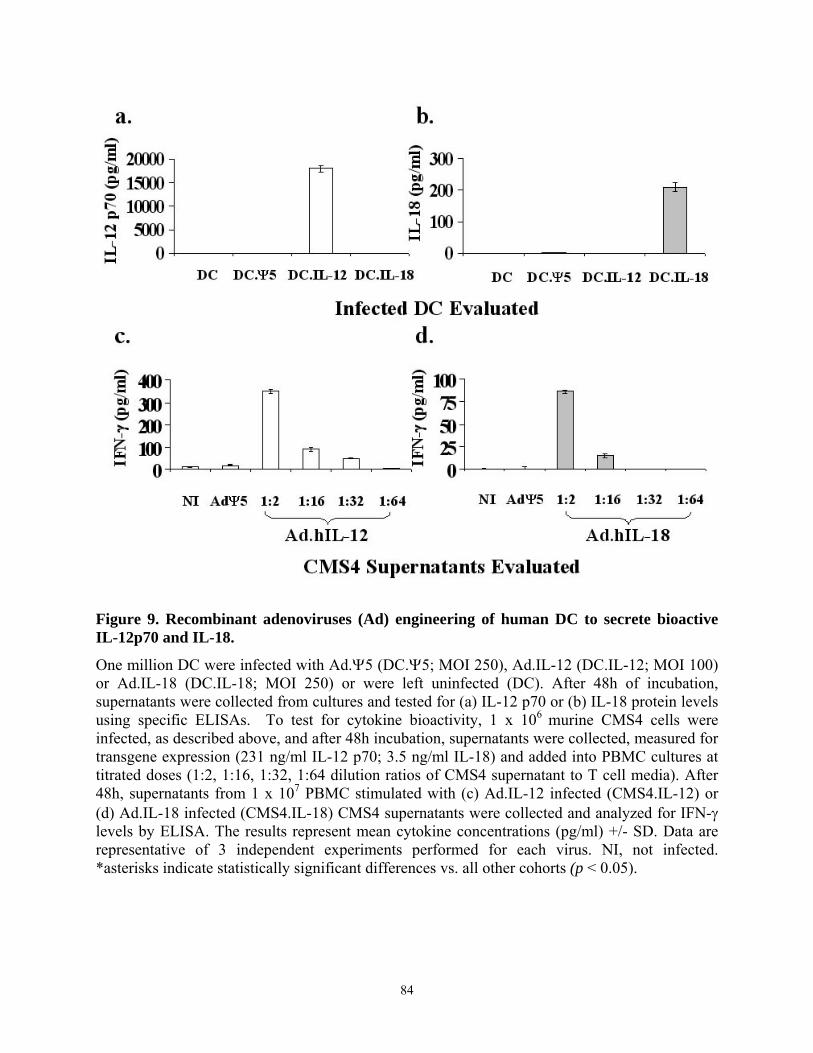
Figure 9. Recombinant adenoviruses (Ad) engineering of human DC to secrete bioactive IL-12p70 and IL-18.
One million DC were infected with Ad.Ψ5 (DC.Ψ5; MOI 250), Ad.IL-12 (DC.IL-12; MOI 100) or Ad.IL-18 (DC.IL-18; MOI 250) or were left uninfected (DC). After 48h of incubation, supernatants were collected from cultures and tested for (a) IL-12 p70 or (b) IL-18 protein levels using specific ELISAs. To test for cytokine bioactivity, 1 x 10⁶ murine CMS4 cells were infected, as described above, and after 48h incubation, supernatants were collected, measured for transgene expression (231 ng/ml IL-12 p70; 3.5 ng/ml IL-18) and added into PBMC cultures at titrated doses (1:2, 1:16, 1:32, 1:64 dilution ratios of CMS4 supernatant to T cell media). After 48h, supernatants from 1 x 10⁷ PBMC stimulated with (c) Ad.IL-12 infected (CMS4.IL-12) or (d) Ad.IL-18 infected (CMS4.IL-18) CMS4 supernatants were collected and analyzed for IFN-γ levels by ELISA. The results represent mean cytokine concentrations (pg/ml) +/- SD. Data are representative of 3 independent experiments performed for each virus. NI, not infected. *asterisks indicate statistically significant differences vs. all other cohorts (p < 0.05).
84
The bioactivity of these transgene products was determined indirectly by assessing the
ability of infected cell supernatant samples to subsequently induce IFN-γ secretion from
responder human PBMC. To minimize the impact of additional non-virus-induced factors on my
assay results (i.e. other than IL-12p70 or IL-18), I analyzed supernatants from infected murine
CMS4 tumor cells (191), to stimulate IFN-γ secretion from freshly-isolated PBMC. CMS4
supernatants were titrated on responder cells in order to discern any dose-dependent induction of
IFN-γ secretion. Both the IL-12p70 (Fig. 9c) and IL-18 (Fig. 9d) transgene products were
observed to induce IFN-γ secretion from PBMC in a dose-dependent manner, while control Ad-
infected CMS4 supernatants did not.
3.4.2. DC co-infected with Ad.IL-12 and Ad.IL-18 exhibit enhanced Th1-type CD4+ T
cell immunostimulatory capacity when compared to control DC.
DC derived from two normal HLA-DR4+ donors (ND) were infected with either Ad.Ψ5 control
vector (DC.Ψ5), Ad.IL-12 (DC.IL-12) or Ad.IL-18 (DC.IL-18) alone, or co-infected with both
Ad.IL-12 and Ad.IL-18 (DC.IL-12/18) at the previously determined, optimized MOIs.
Following infection, DC were pulsed with MAGE-A6121-144, MAGE-A6140-170 and MAGE-A6246-
263 peptides for 3h, and then added to culture wells containing autologous CD4+ T cells. A single
round of in vitro stimulation was employed in order to most directly discern the ability of DC
stimulations to amplify Th1-type responses against these epitopes (i.e. by avoiding extended in
vitro cultures where the impact on existent immunity would be less interpretable). Eleven days
after the initiation of the IVS protocol, IFN-γ ELISPOT assays were performed to enumerate
peptide-specific Th1-type CD4+ T cell responses (Fig. 10). While individual adenoviral
infections had little effect on the induction of responses (except for ND1, where Ad.IL-12
85
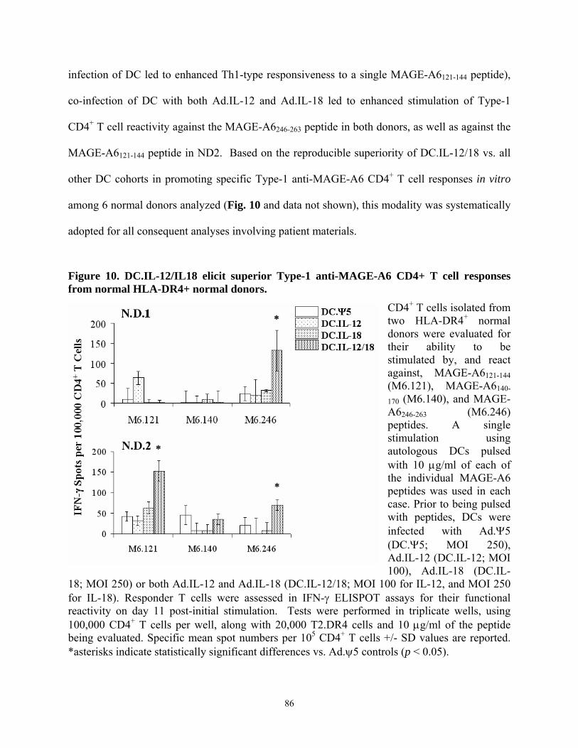
infection of DC led to enhanced Th1-type responsiveness to a single MAGE-A6121-144 peptide),
co-infection of DC with both Ad.IL-12 and Ad.IL-18 led to enhanced stimulation of Type-1
CD4+ T cell reactivity against the MAGE-A6246-263 peptide in both donors, as well as against the
MAGE-A6121-144 peptide in ND2. Based on the reproducible superiority of DC.IL-12/18 vs. all
other DC cohorts in promoting specific Type-1 anti-MAGE-A6 CD4+ T cell responses in vitro
among 6 normal donors analyzed (Fig. 10 and data not shown), this modality was systematically
adopted for all consequent analyses involving patient materials.
Figure 10. DC.IL-12/IL18 elicit superior Type-1 anti-MAGE-A6 CD4+ T cell responses from normal HLA-DR4+ normal donors.
CD4+ T cells isolated from two HLA-DR4+ normal donors were evaluated for their ability to be stimulated by, and react against, MAGE-A6121-144 (M6.121), MAGE-A6140-
170 (M6.140), and MAGE-A6246-263 (M6.246) peptides. A single stimulation using autologous DCs pulsed with 10 μg/ml of each of the individual MAGE-A6 peptides was used in each case. Prior to being pulsed with peptides, DCs were infected with Ad.Ψ5 (DC.Ψ5; MOI 250), Ad.IL-12 (DC.IL-12; MOI 100), Ad.IL-18 (DC.IL-
18; MOI 250) or both Ad.IL-12 and Ad.IL-18 (DC.IL-12/18; MOI 100 for IL-12, and MOI 250 for IL-18). Responder T cells were assessed in IFN-γ ELISPOT assays for their functional reactivity on day 11 post-initial stimulation. Tests were performed in triplicate wells, using 100,000 CD4+ T cells per well, along with 20,000 T2.DR4 cells and 10 μg/ml of the peptide being evaluated. Specific mean spot numbers per 105 CD4+ T cells +/- SD values are reported. *asterisks indicate statistically significant differences vs. Ad.ψ5 controls (p < 0.05).
86
3.4.3. DC.IL-12/18 loaded with MAGE-A6 peptides/protein effectively stimulate
epitope-specific Th1-type responses in melanoma patients following IVS
In these experiments, I evaluated CD4+ T cell responses to cytokine gene-engineered DC-based
stimulations in 8 HLA-DR4+ patients with melanoma (Table 7). DC derived from patient
PBMC were either not treated or co-infected with Ad.IL-12 and Ad.IL-18 (DC.IL-12/18). Both
groups of DCs were either pulsed with a peptide mix (including MAGE-A6121-144, MAGE-A6140-
170, MAGE-A6246-263, and in some indicated cases, MAGE-A6280-302) or with recombinant
MAGE-A6 (rMAGE), then added to cultures of autologous CD4+ T cells. Eleven days after the
initiation of the IVS protocol, IFN-γ ELISPOT assays were performed in order to enumerate the
frequencies of peptide-specific Th1-type CD4+ T cell responders. Peptide-pulsed DC.IL-12/18
enhanced the number/frequency of responder T cells as well as the magnitude of mean
responses, especially against the MAGE-A6121-144 and MAGE-A6280-302 epitopes, while peptide
stimulations with control, uninfected DC were ineffective in modulating specific CD4+ T
responses (Table 7). Similarly, rMAGE-pulsed DC.IL-12/18 were more effective than protein-
pulsed, uninfected DC in stimulating epitope-specific CD4+ T cell responses in vitro. Anti-
MAGE-A6121-144, -MAGE-A6280-302, as well as -MAGE-A6246-263 mean CD4+ T cell responses
were significantly enhanced following stimulation with rMAGE-pulsed DC.IL-12/18 (Table 7).
Indeed, all of the donors evaluated responded to at least one MAGE-A6 epitope in the ELISPOT
assay following stimulation with rMAGE protein-loaded DC.IL-12/18. In the majority of cases,
protein-pulsed uninfected DC appeared more effective than peptide-pulsed, uninfected DC in
promoting Type-1 responses (Table 7). Overall, these results suggest the superior
immunostimulatory effectiveness of DC.IL-12/18 (vs. control DC) loaded with either synthetic
87
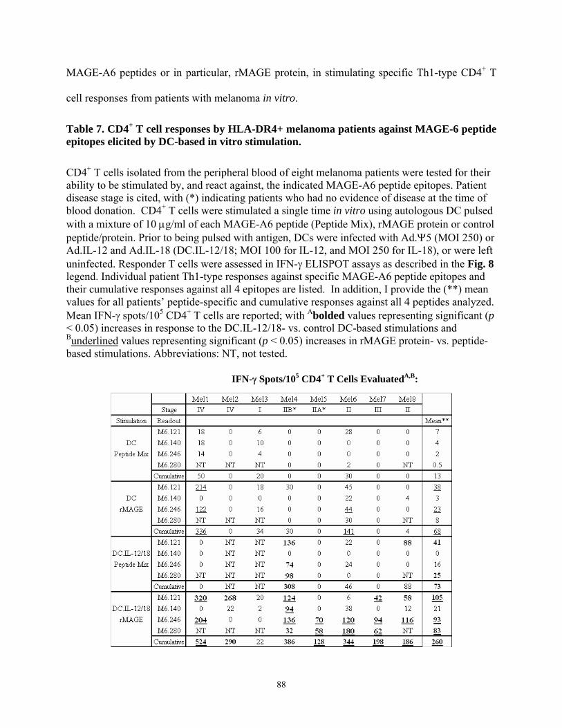
MAGE-A6 peptides or in particular, rMAGE protein, in stimulating specific Th1-type CD4+ T
cell responses from patients with melanoma in vitro.
Table 7. CD4+ T cell responses by HLA-DR4+ melanoma patients against MAGE-6 peptide epitopes elicited by DC-based in vitro stimulation.
CD4+ T cells isolated from the peripheral blood of eight melanoma patients were tested for their ability to be stimulated by, and react against, the indicated MAGE-A6 peptide epitopes. Patient disease stage is cited, with (*) indicating patients who had no evidence of disease at the time of blood donation. CD4+ T cells were stimulated a single time in vitro using autologous DC pulsed with a mixture of 10 μg/ml of each MAGE-A6 peptide (Peptide Mix), rMAGE protein or control peptide/protein. Prior to being pulsed with antigen, DCs were infected with Ad.Ψ5 (MOI 250) or Ad.IL-12 and Ad.IL-18 (DC.IL-12/18; MOI 100 for IL-12, and MOI 250 for IL-18), or were left uninfected. Responder T cells were assessed in IFN-γ ELISPOT assays as described in the Fig. 8 legend. Individual patient Th1-type responses against specific MAGE-A6 peptide epitopes and their cumulative responses against all 4 epitopes are listed. In addition, I provide the (**) mean values for all patients’ peptide-specific and cumulative responses against all 4 peptides analyzed. Mean IFN-γ spots/105 CD4+ T cells are reported; with Abolded values representing significant (p < 0.05) increases in response to the DC.IL-12/18- vs. control DC-based stimulations and Bunderlined values representing significant (p < 0.05) increases in rMAGE protein- vs. peptide-based stimulations. Abbreviations: NT, not tested.
IFN-γ Spots/105 CD4+ T Cells EvaluatedA,B:
88
3.5. Discussion
In this study, I generated a novel, functional adenoviral vector encoding the mature form
of human IL-18 protein, as well as a functional adenoviral vector encoding for human IL-12p70
protein, and analyzed their ability to infect human DC, thereby yielding a superior in vitro
“vaccine” capable of promoting tumor-specific Type-1 CD4+ T cell responses. Consistent with
previous reports by our group and others evaluating adenoviral infection of DC (191, 220, 221),
neither vector induced terminal DC maturation following infection (based on phenotypic
criteria), resulting in engineered DC that were functionally competent to uptake and process
soluble antigens (such as rMAGE).
To directly assess the Th1-stimulating properties associated with Ad.IL-12 and Ad.IL-18
infection on DC, and not alternate stimuli, engineered DC were not concomitantly or
consequently matured using cytokine/prostanoid cocktails. Given the desire to interpret the acute
effects of DC-based stimulations on a perceived Type-1 dysfunctional T cell repertoire in cancer
patients (208, 222), I analyzed peripheral blood CD4+ T cells isolated from HLA-DR4+ normal
donors activated after a single-round of in vitro sensitization. I evaluated normal donor and
patient CD4+ T cells stimulated with uninfected DC or Ad-engineered DC loaded with MAGE-
A6 peptides or protein for their ability to secrete IFN-γ when restimulated (in ELISPOT assays)
with three known HLA-DR4-restricted MAGE-A6-derived (MAGE-A6121-144, MAGE-A6140-170,
MAGE-A6246-263 epitopes (208, 222); and a novel poly-DR-presented MAGE-A6280-3021)
epitopes. My results suggest that DC.IL-12/18 were the most effective antigen presenting cells
for the promotion of Th1-type responses (that may be associated with tumor regression in situ;
ref. (191)) among those tested in this study. In contrast, DC infected with either Ad.IL-12
89
(DC.IL-12) or Ad.IL-18 (DC.IL-18) exhibited immunostimulatory capacities that were no
greater than those noted for DC infected with Ad.Ψ5 (DC.Ψ5). The improved efficacy
associated with DC.IL-12/18 stimulator cells may reflect the knowledge that IL-12p70 is
required to induce T cell surface expression of the IL-18 receptor (IL-18R) on naïve T cells
(167), while the IL-1 family member, IL-18, potentiates the differentiation of Th1 cells instigated
by IL-12p70 (168), and that IL-12 and IL-18 synergize in the activation of T cells and induction
of IFN-γ secretion from T cell responders (165, 166).
To evaluate the efficacy of this approach in the cancer setting, monocyte-derived DC
were generated from eight HLA-DR4+ melanoma patients and used to stimulate autologous
CD4+ T cells in vitro. Uninfected DC were compared to DC.IL-12/18 for their ability to
stimulate Th1-type responses after being pulsed with synthetic peptides or rMAGE protein. I
observed that DC.IL-12/18 were more effective in stimulating peptide-specific Th1-type
responses than uninfected DC, especially when loaded with rMAGE protein, in which case,
every patient (regardless of clinical staging and the presence/absence of disease at the time of
analysis) evaluated responded to at least one of the previously-defined, naturally-
processed/presented MAGE-A6-derived epitopes evaluated following in vitro sensitization. This
suggests that DC.IL-12/18 may be suitable for use in vaccines designed to override the general
Type-1 dysfunction in CD4+ T cell responses to tumor antigens observed in many cancer patients
with advanced stage disease (100, 101, 208, 222), in support of therapeutic immunity in the
clinical setting.
In conjunction with the knowledge that IL-18 is a chemoattractant for myeloid and
plasmacytoid DC (171, 172), one could also hypothesize that protein-pulsed DC.IL-12/18, if
injected s.c. or intratumorally, might not only induce Th1-type responses, but also attract
90
additional host DCs into the vaccine/tumor microenvironment. Within this microenvironment,
such a paradigm could enhance consequent tumor cell apoptosis, tumor antigen uptake and the
cross-priming of anti-tumor T cells in vivo (191).
In summary, I have generated a novel Ad.hIL-18, and have shown that when used in
conjunction with Ad.hIL-12 to co-infect human DC, a vaccine platform (DC.IL-12/18) is
produced that may be loaded with a recombinant tumor antigen or its derivative peptides in order
to effectively induce specific Th1-type CD4+ T cell responses in vitro from normal donors and
patients with melanoma. Further investigation is warranted to determine whether this strategy
can be translated into the clinic as a therapeutic vaccine, either as a stand-alone modality, or
when administered in combination with agents that ablate or neutralize functional regulatory T
cell activity in situ (223-225), in order to optimally potentiate therapeutic Type-1 anti-tumor
immunity in vivo.
91
GENERAL DISCUSSION
While sustained CD8+ T lymphocyte responses are required for effective clearance of
tumors, anti-tumor CD4+ T cells are at least equally important due to their ability to regulate the
quality, magnitude and durability of CD8+ CTL-mediated immunity in vivo. Previous studies of
peptide-specific, CD4+ T cell responses against tumor antigens, including MAGE-A6, have been
traditionally focused on an analysis of a single or very limited set of HLA-DR restriction
elements. Given the extreme polymorphism among HLA-DR alleles, the potential utility of the
previously defined epitopes is limited to only a minority of patients. Optimally, TAA-derived
peptides should be promiscuously binding (i.e. presented by multiple HLA-DR alleles; poly-
DR), which would increase their clinical applicability by making them immunogenic in a high
frequency of responders. Furthermore, any potential clinical modality implementing these
peptides needs to circumvent immuno-inhibitory properties of tumors, and stimulate Th1-type
responses. In these studies I sought to define poly-DR presented MAGE-A6 Th epitopes that
would clinically be broadly applicable. Also, I wanted to know whether I could stimulate Type-1
responses in vitro by engineering antigen-presenting DCs to secrete high levels of the IFN-γ-
inducing cytokines IL-12p70 and IL-18. I defined two highly-immunogenic poly-DR-binding,
MAGE-A6-derived epitopes, one of which shares immunogenicity with a Mycoplasma penetrans
HF-2 permease-derived epitope (MPHF2). Interestingly, the microbial epitope was more
effective at stimulating MAGE-A6-specific responses in vitro than its MAGE-A6 counterpart,
capable of promoting previously deficient patient Th1-type responses. Furthermore, I showed
that human DCs engineered with recombinant adenoviral vectors encoding IL-12p70 and IL-18
represent an efficient in vitro stimulus capable of effectively promoting specific Th1-type
responses.
92
First, using DC1-based in vitro vaccinations as a model system, I wanted to define poly-
DR-binding MAGE-A6-derived epitopes that would be recognized by CD4+ T-lymphocytes in a
high frequency of responders regardless of their HLA-DR phenotype. The MAGE-A6 protein
sequence was analyzed using the ProPred HLA-DR peptide-binding algorithm, and MAGE-A6
peptide sequences were selected based on their predicted ability to bind the broadest HLA-DR
repertoire. I synthesized 4 putative poly-DR peptides, and analyzed Th1 cell responses against
them. Two novel (MAGE-A6172-187 and MAGE-A6280-302) epitopes and a previously-defined
(MAGE-6140-170) HLA-DR4-restricted epitope were recognized by CD4+ T cells isolated from
most normal donors and patients with melanoma that were evaluated, with MAGE-A6172-187 and
MAGE-A6280-302 being the most immunogenic. These epitopes appear to be naturally-processed
and presented in the context of MHC class II molecules, as peptide-specific CD4+ T cells also
recognized autologous monocytes pulsed with full length MAGE-A6 protein.
Interestingly, normal donors mounted detectable Th1-type responses against the MAGE-
A6172-187 and MAGE-A6280-302 peptides after only a single-round of IVS. I hypothesized that the
MAGE-A6 peptides evaluated shared sufficient sequence homologies to microbial proteins to
which many individuals may have become naturally primed against, allowing for functional
cross-reactivity to be detected in my assays. I performed sequence homology searches of the
GenBank database for potential sources of cross-reactive epitopes. These analyses suggested that
the most immunogenic MAGE-A6172-187 and MAGE-A6280-302 epitopes exhibited significant
homologies to a number of known microbial sequences, of which the most likely encountered
candidates are Mycoplasma penetrans HF-2 permease-derived (MPHF2) and Chlamydia
muridarum Nigg conserved hypothetical protein TC0097-derived (CHP) epitopes respectively.
Unlike MAGE-A6172-187 and MAGE-A6280-302, MAGE-A6140-170 (moderately immunogenic) and
93
MAGE-A6192-214 (poorly immunogenic) epitopes did not share any identified microbial
homologies according to the GenBank database search.
CHP homologue peptide did not exhibit any significant cross-stimulatory ability to
MAGE-A6280-302, which was perhaps not surprising as it is derived from a mouse-tropic strain of
Chlamydia and is rarely encountered by humans. Contrary to this, CD4+ T cells primed with the
MPHF2 peptide cross-reacted against autologous monocytes pulsed with the MAGE-A6172-187
peptide or rMAGE protein, and recognized HLA-matched MAGE-A6+ melanoma cell lines in an
MHC class II-restricted fashion. CD4+ T cells generated using the MAGE-A6 peptide recognized
APCs only when pulsed with relatively high concentrations of peptide (with a half-maximal
response associated with a peptide dose of 3-10 μM), while MPHF2-stimulated cells recognized
target cells pulsed with approximately 1000-fold lower concentrations of peptide. Furthermore,
stimulations with MPHF2 were capable of promoting previously deficient patient Th1-type
responses to MAGE-A6, further supporting the potential clinical relevance for this peptide.
Mycoplasma infections have been shown to stimulate Th1, Th2, and Treg responses.
However, in a manner similarly to that of tumors, their effective clearance is associated with
Th1-type responses (226). As stimulations with MPHF2 yielded rapid, heteroclitic MAGE-A6
Th1 responses, it will be of interest to examine whether this is prompted from a resident pool of
memory CD4+ T cells in patients, or from an as yet antigen-inexperienced population of T cells
that could potentially be primed in vivo and provide therapeutic benefit to patients with MAGE-
A6+ lesions. My preliminary studies show that both Th1 and Th2 responses could be observed in
freshly-isolated CD4+ T cells against the MPHF2, but not the CS326-345 or MAGE-A6172-187
peptides (Figure 11). While these results suggest the likelihood of a memory response, this
observation requires further investigation of MPHF2 responsiveness (in ELISPOT assays and/or
94
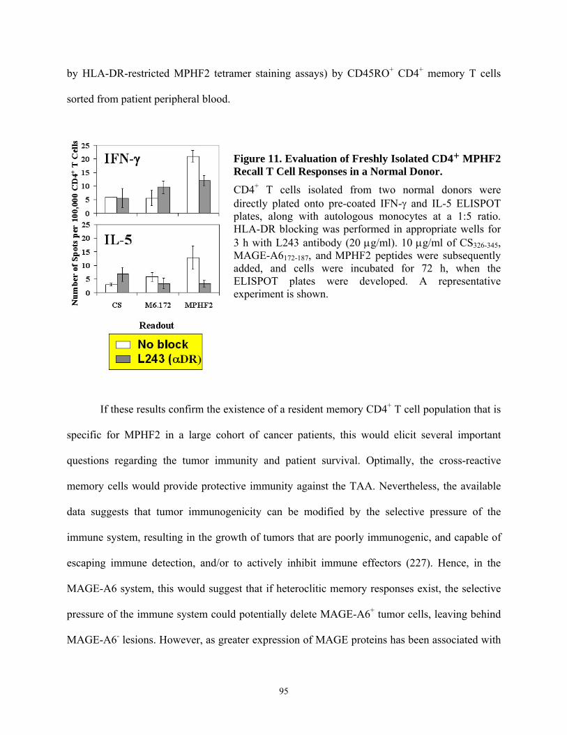
by HLA-DR-restricted MPHF2 tetramer staining assays) by CD45RO+ CD4+ memory T cells
sorted from patient peripheral blood.
Figure 11. Evaluation of Freshly Isolated CD4+ MPHF2 Recall T Cell Responses in a Normal Donor. CD4+ T cells isolated from two normal donors were directly plated onto pre-coated IFN-γ and IL-5 ELISPOT plates, along with autologous monocytes at a 1:5 ratio. HLA-DR blocking was performed in appropriate wells for 3 h with L243 antibody (20 μg/ml). 10 μg/ml of CS326-345, MAGE-A6172-187, and MPHF2 peptides were subsequently added, and cells were incubated for 72 h, when the ELISPOT plates were developed. A representative experiment is shown.
If these results confirm the existence of a resident memory CD4+ T cell population that is
specific for MPHF2 in a large cohort of cancer patients, this would elicit several important
questions regarding the tumor immunity and patient survival. Optimally, the cross-reactive
memory cells would provide protective immunity against the TAA. Nevertheless, the available
data suggests that tumor immunogenicity can be modified by the selective pressure of the
immune system, resulting in the growth of tumors that are poorly immunogenic, and capable of
escaping immune detection, and/or to actively inhibit immune effectors (227). Hence, in the
MAGE-A6 system, this would suggest that if heteroclitic memory responses exist, the selective
pressure of the immune system could potentially delete MAGE-A6+ tumor cells, leaving behind
MAGE-A6- lesions. However, as greater expression of MAGE proteins has been associated with
95
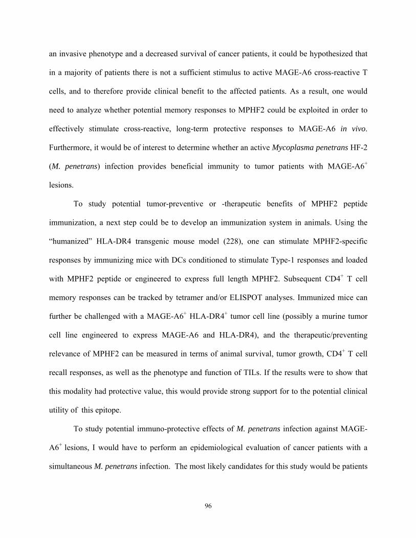
an invasive phenotype and a decreased survival of cancer patients, it could be hypothesized that
in a majority of patients there is not a sufficient stimulus to active MAGE-A6 cross-reactive T
cells, and to therefore provide clinical benefit to the affected patients. As a result, one would
need to analyze whether potential memory responses to MPHF2 could be exploited in order to
effectively stimulate cross-reactive, long-term protective responses to MAGE-A6 in vivo.
Furthermore, it would be of interest to determine whether an active Mycoplasma penetrans HF-2
(M. penetrans) infection provides beneficial immunity to tumor patients with MAGE-A6+
lesions.
To study potential tumor-preventive or -therapeutic benefits of MPHF2 peptide
immunization, a next step could be to develop an immunization system in animals. Using the
“humanized” HLA-DR4 transgenic mouse model (228), one can stimulate MPHF2-specific
responses by immunizing mice with DCs conditioned to stimulate Type-1 responses and loaded
with MPHF2 peptide or engineered to express full length MPHF2. Subsequent CD4+ T cell
memory responses can be tracked by tetramer and/or ELISPOT analyses. Immunized mice can
further be challenged with a MAGE-A6+ HLA-DR4+ tumor cell line (possibly a murine tumor
cell line engineered to express MAGE-A6 and HLA-DR4), and the therapeutic/preventing
relevance of MPHF2 can be measured in terms of animal survival, tumor growth, CD4+ T cell
recall responses, as well as the phenotype and function of TILs. If the results were to show that
this modality had protective value, this would provide strong support for to the potential clinical
utility of this epitope.
To study potential immuno-protective effects of M. penetrans infection against MAGE-
A6+ lesions, I would have to perform an epidemiological evaluation of cancer patients with a
simultaneous M. penetrans infection. The most likely candidates for this study would be patients
96
with spontaneously-regressing melanoma lesions since the majority of them are MAGE-A6+ and
easily accessible for tumor evaluation. As M. penetrans has been suggested to be a primary cause
of non-HIV-related urethritis and respiratory disease, I would need to correlate spontaneously-
regressing melanomas to an active or recently concluded M. penetrans infection. Frequencies of
circulating MPHF2-specific CD4+ T cells can be measured by tetramer analysis and, upon
surgical removal, melanoma lesions could be analyzed for MAGE-A6 expression by RT-PCR.
These results would provide indirect correlation between M. penetrans infection and its possible
protective properties against MAGE-A6.
As patients with malignant tumors exhibit dysfunctional Th1-type responses against
TAA-derived epitopes, it is imperative to re-establish the Th1 functionality in order to enhance
their chances of survival. I hypothesized that patient anti-tumor Th1-type responses might be
recovered/enhanced by in vitro stimulation of CD4+ T cells using vaccines containing autologous
DCs engineered to secrete high levels of the IFN-γ-inducing cytokines, IL-12p70 and/or IL-18.
In the second study, I investigated whether human DCs engineered to secrete IL-12p70 and IL-
18 by recombinant adenoviral infection can promote and enhance Th1-type, MAGE-A6-specific
responses in vitro. I chose this modality as it could not only prove capable of supporting Type-1
immunity, but also would have the potential to limit toxicities previously observed for systemic
application IL-12p70 alone or IL-12p70 combined with IL-18. Initial results showed that DC
infected with both Ad.IL-12 and Ad.IL-18 (DC.IL-12/18) were more effective at stimulating
MAGE-A6-specific Th1-type CD4+ T cell responses than DCs infected with either of the
cytokine vectors alone, Ad.Ψ5 virus (control vector) or uninfected DC. Further tests of this
vaccine strategy showed that DC.IL-12/18 loaded with MAGE-A6 peptides or rMAGE
effectively stimulated epitope-specific Th1-type CD4+ T cell responses in melanoma patients
97
following a single round of IVS regardless of their disease stage or current disease status. In
particular, DC.IL-12/18 loaded with rMAGE were the most effective stimulators of Th1-type
immunity in vitro, with responses frequently being directed against multiple MAGE-A6-derived
epitopes. My results show that DC.IL-12/18 loaded with tumor peptides or recombinant tumor
antigens may represent an effective vaccine capable of selectively promoting Th1-type CD4+ T
cell immunity in patients with cancer who may display existing immune dysfunction.
Using the same modality, I compared the ability of DC.IL-12/18 loaded with MAGE-A6-
derived HLA-A2 peptides or rMAGE to stimulate Type-1 CD8+ T cell responses. My initial
results suggested that DC.IL-12/18 could potentially enhance Type-1 CD8+ T cell responses
when compared to DCs infected with either of the cytokine vectors alone, Ad.Ψ5 virus (control
vector) or uninfected DC (Figure 12).
Figure 12. DC.IL-12/IL18 enhance Type-1 anti-MAGE-A6 CD8+ T cell responses from normal HLA-A2+ normal donors.
CD8+ T cells isolated from two HLA-A2+ normal donors were evaluated for their ability to be stimulated by, and react against, MAGE-A6112-120 (M6.112), and MAGE-A6271-279 (M6.271) peptides. Stimulation was performed as described in Figure 12. Responder T cells were assessed in IFN-γ ELISPOT assays for their functional reactivity on day 11 post-initial stimulation. Tests were performed in triplicate wells, using 100,000 CD8+ T cells per well, along with 20,000 T2.DR4 cells and 10 μg/ml of the peptide being evaluated. Specific mean spot numbers per 105 CD8+ T cells +/- SD values are reported. *asterisks indicate statistically significant differences vs. Ad.Ψ5 controls (p < 0.05).
98
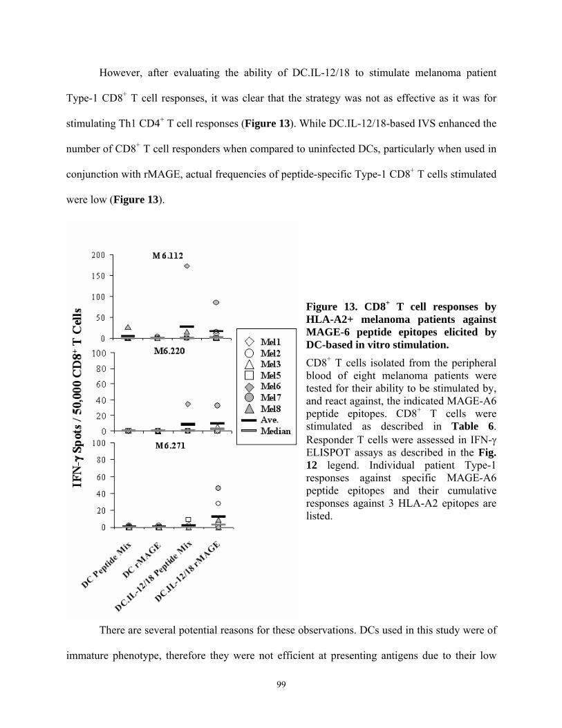
However, after evaluating the ability of DC.IL-12/18 to stimulate melanoma patient
Type-1 CD8+ T cell responses, it was clear that the strategy was not as effective as it was for
stimulating Th1 CD4+ T cell responses (Figure 13). While DC.IL-12/18-based IVS enhanced the
number of CD8+ T cell responders when compared to uninfected DCs, particularly when used in
conjunction with rMAGE, actual frequencies of peptide-specific Type-1 CD8+ T cells stimulated
were low (Figure 13).
Figure 13. CD8+ T cell responses by HLA-A2+ melanoma patients against MAGE-6 peptide epitopes elicited by DC-based in vitro stimulation.
CD8+ T cells isolated from the peripheral blood of eight melanoma patients were tested for their ability to be stimulated by, and react against, the indicated MAGE-A6 peptide epitopes. CD8+ T cells were stimulated as described in Table 6. Responder T cells were assessed in IFN-γ ELISPOT assays as described in the Fig. 12 legend. Individual patient Type-1 responses against specific MAGE-A6 peptide epitopes and their cumulative responses against 3 HLA-A2 epitopes are listed.
There are several potential reasons for these observations. DCs used in this study were of
immature phenotype, therefore they were not efficient at presenting antigens due to their low
99
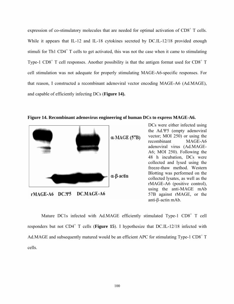
expression of co-stimulatory molecules that are needed for optimal activation of CD8+ T cells.
While it appears that IL-12 and IL-18 cytokines secreted by DC.IL-12/18 provided enough
stimuli for Th1 CD4+ T cells to get activated, this was not the case when it came to stimulating
Type-1 CD8+ T cell responses. Another possibility is that the antigen format used for CD8+ T
cell stimulation was not adequate for properly stimulating MAGE-A6-specific responses. For
that reason, I constructed a recombinant adenoviral vector encoding MAGE-A6 (Ad.MAGE),
and capable of efficiently infecting DCs (Figure 14).
Figure 14. Recombinant adenovirus engineering of human DCs to express MAGE-A6.
DCs were either infected using the Ad.Ψ5 (empty adenoviral vector; MOI 250) or using the recombinant MAGE-A6 adenoviral virus (Ad.MAGE-A6; MOI 250). Following the 48 h incubation, DCs were collected and lysed using the freeze-thaw method. Western Blotting was performed on the collected lysates, as well as the rMAGE-A6 (positive control), using the anti-MAGE mAb 57B against rMAGE, or the anti-β-actin mAb.
Mature DC1s infected with Ad.MAGE efficiently stimulated Type-1 CD8+ T cell
responders but not CD4+ T cells (Figure 15). I hypothesize that DC.IL-12/18 infected with
Ad.MAGE and subsequently matured would be an efficient APC for stimulating Type-1 CD8+ T
cells.
100
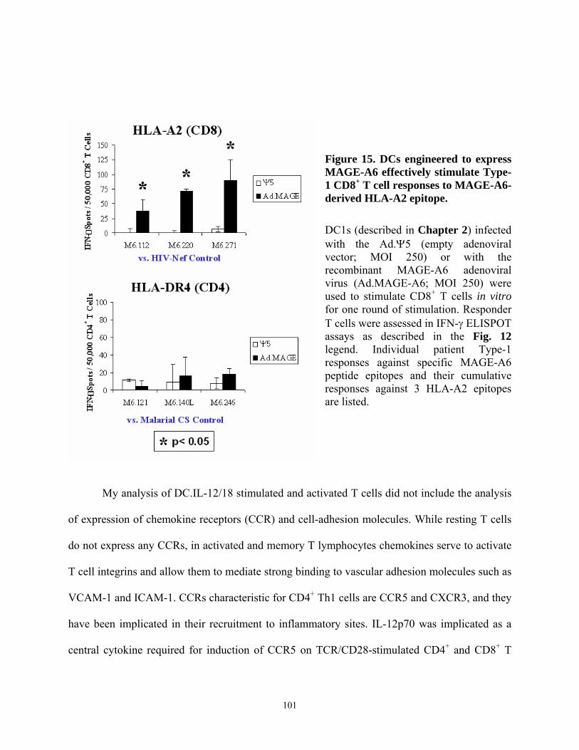
Figure 15. DCs engineered to express MAGE-A6 effectively stimulate Type-1 CD8+ T cell responses to MAGE-A6-derived HLA-A2 epitope. DC1s (described in Chapter 2) infected with the Ad.Ψ5 (empty adenoviral vector; MOI 250) or with the recombinant MAGE-A6 adenoviral virus (Ad.MAGE-A6; MOI 250) were used to stimulate CD8+ T cells in vitro for one round of stimulation. Responder T cells were assessed in IFN-γ ELISPOT assays as described in the Fig. 12 legend. Individual patient Type-1 responses against specific MAGE-A6 peptide epitopes and their cumulative responses against 3 HLA-A2 epitopes are listed.
My analysis of DC.IL-12/18 stimulated and activated T cells did not include the analysis
of expression of chemokine receptors (CCR) and cell-adhesion molecules. While resting T cells
do not express any CCRs, in activated and memory T lymphocytes chemokines serve to activate
T cell integrins and allow them to mediate strong binding to vascular adhesion molecules such as
VCAM-1 and ICAM-1. CCRs characteristic for CD4+ Th1 cells are CCR5 and CXCR3, and they
have been implicated in their recruitment to inflammatory sites. IL-12p70 was implicated as a
central cytokine required for induction of CCR5 on TCR/CD28-stimulated CD4+ and CD8+ T
101
cells. It was also shown in a mouse tumor model that IL-12p70 induces enhanced intratumoral T
cell migration that is dependent on the interaction between LFA-1 and VLA-4 adhesion
molecules with their ligands (ICAM-1 and VCAM-1 respectively) on tumor-associated
vasculature (229). Therefore it will be of interest to see whether T cells stimulated with DC.IL-
12/18 would stimulate expression of CCR5 that would allow for their potential trafficking into
tumor sites.
Multiple vaccination strategies utilizing the innate immunostimulatory ability of DCs
have been developed, however the ideal vaccination protocol with these cells has not yet been
described. Previous clinical trials implementing various DC-based vaccination strategies did not
yield completely satisfactory results, but did show potential that supported their further
development. Effective immunostimulatory strategies described in these studies could provide
potentially novel vaccination modalities that could circumvent immunoinhibitory properties of
certain tumors and enhance Type-1 responses in cancer patients. I have described here three
different MAGE-A6 antigen formats that in a clinical setting could be used alone or in
conjunction with DCs to preferentially stimulate CD4+ or CD8+ T cell responses in patients with
MAGE-A6+ lesions.
An optimal immunization strategy would simultaneously stimulate both of these T cell
subsets in a Type-1 manner. One clinical strategy would require direct immunization of patients
with the rMAGE or MAGE-A6- and/or MPHF2-derived peptides in conjunction with an
immunological adjuvant such as SBAS-2 [the mix of the QS21 saponin and of monophosphoryl
lipid A (MPL)] (230), relying on patient’s immune system to process antigens and stimulate T
cell responses. This strategy would not be labor-intensive, however its clinical benefits, based on
previous studies, would most likely be less than satisfactory (203).
102
As DC-based immunizations were shown to be one of the better vaccination strategies, it
could be hypothesized that antigen-loaded, Type-1-polarizing DC.IL-12/18 modality tested in
these studies has the potential to be implemented in a clinical setting. These cells can be pulsed
with MHC class I and II-restricted peptides, engineered to express MAGE-A6 using Ad.MAGE-
A6, or loaded with rMAGE. The translational utility of most of the peptides defined is clinically
limited to a modest group of patients due to a requirement for expression of specific HLA
haplotypes. While poly-DR-binding MAGE-A6 and MPHF2 Th epitopes described here could be
applicable in an extended cohort of patients to stimulate MAGE-A6-restricted Th1 responses,
previously-described MHC class I-binding peptides were shown to have a greater level of HLA
haplotype restriction. rMAGE-loaded and/or Ad.MAGE-A6-infected DC.IL-12/18 could provide
a way to stimulate CD4+ and CD8+ T cell responses respectively in cancer patients without HLA
haplotype restrictions. Combinational therapy with both rMAGE and Ad.MAGE-A6, that
primarily stimulate CD4+ and CD8+ T cell subsets respectively, could provide for an effective
immunization strategy. Furthermore, as mature DCs were shown to be superior at inducing T-
cell responses than immature DCs in a clinical setting (231), it could be theorized that an optimal
DC.IL-12/18-based immunization strategy should utilize matured, antigen-loaded DC.IL-12/18
capable of migrating to a lymph node and effectively stimulating both T cell subsets. Ultimately
the goal of these vaccinations would be to not only stimulate MAGE-A6-specific responses, but
also promote vaccine-induced epitope spreading, leading to enhanced, clinically-effective T cell
repertoire against other TAAs.
The effectiveness of a DC-based vaccine depends not only on the choice of TAA and its
format (i.e. peptide, recombinant protein or gene transfection), but on the vaccination schedule in
terms of dose (i.e. number of autologous DCs), frequency, number and site of immunization.
103
Reported doses range from 3-20 x 106 DCs per immunization, with an average dose of 10 x 106
cells, with multiple vaccination schedules ranging from 1-6 week intervals. Patents are
immunized either by intratumoral (i.t.), subcutaneous (s.c.) in the leg or arm closest to intact
lymph nodes, or ultrasound-guided intranodal (i.n.) injections (231-233). One thing that is
common in these studies is that patients have to be repeatedly vaccinated in order to stimulate
high enough frequencies of effective Type-1 T cells. While there are no studies that specifically
correlate TAA-specifc T cell numbers to effective tumor rejection, an increase in frequency of
TAA-specific lymphocytes following immunizations is generally correlated to a better survival
of patients (234). Based on these reports, a potential antigen-loaded DC.IL-12/18 strategy could
require a s.c. or i.n. bi-weekly immunizations. Due to high levels of IL-12 and IL-18 being
secreted by DC.IL-12/18, the number of cells used for immunization would have to be
subsequently determined based on any toxicities that could arise as a result of vaccinations. An
additional concern is whether adenovirally-engineered DCs can persist long enough in situ to
stimulate effective T cell responses as most people have developed immune responses to
naturally-encountered adenoviruses that could eliminate adenovirally-infected cells. Due to this
concern, multiple strains of recombinant adenoviral vectors have been developed that, if
engineered to encode for IL-12p70 and IL-18, could allow for multiple vaccinations.
Any potential DC.IL-12/18-based vaccination should also encompass a strategy to
antagonize Treg numbers and function, as it was shown that this subset of cells has a profound
ability to suppress anti-tumor immune responses in cancer patients. While there are no agents
that would affect Treg specifically, there are strategies that could eliminate these cells at a
greater frequency than other T lymphocyte subsets. DC.IL-12/18 could be administered in
combination with agents that ablate or neutralize functional regulatory T cell activity in situ,
104
such as cyclophosphamide, anti-CD25 antibody, GITR ligand (235, 236), or rIL-2 diphtheria
toxin conjugate DAB389IL-2/ONTAK (132). These potential combinational therapies could prove
to be more effective at providing clinical benefit than any of these modalities alone.
As an alternative to maturing antigen-loaded DC ex vivo, immature DC.IL-12/18 without
the antigen load could be injected directly into a tumor pre-treated with chemotherapy or locally
treated with radiotherapy, exploiting the local TAA repertoire to stimulate tumor-specific
lymphocyte responses. These conventional treatments could enhance the efficacy of subsequent
immunotherapy as they induce apoptosis, and apoptotic cells were shown to be a good source of
cross-presented antigens under inflammatory conditions (237, 238). IL-12 and IL-18 generated
by DC.IL-12/18, as well as, proinflammatory mediators such as heat shock proteins and IL-6 that
are released due to chemotherapy-induced apoptosis (237), would provide enough inflammatory
signal for DCs to effectively uptake and cross-present tumor antigens. Similarly, destruction by
radiofrequency ablation creates massive tissue apoptosis/necrosis which the immune system is
unable to completely clear. Because of the failure to clear apoptotic cells, post-apoptotic,
secondary necrosis occurs (238). Necrotic cells are a good source of antigens while
simultaneously providing strong maturation signals (self DNA and RNA, uric acid) to DCs.
Therefore, immature DC.IL-12/18 exposed to necrotic tumor cells could undergo maturation
after antigen uptake, and become effective antigen presenting cells. As the result, DC.IL-12/18
with or without the antigen load could be an effective immunization strategy if used in
combination with conventional cancer therapies.
The work performed in this thesis has defined two novel poly-DR-binding, MAGE-A6-
derived Th epitopes, as well as a mimicking peptide cross-reactive to a MAGE-A6 Th epitope,
that are immunogenic in a high frequency of responders. Furthermore, I have generated rMAGE
105
and Ad.MAGE-A6 that can be effectively used for stimulating CD4+ and CD8+ T cell subsets
respectively. Combined with my findings that DC.IL-12/18 could effectively generate Th1-type
responses in vitro, this thesis introduces the possibility of a novel DC-based therapeutic approach
for the treatment and possibly even prevention of MAGE-A6+ tumors. I hypothesize that DC.IL-
12/18 could be an effective platform for stimulating anti-tumor Th1 responses, particularly if
used in combination with conventional cancer treatments as well as agents that antagonize Treg
numbers and function. They could be used as an immunization modality by themselves, or they
could be loaded with a variety of antigen formats, including the ones described in this
dissertation. This type of therapy could be effective against a wide range of tumor types, and
would not be restricted to a patients HLA types particularly if used in combination with rMAGE
and/or Ad.MAGE-A6. Furthermore, if evidence gives further support to a residing MPHF2
memory CD4+ T cells in a large portion of the population, it could be hypothesized that this
epitope could be potentially employed as a preventive and/or therapeutic agent.
106
BIBLIOGRAPHY 1. Gromet, M. A., W. L. Epstein, and M. S. Blois. 1978. The regressing thin malignant
melanoma: a distinctive lesion with metastatic potential. Cancer 42:2282.
2. Tihan, T., and D. A. Filippa. 1996. Coexistence of renal cell carcinoma and malignant lymphoma. A casual relationship or coincidental occurrence? Cancer 77:2325.
3. van der Bruggen, P., C. Traversari, P. Chomez, C. Lurquin, E. De Plaen, B. J. van den Eynde, A. Knuth, and T. Boon. 1991. A gene encoding an antigen recognized by cytolytic T lymphocytes on a human melanoma. Science 254:1643.
4. van den Eynde, B. J., and P. van der Bruggen. 1997. T cell defined tumor antigens. Current Opinion in Immunology 9:684.
5. Jager, E., D. Jager, and A. Knuth. 1999. CTL-defined cancer vaccines: Perspectives for active immunotherapeutic interventions in minimal residual disease. Cancer and Metastasis Reviews 18:143.
6. Wang, R. F. 1999. Human tumor antigens: implications for cancer vaccine development. J. Mol. Med. 77:640.
7. Novellino, L., C. Castelli, and G. Parmiani. 2005. A listing ofhuman tumor antigens recognized by T cells: March 2004 update. Cancer Immunol. Immunother. 54:187.
8. Luretti, R., P. Pisarra, A. Verrecchia, C. Farina, G. Nicolini, A. Anichini, C. Bordignon, M. Sensi, G. Parmiani, and C. Traversari. 1998. Translation of a retained intron in tyrosinase-related protein (TRP) 2 mRNA generates a new cytotoxic T lymphocyte (CTL) - defined and shared human melanoma antigen not expressed in normal cells of the melanocytic lineage. J. Exp. Med. 188:1005.
9. Chomez, P., O. De Backer, M. Bertrand, E. De Plaen, T. Boon, and S. Lucas. 2001. An overview of the MAGE gene family with the identification of all human members of the family. Cancer Res. 61:5544.
10. Barker, P., and A. Salehi. 2002. The MAGE proteins: emerging roles in cell cycle progression, apoptosis, and neurogenetic disease. J Neurosci Res. 67:705.
107
11. Takahashi, K., S. Shichijo, M. Noguchi, M. Hirohata, and K. Itoh. 1995. Identification of MAGE-1 and MAGE-4 proteins in spermatogonia and primary spermatocytes of testis. Cancer Res. 55:3478.
12. Grobholz, R., C. S. Verbeke, C. Schleger, K.-U. Kohrmann, B. Hein, G. Wolf, U. Bleyl, G. C. Spagnoli, K. Coplan, D. Kolb, K. Iversen, and A. A. Jungbluth. 2000. Expression of MAGE antigens and analysis of the inflammatory T-cell infiltrate in human seminoma. Urol. Res. 28:398.
13. De Plaen, E. 1994. Structure, chromozomal lovalization, and expression of 12 genes of the MAGE family. Immunogenetics 40:360.
14. Furuta, J., Y. Umebayashi, K. Miyamoto, K. Kikuchi, F. Otsuka, T. Sugimura, and T. Ushijima. 2004. Promoter methylation profiling of 30 genes in human malignant melanoma. Cancer Res. 95:962.
15. Janssen, B. L., L. T. van de Locht, A. Fourkour, C. de Smet, E. J. Mensink, G. N. van Muijen, and T. J. de Vries. 1999. Transcription of the MAGE-1 gene and the methylation status of its Ets binding promoter elements: a quantitative analysis in melanoma cell lines using a real-time polymerase chain reaction technique. Melanoma Research 9:213.
16. Barker, P. A., and A. Salehi. 2002. The MAGE proteins: emerging roles in cell cycle progression, apoptosis, and neurogenetic disease. J. Neurosci. Res. 67:705.
17. Takazaki, R., I. Nishimura, and K. Yoshikawa. 2002. Necdin is required for terminal differentiation and survival of primary dorsal root ganglion neurons. Exp. Cell Res. 277:220.
18. Taniura, H., K. Matsumoto, and K. Yoshikawa. 1999. Physical and functional interactions of neuronal growth suppressor necdin with p53. J. Biol. Chem. 274:16242.
19. Wen, C. J., B. Xue, W. X. Qin, M. Yu, M. Y. Zhang, D. H. Zhao, X. Gao, J. R. Gu, and C. J. Li. 2004. hNRAGE, a human neurotrophin receptor interacting MAGE homologue, regulates p53 transcriptional activity and inhibits cell proliferation. FEBS Letters 564:171.
20. Park, J. H., G. H. Kong, and S. W. Lee. 2002. hMAGE-A1 overexpression reduces TNF-alpha cytotoxicityin ME-180 cells. Mol. Cells 14:122.
108
21. Nagao, T., H. Higashitsuji, K. Nonoguchi, T. Sakurai, S. Dawson, R. J. Mayer, K. Itoh, and J. Fujita. 2003. MAGE-A4 interacts with the liver oncoprotein gankyrin and suppresses its tumorigenic activity. J. Biol. Chem. 278:10668.
22. Sakurai, T., K. Itoh, H. Higashitsuji, T. Nagao, K. Nonoguchi, T. Chiba, and J. Fujita. 2004. A cleaved form of MAGE-A4 binds to Miz-1 and induces apoptosis in human cells. J. Biol. Chem. 279:15505.
23. Chomez, P., O. De Backer, M. Bertrand, E. De Plaen, T. Boon, and S. Lucas. 2001. An overview of the MAGE gene family with the identification of all human members of the family. Cancer Res. 61:5544.
24. Kocher, T., E. Schultz-Thater, F. Gudat, C. Schaefer, G. Casorati, A. Juretic, T. Willimann, F. Harder, M. Heberer, and G. C. Spagnoli. 1995. Identification and intracellular location of MAGE-3 gene product. Cancer Res. 55:2236.
25. Sarcevic, B., G. C. Spagnoli, L. Terracciano, E. Schultz-Thater, M. Heberer, M. Gamulin, Z. Krajina, T. Oresic, R. Separovic, and A. Juretic. 2003. Expression of cancer/testis tumor associated antigens in cervical squamous cell carcinoma. Oncology 64:443.
26. Jungbluth, A. A., K. J. Busam, D. Kolb, K. Iversen, K. Coplan, Y.-T. Chen, G. C. Spagnoli, and L. J. Old. 2000. Expression of MAGE-antigens in normal tissues and cancer. International Journal of Cancer 85:460
27. Gibbs, P., A. Hutchins, K. Dorian, H. Vaughan, I. Davis, M. Silvapulle, and J. Cebon. 2000. MAGE-12 and MAGE-6 are frequently expressed in malignant melanoma. Melanoma Res. 10:259.
28. Luftl, M., G. Schuler, and A. A. Jungbluth. 2004. Melanoma or not? Cancer testis antigens may help. Brit. J. Dermatopath. 151:1213.
29. Hofbauer, G. F., C. Schaefer, C. Noppen, R. Boni, J. Kamarashev, F. O. Nestle, G. C. Spagnoli, and R. Dummer. 1997. MAGE-3 immunoreactivity in formalin-fixed, paraffin-embedded primary and metastatic melanoma: frequency and distribution. American Journal of Pathology 151:1549.
30. Kocher, T., M. Zheng, M. Bolli, R. Simon, T. Forster, E. Schultz-Thater, E. Remmel, C. Noppen, U. Schmid, D. Ackermann, M. J. Mihatsch, T. Gasser, M. Heberer, G. Sauter, and G. C. Spagnoli. 2002. Prognostic relevance of MAGE-A4 tumor antigen expression
109
in transitional cell carcinoma of the urinary bladder: a tissue microarray study. Int. J. Cancer 100:702.
31. Schultz, E. S., B. Schuler-Thurner, V. Stroobant, L. Jenne, T. G. Berger, K. Thielmanns, P. v. d. Bruggen, and G. Schuler. 2004. Functional analysis of tumor-specific Th cell responses detected in melanoma patients after dendritic cell-based immunotherapy. J. Immunol. 172:1304.
32. Gajewski, T. F., F. Fallarino, A. Ashikari, and M. Sherman. 2001. Immunization of HLA-A2+ melanoma patients with MAGE-3 or MelanA peptide-pulsed autologous peripheral blood mononuclear cells plus recombinant human interleukin 12. Clin. Cancer Res. 7:895s.
33. Lurquin, C., B. Lethe, E. D. Plaen, V. Corbiere, I. Theate, N. v. Baren, P. G. Coulie, and T. Boon. 2005. Contrasting frequencies of antitumor and anti-vaccine T cells in metastases of a melanoma patient vaccinated with a MAGE tumor antigen. J. Exp. Med. 201:249.
34. Germeau, C., W. Ma, F. Schiavetti, C. Lurquin, E. Henry, N. Vigneron, F. Brasseur, B. Lethe, E. D. Plaen, T. Velu, T. Boon, and P. G. Coulie. 2005. High frequency of antitumor T cells in the blood of melanoma patients before and after vaccination with tumor antigens. J. Exp. Med. 201:241.
35. Ding, M., R. J. Beck, C. J. Keller, and R. G. Fenton. 1994. Cloning and analysis of MAGE-1-related genes. Biochem. Biophys. Res. Commun. 202:549.
36. Tatsumi, T., L. S. Kierstead, E. Ranieri, L. Gesualdo, F. P. Schena, J. H. Finke, R. M. Bukowski, V. Brusic, J. Sidney, A. Sette, T. F. Logan, Y. L. Kasamon, C. L. Slingluff, Jr., J. M. Kirkwood, and W. J. Storkus. 2003. MAGE-6 Encodes HLA-DR{beta}1*0401-presented Epitopes Recognized by CD4+ T Cells from Patients with Melanoma or Renal Cell Carcinoma. Clin Cancer Res 9:947.
37. Rayman, P., A. K. Wesa, A. L. Richmond, T. Das, K. Biswas, G. Raval, W. J. Storkus, C. Tannenbaum, A. Novick, R. Bukowski, and J. Finke. 2004. Effect of Renal Cell Carcinomas on the Development of Type 1 T-Cell Responses. Clin Cancer Res 10:6360S.
38. Liu, B., S. Ye, P. He, Y. Liu, and Z. Tang. 1999. MAGE-1 and related MAGE gene expression may be associated with hepatocellular carcinoma. J. Cancer Res. And Clin. Oncology 125:685.
110
39. Otte, M., M. Zafrakas, L. Riethdorf, U. Pichlmeier, T. Loning, F. Janicke, and K. Pantel. 2001. MAGE-A gene expression pattern in primary breast cancer. Cancer Res. 61:6682.
40. Lin, J., L. Lin, D. Thomas, J. Greenson, T. Giordano, G. Robinson, R. Barve, F. Weishaar, J. Taylor, M. Orringer, and D. Beer. 2004. Melanoma-associated antigens in esophageal adenocarcinoma: identification of novel MAGE-A10 splice variants. Clin. Cancer Res. 10:5708.
41. Zorn, E., and T. Hercend. 1999. A MAGE-6-encoded peptide is recognized by expanded lymphocytes infiltrating a spontaneously regressing human primary melanoma lesion. Eur. J. Immunol. 29:602.
42. Scanlan, M. J., I. Gout, C. M. Gordon, B. Williamson, E. Stockert, A. O. Gure, D. Jager, Y.-T. Chen, A. Mackay, M. J. O'Hare, and L. J. Old. 2001. Humoral immunity to human breast cancer: antigen definition and quantitative analysis of mRNA expression. Cancer Immunity 1:4.
43. Huang, J., H. T. Khong, M. E. Dudley, M. El-Gamil, Y. F. Li, S. A. Rosenberg, and P. F. Robbins. 2005. Survival, Persistence, and Progressive Differentiation of Adoptively Transferred Tumor-Reactive T Cells Associated with Tumor Regression. J. Immunother. 28:258.
44. Rammensee, H. G. 1995. Chemistry of peptides associated with MHC class I and II molecules. Curr. Opin. Immunol. 7:85.
45. van Bergen, J., F. Ossendorp, R. Jordens, A. M. Mommaas, J. W. Drijfhout, and F. Koning. 1999. Get into the groove! Targeting antigens to MHC class II [pathways of antigen processing and presentation]. Immunol. Rev. 172:87.
46. Cresswell, P. 1993. Antigen processing. Ann. Rev. Immunol. 11:259.
47. Rammensee, H. G., T. Fried, and S. Stevanovic. 1995. MHC ligands and peptide motifs: first listing. Immunogenetics 41:178.
48. Yewdell, J. W., and J. R. Bennink. 1999. Immunodominance in major histocompatibility complex class I-restricted T lymphocyte responses. Annu. Rev. Immunol. 17:51.
49. Rescigno, M., F. Granucci, and P. Ricciardi-Castagnoli. 1999. Dendritic cells at the end of the millenium. Immunol. Cell Biol. 77:404.
111
50. Matsumura, M., D. H. Fremont, P. A. Peterson, and I. A. Wilson. 1992. Emerging principles for the recognition of peptide antigens by MHC class I molecules. Science 257:927.
51. Bouvier, M., and D. C. Wiley. 1994. Importance of peptide amino and carboxyl termini tot he stability of MHC class I molecules. Science 265:398.
52. Cerundolo, V., T. Elliot, J. Elvin, J. Bastin, H. B. Rammensee, and A. Townsend. 1991. The binding affinity and dissociation rates of peptides for class I major histocompatibility complex molecules. Eur. J. Immunol. 21:2069.
53. Neefjes, J. J., J. Dierx, and H. L. Ploegh. 1993. The effect of anchor residue modifications on the stability of major histocompatibility complex class I-peptide interactions. Eur. J. Immunol. 23:840.
54. Reits, E., A. Griekspoor, J. Neijssen, T. Groothuis, K. Jalink, P. van Veelen, H. Janssen, J. Calafat, J. W. Drijfhout, and J. Neefjes. 2003. Peptide diffusion, protection, and degradation in nuclear and cytoplasmic compartments before antigen presentation by MHC class I. Immunity 18:97.
55. Groothuis, T. A., A. C. Griekspoor, J. J. Neijssen, C. A. Herberts, and J. J. Neefjes. 2005. MHC class I alleles and their exploration of the antigen-processing machinery. Immunol. Rev. 207:60.
56. Rock, K. L., S. Gamble, and L. Rothstein. 1990. Presentation of exogenous antigen with class I major histocompatibility complex molecules. Science 249:918.
57. Rock, K. L., L. Rothstein, S. Gamble, and C. Fleischacker. 1993. Characterization of antigen-presenting cells that present exogenous antigens in association with class I MHC molecuels. J. Immunol. 150:438.
58. Neijssen, J., C. Herberts, J. W. Drijfhout, E. Reits, L. Janssen, and J. Neefjes. 2005. Cross-presentation by intracellular peptide transfer through gap junctions. Nature 434:83.
59. Groothuis, T. A. M., and J. Neefjes. 2005. The many roads to cross-presentation. J. Exp. Med. 202:1313.
112
60. Gromme, M., F. G. Uytdehaag, H. Janssen, J. Calafat, R. S. van Binnendijk, M. J. Kenter, A. Tulp, D. Verwoerd, and J. Neefjes. 1999. Recycling MHC class I molecules and endosomal peptide loading. Proc. Natl. Acad. Sci. USA 96:10326.
61. Touret, N., P. Paroutis, M. Terbiznik, R. Harrison, S. Trombetta, M. Pypaert, A. Chow, A. Jiang, J. Shaw, and C. Yip. 2005. Quantitative and dynamic assessment of the contribution of the endoplasmic reticulum to phagosome formation. Cell 123:157.
62. Villadangos, J. A., P. Schnorrer, and N. S. Wilson. 2005. Control of MHC class II antigen presentation in dendritic cells: a balance between creative and destructive forces. Immunol. Rev. 207:191.
63. Bryant, P. W., A. M. Lennon-Dumenil, E. Fiebiger, C. Lagaudriere-Gesbert, and H. L. Ploegh. 2002. Proteolysis and antigen presentation by MHC class II molecules. Adv. Immunol. 80:71.
64. Lazarski, C. A., F. A. Chaves, S. A. Jenks, S. Wu, K. A. Richards, J. M. Weaver, and A. J. Sant. 2005. The kinetic stability of MHC class II:peptide complexes is a key parameter that dictates immunodominance. Immunity 23:29.
65. Page, D. M., L. P. Kane, T. M. Onami, and S. M. Hedrick. 1996. Cellular and biochemical requirements for thymocyte negative selection. Semin. Immunol. 8:69.
66. Zacharchuk, C. M., M. Mercep, and J. D. Ashwell. 1991. Thymocyte activation and death: a mechanism for molding the T cell repertoire. Ann. NY Acad. Sci. 636:52.
67. Anderton, S. M., C. G. Radu, P. A. Lowrey, E. S. Ward, and D. C. Wraith. 2001. Negative selection during the peripheral immune response to antigen. J. Exp. Med, 193:1.
68. Weiner, H. L. 2001. Induction and mechanism of action of transforming growth factor-beta-secreting Th3 regulatory cells. Immunol. Rev. 182:207.
69. Hung, K., R. Hayashi, A. Lafond-Walker, C. Lowenstein, D. Pardoll, and H. Levitsky. 1998. The central role of CD4(+) T cells in the antitumor immune response. J.Exp. Med. 188:2357.
70. Fallarino, F., U. Grohmann, R. Bianchi, C. Vacca, M. C. Fioretti, and P. Puccetti. 2000. Th1 and Th2 cell clones to a poorly immunogenic tumor antigen initiate CD8+ T cell-dependent tumor eradication in vivo. J. Immunol. 165:5495.
113
71. Nishimura, T., M. Nakui, K. Sato, K. Iwakabe, H. Kitamura, M. Sekimoto, A. Ohta, T. Koda, and S. Nishimura. 2000. The critical role of Th1-dominant immunity in tumor immunology. Cancer Chemother. Pharmacol. 46:S52.
72. Nestle, F. O., S. Alijagic, M. Gilliet, Y. Sun, S. Grabbe, R. Dummer, G. Burg, and D. Schadendorf. 1998. Vaccination of melanoma patients with peptide- or tumor lysate-pulsed dendritic cells. Nature Medicine 4:328.
73. Liau, L. M., E. R. Jensen, T. J. Kremen, S. K. Odesa, S. N. Sykes, M. C. Soung, J. F. Miller, and J. M. Bronstein. 2002. Tumor Immunity within the Central Nervous System Stimulated by Recombinant Listeria monocytogenes Vaccination. Cancer Res 62:2287.
74. Disis, M. L., K. H. Grabstein, P. R. Sleath, and M. A. Cheever. 1999. Generation of Immunity to the HER-2/neu Oncogenic Protein in Patients with Breast and Ovarian Cancer Using a Peptide-based Vaccine. Clin Cancer Res 5:1289.
75. Bonehill, A., C. Heirman, and K. Thielemans. 2005. Genetic approaches for the induction of a CD4+ T cell response in cancer immunotherapy. J. Gene Med. 7:686.
76. Qin, Z., and T. Blankenstein. 2000. CD4+ T cell--mediated tumor rejection involves inhibition of angiogenesis that is dependent on IFN gamma receptor expression by nonhematopoietic cells. Immunity 12:677.
77. Qin, Z., J. Schwartzkopff, F. Pradera, T. Kammertoens, B. Seliger, H. Pircher, and T. Blankenstein. 2003. A critical requirement of interferon gamma-mediated angiostasis for tumor rejection by CD8+ T cells. Cancer Research 63:4095.
78. Appay, V. 2004. The physiological role of cytotoxic CD4(+) T-cells: the holy grail? Clinical & Experimental Immunology 138:10.
79. Appay, V., J. Zaunders, L. Papagno, J. Sutton, A. Jaramillo, A. Waters, P. Easterbrook, P. Grey, D. Smith, A. McMichael, D. Cooper, S. Rowland-Jones, and A. Kelleher. 2002. Characterization of CD4(+) CTLs ex vivo. Journal of Immunology 168:5954.
80. Blay, J. Y., S. Negrier, V. Combaret, S. Attali, E. Goillot, and Y. Merrouche. 1992. Serum level of interleukin 6 as a prognosis factor in metastatic renal cell carcinoma. Cancer Res. 52:3317.
114
81. Stassen, M., S. Fondel, T. Bopp, C. Richter, C. Muller, J. Kubach, C. Becker, J. Knop, A. H. Enk, S. Schmitt, E. Schmitt, and H. Jonuleit. 2004. Human CD25+ regulatory T cells: two subsets defined by the integrins alpha4beta7 or alpha4beta1 confer distinct suppressive properties upon CD4+ T helper cells. Eur. J. Immunol. 34:1303.
82. Seder, R. A., T. Marth, M. C. Sieve, W. Strober, J. J. Letterio, A. B. Roberts, and B. Kelsall. 1998. Factors involved in the differentiation of TGF-β-producing cells from naïve CD4+ T cells: IL-4 and IFN-γ have opposing effects, while TGF-β positively regulates its own production. J. Immunol. 160:5719.
83. Okamura, H., H. Tsutsui, T. Komatsu, M. Yutsudo, A. Hakura, T. Tanimoto, K. Torigoe, T. Okura, Y. Nukada, K. Hattori, K. Akita, M. Namba, F. Tanabe, K. Konishi, S. Fukuda, and M. Hurioto. 1995. Cloning of a new cytokine that induces IFN-gamma production by T cells. Nature 378:88.
84. Dap, T., K. Ohashi, T. Kayano, M. Kurimoto, and H. Okamura. 1996. Interferon-γ-inducing factor, a novel cytokine, enhances Fas ligand-mediated cytotoxicity of murine T helper 1 cells. Cell. Immunol. 173:230.
85. Osaki, T., J. M. Peron, Q. Cai, H. Okamura, P. D. Robbins, M. Kurimoto, M. T. Lotze, and H. Tahara. 1998. IFN-γ-inducing factor/IL-18 administration mediates IFN-γ- and IL-12-independent antitumor effects. J. Immunol. 160:1742.
86. Hashimoto, W., T. Osaki, H. Okamura, P. D. Robbins, M. Kurimoto, S. Nagata, M. T. Lotze, and H. Tahara. 1999. Differential antitumor effects of administration of recombinant IL-18 or recombinant IL-12 are mediated primarily by Fas-Fas ligand- and perforin-induced tumor apoptosis, respectively. J. Immunol. 163:583.
87. Osaki, T., W. Hashimoto, A. Gambotto, H. Okamura, P. D. Robbins, M. Kurimoto, M. T. Lotze, and H. Tahara. 1999. Potent antitumor effects mediated by local expression of the mature form of the interferon-g inducing factor, interleukin-18 (IL-18). Gene Ther. 6:808.
88. Geginat, G., T. Ruppert, H. Hengel, R. Holtappels, and U. H. Koszinowski. 1997. IFN-gamma is prerequisite for optimal antigen processing of viral peptides in vivo. J. Immunol. 158:3303.
89. Rosa, F. M., and M. Fellous. 1998. Regulation of HLA-DR gene by IFN-gamma. Transcriptional and post-transcriptional control. J. Immunol. 140:1660.
115
90. Hockett, R. D., J. R. Cook, K. Findlay, and C. V. Harding. 1996. Interferon-gamma differentially regulates antigen-processing functions in distinct endocytic compartments of macrophages with constitutive expression of class II major histocompatibility complex molecules. Immunol. 88:68.
91. Maile, R., K. A. Elsegood, T. C. Harding, J. B. Uney, C. E. Stewart, G. Banting, and C. M. Dayan. 2000. Effective formation of major histocompatibility complex class II-peptide complexes from endogenous antigen by thyroid epithelial cels. Immunol. 99:367.
92. Sospedra, M., E. Tolosa, P. Armengol, Y. Ashhab, S. Urlinger, A. Lucas-Martin, M. Foz-Sala, D. Jaraquemada, and R. Pujol-Borrell. 1997. Hyperexpression of transporter in antigen processing-1 (TAP-1) in thyroid glands affected by autoimmunity: a contributing factor to the breach of tolerance to thyroid antigens? Clin. Exp. Immunol. 109:98.
93. Tanaka, K., and M. Kasahara. 1998. The MHC class I ligand-generating system: roles of immunoproteasomes and the interferon-gamma-inducible proteasome activator PA28. Immunol. Rev. 163:161.
94. Sewell, A. K., D. A. Price, H. Teisserenc, B. L. J. Booth, U. Gileadi, F. M. Flavin, J. Trowsdale, R. E. Phillips, and V. Cerundolo. 1999. IFN-gama exposes a cryptic cytotoxic T lymphocyte epitope in HIV-1 reverse transcriptase. J. Immunol. 162:7075.
95. Schultz, E. S., J. Chapiro, C. Lurquin, S. Claverol, O. Burlet-Schiltz, G. Warnier, V. Russo, S. Morel, F. Levy, T. Boon, B. J. V. d. Eynde, and P. v. d. Bruggen. 2002. The production of a new MAGE-3 peptide presented to cytolytic T lymphocytes by HLA-B40 requires the immunoproteasome. J. Exp. Med. 195:391.
96. Dick, T. P., T. Ruppert, M. Groettrup, P. M. Kloetzel, L. Kuehn, U. H. Koszinowski, S. Stevanovic, H. Schild, and H. G. Rammensee. 1996. Coordinated dual cleavages induced by the proteasome regulator PA28 lead to dominant MHC ligands. Cell 86:253.
97. Wittke, F., R. Hoffmann, J. Buer, I. Dallmann, K. Oevermann, S. Sel, T. Wandert, A. Ganser, and J. Atzpodien. 1999. Interleukin 10 (IL-10): an immunosuppressive factor and independent predictor in patients with metastatic renal cell carcinoma. Br. J. Cancer 79:1182.
98. Lowes, M. A., G. A. Bishop, K. Crotty, R. S. Barnetson, and G. M. Halliday. 1997. T helper 1 cytokine mRNA is increased in spontaneously regressing primary melanoma. J. Invest. Derm. 108:914.
116
99. Reichert, T. E., L. Strauss, E. M. Wagner, W. Gooding, and T. L. Whiteside. 2002. Signaling Abnormalities, Apoptosis, and Reduced Proliferation of Circulating and Tumor-infiltrating Lymphocytes in Patients with Oral Carcinoma. Clin Cancer Res 8:3137.
100. Gray, C. P., P. Arosio, and P. Hersey. 2003. Association of Increased Levels of Heavy-Chain Ferritin with Increased CD4+ CD25+ Regulatory T-Cell Levels in Patients with Melanoma. Clin Cancer Res 9:2551.
101. Wolf, A. M., D. Wolf, M. Steurer, G. Gastl, E. Gunsilius, and B. Grubeck-Loebenstein. 2003. Increase of Regulatory T Cells in the Peripheral Blood of Cancer Patients. Clin Cancer Res 9:606.
102. Schwaab, T., J. A. Heaney, A. R. Schned, R. D. Harris, B. F. Cole, R. J. Noelle, D. M. Phillips, L. Stempkowski, and M. S. Ernstoff. 2000. A randomized phase II trial comparing two different sequence combinations of autologous vaccine and human recombinant interferon gamma and human recombinant interferon alpha2B therapy in patients with metastatic renal cell carcinoma: clinical outcome and analysis of immunological parameters. J. Urol. 163:1322.
103. Mailliard, R. B., S. Egawa, Q. Cai, A. Kalinska, S. N. Bykovskaya, M. T. Lotze, M. L. Kapsenberg, W. J. Storkus, and P. Kalinski. 2002. Complementary Dendritic Cell-activating Function of CD8+ and CD4+ T Cells: Helper Role of CD8+ T Cells in the Development of T Helper Type 1 Responses
10.1084/jem.20011662. J. Exp. Med. 195:473.
104. Griffiths, G. M. 1995. The cell biology of CTL killing. Curr. Opin. Immunol. 7:343.
105. Mosmann, T. R., L. Li, and S. Sad. 1997. Functions of CD8 T cell subsets secreting different cytokine patterns. Immunology 9:87.
106. Takeuchi, Y., T. Nishimura, X. H. Gao, K. Watanabe, A. Akatsuka, Y. Shinkai, K. Okumura, and S. Habu. 1992. Perforin is expressed in CTL populations generated in vivo. Immunol. Lett. 31:183.
107. Pena, S. V., and A. M. Krensky. 1997. Granulysin, a new human cytolytic granule-associated protein with possible involvement in cell-mediated cytotoxicity. Semin. Immunol. 9:117.
117
108. Lord, S. J., R. V. Rajotte, G. S. Korbutt, and R. C. Bleackley. 2003. Granzyme B: a natural born killer. Immunol. Rev. 193:31.
109. Thomas, D. A., C. Du, M. Xu, X. Wang, and T. J. Ley. 2000. DFF45/ICAD can be directly processed by granzyme B during the induction of apoptosis. Immunity 12:621.
110. Sarin, A., E. K. Haddad, and P. A. Henkart. 1998. Caspase dependence of target cell damage induced by cytotoxic lymphocytes. J. Immunol. 161:2810.
111. Waterhouse, N. J., and J. A. Trapani. 2002. CTL: Caspases terminate life, but that's not the whole story. Tissue Antigens 59:175.
112. Dempsey, P. W., S. E. Doyle, J. Q. He, and G. Cheng. 2003. The signaling adaptors and pathways activated by TNF superfamily. Cytokine Growth Factor Rev. 14:193.
113. Roth, E., and H. Pircher. 2004. IFN-gamma promotes Fas ligand- and perforin-mediated liver cell destruction by cytotoxic CD8 T cells. J. Immunol. 172:1588.
114. Holler, N., A. Tardivel, M. Kovacsovics-Bankowski, S. Hertig, O. Gaide, F. Martinon, A. Tinel, D. Deperthes, S. Calderara, T. Schulthess, J. Engel, P. Schneider, and J. Tschopp. 2003. Two adjacent trimeric Fas ligands are required for Fas signaling and formation of a death-inducing signaling complex. Mol. Cell. Biol. 23:1428.
115. Hayakawa, A., J. Wu, Y. Kawamoto, Y. W. Zhou, S. Tanuma, I. Nakashima, and H. Suzuki. 2002. Activation of caspase-8 is critical for sensitivity to cytotoxic anti-Fas antibody-induced apoptosis in human ovarian cancer cells. Apoptosis 7:107.
116. Brunkow, M. E. 2001. Disruption of a new forkhead/winged-helix protein, surfin, results in the fatal lymphoproliferative disorder of the scurfy mouse. Nat. Genet. 27:68.
117. Baecher-Allan, C., J. A. Brown, G. J. Freeman, and D. A. Hafler. 2001. CD4+CD25high regulatory cells in human peripheral blood. J. Immunol. 167:1245.
118. Fontenot, J. D. 2005. Regulatory T cell lineage specification by the forkhead transcription factor foxp3. Immunity 22:329.
119. Sakaguchi, S. 2000. Regulatory T cells: key controllers of immunologic self-tolerance. Cell 101:455.
118
120. Sakaguchi, S. 2004. Naturally arising CD4+ regulatory T cells for immunololgic self-tolerance and negative control of immune responses. Annu. Rev. Immunol, 22:531.
121. Bennett, C. L. 2001. The immune dysregulation, polyendocrinopathy, enteropathy, X-linked syndrome (IPEX) is caused by mutations of FOXP3. Nat. Genet. 27:20.
122. Jonuleit, H., and E. Schmitt. 2003. The regulatory T cell family: Distinct subsets and their interrelations. J. Immunol. 171:6323.
123. Francois Bach, J. 2003. Regulatory T cells under scrutiny. Nat. Rev. Immunol. 3:189.
124. Woo, E. Y. 2002. Cutting edge: regulatory T cells from lung cancer patients directly inhibit autologous T cell proliferation. J. Immunol. 168.
125. Curiel, T. J. 2004. Specific recruitment of regulatory T cells in ovarian carcinoma fosters immune privilege and predicts reduced survival. Nat. Med. 10:942.
126. Thornton, A. M., and E. M. Shevach. 1998. CD4+CD25+ immunoregulatory T cells suppress polyclonal T cell activation in vitro by inhibiting interleukin-2 production. J. Exp. Med, 188:287.
127. Takahashi, T., Y. Kuniyasu, M. Toda, N. Sakaguchi, M. Itoh, M. Iwata, J. Shimizu, and S. Sakaguchi. 1998. Immunologic self-tolerance maintained by CD25+CD4+ naturally anergic and suppressive T cells: induction of autoimmune disease by breaking their anergic/suppressive state. Int. Immunol. 10:1969.
128. Zhang, H., K. S. Chua, M. Guimond, V. Kapoor, M. V. Brown, T. A. Fleisher, L. M. Long, D. Bernstein, B. J. Hill, D. C. Douek, J. A. Berzofsky, C. S. Carter, E. J. Read, L. J. Helman, and C. L. Mackall. 2005. Lymphopenia and interleukin-2 therapy alter homeostasis of CD4+CD25+ regulatory T cells. Nat. Med. 11:1238.
129. Sutmuller, R. P. 2001. Synergism of cytotoxic T lymphocyte-associated antigen 4 blocade and depletion of CD25(+) regulatory T cells in antitumor therapy reveals alternative pathways for suppression of autoreactive cytotoxic T lymphocyte responses. J. Exp. Med, 194:823.
130. Shimizu, J., S. Yamazaki, and S. Sakaguchi. 1999. Induction of tumor immunity by removing CD25+CD4+ T cells: a common basis between tumor immunity and autoimmunity. J. Immunol. 163:5211.
119
131. Onizuka, S. 1999. Tumor rejection by in vivo administration of anti-CD25 (interleukin-2 receptor alpha) monoclonal antibody. Cancer Res. 59:3128.
132. Dannull, J., Z. Su, D. Rizzieri, B. K. Yang, D. Coleman, D. Yancey, A. Zhang, P. Dahm, N. Chao, E. Gilboa, and J. Vieweg. 2005. Enhancement of vaccine-mediated antitumor immunity in cancer patients after depletion of regulatory T cells. J. Clin. Invest. 115:3623.
133. Ghiringhelli, F., N. Larmonier, E. Schmitt, A. Parcellier, D. Cathelin, C. Garrido, B. Chauffert, E. Solary, B. Bonnotte, and F. Martin. 2004. CD4+CD25+ regulatory T cells suppress tumor immunity but are sensitive to cyclophosphamide which allows immunotherapy of established tumors to be curative. Eur. J. Immunol. 34:336.
134. Liyanage, U. K., T. T. Moore, H. G. Joo, Y. Tanaka, V. Herrmann, G. Doherty, J. A. Drebin, S. M. Strasberg, T. J. Eberlein, P. S. Goedegebuure, and D. C. Linehan. 2002. Prevalence of regulatory T cells is increased in peripheral blood and tumor microenvironment of patients with pancreas or breast adenocarcinoma. J. Immunol. 169:2756.
135. Ghiringhelli, F., P. E. Puig, S. Roux, A. Parcellier, E. Schmitt, E. Solary, G. Lroemer, F. Martin, B. Chauffert, and L. Zitvogel. 2005. Tumor cells convert immature myeloid dendritic cells into TGF-β-secreting cells inducing CD4+CD25+ regulatory T cell proliferation. J. Exp. Med, 202:919.
136. Fujii, S., K. Shimizu, C. Smith, L. Bonifaz, and R. M. Steinman. 2003. Activation of natural killer T cells by alpha-galactosylceramide rapidly induces the full maturation of dendritic cells in vivo and thereby acts as an adjuvant for combined CD4 and CD8 T cell immunity to a coadministered protein. J. Exp. Med, 198:267.
137. Adams, S., D. W. O'Neill, and N. Bhardwaj. 2005. Recent advances in dendritic cell biology. J. Clin. Immunol. 25:177.
138. Martin, E., B. O'Sullivan, P. Low, and R. Thomas. 2003. Antigen-specific suppression of a primed immune response by dendritic cells mediated by regulatory T cells secreting interleukin-10. Immunity 18:155.
139. Nestle, F. O., S. Alijagic, M. Gilliet, Y. Sun, S. Grabbe, R. Dummer, G. Burg, and D. Schandendorf. 1998. Vaccination of melanoma patients with peptide- or tumor lysate-pulsed dendritic cells. Nat. Med. 4:328.
120
140. Zhong, L., A. Granelli-Piperno, Y. Choi, and R. M. Steinman. 1999. Recombinant adenovirus is an efficient and non-perturbing genetic vector for human dendritic cells. Eur. J. Immunol. 29:964.
141. Tuting, T., C. C. Wilson, D. M. Marting, Y. L. Kasamon, J. Rowles, D. I. Ma, C. L. J. Slingluff, S. N. Wagner, P. van der Bruggen, J. Baar, M. T. Lotze, and W. J. Storkus. 1998. Autologous human monocyte-derived dendritic cells genetically modified to express melanoma antigens elicit primary cytotoxic T cell response in vitro: enhancement by cotransfection of genes encoding the Th1-biasing cytokines IL-12 and IFN-alpha. J. Immunol. 160:1139.
142. Mayordomo, J. I., T. Zorina, W. J. Storkus, L. Zitvigel, M. D. Garcia-Prats, A. B. DeLeo, and M. T. Lotze. 1997. Bone-marrow-derived dendritic cells serve as potent adjuvants for peptide-based antitumor vaccines. Stem Cells 15:94.
143. Celluzzi, C. M., J. I. Mayordomo, W. J. Storkus, M. T. Lotze, and L. D. Falo. 1996. Peptide-pulsed dendritic cells induce antigen-specific, CTL-mediated protective tumor immunity. J. Exp. Med. 183:283.
144. Banchereau, J., and R. M. Steinman. 1998. Dendritic cells and the control of immunity. Nature 392:245.
145. Kapsenberg, M. L. 2003. Dendritic-cell control of pathogen-driven T-cell polarization. Nat. Rev. Immunol. 3:984.
146. Inaba, K., and M. Inaba. 2005. Antigen recognition and presentation by dendritic cells. Int. J. Hematol. 81:181.
147. Hart, D. N. 1997. Dendritic cells: unique leukocyte populations which control the primary immune response. Blood 90:3245.
148. Dieu, M. C., B. Vanbervliet, A. Vicari, J. M. Bridon, E. Oldham, S. Ait-Yahia, F. Briere, A. Zlotnik, S. Lebecque, and C. Caux. 1998. Selective recruitment of immature and mature dendritic cells by distinct chemokines expressed in different anatomic sites. J. Exp. Med, 188:373.
149. Banchereau, J., F. Briere, C. Caux, J. Davoust, S. Lebecque, Y. J. Liu, B. Pulendran, and K. Palucka. 2000. Immunobiology of dendritic cells. Annu. Rev. Immunol, 18:767.
121
150. Yoshida, R., T. Imai, K. Hieshima, J. Kusuda, M. Baba, M. Kitaura, M. Nishimura, M. Kakizaki, H. Nomiyama, and O. Yoshie. 1997. Molecular cloning of a novel human CC chemokine EBI1-ligand chemokine that is a specific functional ligand for EBI1, CCR7. J. Biol. Chem. 272:13803.
151. Benvenuti, F., C. Lagaudriere-Gesbert, I. Grandjean, C. Jancic, C. Hivroz, A. Trautmann, O. Lantz, and S. Amigorena. 2004. Dendritic cell maturation controls adhesion, synapse formation, and the duration of the interactions with naive T lymphocytes. J. Immunol. 172:292.
152. Dzionek, A., Y. Sohma, J. Nagafune, M. Cella, F. Facchetti, G. Gunther, I. Johnston, A. Lanzavecchia, T. Nagasaka, T. Okada, W. Vermi, G. Winkels, T. Yamamoto, M. Zysk, Y. Yamaguchi, and J. Schmitz. 2001. BDCA-2, a novel plasmacytoid dendritic cell-specific type II C-type lectin, mediates antigen capture and is a potent inhibitor of interferon alpha/beta induction. J. Exp. Med. 194:1823.
153. Rissoan, M. C., V. Soumelis, N. Kadowaki, G. Grouard, F. Briere, R. de Waal Malefyt, and Y. J. Liu. 1999. Reciprocal control of T helper cell and dendritic cell differentiation. Science 283:1183.
154. Grouard, G., M. C. Rissoan, L. Filgueira, I. Durand, J. Bancherau, and Y. J. Liu. 1997. The enigmatic plasmacytoid T cells develop into dendritic cells with interleukin (IL)-3 and CD40-ligand. J. Exp. Med. 185:1101.
155. Siegal, F. P., N. Kadowaki, M. Shodell, P. A. Firzgerald-Bocarsly, K. Shah, S. Ho, S. Antonenko, and Y. J. Liu. 1999. The nature of the principal type 1 interferon-producing cells in human blood. Science 284:1835.
156. Re, F., and J. L. Strominger. 2001. Toll-like receptor 2 (TLR2) and TLR4 differentially activate human dendritic cells. J. Biol. Chem. 276:37692.
157. Weigt, H., P. F. Muhlradt, A. Emmendorffer, N. Krug, and A. Braun. 2003. Synthetic mycoplasma-derived lipopeptide MALP-2 induces maturation and function of dendritic cells. Immunobiology 207:223.
158. Trinchieri, G., S. Pflanz, and R. A. Kastelein. 2003. The IL-12 Family of Heterodimeric Cytokines: New Players in the Regulation of T Cell Responses. Immunity 19:641.
159. Athie-Morales, V., H. H. Smits, D. A. Cantrell, and C. M. U. Hilkens. 2004. Sustained IL-12 Signaling is Required for Th1 Development. J. Immunol. 172:61.
122
160. Smits, H. H., J. G. I. v. Rietschoten, C. M. U. Hilkens, R. Sayilir, F. Stiekema, M. L. Kapsenberg, and E. A. Wierenga. 2001. IL-12-induced reversal of human Th2 cells is accompanied by full restoration of IL-12 responsiveness and loss of GATA-3 expression. Eur. J. Immunol. 31:1055.
161. Chang, J., J.-H. Cho, S.-W. Lee, S.-Y. Choi, S.-J. Ha, and Y.-C. Sung. 2004. IL-12 Priming During In Vitro Antigenic Stimulation Changes Properties of CD8 T Cells and Increases Generation of Effector and Memory Cells. J. Immunol. 172:2818.
162. Wan, Y., L. Lu, J. L. Bramson, S. Baral, Q. Zhu, A. Pilon, and K. Dayball. 2001. Dendritic Cell-Derived IL-12 Is Not Required for the Generation of Cytotoxic, IFN-γ-secreting, CD8+ CTL In Vivo. J. Immunol. 167:5027.
163. Okamura, H., S.-i. Kashiwamura, H. Tsutsui, T. Yoshimoto, and K. Nakanishi. 1998. Regulation of interferon-[gamma] production by IL-12 and IL-18. Current Opinion in Immunology 10:259.
164. Reddy, P. 2004. Interleukin-18: recent advances. Curr. Opin. Hematol. 11:405.
165. Ahn, H. J., S. Maruo, M. Tomura, J. Mu, T. Hamaoka, K. Nakanishi, S. Clark, M. Kurimoto, H. Okamura, and H. Fujiwara. 1997. A mechanism underlying synergy between IL-12 and IFN-gamma-inducing factor in enhanced production of IFN-gamma. J. Immunol. 159:2125.
166. Stoll, S., H. Jonuleit, E. Schmitt, G. Müller, H. Yamauchi, M. Kurimoto, J. Knop, and A. H. Enk. 1998. Production of functional IL-18 by different subtypes of murine and human dendritic cells (DC): DC-derived IL-18 enhances IL-12-dependent Th1 development. European Journal of Immunology 28:3231
167. Yoshimoto, T., K. Takeda, T. Tanaka, K. Ohkusu, S. Kashiwamura, H. Okamura, S. Akira, and K. Nakanishi. 1998. IL-12 up-regulates IL-18 receptor expression on T cells, Th1 cells, and B cells:synergism with IL-18 for IFN-gamma production. J. Immunol. 161:3400.
168. Robinson, D., K. Shibuya, A. Mui, F. Zonin, E. Murphy, and T. Sana. 1997. IGIF does not drive Th1 development but synergizes with IL-12 for interferon-gamma production and activates IRAK and NF-kB. Immunity 7:571.
169. Nakanishi, K., T. Yoshimoto, and H. Tsutsui. 2001. Interleukin-18 regulates both Th1 and Th2 responses. Annu. Rev. Immunol. 19:423.
123
170. Yoshimoto, T., B. Min, and T. Sugimoto. 2003. Nonredundant roles for CD1d-restricted natural killer cells and conventional CD4+ T cells in the induction of immunoglobulin E antibodies in response to interleukin 18 treatment of mice. J. Exp. Med, 197:997.
171. Gutzmer, R., K. Langer, S. Mommert, M. Wittmann, A. Kapp, and T. Werfel. 2003. Human Dendritic Cells Express the IL-18R and Are Chemoattracted to IL-18 J Immunol 171:6363.
172. Kaser, A., S. Kaser, N. C. Kaneider, B. Enrich, C. J. Wiedermann, and H. Tilg. 2004. Interleukin-18 attracts plasmacytoid dendritic cells (DC2s) and promotes Th1 induction by DC2s through IL-18 receptor expression. Blood 103:648.
173. Penn, I. 1996. Malignant melanoma in organ allograft recipients. Transplantation 61:274.
174. Clemente, C. G., M. C. Mihm, R. Bufalino, S. Zurrida, P. Collini, and N. Cascinelli. 1996. Prognostic value of tumor infiltrating lymphocytes in the vertical growth phase of primary cutaneous melanoma. Cancer 77:1303.
175. Boon, T., and P. van der Bruggen. 1996. Human tumor antigens recognized by T lymphocytes. J. Exp. Med. 183:725.
176. Old, L. J., and Y.-T. Chen. 1998. New paths in human cancer serology. J. Exp. Med. 187:1163.
177. Rosenberg, S. A., J. J. Mule, P. J. Spiess, C. M. Reichert, and S. L. Schwarz. 1985. Regression of established pulmonary metastases and subcutaneous tumor mediated by the systemic administration of high-dose recombinant interleukin 2. J. Exp. Med. 161:1169.
178. Mitchell, M. S. 2003. Cancer vaccines, a critical review - Part II. Curr. Opin. Invest. Drugs 3:150.
179. Mitchell, M. S. 2003. Cancer vaccines, a critical review - Part I. Curr. Opin. Invest. Drugs 3:140.
180. Lenzi, R., M. Rosenblum, C. Verschraegen, A. P. Kudelka, J. J. Kavanagh, M. E. Hicks, E. A. Lang, M. A. Nash, L. B. Levy, M. E. Garcia, C. D. Platsoucas, J. L. Abbruzzese, and R. S. Freedman. 2002. Phase I Study of Intraperitoneal Recombinant Human Interleukin 12 in Patients with Mullerian Carcinoma, Gastrointestinal Primary Malignancies, and Mesothelioma. Clin Cancer Res 8:3686.
124
181. Lee, P., F. Wang, J. Kuniyoshi, V. Rubio, T. Stuges, S. Groshen, C. Gee, R. Lau, G. Jeffery, K. Margolin, V. Marty, and J. Weber. 2001. Effects of interleukin-12 on the immune response to a multipeptide vaccine for resected metastatic melanoma. Journal of Clinical Oncology 19:3836.
182. Atkins, M. B., M. J. Robertson, M. Gordong, M. T. Lotze, M. DeCoste, J. S. Dubois, J. Ritz, A. B. Sandler, H. D. Edington, P. D. Garzone, J. W. Mier, C. M. Canning, L. Battiato, H. Tahara, and M. L. Sherman. 1997. Phase I evaluation of intravenous recombinant human interleukin 12 in patients with advanced malignancies. Clin. Cancer Res. 3:409.
183. van Herpen, C. M., R. Huijbens, M. Looman, J. de Vries, H. Marres, J. van de Ven, R. Hermsen, G. J. Adema, and P. H. De Mulder. 2003. Pharmacokinetics and Immunological Aspects of a Phase Ib Study with Intratumoral Administration of Recombinant Human Interleukin-12 in Patients with Head and Neck Squamous Cell Carcinoma: A Decrease of T-bet in Peripheral Blood Mononuclear Cells. Clin Cancer Res 9:2950.
184. van Herpen, C. M., M. Looman, M. Zonneveld, N. Scharenborg, P. C. de Wilde, L. van de Locht, M. A. W. Merkx, G. J. Adema, and P. H. De Mulder. 2004. Intratumoral Administration of Recombinant Human Interleukin 12 in Head and Neck Squamous Cell Carcinoma Patients Elicits a T-Helper 1 Profile in the Locoregional Lymph Nodes. Clin Cancer Res 10:2626.
185. Younes, A., B. Pro, M. J. Robertson, I. W. Flinn, J. E. Romaguera, F. Hagemeister, N. H. Dang, P. Fiumara, E. M. Loyer, F. F. Cabanillas, P. W. McLaughlin, M. A. Rodriguez, and F. Samaniego. 2004. Phase II Clinical Trial of Interleukin-12 in Patients with Relapsed and Refractory Non-Hodgkin's Lymphoma and Hodgkin's Disease. Clin Cancer Res 10:5432.
186. Mazzolini, G., I. Narvaiza, A. Perez-Diez, M. Rodriguez-Calvillo, Q. C., B. Sangro, J. Ruiz, J. Prieto, and I. Melero. 2001. Genetic heterogeneity in the toxicity to systemic adenoviral gene transfer of interleukin-12. Gene Therapy 8:259.
187. Carson, W. E., J. E. Dierksheide, S. Jabbour, M. Anghelina, P. Bouchard, G. Ku, H. Yu, H. Baumann, M. H. Shah, M. A. Cooper, J. Durbin, and M. A. Caligiuri. 2000. Coadministration of interleukin-18 and interleukin-12 induces a fatal inflammatory response in mice: critical role of natural killer cell interferon-gamma production and STAT-mediated signal transduction. Blood 96:1465.
125
188. Sangro, B., G. Mazzolini, J. Ruiz, M. Herraiz, J. Quiroga, I. Herrero, A. Benito, J. Larrache, J. Pueyo, J. C. Subtil, C. Olague, J. Sola, B. Sadaba, C. Lacasa, I. Melero, C. Qian, and J. Prieto. 2004. Phase I Trial of Intratumoral Injection of an Adenovirus Encoding Interleukin-12 for Advanced Digestive Tumors. J Clin Oncol 22:1389.
189. Mazzolini, G., C. Alfaro, B. Sangro, E. Feijoo, J. Ruiz, A. Benito, I. Tirapu, A. Arina, J. Sola, M. Herraiz, F. Lucena, C. Olague, J. Subtil, J. Quiroga, I. Herrero, B. Sadaba, M. Bendandi, C. Qian, J. Prieto, and I. Melero. 2005. Intratumoral Injection of Dendritic Cells Engineered to Secrete Interleukin-12 by Recombinant Adenovirus in Patients With Metastatic Gastrointestinal Carcinomas. J Clin Oncol 23:999.
190. Mazzolini, G., J. Prieto, and I. Melero. 2003. Gene Therapy of Cancer with Interleukin-12. Current Pharmaceutical Design 9:1981.
191. Tatsumi, T., J. Huang, W. E. Gooding, A. Gambotto, P. D. Robbins, N. L. Vujanovic, S. M. Alber, S. C. Watkins, H. Okada, and W. J. Storkus. 2003. Intratumoral delivery of dendritic cells engineered to secrete both interleukin (IL)-12 and IL-18 effectively treats local and distant disease in association with broadly reactive Tc1-type immunity. Cancer Res. 63:6378.
192. Kohrt, H. E., C.-T. Shu, T. B. Stuge, S. P. Holmes, J. Weber, and P. P. Lee. 2005. Rapid Assessment of Recognition Efficiency and Functional Capacity of Antigen-Specific T-cell Responses. J. Immunother. 28:297.
193. Hemmer, B., M. Vergelli, C. Pinilla, R. Houghten, and R. Martin. 1998. Probing degeneracy in T-cell recognition using peptide combinatorial libraries. Immunol. Today 19:163.
194. Mason, D. 1998. A very high level of crossreactivity is an essential feature of the T-cell receptor. Immunol. Today 19:395.
195. Hoffmann, T. K., D. J. Loftus, K. Nakano, M. J. Maeurer, K. Chikamatsu, E. Appella, T. L. Whiteside, and A. B. DeLeo. 2002. The ability of variant peptides to reverse the nonresponsiveness of T lymphocytes to the wild-type sequence p53264-272 epitope. J. Immunol. 168:1338.
196. Chiong, B., R. Wong, P. Lee, J. Delto, R. Scotland, R. Lau, and J. Weber. 2004. Characterization of Long-Term Effector-Memory T-Cell Responses in Patients with Resected High-Risk Melanoma Receiving a Melanoma Peptide Vaccine. J. Immunother. 27:368.
126
197. Nielsen, M.-B., A. F. Kirkin, D. Loftus, M. H. Nissen, L. Rivoltini, J. Zeuthen, Carsten, Geisler, and N. Odum. 1999. Amino acid substitutions in the melanoma antigen recognized by T cell 1 peptide modulate cytokine responses in melanoma-specific T cells. J. Immunother. 23:405.
198. Davies, J. M. 1997. Molecular Mimicry: Can Epitope Mimicry Induce Autoimmune Disease? Immunol. Cell Biol. 75:113.
199. Brocke, S., S. Hausmann, L. Steinman, and K. W. Wucherpfennig. 1998. Microbial peptides and superantigens in the pathogenesis of autoimmune diseases of the central nervous system. Seminars in Immunology 10:57.
200. Moriyama, H., L. Wen, N. Abiru, E. Liu, L. Yu, D. Milao, R. Gianani, F. S. Wong, and G. S. Eisenbarth. 2002. Induction and acceleration of insulitis/diabetes in mice with a viral mimic (polyinosinic-polycytidylic acid) and an insulin self-peptide. PNAS 99:5539.
201. Hudrisier, D., J. Riond, O. Biurlet-Schiltz, M. G. von Herrath, H. Lewicki, B. Monsarrat, M. B. A. Oldstone, and J. E. Gairin. 2001. Structural and Functional Identification of Major Histocompatibility Complex Class I-restricted Self-peptides as Naturally Occurring Molecular Mimics of Viral Antigens. J. Biol. Chem. 276:19396.
202. Loftus, D. J., C. Castelli, T. M. Clay, P. Squarcina, M. Marincola, M. I. Nishimura, G. Parmiani, E. Appella, and L. Rivoltini. 1996. Identification of Epitope Mimics Recognized by CTL Reactive to the Melanoma/Melanocyte-derived Peptide MART-1(27-
35). J. Exp. Med. 184:647.
203. Rosenberg, S. A., J. C. Yang, and N. P. Restifo. 2004. Cancer immunotherapy: moving beyond current vaccines. Nat. Med. 10:909.
204. van der Bruggen, P., C. Traversari, P. Chomez, C. Lurquin, E. De Plaen, B. Van den Eynde, A. Knuth, and T. Boon. 1991. A gene encoding an antigen recognized by cytolytic T lymphocytes on a human melanoma. Science 254:1643.
205. Furuta, J., Y. Umebayashi, K. Miyamoto, K. Kikuchi, F. Otsuka, T. Sugimura, and T. Ushijima. 2004. Promoter methylation profiling of 30 genes in human malignant melanoma. Cancer Sci. 95:962.
206. Marchand, M., N. van Baren, P. Weynants, V. Brichard, B. Dreno, M.-H. Tessier, E. Rankin, G. Parmiani, F. Arienti, and Y. Humblet. 1999. Tumor regressions observed in
127
patients with metastatic melanoma treated with an antigenic peptide encoded by gene MAGE-3 and presented by HLA-A1. International Journal of Cancer 80:219.
207. Surman, D., M. Dudley, W. Overwijk, and N. Restifo. 2000. Cutting edge: CD4+ T cell control of CD8+ T cell reactivity to a model tumor antigen. J. Immunol. 164:562.
208. Tatsumi, T., L. S. Kierstead, E. Ranieri, L. Gesualdo, F. P. Schena, J. H. Finke, R. M. Bukowski, J. Mueller-Berghaus, J. M. Kirkwood, W. W. Kwok, and W. J. Storkus. 2002. Disease-associated Bias in T Helper Type 1 (Th1)/Th2 CD4+ T Cell Responses Against MAGE-6 in HLA-DRB10401+ Patients With Renal Cell Carcinoma or Melanoma. J. Exp. Med. 196:619.
209. Choppa, P. C., A. Vojdani, C. Tagle, R. Andrin, and L. Magtoto. 1998. Multiplex PCR for the detection of Mycoplasma fermentans, M. hominis and M. penetrans in cell cultures and blood samples of patients with chronic fatigue syndrome. Molecular and Cellular Probes 12:301.
210. de Jong, E., P. Vieira, P. Kalinski, J. Schuitemaker, Y. Tanaka, E. Wierenga, M. Yazdanbakhsh, and M. Kapsenberg. 2002. Microbial compounds selectively induce Th1 cell-promoting or Th2 cell-promoting dendritic cells in vitro with diverse th cell-polarizing signals. Journal of Immunology 168:1704.
211. Vieira, P., E. de Jong, E. Wierenga, M. Kapsemberg, and P. Kalinski. 2000. Development of Th1-inducing capacity in myeloid dendritic cells requires environmental instruction. J. Immunol. 164:4507.
212. Gibbs, P., A. M. Hutchins, K. T. Dorian, H. A. Vaughan, I. D. Davis, M. Silvapulie, and J. S. Cebon. 2000. MAGE-12 and MAGE-6 are frequently expressed in malignant melanoma. Melanoma Res. 10:259.
213. Sudo, T., T. Kuramoto, S. Komiya, A. Inoue, and K. Itoh. 1997. Expression of MAGE genes in osteosarcoma. J. Orthop. Res. 15:128.
214. Sasaki, Y., J. Ishikawa, A. Yamashita, K. Oshima, T. Kenri, K. Furuya, C. Yoshino, A. Horino, T. Shiba, T. Sasaki, and M. Hattori. 2002. The complete genomic sequence of Mycoplasma penetrans, an intracellular bacterial pathogen in humans. Nucleic Acids Research 30:5293.
215. Read, T. D., R. C. Brunham, C. Shen, S. R. Gill, J. F. Heidelberg, O. White, E. K. Hickey, J. Peterson, T. Utterback, K. Berry, S. Bass, K. Linher, J. Weidman, H. Khouri,
128
B. Craven, C. Bowman, R. Dodson, M. Gwinn, W. Nelson, R. DeBoy, J. Kolonay, G. McClarty, S. L. Salzberg, J. Eisen, and C. M. Fraser. 2000. Genome Sequences of Chlamydia trachomatis MoPn and Chlamydia pneumoniae AR39. Nucleic Acids Research 28:1397.
216. Kierstead, L., E. Ranieri, W. Olson, V. Brusic, J. Sidney, A. Sette, Y. Kasamon, C. J. Slingluff, J. Kirkwood, and W. Storkus. 2001. gp100/pmel17 and tyrosinase encode multiple epitopes recognized by Th1-type CD4+T cells. British Journal of Cancer 85:1738.
217. Parkhurst, M., J. Riley, P. Robbins, and S. Rosenberg. 2004. Induction of CD4+ Th1 lymphocytes that recognize known and novel class II MHC restricted epitopes from the melanoma antigen gp100 by stimulation with recombinant protein. Journal of Immunotherapy 27:79.
218. Hersey, P., S. Menzies, B. Coventry, T. Nguyen, M. Farrelly, S. Collins, D. Hirst, and H. Johnson. 2005. Phase I//II study of immunotherapy with T-cell peptide epitopes in patients with stage IV melanoma. Cancer Immunol. Immunother. 54:208.
219. Hardy, S., M. Kitamura, T. Harris-Stansil, Y. Dai, and M. Phipps. 1997. Construction of adenovirus vectors through Cre-lox recombination. J. Virol. 71:1842.
220. Ranieri, E., W. Herr, A. Gambotto, W. C. Olson, D. Rowe, P. D. Robbins, L. S. Kierstead, S. C. Watkins, L. Gesualdo, and W. J. Storkus. 1999. Dendritic cells transduced with an adenovirus vector encoding Epstein-Barr virus latent membrane protein 2B: a new modality for vaccination. J. Virol. 73:10416.
221. Dietz, A. B., P. A. Bulur, C. A. Brown, V. S. Pankratz, and S. Vuk-Pavlovic. 2001. Maturation of dendritic cells infected by recombinant adenovirus can be delayed without impact on transgene expression. Gene Therapy 8:419.
222. Tatsumi, T., C. J. Herrem, W. C. Olson, J. H. Finke, R. M. Bukowski, and M. S. Kinch. 2003. Disease stage variation in CD4+ and CD8+ T-cell reactivity to the receptor tyrosinase kinase EphA2 in patients with renal cell carcinoma. Cancer Res. 63:4481.
223. Hoover, S. K., S. K. Barrett, T. M. Turk, T. C. Lee, and H. D. Bear. 1990. Cyclophosphamide and abrogation of tumor-induced suppressor T cell activity. Cancer Immunology, Immunotherapy 31:121.
129
224. Machiels, J. P., R. T. Reilly, L. A. Emens, A. M. Ercolini, R. Y. Lei, D. Weintraub, F. I. Okoye, and E. M. Jaffee. 2001. Cyclophosphamide, doxorubicin, and paclitaxel enhance the antitumor immune response of granulocyte/macrophage-colony stimulating factor-secreting whole-cell vaccines in HER-2/neu tolarized mice. Cancer Res 61:3689.
225. North, R. J. 1982. Cyclophosphamide-facilitated adoptive immunotherapy of an established tumor depends on elimination of tumor-induced supressor T cells. J. Exp. Med. 155:1063.
226. Yang, J., W. C. Hooper, D. J. Phillips, and D. F. Talkington. 2004. Cytokines in Mycoplasma pneumoniae infections. Cytokine & Growth Factor Reviews 15:157.
227. Dunn, G. P., A. T. Bruce, and H. Ikeda. 2002. Cancer immunoediting: From immunosurveillance to tumor escape. Nat. Immunol. 3:991.
228. Cope, A. P., S. Patel, M. Congia, N. Hain, F. C. Hall, S. L. Parry, L. H. Fugger, H. Sara Michie, and O. McDevitt. 1999. HLA class II transgenic mice: models of the human CD4+ T-cell immune response
[Pathways Of Antigen Processing And Presentation]. Immunol. Rev. 172:335.
229. Fujiwara, H., and T. Hamaoka. 2001. Coordination of chemokine and adhesion systems in intratumoral T cell migration responsible for the induction of tumor regression. Int. Immunopharm. 1:613.
230. Marchand, M., C. J. A. Punt, S. Aamdal, B. Escudier, W. H. J. Kruit, U. Keilholz, L. Hakansson, N. van Baren, Y. Humblet, P. Mulders, M.-F. Avril, A. M. M. Eggermont, C. Scheibenbogen, J. Uiters, J. Wanders, M. Delire, T. Boon, and G. Stoter. 2003. Immunisation of metastatic cancer patients with MAGE-3 protein combined with adjuvant SBAS-2: a clinical report. Eur. J. of Cancer 39:70.
231. Jonuleit, H., A. Giesecke-Tuettenberg, T. Tuting, B. Thurner-Schuler, T. B. Stuge, L. Paragnik, A. Kandemir, P. P. Lee, G. Schuler, J. Knop, and A. H. Enk. 2001. A comparison of two types of dendritic cell as adjuvants for the induction of melanoma-specific T-cell responses in humans following intranodal injection. Int. J. Cancer 93:243.
232. Barbuto, J. A. M., L. F. C. Ensina, A. R. Neves, P. C. Bergami-Santos, K. R. M. Leite, R. Marques, F. Costa, S. C. Martins, L. H. Camara-Lopes, and A. C. Buzaid. 2004. Dendritic cell-tumor cell hybrid vaccination for metastatic cancer. Cancer Immunol. Immunother. 53:1111.
130
233. Escobar, A., M. Lopez, A. Serrano, M. Ramirez, C. Perez, A. Aguirre, R. Gonzales, J. Alfaro, M. Larrondo, M. Fodor, C. Ferrada, and F. Salazar-Onfray. 2005. Dendritic cell immunizations alone or combined with low doses of interleukin-2 induce specific immune responses in melanoma patients. Clin. Exp. Immunol. 142:555.
234. Peterson, A. C., H. Harlin, and T. F. Gajewski. 2003. Immunization With Melan-A Peptide-Pulsed Peripheral Blood Mononuclear Cells Plus Recombinant Human Interleukin-12 Induces Clinical Activity and T-Cell Responses in Advanced Melanoma. J. Clin. Oncology 21:2342.
235. Stephens, G. L., R. S. McHugh, M. J. Whitters, D. A. Young, D. Luxenberg, B. M. Carreno, M. Collins, and E. M. Shevach. 2004. Engagement of glucocorticoid-induced TNFR family-related receptor on effector T cells by its ligand mediates resistance to suppression by CD4+CD25+ T cells. J. Immunol. 173:5008.
236. Ji, H.-b., G. Liao, W. A. Faubion, A. C. Abadia-Molina, C. Cozzo, F. S. Laroux, A. Caton, and C. Terhorst. 2004. Cutting edge: the natural ligand for glucocorticoid-induced TNF receptor-related protein abrogates regulatory T cell suppression. J. Immunol. 172:5823.
237. van der Most, R. G., A. Currie, B. W. S. Robinson, and R. A. Lake. 2006. Cranking the immunologic engine with chemotherapy: using context to drive tumor antigen cross-presentation towards useful antitumor immunity. Cancer Res. 66:601.
238. Demaria, S., N. Bhardwaj, W. H. McBride, and S. C. Formenti. 2005. Combining Radiotherapy and Immunotherapy: A Revived Partnership. Int. J. Radiation Oncology Biol. Phys. 63:655.
131


![Effects of delayed immune-response in tumor immune-system … · 108 Effects of a delayed immune-response in tumor immune-system interplay delays [11, 13]. As a consequence, numerical](https://img.pdfslide.net/doc/110x75/5c016ec609d3f23b288cb102/effects-of-delayed-immune-response-in-tumor-immune-system-108-effects-of-a-delayed.jpg)