Embed Size (px)
Citation preview

Dissertation on
PROSPECTIVE COMPARISON OF INTUBATING CONDITIONS
WITH AIRTRAQ LARYNGOSCOPE AND MACINTOSH
LARYNGOSCOPE IN RANDOMLY SELECTED ELECTIVE ADULT
SURGICAL PATIENTS
Dissertation submitted in partial fulfilment of
M.D. DEGREE EXAMINATION
BRANCH X – ANAESTHESIOLOGY
MADRAS MEDICAL COLLEGE, CHENNAI.
THE TAMILNADU DR.M.G.R. MEDICAL UNIVERSITY
CHENNAI, TAMIL NADU
APRIL 2011
CERTIFICATE
This is to certify that the dissertation entitled, “PROSPECTIVE COMPARISON OF INTUBATING CONDITIONS WITH AIRTRAQ LARYNGOSCOPE AND MACINTOSH LARYNGOSCOPE IN RANDOMLY SELECTED ELECTIVE ADULT SURGICAL PATIENTS” submitted by Dr. ARAVIND KUMAR. P in partial fulfillment for the award of the degree of Doctor of Medicine in Anaesthesiology by the Tamilnadu Dr.M.G.R. Medical University, Chennai is a bonafide record of the work done by him in the Institute of Anaesthesiology and Critical Care, Madras Medical College, during the academic year 2008 – 2011.
Prof. Dr. C.R.Kanyakumari, M.D.,D.A., Prof.Dr.D.Gandhimathi,M.D.,D.A.,
Professor and Director, Additional Professor & Guide,
Institute of Anaesthesiology & Critical Care, Institute of Anaesthesiology&Critical Care
Madras Medical College, Madras Medical College,
Chennai – 600003. Chennai – 600003.
Dr. J.Mohanasundaram. M.D., D.N.B.,PhD,
The Dean,
Madras Medical College & Govt. General Hospital,
Chennai - 600003

ACKNOWLEDGEMENT
Iam extremely grateful to Dr.J.Mohanasundaram, M.D.,DNB.,PhD.,
Dean, Madras Medical College, for his permission to carry out this study.
Iam immensely grateful to Prof.Dr.C.R.Kanyakumari M.D.,D.A.,
Professor and Director, Institute of Anaesthesiology and Critical Care, for her
concern and support in conducting the study.
Iam very grateful to Dr.T.Venkatachalam,M.D.,D.A.,Dr.Esther
Sudarshini Rajkumar,M.D,D.A., Dr.D.Gandhimathi,M.D,D.A., and
Dr.B.Kala,M.D,D.A., Professors, Institute of Anaesthesiology and Critical
Care, for their constant motivation and valuable suggestions.
Iam greatly indebted to my guide Dr.D.Gandhimathi,M.D.,D.A., and co-
guide Dr.Catherine Ratnasamy,M.D,D.A., for their inspiration, guidance, and
comments at all stages of this study.
Iam thankful to all Assistant Professors for their guidance and help.
Iam thankful to Institutional Ethical Committee for their guidance and
approval for this study.
Iam thankful to all my colleagues for the help rendered in carrying out this
dissertation.
Last, but not least, I thank all the patients who willingly submitted
themselves to this study.

CONTENTS
S.NO
TITLE
1 INTRODUCTION
2 AIM OF THE STUDY
3 UPPER AIRWAY ANATOMY
4 MACINTOSH & AIRTRAQ LARYNGOSCOPE-DEVICE
DESCRIPTION AND INTUBATION TECHNIQUES
5 REVIEW OF LITERATURE
6 MATERIALS AND METHODS
7 OBSERVATION AND RESULTS
8 DISCUSSION
9 SUMMARY
10 CONCLUSION
ANNEXURE
Bibliography
Proforma
Information on the study
Patient consent form
Ethical committee approval
Master chart
Abbreviation

INTRODUCTION
Tracheal intubation using a laryngoscope is considered to be the Gold standard1
of airway management during administration of general anaesthesia and in
critical care settings because of its several advantages including
• Allows delivery of anaesthetic gases and oxygen via positive pressure
ventilation without inflation of stomach
• Isolation of the respiratory tract from GI system and hence minimal risk
of aspiration
• Access to tracheobronchial tree for pulmonary hygiene and drug
administration(e.g.inhaled bronchodilators)
• Improved surgical access to head and neck.
Airway management is important in anaesthesia because adverse respiratory
events are responsible for 75% of ASA closed claims2. Of these inadequate
ventilation is the main culprit(38%), followed by oesophageal placement of
tracheal tube(17%) and difficult intubation(18%). Approximately 600 patients3
die each year in the developed world from complications related to airway
management and the scenario in the underdeveloped world is much grimmer.
AIM OF THE STUDY
To compare the intubating conditions with Airtraq laryngoscope and Macintosh
laryngoscope in respect to
• Advantages and safety
• Effective intubation time
• Airway trauma

STRUCTURE AND FUNCTION OF THE UPPER AIRWAYS4,5,6
Anatomically airway is the passage through which air passes during
respiration. It may be divided into the upper and lower airway. The upper
airway comprises nasal cavity, oral cavity, nasopharynx, oropharynx, pharynx
and larynx.
NASAL CAVITY:
Nasal cavity extends from nares to end of the turbinates. The normal airway
begins functionally at the nares. As air passes through the nose, the important
functions of warming and humidification occur. The nose is the primary
pathway for normal breathing. The nasal cavities are divided by the nasal
septum. The roof is formed by the cribriform plate of the ethmoid bone. The
bony lateral wall is the origin of the three bony turbinates that project into the
nasal cavity. Openings in the lateral wall communicate with paranasal sinuses.
ORAL CAVITY:
It extends from mouth opening to anterior tonsillar pillar. Contracture of
mouth and lips can lead to difficulty in laryngoscopy. The roof of the mouth is
bounded by alveolar arch and teeth and consists of the hard palate anteriorly and
soft palate posteriorly. The tongue makes up most of the mouth, which is
bounded by the mandible and teeth. The ability to achieve good mouth opening

is important for any airway procedure. Initial mouth opening is achieved by
rotation within the temperomandibular joint and subsequent opening by sliding
of the condyles of the mandible within the joint.
PHARYNX:
The pharynx is a fibromuscular tube that extends from the base of the
skull to the lower border of cricoid cartilage. It joins the nasal and oral cavities
above, with larynx and oesophagus below. It is divided into nasopharynx and
oropharynx.
THE NASOPHARYNX:
Extends from the posterior end of the turbinates to posterior pharyngeal
wall above the soft palate and consists of the nasal cavity, septum, turbinates
and adenoids.
THE OROPHARYNX:
Extends from the soft palate above to the epiglottis below, and anteriorly
from the tonsillar pillar to the posterior pharyngeal wall. It includes the tonsils,
uvula and the epiglottis. The tongue is the principal source of oropharyngeal
obstruction, usually because of decreased tone of the genioglossus muscle. The
latter contracts to move the tongue forward during inspiration and thus acts as a
pharyngeal dilator. The vallecula is the space between epiglottis and base of the
tongue. It has paired depressions on both sides of glosso epiglottic fold.
Laryngoscope blade tip is positioned in vallecula during conventional
laryngoscopy. Gentle upward pressure on the vallecula with laryngoscope blade
tensions hyuepiglottic ligament and indirectly elevates the larynx and helps in
the alignment of laryngeal and pharyngeal axes.
LARYNX:
The larynx, which lies at the level of the third through sixth cervical
vertebrae, serves as the organ of phonation and as a valve to protect the lower
airways from the contents of the alimentary tract.
The laryngeal cavity extends from the epiglottis to the lower level of the
cricoid cartilage. The larynx bulges posteriorly into the laryngopharynx, with
the pyriform fossa lying on each side. It is suspended from the hyoid bone by
the thyrohyoid membrane.
The structure consists of muscles, ligaments, and a framework of
cartilages. These include the thyroid, cricoids, arytenoids, corniculates and the
epiglottis. The latter, a fibrous cartilage, has a mucous membrane covering that
reflects as the glossoepiglottic fold onto the pharyngeal surface of the tongue.
The epiglottis projects into the pharynx and overhangs the laryngeal inlet.
However, it is not absolutely essential for sealing off the airway during
swallowing.

The inlet is formed by the epiglottis, which joins to the apex of the
arytenoids cartilages on each side by the aryepiglottic folds. Inside the laryngeal
cavity one first encounters the vestibular folds, which are narrow bands of
fibrous tissue on each side. These extend from the anterolateral surface of each
arytenoids to the angle of the thyroid where the latter attaches to the epiglottis.
These folds are referred to as the false vocal cords and are separated from the
true vocal cords by the laryngeal sinus or ventricle.
The true vocal cords are pale white ligamentous structures that attach to
the angles of th thyroid anteriorly and to the arytenoids posteriorly. The
triangular fissure between these vocal cords is termed the glottic opening, which
represents the narrowest segment of the laryngeal opening in adults.
Cricoid cartilage is a complete ring shaped cartilage and continues with
trachea. In young children(<10 years old), the narrowest segment lies just below
the cords at the level of the cricoid ring.
The mean length of the relaxed open glottis is 23 mm in males & 17 mm
in females.
Conventional laryngoscopy is performed in the supine position. In this
position oral, pharyngeal and laryngeal axes of the patient are offset, making it
difficult to obtain a good view of glottis by the conventional laryngoscope. A
slight neck flexion of 250 – 350 and head extension of approximately 850 at

atlanto occipital joint helps to align the axes called Magill’s ( sniffing )
position7.
As successful direct laryngoscopy and intubation requires the alignment
of oral, pharyngeal and laryngeal axes, the intubation and visual confirmation
are often complicated by the anatomical abnormalities of the upper airway,
comorbid illness, position of the patient as well as by the location and other
external factors.
In recent decades, video techniques using fibreoptic technology and
Airtraq laryngoscopes based on reflecting mirrors are being commonly
employed. They have rigid curved blades to match the anatomical alignment8
thus improving laryngeal view even in patients who can’t be kept in ideal
sniffing position.
OVERVIEW OF LARYNGOSCOPE DESIGN9:
Commonly used laryngoscopes can be classified as
CONVENTIONAL LIGHT LARYNGOSCOPES: The light source is at the
distal end of the blade, powered by batteries at the handle and electrical
connections to illuminate the lamp.
Examples include:
• Macintosh type laryngoscopes (curved blades)
• Miller type laryngoscopes and other straight blade designs
• McCoy laryngoscope and variants (articulating tip )
FIBREOPTIC LIGHT LARYNGOSCOPES10: Advancement in newer
lighting technologies eliminated electric wire, lamps and contacts from blade
thus producing a very dependable, cold and brighter illumination. Now LED/
XENON lamps that produce excellent light, which follows a quartz glass fibre
optic bundle or plastic bundle along the blade to illuminate a patient’s oral
cavity are used.

Laryngoscopes using fibreoptic principle include:
• Rigid fibreoptic Laryngoscopes
1. Bullard laryngoscope
2. Upsher laryngoscope
3. Wu laryngoscope (Wuscope)
• Video laryngoscope (with microminiature TV camera)
• Flexible Fibreoptic laryngoscope (Bronchoscopes)
INDIRECT LARYNGOSCOPES: Here the image is transmitted from the
illuminated tip to the view finder via a series of lenses, prisms and mirrors. A
high-quality, wide-angle view of the glottis and surrounding structures and the
tip of the endotracheal tube is provided.
Example : the Airtraq laryngoscope.
HISTORY OF LARYNGOSCOPES11
The history of the laryngoscope can be traced to the middle of the eighteenth
century; it is only since the early decades of the twentieth century that
visualization of the vocal cords has been important in anaesthesia.
• Vesalius in 1543 reported the first tracheal intubation in an animal.
• First laryngoscope was invented in 1854 by Manuel Patricio Rodriguez
Garcia.
• In the early 1870s, Trendelenburg from Germany performed the first
endotracheal anaesthesia in man.
• In 1913 the first anaesthetic laryngoscope was invented by Jackson.
• Modern day laryngoscope systems began in early 1940s.
• In 1942, Curare was introduced as a muscle relaxant for abdominal
relaxation during general anaesthesia and endotracheal intubation became
routine in major abdominal and other surgeries.
• In 1941, Robert Miller designed a blade with a curve on the bottom and a
curved distal tip, which is now known as the Miller blade.
• Robert Macintosh designed a blade with a conyinous curvature in 1943.
The added curve was designed to lessen the chance of damage to the
patient’s upper teeth.
• Modifications over the years have been made to both the blades for the
purpose of providing more optimal intubating conditions.
• The Airtraq laryngoscope was invented by Dr.Pedro Acha and
manufactured by Prodol Meditec, Vizcaya, Spain and was first presented
to the market in 2006.

DESCRIPTION OF MACINTOSH LARYNGOSCOPE12:
Macintosh laryngoscope consists of a handle and detachable blade. The
light source is energized when the blade and handle are locked in the working
position.
HANDLE:
The handle provides the power source for light. A hook on hinge folding
connection between the handle and the blade is most commonly used. The
handle is fitted with a hinge pin that fits a slot on the base of the blade. This
allows quick and easy attachment and detachment. Handles have a metallic
contact, which completes an electrical circuit when handle and blade are in
working position.
BLADE:
The blade is the rigid component that is inserted into the mouth. The
blade is composed of a base, heel, tongue, flange, web, tip and light source.
The tongue or spatula is the main shaft. It has smooth, gentle curve that
extends to the tip. It serves to compress and manipulate the soft tissues
especially the tongue and lower jaw. The flange projects off the side of the
tongue and is connected to it by the web. It serves to guide instrumentation and
deflect tissues out of the line of vision. The flange determines the cross
sectional shape. In Macintosh blade the cross sections form a reverse Z. The
tip or beak contacts vallecula and helps to elevate the epiglottis. It is usually
blunt to decrease trauma. In Macintosh blade, bulb or fiberoptic light source
can be connected.
INTUBATION WITH MACINTOSH LARYNGOSCOPE13:
Proper preparation should include airway assessment, assembling and
checking airway equipment and finally achieving sniffing position. Positioning
the height of the table at the level of laryngoscopist’s naval helps to achieve a
straight line between the operator’s eye and the patient’s upper airway.
The Macintosh blade should be held with the left hand while the right
thumb and index finger open the mouth. Laryngoscope blade should be
introduced from the right side of the patient’s mouth without engaging the lips
and teeth. When half of the blade is introduced tongue should be swept to the
left as laryngoscope blade is moved to the centre.
On deeper entry into the oral cavity, the blade tip is positioned between
the base of the tongue and the pharyngeal surface of the epiglottis (vallecula).
At that stage th tongue and pharyngeal soft tissues are lifted to expose the
glottis opening.

MAC
AIR
CINTOSH
TRAQ LAR
LARYNGO
RYNGOSCO
OSCOPE
OPE

INTUBATION WITH MACINTOSH LARYNGOSCOPE
INTUBATION WITH AIRTRAQ LARYNGOSCOPE
DESCRIPTION OF AIRTRAQ LARYNGOSCOPE14:
The design of Airtraq laryngoscope is such as to provide a view of the
glottis without alignment of the oral, pharyngeal and tracheal axes.The device
is made of medical grade plastic material. The blade of the Airtraq consists of
two side by side channels. One channel acts as the housing for the placement
and insertion of the tracheal tube, and the other channel terminates in a distal
lens. A battery operated light is present at the tip of the blade. The maximum
thickness of the blade is 18 mm.The image is transmitted to a proximal
viewfinder using a combination of lenses and prisms, rather than fibreoptics.
The viewing lens allows visualization of the glottis and surrounding structures
and the tip of the tracheal tube.
The Airtraq is anatomically shaped and standard tracheal tubes of all sizes
can be used. A clip-on wireless video system15 is also available which allows
viewing on an external screen. This may be particularly useful for teaching
purposes.
USE OF AIRTRAQ LARYNGOSCOPE:
To use the Airtraq device, it is activated 30 seconds before use by pressing
the button located on the left side of the viewfinder which turns on the light and
warms up the distal optical system to prevent fogging; the light stops blinking
when the antifogging mechanism is fully activated. The selected

A
AIRTRAQ –
– DEVICE
E DESCRIP
PTION
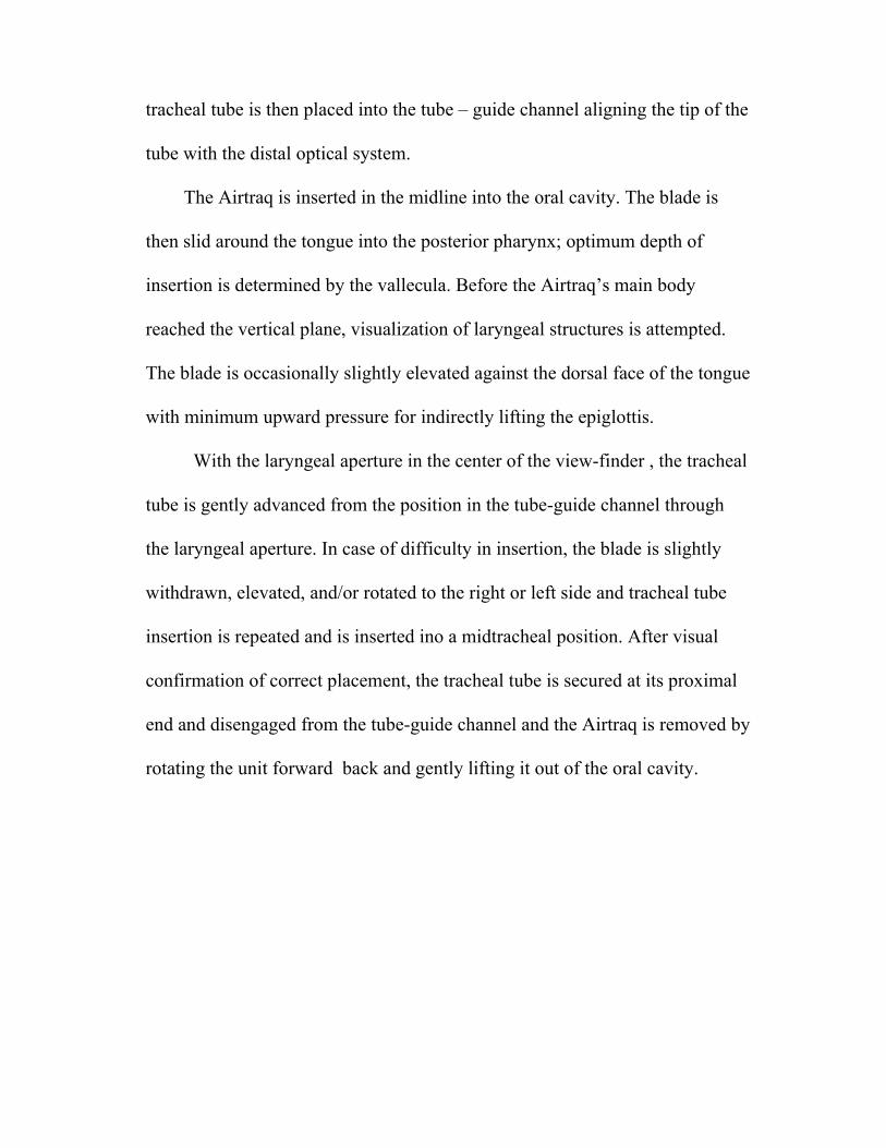
tracheal tube is then placed into the tube – guide channel aligning the tip of the
tube with the distal optical system.
The Airtraq is inserted in the midline into the oral cavity. The blade is
then slid around the tongue into the posterior pharynx; optimum depth of
insertion is determined by the vallecula. Before the Airtraq’s main body
reached the vertical plane, visualization of laryngeal structures is attempted.
The blade is occasionally slightly elevated against the dorsal face of the tongue
with minimum upward pressure for indirectly lifting the epiglottis.
With the laryngeal aperture in the center of the view-finder , the tracheal
tube is gently advanced from the position in the tube-guide channel through
the laryngeal aperture. In case of difficulty in insertion, the blade is slightly
withdrawn, elevated, and/or rotated to the right or left side and tracheal tube
insertion is repeated and is inserted ino a midtracheal position. After visual
confirmation of correct placement, the tracheal tube is secured at its proximal
end and disengaged from the tube-guide channel and the Airtraq is removed by
rotating the unit forward back and gently lifting it out of the oral cavity.

REVIEW OF LITERATURE
The literature was searched and reviewed to seek for advantages and the
problems related to Airtraq aided intubation techniques.
1. Chrisen H. Maharaj , Elma Buckley, Brian.H.Harte and John.G.Laffey,
Department of Anaesthesia, University College Hospital, Galway, Ireland,
conducted study on “Endotracheal intubation in patients with cervical spine
immobilization- A comparison of Macintosh and Airtraq laryngoscopes”16 in
40 patients and found that the Airtraq reduced the duration of intubation
(mean 13.2 seconds vs 20.3 seconds with Macintosh), the need for additional
maneuvers, and the intubation difficulty scale score(0.1 vs 2.7). Tracheal
intubation with the Airtraq caused fewer alterations in blood pressure and heart
rate.
2. Yoshihiro Hirabayashi and Norimasa Seo, Department of Anaesthesiology,
Jichi Medical University, Tochigi, Japan conducted a study on 20 patients
where nasotracheal intubation was performed by a non-anaesthesia physician
with 1-2 months of training in airway management and compared the
intubating conditions between Macintosh and Airtraq laryngoscopes17. It was
found from the study that nasotracheal intubation was achieved in 65 seconds
(mean) using Airtraq laryngoscopy, while it required a significantly longer
time of 123 seconds (mean) using Macintosh laryngoscopy with Magill
forceps. No patient in the Airtraq group experienced esophageal intubation,

while one resident performed an esophageal intubation in the Macintosh
group. It was concluded from the study that in comparison with the Macintosh
laryngoscope, the Airtraq laryngoscope provides superior intubation conditions
for personnel who are training in airway management, resulting in less time to
secure the airway.
3. S.K.Ndoko, R.Amathieu, L.Tual, C.Polliand, W.Kamoun and L.El.Housseini,
Anasthesia and Intensive Care department, Jean Verdler Public University
Hospital, Paris, France, conducted a study on “Tracheal intubation of morbidly
obese patients: a randomized trial comparing performance of Macintosh and
Airtraq laryngoscopes”18 in 106 morbidly obese patients undergoing surgery
and found that in the Airtraq group, tracheal intubation was successfully
carried out in all the patients within 120 seconds and in the Macintosh group,
six patients required intubation with the Airtraq laryngoscope. The mean time
taken for tracheal intubation was 24 seconds and 56 seconds respectively with
the Airtraq and Macintosh laryngoscopes. SpO2 was better maintained in the
Airtraq group than in the Macintosh laryngoscope group with one and nine
patients, respectively, demonstrating drops of SpO2 to 92% or less. They
concluded from the study that the Airtraq laryngoscope shortened the duration
of tracheal intubation and prevented reductions in arterial oxygen saturation in
morbidly obese patients.
4. Schirin M.Missaghi, Klaus Kraser, Hildgard Lackner, Anita Moser and Ernst
Zadrobilek, Department of Anaesthesia and Intensive Care, Empress Elisabeth

Hospital of the city of Vienna, Austria conducted a study “ The Airtraq Optical
Laryngoscope: Experiences with a new disposable device for orotracheal
intubation.” 19214 patients undergoing elective thyroid surgery were
investigated. Patients with previously experienced difficult conventional
tracheal intubation, anatomic features predictive for difficult conventional
laryngoscopy and tracheal intubation and/or obesity were given preferential
enrollment. Cormack Lehane View was performed with a Macintosh
laryngoscope. The laryngeal views obtained with the Airtraq were evaluated
and the tracheal tube was placed and advanced through the laryngeal aperture.
Grades 1 to 5 at Cormack Lehane View were obtained in 74,62,44, 32, and 2
patients respectively in conventional laryngoscopy. The success rate of Airtraq
assisted tracheal intubation at the first attempt was 97%(207/214) with
laryngeal views of grade 1 in all of these patients. Minor problems and
difficulties with impeded blade insertion and impeded tracheal tube insertion
were encountered in 9 and 12 percent respectively. 7 patients required a
second attempt for tracheal intubation; the causes were failed identification of
anatomical structures(1 patient), failed tracheal tube advancement during
laryngeal passage(4 patients), and requirement of downsizing the tracheal tube
for unimpeded and atraumatic laryngeal passage(2 patients). In all patients,
Airtraq assisted tracheal intubation was successful(after a maximum of 2
attempts). They concluded from the study that provided formal instruction,
success of tracheal intubation with Airtraq performed by novice users was not
affected by Cormack Lehane View. The Airtraq proved to be uniquely useful
for routine and difficult laryngoscopy and tracheal intubation.
5. A study titled “ Evaluation of intubation using the Airtraq or Macintosh
laryngoscope by anaesthetists in easy and simulated difficult laryngoscopy- a
manikin study” 20was conducted by C.H.Maharaj, B.D.Higgins, B.H.Harte,
and J.G.Laffey, Department of Anaesthesia, University College
Hospital,Galway, Ireland in which the Airtraq and Macintosh laryngoscope
were compared in simulated easy and difficult laryngoscopy. 25 anaesthetists
were allowed up to 3 attempts to intubate the trachea in each of three
laryngoscopy scenarios using a Laerdal® Intubation Trainer followed by 5
scenarios using a Laerdal SimMan® Manikin. Each anaesthetist then
performed tracheal intubation of the normal airway a second time to
characterise the learning curve. In the simulated easy laryngoscopy scenarios,
there was no difference between the Airtraq and Macintosh in success of
tracheal intubation. The time taken to intubate at the end of the protocol was
significantly lower using the Airtraq(9.5 secs vs 14.2 secs), demonstrating a
rapid acquisition of skills. In the simulated difficult laryngoscopy scenarios,
the Airtraq was more successful in achieving tracheal intubation, required less
time to intubate successfully, caused less dental trauma, and was considered
by the anaesthetists to be easier to use.
6. Zadrobilek.E. et al conducted a study titled “ Success of orotracheal intubation
with the Airtraq optical laryngoscope in patients with difficult conventional

laryngoscopy”21 on 312 patients for elective thyroid surgical procedures with
various conventional laryngoscopic views. Further 20 patients with difficult
conventional laryngoscopy (CL) also for elective thyroid surgery attempted by
using the Airtraq were additionally included in this clinical review. In the
332 patients evaluated, grade 1 to 5 at CL was obtained in 111,90,61,68 and 2
patients respectively. The overall success rate of Airtraq assisted tracheal
intubation at the first attempt was 98%; in all patients, tracheal intubation was
successful after a maximum of 2 attempts. In the 70 patients with difficult CL
(grade 4 or 5), the success rate of Airtraq assisted tracheal intubation at the
first attempt was 94% (66/70 patients). The causes of primary failures of
tracheal intubation were failed identification of anatomical structures (in one
patient) and failed tracheal tube advancement during laryngeal passage (in 3
patients). Visualization of the entire laryngeal aperture was finally obtained in
all patients; downsized tracheal tubes for atraumatic tracheal intubation were
required in 2 patients.
7. A study titled “ The Airtraq laryngoscope for placement of double-lumen
endobronchial tube”22 was conducted by Y.Hirabayashi and N.Seo, Jichi
Medical University, Japan to study the usefulness of Airtraq in the placement
of double-lumen tubes. They found that the Airtraq laryngoscope allowed
placement of 35 and 37 French double lumen tubes in 10 patients without
complications. A regular size Airtraq laryngoscope accepts 35 and 37 French
double lumen tubes, although the latter was somewhat thick against the

channel of the scope. It is probably impossible to insert a 39 French double
lumen tube with an outer diameter of 13 mm. They concluded that despite
this limitation, the Airtraq laryngoscope appears to be an alternative approach
dor double lumen tube placement when the physician encounters cases in
which the conventional Macintosh laryngoscopy results in unsuccessful double
lumen tube placement.
8. A study titled “Comparison of the Airtraq® and Truview® laryngoscopes to the
Macintosh laryngoscope for use by Advanced Paramedics in easy and
simulated difficult intubation in manikins “ 23was performed by Sajid Nasim,
Chrisen H Maharaj, Ihsan Butt, Muhammad A Malik, John O' Donnell,
Brendan D Higgins, Brian H Harte and John G Laffey, Department of
Anaesthesia, Galway University Hospitals, Galway, Ireland in which they
compared the efficacy of these two devices to the Macintosh laryngoscope
when used by 21 paramedics proficient in direct laryngoscopy in a
randomized, controlled, manikin study. Each participant took turns performing
laryngoscopy and intubation with each device, in an easy intubation scenario
and following placement of a hard cervical collar, in a SimMan® manikin.
They found that the Airtraq reduced the number of optimization maneuvers
and reduced the potential for dental trauma when compared to the Macintosh,
in both the normal and simulated difficult intubation scenarios. In contrast, the
Truview increased the duration of intubation attempts and required a greater
number of optimization maneuvers, compared to both the Macintosh and
Airtraq laryngoscope devices.
9. Lange.M., Frommer.M., and Redel.A conducted a study titled “Comparison of
the Glidescope and Airtraq optical laryngoscopes in patients undergoing direct
microlaryngoscopy.”24
In this study, the Airtraq and the Glidescope were compared in 60 ASA I-III
patients with tumours of the upper airway undergoing direct endoscopic
microlaryngoscopy. Patients were randomly assigned to the Airtraq or the
Glidescope group and the Cormack and Lehane grade was assessed by
Macintosh laryngoscopy prior to tracheal intubation. There were no
differences in tracheal intubation success rates or duration of intubation
attempts between both devices. The Cormack and Lehane grade was improved
in 77% and 82% of cases in the Airtraq and Glidescope group, respectively.
Blood traces on the device and traumatic pharyngeal lesions were found more
frequently in the Airtraq group. The Airtraq and Glidescope laryngoscopes are
valuable tools for the management of patients with potentially difficult airways
with the Glidescope appearing to be less traumatic.
10. Emily L.Brown and Ron M.Walls compared the Airway Scope , Airtraq
and Macintosh in 4 simulated difficult airway scenarios, namely normal
airway, cervical spine rigidity, limited mouth opening, and pharyngeal
obstruction25. They concluded that the tracheal intubation success rate was
significantly higher with the Airway Scope and Airtraq than with the

Macintosh laryngoscope(100% and 98% vs 89%). Mean time to intubation and
mean time to first inflation of the lungs were significantly shorter with the
Airway Scope than with either the Airtraq or Macintosh laryngoscopes
(intubation, 10.6 vs 16.2 vs 15.8 seconds, respectively; inflation, 16.1 vs 21.6
vs 23.5 seconds respectively). In the limited mouth-opening scenario, rates of
successful intubation were significantly higher with the Airway Scope and
Airtraq than with the Macintosh laryngoscope (100% and 100% vs 83%).
Successful intubation rates for other scenarios were not statistically significant.
11. Malin.E., Montblanc.J.de., Ynineb.Y., Marret.E., Bonnet.F conducted a case
series on the “Performance of the Airtraq™ laryngoscope after failed
conventional tracheal intubation” 26 .The Airtraq™ was used in 47 patients with
predicted or unpredicted difficult intubation after failed orotracheal intubation
performed by two senior anaesthesiologists with the Macintosh laryngoscope.
Tracheal intubation with Airtraq™ was successful in 36 patients (80%). The
Cormack and Lehane score was IIb-III in 35 patients, and IV in 12 patients,
with the Macintosh laryngoscope, while Cormack and Lehane score was I-IIa
in 40 patients, IIb-III in three and IV in four with Airtraq™. A gum elastic
bougie was used to facilitate tracheal access in one-third (11/36) of the cases.
Orotracheal intubation was not possible with Airtraq™ in nine cases, five of
whom had a pharyngeal, laryngeal or basal lingual tumour. They concluded
that in patients with difficult airway, following failed conventional orotracheal

intubation, Airtraq™ allows securing the airway in 80% of cases mainly by
improving glottis view. However, the Airtraq™ does not guarantee successful
intubation in all instances, especially in case of laryngeal and/or pharyngeal
obstruction.
12.Harald Groeben, Gregor Saint Mont, Roman Pförtner, Ilona Biesler,
Anesthesiology & CCM, Clinics Essen-Mitte, Essen, Germany compared
intubation using a modified Airtraq for nasal intubation and Standard
Macintosh Blade in Difficult Nasal Intubation27 . 80 patients scheduled for
maxillo-facial surgery, requiring nasal endotracheal intubation, with an
expected difficult intubation were enrolled for the study and were randomized
for intubation with a Macintosh (n=40) or Airtraq laryngoscope (n=40). All
patients had one or more risk factors for a difficult intubation (mouth opening
≤ 2.5 cm, Mallampati score of IV, documented history of difficult intubation,
obvious tumor or swelling). Success rate, visualization of the glottis, time for
intubation, and need for optimization maneuvers (cricoid pressure, change of
head position, Eschmann stylet, Magill forceps) were evaluated. It was found
that intubation with the Airtraq was successful in 37 out of 40 patients while
conventional intubation was successful in 26 out of 40 patients. The
visualization of the glottis according to Cormack & Lehane (22/14/1/3 vs.
4/11/11/14), time for intubation (50±61s vs. 91±50s) and the need for
supporting maneuvers (0 to 4 maneuvers: Airtraq 19/10/5/4/0 vs. Macintosh

3/5/11/18/0) were significantly different in favor of the Airtraq technique.
Overall, a Magill forceps was not used to advance the tube and could not even
been brought close to the glottis in 52 patients.
It was concluded that Nasal Airtraq for difficult endotracheal intubations
provided a significantly better view of the glottis with less need for optimizing
maneuvers. Accordingly, the time for intubation was significantly shorter and
the success rate was significantly higher with the Airtraq technique.

METHODOLOGY
It was a Prospective, Randomized, Single blinded (subject), Case control
study conducted in the Institute of Anaesthesiology and Critical Care, Madras
Medical College and Government General Hospital, Chennai. 60 adult patients
satisfying inclusion criteria were enrolled in the study after obtaining informed
consent.
INCLUSION CRITERIA
Elective adult surgical patients requiring general endotracheal anaesthesia
Males and females
ASA physical status 1,2 & 3
Age 18 years of age and older
Who have given valid informed consent
EXCLUSION CRITERIA
Healthy volunteers
Not satisfying inclusion criteria
Patients requiring special techniques for intubation such as rapid
sequence induction

Intubated prior to surgery
Severe cardiovascular, hepatic or renal disease, mental illness
Are unconscious or very severely ill, ASA physical status IV
Need for nasal intubation
MATERIALS
Macintosh laryngoscope- current standard device
Airtraq laryngoscope device- used during laryngoscopy to facilitate
intubation
Weighing machine calibrated to 1 kg
Measuring tape calibrated to 0.5 cm
Goniometer

AIRWAY ASSESSMENT28
Previous anaesthesia records, H/O snoring, H/O voice change, H/O previous
surgery, Trauma, Burns, Tumour in and around the oral cavity, neck or cervical
spine were asked in the history.
H/O systemic illness like Diabetes, Hypertension, Ankylosing spondylitis,
Rheumatoid arthritis were asked and recorded.
General examination included examination for facial anomalies,
Temperomandibular joint pathology, Anomalies of the mouth and tongue,
pathology of nose, pathology of palate.
Height in centimeters and weight in kilograms were recorded and Body Mass
Index was calculated.
Individual airway indices were measured
A-O joint movement: Patient asked to look at the ceiling without raising the
eyebrow and the range of movements were measured with gonioscope.
Neck flexion: Patient was asked to touch the manubrium sterni with chin and
the range of movements measured with gonioscope.
TMJ function: The patient was asked to open his mouth wide open and the
inter incisor distance measured. Examiner’s index finger was placed in front of

the tragus and thumb over the mastoid process and the patient was asked to
open the mouth and sliding movement of the mandibular condyle was assessed.
Upper lip bite test: The patient was asked to bite the upper lip with the lower
incisor and graded as follows:
Class 1 : Lower incisor can bite the upper lip above the vermilion
line.
Class 2 : Lower incisor can bite the upper lip below the vermilion
line
Class 3 : Lower incisor cannot bite the upper lip
Thyromental distance: Distance between the thyoid notch and mental
symphysis when the neck is fully extended and mouth closed.
Sternomental distance: Distance between the sterna notch and mental
symphysis when the neck was fully extended and mouth closed.
Neck circumference: Measured in cm at the level of thyroid notch.
Examination of dentition: Abnormalities like cracking, buck tooth, loose,
artificial and absence of incisors were examined and recorded.

Samson and Young modification of Mallampatti grading29:
The patient kept in sitting position with maximal mouth opening, protruding
tongue, without phonation and the observer’s eye in level with patient’s mouth,
the degree to which the faucial pillars, uvula, soft palate, and hard palate were
visible were recorded and classified as follows:
Grade I : Faucial pillars, uvula, soft palate and hard palate visible
Grade II : Uvula, soft palate and hard palate visible
Grade III : Base of uvula or none, soft palate and hard palate visible
Grade IV : Only hard palate visible.
After assessment patient shifted to operating room.
i.v line started and monitors connected.
Patient allotted to either Airtraq or Macintosh group by way of sealed
envelopes.
Airtraq and Macintosh laryngoscope checked for battery power.
Appropriate size endotracheal tube for the patient selected.
Heart rate, blood pressure and SpO2 measured (preinduction)
Inj. Glycopyrrolate 0.2mg and Inj. Fentanyl 2mcg/kg given as premedication.

Then preoxygenated with 100% oxygen at 6ltr/min for 3 min.
Induction done with Inj.Thiopentone 5mg/kg + NDP neuromuscular blocker.
Ventilated with face mask for 3 min.
Heart rate, blood pressure and SpO2 measured (preintubation).
Intubation attempted with Airtraq/Macintosh laryngoscope.
Observation of Cook’s modification of Cormack and Lehane grading
Cook’s modification of Cormack and Lehane grading and Intubation Difficulty
Score were noted as follows:
CORMACK AND LEHANE GRADING SYSTEM30:
Entire vocal cord visualized - Grade I
Posterior part of vocal cords seen - Grade IIa
Arytenoids only seen - Grade IIb
Epiglottis only seen (liftable) - Grade IIIa
Tip of epiglottis only seen (adherent)- Grade IIIb
No glottis structure seen - Grade IV


INTUBATION DIFFICULTY SCORE :
Seven variables are used.
N1 - No: of supplementary attempts. An attempt is defined as one
advancement of tracheal tube in the direction of the glottis
during direct laryngoscopy.
N2 - No: of supplementary operators directly operating (not assisting)
N3 - No: of supplementary techniques used.
N4 - Cormack Lehane grade minus one.
N5 - Subjectively increased lifting force applied during larynoscopy.
N6 - Need for external laryngeal manipulation
N7 - Position of vocal cords. 0-abduction, 1-adduction.
If intubation with Airtraq failed and saturation maintained, Macintosh blade was
used for intubation and if the saturation decreased, mask ventilation with 100%
oxygen followed by intubation with Macintosh laryngoscope.
Apart from Cormack-Lehane and Intubation Difficulty Score, the following
factors were also noted.

• Intubation time: Measured from entry of the device into the oral cavity
until confirmation of proper placement of tracheal tube.
• Heart rate, blood pressure and SpO2 were measured 1,3 and 5 minutes
post intubation.
• Complication rate: All complications will be recorded, with special
attention to common complications such as upper airway and dental
trauma.

OBSERVATION AND RESULTS
This prospective, randomized, single blind (subject), case controlled
study compared the intubating conditions with Airtraq laryngoscope and
Macintosh laryngoscope and evaluated the advantages and safety, effective
airway time, airway trauma and hemodynamic response.
All data were collected and tabulated.
DEMOGRAPHIC VARIABLES:
60 patients were randomly selected and included in this study. Thirty patients
were randomly assigned to undergo tracheal intubation with Airtraq
laryngoscope (group A) and thirty underwent tracheal intubation with
Macintosh laryngoscope (group B). Mean age, sex distribution and Body Mass
Index of the patients in both the group were compared and there were no
statistically significant differences between the groups.
T Test:
PARAMETER
ASSESSSED
Group A
(AIRTRAQ)
Group B
(MACINTOSH)
P value
Mean SD Mean SD
Age, yr 36.63 13.91 37.4 12.82 0.825
Body Mass Index 25.302 4.375 24.66 3.3787 0.527

AGE and BMI Comparison between the two groups.
Sex distribution in both the groups
36.6
25.3
37.4
24.6
0
5
10
15
20
25
30
35
40
Age BMI
Group A
Group B
Group B
Male
Female
Group A
Male
Female
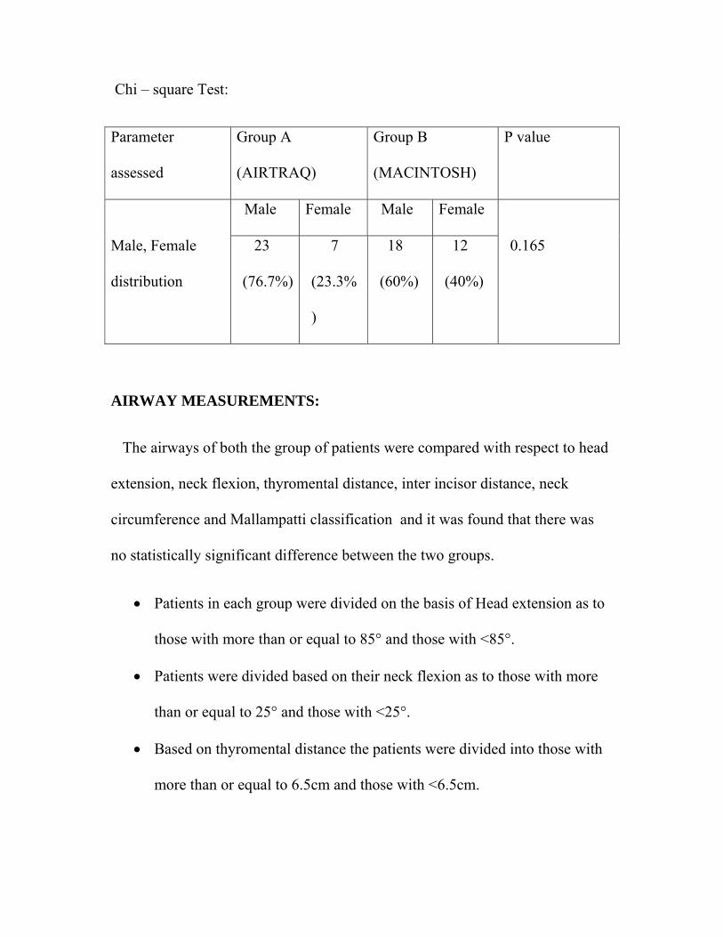
Chi – square Test:
Parameter
assessed
Group A
(AIRTRAQ)
Group B
(MACINTOSH)
P value
Male Female Male Female
Male, Female
distribution
23
(76.7%)
7
(23.3%
)
18
(60%)
12
(40%)
0.165
AIRWAY MEASUREMENTS:
The airways of both the group of patients were compared with respect to head
extension, neck flexion, thyromental distance, inter incisor distance, neck
circumference and Mallampatti classification and it was found that there was
no statistically significant difference between the two groups.
• Patients in each group were divided on the basis of Head extension as to
those with more than or equal to 85° and those with <85°.
• Patients were divided based on their neck flexion as to those with more
than or equal to 25° and those with <25°.
• Based on thyromental distance the patients were divided into those with
more than or equal to 6.5cm and those with <6.5cm.

• Based on inter incisor distance the patients were divided into those with
more than or equal to 3 cm and those with <3cm.
• Neck circumference was measured using inch tape and the mean value of
both the groups was compared using T test and were found to be
statistically insignificant.
• 12 patients in Group A and 20 patients in Group B had a Mallampatti
class 1. There were 17 patients in Group A and 10 patients in Group B
with Mallampatti class 2. Only 1 patient in Group A had a Mallampatti
class 3 and no patient in Group B had a MPC of 3. No patient selected in
either of the group had a MPC of 4.

Chi – square Test:
Prameter
assessed
Group A
(AIRTRAQ)
Group B
(MACINTOSH)
P value
Head extension >85° <85° >85° <85° 1
28(93.3%) 2(6.7%) 28(93.3%) 2(6.7%)
Neck flexion >25° <25° >25° <25° 1
28(93.3%) 2(6.7%) 28(93.3%) 2(6.7%)
Inter Incisor
Distance
>3 cm <3 cm >3 cm <3 cm 1
29(96.7%) 1(3.3%) 29(96.7%) 1(3.3%)
Thyro Mental
Distance
>6.5cm <6.5cm >6.5cm <6.5cm 1
28(93.3%) 2(6.7%) 28(93.3%) 2(6.7%)
T Test:
Parameter
assessed
Group N Mean
(cm)
Std.
deviation
P value
Neck
circumference
A(Airtraq) 30 38.07 3.028 0.087
B(Macintosh) 30 36.83 2.437

MALLA
Chi – sq
Mallam
Classifi
1
2
3
4
MALLA
1
1
1
1
1
2
No.of patients
AMPATTI
quare Test:
mpatti
cation
1
2
3
4
AMPATTI
0
2
4
6
8
10
12
14
16
18
20
M
12
I CLASS
:
Group A
(AIRTRA
12 (40%)
17 (56.7%
1 (3.3%)
0 (0%)
CLASS DI
PC 1
2
20
A
Q)
G
(MA
20 (
%) 10 (
0 (0
0 (0
ISTRIBUT
MPC 2
17
1
Group B
ACINTOS
(66.7%)
(33.3%)
0%)
0%)
ION ACRO
10
SH)
P valu
0.0
OSS THE T
MPC 3
10
ue
09
TWO GRO
OUPS
Group A
Group B
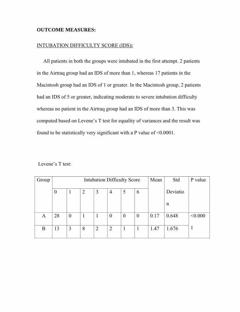
OUTCOME MEASURES:
INTUBATION DIFFICULTY SCORE (IDS):
All patients in both the groups were intubated in the first attempt. 2 patients
in the Airtraq group had an IDS of more than 1, whereas 17 patients in the
Macintosh group had an IDS of 1 or greater. In the Macintosh group, 2 patients
had an IDS of 5 or greater, indicating moderate to severe intubation difficulty
whereas no patient in the Airtraq group had an IDS of more than 3. This was
computed based on Levene’s T test for equality of variances and the result was
found to be statistically very significant with a P value of <0.0001.
Levene’s T test:
Group Intubation Difficulty Score Mean Std
Deviatio
n
P value
0 1 2 3 4 5 6
A 28 0 1 1 0 0 0 0.17 0.648 <0.000
1 B 13 3 8 2 2 1 1 1.47 1.676

IDS Score for Airtraq group
IDS Score for Macintosh group
2928
3028
30 30 30
12
02
0 0 00
5
10
15
20
25
30
35
N1 N2 N3 N4 N5 N6 N7
No.of patients
0
1 and>1
29 29
26
13
23
18
30
1 1
4
17
7
12
00
5
10
15
20
25
30
35
N1 N2 N3 N4 N5 N6 N7
No.of patients
0
1 and >1

Total IDS score for both the groups
Comparison of intubation difficulty scale score distributions with the Airtraq versus Macintosh laryngoscopes. Number of patients is shown above each bar. P<0.0001 between groups, Mann‐Whitney U test.
28
01 1
0 0 0
13
3
8
2 21 1
0
5
10
15
20
25
30
0 1 2 3 4 5 6
No. of p
atients
IDS SCORE
Group A
Group B

CORMACK and LEHANE grading:
Cormack and Lehane grade of both the group of patients were compared to
grade the laryngeal view.
93.33% of patients in the Airtraq group had a CL grade of 1, compared to
43.33% of patients in the Macintosh group.
In the Airtraq group 6.67% of patients had a CL grade of 2 compared to 50% of
patients in the Macintosh group.
No patient in the Airtraq group had a CL grade of 3 or 4, whereas 6.67% in the
Macintosh group had a CL grade of 3 and none with a grade of 4.
The differences between the two groups were statistically significant.
Pearson Chi - square test:
Group CL 1 CL2 CL3 CL4 P value
Airtraq 28(93.33%) 2(6.67%) 0(0%) 0(0%) <0.0001
Macintosh 13(43.33%) 15(50%) 2(6.67%) 0(0%)

Co
1
1
2
2
3
No.of patients
ormack an
0
5
10
15
20
25
30
CL1
28
nd Lehan
1
13
e grade d
CL2
2
15
distributio
CL3
02
on in both
CL4
0 0
h groups
Group A
Group B

DURA
Mean d
Macinto
T test an
Levene
Paramet
assessed
Duratio
1
1
1
1
1
Second
s
ATION OF
uration of
osh group i
nd was fou
’s T test:
ter
d
Gro
n Airt
Mac
Dura
0
2
4
6
8
10
12
14
16
18
INTUBAT
intubation
it was foun
und to be s
oup
traq
cintosh
ation of in
1
TION:
n with the A
nd to be 17
tatistically
N
30
30
ntubation
1.05
Airtraq gro
7.2 secs. It
y significan
Mean
11.03
17.2
n in both
17.2
oup was 11
was comp
nt.
S.D
6.07
5.04
the group
.03 secs in
puted using
D P
71 <
47
ps
n the
g Levene’s
P value
<0.0001
Group A
Group B
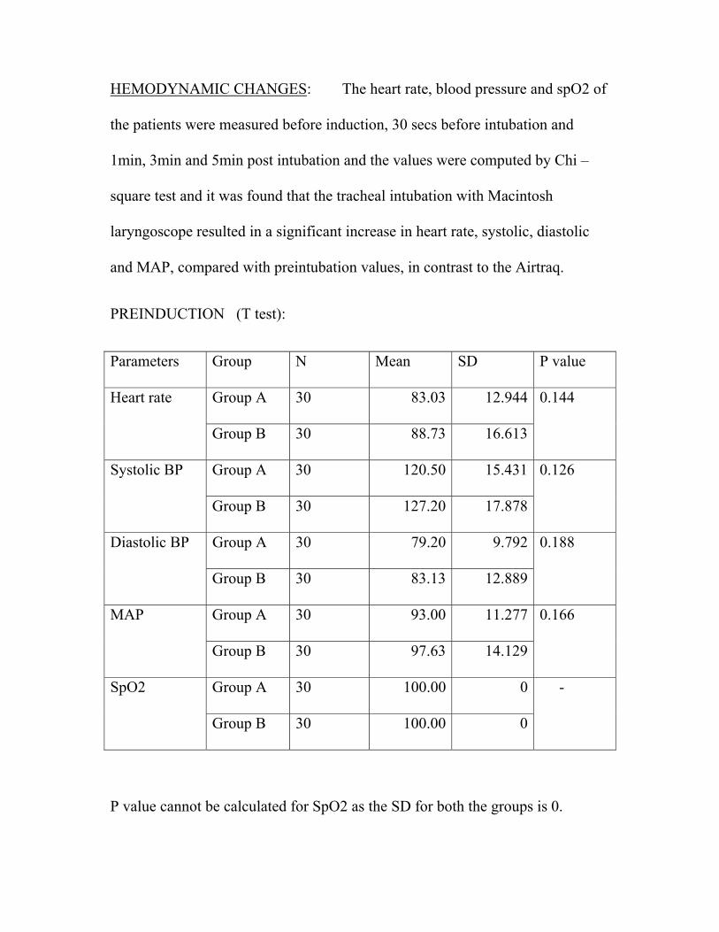
HEMODYNAMIC CHANGES: The heart rate, blood pressure and spO2 of
the patients were measured before induction, 30 secs before intubation and
1min, 3min and 5min post intubation and the values were computed by Chi –
square test and it was found that the tracheal intubation with Macintosh
laryngoscope resulted in a significant increase in heart rate, systolic, diastolic
and MAP, compared with preintubation values, in contrast to the Airtraq.
PREINDUCTION (T test):
Parameters Group N Mean SD P value
Heart rate Group A 30 83.03 12.944 0.144
Group B 30 88.73 16.613
Systolic BP Group A 30 120.50 15.431 0.126
Group B 30 127.20 17.878
Diastolic BP Group A 30 79.20 9.792 0.188
Group B 30 83.13 12.889
MAP Group A 30 93.00 11.277 0.166
Group B 30 97.63 14.129
SpO2 Group A 30 100.00 0 -
Group B 30 100.00 0
P value cannot be calculated for SpO2 as the SD for both the groups is 0.

PREINTUBATION
T test:
Parameters Group N Mean SD P value
Heart rate Group A 30 86.87 10.734 0.556
Group B 30 88.83 14.697
Systolic BP Group A 30 111.50 15.136 0.405
Group B 30 115.13 18.256
Diastolic BP Group A 30 74.17 11.618 0.921
Group B 30 73.87 11.578
MAP Group A 30 86.57 12.227 0.749
Group B 30 87.63 13.479
SpO2 Group A 30 100.00 0 -
Group B 30 100.00 0
P value for SpO2 cannot be calculated as the SD of both the groups is 0.
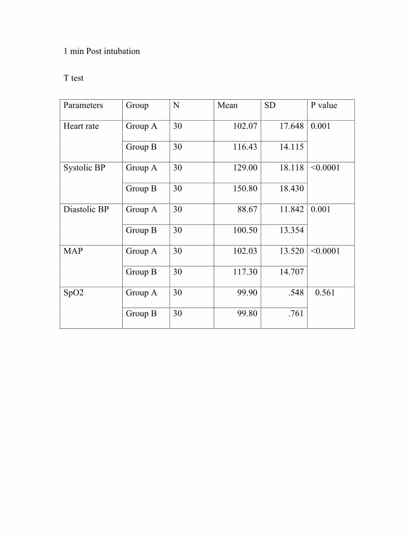
1 min Post intubation
T test
Parameters Group N Mean SD P value
Heart rate Group A 30 102.07 17.648 0.001
Group B 30 116.43 14.115
Systolic BP Group A 30 129.00 18.118 <0.0001
Group B 30 150.80 18.430
Diastolic BP Group A 30 88.67 11.842 0.001
Group B 30 100.50 13.354
MAP Group A 30 102.03 13.520 <0.0001
Group B 30 117.30 14.707
SpO2 Group A 30 99.90 .548 0.561
Group B 30 99.80 .761

3 min Post intubation
T test
Parameters Group N Mean SD P value
Heart rate Group A 30 92.30 14.003 0.004
Group B 30 103.40 14.483
Systolic BP Group A 30 120.43 16.913 0.006
Group B 30 133.57 18.578
Diastolic BP Group A 30 80.83 11.546 0.018
Group B 30 88.43 12.506
MAP Group A 30 94.07 12.881 0.008
Group B 30 103.60 14.036
SpO2 Group A 30 100.00 .000 0.321
Group B 30 99.97 .183

5 min Post intubation
T test
Parameters Group N Mean SD P value
Heart rate Group A 30 84.80 10.506 0.089
Group B 30 90.30 13.899
Systolic BP Group A 30 112.73 12.188 0.033
Group B 30 120.70 15.825
Diastolic BP Group A 30 75.07 10.123 0.435
Group B 30 77.20 10.867
MAP Group A 30 87.53 10.644 0.167
Group B 30 91.70 12.349
SpO2 Group A 30 100.00 0 -
Group B 30 100.00 0
P value cannot be calculated for SpO2 as the SD for both the groups is 0 .
The differences in heart rate, and blood pressure in both the groups was
statistically significant in the 1min and 3 min post intubation measurements and
not significant in the 5 min post intubation measurement.

Heart rate changes
Systolic BP changes
0
20
40
60
80
100
120
140Heart rate in
Beats/m
in
Group A
Group B
0
20
40
60
80
100
120
140
160
Systolic BP mm Hg
Group A
Group B

Diastolic BP changes
Mean Arterial Pressure changes
The SpO2 changes in the pre and post intubation periods in both the groups was
0
20
40
60
80
100
120
Diastolic BP in m
m Hg
Group A
Group B
0
20
40
60
80
100
120
140
MAP in m
m Hg
Group A
Group B

not statistically significant.
AIRWAY TRAUMA:
2 patients in the Airtraq group and 3 patients in the Macintosh group
experienced trauma to the airways and all the injuries were to the soft tissues.
Pearson’s Chi – square test:
Group Trauma P value
Yes No
Airtraq 2(6.67%) 28(93.33%) 0.64
Macintosh 3(10%) 27(90%)
Trauma in Airtraq group
YES
NO

OPERATOR GRADING:
The operator graded the ease of intubation in an increasing grade of difficulty
from grade 1 to grade 5.
Grade 1 : Easy intubation
Grade 2 : Mild difficulty
Grade 3 : Moderate difficulty
Grade 4 : Extremely difficult
Grade 5 : Cannot intubate
28 patients in the Airtraq group had a grade 1 ease of intubation , compared to
20 patients in the Macintosh group.
Trauma in Macintosh group
YES
NO

In the Airtraq group 1 patient had a grade 2 ease of intubation, compared to 7
patients in the Macintosh group.
1 patient in the Airtraq group had a grade 3 ease of intubation , compared to 3
patients in the Macintosh group.
Pearson Chi –square test:
Operator
Grading
Group P value
Airtraq Macintosh
1 28(93.33%) 20(66.67%) 0.033
2 1(3.33%) 7(23.33%)
3 1(3.33%) 3(10%)
4 0(0%) 0(0%)
5 0(0%) 0(0%)

O
Op
Operato
perator g
r gradin
grading i
g in Airt
in Macin
traq grou
ntosh gro
up
oup
Grade1
Grade2
Grade3
Grade 1
Grade 2
Grade 3

DISCUSSION
Expert airway management is an essential skill of an Anaesthesiologist.
Difficulties with tracheal intubation are mostly caused by difficult direct
laryngoscopy with impaired view to the vocal cords31. Unfortunately, despite
all the information currently available, no single factor reliably predicts these
difficulties32.
Consequently, many difficult intubations will not be recognized until after
induction of anaesthesia. Unanticipated difficult intubation can lead to critical
situations, especially in those patients who are at risk for gastric regurgitation,
who are difficult to ventilate by mask or who have limited cardiopulmonary
reserves.
When a person is in supine position and head in neutral position, the
laryngeal axis is almost horizontal. The pharyngeal axis is approximately 30 –
450 from the horizontal axis and the oral axis almost perpendicular to the
laryngeal axis33.
Successful direct laryngoscopy for the exposure of the glottis opening
requires the alignment of oral, pharyngeal and laryngeal axes. Elevation of head
about 10 cm with pads below the occiput aligns the laryngeal and pharyngeal
axes.

Subsequent head extension at the atlanto occipital joint creates the
shortest distance and most nearly straight line from the incisors to glottic
opening.
The degree of head and neck movements that can facilitate intubation
with conventional aids are:
• Head extension > 80 – 850
• Neck flexion > 25 – 300
• Head/neck rotation > 70 – 750
• Normal lateral bending movements at cervical spines
Include 5 – 100 at each cervical spine below C2 level.
Presence of factors like Ankylosing spondylitis, Rheumatoid arthritis,
Cervical spine fusion, Cervical spondylitis, Cervical spine injuries,
Scleroderma, Fibrosis of neck region due to burns will prevent ideal positioning
and intubation may be difficult with conventional aids.
Many endoscopic intubation laryngoscopes such as the Bullard
laryngoscope, the Upsher laryngoscope, the Wuscope and the Airtraq
laryngoscope have been designed to visualize the vocal cords through
a proximal viewfinder that overcomes the curved anatomical axis by prism or
mirror. They have been found to be useful in situations where conventional

laryngoscopy fails to get desired laryngeal view. Trial reports so far have shown
improvement in laryngeal view and ease of intubation.
The advantages of Airtraq laryngoscope from the available literatures
include34,35,36:
• As the axis of Airtraq laryngoscope is curved and the image is
transmitted through lenses and mirrors, the alignment of the axes may
not be needed – improved intubating conditions in patients.
• Useful when there is altered anatomy and when contraindications for
Magill’s positioning are present.
• The displayed anatomy is magnified.
• Recognition of the anatomical structures and anomalies is easier.
• Since fewer manipulations of the airway is needed, it is associated
with fewer hemodynamic changes during intubation.
• Significantly reduces the duration of intubation.
• A clip-on wireless video system is also available which allows
viewing on an external screen. This may be particularly useful for
teaching purposes.
• Shortens the learning curve in novice personnel learning to intubate.
Our study was designed to compare the intubating conditions of Airtraq
laryngoscope with conventionally used Macintosh laryngoscope. 60 patients
were randomly selected and included in the study.

INTUBATION DIFFICULTY SCORE37:
Intubation difficulty score was used to evaluate intubating conditions.
It was developed by Adnet et al in 1997. It is a blend of subjective and
objective criteria that permit a qualitative and quantitative approach to the
progressive nature of the difficulty in intubation and appears to be the best
indicator till date.
In this scale, the value of IDS is ‘0’ if full visualization of the
laryngeal aperture is possible during laryngoscopy and vocal cords are seen
to be nicely abducted. Each variation from this defined ‘ideal’ intubation
increases the degree of difficulty, the overall score being the sum of all
variations from the definition.
It was generally easy to insert the Airtraq laryngoscope, to obtain a full
view of the glottis, and to intubate the trachea without major complications.
In this device, the tracheal tube can be attached to the side of the blade and
the tip of the tube is visible on the viewfinder. Once the glottis was
positioned in the centre of the viewfinder, it was easy to advance the tube into
the trachea.
There was one difficulty though. Inserting the Airtraq too close to
the glottis will only allow the initial posterior movement of the tube and
result in a failure to intubate. The ‘back and up manoeuvre’ which
involves withdrawing the device away from the glottis and lifting the device
up before attempting to intubate helps to overcome this problem.
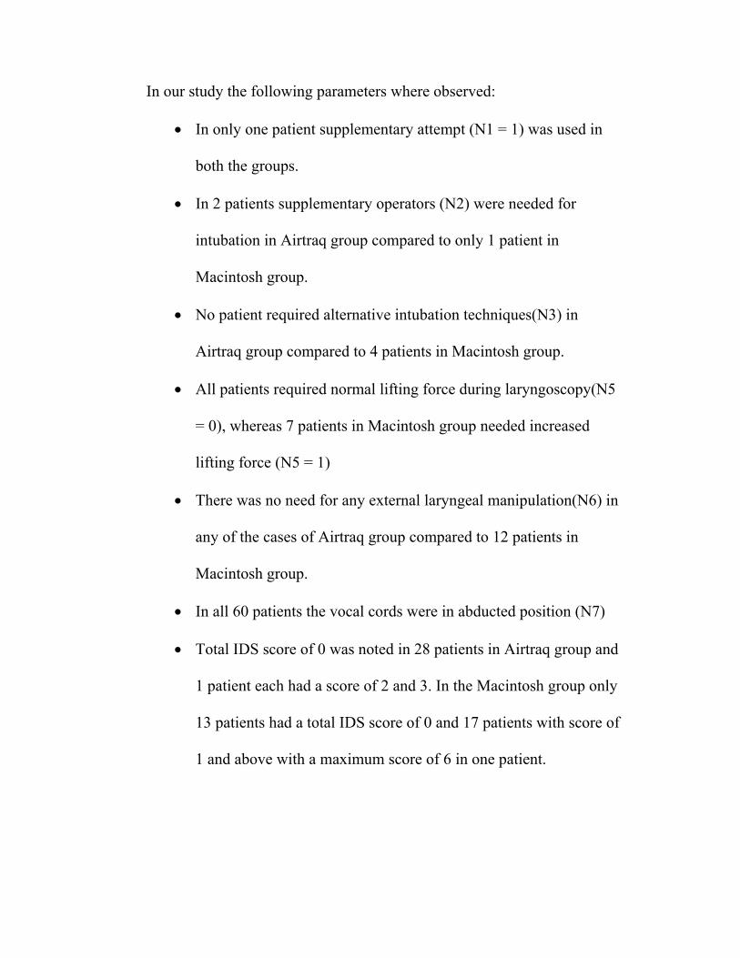
In our study the following parameters where observed:
• In only one patient supplementary attempt (N1 = 1) was used in
both the groups.
• In 2 patients supplementary operators (N2) were needed for
intubation in Airtraq group compared to only 1 patient in
Macintosh group.
• No patient required alternative intubation techniques(N3) in
Airtraq group compared to 4 patients in Macintosh group.
• All patients required normal lifting force during laryngoscopy(N5
= 0), whereas 7 patients in Macintosh group needed increased
lifting force (N5 = 1)
• There was no need for any external laryngeal manipulation(N6) in
any of the cases of Airtraq group compared to 12 patients in
Macintosh group.
• In all 60 patients the vocal cords were in abducted position (N7)
• Total IDS score of 0 was noted in 28 patients in Airtraq group and
1 patient each had a score of 2 and 3. In the Macintosh group only
13 patients had a total IDS score of 0 and 17 patients with score of
1 and above with a maximum score of 6 in one patient.

In the study conducted by Chrisen H. Maharaj, Elma Buckley, Brian
H. Harte and John G. Laffey titled “Endotracheal intubation in patients
with cervical spine immobilization-A comparison of Macintosh and
Airtraq laryngoscopes” it was found that 14 out of the 20 in Macintosh
group had an IDS score of 1 or more, compared with 1 in the Airtraq
group. In the Macintosh group 4 patients had an IDS score of 5 or greater,
indicating moderate to severe intubation difficulty. These findings are
comparable to our study.
IMPROVEMENT IN LARYNGEAL VIEW:
The laryngoscopic view was graded by Cormack and Lehane
classification. Cormack and Lehane score (1,2,3,4) with Airtraq was
(28,2,0,0) and with Macintosh blade was (13,15,2,0). The difference was
statistically significant when analysed with Pearson chi square test and
paired T test.
Cormack and Lehane grade of 1 was seen in 93.3% of cases in the Airtraq
group which represents best intubating conditions.
This result is comparable to the study titled “Endotracheal intubation in
patients with cervical spine immobilization – A comparison of Macintosh
and Airtraq laryngoscopes” conducted by Chrisen H.Maharaj et al at the
University College Hospital, Galway, Ireland in which 19 out of the 20
patients intubated with Airtraq had a Cormack and Lehane grade of 1 and
1 patient had a grade of 2 when compared to 6,7,7 patients with CL grade
of 1,2 and 3 respectively in the Macintosh group.
DURATION OF INTUBATION ATTEMPT:
The mean time to intubate with the Airtraq group was 11.03 seconds
and in the Macintosh group it was 17.2 seconds and it was found to be
statistically significant when computed with Levene’s test for equality of
variances.
In the test conducted by Chrisen Maharaj et al in Ireland in live
patients it was 20.3 seconds with Macintosh and 13.2 seconds with the
Airtraq laryngoscopes.
In a different study conducted by Maharaj et al in manikins it was
found that the time for intubation with the macintosh group was 14.2
seconds and in the Airtraq group it was 9.5 seconds.
In a study conducted by S.K.Ndoko et al in the Jean Verdler Public
University Hospital, France in 106 morbidly obese patients the mean time
to intubate using Airtraq was 24 seconds and with Macintosh laryngoscope
was 56 seconds.

All the above studies demonstrate that Airtraq considerably reduces
the time to intubate the patients.
HEMODYNAMIC CHANGES:
The mean increase in heart rate from the preintubation values was 15
per min at 1 min post intubation in the Airtraq group and 27 per min in the
Macintosh group.
The mean increase in Mean arterial pressure from the preintubation
values was 15 mm Hg in the Airtraq group and 29 mm Hg in the
Macintosh group at 1 min post intubation.
The above findings suggest that the Airtraq resulted in less
stimulation of heart rate and blood pressure after tracheal intubation in
comparison with the Macintosh laryngoscope. This finding probably
reflects the fact that the Airtraq provides a view of the glottis without a
need to align the oral, pharyngeal and tracheal axes and therefore requires
less force to be applied during laryngoscopy. These findings were similar
to those obtained in the study conducted by Chrisen H.Maharaj et al in
Ireland.
The potential of the Airtraq to produce less stimulation of heart rate
and blood pressure may be particularly advantageous in clinical situations
such as coronary artery disease or arrhythmias.

AIRWAY TRAUMA:
Minor degree of airway trauma was noted in 2 out of the 30
patients in the Airtraq group and 3 out of the 30 patients in the Macintosh
group. All injuries were to the soft tissues. These findings were not
statistically significant.
In the study conducted by Maharaj et al it was found that intubation
attempts with Airtraq significantly reduced the incidence of airway trauma
in Laerdal Airway Trainer and SimMan Manikin in easy and simulated
difficult airway scenarios when compared to Macintosh laryngoscope.
Acute traumatic complications like injury to the lips, tongue, nose,
pharynx, larynx and trachea can occur during laryngoscopy and intubation.
Traumatic complications have been extensively described in two excellent
reviews.
1. Weber S. Traumatic complications of airway management.,
Anaesthesiology clinics of North America 2002;20:503-512.
2. Loh KS, Irish JC. Traumatic complications of intubation and other
airway management procedures.Anaesthesiology 2002;20: quoting that
“ Minor trauma to the airway is common and incidence increases with
increasing duration, increasing grade of difficulty, female gender and >
60 yrs of age. Most traumatic complications do not result in major
morbidity or mortality. However, some require immediate recognition
and management.”
SUMMARY
Airtraq laryngoscope significantly improves laryngeal exposure and
facilitates rapid, easy and reliable intubation.
It can be useful in routine anaesthesia care and also in anticipated and
unanticipated difficult intubation scenarios.
The reduction in hemodynamic changes during laryngoscopy can be
useful in patients with coronary artery disease and arrhythmias.
It significantly reduces the learning curve in novice laryngoscopists
and the provision for additional video attachment also helps in this regard.
It can be considered that the Airtraq will be a useful addition to the
range of difficult airway devices available and it may obviate the need for
more sophisticated and complex airway instruments like flexible fiber optic
bronchoscope to a particular extent.

CONCLUSION
In conclusion, the Airtraq laryngoscope offers a new approach to tracheal
intubation of patients with anticipated and unanticipated difficult airway. The
Airtraq reduced the difficulty of tracheal intubation and the degree of
hemodynamic stimulation compared with the Macintosh laryngoscope. These
findings demonstrate the efficacy of the Airtraq in many clinically relevant
contexts and adds to the evolving body of knowledge regarding this potentially
useful device.
BIBLIOGRAPHY
1. Miller’s Anaesthesia, 6th edition; chapter 42; Airway management,
Thomas J.Gal
2. Miller,CG: Management of the Difficult Intubation. ASA Newsletter
64(6):13-16 and 19, 2000
3. Caplan RA, Posner KL, Ward RJ, Cheney FW: Adverse respiratory
events in anaesthesia: a closed claims analysis. Anaesthsiology 72:828-
833, 1990
4. Miller’s anaesthesia, 6th edition; chapter 42; Airway management,
Thomas J.Gal
5. Airway management: Rashid M Khan MD, Professor, Dept of
Anaesthesiology, JN Medical college, Aligarh, India
6. Clinical anaesthesia – 5th edition by MD Paul G.Barash, MD Bruce
F.Cullen, MD Robert K.Stoelting by Lippincott Williams and Wilkins
Publishers
7. Levintan R, Andrew O. Airway management and direct laryngoscopy.
Critical care clinics 2007;16;373-86
8. Dorsh JE and Dorsh SE: Understanding Anaesthesia Equipment, 4th
edition. Baltimore , Williams and Wilkins

9. Pentax news release : launch of the Airway scope AWS-S100, a rigid
video laryngoscope for intubation, and the Intlock ITL-S, a specialized
laryngoscope blade
10. Fulling PD, Roberts JT, Fibreoptic intubation. International
anaesthesiology clinics 2000; 34; 189-217
11. Hurford WE. Techniques for tracheal intubation. International
anaesthesiology clinics.2004;38;1-28
12. Dorsh JE & Dorsh SE: Understanding Anaesthesia equipment, 4th
edition. Baltimore, Williams and Wilkins
13. Airway management :Rashid M Khan MD, Professor , Dept of
Anaesthesiology, JN Medical College, Aligarh, India
14. The Airtraq Optical Laryngoscope: Experiences with a new disposable
device for orotracheal intubation: Schirin M.Missaghi M.D., Klaus
Krasser,M.D., Hildegard Lackner-Ausserhofer,M.D., Anita Moser,
M.D., and Ernst Zadrobilek. Internet Journal of Airway Management,
Volume 4, Jan 2006 to Dec 2007.
15. Hirabayashi Y, Seo N. A monitor to facilitate use of the Airtraq
laryngoscope. Anaesthesia. 2007; 62:1081.
16. Chrisen H.Maharaj, Elma Buckley, Brian H.Harte, John
G.Laffey.Endotracheal intubation in patients with cervical spine
immobilization- A comparison of Macintosh and Airtraq
laryngoscopes. Anaesthesiology 2007;107:53-9.

17. Yoshihiro Hirabayashi and Norimasa Seo. Airtraq laryngoscope has
an advantage over Macintosh laryngoscope for nasotracheal intubation
by novice laryngoscopists. J Anaesth (2009) 23:172-173.
18. S.K.Ndoko et al. Tracheal intubation of morbidly obese patients: a
randomized trial comparing performance of Macintosh and Airtraq.
British journal of Anaesthesia 100(2):263-8(2008).
19. Schirin M.Missaghi, Klaus Kraser,Hildgard Lackner, Anita Moser,
Ernst Zadrobilek. The Airtraq optical laryngoscope: experiences with a
new disposable device for orotracheal intubation. Internet Journal of
Airway Management. Vol 4 (Jan 2006 – Dec 2007).
20. C.H.Maharaj, B.D.Higgins, B.H.Harte, J.G.Laffey. Evaluation of
intubation using the Airtraq or Macintosh laryngoscope by
anaesthetists in easy and simulated difficult laryngoscopy – a manikin
study. Anaesthesia,2006,61, pages 469-477.
21. Zabrobilek E, Schirin.M.Missaghi. Success of orotracheal intubation
with the Airtraq optical laryngoscope in patients with difficult
conventional laryngoscopy. Internet Journal of Airway Management,
vol 5, (Jan 2008 to Dec 2009).
22. Y.Hirabayashi and N.Seo. The Airtraq laryngoscope for placement of
double – lumen endobronchial tube. Canadian Journal of Anaesthesia
54:955-957 (2007).

23. Sajid Nasim, Chrisen H.Maharaj,Ihsan Butt, Muhammad A.Malik,
John O’Donnel, Brendan D.Higgins,Brian H.Harte, John G.Laffey.
Comparison of the Airtraq and Truview laryngoscopes to the
Macintosh laryngoscope for use by advanced paramedics in easy and
simulated difficult intubation in manikins. BMC Emergency Medicine
2009:9:2
24. Lange.M,Frommer.M, Redel.A. Comparison of the Glidescope and
Airtraq optical laryngoscopes in patients undergoing direct
microlaryngoscopy. Anaesthesia. Vol 64,323-328, 2009.
25. Emily L.Brown,Ron M.Walls. Comparison of the Airway Scope,
Airtraq and Macintosh laryngoscopes in simulated difficult airway
scenarios in manikins. Journal Watch Emergency Medicine, April 16,
2010.
26. Malin.E, Montblanc.J.de, Ynineb.Y, Marret.E, Bonnet.F. Performance
of the Airtraq laryngoscope after failed conventional tracheal
intubation. Acta Anaesthesia Scand 2010.Feb;54(2): 256-7.
27. Harald Groeben, Gregor Saint Mont, Roman Pfortner, Ilona Biesler.
Proceedings of the 2009 Annual meeting of the American Society of
Anaesthesiologists, Oct 17,2009.
28. Predicting Difficult Intubation in Apparently Normal Patients: A Meta
analysis of Bedside Screening Test Performance :Shiga, Toshiya

M.D.,Ph.D., Wajima, Zen’ichiro M.D.,Ph.D., Inoue, Tetsuo
M.D.,Ph.D., Sakamoto, Atsuhiro M.D.,Ph.D.
29. MAroof M,Khan RM. Modified Mallampatti technique correlates
better with Cormack & Lehane grades of laryngoscopic view.
Anaesthesia & Analgesia;2002
30. The modified Cormack & Lehane score for the grading of direct
laryngosopy : evaluation in the Asian population: Koh LK, Kong CE,
Ip-Yam PC. Department of Anaesthesia and Surgical Intensive Care,
Singapore General Hospital, Singapore: Anaesth Intensive Care. 2002
Feb; 30(1):48-51
31. Yamamoto K, Tsubokawa T, Shibata K et al. Predicting Difficult
Intubation with Direct Laryngosopy. Anaesthesiology 1997;86; 316-
321
32. O’Connor MF. Airway assessment of 25,000 patients in a preoperative
clinic. Anaesthesia and Analgesia. 2002; 94; S 113
33. Hurford WE. Techniques for tracheal intubation. International
Anaesthesiology clinics. 2004; 38;1-28
34. Maharaj CH, Higgins B, Harte BH, Laffey JG: Evaluation of ease of
intubation with the Airtraq or Macintosh laryngoscope by anaesthetists
in easy and simulated difficult laryngoscopy: A manikin study.
Anaesthesia 2006;61:469-77

35. Maharaj CH, Ni Chonghaile M, Higgins B, Harte BH, Harte JG:
Tracheal intubation by inexperienced medical residents using the
Airtraq and Macintosh laryngoscope: A manikin study. Am J
Emergency Med 2006; 24:769-74
36. Maharaj CH, O’Croinin D, Curtley G, Harte BH, Laffey JG: A
comparison of tracheal intubation using the Airtraq and Macintosh
laryngoscope in routine airway management: A randomized, controlled
clinical trial. Anaesthesia 2006;61: 1093-9
37. Adnet F, Borron SW, Racine SX, Clemessy JL, Fournier JL, Plaisance
P, Lapandry C: The intubation difficulty scale (IDS): Proposal and
evaluation of a new score characterizing the complexity of
endotracheal intubation. Anaesthesiology 1997 Dec;87(6): 1290-7

PROFORMA
PROSPECTIVE COMPARISON OF INTUBATING CONDITIONS WITH AIRTRAQ LARYNGOSCOPE AND MACINTOSH LARYNGOSCOPE IN RANDOMLY SELECTED ELECTIVE ADULT SURGICAL PATIENTS
NAME : AGE: SEX: I.P.No:
DIAGNOSIS: SURGERY PLANNED:
INTUBATED WITH:
PREOPERATIVE ASSESSMENT:
HISTORY:
CO‐MORBID ILLNESS & TREATMENT DETAILS:
EFFORT TOLERANCE‐ _______ METS
H/O PREVIOUS SURGERY(ANY DOCUMENTED DIFFICULT AIRWAY)‐
H/O TRAUMA/BURNS/TUMOURS INVOLVING AIRWAY‐
H/O SNORING‐
H/O VOICE CHANGE‐
GENERAL EXAMINATION:
HEIGHT: WEIGHT: BMI:
ANAEMIA‐ JAUNDICE‐ CERVICAL SPINE‐ TONGUE‐
PULSE‐ BP‐ CVS‐ RS‐
AIRWAY EXAMINATION:
GROSS ALTERATION IN AIRWAY ANATOMY:
HAIR BUN: YES/NO BEARD: YES/NO
NECK FLEXION: >250/<250 NECK EXTENSION: >800/<800
INTER INCISOR DISTANCE: >3cm/<3cm THYROMENTAL DISTANCE: >6cm/<6cm
UPPER LIP BITE TEST; I/II/III NECK CIRCUMFERANCE:
RECEDING MANDIBLE: None/Mod/Sev PALATE CONFIGURATION:Narrow/Normal
DENTURES:
ARTIFICIAL (REMOVABLE/FIXED): YES/NO BUCK TEETH: None/Mod/Sev
LOOSE TEETH : YES/NO CRACKED TEETH : YES/NO ABSENT TEETH : YES/NO

MODIFIED MALLAMPATTI CLASSIFICATION (MARK‐0)
CORMACK & LEHANE GRADING:
INTUBATION DIFFICULTY SCORE:
1. NUMBER OF ATTEMPTS: 2. NUMBER OF SUPPLEMENTARY OPERATORS: 3. NUMBER OF ALTERNATIVE TECHNIQUES: (change of blade/ use of bougie): 4. CORMACK & LEHANE GRADE minus 1: 5. LIFTING FORCE: 6. EXTERNAL LARYNGEAL MANIPULATION: (needed/not needed): 7. POSITION OF VOCAL CORDS: (abducted/adducted):
N1 N2 N3 N4 N5 N6 N7
DURATION: ______ SECONDS
OPERATOR GRADING OF EASE OF INTUBATION : 1 – 2 – 3 – 4 – 5
VITAL PARAMETERS:
PR BP SPO2
PRE INDUCTION (30 SECS)
PREINTUBATION (30 SECS)
POST INTUBATION
1 min
3 min
5 min
PATIENT CONSENT FORM
STUDY TITLE: Prospective, randomized comparison of intubating conditions with Airtraq laryngoscope & Macintosh laryngoscope in randomly selected elective adult surgical patients.
STUDY CENTRE: Institute of Anaesthesiology & Critical Care, Madras Medical College.
PARTICIPANT NAME: AGE: SEX:
I.P.NO:
I confirm that I have understood the purpose of procedure for the above study. I had the opportunity to ask questions and all my questions and doubts have been answered to my satisfaction.
I have been explained about the possible complications that may occur during the procedure like traumatic injury to the throat. I understand that every precaution will be taken to prevent such an injury and if it happens will be treated accordingly. I have been informed that no other major complication has been reported so far with the use of Airtraq.
I understand that my participation in the study is voluntary and that I am free to withdraw at any time without giving any reason.
I understand that investigator, regulatory authorities and the Ethics committee will not need my permission to look at my health records both in respect to the current study and any further research that may be conducted in relation to it, even if I withdraw from the study. I understand that my identity will not be revealed in any information released to third parties or published, unless as required under the law. I agree not to restrict the use of any data or results that arise from the study.
I hereby consent to participate in this study of comparison of intubating conditions with Airtraq and Macintosh laryngoscope.
Time:
Date: Signature/ Thumb impression of patient
Place: Patient name: ____________________
Signature of the investigator:
Name of the investigator :


GROUP S.NO NAME AGE SEX SURGERY
A 1 CHINNASAMY 61 MALE LUMBAR LAMINECTOMYA 2 USHA 43 FEMALE CRANIOTOMYA 3 PREMKUMAR 25 MALE CRANIOTOMYA 4 JAGADEESAN 30 MALE CRANIOTOMYA 5 PREMKUMAR 23 MALE BURR HOLEA 6 MARIMUTHU 56 MALE LUMBAR LAMINECTOMYA 7 ARUNACHALAM 50 MALE CRANIOTOMYA 8 PREMKUMAR 28 MALE CRANIOTOMYA 9 RAMESH 24 MALE DEPRESSED FRACTUREA 10 PARIMELALAGAN 19 MALE CRANIOTOMYA 11 RAMESH 33 MALE CRANIOTOMYA 12 MAHESH KUMAR 23 MALE ACF‐ REPAIRA 13 VEERAN 42 MALE CORPECTOMY AND STABILISATA 14 CHENGAMALAM 28 MALE CRANIOTOMYA 15 PERIASAMY 25 MALE CRANIOTOMYA 16 ELUMALAI 35 MALE BONY TUMOR EXCISIONA 17 MARIAMBANU 36 FEMALE LUMBAR LAMINECTOMYA 18 DHARMALINGAM 55 MALE VP SHUNTA 19 AMARNATH 28 MALE LUMBAR LAMINECTOMYA 20 ETTIAPPAN 65 MALE LUMBAR LAMINECTOMYA 21 VIVEGA 18 FEMALE CRANIOTOMYA 22 SETTU 35 MALE CYSTOPERITONEAL SHUNTA 23 LALITHA 40 FEMALE VP SHUNTA 24 GIRIBABU 19 MALE LUMBAR LAMINECTOMYA 25 SIVAGAMI 43 FEMALE CRANIOTOMYA 26 SANGITA 32 FEMALE VP SHUNTA 27 SHANKAR 60 MALE CRANIOTOMYA 28 RAVIKUMAR 24 MALE VP SHUNTA 29 SRINIVASAN 56 MALE CRANIOPLASTYA 30 MEENA 43 FEMALE BONY TUMOR EXCISIONB 31 SAKUNTHALA 40 FEMALE VP SHUNTB 32 KUMAR 29 MALE CRANIOTOMYB 33 SUREKHA 29 FEMALE FLAP COVERB 34 LAKSHMANAN 42 MALE LUMBAR LAMINECTOMYB 35 ANNAMUTHU 37 MALE D10‐L3 SCHWANNOMA‐EXCISIB 36 MAHENDRARAJU 27 MALE LUMBAR LAMINECTOMYB 37 NAVEEN 19 MALE CIRSOID ANEURYSM LIGATIONB 38 AMARNATH 28 MALE REDO LAMINECTOMYB 39 VENU 50 MALE CRANIOTOMYB 40 MUTHU 32 MALE CRANIOPLASTYB 41 SIVAGAMI 48 FEMALE CRANIOTOMYB 42 SAROJA 60 FEMALE CRANIOTOMYB 43 DEVI 21 FEMALE ENDONASAL EXCISIONB 44 SAKUNTHALA 40 FEMALE CRANIOTOMYB 45 SELVI 48 FEMALE LUMBAR LAMINECTOMY

B 46 KUBERAN 30 MALE LUMBAR LAMINECTOMYB 47 CHITHRAKUMAR 33 MALE CRANIOTOMYB 48 PERUMAL 60 MALE CRANIOTOMYB 49 REKHA 22 FEMALE CRANIOTOMYB 50 BHARATHI 25 FEMALE CRANIOTOMYB 51 NIRMALADEVI 41 FEMALE LUMBAR LAMINECTOMYB 52 USMAN 55 MALE LUMBAR LAMINECTOMYB 53 SARALA 32 FEMALE VP SHUNTB 54 KAMATCHI 59 FEMALE CRANIOTOMYB 55 RAJESH 25 MALE CRANIOTOMYB 56 ARUN 27 MALE CRANIOTOMYB 57 VEERAPANDIAN 54 MALE CRANIOPLASTYB 58 SIVAKUMAR 22 MALE VP SHUNTB 59 PANDURANGAN 33 MALE SCHWANNOMA EXCISIONB 60 RANGASAMY 54 MALE LUMBAR LAMINECTOMY

Ht(cm) Wt(kg) BMI MPC N1 N2 N3 N4 N5
155 50 20.8 2 0 0 0 0 0150 50 22.22 2 0 0 0 0 0162 80 30.48 2 0 0 0 0 0162 55 20.95 2 0 0 0 0 0173 65 21.71 1 0 0 0 0 0158 59 23.63 2 0 0 0 0 0165 60 22.03 2 0 0 0 0 0162 80 30.48 2 0 0 0 0 0165 60 22.03 1 0 0 0 0 0169 65 22.76 1 0 0 0 0 0175 72 23.51 2 0 0 0 0 0155 55 22.9 1 0 0 0 0 0162 70 26.67 2 0 0 0 0 0172 70 23.66 2 0 0 0 0 0172 82 27.72 2 0 0 0 0 0170 85 29.41 2 0 0 0 0 0155 78 32.46 2 0 0 0 0 0155 80 33.3 1 0 0 0 0 0172 80 27.04 1 0 0 0 0 0161 76 29.32 2 0 0 0 0 0145 42 19.98 1 0 0 0 0 0163 65 24.46 1 0 0 0 0 0155 55 22.9 1 0 0 0 0 0170 65 22.49 1 0 0 0 0 0165 75 27.54 2 0 1 0 1 0158 55 22.03 1 0 0 0 0 0162 88 33.53 3 1 1 0 1 0178 65 20.51 2 0 0 0 0 0154 78 32.89 2 0 0 0 0 0153 46 19.65 1 0 0 0 0 0152 60 25.97 2 0 0 0 1 0160 70 27.34 2 1 1 1 1 1152 45 19.48 1 0 0 0 1 0165 60 22.04 1 0 0 0 1 1165 68 24.98 1 0 0 0 0 0163 85 31.99 2 0 0 1 1 1158 60 24.03 1 0 0 0 0 0172 73 24.67 1 0 0 0 1 0167 65 23.3 1 0 0 0 0 0175 65 21.22 1 0 0 0 0 0152 45 19.48 1 0 0 1 2 1156 70 28.76 2 0 0 0 0 0155 55 22.89 1 0 0 0 1 0151 52 22.8 2 0 0 0 1 0155 68 28.3 1 0 0 0 0 0
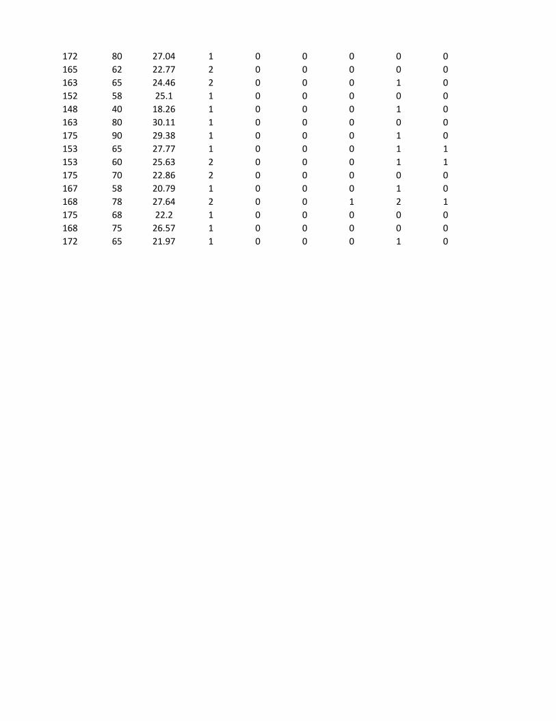
172 80 27.04 1 0 0 0 0 0165 62 22.77 2 0 0 0 0 0163 65 24.46 2 0 0 0 1 0152 58 25.1 1 0 0 0 0 0148 40 18.26 1 0 0 0 1 0163 80 30.11 1 0 0 0 0 0175 90 29.38 1 0 0 0 1 0153 65 27.77 1 0 0 0 1 1153 60 25.63 2 0 0 0 1 1175 70 22.86 2 0 0 0 0 0167 58 20.79 1 0 0 0 1 0168 78 27.64 2 0 0 1 2 1175 68 22.2 1 0 0 0 0 0168 75 26.57 1 0 0 0 0 0172 65 21.97 1 0 0 0 1 0

N6 N7 TOTAL Duration OperatorN (sec) grading HR Sys BP Diast BP MAP
0 0 0 13 1 84 112 72 850 0 0 14 1 92 115 84 940 0 0 11 1 90 142 90 1070 0 0 8 1 104 124 84 970 0 0 6 1 68 130 80 970 0 0 7 1 59 114 74 870 0 0 12 1 60 109 76 870 0 0 8 1 90 142 90 1070 0 0 11 1 83 103 78 860 0 0 6 1 65 91 69 760 0 0 8 1 85 138 93 1080 0 0 6 1 88 119 73 880 0 0 10 1 82 92 62 720 0 0 7 1 88 110 79 890 0 0 7 1 75 119 84 960 0 0 6 1 92 140 87 1050 0 0 8 1 71 131 82 990 0 0 9 1 91 108 73 850 0 0 18 1 80 148 102 1170 0 0 8 1 94 124 73 900 0 0 10 1 112 98 63 750 0 0 7 1 61 110 72 850 0 0 9 1 83 119 69 860 0 0 13 1 78 134 89 1040 0 2 25 2 94 114 70 850 0 0 12 1 78 112 74 870 0 3 35 3 102 145 95 1120 0 0 12 1 87 132 88 1030 0 0 11 1 85 128 83 980 0 0 14 1 70 112 68 831 0 2 14 1 90 152 104 1201 0 6 25 2 85 114 73 871 0 2 18 2 112 120 81 940 0 2 16 1 128 150 90 1100 0 0 14 1 102 112 72 851 0 4 32 3 112 152 102 1150 0 0 15 1 94 92 61 711 0 2 23 2 77 142 92 1080 0 0 12 1 71 142 92 1080 0 0 13 1 85 110 70 831 0 5 25 3 105 119 78 920 0 0 12 1 107 150 103 1190 0 1 11 1 86 109 68 821 0 2 19 2 94 136 95 1090 0 0 13 1 89 130 82 98
PREINDUCTION

0 0 0 21 1 82 112 75 870 0 0 12 1 83 101 71 810 0 1 16 1 65 102 62 750 0 0 12 1 89 118 63 811 0 2 16 1 84 94 68 770 0 0 15 1 72 132 85 1011 0 2 18 1 58 142 103 1161 0 3 21 2 110 132 84 1001 0 3 21 2 72 153 95 1140 0 0 18 1 114 124 81 950 0 1 18 1 86 132 84 1000 0 4 23 3 78 142 95 1110 0 0 10 1 65 132 83 990 0 0 14 1 78 138 87 1041 0 2 19 2 89 132 95 107
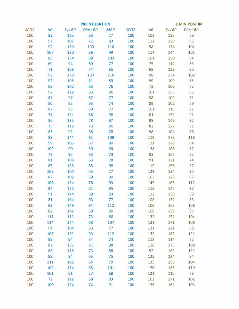
SPO2 HR Sys BP Diast BP MAP SPO2 HR Sys BP Diast BP100 82 105 63 77 100 103 125 79100 97 107 71 83 100 112 129 96100 92 130 100 110 100 98 134 102100 107 130 86 99 100 114 144 101100 85 134 88 103 100 101 150 94100 69 94 69 77 100 75 112 83100 71 108 74 85 100 68 128 90100 92 130 100 110 100 98 134 102100 92 105 81 89 100 99 109 85100 69 102 63 76 100 72 106 72100 92 122 83 96 100 101 135 95100 87 97 67 77 100 98 109 71100 80 95 63 74 100 89 102 69100 82 95 60 72 100 101 115 81100 74 121 86 98 100 81 132 91100 86 135 78 97 100 98 146 92100 75 112 73 86 100 82 122 81100 83 92 68 76 100 98 104 86100 89 144 91 109 100 119 172 118100 99 105 67 80 100 121 128 84100 102 90 59 69 100 128 108 82100 72 92 63 73 100 83 107 72100 81 108 63 78 100 91 121 74100 85 125 85 98 100 119 156 97100 102 100 65 77 100 129 134 95100 87 102 69 80 100 103 128 87100 108 124 78 93 100 143 165 112100 94 123 81 95 100 118 145 97100 91 114 68 83 100 112 138 89100 81 104 63 77 100 108 132 83100 83 149 94 112 100 108 163 108100 82 102 69 80 100 108 139 93100 111 111 73 86 100 132 154 104100 114 144 88 107 100 131 171 108100 94 104 63 77 100 121 121 69100 106 151 93 112 100 132 182 121100 99 94 64 74 100 112 114 72100 82 131 81 98 100 118 173 108100 68 118 73 88 100 92 162 121100 89 94 65 75 100 125 124 94100 111 108 64 79 100 135 158 104100 102 134 85 101 100 128 165 119100 101 91 57 68 100 121 115 78100 72 112 68 83 100 102 171 102100 100 124 74 91 100 126 162 103
PREINTUBATION 1 MIN POST IN

100 77 121 82 95 100 98 159 102100 81 92 63 73 100 111 138 92100 61 93 59 70 100 92 132 93100 92 105 61 76 100 121 142 95100 94 85 59 68 100 122 125 87100 71 121 79 93 100 98 162 103100 67 138 99 112 100 99 172 123100 100 94 67 76 100 125 152 103100 75 112 72 85 100 105 165 112100 108 102 63 76 100 132 142 95100 93 121 75 90 100 121 149 102100 83 133 84 100 100 145 165 112100 71 118 72 87 100 101 146 95100 83 126 81 96 100 105 143 89100 95 126 89 101 100 127 158 108

MAP SPO2 HR Sys BP Diast BP MAP SPO2 HR Sys BP94 100 93 114 71 85 100 81 108107 100 93 115 85 95 100 87 109112 100 99 128 85 99 100 92 125115 100 108 137 82 100 100 101 110113 100 98 142 88 106 100 88 13493 100 65 108 69 82 100 59 102103 100 74 133 91 105 100 70 123113 100 99 128 89 102 100 92 12193 100 93 103 81 88 100 87 10183 100 73 103 70 81 100 68 96108 100 92 121 81 94 100 85 11784 100 84 97 68 78 100 88 10280 100 81 93 61 72 100 83 9592 100 99 112 81 91 100 81 117105 100 70 124 93 103 100 75 122110 100 92 144 94 111 100 93 13291 100 73 111 72 85 100 75 10892 100 92 112 89 97 100 81 116136 100 105 162 102 122 100 95 13499 100 97 108 69 82 100 92 10291 100 114 102 73 83 100 102 9384 100 71 101 65 77 100 65 9790 100 83 111 63 79 100 81 108117 100 92 147 98 116 100 87 126108 100 112 121 87 98 100 98 108101 100 90 114 78 90 100 77 105130 97 122 148 102 117 100 98 134113 100 105 132 91 105 100 91 118105 100 101 124 76 92 100 89 11299 100 99 118 71 87 100 83 107126 100 101 141 93 109 100 90 136108 97 92 121 81 94 99 83 108121 100 118 132 94 107 100 108 118129 100 108 164 102 123 100 94 15286 100 108 105 61 76 100 93 102141 97 122 161 113 129 100 108 14286 100 98 102 68 79 100 92 98130 100 101 158 93 115 100 91 143135 100 81 143 91 108 100 72 131104 100 104 114 81 92 100 91 110122 100 112 128 92 104 100 108 104134 100 112 152 102 119 100 101 14290 100 112 111 75 87 100 103 109125 100 81 161 103 122 100 72 133123 100 133 151 91 111 100 102 132
TUBATION 3 MIN POST INTUBATION 5 MIN

121 100 83 132 93 106 100 75 121107 100 102 124 74 91 100 91 103106 100 89 112 77 89 100 71 105111 100 108 121 74 90 100 92 103100 100 118 102 69 80 100 101 93123 100 83 142 92 109 100 71 135139 100 83 152 101 118 100 61 131119 100 111 112 83 96 100 108 104130 100 99 149 103 118 100 81 125111 100 118 121 83 96 100 104 108118 100 108 133 93 106 100 87 122130 100 131 148 105 119 100 114 132112 100 87 135 86 102 100 73 121107 100 91 137 83 101 100 81 126125 100 108 143 97 112 100 91 132

TRAUMADiast BP MAP SPO2 (Y/N)
63 78 100 N79 89 100 N81 96 100 N69 83 100 N86 102 100 N76 85 100 N83 96 100 N83 96 100 N79 86 100 N58 67 100 N75 89 100 N69 80 100 N66 76 100 N79 92 100 N86 98 100 N91 105 100 N69 82 100 N89 98 100 N92 106 100 N71 81 100 N59 70 100 N61 73 100 N61 77 100 N84 98 100 N75 86 100 Y67 80 100 N91 105 100 Y72 87 100 N71 85 100 N67 A 100 N91 106 100 N62 77 100 N75 89 100 N93 113 100 N60 74 100 N92 109 100 Y64 75 100 N93 110 100 N85 100 100 N68 82 100 N72 83 100 Y93 109 100 N79 89 100 N85 101 100 N85 101 100 N
POST INTUBATION

75 90 100 N65 78 100 N63 77 100 N61 75 100 N61 72 100 N87 103 100 N83 99 100 N75 85 100 N79 94 100 N71 83 100 N75 91 100 N87 102 100 Y75 90 100 N75 92 100 N87 102 100 N