Embed Size (px)
Citation preview
Turk J Med Sci2006; 36 (4): 251-253© TÜB‹TAKE-mail: [email protected]
251
CASE REPORT
Early Onset Stroke Due To Fibromuscular Dysplasia in aChild
Abstract: Fibromuscular dysplasia (FMD) is a rare, idiopathic and nonatheromatous disease. Definitivediagnosis is made only by angiography, which is characterized by a “string of beads” appearance. In this article,we present the rare entity of early-onset thromboembolic stroke due to FMD in a case who was found to havea carotid artery lesion in angiography. Antiaggregant therapy was given (300 mg/oral/day) and partialimprovement was observed on follow-up.
Key Words: Fibromuscular dysplasia, carotid artery, child, stroke, angiography
Çocukta Fibromüsküler Displaziye Ba¤l› Erken Bafllang›çl› Stroke Olgusu
Özet: Fibromüsküler displazi (FMD) nadir görülen, idiopatik, ve nonatheramatöz bir hastal›kt›r. Kesin tan›anjiografi ile dizilmifl tesbih tanesi fleklindeki görünümle konur. Bu makalede çocukluk döneminde nadirgörülen, tromboembolik strokla prezente, karotid arter lezyonlu olgu sunulmaktad›r. Antiagregan tedavi300mg/oral/gün verildi. Kontrolde k›smi iyileflme gözlendi.
Anahtar Sözcükler: Fibromüsküler displazi, korotis, çocuk, inme, anjiyografi
Selçuk ÇOMO⁄LU1
Yavuz GÜRER2
1 Department of Neurology,D›flkap› Y›ld›r›m Beyaz›t Teachingand Research Hospital,Ankara - TURKEY
2 Department of Pediatric Neurology,Dr. Sami Ulus Pediatric Hospital,Ankara - TURKEY
Received: September 19, 2005Accepted: June 15, 2006
Correspondence
Selçuk ÇOMO⁄LUBirlik mahallesi, 19. sokak, 10/3, Gaziosmanpafla, Ankara, TURKEY
Fibromuscular dysplasia (FMD) is a nonatheromatous angiopathy primarily affectingmedium-sized arteries (1-13). The etiology of the disease is not known, but variousopinions have been put forward about the genesis of FMD, such as geneticpredisposition, trauma, and underlying connective tissue disease (1,2). The symptomsvary in correlation to the affected arterial region. In patients whose carotids have beeninvolved, most frequently ischemic symptoms, and rarely bleeding, have been reported(4). Definitive diagnosis is made by angiography, which is characterized by a “string ofbeads” appearance (4,5). In this article, we present a FMD case who had early-onsetischemic stroke due to involvement of the carotid artery.
Case Report
A 12-year-old female patient was admitted to the hospital with the complaint ofsudden-onset left-side weakness and speech disorder. Her past and family history wasnormal and there was no previous systemic disease, transient ischemic attack (TIA) ordrug abuse. On physical examination, her blood pressure, heart rate and systemicfunctions were all within normal limits. On neurologic examination, the patient was fullyconscious and cooperative; left central facial paralysis, dysarthric speech and lefthemiparesis (3/5) were present. In laboratory investigations, routine blood, urine andbiochemical tests were normal. A hypodense infarct area in a right basal ganglia andcapsula interna was detected in computed tomography (CT). She had been investigatedfor the young stroke etiology: hemostatic parameters such as prothrombin time, partialthromboplastin time, bleeding time associated with protein C and S, antithrombin III,and fibrinogen levels; and collagen markers such as rheumatoid factor, antinuclearantibody (ANA), and antiDNA were normal, as were cardiac tests includingelectrocardiography and echocardiography. In the cerebral angiography, there was a
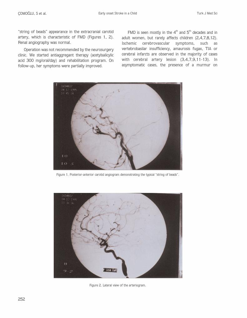
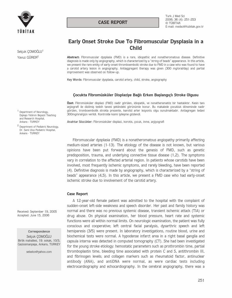
“string of beads” appearance in the extracranial carotidartery, which is characteristic of FMD (Figures 1, 2).Renal angiography was normal.
Operation was not recommended by the neurosurgeryclinic. We started antiaggregant therapy (acetylsalicylicacid 300 mg/oral/day) and rehabilitation program. Onfollow-up, her symptoms were partially improved.
FMD is seen mostly in the 4th and 5th decades and inadult women, but rarely affects children (2,4,7,8,12).Ischemic cerebrovascular symptoms, such asvertebrobasilar insufficiency, amaurosis fugax, TIA orcerebral infarcts are observed in the majority of caseswith cerebral artery lesion (3,4,7,9,11-13). Inasymptomatic cases, the presence of a murmur on
252
ÇOMO⁄LU, S et al. Early onset Stroke in a Child Turk J Med Sci
Figure 1. Posterior-anterior carotid angiogram demonstrating the typical “string of beads”.
Figure 2. Lateral view of the arteriogram.
auscultation of the carotid artery can be revealed, but inour case, no pathology on carotid auscultation wasdetected. It is reported that the frequency of aneurysmsin FMD is three times higher and subarachnoidhemorrhages more frequent than in the normalpopulation. However, the majority of stroke cases consistof ischemic cerebrovascular events (1,3,4,6,11).
In histopathologic examinations, three different typesof disorder in vessel walls are described: intimalfibroplasia, medial fibroplasia and subadventitialhyperplasia. Medial fibroplasia is the most frequent and itis accepted as a characteristic feature of FMD (2,4).
FMD diagnosis can be made only by the characteristicangiographic appearance of a “string of beads” (1-11).Angiographic appearance in our case was also in harmonywith FMD. Although the true incidence of FMD is notknown, in patients in whom carotid angiography wasperformed, FMD has been reported at a rate of
approximately 0.3-0.9% and bilateral involvement as60%.
Most of the patients can be effectively treated withantiplatelet agents. However, if the patients havepersistent or progressive symptoms, dilatation of thecarotid artery may be indicated (1,3,4,6). Surgicalintervention is recommended for FMD of the carotidarteries associated with intracranial aneurysms becauseantiplatelet medication is contraindicated (1,3,4). Wepreferred the medical approach for our case, andobserved partial improvement in her follow-up.
In this article, we emphasize that fibromusculardysplasia should also be considered in the differentialdiagnosis of childhood stroke. For this purpose,angiography must be planned for diagnosis. Additionally,patients with FMD must be followed carefully forprogression of the central vascular pathology andhypertension.
253
Vol: 36 No: 4 Early onset Stroke in a Child August 2006
References1. Difazio M, Hinds SR, Depper M, Tom B, Davis R. Intracranial
fibromuscular dysplasia in a six-year-old: a rare cause of childhoodstroke. J Child Neurol 2000; 15 (8): 559-562.
2. Puri V, Riggs G. Case report of fibromuscular dysplasia presentingas stroke in a 16-year-old boy. J Child Neurol 1999; 14: 233-238.
3. Chive NC, DeLong GR, Heinz ER. Intracranial fibromusculardysplasia in a 5-year-old child. Pediatr Neurol 1996; 14: 262-264.
4. Wesen CA, Elliot BM. Fibromuscular dysplasia of the carotidarteries. Am J Surg 1986; 151: 448-451.
5. Shields WD, Ziter FA, Osborn AG, Allen J. Fibromuscular dysplasiaas a cause of stroke in infancy and childhood. Pediatrics 1977;59: 899-901.
6. Collins GJ, Clagett GP. Fibromuscular dysplasia of the internalcarotid arteries. Ann Surg 1981; 194: 89-96.
7. Balaji MR, James AD. Fibromuscular dysplasia of the internalcarotid artery. Arch Surg 1980; 115: 984-986.
8. Gumerlock MK, Coull BM, Howieson J, Buchan C, Neuwelt EA.Late stenosis of a superficial temporal-middle cerebral arterybypass: angiographic and histological findings. Neurosurgery1985; 16: 650-657.
9. Pozzati E, Giuliani G, Acciarri N, Nuzzo G. Long-term follow-up ofocclusive cervical carotid dissection. Stroke 1990; 21: 528-531.
10. Saygı S, Bolay H, Tekkok IH, Cila A, Zileli T. Fibromusculardysplasia of the basilar artery: a case with brain stem stroke.Angiology 1990; 41(8): 658-661.
11. Emparanza JI, Aldamiz-Echevarria L, Peren-Yerza E, HernandezJ, Pena B, Gaztanega R. Ischemic stroke due to fibromusculardysplasia. Neuropediatrics 1989; 20: 181-182.
12. Bowen MD, Burak CR, Baron TF. Childhood ischemic stroke in anon-urban population. J Child Neurol 2005; 20: 194-197.
13. Dziewas R, Konrad C, Drager B, Evers S, Besselmann M,Ludemann P. B et al. Cervical artery dissection - clinical features,risk factors, therapy and outcome in 126 patients. J Neurol2003; 250: 1179-1184.