Embed Size (px)
Citation preview
Endogenous nitric oxide enhancesprostaglandin production in a model of renalinflammation.
D Salvemini, … , M G Currie, P Needleman
J Clin Invest. 1994;93(5):1940-1947. https://doi.org/10.1172/JCI117185.
The interaction between nitric oxide (NO) and cyclooxygenase (COX) was studied in arabbit model of renal inflammation, the ureteral obstructed hydronephrotic kidney (HNK). Exvivo perfusion of the HNK but not the control kidney (e.g., unobstructed contralateral kidney,CLK), led to a time-dependent release of nitrite (NO2-), a breakdown product of NO.Stimulation of the HNK with bradykinin (BK) evoked a time-dependent increase inprostaglandin E2 (PGE2) production. NG-monomethyl-L-arginine (L-NMMA), which blocksthe activity of both constitutive and inducible nitric oxide synthase (cNOS and iNOS),aminoguanidine, a recently described selective iNOS inhibitor, dexamethasone, orcycloheximide abolished the release of NO2- and attenuated the exaggerated BK-inducedPGE2 production. This supports the existence of iNOS and COX-2 in the HNK. In the CLK,BK elicited release of both NO2- and PGE2 but this did not augment with time. L-NMMA butnot aminoguanidine, dexamethasone, or cycloheximide attenuated NO2- and PGE2 releaseindicative of the presence of constitutive but not inducible NOS or COX. The current studysuggests that the endogenous release of NO from cNOS in the CLK activates a constitutiveCOX resulting in optimal PGE2 release by BK. In addition, in the HNK, NO release fromiNOS activates the induced COX resulting in markedly increased release ofproinflammatory prostaglandin. The broader implication of this study is that thecyclooxygenase isozymes are […]
Research Article
Find the latest version:
http://jci.me/117185-pdf
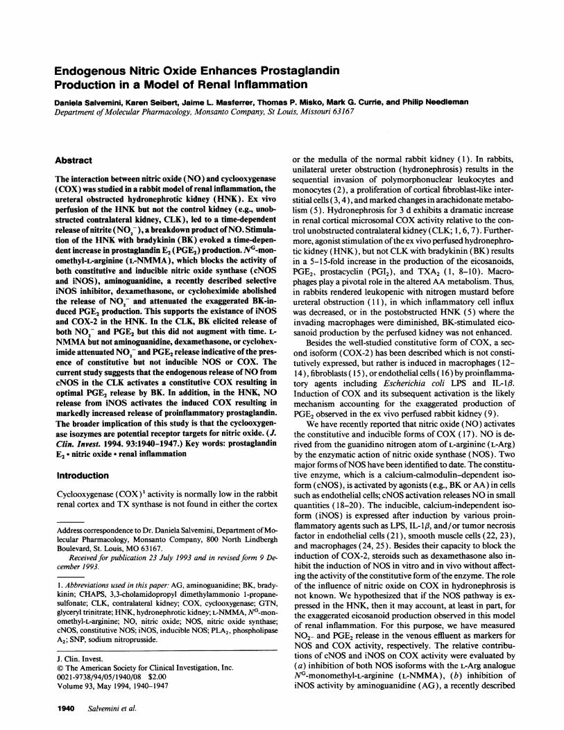
Endogenous Nitric Oxide Enhances ProstaglandinProduction in a Model of Renal InflammationDaniela Salvemini, Karen Seibert, Jaime L. Masferrer, Thomas P. Misko, Mark G. Currie, and Philip NeedlemanDepartment of Molecular Pharmacology, Monsanto Company, St Louis, Missouri 63167
Abstract
The interaction between nitric oxide (NO) and cyclooxygenase(COX) was studied in a rabbit model of renal inflammation, theureteral obstructed hydronephrotic kidney (HNK). Ex vivoperfusion of the HNKbut not the control kidney (e.g., unob-structed contralateral kidney, CLK), led to a time-dependentrelease of nitrite (NO;-), a breakdown product of NO. Stimula-tion of the HNKwith bradykinin (BK) evoked a time-depen-dent increase in prostaglandin E2 (PGE2) production. N-mon-omethyl-L-arginine (L-NMMA), which blocks the activity ofboth constitutive and inducible nitric oxide synthase (cNOSand iNOS), aminoguanidine, a recently described selectiveiNOS inhibitor, dexamethasone, or cycloheximide abolishedthe release of NO2- and attenuated the exaggerated BK-in-duced PGE2production. This supports the existance of iNOSand COX-2 in the HNK. In the CLK, BK elicited release ofboth NO2- and PGE2 but this did not augment with time. L-NMMAbut not aminoguanidine, dexamethasone, or cyclohex-imide attenuated NO2- and PGE2release indicative of the pres-ence of constitutive but not inducible NOSor COX. Thecurrent study suggests that the endogenous release of NOfromcNOS in the CLK activates a constitutive COXresulting inoptimal PGE2 release by BK. In addition, in the HNK, NOrelease from iNOS activates the induced COXresulting inmarkedly increased release of proinflammatory prostaglandin.The broader implication of this study is that the cyclooxygen-ase isozymes are potential receptor targets for nitric oxide. (J.Clin. Invest. 1994. 93:1940-1947.) Key words: prostaglandinE2 'nitric oxide * renal inflammation
Introduction
Cyclooxygenase (COX)' activity is normally low in the rabbitrenal cortex and TX synthase is not found in either the cortex
Address correspondence to Dr. Daniela Salvemini, Department of Mo-lecular Pharmacology, Monsanto Company, 800 North LindberghBoulevard, St. Louis, MO63167.
Received for publication 23 July 1993 and in revised form 9 De-cember 1993.
1. Abbreviations used in this paper: AG, aminoguanidine; BK, brady-kinin; CHAPS, 3,3-cholamidopropyl dimethylammonio 1-propane-sulfonate; CLK, contralateral kidney; COX, cyclooxygenase; GTN,glyceryl trinitrate; HNK, hydronephrotic kidney; L-NMMA, NG-mon-omethyl-L-arginine; NO, nitric oxide; NOS, nitric oxide synthase;cNOS, constitutive NOS; iNOS, inducible NOS; PLA2, phospholipaseA2; SNP, sodium nitroprusside.
or the medulla of the normal rabbit kidney ( 1). In rabbits,unilateral ureter obstruction (hydronephrosis) results in thesequential invasion of polymorphonuclear leukocytes andmonocytes (2), a proliferation of cortical fibroblast-like inter-stitial cells ( 3, 4), and marked changes in arachidonate metabo-lism (5). Hydronephrosis for 3 d exhibits a dramatic increasein renal cortical microsomal COXactivity relative to the con-trol unobstructed contralateral kidney (CLK; 1, 6, 7). Further-more, agonist stimulation of the ex vivo perfused hydronephro-tic kidney (HNK), but not CLKwith bradykinin (BK) resultsin a 5-15-fold increase in the production of the eicosanoids,PGE2, prostacyclin (PGI2), and TXA2 (1, 8-10). Macro-phages play a pivotal role in the altered AA metabolism. Thus,in rabbits rendered leukopenic with nitrogen mustard beforeureteral obstruction ( 11), in which inflammatory cell influxwas decreased, or in the postobstructed HNK(5) where theinvading macrophages were diminished, BK-stimulated eico-sanoid production by the perfused kidney was not enhanced.
Besides the well-studied constitutive form of COX, a sec-ond isoform (COX-2) has been described which is not consti-tutively expressed, but rather is induced in macrophages ( 12-14), fibroblasts ( 15), or endothelial cells ( 16) by proinflamma-tory agents including Escherichia coli LPS and IL- 1,.Induction of COXand its subsequent activation is the likelymechanism accounting for the exaggerated production ofPGE2observed in the ex vivo perfused rabbit kidney (9).
Wehave recently reported that nitric oxide (NO) activatesthe constitutive and inducible forms of COX( 17). NOis de-rived from the guanidino nitrogen atom of L-arginine (L-Arg)by the enzymatic action of nitric oxide synthase (NOS). Twomajor forms of NOShave been identified to date. The constitu-tive enzyme, which is a calcium-calmodulin-dependent iso-form (cNOS), is activated by agonists (e.g., BKor AA) in cellssuch as endothelial cells; cNOSactivation releases NOin smallquantities ( 18-20). The inducible, calcium-independent iso-form (iNOS) is expressed after induction by various proin-flammatory agents such as LPS, IL- Ifl, and/or tumor necrosisfactor in endothelial cells (21), smooth muscle cells (22, 23),and macrophages (24, 25). Besides their capacity to block theinduction of COX-2, steroids such as dexamethasone also in-hibit the induction of NOSin vitro and in vivo without affect-ing the activity of the constitutive form of the enzyme. The roleof the influence of nitric oxide on COXin hydronephrosis isnot known. Wehypothesized that if the NOSpathway is ex-pressed in the HNK, then it may account, at least in part, forthe exaggerated eicosanoid production observed in this modelof renal inflammation. For this purpose, we have measuredNO2- and PGE2 release in the venous effluent as markers forNOSand COXactivity, respectively. The relative contribu-tions of cNOSand iNOS on COXactivity were evaluated by(a) inhibition of both NOSisoforms with the L-Arg analogueNG-monomethyl-L-arginine (L-NMMA), (b) inhibition ofiNOS activity by aminoguanidine (AG), a recently described
1940 Salvemini et al.
J. Clin. Invest.©The American Society for Clinical Investigation, Inc.0021-9738/94/05/1940/08 $2.00Volume 93, May 1994, 1940-1947
selective iNOS inhibitor (26-28), and (c) inhibition of the in-duction of iNOS by cycloheximide or dexamethasone.
Methods
Ureteral obstruction. Male NewZealand white rabbits (2-3 kg) wereobtained from Mohican Valley Rabbitry (Loudenville, OH) andhoused for at least 1 wk before the experiments. All animals were fednormal rabbit Chow (Ralston Purina Co., St. Louis, MO)and allowedfree access to water. Unilateral ureteral obstruction was carried out by apreviously described procedure (8). Briefly, the animals were anesthe-tized with ketamine/xylazine (35/3 mg/kg, i.m.), and a small abdomi-nal incision was made. A silk suture was tied around one of the ureternear the bladder. 150,000 IU of penicillin antibiotic was administered(s.c.) postoperatively, and the rabbit allowed to return to dorsal recum-bancy before returning to its cage. 3 d after obstruction (8), rabbitswere anesthetized again (ketamine/xylazine 35/3 mg/kg, i.m.) andheparinized (250 U/kg, i.v.), and the abdominal cavity was openedand the renal arteries were cannulated. Kidneys were removed, flushedwith 30 ml ice-cold Krebs-Henseleit buffer, and perfused at 10 ml/minwith oxygenated (95%02/5% C02, pH 7.4) Krebs-Henseleit buffermaintained at 370C.
Ex vivo kidney perfusion. Changes in perfusion pressures (mea-sured with a Cobe disposable pressure transducer on a Grass model 7Dpolygraph) reflect changes in renal resistance. Perfusion of the kidneysat a rate of 10 ml/min gave pressures of 78±1 mmHgin the CLKand75±2 mmHgin the HNK(n = 36). Under these experimental proce-dures, papaverine (0.1 mM) did not affect basal perfusion pressure,which demonstrates that the perfused rabbit kidney lacks intrinsictone. Tone could be increased by adding noradrenaline (0.3 ttM) in theperfusion buffer and this allowed vasodilator responses to BKor AA tobe monitored. Wedid not increase tone in our experiments so as not tocomplicate the model and thus the interpretation of the data. Therewas no need for the addition of albumin or other osmotic agents to thebuffer because the perfusion pressure recordings were stable for theduration of the experiment (6 h). The drugs used in our study did notaffect perfusion pressure. No change in kidney weights were observedafter perfusion for up to 6 h (from 15±0.1 to 15±1 g in the CLKandfrom 26±1 to 27±1 g in the HNK, n = 36), suggesting the absence ofsignificant edema formation over this time period.
Drug administration. BK was used to stimulate NO2- and PGE2.Release of PGE2 by BK requires activation of phospholipase A2(PLA2), which in turn releases AA from endogenous phospholipid forconversion to prostaglandin endoperoxide by COX. Therefore, to de-termine whether or not the actions of NOon PGE2 release were aconsequence of an effect on PLA2 activity we also tested the effects ofNOon AA-mediated release of PGE2 in the CLK. The isolated rabbitkidneys were allowed to equilibrate for 30 min before experimentalmanipulations. All drugs used in this study, except for sodium nitro-prusside (SNP) or 3,3-cholamidopropyl dimethylammonio 1-propane-sulfonate (CHAPS), were infused for 30 min after the equilibrationperiod and before the first BK ( 1 Mg) or AA (30 MM) injection (this isreferred to as time 0). Drugs were infused for the entire duration of theexperiment (e.g., 6 h). Bradykinin or AA were injected as a bolusintraarterially every hour from 0 to 6 h of perfusion to stimulate NO2-and PGE2release from the kidney. These agonists were injected with a10-min lag period from each other. The profile of PGE2release by BKor AA revealed that maximal PGE2release peaked at the third minuteafter agonist injection and declined between the fourth and fifth min-utes (n = 8). Therefore, in all our experiments we collected the venouseffluents from the CLK/HNK for 3 min both before and after each BKor AA injection. The effects of the NOdonors, SNPor glyceryl trini-trate (GTN), or of the detergent, CHAPS(4.7 mg/ml) on BK(1 Mg) orAA (30 MM)-induced PGE2release from the CLKor HNKwere testedat the sixth hour of perfusion. At this time point, after the bolus injec-tion of BK or AA, the kidneys were perfused with SNPfor 30 mmnorwith CHAPSfor 30 s and then rechallenged with the same dose of BK
or AA. The concentration of CHAPSused was sufficient to removerenal vascular endothelium and thus endothelium-dependent re-sponses without altering non-endothelium-mediated effects (29). Insome experiments, indomethacin (1 MM) was perfused through thekidneys together with SNP. The venous effluent was assayed for PGE2by radioimmunoassay (30) or for NO2- release using the diaminona-phthalene assay (31 ). The production of NO2-, a breakdown productof NO, or PGE2was used respectively as a marker for NOSand COXactivity.
Materials. The composition of Krebs-Henseleit buffer was (mM):NaCl 120, KCI 4.7, MgSO4- 7H20 1.2, CaCl2 - 2H20 2.5, KH2PO41.2,NaHCO325, dextrose 10. All drugs were obtained from Sigma Chemi-cal Co. (St Louis, MO).
Statistical analysis. Results are expressed as mean±SEMfor (n)experiments. The results were analyzed by Student's unpaired t test todetermine the significant difference between means, or by a two-wayANOVAfollowed by a least significance procedure to determine thenature of this response. This will be specified when appropriate. A Pvalue of < 0.05 was taken as significant.
Results
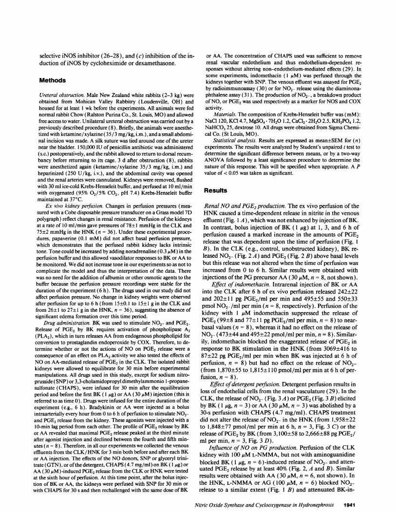
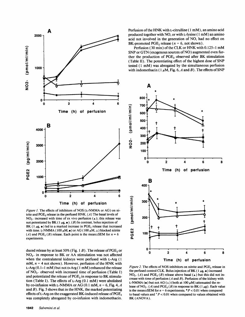
Renal NOand PGE2production. The ex vivo perfusion of theHNKcaused a time-dependent release in nitrite in the venouseffluent (Fig. 1 A), which was not enhanced by injection of BK.In contrast, bolus injection of BK (1 Mg) at 1, 3, and 6 h ofperfusion caused a marked increase in the amounts of PGE2release that was dependent upon the time of perfusion (Fig. 1B). In the CLK (e.g., control, unobstructed kidney), BK re-leased NO2- (Fig. 2 A) and PGE2(Fig. 2 B) above basal levelsbut this release was not altered when the time of perfusion wasincreased from 0 to 6 h. Similar results were obtained withinjections of the PGprecursor AA(30 ,M, n = 8, not shown).
Effect of indomethacin. Intrarenal injection of BK or AAinto the CLK after 6 h of ex vivo perfusion released 242±22and 202±11 pg PGE2/ml per min and 495±55 and 550±33pmol N02-/ml per min (n = 8, respectively). Perfusion of thekidney with 1 MuM indomethacin suppressed the release ofPGE2(99±8 and 77±11 pg PGE2/ml per min, n = 8) to near-basal values (n = 8), whereas it had no effect on the release ofNO2- (473+44 and 495±22 pmol/ml per min, n = 8). Similar-ily, indomethacin blocked the exaggerated release of PGE2inresponse to BK stimulation in the HNK(from 3069±416 to87±22 pg PGE2/ml per min when BK was injected at 6 h ofperfusion, n = 8) but had no effect on the release of NO2-(from 1,870±55 to 1,815±110 pmol/ml per min at 6 h of per-fusion, n = 8).
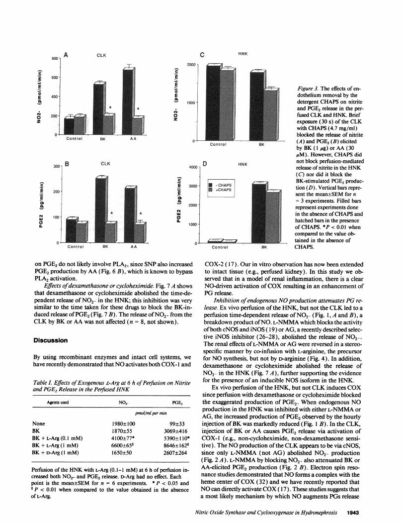
Effect of detergent perfusion. Detergent perfusion results inloss of endothelial cells from the renal vasculature (29). In theCLK, the release of NO2- (Fig. 3 A) or PGE2(Fig. 3 B) elicitedby BK (1 g, n = 3) or AA (30 MM, n = 3) was abolished by a30-s perfusion with CHAPS(4.7 mg/ml). CHAPStreatmentdid not alter the release of NO2- in the HNK(from 1,958±22to 1,848±77 pmol/ml per min at 6 h, n = 3, Fig. 3 C) or therelease of PGE2by BK(from 3,100±58 to 2,666±88 pg PGE2/ml per min, n = 3, Fig. 3 D).
Influence of NOon PGproduction. Perfusion of the CLKkidney with 100 MML-NMMA, but not with aminoguanidineblocked BK (1 Ig, n = 6)-induced release of NO2- and atten-uated PGE2 release by at least 40% (Fig. 2, A and B). Similarresults were obtained with AA (30,MM, n = 6, not shown). Inthe HNK, L-NMMAor AG(100 MM, n = 6) blocked NO2-release to a similar extent (Fig. 1 B) and attenuated BK-in-
Nitric Oxide Synthase and Cyclooxygenase in Hydronephrosis 1941
Perfusion of the HNKwith L-citrulline ( 1 mM), an amino acidproduced together with NO, or with L-lysine ( 1 mM)an aminoacid not involved in the generation of NO, had no effect onBK-promoted PGE2release (n = 6, not shown).
Perfusion (30 min) of the CLKor HNKwith 0.125-1 mMSNPor GTN(exogenous sources of NO) augmented even fur-ther the production of PGE2 observed after BK stimulation(Table II). The potentiating effect of the highest dose of SNPtested (1 mM)was abrogated by the simultaneous perfusionwith indomethacin (1 ,uM, Fig. 6, A and B). The effects of SNP
A*
2 4 6
Time (h) of perfusiona
EE
0E-.
CJ0z
0 2 4
Time (h) of perfusion
B
0 2 4 6
Time (h) of perfusion
Figure 1. The effects of inhibitors of NOS(L-NMMA or AG) on ni-trite and PGE2release in the perfused HNK. (A) The basal levels ofNO2- increased with time of ex vivo perfusion (A); this release was
not potentiated by BK ( 1 Mg, .). (B) In contrast, bolus injection ofBK (1 qg, * ) led to a marked increase in PGE2 release that increasedwith time. L-NMMA( 100MgM, *) or AG( 100 ,uM, A) blocked nitrite(A) and PGE2(B) release. Each point is the mean±SEMfor n = 6experiments.
duced release by at least 50% (Fig. 1 B). The release of PGE2or
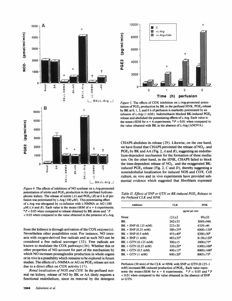
NO2- in response to BK or AA stimulation was not affectedwhen the contralateral kidneys were perfused with L-Arg (1mM, n = 4 not shown). However, perfusion of the HNKwithL-Arg (0.1- 1 mM)but not D-Arg ( 1 mM)enhanced the releaseof NO2- observed with increased time of perfusion (Table I)and potentiated the release of PGE2in response to BKstimula-tion (Table I). The effects of L-Arg (0.1 mM)were abolishedby co-infusion with L-NMMAor AG(0.1 mM,n = 6, Fig. 4, Aand B). Fig. 5 shows that in the HNK, the marked potentiatingeffects of L-Arg on the exaggerated BK-induced release of PGE2was completely abrogated by co-infusion with indomethacin.
E
E
0.
cs4LU
0a.
0 2 4 6
Time (h) of perfusion
8
Figure 2. The effects of NOSinhibitors on nitrite and PGE2release inthe perfused control CLK. Bolus injection of BK(1 Mug, .) increasedNO2- (A) and PGE2(B) release above basal (A) but this did not in-crease with time of perfusion (A and B). Perfusion of the kidney withL-NMMA(m) but not AG(A) (both at 100 MuM) attenuated the re-
lease of NO2- (A) and PGE2(B) in response to BK (1 Mg). Each valueis the mean±SEMfor n = 6 experiments. *P < 0.01 when comparedto basal values and +P < 0.01 when compared to values obtained withBK (ANOVA).
1942 Salvemini et al.
A2000
E
E0
csJ0z
B4000
3000EE
0.
cm4
0.
6 8
C HNK
c
EE0Ea
0z
600-i
400
200
0-
2000 1c
EE
0
E
CM0z
1000 -
Control BK AA
3001 B
C
E2E
CNCL
C,a.
200
100-
01
CLK4000 1 D
C
EE
C,
a.
3000 -
2000 -
1 000 I
on PGE2do not likely involve PLA2, since SNPalso increasedPGE2production by AA (Fig. 6 B), which is known to bypassPLA2 activation.
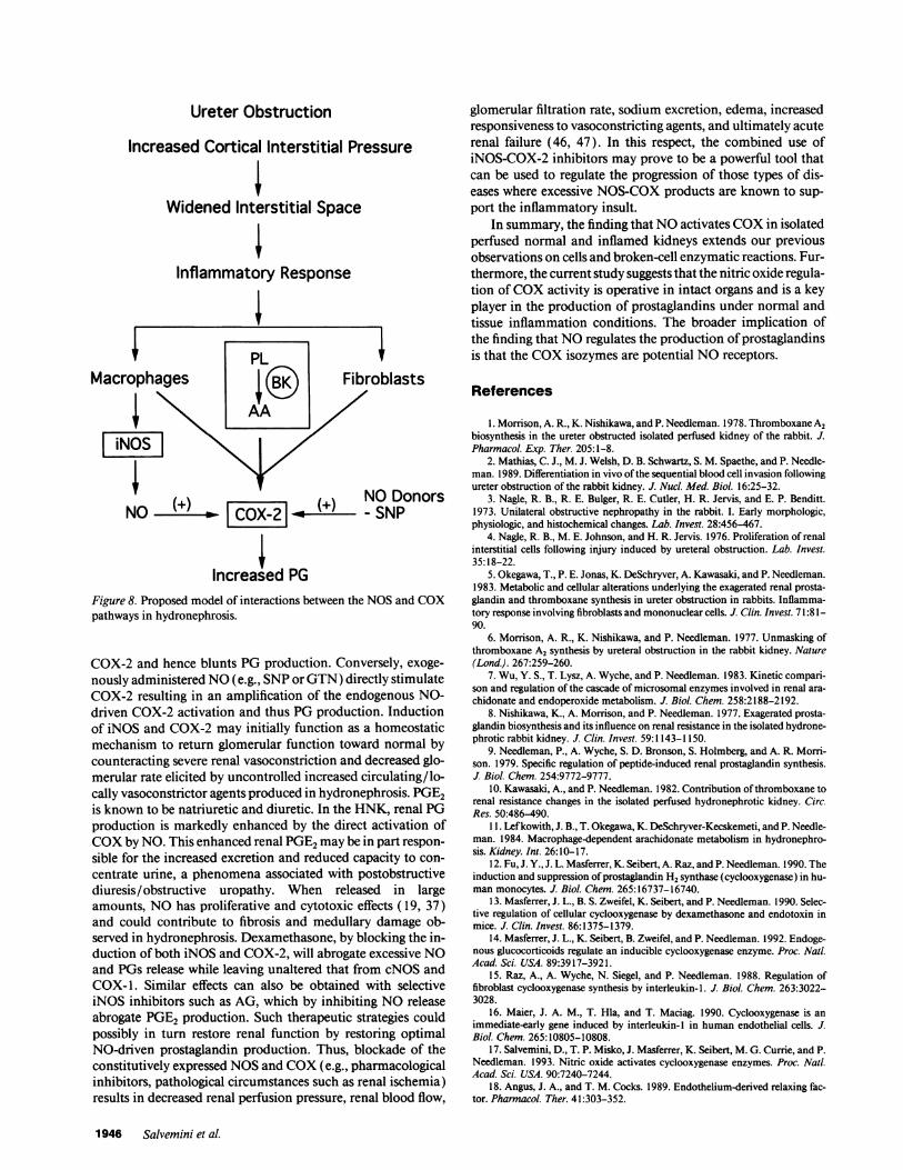
Effects of dexamethasone or cycloheximide. Fig. 7 A showsthat dexamethasone or cycloheximide abolished the time-de-pendent release of NO2- in the HNK; this inhibition was verysimilar to the time taken for these drugs to block the BK-in-duced release of PGE2(Fig. 7 B). The release of NO2- from theCLKby BK or AAwas not affected (n = 8, not shown).
Discussion
By using recombinant enzymes and intact cell systems, wehave recently demonstrated that NOactivates both COX-1 and
Table I. Effects of Exogenous L-Arg at 6 h of Perfusion on Nitriteand PGE2Release in the Perfused HNK
Agents used NO2- PGE2
pmol/ml per min
None 1980±100 99±33BK 1870±55 3069±416BK + L-Arg (0.1 mM) 4100±77* 5390±110*BK + L-Arg (I mM) 6600±65* 8646±162*BK + D-Arg (I mM) 1650±50 2607±264
Perfusion of the HNKwith L-Arg (0.1 -1 mM)at 6 h of perfusion in-creased both NO2- and PGE2 release. D-Arg had no effect. Eachpoint is the mean±SEM for n = 6 experiments. * P < 0.05 and* P < 0.01 when compared to the value obtained in the absenceof L-Arg.
Conirol
HNK
* - CHAPS+CHAPS
Control
Figure 3. The effects of en-dothelium removal by thedetergent CHAPSon nitriteand PGE2 release in the per-fused CLKand HNK. Briefexposure (30 s) of the CLKwith CHAPS(4.7 mg/ml)blocked the release of nitrite(A) and PGE2(B) elicitedby BK (I jg) or AA (30sM). However, CHAPSdid
not block perfusion-mediatedrelease of nitrite in the HNK(C) nor did it block theBK-stimulated PGE2produc-tion (D). Vertical bars repre-sent the mean±SEMfor n= 3 experiments. Filled barsrepresent experiments donein the absence of CHAPSandhatched bars in the presenceof CHAPS. *P < 0.01 whencompared to the value ob-tained in the absence ofCHAPS.
COX-2 (17). Our in vitro observation has now been extendedto intact tissue (e.g., perfused kidney). In this study we ob-served that in a model of renal inflammation, there is a clearNO-driven activation of COXresulting in an enhancement ofPGrelease.
Inhibition of endogenous NOproduction attenuates PGre-lease. Ex vivo perfusion of the HNK, but not the CLK led to aperfusion time-dependent release of NO2- (Fig. 1, A and B), abreakdown product of NO. L-NMMAwhich blocks the activityofboth cNOSand iNOS ( 19) or AG, a recently described selec-tive iNOS inhibitor (26-28), abolished the release of NO2-.The renal effects of L-NMMAor AGwere reversed in a stereo-specific manner by co-infusion with L-arginine, the precursorfor NOsynthesis, but not by D-arginine (Fig. 4). In addition,dexamethasone or cycloheximide abolished the release ofNO2- in the HNK(Fig. 7 A), further supporting the evidencefor the presence of an inducible NOSisoform in the HNK.
Ex vivo perfusion of the HNK, but not CLK induces COXsince perfusion with dexamethasone or cycloheximide blockedthe exaggerated production of PGE2. When endogenous NOproduction in the HNKwas inhibited with either L-NMMAorAG, the increased production of PGE2observed by the hourlyinjection of BKwas markedly reduced (Fig. 1 B). In the CLK,injection of BK or AA causes PGE2 release via activation ofCOX-1 (e.g., non-cycloheximide, non-dexamethasone sensi-tive). The NOproduction of the CLKappears to be via cNOS,since only L-NMMA(not AG) abolished NO2- production(Fig. 2 A). L-NMMAby blocking NO2- also attenuated BKorAA-elicited PGE2 production (Fig. 2 B). Electron spin reso-nance studies demonstrated that NOforms a complex with theheme center of COX(32) and we have recently reported thatNOcan directly activate COX( 17). These studies suggests thata most likely mechanism by which NOaugments PGs release
Nitric Oxide Synthase and Cyclooxygenase in Hydronephrosis 1943
CLK
c
| +L-Arg+L-Arg+Indo
8000
6000
4000
*
*
2000
A-+
Yi z
mo +
L BK+L-Arg -1
*
<:
I
E +
m + z
m +
L BK+L-Arg I
0 1 3 6
Time (h) perfusionFigure 5. The effects of COXinhibition on L-Arg-promoted poten-tiation of PGE2production by BK in the perfused HNK. PGE2releaseby BKat 0, 1, 3, and 6 h of perfusion is markedly potentiated by aninfusion of L-Arg (1 mM). Indomethacin blocked BK-induced PGE2release and abolished the potentiating effects of L-Arg. Each value isthe mean±SEMfor n = 6 experiments. *fP < 0.01 when compared tothe value obtained with BK in the absence of L-Arg (ANOVA).
CHAPSabolishes its release (29). Likewise, on the one hand,we have found that CHAPSprevented the release of NO2- andPGE2by BKand AA (Fig. 2, A and B), suggesting an endothe-lium-dependent mechanism for the formation of these media-tors. On the other hand, in the HNK, CHAPSfailed to blockthe time-dependent release of NO2- and the exaggerated BK-induced PGE2release (Fig. 2, Cand D), thereby suggesting anonendothelial localization for induced NOSand COX. Cellculture, ex vivo and in vivo experiments have provided sub-stantial evidence which suggested that fibroblasts expressed
Figure 4. The effects of inhibitors of NOsynthase on L-Arg-promotedpotentiation of nitrite and PGE2production in the perfused hydrone-phrotic kidney. The release of nitrite (A) and PGE2(B) at 6 h of per-
fusion was potentiated by L-Arg (100 gM). This potentiating effectof L-Arg was abrogated by co-infusion with L-NMMAor AG(100MM) (A and B). Each value is the mean±SEMof n = 6 experiments.
*P < 0.05 when compared to release obtained by BKalone and +P< 0.05 when compared to the value obtained in the presence of L-Arg.
from the kidneys is through activation of the COXenzyme(s).Nevertheless other possibilities exist. For instance, NOinter-acts with oxygen-derived free radicals and as such NOcan beconsidered a free radical scavenger (33). Free radicals are
known to modulate the COXpathways (34). Whether this or
other properties of NOaccount for part of the mechanism bywhich NOincreases prostaglandin production in whole organs
or in vivo is a possibility which remains to be explored in futurestudies. The effects of L-NMMAor AGon PGE2release are notdue to a direct effect on COXactivity ( 17).
Renal localization of NOSand COX. In the perfused nor-
mal rat kidney, release of NOby BK or AA likely requires a
functional endothelium, since its removal by the detergent
Table II. Effects of SNPor GTNon BK-induced PGE2Release inthe Perfused CLKand HNK
Additions CLK HNK
pg/ml per min
None 121±2 99±33BK 242±33 3069±946BK + SNP(0.125 mM) 223±20 4329±40BK + SNP(0.25 mM) 380±25* 6000±120*BK + SNP(0.5 mM) 455±40* 8200±50$BK + SNP(1 mM) 485±35$ 8138±120$BK + GTN(0.125 mM) 300±15 5000±75*BK + GTN(0.25 mM) 320±20* 6300±100*BK + GTN(0.5 mM) 400±15* 8000±100$BK + GTN(1 mM) 600±20* 8005±75*
Perfusion (30 min) of the CLKor HNKwith SNPor GTN(0.125-1mM)increased BK-induced production of PGE2. Each value repre-sents the mean±SEMfor n = 6 experiments. * P < 0.05 and * P< 0.01 when compared to the value obtained in the absence of SNPor GTN.
1944 Salvemini et al.
5000 I A
4000 -
3000
2000
1000
w
Cum
EE
Qa.
w
0.
a
EE-.0
z
Ea
Aw
C,0EE0
cL
AL
LI)-CJ4e+
0
z-J
0
B6000 -
5000-
4000-
3000 -
2000
100:1-
0
4cm E
z-J
m
1 0000 *
-
changes occurring in the HNKthat were proposed earlier (5,11 ) can be updated as shown in Fig. 8. Unilateral ureter ob-struction causes a mechanical disruption and/or an immuno-logic stimulus in the cortex that triggers a regional inflamma-tory response resulting in accumulation of macrophages, fibro-blast proliferation, and markedly increased AA metabolism.Wepropose that macrophages are activated by ex vivo kidneyperfusion to release factor(s) that stimulate fibroblast prolifera-tion and that induces COX-2 presumably in the fibroblast. Atthe same time, induction of iNOS takes place in the macro-phage, NOis released and COX-2 activated. Release of endoge-nous renal AA after stimulation with BK leads to an exagger-ated release of PGE2. Removal of endogenous NO(e.g., by theuse of NOSinhibitors) prevents the NO-driven activation of
600 -
c 500
EE 400cm
300 -
0 200
100-
0-
B
T 2000
CLKControl*SNP
* *SNPIndo
Basal
E
E
Ea
%-I
1000'
BK AA
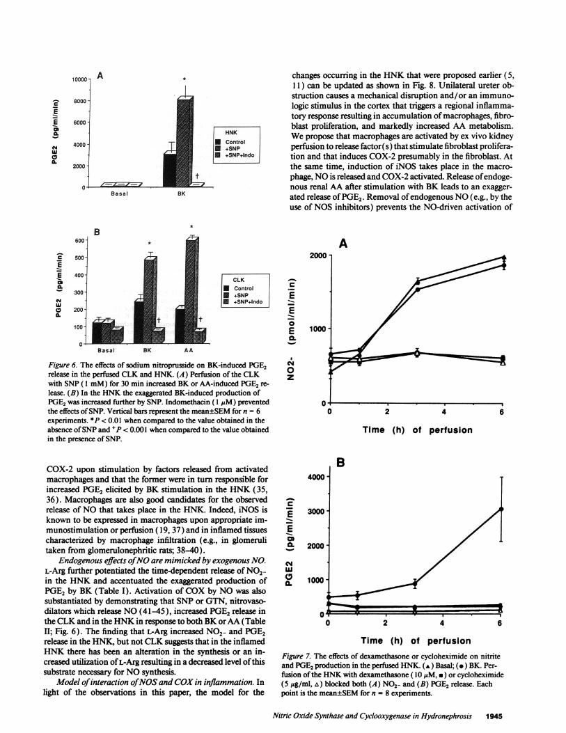
Figure 6. The effects of sodium nitroprusside on BK-induced PGE2release in the perfused CLK and HNK. (A) Perfusion of the CLKwith SNP(1 mM) for 30 min increased BKor AA-induced PGE2re-
lease. (B) In the HNKthe exaggerated BK-induced production ofPGE2was increased further by SNP. Indomethacin ( I gM) preventedthe effects of SNP. Vertical bars represent the mean±SEMfor n = 6experiments. *P < 0.01 when compared to the value obtained in theabsence of SNPand +P < 0.001 when compared to the value obtainedin the presence of SNP.
COX-2 upon stimulation by factors released from activatedmacrophages and that the former were in turn responsible forincreased PGE2 elicited by BK stimulation in the HNK(35,36). Macrophages are also good candidates for the observedrelease of NOthat takes place in the HNK. Indeed, iNOS isknown to be expressed in macrophages upon appropriate im-munostimulation or perfusion ( 19, 37) and in inflamed tissuescharacterized by macrophage infiltration (e.g., in glomerulitaken from glomerulonephritic rats; 38-40).
Endogenous effects of NOare mimicked by exogenous NO.L-Arg further potentiated the time-dependent release of NO2-in the HNKand accentuated the exaggerated production ofPGE2 by BK (Table I). Activation of COXby NOwas alsosubstantiated by demonstrating that SNPor GTN, nitrovaso-dilators which release NO(41-45), increased PGE2 release inthe CLKand in the HNKin response to both BKor AA(TableII; Fig. 6). The finding that L-Arg increased NO2- and PGE2release in the HNK, but not CLKsuggests that in the inflamedHNKthere has been an alteration in the synthesis or an in-creased utilization of L-Arg resulting in a decreased level of thissubstrate necessary for NOsynthesis.
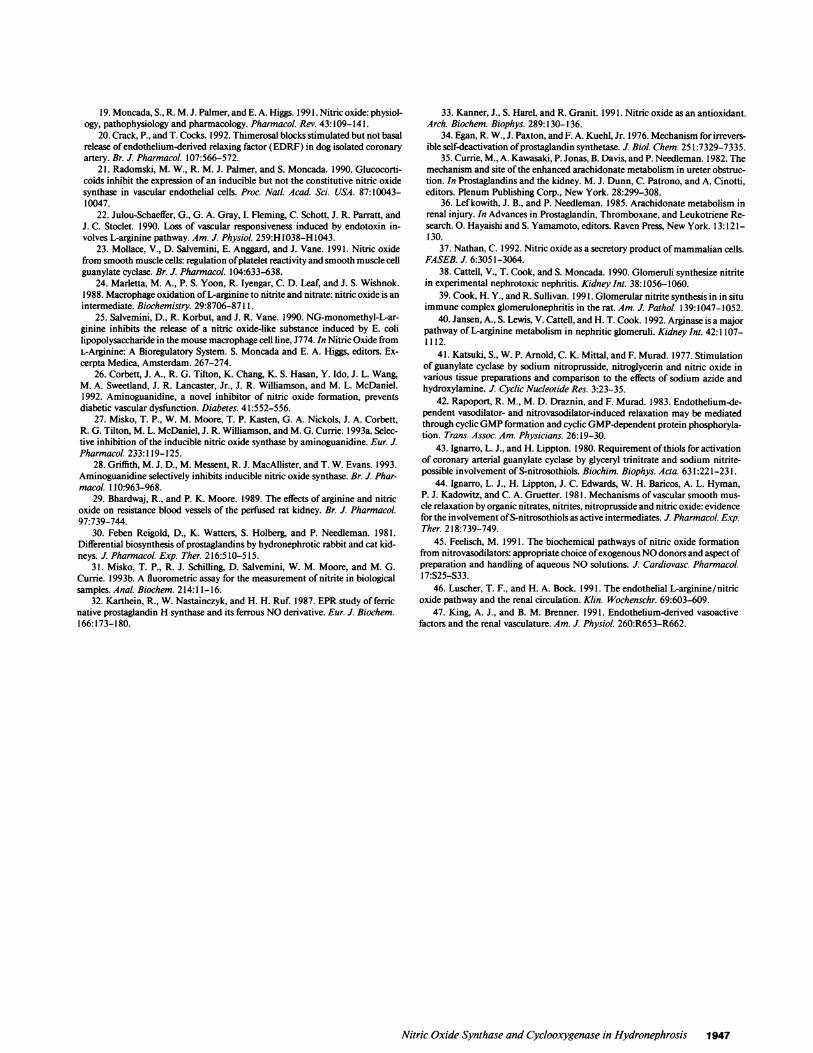
Model of interaction of NOSand COXin inflammation. Inlight of the observations in this paper, the model for the
CM0z
A
Time (h) of perfusion
E
EI--
c*IC,
a.
B
0 2 4 6
Time (h) of perfusion
Figure 7. The effects of dexamethasone or cycloheximide on nitriteand PGE2production in the perfused HNK. (A) Basal; (.) BK. Per-fusion of the HNKwith dexamethasone (10 jiM, *) or cycloheximide(5 jg/ml, &) blocked both (A) NO2- and (B) PGE2 release. Eachpoint is the mean±SEMfor n = 8 experiments.
Nitric Oxide Synthase and Cyclooxygenase in Hydronephrosis 1945
A *
c
EE0
0.
0.2000
HNK* Control* +SNP* *SNPINlndo
Basal BK
Ureter Obstruction
Increased Cortical Interstitial Pressure
Widened Interstitial Space
Inflammatory Response
Macrophages
iNOS
Fibroblasts
V V ~~,~ NODonorsNO ()- COX-21 ( - SNP
Increased PGFigure 8. Proposed model of interactions between the NOSand COXpathways in hydronephrosis.
COX-2 and hence blunts PGproduction. Conversely, exoge-nously administered NO(e.g., SNPor GTN) directly stimulateCOX-2 resulting in an amplification of the endogenous NO-driven COX-2 activation and thus PGproduction. Inductionof iNOS and COX-2 may initially function as a homeostaticmechanism to return glomerular function toward normal bycounteracting severe renal vasoconstriction and decreased glo-merular rate elicited by uncontrolled increased circulating/lo-cally vasoconstrictor agents produced in hydronephrosis. PGE2is known to be natriuretic and diuretic. In the HNK, renal PGproduction is markedly enhanced by the direct activation ofCOXby NO. This enhanced renal PGE2maybe in part respon-sible for the increased excretion and reduced capacity to con-centrate urine, a phenomena associated with postobstructivediuresis/obstructive uropathy. When released in largeamounts, NOhas proliferative and cytotoxic effects (19, 37)and could contribute to fibrosis and medullary damage ob-served in hydronephrosis. Dexamethasone, by blocking the in-duction of both iNOS and COX-2, will abrogate excessive NOand PGs release while leaving unaltered that from cNOSandCOX- 1. Similar effects can also be obtained with selectiveiNOS inhibitors such as AG, which by inhibiting NOreleaseabrogate PGE2 production. Such therapeutic strategies couldpossibly in turn restore renal function by restoring optimalNO-driven prostaglandin production. Thus, blockade of theconstitutively expressed NOSand COX(e.g., pharmacologicalinhibitors, pathological circumstances such as renal ischemia)results in decreased renal perfusion pressure, renal blood flow,
glomerular filtration rate, sodium excretion, edema, increasedresponsiveness to vasoconstricting agents, and ultimately acuterenal failure (46, 47). In this respect, the combined use ofiNOS-COX-2 inhibitors may prove to be a powerful tool thatcan be used to regulate the progression of those types of dis-eases where excessive NOS-COXproducts are known to sup-port the inflammatory insult.
In summary, the finding that NOactivates COXin isolatedperfused normal and inflamed kidneys extends our previousobservations on cells and broken-cell enzymatic reactions. Fur-thermore, the current study suggests that the nitric oxide regula-tion of COXactivity is operative in intact organs and is a keyplayer in the production of prostaglandins under normal andtissue inflammation conditions. The broader implication ofthe finding that NOregulates the production of prostaglandinsis that the COXisozymes are potential NOreceptors.
References
1. Morrison, A. R., K. Nishikawa, and P. Needleman. 1978. Thromboxane A2biosynthesis in the ureter obstructed isolated perfused kidney of the rabbit. J.Pharmacol. Exp. Ther. 205:1-8.
2. Mathias, C. J., M. J. Welsh, D. B. Schwartz, S. M. Spaethe, and P. Needle-man. 1989. Differentiation in vivo of the sequential blood cell invasion followingureter obstruction of the rabbit kidney. J. Nucl. Med. Bio. 16:25-32.
3. Nagle, R. B., R. E. Bulger, R. E. Cutler, H. R. Jervis, and E. P. Benditt.1973. Unilateral obstructive nephropathy in the rabbit. I. Early morphologic,physiologic, and histochemical changes. Lab. Invest. 28:456-467.
4. Nagle, R. B., M. E. Johnson, and H. R. Jervis. 1976. Proliferation of renalinterstitial cells following injury induced by ureteral obstruction. Lab. Invest.35:18-22.
5. Okegawa, T., P. E. Jonas, K. DeSchryver, A. Kawasaki, and P. Needleman.1983. Metabolic and cellular alterations underlying the exagerated renal prosta-glandin and thromboxane synthesis in ureter obstruction in rabbits. Inflamma-tory response involving fibroblasts and mononuclear cells. J. Clin. Invest. 71:81-90.
6. Morrison, A. R., K. Nishikawa, and P. Needleman. 1977. Unmasking ofthromboxane A2 synthesis by ureteral obstruction in the rabbit kidney. Nature(Lond.). 267:259-260.
7. Wu, Y. S., T. Lysz, A. Wyche, and P. Needleman. 1983. Kinetic compari-son and regulation of the cascade of microsomal enzymes involved in renal ara-chidonate and endoperoxide metabolism. J. BioL. Chem. 258:2188-2192.
8. Nishikawa, K., A. Morrison, and P. Needleman. 1977. Exagerated prosta-glandin biosynthesis and its influence on renal resistance in the isolated hydrone-phrotic rabbit kidney. J. Clin. Invest. 59:1143-1150.
9. Needleman, P., A. Wyche, S. D. Bronson, S. Holmberg, and A. R. Morri-son. 1979. Specific regulation of peptide-induced renal prostaglandin synthesis.J. Biol. Chem. 254:9772-9777.
10. Kawasaki, A., and P. Needleman. 1982. Contribution of thromboxane torenal resistance changes in the isolated perfused hydronephrotic kidney. Circ.Res. 50:486-490.
1 1. Lefkowith, J. B., T. Okegawa, K. DeSchryver-Kecskemeti, and P. Needle-man. 1984. Macrophage-dependent arachidonate metabolism in hydronephro-sis. Kidney. Int. 26:10-17.
12. Fu, J. Y., J. L. Masferrer, K. Seibert, A. Raz, and P. Needleman. 1990. Theinduction and suppression of prostaglandin H2 synthase (cyclooxygenase) in hu-man monocytes. J. Biol. Chem. 265:16737-16740.
13. Masferrer, J. L., B. S. Zweifel, K. Seibert, and P. Needleman. 1990. Selec-tive regulation of cellular cyclooxygenase by dexamethasone and endotoxin inmice. J. Clin. Invest. 86:1375-1379.
14. Masferrer, J. L., K. Seibert, B. Zweifel, and P. Needleman. 1992. Endoge-nous glucocorticoids regulate an inducible cyclooxygenase enzyme. Proc. Natl.Acad. Sci. USA. 89:3917-3921.
15. Raz, A., A. Wyche, N. Siegel, and P. Needleman. 1988. Regulation offibroblast cyclooxygenase synthesis by interleukin-l. J. Biol. Chem. 263:3022-3028.
16. Maier, J. A. M., T. Hla, and T. Maciag. 1990. Cyclooxygenase is animmediate-early gene induced by interleukin-l in human endothelial cells. J.Biol. Chem. 265:10805-10808.
17. Salvemini, D., T. P. Misko, J. Masferrer, K. Seibert, M. G. Currie, and P.Needleman. 1993. Nitric oxide activates cyclooxygenase enzymes. Proc. Natl.Acad. Sci. USA. 90:7240-7244.
18. Angus, J. A., and T. M. Cocks. 1989. Endothelium-derived relaxing fac-tor. Pharmacol. Ther. 41:303-352.
1946 Salvemini et al.
19. Moncada, S., R. M. J. Palmer, and E. A. Higgs. 1991. Nitric oxide: physiol-ogy, pathophysiology and pharmacology. Pharmacol. Rev. 43:109-141.
20. Crack, P., and T. Cocks. 1992. Thimerosal blocks stimulated but not basalrelease of endothelium-derived relaxing factor (EDRF) in dog isolated coronaryartery. Br. J. Pharmacol. 107:566-572.
21. Radomski, M. W., R. M. J. Palmer, and S. Moncada. 1990. Glucocorti-coids inhibit the expression of an inducible but not the constitutive nitric oxidesynthase in vascular endothelial cells. Proc. Nati. Acad. Sci. USA. 87:10043-10047.
22. Julou-Schaeffer, G., G. A. Gray, I. Fleming, C. Schott, J. R. Parratt, andJ. C. Stoclet. 1990. Loss of vascular responsiveness induced by endotoxin in-volves L-arginine pathway. Am. J. Physiol. 259:H 1038-H 1043.
23. Mollace, V., D. Salvemini, E. Anggard, and J. Vane. 1991. Nitric oxidefrom smooth muscle cells: regulation of platelet reactivity and smooth muscle cellguanylate cyclase. Br. J. Pharmacol. 104:633-638.
24. Marletta, M. A., P. S. Yoon, R. Iyengar, C. D. Leaf, and J. S. Wishnok.1988. Macrophage oxidation of L-arginine to nitrite and nitrate: nitric oxide is anintermediate. Biochemistry. 29:8706-871 1.
25. Salvemini, D., R. Korbut, and J. R. Vane. 1990. NG-monomethyl-L-ar-ginine inhibits the release of a nitric oxide-like substance induced by E. colilipopolysaccharide in the mouse macrophage cell line, J774. In Nitric Oxide fromL-Arginine: A Bioregulatory System. S. Moncada and E. A. Higgs, editors. Ex-cerpta Medica, Amsterdam. 267-274.
26. Corbett, J. A., R. G. Tilton, K. Chang, K. S. Hasan, Y. Ido, J. L. Wang,M. A. Sweetland, J. R. Lancaster, Jr., J. R. Williamson, and M. L. McDaniel.1992. Aminoguanidine, a novel inhibitor of nitric oxide formation, preventsdiabetic vascular dysfunction. Diabetes. 41:552-556.
27. Misko, T. P., W. M. Moore, T. P. Kasten, G. A. Nickols, J. A. Corbett,R. G. Tilton, M. L. McDaniel, J. R. Williamson, and M. G. Currie. 1993a. Selec-tive inhibition of the inducible nitric oxide synthase by aminoguanidine. Eur. J.Pharmacol. 233:119-125.
28. Griffith, M. J. D., M. Messent, R. J. MacAllister, and T. W. Evans. 1993.Aminoguanidine selectively inhibits inducible nitric oxide synthase. Br. J. Phar-macol. 110:963-968.
29. Bhardwaj, R., and P. K. Moore. 1989. The effects of arginine and nitricoxide on resistance blood vessels of the perfused rat kidney. Br. J. Pharmacol.97:739-744.
30. Feben Reigold, D., K. Watters, S. Holberg, and P. Needleman. 1981.Differential biosynthesis of prostaglandins by hydronephrotic rabbit and cat kid-neys. J. Pharmacol. Exp. Ther. 216:510-515.
31. Misko, T. P., R. J. Schilling, D. Salvemini, W. M. Moore, and M. G.Currie. 1 993b. A fluorometric assay for the measurement of nitrite in biologicalsamples. Anal. Biochem. 214:11-16.
32. Karthein, R., W. Nastainczyk, and H. H. Ruf. 1987. EPRstudy of ferricnative prostaglandin H synthase and its ferrous NOderivative. Eur. J. Biochem.166:173-180.
33. Kanner, J., S. Harel, and R. Granit. 1991. Nitric oxide as an antioxidant.Arch. Biochem. Biophys. 289:130-136.
34. Egan, R. W., J. Paxton, and F. A. Kuehl, Jr. 1976. Mechanism for irrevers-ible self-deactivation of prostaglandin synthetase. J. Biol. Chem. 251:7329-7335.
35. Currie, M., A. Kawasaki, P. Jonas, B. Davis, and P. Needleman. 1982. Themechanism and site of the enhanced arachidonate metabolism in ureter obstruc-tion. In Prostaglandins and the kidney. M. J. Dunn, C. Patrono, and A. Cinotti,editors. Plenum Publishing Corp., NewYork. 28:299-308.
36. Lefkowith, J. B., and P. Needleman. 1985. Arachidonate metabolism inrenal injury. In Advances in Prostaglandin, Thromboxane, and Leukotriene Re-search. 0. Hayaishi and S. Yamamoto, editors. Raven Press, NewYork. 13:121-130.
37. Nathan, C. 1992. Nitric oxide as a secretory product of mammalian cells.FASEB. J. 6:3051-3064.
38. Cattell, V., T. Cook, and S. Moncada. 1990. Glomeruli synthesize nitritein experimental nephrotoxic nephritis. Kidney Int. 38:1056-1060.
39. Cook, H. Y., and R. Sullivan. 1991. Glomerular nitrite synthesis in in situimmune complex glomerulonephritis in the rat. Am. J. Pathol. 139:1047-1052.
40. Jansen, A., S. Lewis, V. Cattell, and H. T. Cook. 1992. Arginase is a majorpathway of L-arginine metabolism in nephritic glomeruli. Kidney Int. 42:1107-1112.
41. Katsuki, S., W. P. Arnold, C. K. Mittal, and F. Murad. 1977. Stimulationof guanylate cyclase by sodium nitroprusside, nitroglycerin and nitric oxide invarious tissue preparations and comparison to the effects of sodium azide andhydroxylamine. J. Cyclic Nucleotide Res. 3:23-35.
42. Rapoport, R. M., M. D. Draznin, and F. Murad. 1983. Endothelium-de-pendent vasodilator- and nitrovasodilator-induced relaxation may be mediatedthrough cyclic GMPformation and cyclic GMP-dependent protein phosphoryla-tion. Trans. Assoc. Am. Physicians. 26:19-30.
43. Ignarro, L. J., and H. Lippton. 1980. Requirement of thiols for activationof coronary arterial guanylate cyclase by glyceryl trinitrate and sodium nitrite-possible involvement of S-nitrosothiols. Biochim. Biophys. Acta. 631:221-231.
44. Ignarro, L. J., H. Lippton, J. C. Edwards, W. H. Baricos, A. L. Hyman,P. J. Kadowitz, and C. A. Gruetter. 1981. Mechanisms of vascular smooth mus-cle relaxation by organic nitrates, nitrites, nitroprusside and nitric oxide: evidencefor the involvement of S-nitrosothiols as active intermediates. J. Pharmacol. Exp.Ther. 218:739-749.
45. Feelisch, M. 1991. The biochemical pathways of nitric oxide formationfrom nitrovasodilators: appropriate choice of exogenous NOdonors and aspect ofpreparation and handling of aqueous NOsolutions. J. Cardiovasc. Pharmacol.17:S25-S33.
46. Luscher, T. F., and H. A. Bock. 1991. The endothelial L-arginine/nitricoxide pathway and the renal circulation. KMin. Wochenschr. 69:603-609.
47. King, A. J., and B. M. Brenner. 1991. Endothelium-derived vasoactivefactors and the renal vasculature. Am. J. Physiol. 260:R653-R662.
Nitric Oxide Synthase and Cyclooxygenase in Hydronephrosis 1947


![RoleofPGE inAsthmaandNonasthmatic EosinophilicBronchitis2) by COXs, and metabolism of prostaglandin H 2 to prostaglandin E 2 via prostaglandin E synthase [12]. There are three enzymes](https://img.pdfslide.net/doc/110x75/60d522031e41432a8f254505/roleofpge-inasthmaandnonasthmatic-eosinophilicbronchitis-2-by-coxs-and-metabolism.jpg)