Embed Size (px)
Citation preview
Prosthetic Gait analysis for PhysiotheraPistsICRC PhysIotheRaPy RefeRenCe Manual
International Committee of the Red Cross19, avenue de la Paix1202 Geneva, SwitzerlandT +41 22 734 6001 F +41 22 733 2057Email: [email protected] www.icrc.org© ICRC, January 2014
Prosthetic Gait analysis for PhysiotheraPistsICRC PhysIotheRaPy RefeRenCe Manual

table of contents
Acknowledgements 4
Foreword 5
Lower-limb amputations and general prosthetic knowledge 7
Introduction 7
Terminology and definitions 8
What is a lower-limb prosthesis? 8
General points about lower-limb amputations 11
Surgery 11
Causes of amputation 15
The various types of prostheses for lower-limb amputations 16
Transtibial prostheses 16
Transfemoral prostheses 18
Knee disarticulation prostheses 20
Hip disarticulation prostheses 20
Hemipelvectomy prostheses 20
Symes prostheses 21
Partial foot prostheses 21
Polypropylene technology 23
Introduction 23
Raw materials and orthopaedic components 24
Raw materials 24
Orthopaedic components 25
Basic principles of alignment 27
Initial transtibial alignment 27
Initial transfemoral alignment 32
Clinical decisions and prescriptions 39
Introduction 39
General considerations 40
Specific considerations 40
Stump conditions 40
Ideal stump conditions 40
Problematic stump conditions 42
Amputee conditions 43
Multiple amputations 44
In brief 45
Prosthetic Gait analysis for PhysiotheraPists2
Materials and equipment 47
Introduction 47
Materials and equipment 48
Essential equipment 48
Advanced equipment 49
Optional equipment 49
Pre-prosthetic rehabilitation 51
Introduction 51
Aim of pre-prosthetic rehabilitation 52
Immediate post-surgical management 53
Advice and patient education 56
Pre-prosthetic training 59
Fitting a prosthesis 61
Introduction 61
First fitting principles 62
Fitting a prosthesis 63
TT prostheses 63
TF prostheses (quad socket) 66
Normal gait and prosthetic gait 71
Introduction 71
Normal gait 72
Gait terminology 72
Brief physiological recapitulation 73
Prosthetic gait 75
Quality of the prosthetic components 75
Condition of the stump – energy expenditure 77
Interface between the body and the prosthesis 78
Characteristics of prosthetic gait 80
Gait analysis and gait deviations 83
Introduction 83
General matters 84
Weakness 84
Deformity 85
Impaired control and sensory loss 86
Fear and anxiety 86
Pain 86
TT gait deviations 87
Lateral trunk bending 87
Rotation of the foot at heel strike 87
Knee hyperextension 88
Drop off/early knee flexion 88
Delayed knee flexion 89
Excessive knee flexion 89
Lateral shift of the prosthesis 89
table of contents 3
Knee instability 90
Wide-based gait 90
Pistoning 90
Uneven step length 91
Other deviations 91
TF gait deviations (free knee gait only)37 92
Lateral trunk bending (towards the prosthetic side) 92
Rotation of the foot at heel strike 92
Terminal swing impact 93
Drop off 93
Medial whip/Lateral whip 93
Pelvic rise (hip hiking) 94
Excessive heel rise 95
Knee instability 95
Vaulting 95
Abducted gait 96
Lumbar lordosis 96
Forward trunk 97
Uneven step length 97
Circumduction 98
Other deviations 99
Post-fitting rehabilitation 101
Introduction 101
Aim of post-fitting rehabilitation 102
Weight-bearing and balance 102
Gait training 103
Coordination and endurance 105
Functional activities and ADL 105
Advice and follow-up 105
References 109
Introduction 109
References 110
Bibliography 110
Articles in journals 111
Videos 112
ICRC/SFD internal documents 112
Training courses 112
Annexes 113
Prosthetic Gait analysis for PhysiotheraPists4
acknowledGements
AuthorsCatherine MorvanVenkatakannan PackirisamyMichael RechsteinerFrançois Friedel
The authors would like to thank all those who edited the text and who provided illustrations and photographs.
foreword 5
The ICRC’s Physical Rehabilitation Department has designed a course for physiotherapists on prosthetic gait analysis.
Physiotherapists who work in ICRC physical rehabilitation programmes are involved in the rehabilitation of lower-limb amputees on a daily basis. In conjunction with the patients and the ortho-prosthetists, physiotherapists usually define the objectives of the rehabilitation process and participate in the fitting of prostheses. After ortho-prosthetists have manufactured and adjusted the required orthopaedic devices, physiotherapists are in charge of making sure that the amputees are able to use them properly, can manage them easily and are ready to participate fully in society again. To achieve that, ICRC physiotherapists need to be familiar with the work of the ortho-prosthetists among their colleagues and to understand the specificities of prosthetic gait and prosthetic gait deviations.
All these duties and responsibilities are only referred to briefly in standard international training courses for physiotherapists, and ICRC physiotherapists therefore usually start their first assignment with very little knowledge about amputees’ rehabilitation or prosthetic gait deviations. For that reason, the ICRC has set up a short training course that can be taken by physiotherapists before they are sent to join an ICRC physical rehabilitation programme.
The purpose of the course is to give physiotherapists some initial insight into the prosthetic profession, general skills in rehabilitation for lower-limb amputees and preliminary knowledge of prosthetic gait in a way that will help prepare them for ICRC physical rehabilitation programmes.
This manual presents the course content and adds a wealth of commentary and advice from ortho-prosthetists and physiotherapists with extensive experience in the rehabilitation of amputees.
We trust that this manual will be of use to physiotherapists in carrying out their duties as part of the ICRC physical rehabilitation team.
ICRC Physical Rehabilitation Department
foreword

Prosthetic Gait analysis for PhysiotheraPists6
lower-limb amPutations and General Prosthetic knowledGe 7
lower-limb amPutations and General Prosthetic knowledGe
Introduction
Content
This chapter outlines the different causes of lower-limb amputations, especially in the context of the ICRC’s physical rehabilitation activities. It also discusses the different types of lower-limb prostheses manufactured in ICRC-supported programmes.
Rationale
It is logical to review the surgical aspects of amputations and to be familiar with the basics of prosthetics before exploring the issues of gait problems, their prosthetic or amputee-related causes and possible rectification. Hence, this chapter attempts to guide readers along the same path.
Prosthetic Gait analysis for PhysiotheraPists8
Terminology and definitions
Physical rehabilitation refers to a process aimed at removing – or reducing as far as possible – restrictions to the activities of people with disabilities and at enabling them to become more independent and to enjoy the highest possible quality of life in physical, psychological, social and professional terms.
Orthoses are externally applied devices used to modify the structural and functional characteristics of the neuromuscular and skeletal systems (ISO 9999 06 Orthoses and Prostheses).
Prostheses are externally applied devices used to replace, wholly or in part, an absent or deficient body part (ISO 9999 06 Orthoses and Prostheses).
The terms also cover, for example, body-powered and externally powered external orthoses, prostheses, cosmetic prostheses and orthopaedic footwear.
Endoprostheses, which are not covered by this international standard, are excluded.
What is a lower-limb prosthesis? 1
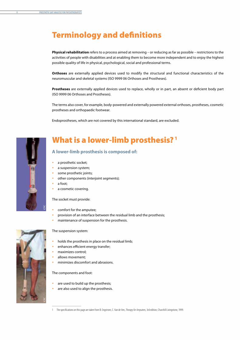
A lower-limb prosthesis is composed of:
y a prosthetic socket; y a suspension system; y some prosthetic joints; y other components (interjoint segments); y a foot; y a cosmetic covering.
The socket must provide:
y comfort for the amputee; y provision of an interface between the residual limb and the prosthesis; y maintenance of suspension for the prosthesis.
The suspension system:
y holds the prosthesis in place on the residual limb; y enhances efficient energy transfer; y maximizes control; y allows movement; y minimizes discomfort and abrasions.
The components and foot:
y are used to build up the prosthesis; y are also used to align the prosthesis.
1 The specifications on this page are taken from B. Engstrom, C. Van de Ven, Therapy for Amputees, 3rd edition, Churchill Livingstone, 1999.
ICRC
ICRC
lower-limb amPutations and General Prosthetic knowledGe 9
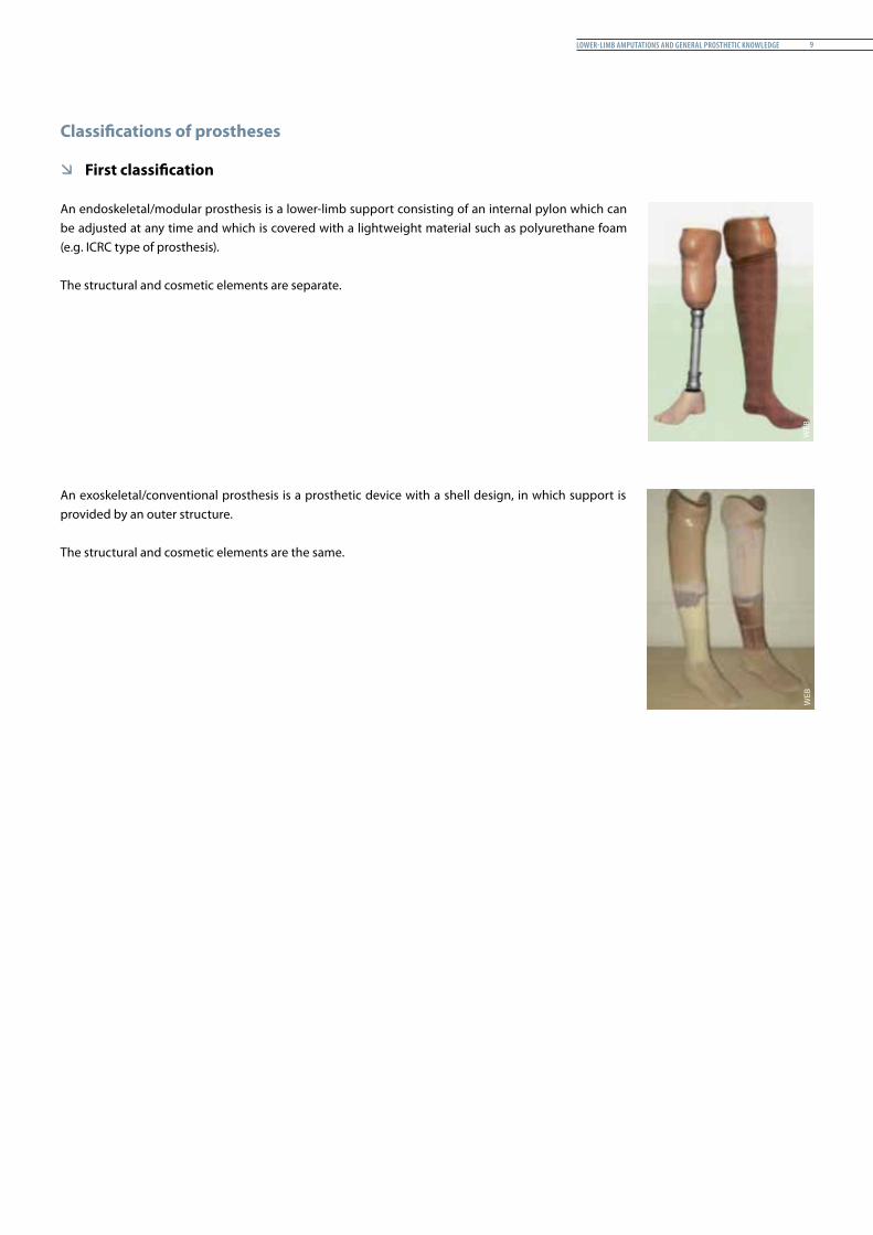
Classifications of prostheses
Ì First classification
An endoskeletal/modular prosthesis is a lower-limb support consisting of an internal pylon which can be adjusted at any time and which is covered with a lightweight material such as polyurethane foam (e.g. ICRC type of prosthesis).
The structural and cosmetic elements are separate.
An exoskeletal/conventional prosthesis is a prosthetic device with a shell design, in which support is provided by an outer structure.
The structural and cosmetic elements are the same.
WEB
WEB
Prosthetic Gait analysis for PhysiotheraPists10
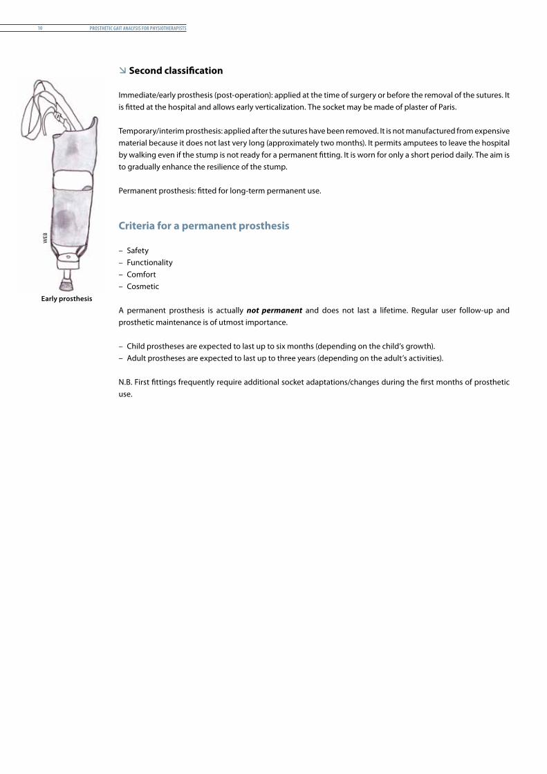
Ì Second classification
Immediate/early prosthesis (post-operation): applied at the time of surgery or before the removal of the sutures. It is fitted at the hospital and allows early verticalization. The socket may be made of plaster of Paris.
Temporary/interim prosthesis: applied after the sutures have been removed. It is not manufactured from expensive material because it does not last very long (approximately two months). It permits amputees to leave the hospital by walking even if the stump is not ready for a permanent fitting. It is worn for only a short period daily. The aim is to gradually enhance the resilience of the stump.
Permanent prosthesis: fitted for long-term permanent use.
Criteria for a permanent prosthesis
– Safety– Functionality– Comfort– Cosmetic
A permanent prosthesis is actually not permanent and does not last a lifetime. Regular user follow-up and prosthetic maintenance is of utmost importance.
– Child prostheses are expected to last up to six months (depending on the child’s growth).– Adult prostheses are expected to last up to three years (depending on the adult’s activities).
N.B. First fittings frequently require additional socket adaptations/changes during the first months of prosthetic use.
Early prosthesis
WEB
lower-limb amPutations and General Prosthetic knowledGe 11
General points about lower-limb amputations
Surgery
General indications for amputation in trauma and war-related situations2
Amputation is generally indicated when there is:– severe damage, with mangled, grossly contaminated wounds;– overwhelming infection;– established gangrene;– continued infection associated with severe nerve and bone injury;– secondary haemorrhage, uncontrollable by other measures;– multiple injuries, where amputation is the fastest means of saving life.
Conservation of the knee joint is of great benefit because of the greater function of a limb with a normal knee. This is particularly important when both lower limbs are injured.
Level of amputation
The level of amputation should be at the lowest possible level of viable tissue. Good viable skin and soft tissue distal to the point of bone division should be saved for use in subsequent stump closure.
Guillotine amputation should not be performed. Long posterior flaps of skin, fascia and obliquely dissected muscles give a much better stump.
Formal amputation should ideally be performed at the site of election decided in conjunction with the physiotherapist or the prosthetist.
Considerations influencing prosthetic management:
y The longer the stump, the longer the lever arm and consequently the control of movement;
y The more intact tissue and skin area, the better the distribution of pressure; y The quality of the stump is more important than its length.
The ideal sites of election for amputations are:
– tibia: 12-14 cm from the tibial tuberosity;– knee disarticulation to minimize surgical trauma in weak patients;– femur: 25-28 cm from the top of the greater trochanter.
Short stumps are always difficult to fit and, due to muscular imbalance, often develop contracture.
2 The information in this section is based on D. Dufour et al., Surgery for Victims of War, 3rd edition, ICRC, Geneva, 1998.
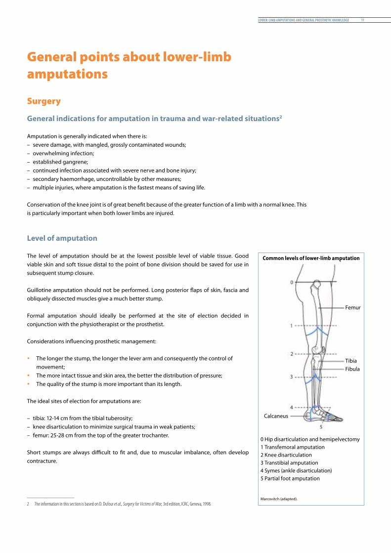
0 Hip disarticulation and hemipelvectomy1 Transfemoral amputation2 Knee disarticulation3 Transtibial amputation4 Symes (ankle disarticulation)5 Partial foot amputation
Marcovitch (adapted).
Femur
Common levels of lower-limb amputation
TibiaFibula
Calcaneus

Prosthetic Gait analysis for PhysiotheraPists12
The minimum length for a tibial stump is 5 cm.
In a short transtibial (TT) amputation (at the level of the tibial tubercle) the fibular head should be removed, otherwise prosthetic use may cause pain. A short TT stump does not provide for adequate control or mobility.
Very short stump (at the level of the tibial tubercle)
Short stump (1∕3 proximal):– supports the most weight (up to 60%)
Medium-length stump (1∕3 medial):– most adequate– best muscle action
Long stump (1∕3 distal):– best lever
The minimum length for a femoral stump is 10 cm.
Long stumps enhance better control of the prosthetic knee unit. Moreover, the shorter the stump, the higher the energy needed to walk.
On the other hand, if the stump is too long it might affect the correct placement of the prosthetic knee and therefore influence the construction/alignment of the prosthesis.
(a) Long stump (1∕3 distal)– best lever– better muscular balance while preserving
the strength of the adductor– energy efficient
(b) Medium-length stump (1∕3 medial)– reduced strength of the adductors– increased flexion and abduction– increased energy expenditure
(c) Short stump (1∕3 proximal)– weak adductor muscle, causing severe imbalance– position of the stump in flexion and abduction– causes massive energy expenditure (effort)
Short TT stump
Short TF stump
Medium TF stumpR. B
aum
gart
ner/
P. B
otta
ICRC
T. B
lum
/VIR
TCO
TW
EB
lower-limb amPutations and General Prosthetic knowledGe 13
Surgical procedure
Amputations for victims of war (open amputations)In the primary amputation, as much bone and soft tissue as possible are conserved. Standard flaps may not be possible and “flaps of opportunity” may have to be made. If the wound/stump is contaminated, delayed primary closure (DPC) is required in war surgery. In DPC, the fascia should be closed over the bone to make sure that there is a mobile flap of skin over subcutaneous fat at the end of the stump. It is important that DPC is not delayed as skin retraction occurs rapidly; if performed within 5-7 days of the injury, this is not a problem. Skin grafting may be necessary if the flaps retract. DPC must never be performed over pus. However, the presence of fibrin will not adversely affect healing.
Standard amputations (closed amputations)In standard amputations, skin flaps should be cut longer than the thickness of the limb, from the level of bone section.
The following guidelines are based on experience:– fascia should be cut at the same level as the skin;– muscle should be cut obliquely back to the level of the bone section;– periosteum should not be reflected proximal to the site of bone division;– major blood vessels should be doubly ligated; arteries and veins should be ligated
individually;– nerves should be divided as high as possible without strong traction and should not be
ligated;– the fibula should be cut ~15 mm shorter than the tibia and the sharp edges at the end of
the tibia should be smoothened;– menisci should be removed in knee disarticulation.
y The quality of the stump is more important than its length. y The scar must be painless, flexible and not adherent to underlying tissues. y For appropriate prosthetic management, the stump should be built to tolerate total
distal end contact with the prosthetic socket, which:– improves blood circulation and wound healing;– promotes lymphatic-venous reflux;– minimizes muscle atrophy and phantom limb pain;– increases pressure distribution;– improves proprioception;– reduces sweating.
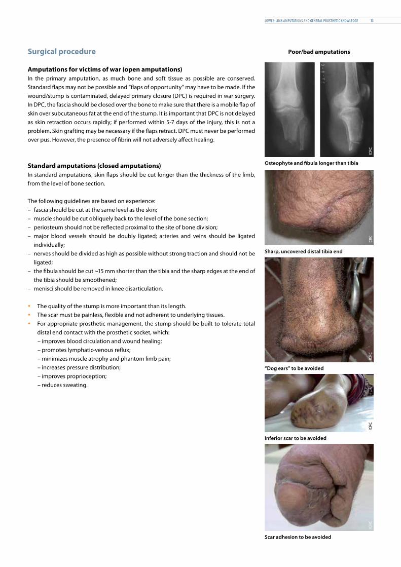
Poor/bad amputations
Osteophyte and fibula longer than tibia
Sharp, uncovered distal tibia end
“Dog ears” to be avoided
Inferior scar to be avoided
Scar adhesion to be avoided
ICRC
ICRC
ICRC
ICRC
ICRC
Prosthetic Gait analysis for PhysiotheraPists14
Recommended surgical procedures for premeditated amputations to endorse adequate prosthetic management
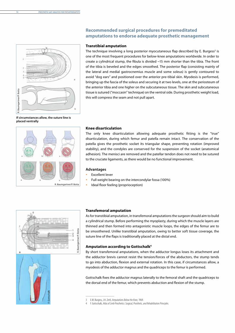
Transtibial amputationThe technique involving a long posterior myocutaneous flap described by E. Burgess3 is one of the most frequent procedures for below-knee amputations worldwide. In order to create a cylindrical stump, the fibula is divided ~15 mm shorter than the tibia. The front of the tibia is beveled and the edges smoothed. The posterior flap (consisting mainly of the lateral and medial gastrocnemius muscle and some soleus) is gently contoured to avoid “dog ears” and positioned over the anterior pre-tibial skin. Myodesis is performed, bringing up the fascia of the soleus and securing it at two levels, one at the periosteum of the anterior tibia and one higher on the subcutaneous tissue. The skin and subcutaneous tissue is sutured (“moccasin” technique) on the ventral side. During prosthetic weight load, this will compress the seam and not pull apart.
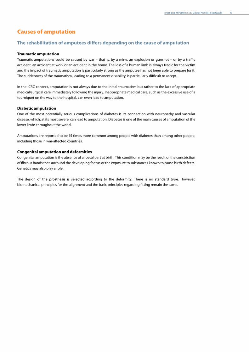
Knee disarticulationThe only knee disarticulation allowing adequate prosthetic fitting is the “true” disarticulation, during which femur and patella remain intact. The conservation of the patella gives the prosthetic socket its triangular shape, preventing rotation (improved stability), and the condyles are conserved for the suspension of the socket (anatomical adhesion). The menisci are removed and the patellar tendon does not need to be sutured to the cruciate ligaments, as there would be no functional improvement.
Advantages y Excellent lever y Full weight bearing on the intercondylar fossa (100%) y Ideal floor feeling (proprioception)
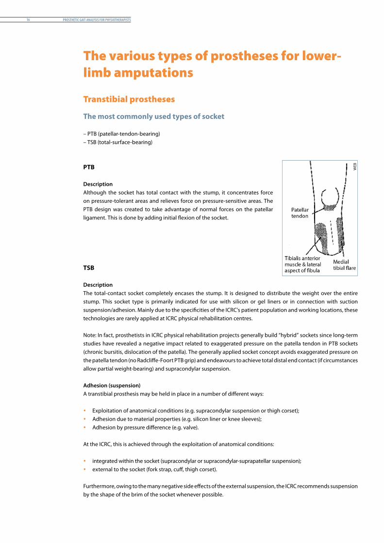
Transfemoral amputationAs for transtibial amputation, in transfemoral amputations the surgeon should aim to build a cylindrical stump. Before performing the myoplasty, during which the muscle layers are thinned and then formed into antagonistic muscle loops, the edges of the femur are to be smoothened. Unlike transtibial amputation, owing to better soft tissue coverage, the suture line of the flaps is traditionally placed at the distal end.
Amputation according to Gottschalk4
By short transfemoral amputations, when the adductor longus loses its attachment and the adductor brevis cannot resist the tension/forces of the abductors, the stump tends to go into abduction, flexion and external rotation. In this case, if circumstances allow, a myodesis of the adductor magnus and the quadriceps to the femur is performed.
Gottschalk fixes the adductor magnus laterally to the femoral shaft and the quadriceps to the dorsal end of the femur, which prevents abduction and flexion of the stump.
3 E.M. Burgess, J.H. Zettl, Amputations Below the Knee, 1969.4 F. Gottschalk, Atlas of Limb Prosthetics: Surgical, Prosthetic, and Rehabilitation Principles.
If circumstances allow, the suture line is placed ventrally
R. B
aum
gart
ner/
P. B
otta
R. B
aum
gart
ner/
P. B
otta
R. Baumgartner/P. Botta
F. G
otts
chal
k
lower-limb amPutations and General Prosthetic knowledGe 15
Causes of amputation
The rehabilitation of amputees differs depending on the cause of amputation
Traumatic amputation Traumatic amputations could be caused by war – that is, by a mine, an explosion or gunshot – or by a traffic accident, an accident at work or an accident in the home. The loss of a human limb is always tragic for the victim and the impact of traumatic amputation is particularly strong as the amputee has not been able to prepare for it. The suddenness of the traumatism, leading to a permanent disability, is particularly difficult to accept.
In the ICRC context, amputation is not always due to the initial traumatism but rather to the lack of appropriate medical/surgical care immediately following the injury. Inappropriate medical care, such as the excessive use of a tourniquet on the way to the hospital, can even lead to amputation. Diabetic amputationOne of the most potentially serious complications of diabetes is its connection with neuropathy and vascular disease, which, at its most severe, can lead to amputation. Diabetes is one of the main causes of amputation of the lower limbs throughout the world.
Amputations are reported to be 15 times more common among people with diabetes than among other people, including those in war-affected countries.
Congenital amputation and deformities Congenital amputation is the absence of a foetal part at birth. This condition may be the result of the constriction of fibrous bands that surround the developing foetus or the exposure to substances known to cause birth defects. Genetics may also play a role.
The design of the prosthesis is selected according to the deformity. There is no standard type. However, biomechanical principles for the alignment and the basic principles regarding fitting remain the same.
Prosthetic Gait analysis for PhysiotheraPists16
The various types of prostheses for lower-limb amputations
Transtibial prostheses
The most commonly used types of socket
– PTB (patellar-tendon-bearing)– TSB (total-surface-bearing)
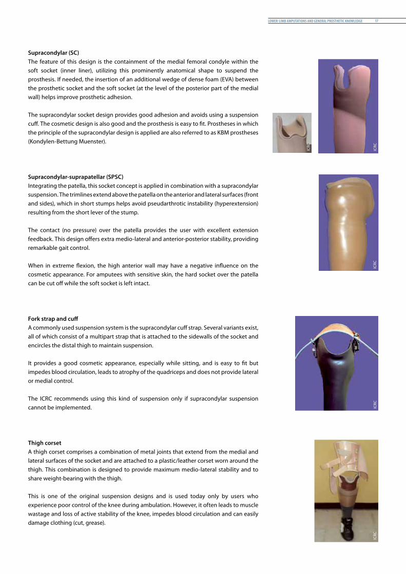
PTB
DescriptionAlthough the socket has total contact with the stump, it concentrates force on pressure-tolerant areas and relieves force on pressure-sensitive areas. The PTB design was created to take advantage of normal forces on the patellar ligament. This is done by adding initial flexion of the socket.
TSB
DescriptionThe total-contact socket completely encases the stump. It is designed to distribute the weight over the entire stump. This socket type is primarily indicated for use with silicon or gel liners or in connection with suction suspension/adhesion. Mainly due to the specificities of the ICRC’s patient population and working locations, these technologies are rarely applied at ICRC physical rehabilitation centres.
Note: In fact, prosthetists in ICRC physical rehabilitation projects generally build “hybrid” sockets since long-term studies have revealed a negative impact related to exaggerated pressure on the patella tendon in PTB sockets (chronic bursitis, dislocation of the patella). The generally applied socket concept avoids exaggerated pressure on the patella tendon (no Radcliffe-Foort PTB grip) and endeavours to achieve total distal end contact (if circumstances allow partial weight-bearing) and supracondylar suspension. Adhesion (suspension) A transtibial prosthesis may be held in place in a number of different ways:
y Exploitation of anatomical conditions (e.g. supracondylar suspension or thigh corset); y Adhesion due to material properties (e.g. silicon liner or knee sleeves); y Adhesion by pressure difference (e.g. valve).
At the ICRC, this is achieved through the exploitation of anatomical conditions:
y integrated within the socket (supracondylar or supracondylar-suprapatellar suspension); y external to the socket (fork strap, cuff, thigh corset).
Furthermore, owing to the many negative side effects of the external suspension, the ICRC recommends suspension by the shape of the brim of the socket whenever possible.
WEB
lower-limb amPutations and General Prosthetic knowledGe 17
Supracondylar (SC)The feature of this design is the containment of the medial femoral condyle within the soft socket (inner liner), utilizing this prominently anatomical shape to suspend the prosthesis. If needed, the insertion of an additional wedge of dense foam (EVA) between the prosthetic socket and the soft socket (at the level of the posterior part of the medial wall) helps improve prosthetic adhesion.
The supracondylar socket design provides good adhesion and avoids using a suspension cuff. The cosmetic design is also good and the prosthesis is easy to fit. Prostheses in which the principle of the supracondylar design is applied are also referred to as KBM prostheses (Kondylen-Bettung Muenster).
Supracondylar-suprapatellar (SPSC)Integrating the patella, this socket concept is applied in combination with a supracondylar suspension. The trimlines extend above the patella on the anterior and lateral surfaces (front and sides), which in short stumps helps avoid pseudarthrotic instability (hyperextension) resulting from the short lever of the stump.
The contact (no pressure) over the patella provides the user with excellent extension feedback. This design offers extra medio-lateral and anterior-posterior stability, providing remarkable gait control.
When in extreme flexion, the high anterior wall may have a negative influence on the cosmetic appearance. For amputees with sensitive skin, the hard socket over the patella can be cut off while the soft socket is left intact.
Fork strap and cuffA commonly used suspension system is the supracondylar cuff strap. Several variants exist, all of which consist of a multipart strap that is attached to the sidewalls of the socket and encircles the distal thigh to maintain suspension.
It provides a good cosmetic appearance, especially while sitting, and is easy to fit but impedes blood circulation, leads to atrophy of the quadriceps and does not provide lateral or medial control.
The ICRC recommends using this kind of suspension only if supracondylar suspension cannot be implemented.
Thigh corsetA thigh corset comprises a combination of metal joints that extend from the medial and lateral surfaces of the socket and are attached to a plastic/leather corset worn around the thigh. This combination is designed to provide maximum medio-lateral stability and to share weight-bearing with the thigh.
This is one of the original suspension designs and is used today only by users who experience poor control of the knee during ambulation. However, it often leads to muscle wastage and loss of active stability of the knee, impedes blood circulation and can easily damage clothing (cut, grease).
ICRC
ICRC
ICRC
ICRC
ICRC
Prosthetic Gait analysis for PhysiotheraPists18
Transfemoral prostheses
The most commonly used types of socket
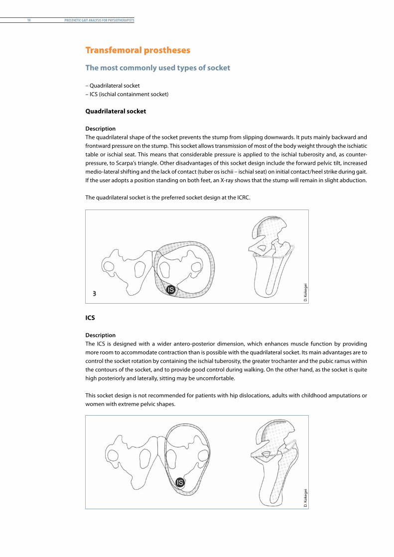
– Quadrilateral socket– ICS (ischial containment socket) Quadrilateral socket
DescriptionThe quadrilateral shape of the socket prevents the stump from slipping downwards. It puts mainly backward and frontward pressure on the stump. This socket allows transmission of most of the body weight through the ischiatic table or ischial seat. This means that considerable pressure is applied to the ischial tuberosity and, as counter-pressure, to Scarpa’s triangle. Other disadvantages of this socket design include the forward pelvic tilt, increased medio-lateral shifting and the lack of contact (tuber os ischii – ischial seat) on initial contact/heel strike during gait. If the user adopts a position standing on both feet, an X-ray shows that the stump will remain in slight abduction.
The quadrilateral socket is the preferred socket design at the ICRC.
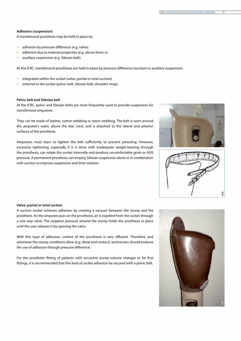
ICS
DescriptionThe ICS is designed with a wider antero-posterior dimension, which enhances muscle function by providing more room to accommodate contraction than is possible with the quadrilateral socket. Its main advantages are to control the socket rotation by containing the ischial tuberosity, the greater trochanter and the pubic ramus within the contours of the socket, and to provide good control during walking. On the other hand, as the socket is quite high posteriorly and laterally, sitting may be uncomfortable.
This socket design is not recommended for patients with hip dislocations, adults with childhood amputations or women with extreme pelvic shapes.
D. K
okeg
eiD
. Kok
egei
lower-limb amPutations and General Prosthetic knowledGe 19
Adhesion (suspension)A transfemoral prosthesis may be held in place by:
y adhesion by pressure difference (e.g. valve); y adhesion due to material properties (e.g. silicon liner); or y auxiliary suspension (e.g. Silesian belt).
At the ICRC, transfemoral prostheses are held in place by pressure difference (suction) or auxiliary suspension:
y integrated within the socket (valve: partial or total suction); y external to the socket (pelvic belt, Silesian belt, shoulder strap).
Pelvic belt and Silesian beltAt the ICRC, pelvic and Silesian belts are most frequently used to provide suspension for transfemoral amputees.
They can be made of leather, cotton webbing or nylon webbing. The belt is worn around the amputee’s waist, above the iliac crest, and is attached to the lateral and anterior surfaces of the prosthesis.
Amputees must learn to tighten the belt sufficiently to prevent pistoning. However, excessive tightening, especially if it is done with inadequate weight-bearing through the prosthesis, can rotate the socket internally and produce uncomfortable groin or ASIS pressure. A permanent prosthesis can employ Silesian suspension alone or in combination with suction to improve suspension and limit rotation.
Valve: partial or total suctionA suction socket achieves adhesion by creating a vacuum between the stump and the prosthesis. As the amputee puts on the prosthesis, air is expelled from the socket through a one-way valve. The negative pressure around the stump holds the prosthesis in place until the user releases it by opening the valve.
With this type of adhesion, control of the prosthesis is very efficient. Therefore, and whenever the stump conditions allow (e.g. distal end contact), technicians should endorse the use of adhesion through pressure difference.
For the prosthetic fitting of patients with recurrent stump-volume changes or for first fittings, it is recommended that this kind of socket adhesion be secured with a pelvic belt.
WEB
ICRC
ICRC
Prosthetic Gait analysis for PhysiotheraPists20
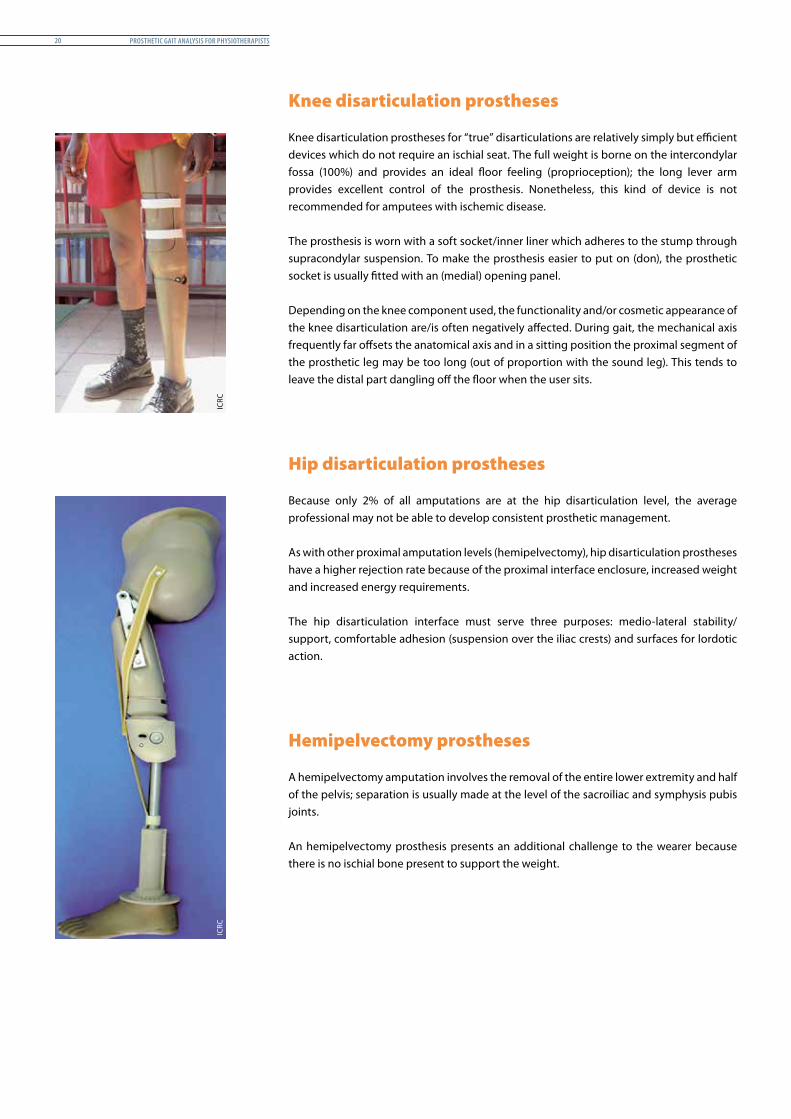
Knee disarticulation prostheses
Knee disarticulation prostheses for “true” disarticulations are relatively simply but efficient devices which do not require an ischial seat. The full weight is borne on the intercondylar fossa (100%) and provides an ideal floor feeling (proprioception); the long lever arm provides excellent control of the prosthesis. Nonetheless, this kind of device is not recommended for amputees with ischemic disease.
The prosthesis is worn with a soft socket/inner liner which adheres to the stump through supracondylar suspension. To make the prosthesis easier to put on (don), the prosthetic socket is usually fitted with an (medial) opening panel.
Depending on the knee component used, the functionality and/or cosmetic appearance of the knee disarticulation are/is often negatively affected. During gait, the mechanical axis frequently far offsets the anatomical axis and in a sitting position the proximal segment of the prosthetic leg may be too long (out of proportion with the sound leg). This tends to leave the distal part dangling off the floor when the user sits.
Hip disarticulation prostheses
Because only 2% of all amputations are at the hip disarticulation level, the average professional may not be able to develop consistent prosthetic management.
As with other proximal amputation levels (hemipelvectomy), hip disarticulation prostheses have a higher rejection rate because of the proximal interface enclosure, increased weight and increased energy requirements.
The hip disarticulation interface must serve three purposes: medio-lateral stability/support, comfortable adhesion (suspension over the iliac crests) and surfaces for lordotic action.
Hemipelvectomy prostheses
A hemipelvectomy amputation involves the removal of the entire lower extremity and half of the pelvis; separation is usually made at the level of the sacroiliac and symphysis pubis joints.
An hemipelvectomy prosthesis presents an additional challenge to the wearer because there is no ischial bone present to support the weight.
ICRC
ICRC
lower-limb amPutations and General Prosthetic knowledGe 21
Symes prostheses
Because of the unique aspects of the ankle-joint disarticulation, Symes prostheses are challenging to manufacture. Aspects that are beneficial for the user are the intact tibia and fibula, which create a long lever for excellent control of the prosthesis.
Depending on the stump conditions, amputees should benefit from full distal end contact and full weight-bearing (100%) and hence an ideal floor feeling (proprioception). To relieve pressure on sensitive stump ends, the proximal brim design can be made in accordance with the principles of a PTB (patellar-tendon-bearing) prosthesis.
Because of the short space between the end of the stump and the floor, usually a special foot, a modified SACH foot, has to be used. Prosthetic adhesion is granted through the anatomical conditions of the distal stump end (supramaleolar suspension).
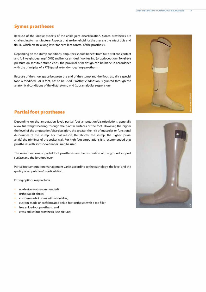
Partial foot prostheses
Depending on the amputation level, partial foot amputation/disarticulations generally allow full weight-bearing through the plantar surfaces of the foot. However, the higher the level of the amputation/disarticulation, the greater the risk of muscular or functional deformities of the stump. For that reason, the shorter the stump, the higher (cross-ankle) the trimlines of the socket wall. For high foot amputations it is recommended that prostheses with soft socket (inner liner) be used.
The main functions of partial foot prostheses are the restoration of the ground support surface and the forefoot lever.
Partial foot amputation management varies according to the pathology, the level and the quality of amputation/disarticulation.
Fitting options may include:
y no device (not recommended); y orthopaedic shoes; y custom-made insoles with a toe filler; y custom-made or prefabricated ankle-foot orthoses with a toe filler; y free ankle-foot prosthesis; and y cross-ankle foot prosthesis (see picture).
Rech
stei
ner/
VIET
COT
ICRC

Prosthetic Gait analysis for PhysiotheraPists22
PolyProPylene technoloGy 23
PolyProPylene technoloGy
Introduction
Content
This chapter provides details of the polypropylene technology which is indigenously developed and applied by the ICRC in the manufacture of prosthetic and orthotic devices. It covers the raw materials, their quality and the parts of the devices (in this case lower-limb prostheses). It also looks at the principles of alignment for transtibial and transfemoral prostheses.
Rationale
Experts working in the field of physiotherapy and physical rehabilitation may be aware of different technologies used in the manufacture of orthopaedic devices. This chapter focuses on polypropylene technology and draws the readers’ attention to the ICRC context, taking account of the fact that they have previous knowledge in the field, most probably using other technologies.
Prosthetic Gait analysis for PhysiotheraPists24
Raw materials and orthopaedic components
The ICRC technology – polypropylene technology – uses PP (polypropylene) and EVA (ethylene vinyl acetate).
Raw materials
PP and EVA are thermoplastics, meaning plastic materials which, when subjected to heat, become plastically formable and when cooled down again, regain solidity and can bear weight.
Thermoplastics processing involves three stages:1. The material is first heated enough to soften it.2. It is forced into the desired shape.3. It is left to cool while it is still held in its new shape.
PP
Specificities y PP is a thermoplastic; y It softens when heated to 180°C (324°F); y It melts when heated above 200°C (360°F); y It is light (floats on water); y It is difficult to glue; y It is easy to weld; y It is elastic and rigid at the same time; y PP for P&O is available in plates of different thickness.
Note: The PP used by the ICRC is coloured. The colouring provides better protection against brittleness when excessively exposed to UV radiation and provides a better cosmetic appearance.
Advantages y Fairly inexpensive; y Long shelf life (providing it is kept out of direct sunlight); y No restrictions with regard to transport; y Allows a reduction in the number of different material items, thereby making stock management less
complicated and costly; y Compared to polyester laminated prosthetic sockets, PP sockets are easier and quicker to manufacture; y Grinding dust is non-hazardous; y One of the most easily recyclable plastics.
DisadvantagesThe ICRC is not aware of any disadvantages related to the characteristics of PP. The rare difficulties encountered tend to be related to poor craftsmanship or inappropriate handling of the material due to the lack of proper equipment.
Some of the advantages can become disadvantages if the material is not handled correctly.
PolyProPylene technoloGy 25
EVA
Specificities y EVA foam is an expanded copolymer (foam); y EVA has a closed cell structure that slowly reverts into its original shape after strong compression; y EVA allows excellent gluing; y EVA for prostheses and orthoses is sold in plates of different thickness.
The ICRC has been using EVA foam for the manufacturing of: y soft sockets (inner liner) for TT prostheses; y prosthetic feet production; y cosmetic calves; y press vacuum moulding of hands and feet.
Advantages y The closed cell structure does not permit moisture penetration (less apt to rot or increase in weight); y Optimal weight/strength relationship; y No toxic reaction by the skin; y Excellent for thermoforming of soft sockets and cosmetic calves; y No restrictions with regard to transport: y Grinding dust is non-hazardous.
Disadvantages y Rather more expensive than PP
Important y Store PP and EVA in a dark, dry, dust-free and ventilated room (no need for air-conditioning). y Inspect regularly for signs of deterioration. y Store PP and EVA sheets horizontally, allowing them to keep their initial shape. y Do not expose PP and EVA to extreme light (UV radiation). y Do not store EVA in a humid or unventilated place. y Do not clean PP with solvents or other household cleaning products, unless the long-term effect on polymers
is known and the material characteristics are not affected.
Orthopaedic components
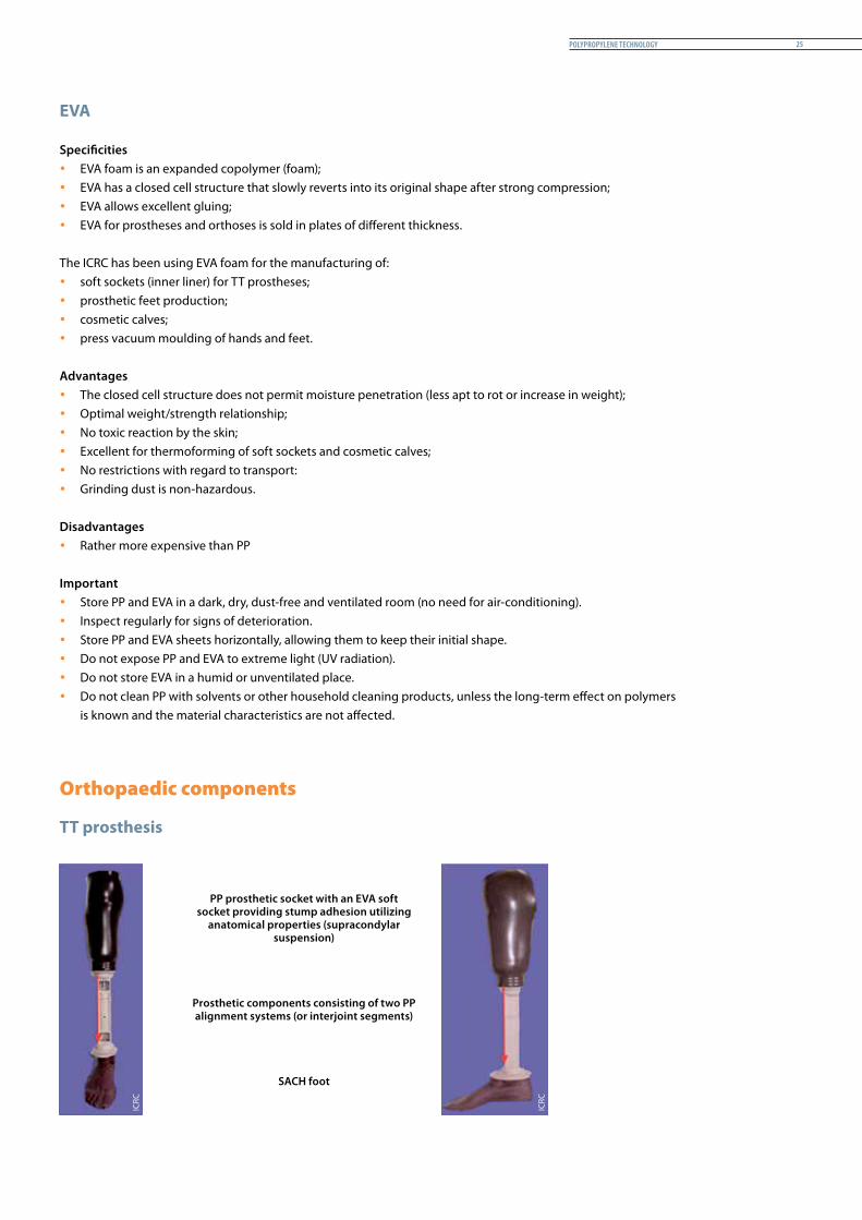
TT prosthesis
PP prosthetic socket with an EVA soft socket providing stump adhesion utilizing
anatomical properties (supracondylar suspension)
Prosthetic components consisting of two PP alignment systems (or interjoint segments)
SACH foot
ICRC
ICRC
Prosthetic Gait analysis for PhysiotheraPists26
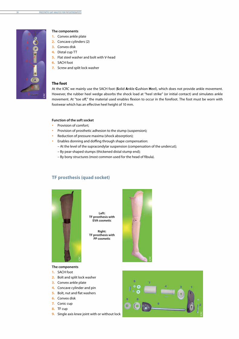
The components1. Convex ankle plate2. Concave cylinders (2)3. Convex disk4. Distal cup TT5. Flat steel washer and bolt with V-head6. SACH foot7. Screw and split lock washer
The footAt the ICRC we mainly use the SACH foot (Solid Ankle Cushion Heel), which does not provide ankle movement. However, the rubber heel wedge absorbs the shock load at “heel strike” (or initial contact) and simulates ankle movement. At “toe off,” the material used enables flexion to occur in the forefoot. The foot must be worn with footwear which has an effective heel height of 10 mm.
Function of the soft socket y Provision of comfort; y Provision of prosthetic adhesion to the stump (suspension); y Reduction of pressure maxima (shock absorption); y Enables donning and doffing through shape compensation:
– At the level of the supracondylar suspension (compensation of the undercut);– By pear-shaped stumps (thickened distal stump end);– By bony structures (most common used for the head of fibula).
TF prosthesis (quad socket)
The components1. SACH foot2. Bolt and split lock washer3. Convex ankle plate4. Concave cylinder and pin5. Bolt, nut and flat washers6. Convex disk7. Conic cup8. TF cup9. Single axis knee joint with or without lock
Left: TF prosthesis with
EVA cosmetic
Right: TF prosthesis with
PP cosmetic
ICRC
ICRC
ICRC
ICRC
PolyProPylene technoloGy 27
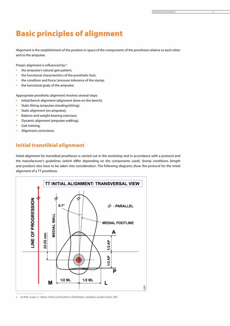
Basic principles of alignment
Alignment is the establishment of the position in space of the components of the prosthesis relative to each other and to the amputee.
Proper alignment is influenced by 5:1
y the amputee’s natural gait pattern; y the functional characteristics of the prosthetic foot; y the condition and force/ pressure tolerance of the stump; y the functional goals of the amputee.
Appropriate prosthetic alignment involves several steps: y Initial/bench alignment (alignment done on the bench); y Static fitting (amputee standing/sitting); y Static alignment (on amputee); y Balance and weight-bearing exercises; y Dynamic alignment (amputee walking); y Gait training; y Alignment corrections.
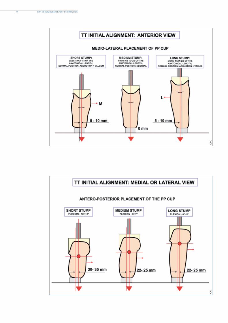
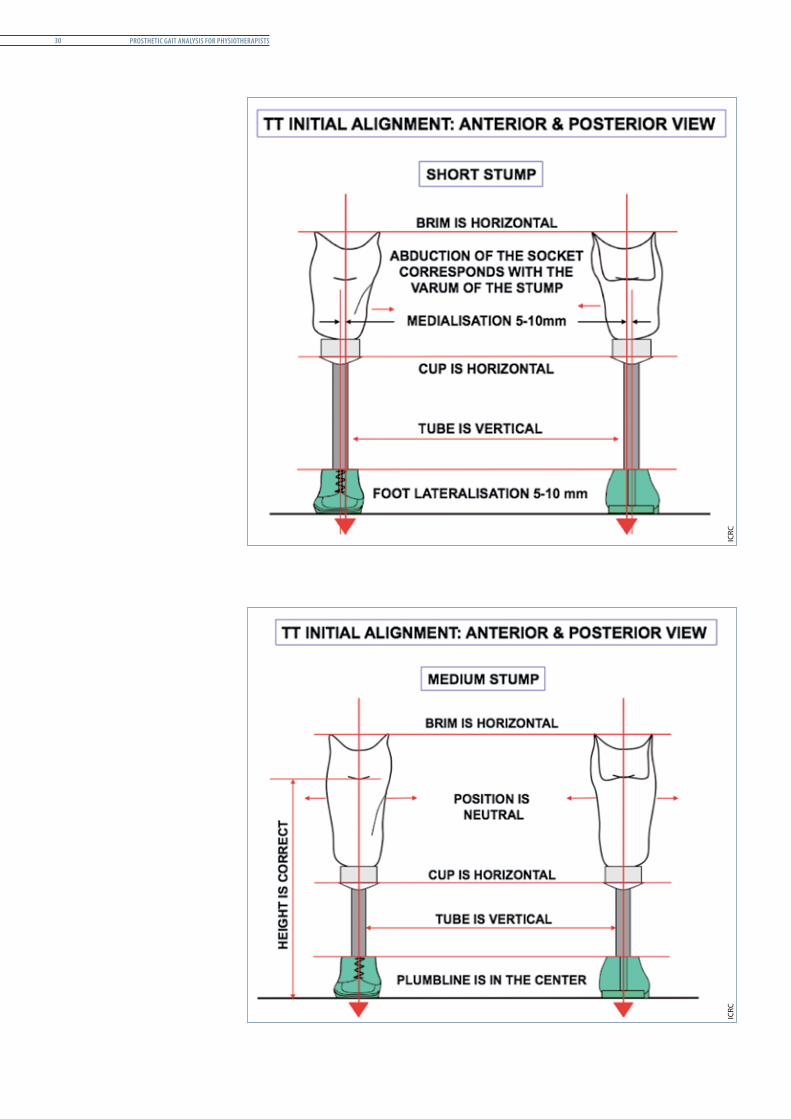
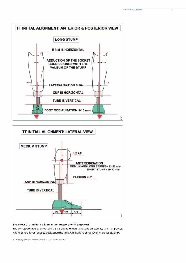
Initial transtibial alignment
Initial alignment for transtibial prostheses is carried out in the workshop and in accordance with a protocol and the manufacturer’s guidelines (which differ depending on the components used). Stump conditions (length and position) also have to be taken into consideration. The following diagrams show the protocol for the initial alignment of a TT prosthesis.
5 See M.M. Lusardi, C.C. Nielsen, Orthotics and Prosthetics in Rehabilitation, 2nd edition, Saunders Elsevier, 2007.
ICRC
Prosthetic Gait analysis for PhysiotheraPists28
ICRC
ICRC
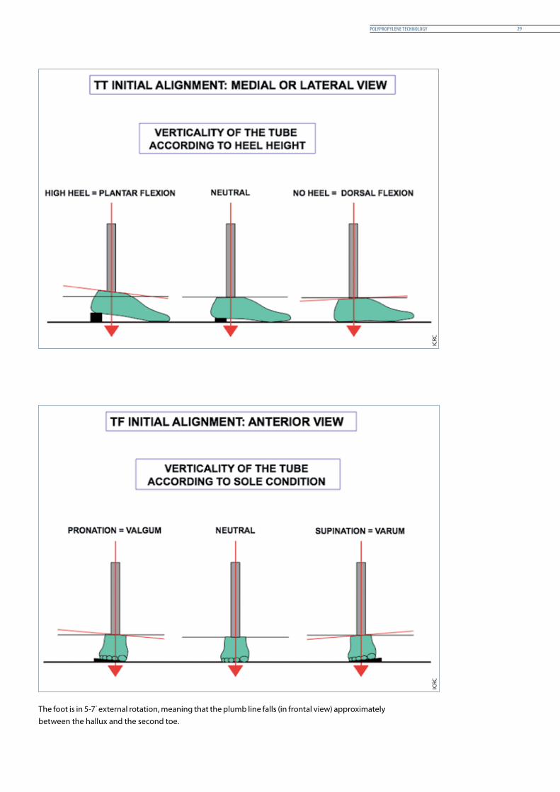
PolyProPylene technoloGy 29
The foot is in 5-7° external rotation, meaning that the plumb line falls (in frontal view) approximately between the hallux and the second toe.
ICRC
ICRC
Prosthetic Gait analysis for PhysiotheraPists30
ICRC
ICRC
PolyProPylene technoloGy 31
The effect of prosthetic alignment on support for TT amputees61The concept of heel and toe levers is helpful to understand support stability in TT amputees. A longer heel lever tends to destabilize the limb, while a longer toe lever improves stability.
6 C. Kirtley, Clinical Gait Analysis, Churchill Livingstone Elsevier, 2006.
ICRC
ICRC
Prosthetic Gait analysis for PhysiotheraPists32
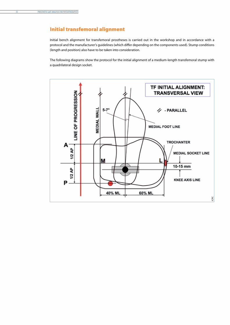
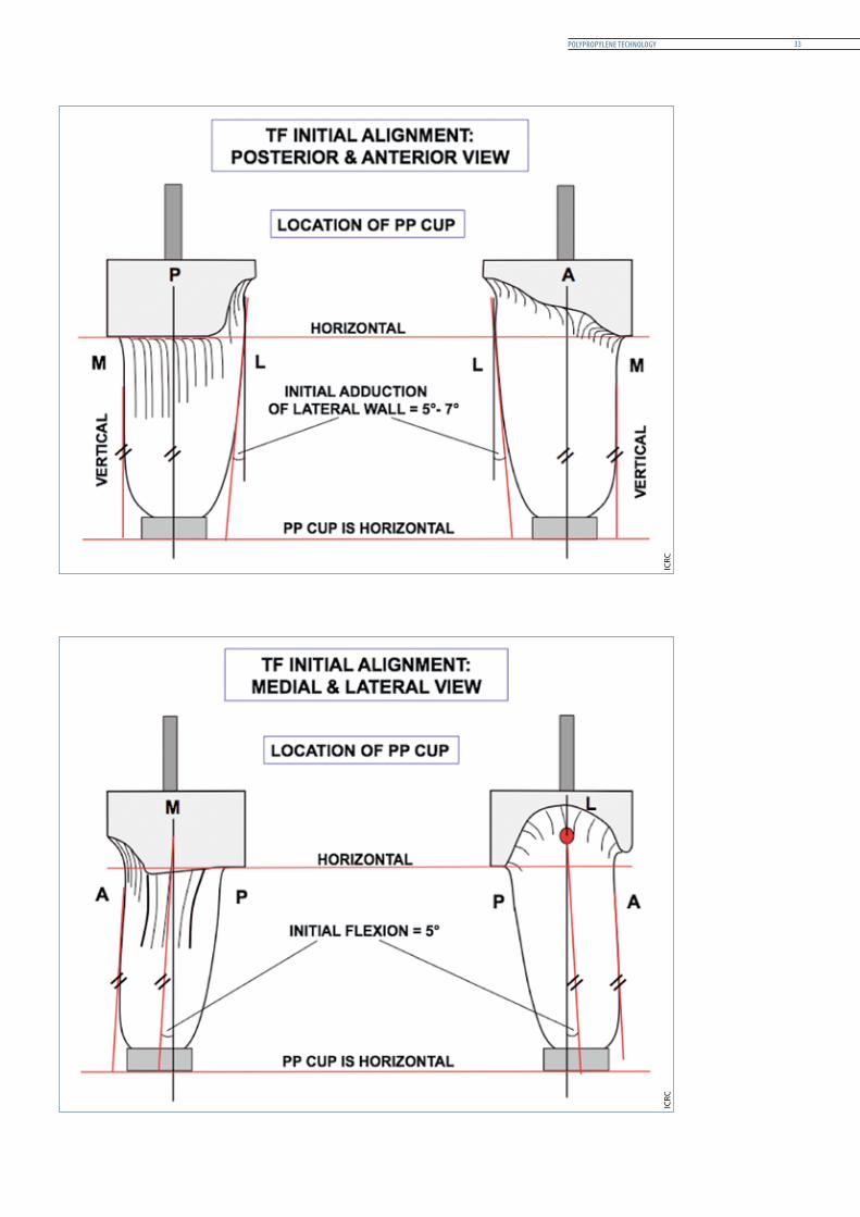
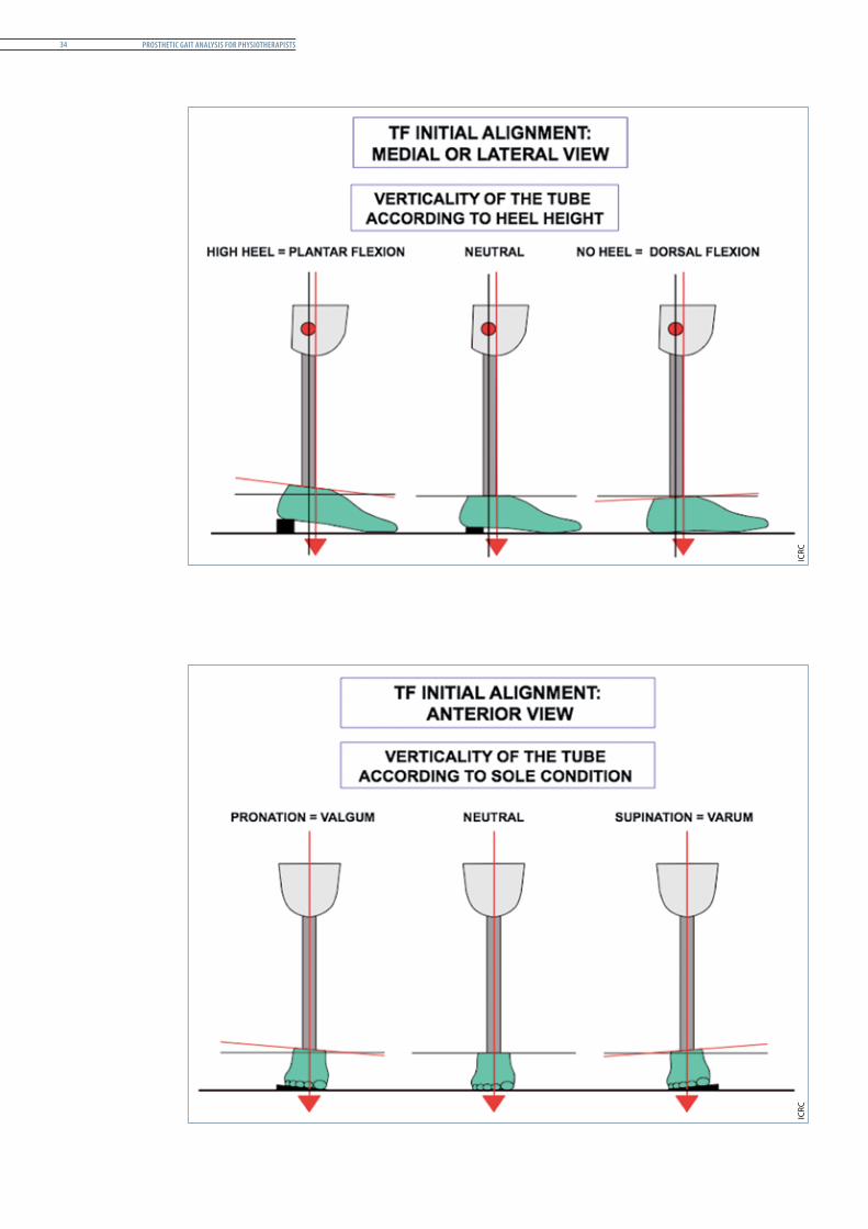
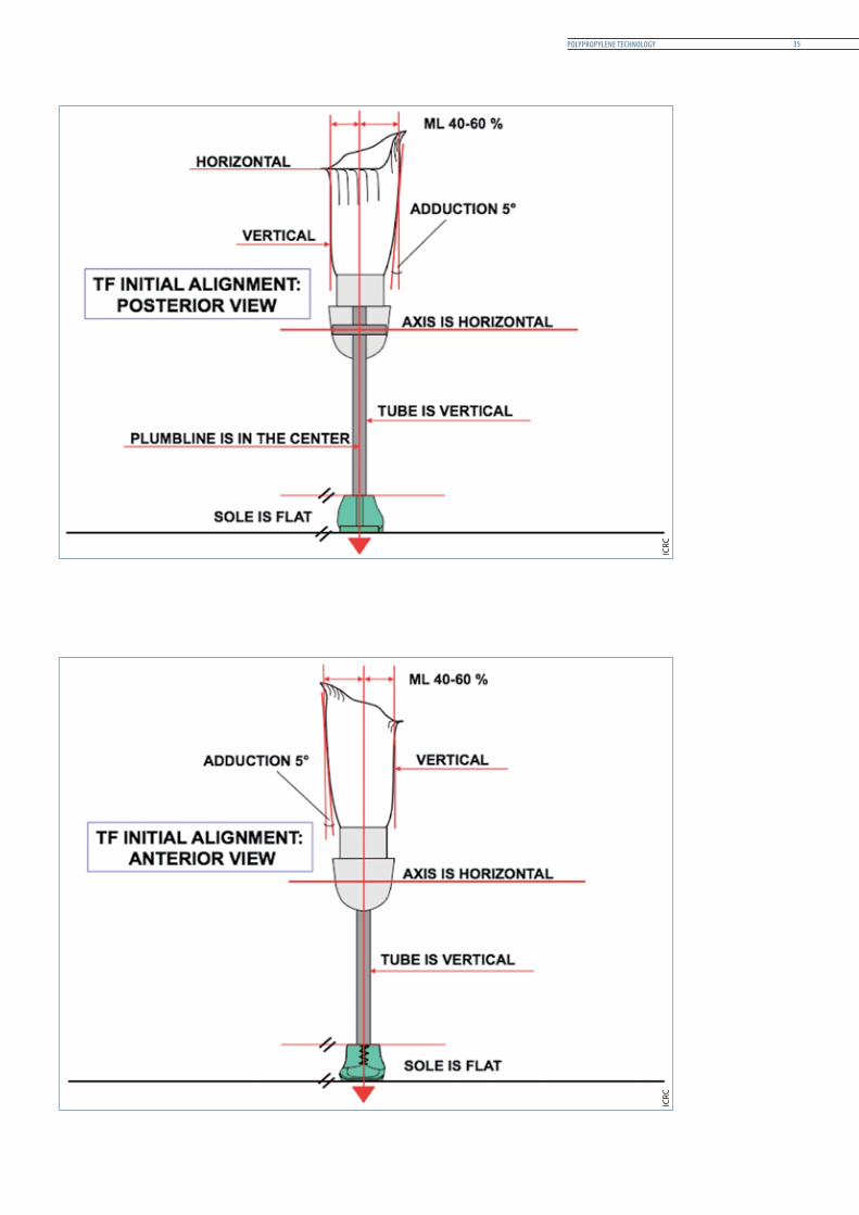
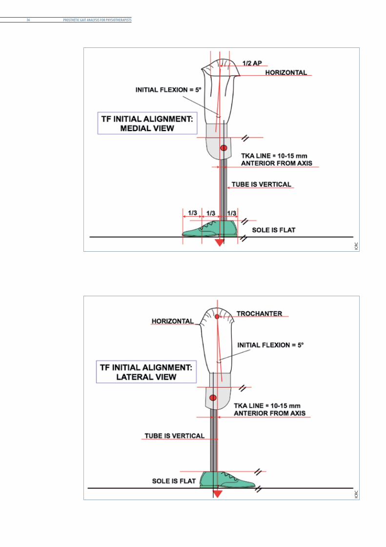
Initial transfemoral alignment
Initial bench alignment for transfemoral prostheses is carried out in the workshop and in accordance with a protocol and the manufacturer’s guidelines (which differ depending on the components used). Stump conditions (length and position) also have to be taken into consideration.
The following diagrams show the protocol for the initial alignment of a medium-length transfemoral stump with a quadrilateral design socket.
ICRC
PolyProPylene technoloGy 33
ICRC
ICRC
Prosthetic Gait analysis for PhysiotheraPists34
ICRC
ICRC
PolyProPylene technoloGy 35
ICRC
ICRC
Prosthetic Gait analysis for PhysiotheraPists36
ICRC
ICRC
PolyProPylene technoloGy 37
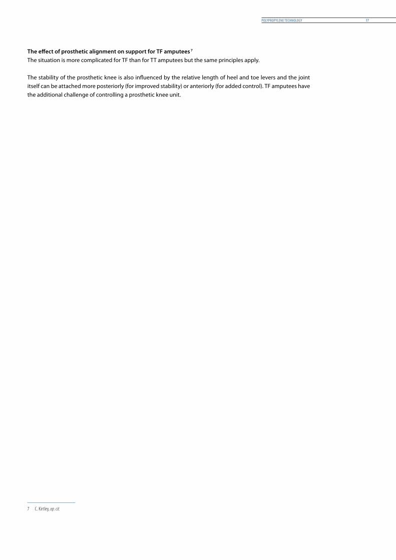
The effect of prosthetic alignment on support for TF amputees 72The situation is more complicated for TF than for TT amputees but the same principles apply.
The stability of the prosthetic knee is also influenced by the relative length of heel and toe levers and the joint itself can be attached more posteriorly (for improved stability) or anteriorly (for added control). TF amputees have the additional challenge of controlling a prosthetic knee unit.
7 C. Kirtley, op. cit.

Prosthetic Gait analysis for PhysiotheraPists38
clinical decisions and PrescriPtions 39
clinical decisions and PrescriPtions
Introduction
Content
This chapter recalls the main points relating to prosthetic fitting and clinical decisions concerning lower-limb amputations following the clinical assessment.
Rationale
This chapter discusses the unique situations that might arise in the field, which could be more challenging than in usual cases. Although the focus is on the ICRC’s working environment, the matter of providing rehabilitation for people with amputations that have not been the result of conflict situations is also addressed.
Although it has not proved possible to establish a general rule for the exact prescription of a device, its type and description, attempts are made in this chapter to give guidelines for those working in this field.
Specific clinical cases and the appropriate prosthetic prescription are covered to a certain extent in the section of this chapter entitled “Specific considerations.”
Prosthetic Gait analysis for PhysiotheraPists40
General considerations
If an early fitting is physically and psychologically beneficial to the user, no general rule needs to be applied when deciding to fit the person with a prosthesis. Nevertheless, some points should be borne in mind in the clinical decision-making process. These are outlined below.
The amputee’s motivation
The amputee is part of the decision-making process but it is not sufficient merely to want a prosthesis. Moreover, the amputee’s family may insist that he or she be fitted with a prosthesis but family motivation does not translate directly into user motivation.
Energy expenditure
Many people are unaware of the physiological demands of prosthetic ambulation (see “Condition of the stump – energy expenditure,” p. 77).
Level of amputation
Amputees exhibit a decrease in walking speed and walking efficiency commensurate with the level of amputation. The higher the level of amputation, the heavier the prosthesis will be.
Specific considerations
Stump conditions
Ideal stump conditions
The conditions of the amputation and – just as important – lack of care after the amputation could lead to problems that delay or even rule out the fitting of a prosthesis.
The ideal stump should be
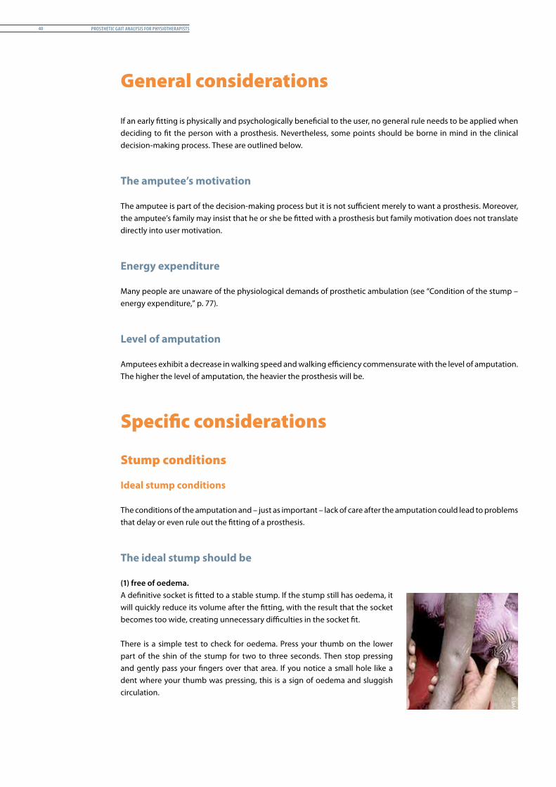
(1) free of oedema.A definitive socket is fitted to a stable stump. If the stump still has oedema, it will quickly reduce its volume after the fitting, with the result that the socket becomes too wide, creating unnecessary difficulties in the socket fit.
There is a simple test to check for oedema. Press your thumb on the lower part of the shin of the stump for two to three seconds. Then stop pressing and gently pass your fingers over that area. If you notice a small hole like a dent where your thumb was pressing, this is a sign of oedema and sluggish circulation. W
EB
clinical decisions and PrescriPtions 41
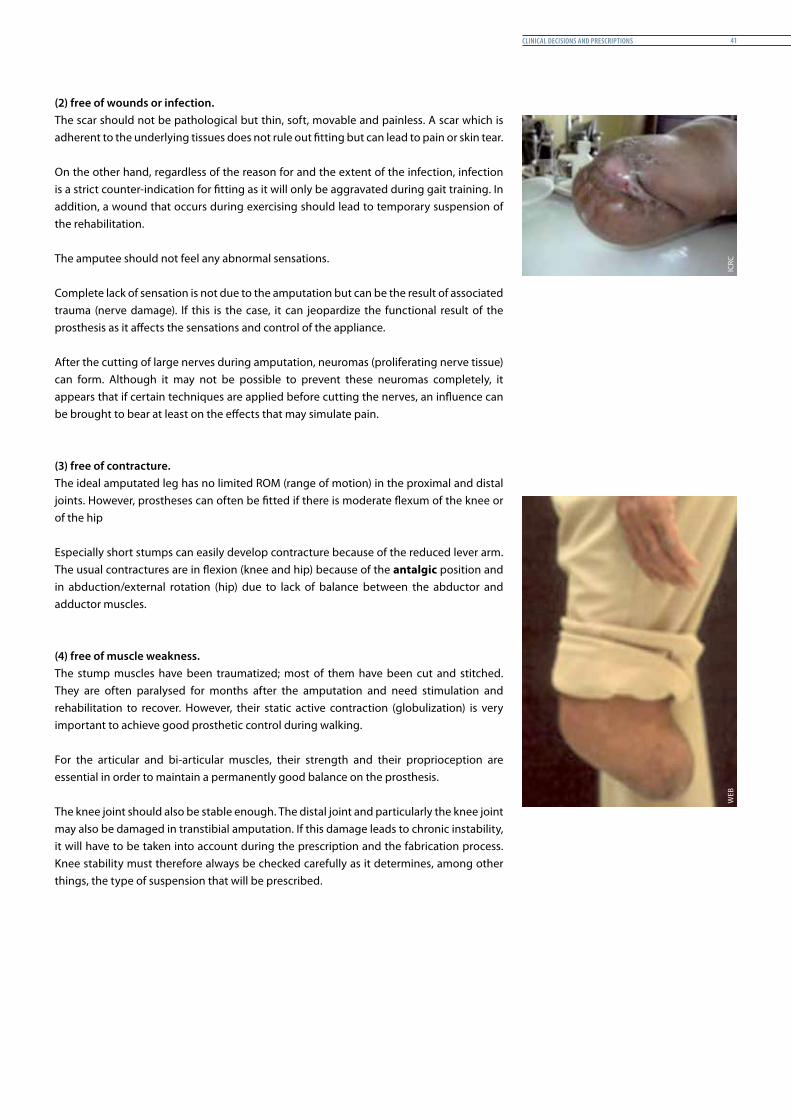
(2) free of wounds or infection.The scar should not be pathological but thin, soft, movable and painless. A scar which is adherent to the underlying tissues does not rule out fitting but can lead to pain or skin tear.
On the other hand, regardless of the reason for and the extent of the infection, infection is a strict counter-indication for fitting as it will only be aggravated during gait training. In addition, a wound that occurs during exercising should lead to temporary suspension of the rehabilitation.
The amputee should not feel any abnormal sensations.
Complete lack of sensation is not due to the amputation but can be the result of associated trauma (nerve damage). If this is the case, it can jeopardize the functional result of the prosthesis as it affects the sensations and control of the appliance.
After the cutting of large nerves during amputation, neuromas (proliferating nerve tissue) can form. Although it may not be possible to prevent these neuromas completely, it appears that if certain techniques are applied before cutting the nerves, an influence can be brought to bear at least on the effects that may simulate pain.
(3) free of contracture.The ideal amputated leg has no limited ROM (range of motion) in the proximal and distal joints. However, prostheses can often be fitted if there is moderate flexum of the knee or of the hip
Especially short stumps can easily develop contracture because of the reduced lever arm. The usual contractures are in flexion (knee and hip) because of the antalgic position and in abduction/external rotation (hip) due to lack of balance between the abductor and adductor muscles.
(4) free of muscle weakness.The stump muscles have been traumatized; most of them have been cut and stitched. They are often paralysed for months after the amputation and need stimulation and rehabilitation to recover. However, their static active contraction (globulization) is very important to achieve good prosthetic control during walking.
For the articular and bi-articular muscles, their strength and their proprioception are essential in order to maintain a permanently good balance on the prosthesis.
The knee joint should also be stable enough. The distal joint and particularly the knee joint may also be damaged in transtibial amputation. If this damage leads to chronic instability, it will have to be taken into account during the prescription and the fabrication process. Knee stability must therefore always be checked carefully as it determines, among other things, the type of suspension that will be prescribed.
ICRC
WEB
Prosthetic Gait analysis for PhysiotheraPists42
Problematic stump conditions
Contractures and limited joint ROM
Most amputees can be fitted with a prosthesis but severe hip flexion contractures, weakness or paralysis of the hip musculature, poor balance8 and coordination, and severe brain damage may mitigate against successful ambulation.1
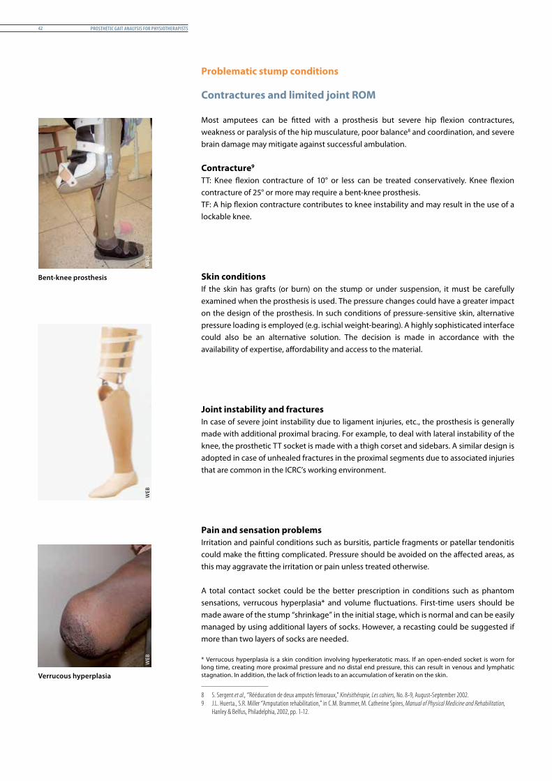
Contracture9 TT: Knee flexion contracture of 10° or less can be treated conservatively. Knee flexion contracture of 25° or more may require a bent-knee prosthesis.TF: A hip flexion contracture contributes to knee instability and may result in the use of a lockable knee.2
Skin conditions If the skin has grafts (or burn) on the stump or under suspension, it must be carefully examined when the prosthesis is used. The pressure changes could have a greater impact on the design of the prosthesis. In such conditions of pressure-sensitive skin, alternative pressure loading is employed (e.g. ischial weight-bearing). A highly sophisticated interface could also be an alternative solution. The decision is made in accordance with the availability of expertise, affordability and access to the material.
Joint instability and fractures In case of severe joint instability due to ligament injuries, etc., the prosthesis is generally made with additional proximal bracing. For example, to deal with lateral instability of the knee, the prosthetic TT socket is made with a thigh corset and sidebars. A similar design is adopted in case of unhealed fractures in the proximal segments due to associated injuries that are common in the ICRC’s working environment.
Pain and sensation problems Irritation and painful conditions such as bursitis, particle fragments or patellar tendonitis could make the fitting complicated. Pressure should be avoided on the affected areas, as this may aggravate the irritation or pain unless treated otherwise.
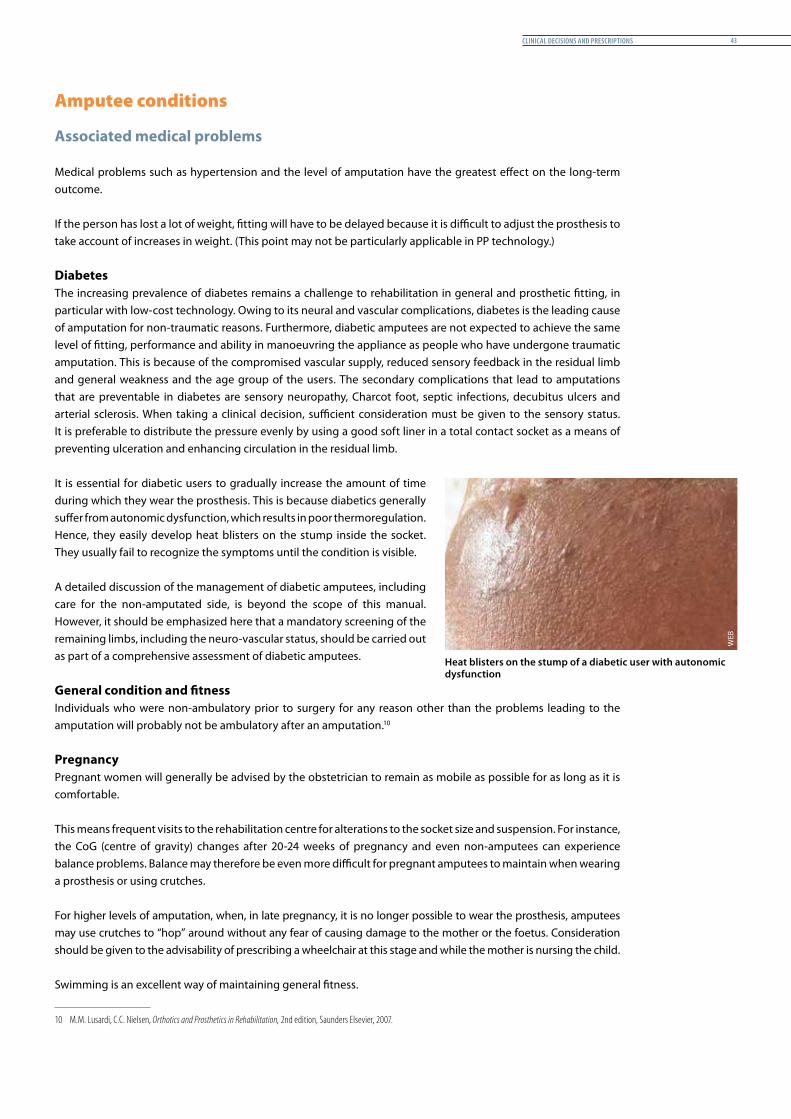
A total contact socket could be the better prescription in conditions such as phantom sensations, verrucous hyperplasia* and volume fluctuations. First-time users should be made aware of the stump “shrinkage” in the initial stage, which is normal and can be easily managed by using additional layers of socks. However, a recasting could be suggested if more than two layers of socks are needed.
* Verrucous hyperplasia is a skin condition involving hyperkeratotic mass. If an open-ended socket is worn for long time, creating more proximal pressure and no distal end pressure, this can result in venous and lymphatic stagnation. In addition, the lack of friction leads to an accumulation of keratin on the skin.
8 S. Sergent et al., “Rééducation de deux amputés fémoraux,” Kinésithérapie, Les cahiers, No. 8-9, August-September 2002.9 J.L. Huerta., S.R. Miller “Amputation rehabilitation,” in C.M. Brammer, M. Catherine Spires, Manual of Physical Medicine and Rehabilitation,
Hanley & Belfus, Philadelphia, 2002, pp. 1-12.
Bent-knee prosthesis
WEB
WEB
Verrucous hyperplasia
WEB
clinical decisions and PrescriPtions 43
Amputee conditions
Associated medical problems
Medical problems such as hypertension and the level of amputation have the greatest effect on the long-term outcome.
If the person has lost a lot of weight, fitting will have to be delayed because it is difficult to adjust the prosthesis to take account of increases in weight. (This point may not be particularly applicable in PP technology.)
DiabetesThe increasing prevalence of diabetes remains a challenge to rehabilitation in general and prosthetic fitting, in particular with low-cost technology. Owing to its neural and vascular complications, diabetes is the leading cause of amputation for non-traumatic reasons. Furthermore, diabetic amputees are not expected to achieve the same level of fitting, performance and ability in manoeuvring the appliance as people who have undergone traumatic amputation. This is because of the compromised vascular supply, reduced sensory feedback in the residual limb and general weakness and the age group of the users. The secondary complications that lead to amputations that are preventable in diabetes are sensory neuropathy, Charcot foot, septic infections, decubitus ulcers and arterial sclerosis. When taking a clinical decision, sufficient consideration must be given to the sensory status. It is preferable to distribute the pressure evenly by using a good soft liner in a total contact socket as a means of preventing ulceration and enhancing circulation in the residual limb.
It is essential for diabetic users to gradually increase the amount of time during which they wear the prosthesis. This is because diabetics generally suffer from autonomic dysfunction, which results in poor thermoregulation. Hence, they easily develop heat blisters on the stump inside the socket. They usually fail to recognize the symptoms until the condition is visible.
A detailed discussion of the management of diabetic amputees, including care for the non-amputated side, is beyond the scope of this manual. However, it should be emphasized here that a mandatory screening of the remaining limbs, including the neuro-vascular status, should be carried out as part of a comprehensive assessment of diabetic amputees.
General condition and fitnessIndividuals who were non-ambulatory prior to surgery for any reason other than the problems leading to the amputation will probably not be ambulatory after an amputation.103
PregnancyPregnant women will generally be advised by the obstetrician to remain as mobile as possible for as long as it is comfortable.
This means frequent visits to the rehabilitation centre for alterations to the socket size and suspension. For instance, the CoG (centre of gravity) changes after 20-24 weeks of pregnancy and even non-amputees can experience balance problems. Balance may therefore be even more difficult for pregnant amputees to maintain when wearing a prosthesis or using crutches.
For higher levels of amputation, when, in late pregnancy, it is no longer possible to wear the prosthesis, amputees may use crutches to “hop” around without any fear of causing damage to the mother or the foetus. Consideration should be given to the advisability of prescribing a wheelchair at this stage and while the mother is nursing the child.
Swimming is an excellent way of maintaining general fitness.
10 M.M. Lusardi, C.C. Nielsen, Orthotics and Prosthetics in Rehabilitation, 2nd edition, Saunders Elsevier, 2007.
Heat blisters on the stump of a diabetic user with autonomic dysfunction
WEB
Prosthetic Gait analysis for PhysiotheraPists44
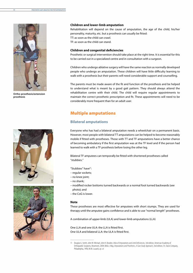
Children and lower-limb amputationRehabilitation will depend on the cause of amputation, the age of the child, his/her personality, maturity, etc. but a prosthesis can usually be fitted:TT: as soon as the child can crawl.TF: as soon as the child can stand.
Children and congenital deficienciesProsthetic or surgical intervention should take place at the right time. It is essential for this to be carried out in a specialized centre and in consultation with a surgeon.
Children who undergo ablative surgery will have the same reaction as normally developed people who undergo an amputation. These children will have little difficulty learning to walk with a prosthesis but their parents will need considerable support and counselling.
The parents must be made aware of the fit and function of the prosthesis and be helped to understand what is meant by a good gait pattern. They should always attend the rehabilitation centre with their child. The child will require regular appointments to maintain the correct prosthetic prescription and fit. These appointments will need to be considerably more frequent than for an adult user.
Multiple amputations
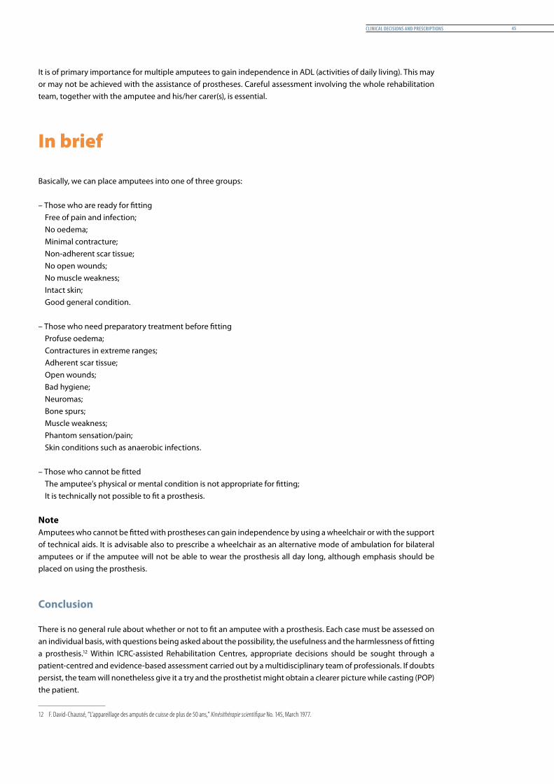
Bilateral amputations
Everyone who has had a bilateral amputation needs a wheelchair on a permanent basis. However, most people with bilateral TT amputations can be helped to become reasonably mobile if fitted with prostheses. Those with TT and TF amputations have a better chance of becoming ambulatory if the first amputation was at the TF level and if the person had learned to walk with a TF prosthesis before losing the other leg.
Bilateral TF amputees can temporally be fitted with shortened prostheses called “stubbies.”
“Stubbies” have11:4
– regular sockets;– no knee joint;– no shank;– modified rocker bottoms turned backwards or a normal foot turned backwards (see
photo); and – the CoG is lower.
NoteThese prostheses are most effective for amputees with short stumps. They are used for therapy until the amputee gains confidence and is able to use “normal length” prostheses.
A combination of upper-limb (ULA) and lower-limb amputations (LLA)
One LLA and one ULA: the LLA is fitted first.One ULA and bilateral LLA: the ULA is fitted first.
11 Douglas G. Smith, John W. Michael, John H. Bowker, Atlas of Amputations and Limb Deficiencies, 3rd edition, American Academy of Orthopaedic Surgeons, Rosemont, 2004; Bella J. May, Amputations and Prosthetics, A Case Study Approach, 2nd edition, F.A. Davis Company, Philadelphia, 1996; M.M. Lusardi, op. cit.
Ortho-prosthesis/extension prosthesis
WEB
ICRC
ICRC
clinical decisions and PrescriPtions 45
It is of primary importance for multiple amputees to gain independence in ADL (activities of daily living). This may or may not be achieved with the assistance of prostheses. Careful assessment involving the whole rehabilitation team, together with the amputee and his/her carer(s), is essential.
In brief
Basically, we can place amputees into one of three groups:
– Those who are ready for fittingFree of pain and infection;No oedema;Minimal contracture;Non-adherent scar tissue;No open wounds;No muscle weakness;Intact skin;Good general condition.
– Those who need preparatory treatment before fitting
Profuse oedema;Contractures in extreme ranges;Adherent scar tissue; Open wounds;Bad hygiene;Neuromas;Bone spurs;Muscle weakness;Phantom sensation/pain;Skin conditions such as anaerobic infections.
– Those who cannot be fitted
The amputee’s physical or mental condition is not appropriate for fitting;It is technically not possible to fit a prosthesis.
Note Amputees who cannot be fitted with prostheses can gain independence by using a wheelchair or with the support of technical aids. It is advisable also to prescribe a wheelchair as an alternative mode of ambulation for bilateral amputees or if the amputee will not be able to wear the prosthesis all day long, although emphasis should be placed on using the prosthesis.
Conclusion
There is no general rule about whether or not to fit an amputee with a prosthesis. Each case must be assessed on an individual basis, with questions being asked about the possibility, the usefulness and the harmlessness of fitting a prosthesis.12 Within ICRC-assisted Rehabilitation Centres, appropriate decisions should be sought through a patient-centred and evidence-based assessment carried out by a multidisciplinary team of professionals. If doubts persist, the team will nonetheless give it a try and the prosthetist might obtain a clearer picture while casting (POP) the patient.5
12 F. David-Chaussé, “L’appareillage des amputés de cuisse de plus de 50 ans,” Kinésithérapie scientifique No. 145, March 1977.

Prosthetic Gait analysis for PhysiotheraPists46
materials and equiPment 47
materials and equiPment
Introduction
Content
This chapter briefly presents the materials and equipment needed for the physiotherapy management of lower-limb amputees.
The items marked with an asterisk (*) can be ordered via ICRC Log/GVA, and the ICRC’s PRP standard catalogue. The items marked with a copyright symbol (©) can often be produced locally and a data sheet (taken from the ICRC’s locally made equipment catalogue) has therefore been added for each of them in order to facilitate their manufacture.
Rationale
This chapter has been included in the manual to provide an initial guide for planning purposes when consideration is being given to opening or refurbishing a physiotherapy department for LLA rehabilitation. Hence, an indication is also given of where the equipment could be procured (as explained above).
Prosthetic Gait analysis for PhysiotheraPists48
Materials and equipment (in order of importance)
y The essential equipment listed below must be available in a physiotherapy clinic attached to a prosthetic service.
y The advanced equipment listed below is mostly needed to supplement basic pre/post-prosthetic rehabilitation and to extend the training area to an outdoor or an advanced gait area.
y The optional equipment listed below is good to have in the physiotherapy department but is generally not required for most of the cases.
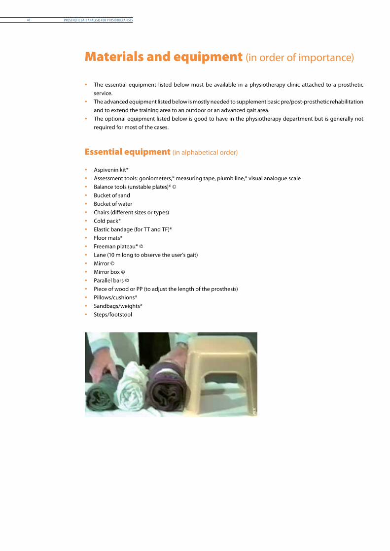
Essential equipment (in alphabetical order)
y Aspivenin kit* y Assessment tools: goniometers,* measuring tape, plumb line,* visual analogue scale y Balance tools (unstable plates)* © y Bucket of sand y Bucket of water y Chairs (different sizes or types) y Cold pack* y Elastic bandage (for TT and TF)* y Floor mats* y Freeman plateau* © y Lane (10 m long to observe the user’s gait) y Mirror © y Mirror box © y Parallel bars © y Piece of wood or PP (to adjust the length of the prosthesis) y Pillows/cushions* y Sandbags/weights* y Steps/footstool
WEB
materials and equiPment 49
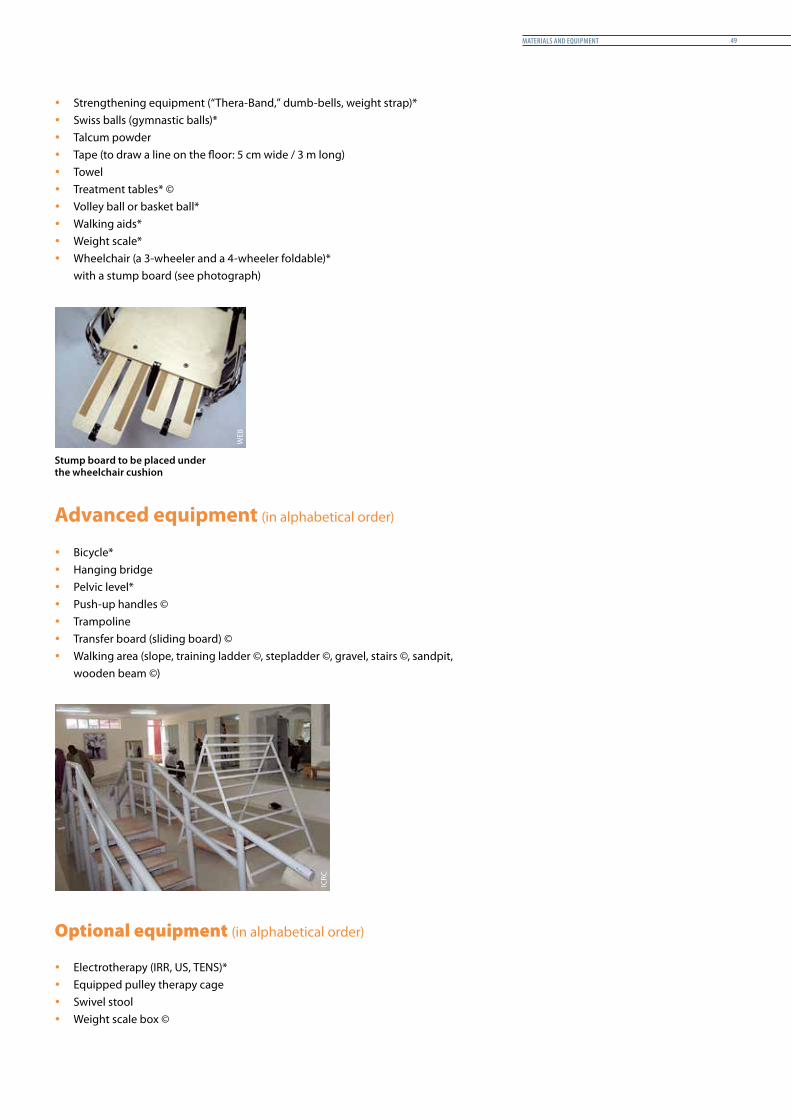
y Strengthening equipment (“Thera-Band,” dumb-bells, weight strap)* y Swiss balls (gymnastic balls)* y Talcum powder y Tape (to draw a line on the floor: 5 cm wide / 3 m long) y Towel y Treatment tables* © y Volley ball or basket ball* y Walking aids* y Weight scale* y Wheelchair (a 3-wheeler and a 4-wheeler foldable)*
with a stump board (see photograph)
Advanced equipment (in alphabetical order)
y Bicycle* y Hanging bridge y Pelvic level* y Push-up handles © y Trampoline y Transfer board (sliding board) © y Walking area (slope, training ladder ©, stepladder ©, gravel, stairs ©, sandpit,
wooden beam ©)
Optional equipment (in alphabetical order)
y Electrotherapy (IRR, US, TENS)* y Equipped pulley therapy cage y Swivel stool y Weight scale box ©
Stump board to be placed under the wheelchair cushion
WEB
ICRC

Prosthetic Gait analysis for PhysiotheraPists50
Pre-Prosthetic rehabilitation 51
Pre-Prosthetic rehabilitation
Introduction
Content
This chapter describes the physiotherapy provided after a patient’s discharge from the surgical unit. The professionals involved in the rehabilitation process should bear in mind that maximum benefit for users can be achieved if they work in such a way as to complement each other’s tasks. The members of the interdisciplinary team should therefore discuss every possible stage in the process with each other and with their patients in order to prepare them well for casting, fitting and post-fitting training and to help them understand the expectations of each clinical department (PT and P&O) regarding the outcome of the treatment.
Rationale
All lower-limb amputees need to be aware that their ability to walk and the quality of the prosthetic gait depend on the condition of the residual limb, the strength of the unaffected limb and general physical fitness. Therefore and according to the individual needs, they need to be prepared for an ideal prosthetic fitting. Careful attention and effort during the pre-prosthetic phase will not only ensure an ideal prosthetic fit but also will make the post-fitting phase easier and minimize gait deviations.
Prosthetic Gait analysis for PhysiotheraPists52
Aim of pre-prosthetic rehabilitation
The aim of pre-prosthetic rehabilitation training with the physiotherapist is to help the patient to recover from the surgical intervention physically and psychologically and to prepare his/her body and mind for the prosthesis.
The rehabilitation process is designed to provide assistance in the transition period immediately following surgery. Physiotherapists work with amputees to reduce swelling, to prevent contractures and to manage any pain, with a particular focus on decreasing residual limb pain (also known as “phantom pain”). Physiotherapists later help prepare amputees for the use of a prosthesis by working with them to build strength, increase endurance, improve mobility and enhance their ability to perform the activities of daily living.
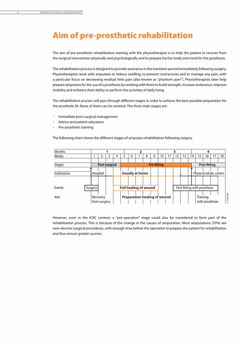
The rehabilitation process will pass through different stages in order to achieve the best possible preparation for the prosthetic fit. None of them can be omitted. The three main stages are:
y Immediate post-surgical management y Advice and patient education y Pre-prosthetic training
The following chart shows the different stages of amputee rehabilitation following surgery.
Months 11 2 3 4 5 6 7 8 9 10 11 12 13 14 15 16 17 18
2 3 4Weeks
Stages Post-surgical Post-�ttingPre-�tting
Institutions
Events
Aim
Hospital
Recovery from surgery
Training with prosthesis
Preparation healing of wound
Usually at home Physical rehab. centre
Surgery First �tting with prosthesisFull healing of wound
However, even in the ICRC context, a “pre-operative” stage could also be considered to form part of the rehabilitation process. This is because of the change in the causes of amputation. Most amputations (70%) are now elective surgical procedures, with enough time before the operation to prepare the patient for rehabilitation and thus ensure greater success.
C. S
chm
id
Pre-Prosthetic rehabilitation 53
Immediate post-surgical management
The main post-surgical problems include surgical and residual limb pain, phantom sensation/pain, oedema, contractures, wound dehiscence and cardio-vascular complications.
Positioning and bed mobility
It is important to remind the amputee about the correct positions when lying, sitting or standing so as to prevent contractures:– For TT amputees: How to prevent knee flexion.– For TF amputees: How to prevent hip flexion or abduction.
In addition to simple positional adjustments, the amputee must learn how to get in and out of bed.
Strength and range of motion
A functional assessment should be made of the movement of both upper and lower limbs. During the ROM assessment the therapist should determine whether the amputee has a fixed contracture or merely soft-tissue tightness on the residual limb. This may affect the manner in which the prosthesis is manufactured.
The functional strength of the major muscle groups should be assessed by manual muscle-testing of all limbs including the residual limb and the trunk. This will help determine the patient’s potential level of ability to perform activities such as transfer, wheelchair management and ambulation with and without the prosthesis.
A programme of exercises should be put together accordingly.
Transfers
Amputees should be made aware of transfer techniques. This includes floor to chair, chair to chair/bed and vice versa.
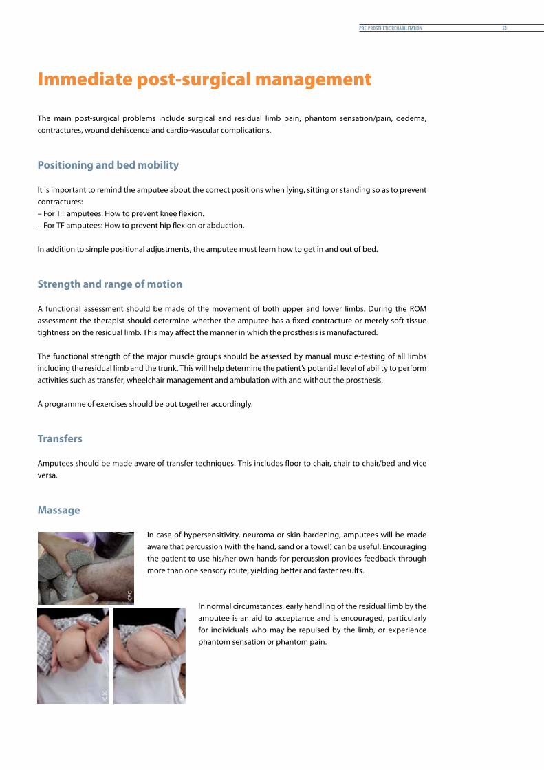
Massage
In case of hypersensitivity, neuroma or skin hardening, amputees will be made aware that percussion (with the hand, sand or a towel) can be useful. Encouraging the patient to use his/her own hands for percussion provides feedback through more than one sensory route, yielding better and faster results.
In normal circumstances, early handling of the residual limb by the amputee is an aid to acceptance and is encouraged, particularly for individuals who may be repulsed by the limb, or experience phantom sensation or phantom pain.
ICRC
ICRC
ICRC
Prosthetic Gait analysis for PhysiotheraPists54
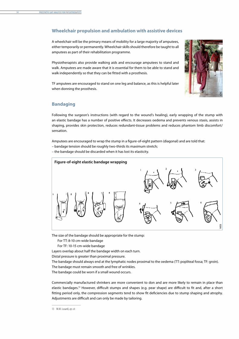
Wheelchair propulsion and ambulation with assistive devices
A wheelchair will be the primary means of mobility for a large majority of amputees, either temporarily or permanently. Wheelchair skills should therefore be taught to all amputees as part of their rehabilitation programme.
Physiotherapists also provide walking aids and encourage amputees to stand and walk. Amputees are made aware that it is essential for them to be able to stand and walk independently so that they can be fitted with a prosthesis.
TF amputees are encouraged to stand on one leg and balance, as this is helpful later when donning the prosthesis.
Bandaging
Following the surgeon’s instructions (with regard to the wound’s healing), early wrapping of the stump with an elastic bandage has a number of positive effects. It decreases oedema and prevents venous stasis, assists in shaping, provides skin protection, reduces redundant-tissue problems and reduces phantom limb discomfort/sensation.
Amputees are encouraged to wrap the stump in a figure-of-eight pattern (diagonal) and are told that:– bandage tension should be roughly two-thirds its maximum stretch;– the bandage should be discarded when it has lost its elasticity.
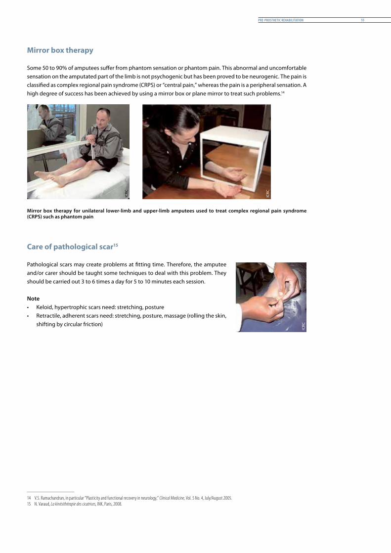
Figure-of-eight elastic bandage wrapping
The size of the bandage should be appropriate for the stump:For TT: 8-10 cm-wide bandageFor TF: 10-15 cm-wide bandage
Layers overlap about half the bandage width on each turn.Distal pressure is greater than proximal pressure.The bandage should always end at the lymphatic nodes proximal to the oedema (TT: popliteal fossa; TF: groin). The bandage must remain smooth and free of wrinkles.The bandage could be worn if a small wound occurs.
Commercially manufactured shrinkers are more convenient to don and are more likely to remain in place than elastic bandages.13 However, difficult stumps and shapes (e.g. pear shape) are difficult to fit and, after a short fitting period only, the compression segments tend to show fit deficiencies due to stump shaping and atrophy. Adjustments are difficult and can only be made by tailoring. 1
13 M.M. Lusardi, op. cit.
WEB
ICRC
Pre-Prosthetic rehabilitation 55
Mirror box therapy
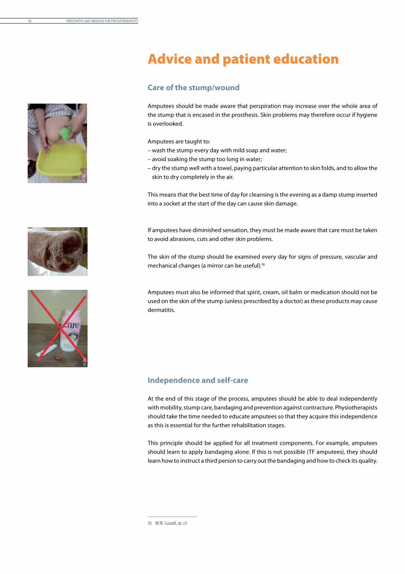
Some 50 to 90% of amputees suffer from phantom sensation or phantom pain. This abnormal and uncomfortable sensation on the amputated part of the limb is not psychogenic but has been proved to be neurogenic. The pain is classified as complex regional pain syndrome (CRPS) or “central pain,” whereas the pain is a peripheral sensation. A high degree of success has been achieved by using a mirror box or plane mirror to treat such problems.142
Mirror box therapy for unilateral lower-limb and upper-limb amputees used to treat complex regional pain syndrome (CRPS) such as phantom pain
Care of pathological scar15
Pathological scars may create problems at fitting time. Therefore, the amputee and/or carer should be taught some techniques to deal with this problem. They should be carried out 3 to 6 times a day for 5 to 10 minutes each session.
Note• Keloid, hypertrophic scars need: stretching, posture• Retractile, adherent scars need: stretching, posture, massage (rolling the skin,
shifting by circular friction)
14 V.S. Ramachandran, in particular “Plasticity and functional recovery in neurology,” Clinical Medicine, Vol. 5 No. 4, July/August 2005.15 N. Varaud, La kinésithérapie des cicatrices, INK, Paris, 2008.
ICRC
ICRC
ICRC
Prosthetic Gait analysis for PhysiotheraPists56
Advice and patient education
Care of the stump/wound
Amputees should be made aware that perspiration may increase over the whole area of the stump that is encased in the prosthesis. Skin problems may therefore occur if hygiene is overlooked.
Amputees are taught to:– wash the stump every day with mild soap and water;– avoid soaking the stump too long in water;– dry the stump well with a towel, paying particular attention to skin folds, and to allow the
skin to dry completely in the air.
This means that the best time of day for cleansing is the evening as a damp stump inserted into a socket at the start of the day can cause skin damage.
If amputees have diminished sensation, they must be made aware that care must be taken to avoid abrasions, cuts and other skin problems.
The skin of the stump should be examined every day for signs of pressure, vascular and mechanical changes (a mirror can be useful).161
Amputees must also be informed that spirit, cream, oil balm or medication should not be used on the skin of the stump (unless prescribed by a doctor) as these products may cause dermatitis.
Independence and self-care
At the end of this stage of the process, amputees should be able to deal independently with mobility, stump care, bandaging and prevention against contracture. Physiotherapists should take the time needed to educate amputees so that they acquire this independence as this is essential for the further rehabilitation stages.
This principle should be applied for all treatment components. For example, amputees should learn to apply bandaging alone. If this is not possible (TF amputees), they should learn how to instruct a third person to carry out the bandaging and how to check its quality.
16 M.M. Lusardi, op. cit.
ICRC
ICRC
ICRC
Pre-Prosthetic rehabilitation 57
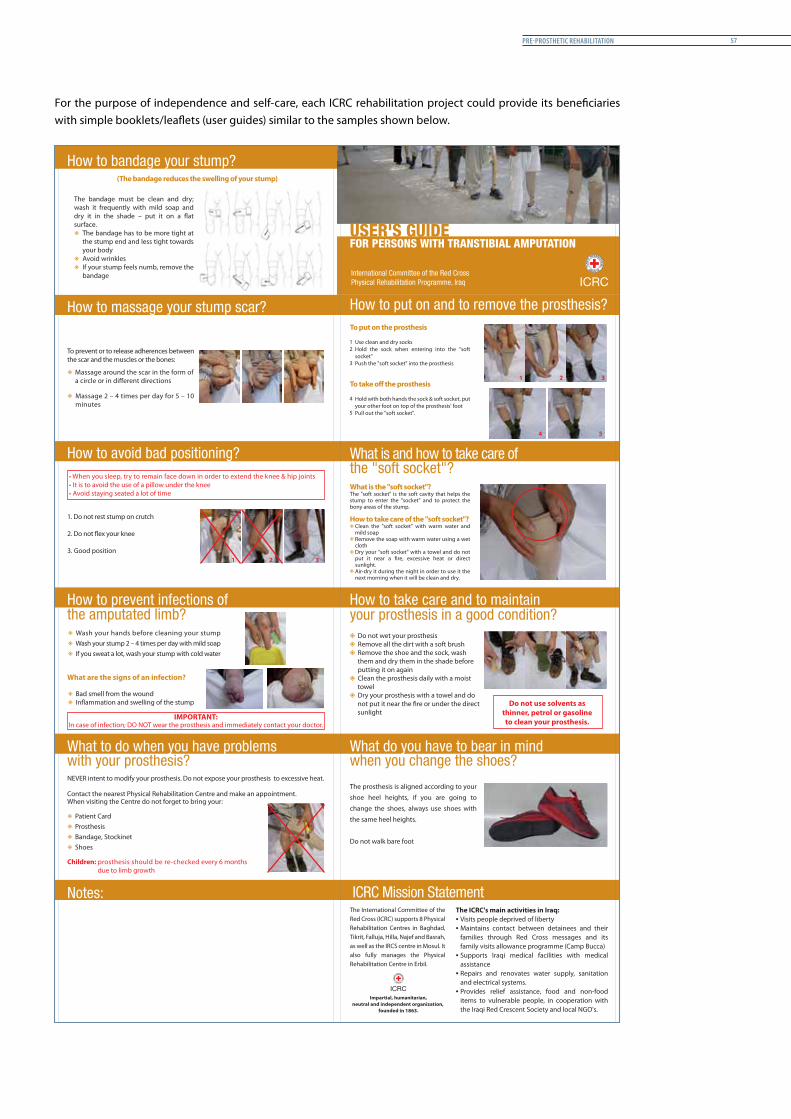
For the purpose of independence and self-care, each ICRC rehabilitation project could provide its beneficiaries with simple booklets/leaflets (user guides) similar to the samples shown below.
What is and how to take care ofthe "soft socket"?
Do not use solvents as thinner, petrol or gasoline to clean your prosthesis.
What do you have to bear in mindwhen you change the shoes?
ICRC Mission Statement
How to take care and to maintain
Do not wet your prosthesis Remove all the dirt with a soft brushRemove the shoe and the sock, wash them and dry them in the shade before putting it on againClean the prosthesis daily with a moist towelDry your prosthesis with a towel and do not put it near the �re or under the direct sunlight
your prosthesis in a good condition?
To put on the prosthesis
Use clean and dry socksHold the sock when entering into the "soft socket"Push the "soft socket" into the prosthesis
To take o� the prosthesis
Hold with both hands the sock & soft socket, put your other foot on top of the prosthesis' footPull out the "soft socket".
What is the "soft socket"?The "soft socket" is the soft cavity that helps the stump to enter the "socket" and to protect the bony areas of the stump.
How to take care of the "soft socket"?Clean the "soft socket" with warm water and mild soapRemove the soap with warm water using a wet clothDry your "soft socket" with a towel and do not put it near a �re, excessive heat or direct sunlight. Air-dry it during the night in order to use it the next morning when it will be clean and dry.
12
3
4
5
1 2 3
4 5
How to put on and to remove the prosthesis?
USER'S GUIDEFOR PERSONS WITH TRANSTIBIAL AMPUTATION
The prosthesis is aligned according to your
shoe heel heights, if you are going to
change the shoes, always use shoes with
the same heel heights.
Do not walk bare foot
How to bandage your stump?(The bandage reduces the swelling of your stump)
How to massage your stump scar?
To prevent or to release adherences between the scar and the muscles or the bones:
Massage around the scar in the form ofa circle or in di�erent directions
Massage 2 – 4 times per day for 5 – 10minutes
How to avoid bad positioning?
1. Do not rest stump on crutch
2. Do not �ex your knee
3. Good position
How to prevent infections of
Wash your hands before cleaning your stumpWash your stump 2 – 4 times per day with mild soapIf you sweat a lot, wash your stump with cold water
What are the signs of an infection?
Bad smell from the woundIn�ammation and swelling of the stump
the amputated limb?
What to do when you have problems with your prosthesis?
Notes:
ProsthesisPatient Card
Bandage, Stockinet Shoes
1 2 3
The ICRC's main activities in Iraq:Visits people deprived of liberty Maintains contact between detainees and their families through Red Cross messages and its family visits allowance programme (Camp Bucca)Supports Iraqi medical facilities with medical assistanceRepairs and renovates water supply, sanitation and electrical systems. Provides relief assistance, food and non-food items to vulnerable people, in cooperation with the Iraqi Red Crescent Society and local NGO's.
International Committee of the Red CrossPhysical Rehabilitation Programme, Iraq
••
•
•
•
• When you sleep, try to remain face down in order to extend the knee & hip joints• It is to avoid the use of a pillow under the knee• Avoid staying seated a lot of time
The bandage must be clean and dry; wash it frequently with mild soap and dry it in the shade – put it on a �at surface.
The bandage has to be more tight at the stump end and less tight towards your bodyAvoid wrinklesIf your stump feels numb, remove the bandage
IMPORTANT:In case of infection; DO NOT wear the prosthesis and immediately contact your doctor.
NEVER intent to modify your prosthesis. Do not expose your prosthesis to excessive heat.
Contact the nearest Physical Rehabilitation Centre and make an appointment. When visiting the Centre do not forget to bring your:
Children: prosthesis should be re-checked every 6 months due to limb growth
The International Committee of the Red Cross (ICRC) supports 8 Physical Rehabilitation Centres in Baghdad, Tikrit, Falluja, Hilla, Najef and Basrah, as well as the IRCS centre in Mosul. It also fully manages the Physical Rehabilitation Centre in Erbil.
Impartial, humanitarian,neutral and independent organization,
founded in 1863.
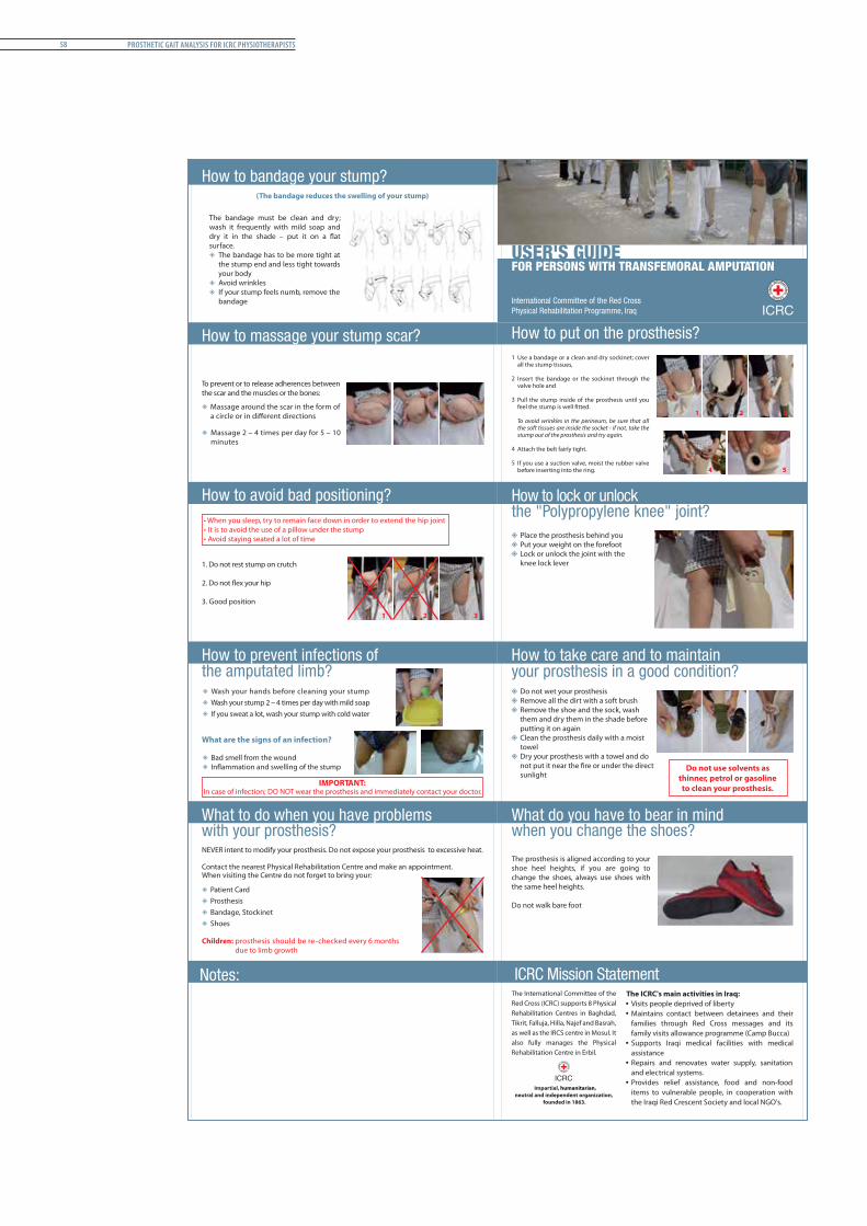
Prosthetic Gait analysis for icrc PhysiotheraPists58
How to lock or unlock la
Place the prosthesis behind you Put your weight on the forefoot Lock or unlock the joint with the knee lock lever
the "Polypropylene knee" joint?
Do not use solvents as thinner, petrol or gasoline to clean your prosthesis.
What do you have to bear in mindwhen you change the shoes?
The ICRC's main activities in Iraq:Visits people deprived of liberty Maintains contact between detainees and their families through Red Cross messages and its family visits allowance programme (Camp Bucca)Supports Iraqi medical facilities with medical assistanceRepairs and renovates water supply, sanitation and electrical systems. Provides relief assistance, food and non-food items to vulnerable people, in cooperation with the Iraqi Red Crescent Society and local NGO's.
ICRC Mission Statement
How to take care and to maintain
Do not wet your prosthesis Remove all the dirt with a soft brushRemove the shoe and the sock, wash them and dry them in the shade before putting it on againClean the prosthesis daily with a moist towelDry your prosthesis with a towel and do
sunlight
your prosthesis in a good condition?
Use a bandage or a clean and dry sockinet; cover all the stump tissues,
Insert the bandage or the sockinet through the valve hole and
Pull the stump inside of the prosthesis until you
To avoid wrinkles in the perineum, be sure that all the soft tissues are inside the socket - if not, take the stump out of the prosthesis and try again.
Attach the belt fairly tight.
If you use a suction valve, moist the rubber valve before inserting into the ring.
1
2
3
4
5
1 2 3
4 5
International Committee of the Red CrossPhysical Rehabilitation Programme, Iraq
How to put on the prosthesis?
USER'S GUIDEFOR PERSONS WITH TRANSFEMORAL AMPUTATION
The prosthesis is aligned according to your shoe heel heights, if you are going to change the shoes, always use shoes with the same heel heights.
Do not walk bare foot
••
•
•
•
How to bandage your stump?(The bandage reduces the swelling of your stump)
How to massage your stump scar?
To prevent or to release adherences between the scar and the muscles or the bones:
Massage around the scar in the form of
Massage 2 – 4 times per day for 5 – 10minutes
How to avoid bad positioning?• When you sleep, try to remain face down in order to extend the hip joint• It is to avoid the use of a pillow under the stump• Avoid staying seated a lot of time
1. Do not rest stump on crutch
2. Do not �ex your hip
3. Good position
How to prevent infections of
Wash your hands before cleaning your stumpWash your stump 2 – 4 times per day with mild soapIf you sweat a lot, wash your stump with cold water
What are the signs of an infection?
Bad smell from the wound
the amputated limb?
IMPORTANT:In case of infection; DO NOT wear the prosthesis and immediately contact your doctor.
NEVER intent to modify your prosthesis. Do not expose your prosthesis to excessive heat.
Contact the nearest Physical Rehabilitation Centre and make an appointment. When visiting the Centre do not forget to bring your:
What to do when you have problems with your prosthesis?
Notes:
Children: prosthesis should be re-checked every 6 months due to limb growth
ProsthesisPatient Card
Bandage, Stockinet Shoes
1 2 3
The bandage must be clean and dry; wash it frequently with mild soap and
surface. The bandage has to be more tight at the stump end and less tight towards your bodyAvoid wrinklesIf your stump feels numb, remove the bandage
The International Committee of the Red Cross (ICRC) supports 8 Physical Rehabilitation Centres in Baghdad, Tikrit, Falluja, Hilla, Najef and Basrah, as well as the IRCS centre in Mosul. It also fully manages the Physical Rehabilitation Centre in Erbil.
Impartial, humanitarian,neutral and independent organization,
founded in 1863.
Pre-Prosthetic rehabilitation 59
Pre-prosthetic training
Pre-prosthetic training is probably the most important phase of the rehabilitation process. It ends the post-surgical period and prepares for prosthetic rehabilitation. The quality of the training and the effort put in to the exercises will enhance the amputees’ functional ability to face the challenge of wearing a prosthesis and to use it to its maximal potential.
Loss of part of the body causes a change in the position of the amputee’s centre of gravity. His/her entire balance is disrupted and it takes time and effort to regain good control. Pre-prosthetic training also sets out to help amputees recover the good coordination that is essential for prosthetic rehabilitation.
At ICRC-assisted physical rehabilitation centres, we generally see amputees for the first time when they are referred for prosthesis fitting. The assessment made at the time of an amputee’s consultation is therefore an essential part of the process and, if necessary, the rehabilitation team should not hesitate to delay the appointment for casting in order to provide extra time for pre-prosthetic rehabilitation. Ideally, physical rehabilitation centres should have the capacity (space, staff, accommodation) for pre-prosthetic rehabilitation because it is often overlooked at the hospital, amputees being given only general advice before they are discharged.
Pre-prosthetic rehabilitation includes strengthening exercises, ROM and stretching exercises, balance and coordination, and functional activities.
Strengthening exercises
Strengthening exercises are very important to enable amputees to acquire many of the activities required for transfer, functional skills and ambulation. The selection of the type of strengthening exercise is dependent on initial muscular strength and endurance, the level of independence, access to equipment, time constraints, safety and several other issues.
Every amputee should be taught a set of strengthening exercises which will continue the strengthening process after discharge from the rehabilitation centre, thus fostering a level of fitness that will allow good ambulation.
The most usual techniques employed are:– isometric strengthening;– manual resistance strengthening; and– isotonic (concentric) strengthening.
The exercises should focus on the muscles of the amputated leg, the sound leg, the upper extremities and the muscles of the trunk.
ICRC
Prosthetic Gait analysis for PhysiotheraPists60
Range of motion and stretching exercises
Most limitations of joint movement that are severe enough to affect prosthetic fit can be prevented if the therapist performs stretching or passive range of motion (PROM) exercises. If loss of ROM occurs as a result of muscular shortening, manual stretching is one alternative employed to restore ROM.
The most usual techniques employed are: – passive joint mobilization;– passive or self-stretching; and– contract-relax stretching.
Movements should be slow, gentle and specific. Preference should always be given to long soft stretching over short powerful stretching.
Balance
There is no need to wait for the first prosthetic fitting before working to improve amputees’ balance and coordination. Many exercises can be performed in lying, sitting and standing positions, and by moving from one position to another, to help amputees to recover good balance. A great deal can be achieved in the pre-fitting stage. Two types of exercises are essential to prepare for ambulation (taking into consideration the general condition of the amputee). They are:– standing unsupported while balancing on one leg;– pelvic control on a gymnastic ball.
It is important for amputees to exercise in a safe environment (parallel bars, hand control, soft mattress). Any fall will increase apprehension and delay the success of the exercises.
Functional activities
The greater the degree of mobility and activity acquired by amputees without a prosthesis, the greater the functional result with the prosthesis will be. Amputees can already achieve a great deal in transfer and ambulation without a prosthesis. Physiotherapists need to motivate, guide and support amputees in achieving ADL independence in accordance with their general condition.
In the period until amputees are ready to be fitted with a prosthesis, rehabilitation should also continue outside the centre with a well-defined home programme of exercises.
ICRC
ICRC
fittinG a Prosthesis 61
fittinG a Prosthesis
Introduction
Content
This chapter explains the first fitting process, which involves continuous interaction between the patient, the prosthetist and the physiotherapist. It briefly describes points to consider in the prevention, detection and correction of prosthetically caused errors affecting the prosthetic gait (socket design/alignment) and fitting mistakes.
Rationale
This document resumes and synthesizes the information and guidelines found in various recommended reference documents. It does not aim to cover fully the complexity of fitting prostheses but rather sets out to address the main issues that need to be resolved before to proceeding to post-fitting rehabilitation.
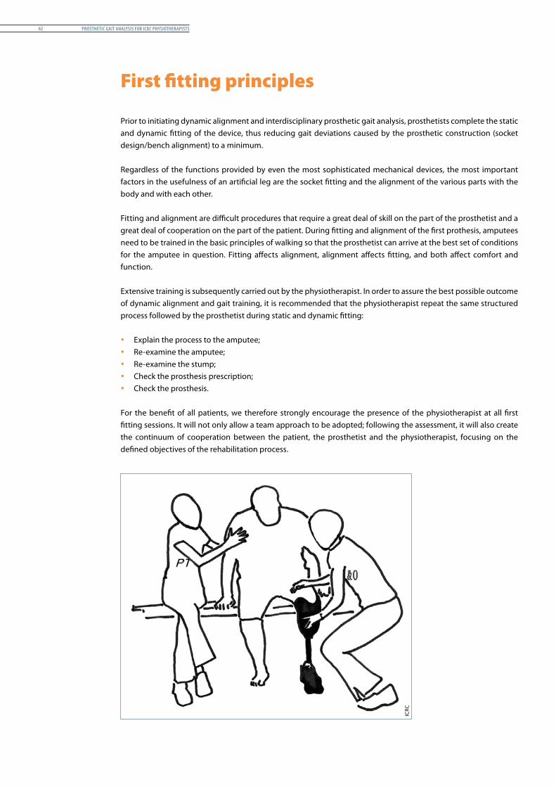
Prosthetic Gait analysis for icrc PhysiotheraPists62
First fitting principles
Prior to initiating dynamic alignment and interdisciplinary prosthetic gait analysis, prosthetists complete the static and dynamic fitting of the device, thus reducing gait deviations caused by the prosthetic construction (socket design/bench alignment) to a minimum.
Regardless of the functions provided by even the most sophisticated mechanical devices, the most important factors in the usefulness of an artificial leg are the socket fitting and the alignment of the various parts with the body and with each other.
Fitting and alignment are difficult procedures that require a great deal of skill on the part of the prosthetist and a great deal of cooperation on the part of the patient. During fitting and alignment of the first prothesis, amputees need to be trained in the basic principles of walking so that the prosthetist can arrive at the best set of conditions for the amputee in question. Fitting affects alignment, alignment affects fitting, and both affect comfort and function.
Extensive training is subsequently carried out by the physiotherapist. In order to assure the best possible outcome of dynamic alignment and gait training, it is recommended that the physiotherapist repeat the same structured process followed by the prosthetist during static and dynamic fitting:
y Explain the process to the amputee; y Re-examine the amputee; y Re-examine the stump; y Check the prosthesis prescription; y Check the prosthesis.
For the benefit of all patients, we therefore strongly encourage the presence of the physiotherapist at all first fitting sessions. It will not only allow a team approach to be adopted; following the assessment, it will also create the continuum of cooperation between the patient, the prosthetist and the physiotherapist, focusing on the defined objectives of the rehabilitation process.
ICRC
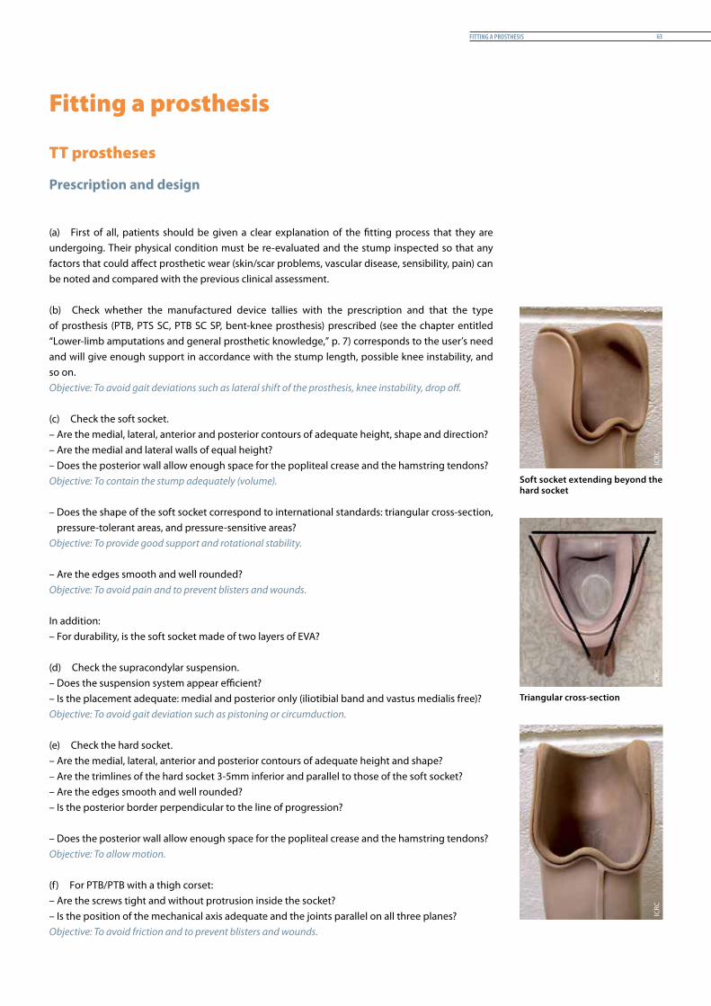
fittinG a Prosthesis 63
Fitting a prosthesis
TT prostheses
Prescription and design
(a) First of all, patients should be given a clear explanation of the fitting process that they are undergoing. Their physical condition must be re-evaluated and the stump inspected so that any factors that could affect prosthetic wear (skin/scar problems, vascular disease, sensibility, pain) can be noted and compared with the previous clinical assessment.
(b) Check whether the manufactured device tallies with the prescription and that the type of prosthesis (PTB, PTS SC, PTB SC SP, bent-knee prosthesis) prescribed (see the chapter entitled “Lower-limb amputations and general prosthetic knowledge,” p. 7) corresponds to the user’s need and will give enough support in accordance with the stump length, possible knee instability, and so on.Objective: To avoid gait deviations such as lateral shift of the prosthesis, knee instability, drop off.
(c) Check the soft socket.– Are the medial, lateral, anterior and posterior contours of adequate height, shape and direction?– Are the medial and lateral walls of equal height?– Does the posterior wall allow enough space for the popliteal crease and the hamstring tendons?Objective: To contain the stump adequately (volume).
– Does the shape of the soft socket correspond to international standards: triangular cross-section, pressure-tolerant areas, and pressure-sensitive areas?
Objective: To provide good support and rotational stability.
– Are the edges smooth and well rounded?Objective: To avoid pain and to prevent blisters and wounds.
In addition:– For durability, is the soft socket made of two layers of EVA?
(d) Check the supracondylar suspension.– Does the suspension system appear efficient?– Is the placement adequate: medial and posterior only (iliotibial band and vastus medialis free)?Objective: To avoid gait deviation such as pistoning or circumduction.
(e) Check the hard socket.– Are the medial, lateral, anterior and posterior contours of adequate height and shape?– Are the trimlines of the hard socket 3-5mm inferior and parallel to those of the soft socket?– Are the edges smooth and well rounded?– Is the posterior border perpendicular to the line of progression?
– Does the posterior wall allow enough space for the popliteal crease and the hamstring tendons?Objective: To allow motion.
(f) For PTB/PTB with a thigh corset:– Are the screws tight and without protrusion inside the socket?– Is the position of the mechanical axis adequate and the joints parallel on all three planes?Objective: To avoid friction and to prevent blisters and wounds.
Soft socket extending beyond the hard socket
Triangular cross-section
ICRC
ICRC
ICRC
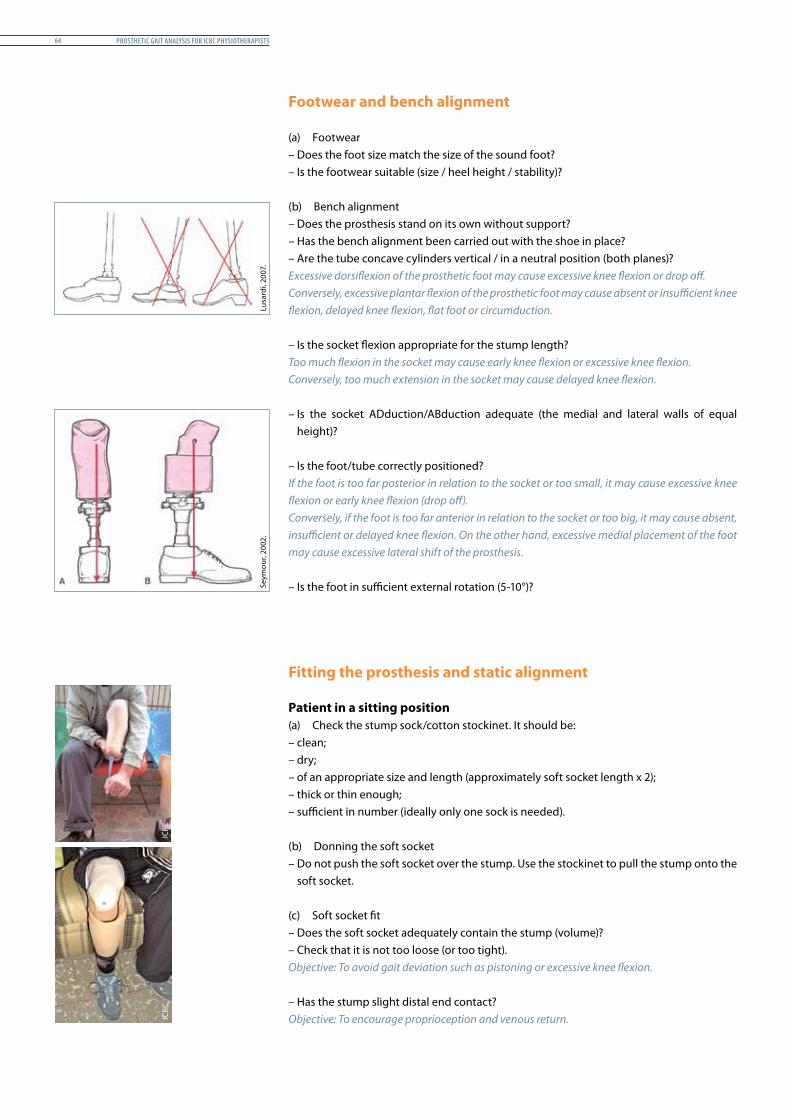
Prosthetic Gait analysis for icrc PhysiotheraPists64
Footwear and bench alignment
(a) Footwear– Does the foot size match the size of the sound foot?– Is the footwear suitable (size / heel height / stability)?
(b) Bench alignment– Does the prosthesis stand on its own without support?– Has the bench alignment been carried out with the shoe in place?– Are the tube concave cylinders vertical / in a neutral position (both planes)?Excessive dorsiflexion of the prosthetic foot may cause excessive knee flexion or drop off.Conversely, excessive plantar flexion of the prosthetic foot may cause absent or insufficient knee flexion, delayed knee flexion, flat foot or circumduction.
– Is the socket flexion appropriate for the stump length?Too much flexion in the socket may cause early knee flexion or excessive knee flexion.Conversely, too much extension in the socket may cause delayed knee flexion.
– Is the socket ADduction/ABduction adequate (the medial and lateral walls of equal height)?
– Is the foot/tube correctly positioned? If the foot is too far posterior in relation to the socket or too small, it may cause excessive knee flexion or early knee flexion (drop off).Conversely, if the foot is too far anterior in relation to the socket or too big, it may cause absent, insufficient or delayed knee flexion. On the other hand, excessive medial placement of the foot may cause excessive lateral shift of the prosthesis.
– Is the foot in sufficient external rotation (5-10°)?
Fitting the prosthesis and static alignment
Patient in a sitting position(a) Check the stump sock/cotton stockinet. It should be:– clean;– dry;– of an appropriate size and length (approximately soft socket length x 2);– thick or thin enough;– sufficient in number (ideally only one sock is needed).
(b) Donning the soft socket– Do not push the soft socket over the stump. Use the stockinet to pull the stump onto the
soft socket. (c) Soft socket fit – Does the soft socket adequately contain the stump (volume)?– Check that it is not too loose (or too tight).Objective: To avoid gait deviation such as pistoning or excessive knee flexion.
– Has the stump slight distal end contact?Objective: To encourage proprioception and venous return.
Seym
our,
2002
.Lu
sard
i, 20
07.
ICRC
ICRC
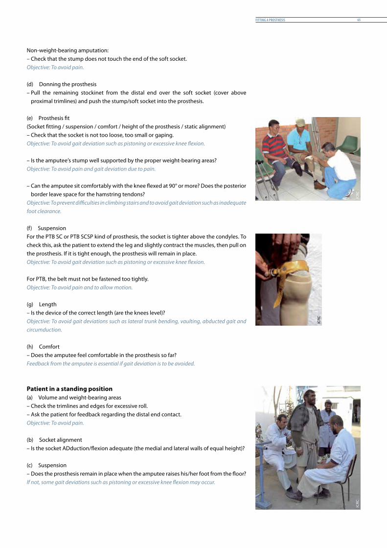
fittinG a Prosthesis 65
Non-weight-bearing amputation:– Check that the stump does not touch the end of the soft socket.Objective: To avoid pain.
(d) Donning the prosthesis– Pull the remaining stockinet from the distal end over the soft socket (cover above
proximal trimlines) and push the stump/soft socket into the prosthesis.
(e) Prosthesis fit (Socket fitting / suspension / comfort / height of the prosthesis / static alignment)– Check that the socket is not too loose, too small or gaping.Objective: To avoid gait deviation such as pistoning or excessive knee flexion.
– Is the amputee’s stump well supported by the proper weight-bearing areas?Objective: To avoid pain and gait deviation due to pain.
– Can the amputee sit comfortably with the knee flexed at 90° or more? Does the posterior border leave space for the hamstring tendons?
Objective: To prevent difficulties in climbing stairs and to avoid gait deviation such as inadequate foot clearance.
(f) SuspensionFor the PTB SC or PTB SCSP kind of prosthesis, the socket is tighter above the condyles. To check this, ask the patient to extend the leg and slightly contract the muscles, then pull on the prosthesis. If it is tight enough, the prosthesis will remain in place.Objective: To avoid gait deviation such as pistoning or excessive knee flexion.
For PTB, the belt must not be fastened too tightly.Objective: To avoid pain and to allow motion.
(g) Length– Is the device of the correct length (are the knees level)?Objective: To avoid gait deviations such as lateral trunk bending, vaulting, abducted gait and circumduction.
(h) Comfort– Does the amputee feel comfortable in the prosthesis so far?Feedback from the amputee is essential if gait deviation is to be avoided.
Patient in a standing position(a) Volume and weight-bearing areas– Check the trimlines and edges for excessive roll.– Ask the patient for feedback regarding the distal end contact.Objective: To avoid pain.
(b) Socket alignment– Is the socket ADduction/flexion adequate (the medial and lateral walls of equal height)?
(c) Suspension– Does the prosthesis remain in place when the amputee raises his/her foot from the floor?If not, some gait deviations such as pistoning or excessive knee flexion may occur.
ICRC
ICRC
ICRC
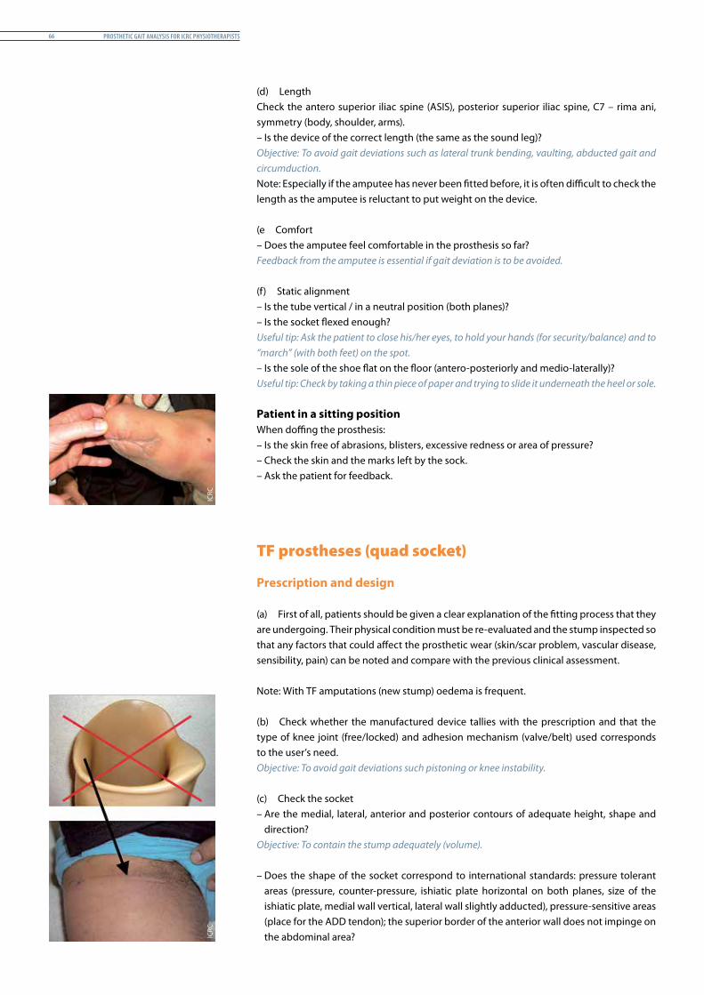
Prosthetic Gait analysis for icrc PhysiotheraPists66
(d) Length Check the antero superior iliac spine (ASIS), posterior superior iliac spine, C7 – rima ani, symmetry (body, shoulder, arms).– Is the device of the correct length (the same as the sound leg)?Objective: To avoid gait deviations such as lateral trunk bending, vaulting, abducted gait and circumduction.Note: Especially if the amputee has never been fitted before, it is often difficult to check the length as the amputee is reluctant to put weight on the device.
(e Comfort– Does the amputee feel comfortable in the prosthesis so far?Feedback from the amputee is essential if gait deviation is to be avoided.
(f) Static alignment– Is the tube vertical / in a neutral position (both planes)?– Is the socket flexed enough?Useful tip: Ask the patient to close his/her eyes, to hold your hands (for security/balance) and to “march” (with both feet) on the spot.– Is the sole of the shoe flat on the floor (antero-posteriorly and medio-laterally)?Useful tip: Check by taking a thin piece of paper and trying to slide it underneath the heel or sole.
Patient in a sitting positionWhen doffing the prosthesis:– Is the skin free of abrasions, blisters, excessive redness or area of pressure?– Check the skin and the marks left by the sock.– Ask the patient for feedback.
TF prostheses (quad socket)
Prescription and design
(a) First of all, patients should be given a clear explanation of the fitting process that they are undergoing. Their physical condition must be re-evaluated and the stump inspected so that any factors that could affect the prosthetic wear (skin/scar problem, vascular disease, sensibility, pain) can be noted and compare with the previous clinical assessment.
Note: With TF amputations (new stump) oedema is frequent. (b) Check whether the manufactured device tallies with the prescription and that the type of knee joint (free/locked) and adhesion mechanism (valve/belt) used corresponds to the user’s need.Objective: To avoid gait deviations such pistoning or knee instability.
(c) Check the socket– Are the medial, lateral, anterior and posterior contours of adequate height, shape and
direction?Objective: To contain the stump adequately (volume).
– Does the shape of the socket correspond to international standards: pressure tolerant areas (pressure, counter-pressure, ishiatic plate horizontal on both planes, size of the ishiatic plate, medial wall vertical, lateral wall slightly adducted), pressure-sensitive areas (place for the ADD tendon); the superior border of the anterior wall does not impinge on the abdominal area?
ICRC
ICRC
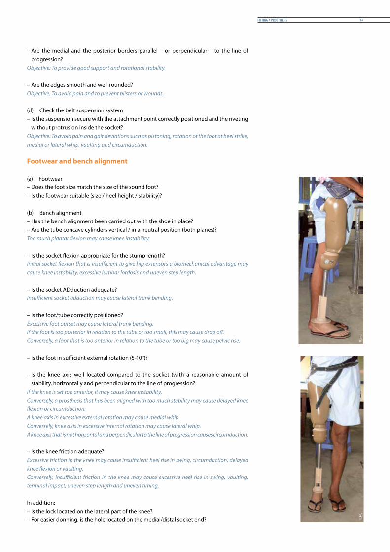
fittinG a Prosthesis 67
– Are the medial and the posterior borders parallel – or perpendicular – to the line of progression?
Objective: To provide good support and rotational stability.
– Are the edges smooth and well rounded?Objective: To avoid pain and to prevent blisters or wounds.
(d) Check the belt suspension system– Is the suspension secure with the attachment point correctly positioned and the riveting
without protrusion inside the socket?Objective: To avoid pain and gait deviations such as pistoning, rotation of the foot at heel strike, medial or lateral whip, vaulting and circumduction.
Footwear and bench alignment
(a) Footwear– Does the foot size match the size of the sound foot?– Is the footwear suitable (size / heel height / stability)?
(b) Bench alignment– Has the bench alignment been carried out with the shoe in place?– Are the tube concave cylinders vertical / in a neutral position (both planes)?Too much plantar flexion may cause knee instability.
– Is the socket flexion appropriate for the stump length?Initial socket flexion that is insufficient to give hip extensors a biomechanical advantage may cause knee instability, excessive lumbar lordosis and uneven step length.
– Is the socket ADduction adequate?Insufficient socket adduction may cause lateral trunk bending.
– Is the foot/tube correctly positioned? Excessive foot outset may cause lateral trunk bending.If the foot is too posterior in relation to the tube or too small, this may cause drop off.Conversely, a foot that is too anterior in relation to the tube or too big may cause pelvic rise.
– Is the foot in sufficient external rotation (5-10°)?
– Is the knee axis well located compared to the socket (with a reasonable amount of stability, horizontally and perpendicular to the line of progression?
If the knee is set too anterior, it may cause knee instability.Conversely, a prosthesis that has been aligned with too much stability may cause delayed knee flexion or circumduction.A knee axis in excessive external rotation may cause medial whip. Conversely, knee axis in excessive internal rotation may cause lateral whip.A knee axis that is not horizontal and perpendicular to the line of progression causes circumduction.
– Is the knee friction adequate?Excessive friction in the knee may cause insufficient heel rise in swing, circumduction, delayed knee flexion or vaulting.Conversely, insufficient friction in the knee may cause excessive heel rise in swing, vaulting, terminal impact, uneven step length and uneven timing.
In addition:– Is the lock located on the lateral part of the knee?– For easier donning, is the hole located on the medial/distal socket end?
ICRC
ICRC
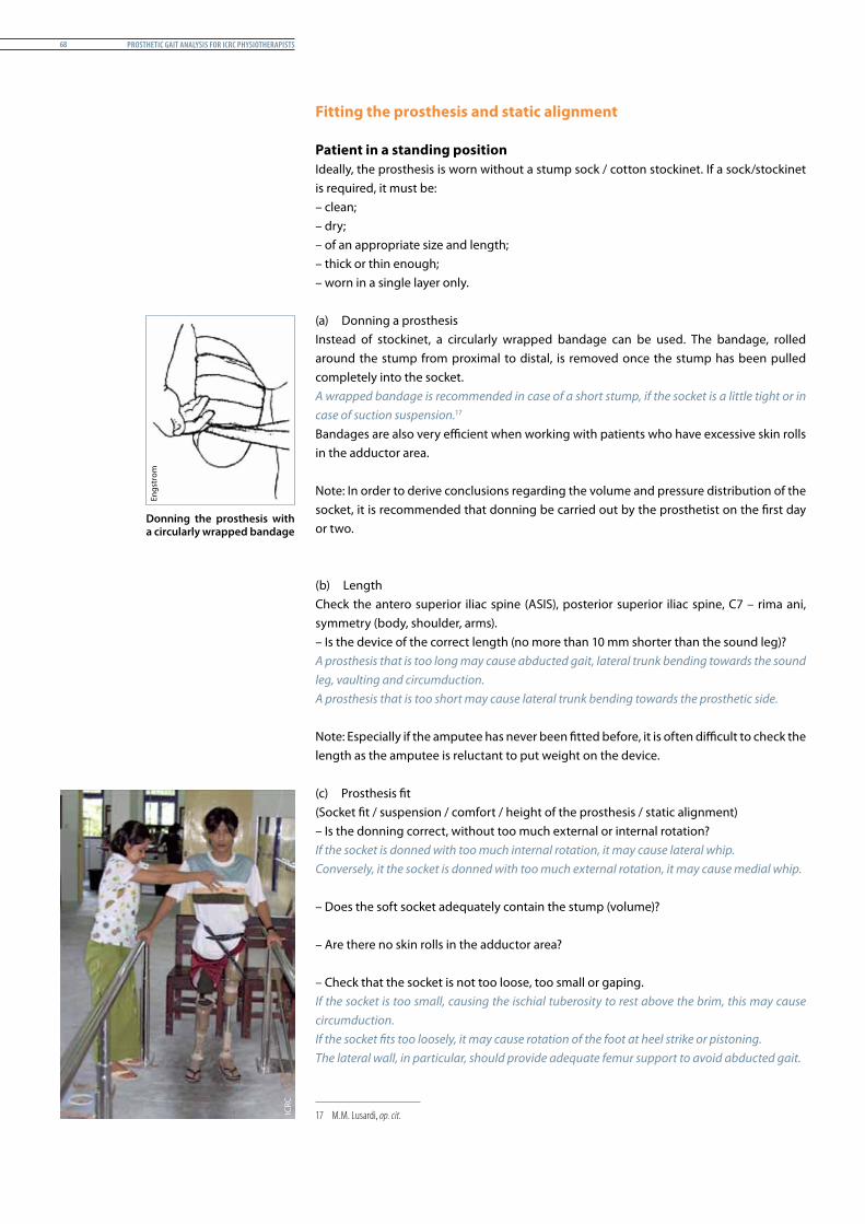
Prosthetic Gait analysis for icrc PhysiotheraPists68
Fitting the prosthesis and static alignment
Patient in a standing positionIdeally, the prosthesis is worn without a stump sock / cotton stockinet. If a sock/stockinet is required, it must be:– clean;– dry;– of an appropriate size and length;– thick or thin enough;– worn in a single layer only.
(a) Donning a prosthesisInstead of stockinet, a circularly wrapped bandage can be used. The bandage, rolled around the stump from proximal to distal, is removed once the stump has been pulled completely into the socket.A wrapped bandage is recommended in case of a short stump, if the socket is a little tight or in case of suction suspension.171 Bandages are also very efficient when working with patients who have excessive skin rolls in the adductor area.
Note: In order to derive conclusions regarding the volume and pressure distribution of the socket, it is recommended that donning be carried out by the prosthetist on the first day or two.
(b) LengthCheck the antero superior iliac spine (ASIS), posterior superior iliac spine, C7 – rima ani, symmetry (body, shoulder, arms).– Is the device of the correct length (no more than 10 mm shorter than the sound leg)?A prosthesis that is too long may cause abducted gait, lateral trunk bending towards the sound leg, vaulting and circumduction.A prosthesis that is too short may cause lateral trunk bending towards the prosthetic side.
Note: Especially if the amputee has never been fitted before, it is often difficult to check the length as the amputee is reluctant to put weight on the device.
(c) Prosthesis fit(Socket fit / suspension / comfort / height of the prosthesis / static alignment)– Is the donning correct, without too much external or internal rotation?If the socket is donned with too much internal rotation, it may cause lateral whip. Conversely, it the socket is donned with too much external rotation, it may cause medial whip.
– Does the soft socket adequately contain the stump (volume)?
– Are there no skin rolls in the adductor area?
– Check that the socket is not too loose, too small or gaping.If the socket is too small, causing the ischial tuberosity to rest above the brim, this may cause circumduction.If the socket fits too loosely, it may cause rotation of the foot at heel strike or pistoning.The lateral wall, in particular, should provide adequate femur support to avoid abducted gait.
17 M.M. Lusardi, op. cit.
Donning the prosthesis with a circularly wrapped bandage
Engs
trom
ICRC
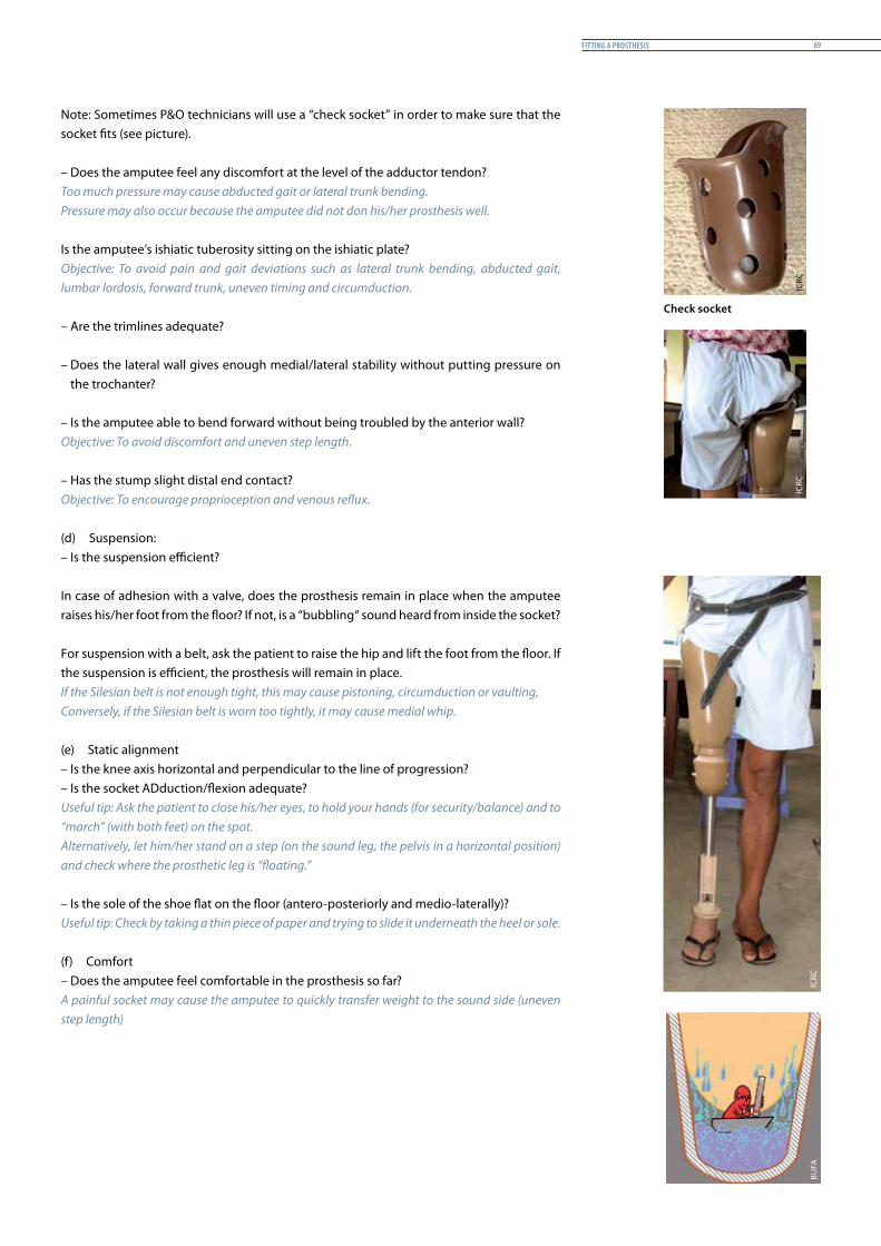
fittinG a Prosthesis 69
Note: Sometimes P&O technicians will use a “check socket” in order to make sure that the socket fits (see picture).
– Does the amputee feel any discomfort at the level of the adductor tendon?Too much pressure may cause abducted gait or lateral trunk bending.Pressure may also occur because the amputee did not don his/her prosthesis well.
Is the amputee’s ishiatic tuberosity sitting on the ishiatic plate?Objective: To avoid pain and gait deviations such as lateral trunk bending, abducted gait, lumbar lordosis, forward trunk, uneven timing and circumduction. – Are the trimlines adequate?
– Does the lateral wall gives enough medial/lateral stability without putting pressure on the trochanter?
– Is the amputee able to bend forward without being troubled by the anterior wall?Objective: To avoid discomfort and uneven step length. – Has the stump slight distal end contact?Objective: To encourage proprioception and venous reflux.
(d) Suspension:– Is the suspension efficient?
In case of adhesion with a valve, does the prosthesis remain in place when the amputee raises his/her foot from the floor? If not, is a “bubbling“ sound heard from inside the socket?
For suspension with a belt, ask the patient to raise the hip and lift the foot from the floor. If the suspension is efficient, the prosthesis will remain in place.If the Silesian belt is not enough tight, this may cause pistoning, circumduction or vaulting,Conversely, if the Silesian belt is worn too tightly, it may cause medial whip. (e) Static alignment– Is the knee axis horizontal and perpendicular to the line of progression?– Is the socket ADduction/flexion adequate?Useful tip: Ask the patient to close his/her eyes, to hold your hands (for security/balance) and to “march” (with both feet) on the spot.Alternatively, let him/her stand on a step (on the sound leg, the pelvis in a horizontal position) and check where the prosthetic leg is “floating.”
– Is the sole of the shoe flat on the floor (antero-posteriorly and medio-laterally)?Useful tip: Check by taking a thin piece of paper and trying to slide it underneath the heel or sole. (f) Comfort– Does the amputee feel comfortable in the prosthesis so far?A painful socket may cause the amputee to quickly transfer weight to the sound side (uneven step length)
Check socket
BUFA
ICRC
ICRC
ICRC
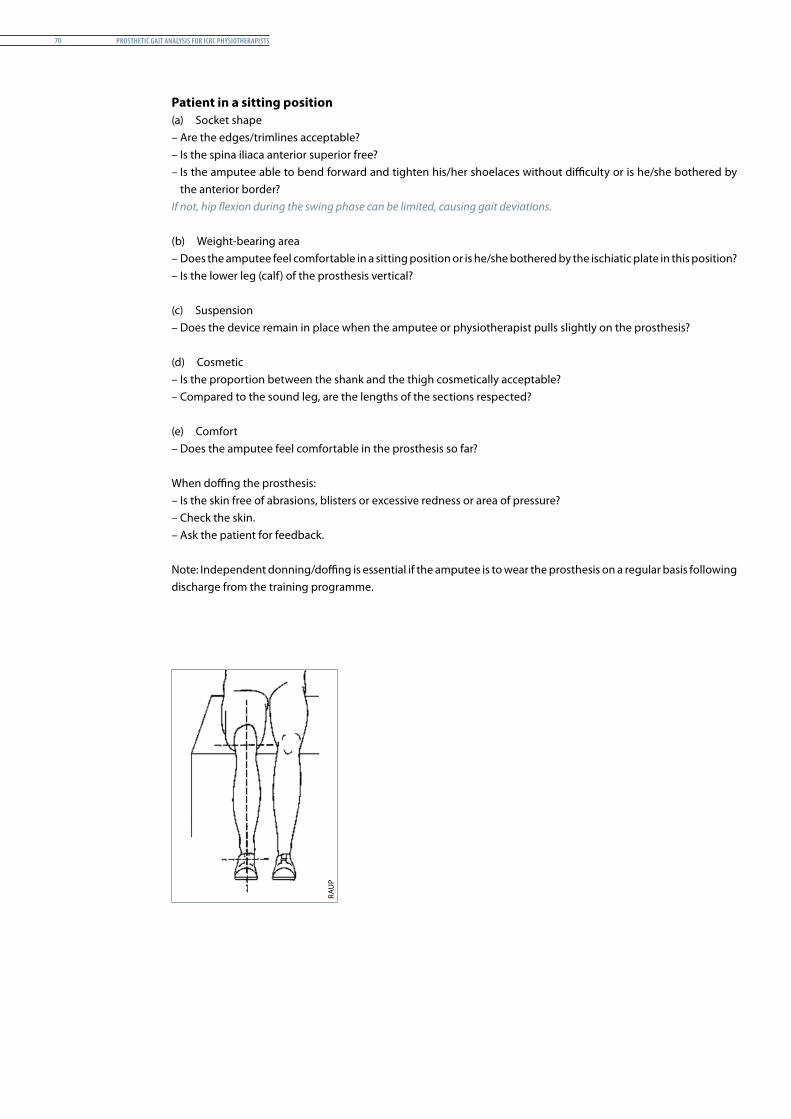
Prosthetic Gait analysis for icrc PhysiotheraPists70
Patient in a sitting position(a) Socket shape– Are the edges/trimlines acceptable?– Is the spina iliaca anterior superior free?– Is the amputee able to bend forward and tighten his/her shoelaces without difficulty or is he/she bothered by
the anterior border? If not, hip flexion during the swing phase can be limited, causing gait deviations.
(b) Weight-bearing area– Does the amputee feel comfortable in a sitting position or is he/she bothered by the ischiatic plate in this position?– Is the lower leg (calf) of the prosthesis vertical?
(c) Suspension– Does the device remain in place when the amputee or physiotherapist pulls slightly on the prosthesis?
(d) Cosmetic– Is the proportion between the shank and the thigh cosmetically acceptable?– Compared to the sound leg, are the lengths of the sections respected?
(e) Comfort– Does the amputee feel comfortable in the prosthesis so far?
When doffing the prosthesis:– Is the skin free of abrasions, blisters or excessive redness or area of pressure?– Check the skin.– Ask the patient for feedback.
Note: Independent donning/doffing is essential if the amputee is to wear the prosthesis on a regular basis following discharge from the training programme.
RAU
P
normal Gait and Prosthetic Gait 71
normal Gait and Prosthetic Gait
Introduction
Content
This chapter recalls the main aspects of normal human gait and prosthetic gait. Prosthetic gait is explained so as to promote an understanding of the optimal expectations for an amputee with a prosthesis, depending on the type of amputation and the kind of components used in the prosthetic device.
Rationale
It is important to understand normal gait and prosthetic gait before proceeding to identify the gait deviations of a prosthesis user. The user’s gait will be compared with the theoretical optimum prosthetic gait.
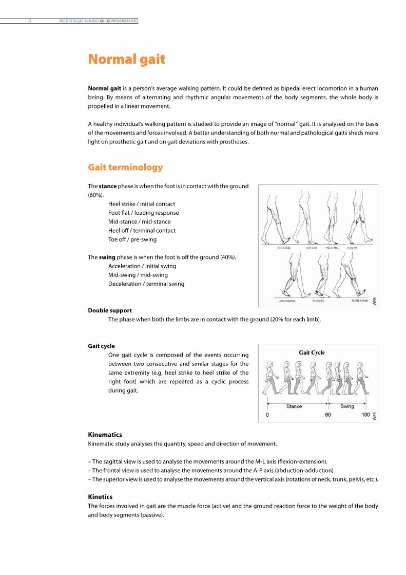
Prosthetic Gait analysis for icrc PhysiotheraPists72
Normal gait
Normal gait is a person’s average walking pattern. It could be defined as bipedal erect locomotion in a human being. By means of alternating and rhythmic angular movements of the body segments, the whole body is propelled in a linear movement.
A healthy individual’s walking pattern is studied to provide an image of “normal” gait. It is analysed on the basis of the movements and forces involved. A better understanding of both normal and pathological gaits sheds more light on prosthetic gait and on gait deviations with prostheses.
Gait terminology
The stance phase is when the foot is in contact with the ground (60%).
Heel strike / initial contactFoot flat / loading responseMid-stance / mid-stanceHeel off / terminal contactToe off / pre-swing
The swing phase is when the foot is off the ground (40%).Acceleration / initial swingMid-swing / mid-swingDeceleration / terminal swing
Double supportThe phase when both the limbs are in contact with the ground (20% for each limb).
Gait cycleOne gait cycle is composed of the events occurring between two consecutive and similar stages for the same extremity (e.g. heel strike to heel strike of the right foot) which are repeated as a cyclic process during gait.
KinematicsKinematic study analyses the quantity, speed and direction of movement.
– The sagittal view is used to analyse the movements around the M-L axis (flexion-extension).– The frontal view is used to analyse the movements around the A-P axis (abduction-adduction).– The superior view is used to analyse the movements around the vertical axis (rotations of neck, trunk, pelvis, etc.).
KineticsThe forces involved in gait are the muscle force (active) and the ground reaction force to the weight of the body and body segments (passive).
WEB
WEB
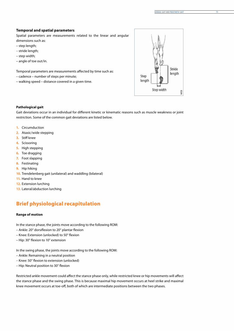
normal Gait and Prosthetic Gait 73
Temporal and spatial parametersSpatial parameters are measurements related to the linear and angular dimensions such as:– step length;– stride length; – step width; – angle of toe out/in.
Temporal parameters are measurements affected by time such as:– cadence – number of steps per minute;– walking speed – distance covered in a given time.
Pathological gaitGait deviations occur in an individual for different kinetic or kinematic reasons such as muscle weakness or joint restriction. Some of the common gait deviations are listed below.
1. Circumduction 2. Ataxic/wide stepping3. Stiff knee4. Scissoring5. High stepping6. Toe dragging7. Foot slapping8. Festinating9. Hip hiking10. Trendelenberg gait (unilateral) and waddling (bilateral) 11. Hand to knee12. Extension lurching13. Lateral/abduction lurching
Brief physiological recapitulation
Range of motion
In the stance phase, the joints move according to the following ROM:– Ankle: 20° dorsiflexion to 20° plantar flexion– Knee: Extension (unlocked) to 50° flexion– Hip: 30° flexion to 10° extension
In the swing phase, the joints move according to the following ROM:– Ankle: Remaining in a neutral position– Knee: 50° flexion to extension (unlocked)– Hip: Neutral position to 30° flexion
Restricted ankle movement could affect the stance phase only, while restricted knee or hip movements will affect the stance phase and the swing phase. This is because maximal hip movement occurs at heel strike and maximal knee movement occurs at toe-off, both of which are intermediate positions between the two phases.
WEB
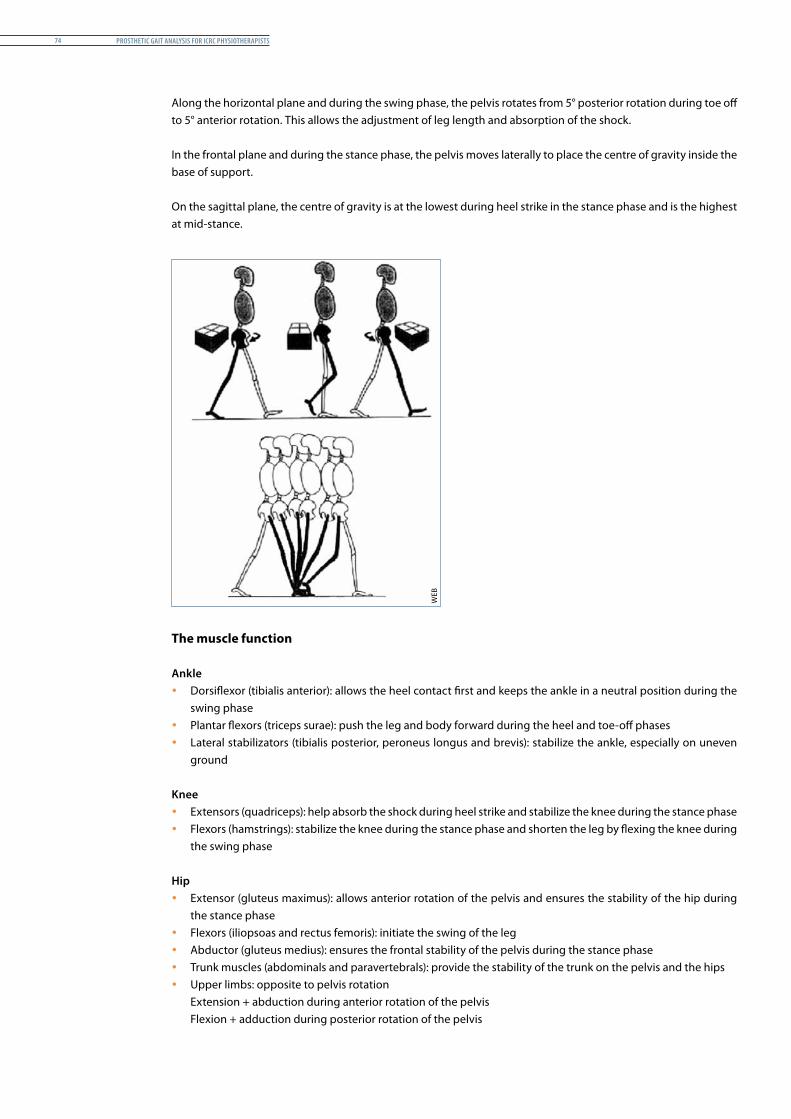
Prosthetic Gait analysis for icrc PhysiotheraPists74
Along the horizontal plane and during the swing phase, the pelvis rotates from 5° posterior rotation during toe off to 5° anterior rotation. This allows the adjustment of leg length and absorption of the shock.
In the frontal plane and during the stance phase, the pelvis moves laterally to place the centre of gravity inside the base of support.
On the sagittal plane, the centre of gravity is at the lowest during heel strike in the stance phase and is the highest at mid-stance.
The muscle function
Ankle y Dorsiflexor (tibialis anterior): allows the heel contact first and keeps the ankle in a neutral position during the
swing phase y Plantar flexors (triceps surae): push the leg and body forward during the heel and toe-off phases y Lateral stabilizators (tibialis posterior, peroneus longus and brevis): stabilize the ankle, especially on uneven
ground
Knee y Extensors (quadriceps): help absorb the shock during heel strike and stabilize the knee during the stance phase y Flexors (hamstrings): stabilize the knee during the stance phase and shorten the leg by flexing the knee during
the swing phase
Hip y Extensor (gluteus maximus): allows anterior rotation of the pelvis and ensures the stability of the hip during
the stance phase y Flexors (iliopsoas and rectus femoris): initiate the swing of the leg y Abductor (gluteus medius): ensures the frontal stability of the pelvis during the stance phase y Trunk muscles (abdominals and paravertebrals): provide the stability of the trunk on the pelvis and the hips y Upper limbs: opposite to pelvis rotation
Extension + abduction during anterior rotation of the pelvis Flexion + adduction during posterior rotation of the pelvis
WEB
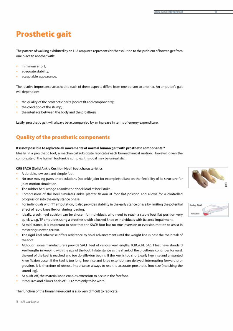
normal Gait and Prosthetic Gait 75
Prosthetic gait
The pattern of walking exhibited by an LLA amputee represents his/her solution to the problem of how to get from one place to another with:
y minimum effort; y adequate stability; y acceptable appearance.
The relative importance attached to each of these aspects differs from one person to another. An amputee’s gait will depend on:
y the quality of the prosthetic parts (socket fit and components); y the condition of the stump; y the interface between the body and the prosthesis.
Lastly, prosthetic gait will always be accompanied by an increase in terms of energy expenditure.
Quality of the prosthetic components
It is not possible to replicate all movements of normal human gait with prosthetic components.181
Ideally, in a prosthetic foot, a mechanical substitute replicates each biomechanical motion. However, given the complexity of the human foot-ankle complex, this goal may be unrealistic.
CRE SACH (Solid Ankle Cushion Heel) foot characteristics y A durable, low cost and simple foot. y No true moving parts or articulations (no ankle joint for example); reliant on the flexibility of its structure for
joint motion simulation. y The rubber heel wedge absorbs the shock load at heel strike. y Compression of the heel simulates ankle plantar flexion at foot flat position and allows for a controlled
progression into the early stance phase. y For individuals with TT amputation, it also provides stability in the early stance phase by limiting the potential
effect of rapid knee flexion during loading. y Ideally, a soft heel cushion can be chosen for individuals who need to reach a stable foot flat position very
quickly, e.g. TF amputees using a prosthesis with a locked knee or individuals with balance impairment. y At mid-stance, it is important to note that the SACH foot has no true inversion or eversion motion to assist in
mastering uneven terrain. y The rigid keel otherwise offers resistance to tibial advancement until the weight line is past the toe break of
the foot. y Although some manufacturers provide SACH feet of various keel lengths, ICRC/CRE SACH feet have standard
keel lengths in keeping with the size of the foot. In late stance as the shank of the prosthesis continues forward, the end of the keel is reached and toe dorsiflexion begins. If the keel is too short, early heel rise and unwanted knee flexion occur. If the keel is too long, heel rise and knee extension are delayed, interrupting forward pro-gression. It is therefore of utmost importance always to use the accurate prosthetic foot size (matching the sound leg).
y At push-off, the material used enables extension to occur in the forefoot. y It requires and allows heels of 10-12 mm only to be worn.
The function of the human knee joint is also very difficult to replicate.
18 M.M. Lusardi, op. cit.
ICRC
Kirtley, 2006.
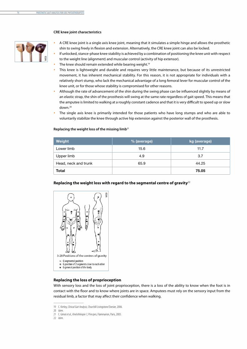
Prosthetic Gait analysis for icrc PhysiotheraPists76
CRE knee joint characteristics
y A CRE knee joint is a single axis knee joint, meaning that it simulates a simple hinge and allows the prosthetic shin to swing freely in flexion and extension. Alternatively, the CRE knee joint can also be locked.
y If unlocked, stance-phase knee stability is achieved by a combination of positioning the knee unit with respect to the weight line (alignment) and muscular control (activity of hip extensor).
y The knee should remain extended while bearing weight.192
y This knee is lightweight and durable and requires very little maintenance, but because of its unrestricted movement, it has inherent mechanical stability. For this reason, it is not appropriate for individuals with a relatively short stump, who lack the mechanical advantage of a long femoral lever for muscular control of the knee unit, or for those whose stability is compromised for other reasons.
y Although the rate of advancement of the shin during the swing phase can be influenced slightly by means of an elastic strap, the shin of the prosthesis will swing at the same rate regardless of gait speed. This means that the amputee is limited to walking at a roughly constant cadence and that it is very difficult to speed up or slow down.203
y The single axis knee is primarily intended for those patients who have long stumps and who are able to voluntarily stabilize the knee through active hip extension against the posterior wall of the prosthesis.
Replacing the weight loss of the missing limb214
Weight % (average) kg (average)
Lower limb 15.6 11.7
Upper limb 4.9 3.7
Head, neck and trunk 65.9 44.25
Total 75.05
Replacing the weight loss with regard to the segmental centre of gravity225
Replacing the loss of proprioceptionWith sensory loss and the loss of joint proprioception, there is a loss of the ability to know when the foot is in contact with the floor and to know where joints are in space. Amputees must rely on the sensory input from the residual limb, a factor that may affect their confidence when walking.
19 C. Kirtley, Clinical Gait Analysis, Churchill Livingstone Elvesier, 2006.20 Idem.21 C. Génot et al., Kinésithérapie 1, Principes, Flammarion, Paris, 2003.22 Idem.
WEB
WEB
normal Gait and Prosthetic Gait 77
Condition of the stump – energy expenditure
Major lower limb amputation increases the gait energy expenditure
The amount of energy required by amputees depends on the cause of the amputation: traumatic amputation is less costly in terms of energy than amputation due to vascular disease.
Amputation level % increase in oxygen consumption
Transtibial (vascular) 33
Transtibial (traumatic) 7
Knee disarticulation (traumatic) 53
Transfemoral vascular 87
The amount of energy required by amputees depends on the level of amputation (including the length of the stump).
Non-disabled Unilateral BK
Bilateral BK
Unilateral AK
Bilateral AK
Polio with unilateral
KAFO
Energy expenditure
100% 110-125% 180-200% 150-170% 300-400% 190-210%
Steps/minute 70-90 60-80 35-45 40-50 15-25 35-45
Distance/minute 70-80 m 60-70 m 25-35 m 30-40 m 15-25 m 20-30
The different energy expenditure of TT and TF amputees explains why surgeons will always try to save the knee joint whenever possible. The knee joint is fundamental in terms of energy expenditure as it minimizes variations in the displacement of the CoM (centre of mass) that are very costly, and plays a role in shock absorption.
Contractions of the gluteus and psoas muscles increase on the amputated side and are also very costly in terms of energy expenditure:– The hamstrings are no longer polyarticular and powerful. Therefore, increased contraction of the gluteus occurs
on the amputated side. – The hip has to flex just before toe raise in order to flex the prosthetic knee. This is mainly effected by the psoas,
which is not as active during normal gait (as the movement is performed by the TFL, sartorius, add magnus and rectus femoris).
– In case of amputation, the hip flexion is increased (psoas) to compensate for the fact that there is no ankle movement and to bring the knee into extension.
Age: For amputees and non-amputees, gait becomes less efficient with age.
The matter of knee locking must be mentioned here. When the prosthetic knee remains unlocked, the energy expenditure is increased for some users but not for others.
In fact, a distinction may be made between two groups of TF amputees:– older users who can walk without too much effort and faster when the knee is locked;– younger users who walk faster when the knee is unlocked but with higher energy expenditure.
Crutches/prosthesis: Non-vascular TT amputees expend less energy when walking with a prosthesis than when
Hue
rta
and
Mill
er, 2
002.
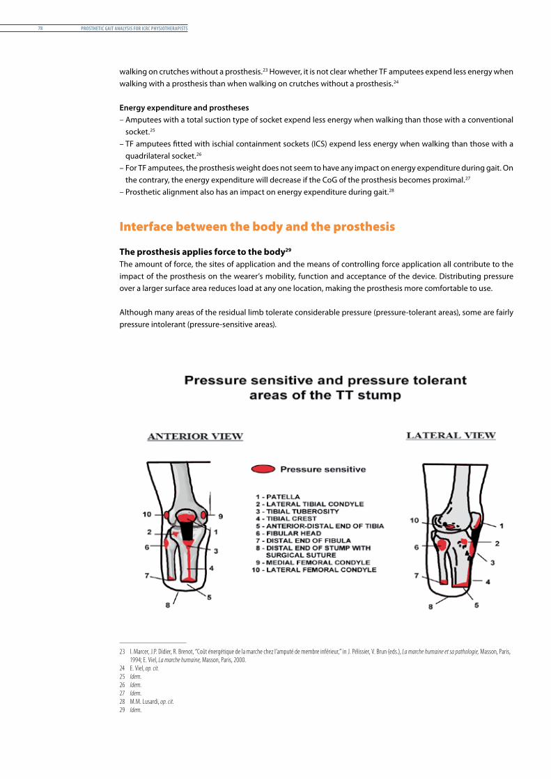
Prosthetic Gait analysis for icrc PhysiotheraPists78
walking on crutches without a prosthesis.23 However, it is not clear whether TF amputees expend less energy when walking with a prosthesis than when walking on crutches without a prosthesis.24 67
Energy expenditure and prostheses– Amputees with a total suction type of socket expend less energy when walking than those with a conventional
socket.258
– TF amputees fitted with ischial containment sockets (ICS) expend less energy when walking than those with a quadrilateral socket.269
– For TF amputees, the prosthesis weight does not seem to have any impact on energy expenditure during gait. On the contrary, the energy expenditure will decrease if the CoG of the prosthesis becomes proximal.2710
– Prosthetic alignment also has an impact on energy expenditure during gait.2811
Interface between the body and the prosthesis
The prosthesis applies force to the body2912
The amount of force, the sites of application and the means of controlling force application all contribute to the impact of the prosthesis on the wearer’s mobility, function and acceptance of the device. Distributing pressure over a larger surface area reduces load at any one location, making the prosthesis more comfortable to use.
Although many areas of the residual limb tolerate considerable pressure (pressure-tolerant areas), some are fairly pressure intolerant (pressure-sensitive areas).
23 I. Marcer, J.P. Didier, R. Brenot, “Coût énergétique de la marche chez l’amputé de membre inférieur,” in J. Pélissier, V. Brun (eds.), La marche humaine et sa pathologie, Masson, Paris, 1994; E. Viel, La marche humaine, Masson, Paris, 2000.
24 E. Viel, op. cit.25 Idem.26 Idem.27 Idem.28 M.M. Lusardi, op. cit.29 Idem.
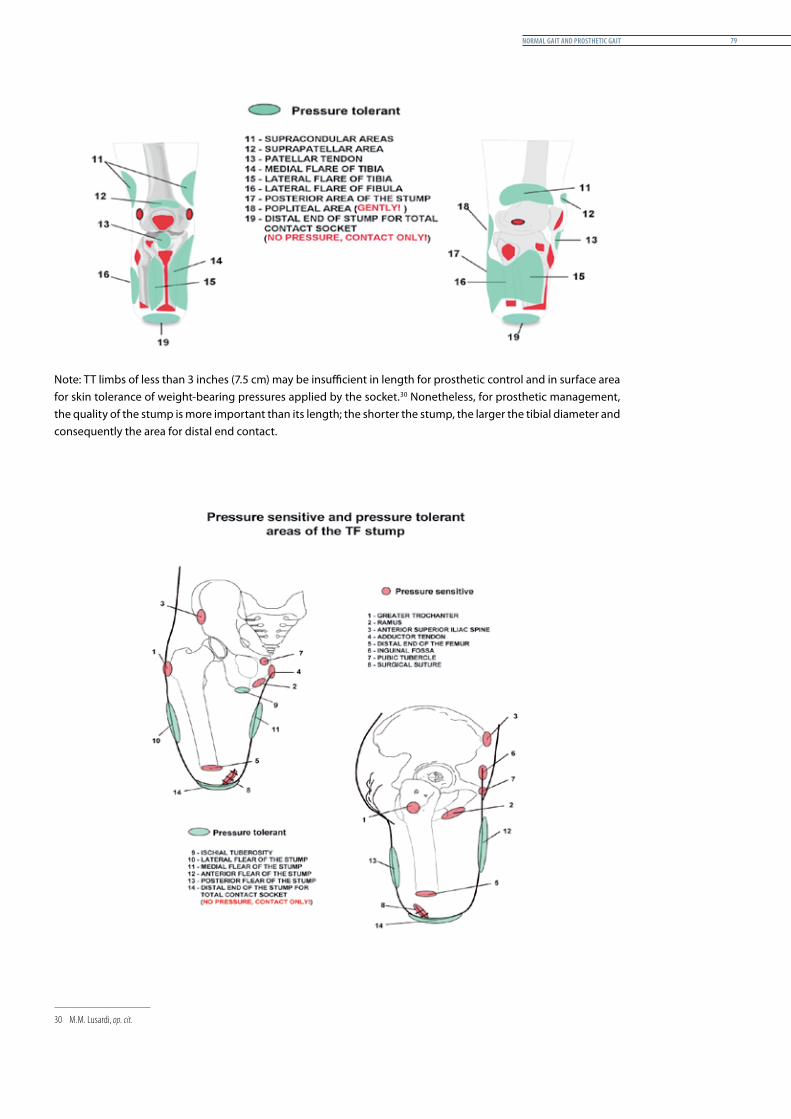
normal Gait and Prosthetic Gait 79
Note: TT limbs of less than 3 inches (7.5 cm) may be insufficient in length for prosthetic control and in surface area for skin tolerance of weight-bearing pressures applied by the socket.30 Nonetheless, for prosthetic management, the quality of the stump is more important than its length; the shorter the stump, the larger the tibial diameter and consequently the area for distal end contact.1
30 M.M. Lusardi, op. cit.
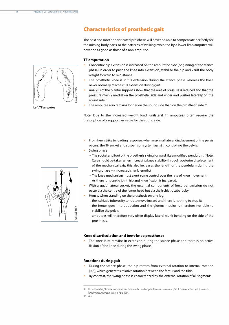
Prosthetic Gait analysis for icrc PhysiotheraPists80
Characteristics of prosthetic gait
The best and most sophisticated prosthesis will never be able to compensate perfectly for the missing body parts so the patterns of walking exhibited by a lower-limb amputee will never be as good as those of a non-amputee.
TF amputation y Concentric hip extension is increased on the amputated side (beginning of the stance
phase) in order to push the knee into extension, stabilize the hip and vault the body weight forward to mid-stance.
y The prosthetic knee is in full extension during the stance phase whereas the knee never normally reaches full extension during gait.
y Analysis of the plantar supports show that the area of pressure is reduced and that the pressure mainly medial on the prosthetic side and wider and pushes laterally on the sound side.311
y The amputee also remains longer on the sound side than on the prosthetic side.322
Note: Due to the increased weight load, unilateral TF amputees often require the prescription of a supportive insole for the sound side.
y From heel strike to loading response, when maximal lateral displacement of the pelvis occurs, the TF socket and suspension system assist in controlling the pelvis.
y Swing phase– The socket and foot of the prosthesis swing forward like a modified pendulum. (Note:
Care should be taken when increasing knee stability through posterior displacement of the mechanical axis; this also increases the length of the pendulum during the swing phase => increased shank length.)
– The knee mechanism must exert some control over the rate of knee movement.– As there is no ankle joint, hip and knee flexion is increased.
y With a quadrilateral socket, the essential components of force transmission do not occur via the centre of the femur head but via the ischiatic tuberosity.
y Hence, when standing on the prosthesis on one leg:– the ischiatic tuberosity tends to move inward and there is nothing to stop it;– the femur goes into abduction and the gluteus medius is therefore not able to
stabilize the pelvis;– amputees will therefore very often display lateral trunk bending on the side of the
prosthesis.
Knee disarticulation and bent-knee prostheses y The knee joint remains in extension during the stance phase and there is no active
flexion of the knee during the swing phase.
Rotations during gait y During the stance phase, the hip rotates from external rotation to internal rotation
(10°), which generates relative rotation between the femur and the tibia. y By contrast, the swing phase is characterized by the external rotation of all segments.
31 M. Enjalbert et al., “Cinématique et cinétique de la marche chez l’amputé des membres inférieurs,” in: J. Pelissier, V. Brun (eds.), La marche humaine et sa pathologie, Masson, Paris, 1994.
32 Idem.
Left TF amputee
Enja
lber
t, 19
94.
Koke
gei,
2007
.
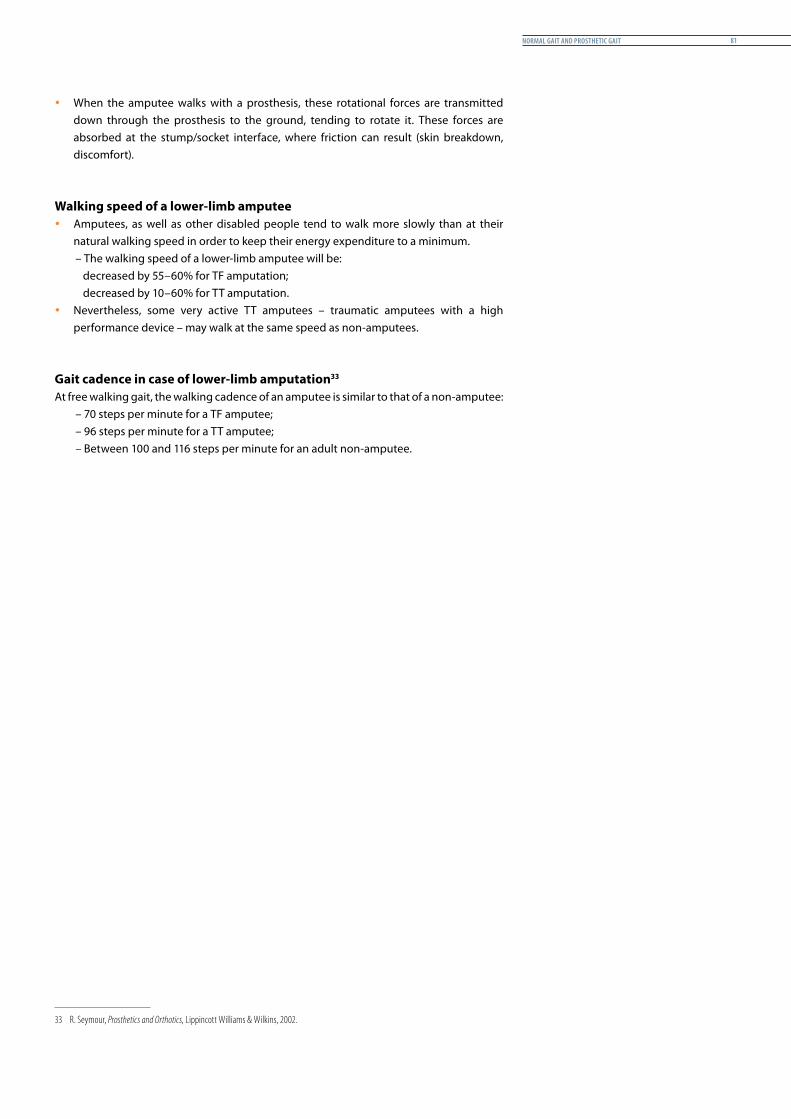
normal Gait and Prosthetic Gait 81
y When the amputee walks with a prosthesis, these rotational forces are transmitted down through the prosthesis to the ground, tending to rotate it. These forces are absorbed at the stump/socket interface, where friction can result (skin breakdown, discomfort).
Walking speed of a lower-limb amputee y Amputees, as well as other disabled people tend to walk more slowly than at their
natural walking speed in order to keep their energy expenditure to a minimum.– The walking speed of a lower-limb amputee will be:
decreased by 55–60% for TF amputation;decreased by 10–60% for TT amputation.
y Nevertheless, some very active TT amputees – traumatic amputees with a high performance device – may walk at the same speed as non-amputees.
Gait cadence in case of lower-limb amputation333
At free walking gait, the walking cadence of an amputee is similar to that of a non-amputee:– 70 steps per minute for a TF amputee;– 96 steps per minute for a TT amputee;– Between 100 and 116 steps per minute for an adult non-amputee.
33 R. Seymour, Prosthetics and Orthotics, Lippincott Williams & Wilkins, 2002.

Prosthetic Gait analysis for icrc PhysiotheraPists82
Gait analysis and Gait deviations 83
Gait analysis and Gait deviations
Introduction
Content
This chapter recalls the general principles of gait analysis and describes the main gait deviations seen in cases of TT or TF amputation. An explanation is given of the deviations that occur when the actual gait of a prosthesis user fails to comply with normal expectations and accepted deviations.
Rationale
The reader is expected to be able to analyse, in a multidisciplinary approach, the gait of an amputee walking with his/her prosthesis, to compare the gait of the evaluated user, to determine the cause(s) of the deviations and to recommend possible solutions for rectifying or minimizing the faults.
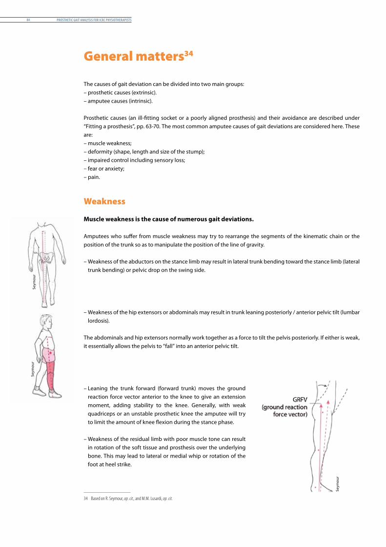
Prosthetic Gait analysis for icrc PhysiotheraPists84
General matters341
The causes of gait deviation can be divided into two main groups:– prosthetic causes (extrinsic).– amputee causes (intrinsic).
Prosthetic causes (an ill-fitting socket or a poorly aligned prosthesis) and their avoidance are described under “Fitting a prosthesis”, pp. 63-70. The most common amputee causes of gait deviations are considered here. These are:– muscle weakness; – deformity (shape, length and size of the stump);– impaired control including sensory loss;– fear or anxiety; – pain.
Weakness
Muscle weakness is the cause of numerous gait deviations.
Amputees who suffer from muscle weakness may try to rearrange the segments of the kinematic chain or the position of the trunk so as to manipulate the position of the line of gravity.
– Weakness of the abductors on the stance limb may result in lateral trunk bending toward the stance limb (lateral trunk bending) or pelvic drop on the swing side.
– Weakness of the hip extensors or abdominals may result in trunk leaning posteriorly / anterior pelvic tilt (lumbar lordosis).
The abdominals and hip extensors normally work together as a force to tilt the pelvis posteriorly. If either is weak, it essentially allows the pelvis to “fall” into an anterior pelvic tilt.
– Leaning the trunk forward (forward trunk) moves the ground reaction force vector anterior to the knee to give an extension moment, adding stability to the knee. Generally, with weak quadriceps or an unstable prosthetic knee the amputee will try to limit the amount of knee flexion during the stance phase.
– Weakness of the residual limb with poor muscle tone can result in rotation of the soft tissue and prosthesis over the underlying bone. This may lead to lateral or medial whip or rotation of the foot at heel strike.
34 Based on R. Seymour, op. cit., and M.M. Lusardi, op. cit.
Seym
our
Seym
our
Seym
our
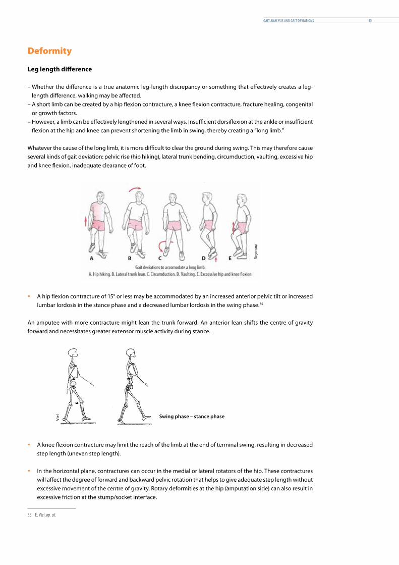
Gait analysis and Gait deviations 85
Deformity
Leg length difference
– Whether the difference is a true anatomic leg-length discrepancy or something that effectively creates a leg-length difference, walking may be affected.
– A short limb can be created by a hip flexion contracture, a knee flexion contracture, fracture healing, congenital or growth factors.
– However, a limb can be effectively lengthened in several ways. Insufficient dorsiflexion at the ankle or insufficient flexion at the hip and knee can prevent shortening the limb in swing, thereby creating a “long limb.”
Whatever the cause of the long limb, it is more difficult to clear the ground during swing. This may therefore cause several kinds of gait deviation: pelvic rise (hip hiking), lateral trunk bending, circumduction, vaulting, excessive hip and knee flexion, inadequate clearance of foot.
y A hip flexion contracture of 15° or less may be accommodated by an increased anterior pelvic tilt or increased lumbar lordosis in the stance phase and a decreased lumbar lordosis in the swing phase.352
An amputee with more contracture might lean the trunk forward. An anterior lean shifts the centre of gravity forward and necessitates greater extensor muscle activity during stance.
y A knee flexion contracture may limit the reach of the limb at the end of terminal swing, resulting in decreased step length (uneven step length).
y In the horizontal plane, contractures can occur in the medial or lateral rotators of the hip. These contractures will affect the degree of forward and backward pelvic rotation that helps to give adequate step length without excessive movement of the centre of gravity. Rotary deformities at the hip (amputation side) can also result in excessive friction at the stump/socket interface.
35 E. Viel, op. cit.
Swing phase – stance phase
Seym
our
Viel
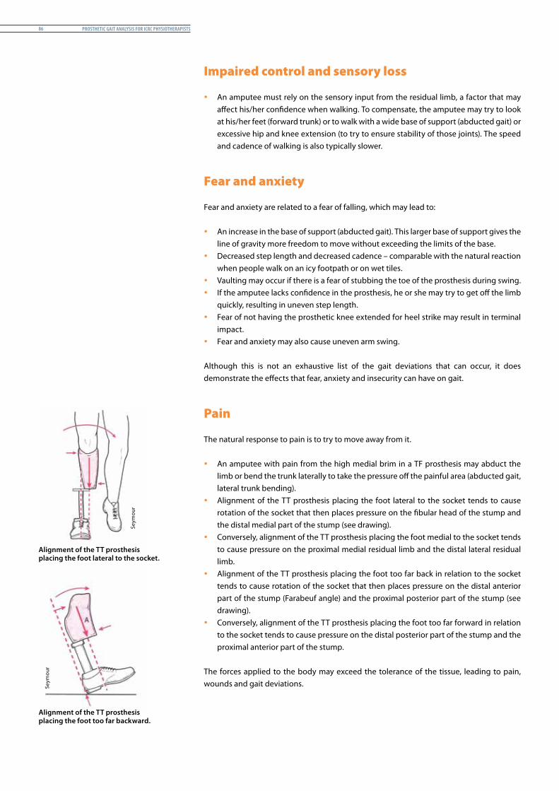
Prosthetic Gait analysis for icrc PhysiotheraPists86
Impaired control and sensory loss
y An amputee must rely on the sensory input from the residual limb, a factor that may affect his/her confidence when walking. To compensate, the amputee may try to look at his/her feet (forward trunk) or to walk with a wide base of support (abducted gait) or excessive hip and knee extension (to try to ensure stability of those joints). The speed and cadence of walking is also typically slower.
Fear and anxiety
Fear and anxiety are related to a fear of falling, which may lead to:
y An increase in the base of support (abducted gait). This larger base of support gives the line of gravity more freedom to move without exceeding the limits of the base.
y Decreased step length and decreased cadence – comparable with the natural reaction when people walk on an icy footpath or on wet tiles.
y Vaulting may occur if there is a fear of stubbing the toe of the prosthesis during swing. y If the amputee lacks confidence in the prosthesis, he or she may try to get off the limb
quickly, resulting in uneven step length. y Fear of not having the prosthetic knee extended for heel strike may result in terminal
impact. y Fear and anxiety may also cause uneven arm swing.
Although this is not an exhaustive list of the gait deviations that can occur, it does demonstrate the effects that fear, anxiety and insecurity can have on gait.
Pain
The natural response to pain is to try to move away from it.
y An amputee with pain from the high medial brim in a TF prosthesis may abduct the limb or bend the trunk laterally to take the pressure off the painful area (abducted gait, lateral trunk bending).
y Alignment of the TT prosthesis placing the foot lateral to the socket tends to cause rotation of the socket that then places pressure on the fibular head of the stump and the distal medial part of the stump (see drawing).
y Conversely, alignment of the TT prosthesis placing the foot medial to the socket tends to cause pressure on the proximal medial residual limb and the distal lateral residual limb.
y Alignment of the TT prosthesis placing the foot too far back in relation to the socket tends to cause rotation of the socket that then places pressure on the distal anterior part of the stump (Farabeuf angle) and the proximal posterior part of the stump (see drawing).
y Conversely, alignment of the TT prosthesis placing the foot too far forward in relation to the socket tends to cause pressure on the distal posterior part of the stump and the proximal anterior part of the stump.
The forces applied to the body may exceed the tolerance of the tissue, leading to pain, wounds and gait deviations.
Alignment of the TT prosthesis placing the foot too far backward.
Alignment of the TT prosthesis placing the foot lateral to the socket.
Seym
our
Seym
our
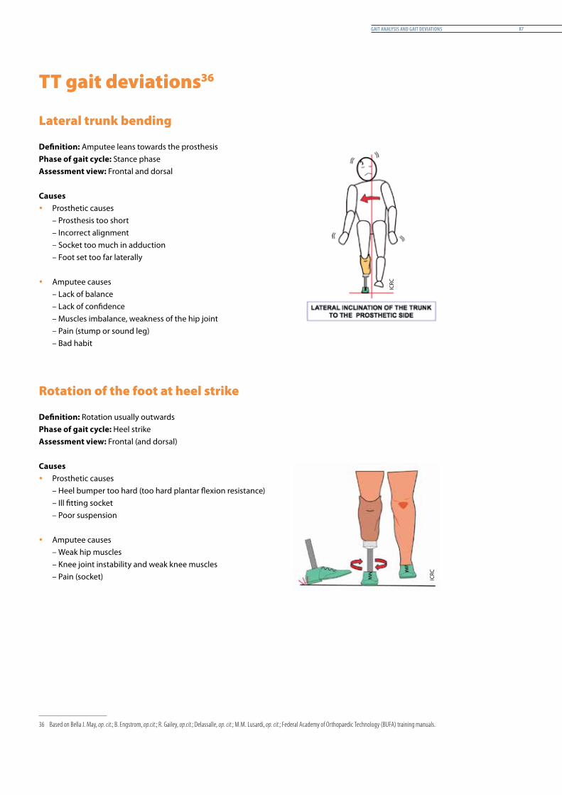
Gait analysis and Gait deviations 87
TT gait deviations363
Lateral trunk bending
Definition: Amputee leans towards the prosthesisPhase of gait cycle: Stance phaseAssessment view: Frontal and dorsal
Causes y Prosthetic causes
– Prosthesis too short– Incorrect alignment – Socket too much in adduction– Foot set too far laterally
y Amputee causes– Lack of balance– Lack of confidence– Muscles imbalance, weakness of the hip joint– Pain (stump or sound leg)– Bad habit
Rotation of the foot at heel strike
Definition: Rotation usually outwardsPhase of gait cycle: Heel strikeAssessment view: Frontal (and dorsal)
Causes y Prosthetic causes
– Heel bumper too hard (too hard plantar flexion resistance)– Ill fitting socket– Poor suspension
y Amputee causes– Weak hip muscles– Knee joint instability and weak knee muscles– Pain (socket)
36 Based on Bella J. May, op. cit.; B. Engstrom, op.cit.; R. Gailey, op.cit.; Delassalle, op. cit.; M.M. Lusardi, op. cit.; Federal Academy of Orthopaedic Technology (BUFA) training manuals.
ICRC
ICRC
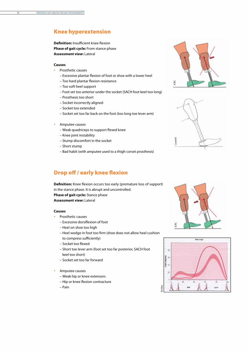
Prosthetic Gait analysis for icrc PhysiotheraPists88
Knee hyperextension
Definition: Insufficient knee flexionPhase of gait cycle: From stance phaseAssessment view: Lateral
Causes y Prosthetic causes
– Excessive plantar flexion of foot or shoe with a lower heel– Too hard plantar flexion resistance– Too soft heel support– Foot set too anterior under the socket (SACH foot keel too long)– Prosthesis too short– Socket incorrectly aligned– Socket too extended– Socket set too far back on the foot (too long toe lever arm)
y Amputee causes– Weak quadriceps to support flexed knee– Knee joint instability– Stump discomfort in the socket– Short stump– Bad habit (with amputee used to a thigh corset prosthesis)
Drop off / early knee flexion
Definition: Knee flexion occurs too early (premature loss of support) in the stance phase. It is abrupt and uncontrolled.Phase of gait cycle: Stance phaseAssessment view: Lateral
Causes y Prosthetic causes
– Excessive dorsiflexion of foot– Heel on shoe too high– Heel wedge in foot too firm (shoe does not allow heel cushion
to compress sufficiently)– Socket too flexed– Short toe lever arm (foot set too far posterior, SACH foot
keel too short)– Socket set too far forward
y Amputee causes– Weak hip or knee extensors– Hip or knee flexion contracture– Pain
ICRC
Lusa
rdi
Kirt
ley
ICRC
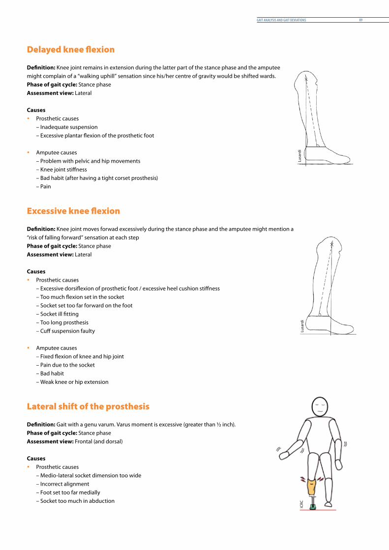
Gait analysis and Gait deviations 89
Delayed knee flexion
Definition: Knee joint remains in extension during the latter part of the stance phase and the amputee might complain of a “walking uphill” sensation since his/her centre of gravity would be shifted wards.Phase of gait cycle: Stance phaseAssessment view: Lateral
Causes y Prosthetic causes
– Inadequate suspension– Excessive plantar flexion of the prosthetic foot
y Amputee causes– Problem with pelvic and hip movements– Knee joint stiffness– Bad habit (after having a tight corset prosthesis)– Pain
Excessive knee flexion
Definition: Knee joint moves forwad excessively during the stance phase and the amputee might mention a “risk of falling forward” sensation at each step Phase of gait cycle: Stance phaseAssessment view: Lateral
Causes y Prosthetic causes
– Excessive dorsiflexion of prosthetic foot / excessive heel cushion stiffness– Too much flexion set in the socket– Socket set too far forward on the foot– Socket ill fitting– Too long prosthesis– Cuff suspension faulty
y Amputee causes– Fixed flexion of knee and hip joint– Pain due to the socket– Bad habit– Weak knee or hip extension
Lateral shift of the prosthesis
Definition: Gait with a genu varum. Varus moment is excessive (greater than ½ inch).Phase of gait cycle: Stance phaseAssessment view: Frontal (and dorsal)
Causes y Prosthetic causes
– Medio-lateral socket dimension too wide– Incorrect alignment– Foot set too far medially– Socket too much in abduction
Lusa
rdi
Lusa
rdi
ICRC
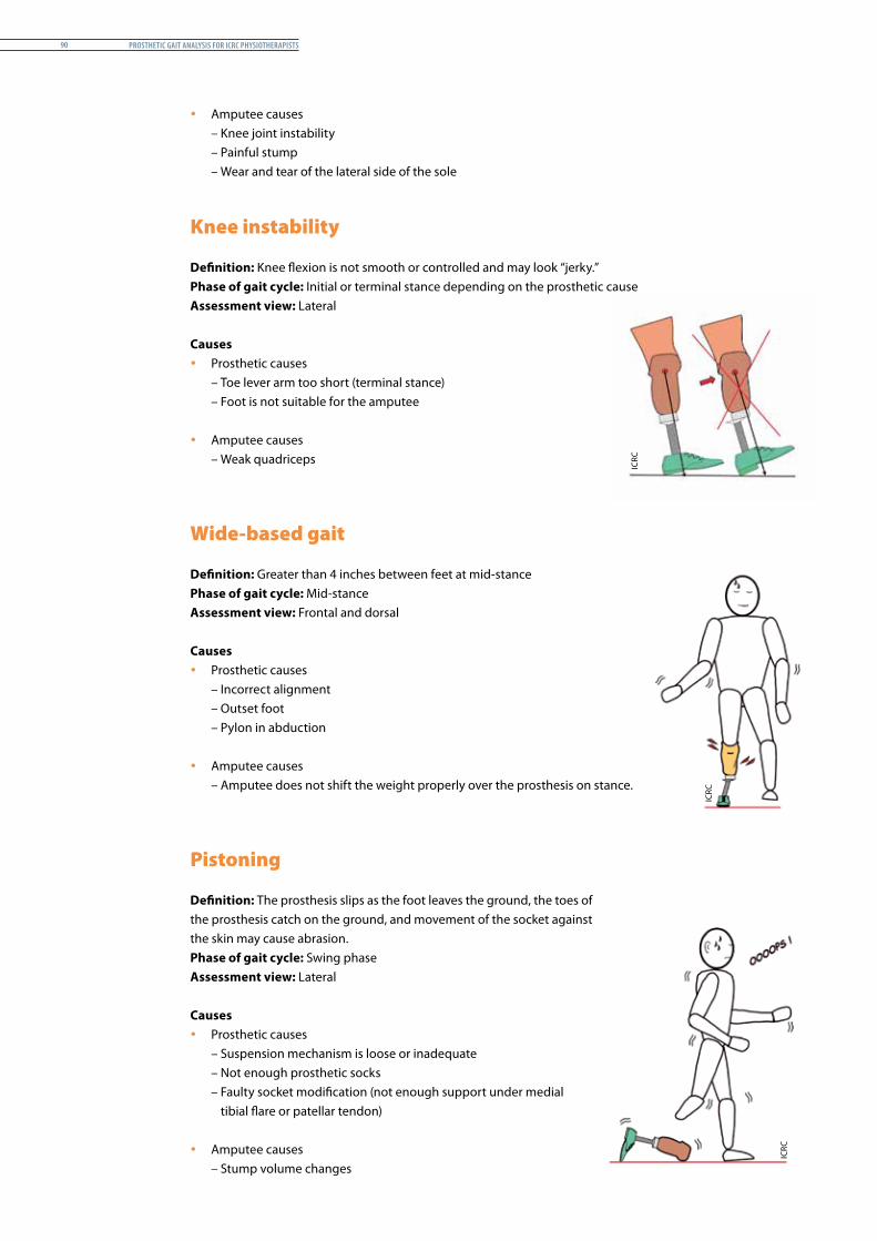
Prosthetic Gait analysis for icrc PhysiotheraPists90
y Amputee causes– Knee joint instability– Painful stump– Wear and tear of the lateral side of the sole
Knee instability
Definition: Knee flexion is not smooth or controlled and may look “jerky.”Phase of gait cycle: Initial or terminal stance depending on the prosthetic causeAssessment view: Lateral
Causes y Prosthetic causes
– Toe lever arm too short (terminal stance)– Foot is not suitable for the amputee
y Amputee causes – Weak quadriceps
Wide-based gait
Definition: Greater than 4 inches between feet at mid-stancePhase of gait cycle: Mid-stanceAssessment view: Frontal and dorsal
Causes y Prosthetic causes
– Incorrect alignment– Outset foot– Pylon in abduction
y Amputee causes– Amputee does not shift the weight properly over the prosthesis on stance.
Pistoning
Definition: The prosthesis slips as the foot leaves the ground, the toes of the prosthesis catch on the ground, and movement of the socket against the skin may cause abrasion. Phase of gait cycle: Swing phaseAssessment view: Lateral
Causes y Prosthetic causes
– Suspension mechanism is loose or inadequate– Not enough prosthetic socks– Faulty socket modification (not enough support under medial
tibial flare or patellar tendon)
y Amputee causes– Stump volume changes
ICRC
ICRC
ICRC
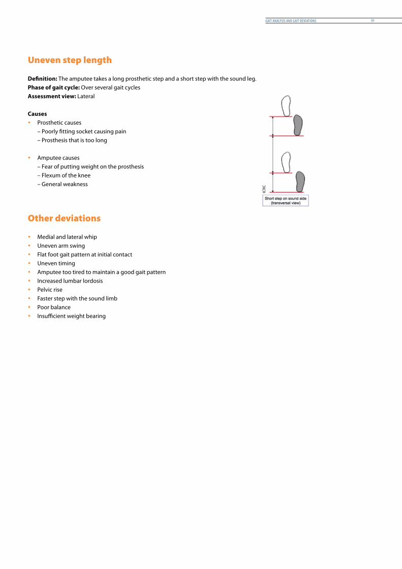
Gait analysis and Gait deviations 91
Uneven step length
Definition: The amputee takes a long prosthetic step and a short step with the sound leg.Phase of gait cycle: Over several gait cyclesAssessment view: Lateral
Causes y Prosthetic causes
– Poorly fitting socket causing pain– Prosthesis that is too long
y Amputee causes– Fear of putting weight on the prosthesis– Flexum of the knee– General weakness
Other deviations
y Medial and lateral whip y Uneven arm swing y Flat foot gait pattern at initial contact y Uneven timing y Amputee too tired to maintain a good gait pattern y Increased lumbar lordosis y Pelvic rise y Faster step with the sound limb y Poor balance y Insufficient weight bearing
ICRC
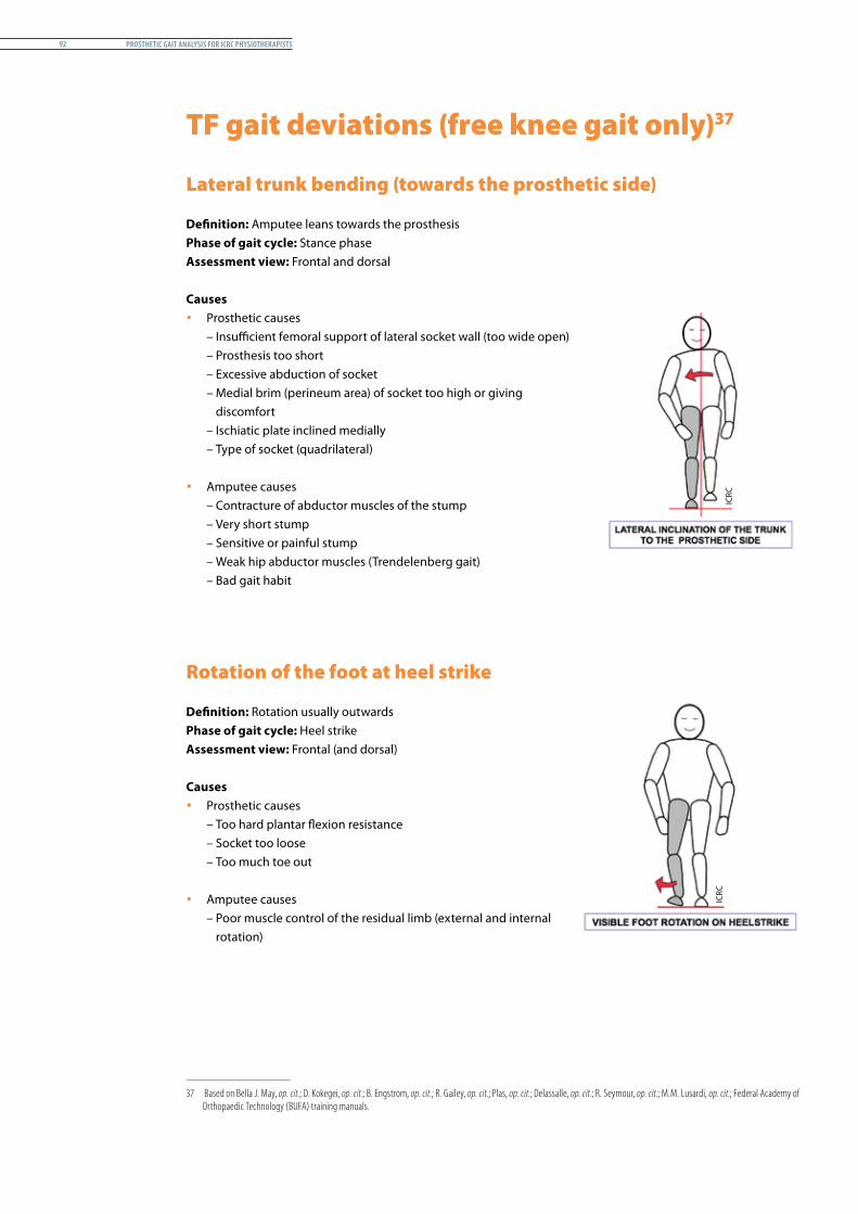
Prosthetic Gait analysis for icrc PhysiotheraPists92
TF gait deviations (free knee gait only)371
Lateral trunk bending (towards the prosthetic side)
Definition: Amputee leans towards the prosthesisPhase of gait cycle: Stance phaseAssessment view: Frontal and dorsal
Causes y Prosthetic causes
– Insufficient femoral support of lateral socket wall (too wide open)– Prosthesis too short– Excessive abduction of socket– Medial brim (perineum area) of socket too high or giving
discomfort– Ischiatic plate inclined medially– Type of socket (quadrilateral)
y Amputee causes– Contracture of abductor muscles of the stump– Very short stump– Sensitive or painful stump– Weak hip abductor muscles (Trendelenberg gait)– Bad gait habit
Rotation of the foot at heel strike
Definition: Rotation usually outwardsPhase of gait cycle: Heel strikeAssessment view: Frontal (and dorsal)
Causes y Prosthetic causes
– Too hard plantar flexion resistance– Socket too loose– Too much toe out
y Amputee causes– Poor muscle control of the residual limb (external and internal
rotation)
37 Based on Bella J. May, op. cit.; D. Kokegei, op. cit.; B. Engstrom, op. cit.; R. Gailey, op. cit.; Plas, op. cit.; Delassalle, op. cit.; R. Seymour, op. cit.; M.M. Lusardi, op. cit.; Federal Academy of Orthopaedic Technology (BUFA) training manuals.
ICRC
ICRC
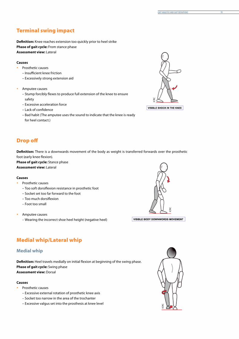
Gait analysis and Gait deviations 93
Terminal swing impact
Definition: Knee reaches extension too quickly prior to heel strikePhase of gait cycle: From stance phaseAssessment view: Lateral
Causes y Prosthetic causes
– Insufficient knee friction– Excessively strong extension aid
y Amputee causes– Stump forcibly flexes to produce full extension of the knee to ensure
safety– Excessive acceleration force– Lack of confidence– Bad habit (The amputee uses the sound to indicate that the knee is ready
for heel contact.)
Drop off
Definition: There is a downwards movement of the body as weight is transferred forwards over the prosthetic foot (early knee flexion).Phase of gait cycle: Stance phaseAssessment view: Lateral
Causes y Prosthetic causes
– Too soft dorsiflexion resistance in prosthetic foot– Socket set too far forward to the foot– Too much dorsiflexion– Foot too small
y Amputee causes– Wearing the incorrect shoe heel height (negative heel)
Medial whip/Lateral whip
Medial whip
Definition: Heel travels medially on initial flexion at beginning of the swing phase.Phase of gait cycle: Swing phaseAssessment view: Dorsal
Causes y Prosthetic causes
– Excessive external rotation of prosthetic knee axis– Socket too narrow in the area of the trochanter– Excessive valgus set into the prosthesis at knee level
ICRC
ICRC
ICRC
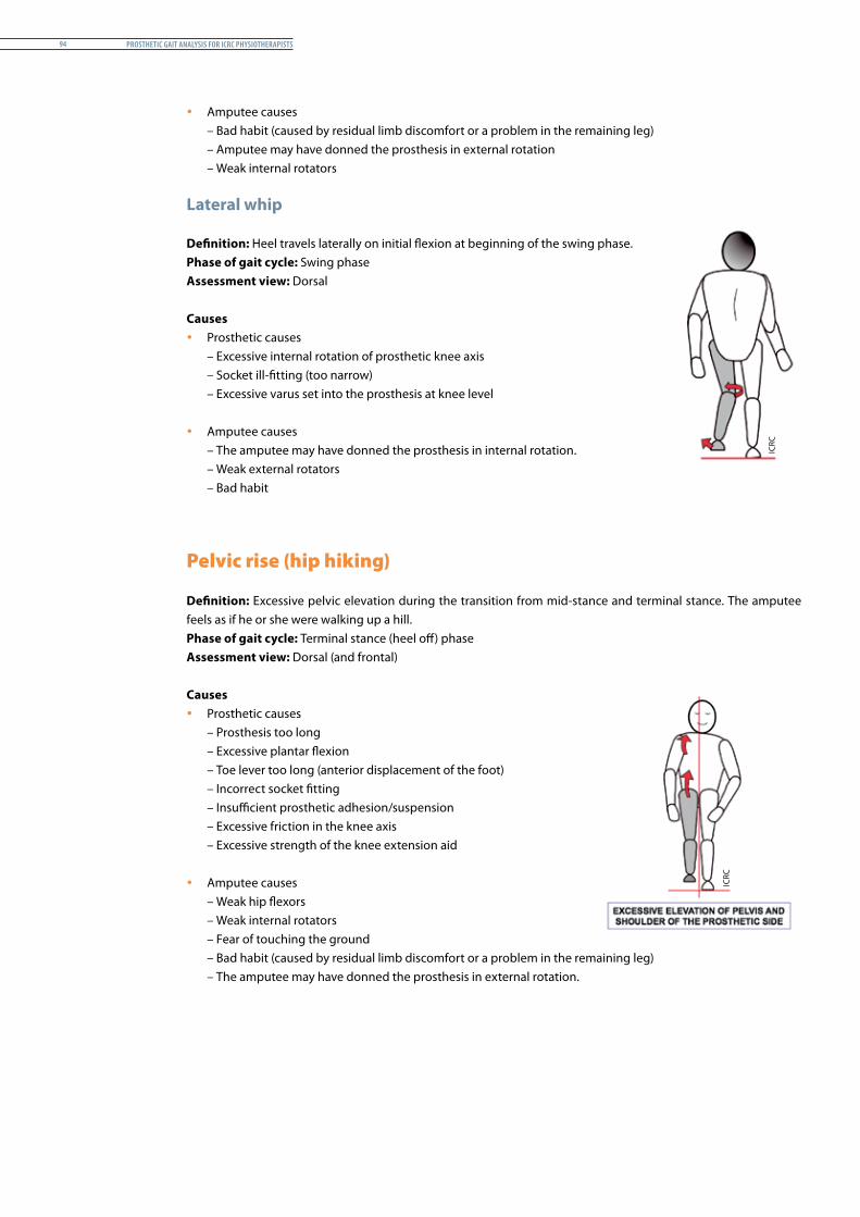
Prosthetic Gait analysis for icrc PhysiotheraPists94
y Amputee causes– Bad habit (caused by residual limb discomfort or a problem in the remaining leg)– Amputee may have donned the prosthesis in external rotation– Weak internal rotators
Lateral whip
Definition: Heel travels laterally on initial flexion at beginning of the swing phase.Phase of gait cycle: Swing phaseAssessment view: Dorsal
Causes y Prosthetic causes
– Excessive internal rotation of prosthetic knee axis– Socket ill-fitting (too narrow)– Excessive varus set into the prosthesis at knee level
y Amputee causes– The amputee may have donned the prosthesis in internal rotation. – Weak external rotators– Bad habit
Pelvic rise (hip hiking)
Definition: Excessive pelvic elevation during the transition from mid-stance and terminal stance. The amputee feels as if he or she were walking up a hill.Phase of gait cycle: Terminal stance (heel off) phaseAssessment view: Dorsal (and frontal)
Causes y Prosthetic causes
– Prosthesis too long– Excessive plantar flexion– Toe lever too long (anterior displacement of the foot)– Incorrect socket fitting– Insufficient prosthetic adhesion/suspension– Excessive friction in the knee axis– Excessive strength of the knee extension aid
y Amputee causes– Weak hip flexors– Weak internal rotators– Fear of touching the ground– Bad habit (caused by residual limb discomfort or a problem in the remaining leg)– The amputee may have donned the prosthesis in external rotation.
ICRC
ICRC

Gait analysis and Gait deviations 95
Excessive heel rise
Definition: Heel of prosthesis rises upwards excessively when knee flexes at the beginning of the swing phase.Phase of gait cycle: Swing phaseAssessment view: Lateral and dorsal
Causes y Prosthetic causes
– Forefoot lever too short (knee flexion initiates too quickly)– Insufficient friction in the knee axis– Insufficient strength of the knee extension aid
y Amputee causes– Too much hip flexor muscle power used to flex the prosthetic knee.– Too fast (uncontrolled) gait
Knee instability
Definition: Prosthetic knee flexion is not stable or controlled and may look “jerky”Phase of gait cycle: From heel contact to mid-stanceAssessment view: Lateral
Causes y Prosthetic causes
– The gravity line falls behind the knee.– The socket is placed too far anteriorly (long heel lever arm).– Lack of adequate socket flexion.– Heel support is too hard that “unlocks” the knee at heel strike– Hip flexion contracture not accommodated in the socket.– Inadequate (too high) heel height on the footwear– Forefoot lever too short
y Amputee causes– Weak hip extensors.
Vaulting
Definition: The amputee rises up on the toe of the sound leg to swing the prosthesis through from toe off to heel strike.Phase of gait cycle: Swing phase (prosthetic side)Assessment view: Lateral and dorsal
Causes y Prosthetic causes
– Prosthesis too long– Excessive plantar flexion– Forefoot lever too long (anterior displacement of the foot)– Ill-fitting socket causing discomfort– Insufficient prosthetic adhesion/suspension– Excessive friction in the knee axis– Excessive strength of the knee extension aid
ICRC
ICRC
ICRC

Prosthetic Gait analysis for icrc PhysiotheraPists96
y Amputee causes– Weak hip flexors– Weak internal rotators– Very short stump– Fear of touching ground (prosthetic toes “hooking” on the floor) – Bad habit (caused by residual limb discomfort or a problem in the remaining leg)– The amputee may have donned the prosthesis in external rotation.
Abducted gait
Definition: Prosthesis held away from midline throughout the gait cyclePhase of gait cycle: Swing phaseAssessment view: Dorsal (and frontal)
Causes y Prosthetic causes
– Prosthesis too long– Insufficient socket abduction– Medial brim (perineum area) of the socket too high– Insufficient femoral support of the lateral socket wall (too wide
open)– Excessive suspension or incorrect location of the “Silesian belt”– Components aligned too far laterally (only if knee axis is
horizontal)
y Amputee causes– Contracture of the abductor muscles (stump side)– Weak hip adductor muscles– Short stump– Pain in the perineum– Lack of balance– Insecurity– Bad habit
Lumbar lordosis
Definition: The amputee creates an active lumbar lordosis.Phase of gait cycle: Stance phase to heel riseAssessment view: Lateral
Causes y Prosthetic causes
– Insufficient stability in knee mechanism– Insufficient socket flexion– Discomfort on ischial plate (too high or anteriorly inclined)– Prosthesis too long– Heel of shoe on prosthesis too high
ICRC
ICRC
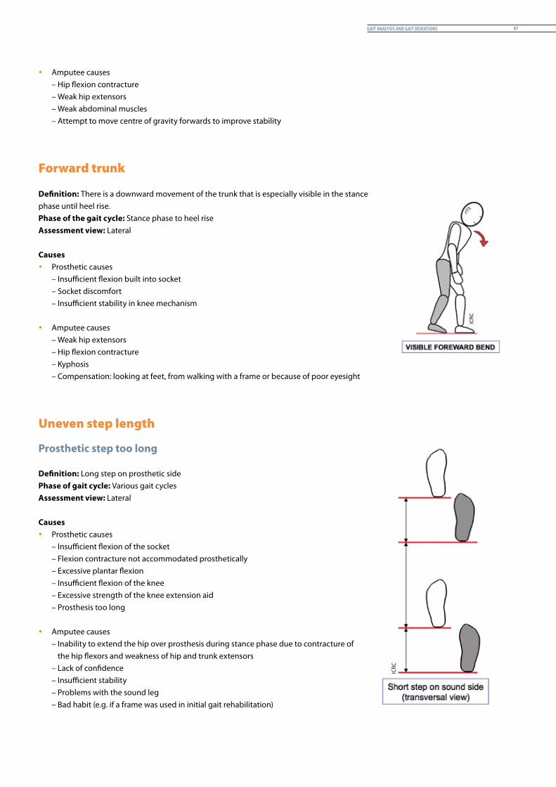
Gait analysis and Gait deviations 97
y Amputee causes– Hip flexion contracture– Weak hip extensors– Weak abdominal muscles– Attempt to move centre of gravity forwards to improve stability
Forward trunk
Definition: There is a downward movement of the trunk that is especially visible in the stance phase until heel rise.Phase of the gait cycle: Stance phase to heel riseAssessment view: Lateral
Causes y Prosthetic causes
– Insufficient flexion built into socket– Socket discomfort– Insufficient stability in knee mechanism
y Amputee causes– Weak hip extensors– Hip flexion contracture– Kyphosis– Compensation: looking at feet, from walking with a frame or because of poor eyesight
Uneven step length
Prosthetic step too long
Definition: Long step on prosthetic sidePhase of gait cycle: Various gait cyclesAssessment view: Lateral
Causes y Prosthetic causes
– Insufficient flexion of the socket– Flexion contracture not accommodated prosthetically– Excessive plantar flexion– Insufficient flexion of the knee– Excessive strength of the knee extension aid– Prosthesis too long
y Amputee causes– Inability to extend the hip over prosthesis during stance phase due to contracture of
the hip flexors and weakness of hip and trunk extensors– Lack of confidence– Insufficient stability– Problems with the sound leg– Bad habit (e.g. if a frame was used in initial gait rehabilitation)
ICRC
ICRC
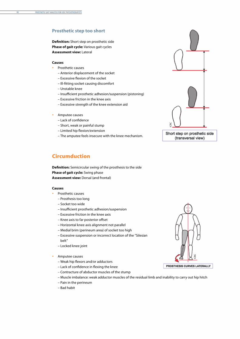
Prosthetic Gait analysis for icrc PhysiotheraPists98
Prosthetic step too short
Definition: Short step on prosthetic sidePhase of gait cycle: Various gait cyclesAssessment view: Lateral
Causes y Prosthetic causes
– Anterior displacement of the socket– Excessive flexion of the socket– Ill-fitting socket causing discomfort– Unstable knee– Insufficient prosthetic adhesion/suspension (pistoning)– Excessive friction in the knee axis– Excessive strength of the knee extension aid
y Amputee causes– Lack of confidence– Short, weak or painful stump– Limited hip flexion/extension– The amputee feels insecure with the knee mechanism.
Circumduction
Definition: Semicircular swing of the prosthesis to the side Phase of gait cycle: Swing phaseAssessment view: Dorsal (and frontal)
Causes y Prosthetic causes
– Prosthesis too long– Socket too wide– Insufficient prosthetic adhesion/suspension– Excessive friction in the knee axis– Knee axis to far posterior offset– Horizontal knee axis alignment not parallel– Medial brim (perineum area) of socket too high– Excessive suspension or incorrect location of the “Silesian
belt”– Locked knee joint
y Amputee causes– Weak hip flexors and/or adductors– Lack of confidence in flexing the knee– Contracture of abductor muscles of the stump– Muscle imbalance: weak adductor muscles of the residual limb and inability to carry out hip hitch– Pain in the perineum– Bad habit
ICRC
ICRC
Gait analysis and Gait deviations 99
Other deviations
y Excessive uneven timing y Uneven arm swing y Excessive pistoning within the socket y Insufficient heel rise y Continuing whip (medial to lateral whip during the swing) y Poor balance y Foot slap y Pelvic rise

Prosthetic Gait analysis for icrc PhysiotheraPists100
Post-fittinG rehabilitation 101
Post-fittinG rehabilitation
Introduction
Content
This chapter describes briefly the rehabilitation that is available to amputees after the prosthetic fitting. In ICRC projects, this rehabilitation is provided by a physiotherapist who will have to define, together with the prosthetist and the amputee, what could be achieved with the proposed appliance.
The fitting of the prosthesis needs to be followed by functional training; in the case of lower-limb amputation, this is essentially gait training. The progressive stages of gait training are described on pages 103-104.
Rationale
Attaining functional independence is the ultimate aim of rehabilitation for lower-limb amputees. By comparison, upper-limb amputees may want a prosthesis for cosmetic purposes only. Hence, gait training for the prosthesis user has maximum significance in the rehabilitation process. Apart from aiming for clear functional goals, users also need to be taught about prosthesis maintenance and care and made aware of the need to report back to the physical rehabilitation centre for follow-up.
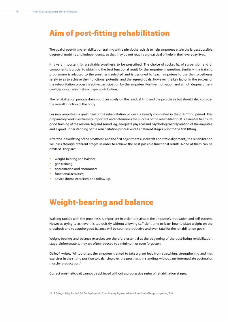
Prosthetic Gait analysis for icrc PhysiotheraPists102
Aim of post-fitting rehabilitation
The goal of post-fitting rehabilitation training with a physiotherapist is to help amputees attain the largest possible degree of mobility and independence, so that they do not require a great deal of help in their everyday lives.
It is very important for a suitable prosthesis to be prescribed. The choice of socket fit, of suspension and of components is crucial to obtaining the best functional result for the amputee in question. Similarly, the training programme is adapted to the prosthesis selected and is designed to teach amputees to use their prostheses safely so as to achieve their functional potential and the agreed goals. However, the key factor in the success of the rehabilitation process is active participation by the amputee. Positive motivation and a high degree of self-confidence can also make a major contribution.
The rehabilitation process does not focus solely on the residual limb and the prosthesis but should also consider the overall function of the body.
For new amputees, a great deal of the rehabilitation process is already completed in the pre-fitting period. This preparatory work is extremely important and determines the success of the rehabilitation. It is essential to ensure good training of the residual leg and sound leg, adequate physical and psychological preparation of the amputee and a good understanding of the rehabilitation process and its different stages prior to the first fitting. After the initial fitting of the prosthesis and the first adjustments (socket fit and static alignment), the rehabilitation will pass through different stages in order to achieve the best possible functional results. None of them can be omitted. They are:
y weight-bearing and balance; y gait training; y coordination and endurance; y functional activities; y advice (home exercises) and follow-up.
Weight-bearing and balance
Walking rapidly with the prosthesis is important in order to maintain the amputee’s motivation and self-esteem. However, trying to achieve this too quickly without allowing sufficient time to learn how to place weight on the prosthesis and to acquire good balance will be counterproductive and even fatal for the rehabilitation goals.
Weight-bearing and balance exercises are therefore essential at the beginning of the post-fitting rehabilitation stage. Unfortunately, they are often reduced to a minimum or even forgotten.
Gailey38 writes, “All too often, the amputee is asked to take a giant leap from stretching, strengthening and mat exercises in the sitting position to balancing over the prosthesis in standing, without any intermediate postural or muscle re-education.” 1
Correct prosthetic gait cannot be achieved without a progressive series of rehabilitation stages.
38 R. Gailey, A. Gailey, Prosthetic Gait Training Program for Lower Extremity Amputees, Advanced Rehabilitation Therapy Incorporated, 1989.
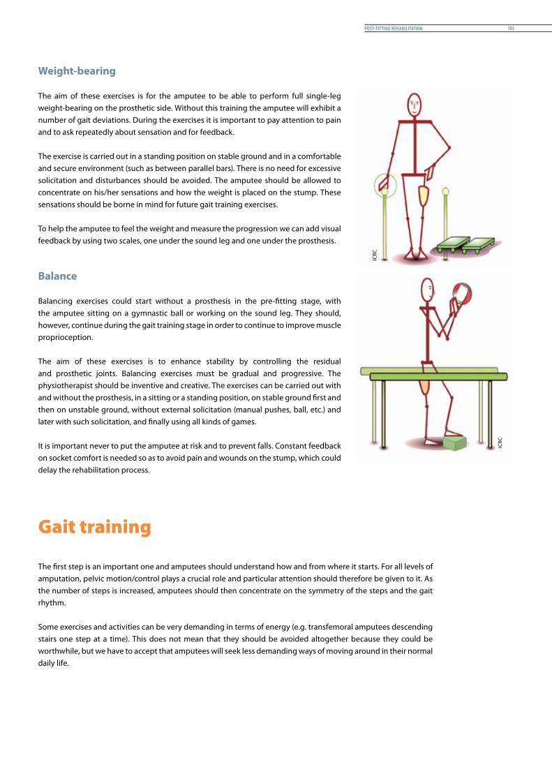
Post-fittinG rehabilitation 103
Weight-bearing
The aim of these exercises is for the amputee to be able to perform full single-leg weight-bearing on the prosthetic side. Without this training the amputee will exhibit a number of gait deviations. During the exercises it is important to pay attention to pain and to ask repeatedly about sensation and for feedback.
The exercise is carried out in a standing position on stable ground and in a comfortable and secure environment (such as between parallel bars). There is no need for excessive solicitation and disturbances should be avoided. The amputee should be allowed to concentrate on his/her sensations and how the weight is placed on the stump. These sensations should be borne in mind for future gait training exercises. To help the amputee to feel the weight and measure the progression we can add visual feedback by using two scales, one under the sound leg and one under the prosthesis.
Balance
Balancing exercises could start without a prosthesis in the pre-fitting stage, with the amputee sitting on a gymnastic ball or working on the sound leg. They should, however, continue during the gait training stage in order to continue to improve muscle proprioception.
The aim of these exercises is to enhance stability by controlling the residual and prosthetic joints. Balancing exercises must be gradual and progressive. The physiotherapist should be inventive and creative. The exercises can be carried out with and without the prosthesis, in a sitting or a standing position, on stable ground first and then on unstable ground, without external solicitation (manual pushes, ball, etc.) and later with such solicitation, and finally using all kinds of games.
It is important never to put the amputee at risk and to prevent falls. Constant feedback on socket comfort is needed so as to avoid pain and wounds on the stump, which could delay the rehabilitation process.
Gait training
The first step is an important one and amputees should understand how and from where it starts. For all levels of amputation, pelvic motion/control plays a crucial role and particular attention should therefore be given to it. As the number of steps is increased, amputees should then concentrate on the symmetry of the steps and the gait rhythm.
Some exercises and activities can be very demanding in terms of energy (e.g. transfemoral amputees descending stairs one step at a time). This does not mean that they should be avoided altogether because they could be worthwhile, but we have to accept that amputees will seek less demanding ways of moving around in their normal daily life.
ICRC
ICRC
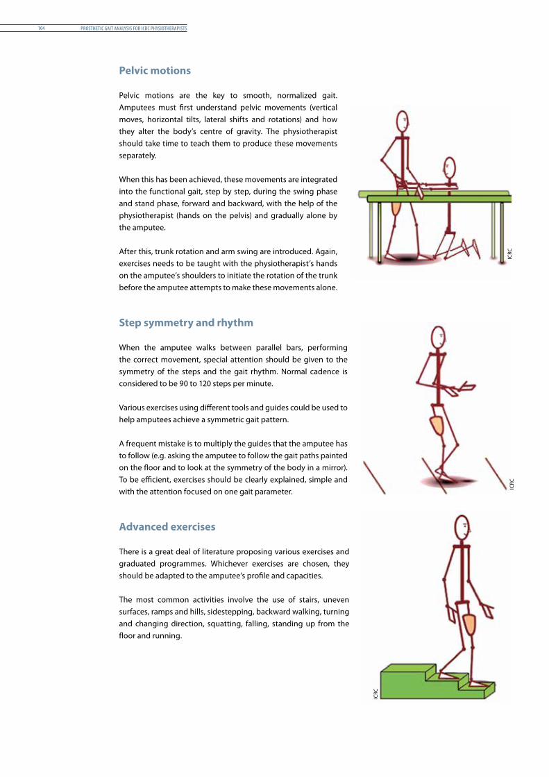
Prosthetic Gait analysis for icrc PhysiotheraPists104
Pelvic motions
Pelvic motions are the key to smooth, normalized gait. Amputees must first understand pelvic movements (vertical moves, horizontal tilts, lateral shifts and rotations) and how they alter the body’s centre of gravity. The physiotherapist should take time to teach them to produce these movements separately.
When this has been achieved, these movements are integrated into the functional gait, step by step, during the swing phase and stand phase, forward and backward, with the help of the physiotherapist (hands on the pelvis) and gradually alone by the amputee.
After this, trunk rotation and arm swing are introduced. Again, exercises needs to be taught with the physiotherapist’s hands on the amputee’s shoulders to initiate the rotation of the trunk before the amputee attempts to make these movements alone.
Step symmetry and rhythm
When the amputee walks between parallel bars, performing the correct movement, special attention should be given to the symmetry of the steps and the gait rhythm. Normal cadence is considered to be 90 to 120 steps per minute.
Various exercises using different tools and guides could be used to help amputees achieve a symmetric gait pattern.
A frequent mistake is to multiply the guides that the amputee has to follow (e.g. asking the amputee to follow the gait paths painted on the floor and to look at the symmetry of the body in a mirror). To be efficient, exercises should be clearly explained, simple and with the attention focused on one gait parameter.
Advanced exercises
There is a great deal of literature proposing various exercises and graduated programmes. Whichever exercises are chosen, they should be adapted to the amputee’s profile and capacities.
The most common activities involve the use of stairs, uneven surfaces, ramps and hills, sidestepping, backward walking, turning and changing direction, squatting, falling, standing up from the floor and running.
ICRC
ICRC
ICRC
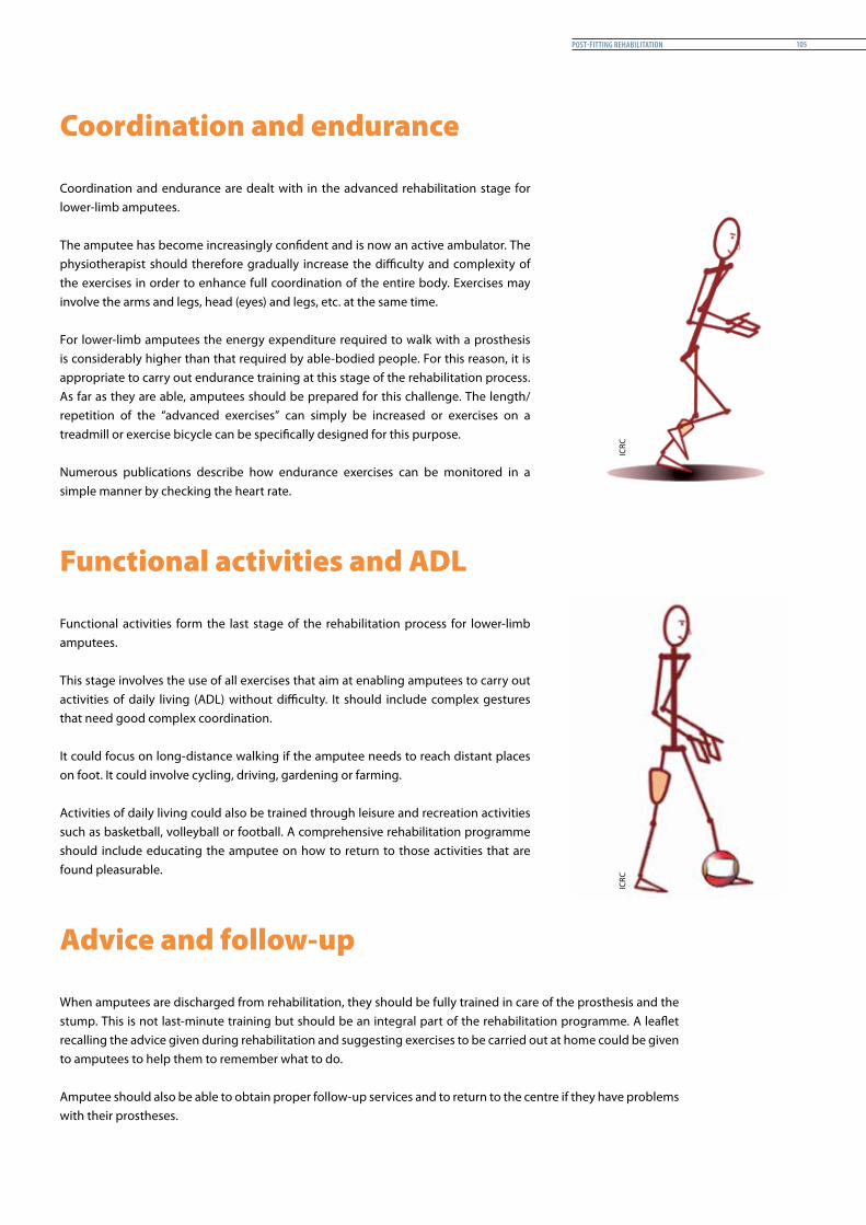
Post-fittinG rehabilitation 105
Coordination and endurance
Coordination and endurance are dealt with in the advanced rehabilitation stage for lower-limb amputees.
The amputee has become increasingly confident and is now an active ambulator. The physiotherapist should therefore gradually increase the difficulty and complexity of the exercises in order to enhance full coordination of the entire body. Exercises may involve the arms and legs, head (eyes) and legs, etc. at the same time.
For lower-limb amputees the energy expenditure required to walk with a prosthesis is considerably higher than that required by able-bodied people. For this reason, it is appropriate to carry out endurance training at this stage of the rehabilitation process. As far as they are able, amputees should be prepared for this challenge. The length/repetition of the “advanced exercises” can simply be increased or exercises on a treadmill or exercise bicycle can be specifically designed for this purpose.
Numerous publications describe how endurance exercises can be monitored in a simple manner by checking the heart rate.
Functional activities and ADL
Functional activities form the last stage of the rehabilitation process for lower-limb amputees.
This stage involves the use of all exercises that aim at enabling amputees to carry out activities of daily living (ADL) without difficulty. It should include complex gestures that need good complex coordination.
It could focus on long-distance walking if the amputee needs to reach distant places on foot. It could involve cycling, driving, gardening or farming.
Activities of daily living could also be trained through leisure and recreation activities such as basketball, volleyball or football. A comprehensive rehabilitation programme should include educating the amputee on how to return to those activities that are found pleasurable.
Advice and follow-up
When amputees are discharged from rehabilitation, they should be fully trained in care of the prosthesis and the stump. This is not last-minute training but should be an integral part of the rehabilitation programme. A leaflet recalling the advice given during rehabilitation and suggesting exercises to be carried out at home could be given to amputees to help them to remember what to do.
Amputee should also be able to obtain proper follow-up services and to return to the centre if they have problems with their prostheses.
ICRC
ICRC
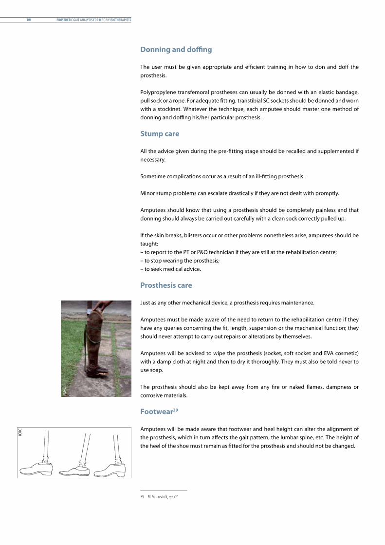
Prosthetic Gait analysis for icrc PhysiotheraPists106
Donning and doffing
The user must be given appropriate and efficient training in how to don and doff the prosthesis.
Polypropylene transfemoral prostheses can usually be donned with an elastic bandage, pull sock or a rope. For adequate fitting, transtibial SC sockets should be donned and worn with a stockinet. Whatever the technique, each amputee should master one method of donning and doffing his/her particular prosthesis.
Stump care
All the advice given during the pre-fitting stage should be recalled and supplemented if necessary.
Sometime complications occur as a result of an ill-fitting prosthesis.
Minor stump problems can escalate drastically if they are not dealt with promptly.
Amputees should know that using a prosthesis should be completely painless and that donning should always be carried out carefully with a clean sock correctly pulled up.
If the skin breaks, blisters occur or other problems nonetheless arise, amputees should be taught:– to report to the PT or P&O technician if they are still at the rehabilitation centre;– to stop wearing the prosthesis;– to seek medical advice.
Prosthesis care
Just as any other mechanical device, a prosthesis requires maintenance.
Amputees must be made aware of the need to return to the rehabilitation centre if they have any queries concerning the fit, length, suspension or the mechanical function; they should never attempt to carry out repairs or alterations by themselves.
Amputees will be advised to wipe the prosthesis (socket, soft socket and EVA cosmetic) with a damp cloth at night and then to dry it thoroughly. They must also be told never to use soap.
The prosthesis should also be kept away from any fire or naked flames, dampness or corrosive materials.
Footwear392
Amputees will be made aware that footwear and heel height can alter the alignment of the prosthesis, which in turn affects the gait pattern, the lumbar spine, etc. The height of the heel of the shoe must remain as fitted for the prosthesis and should not be changed.
39 M.M. Lusardi, op. cit.
ICRC
ICRC
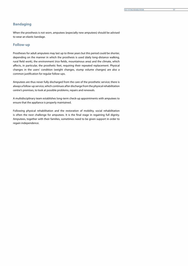
Post-fittinG rehabilitation 107
Bandaging
When the prosthesis is not worn, amputees (especially new amputees) should be advised to wear an elastic bandage.
Follow-up
Prostheses for adult amputees may last up to three years but this period could be shorter, depending on the manner in which the prosthesis is used (daily long-distance walking, rural field work), the environment (rice fields, mountainous area) and the climate, which affects, in particular, the prosthetic feet, requiring their repeated replacement. Physical changes in the users’ condition (weight changes, stump volume changes) are also a common justification for regular follow-ups.
Amputees are thus never fully discharged from the care of the prosthetic service; there is always a follow-up service, which continues after discharge from the physical rehabilitation centre’s premises, to look at possible problems, repairs and renewals.
A multidisciplinary team establishes long-term check-up appointments with amputees to ensure that the appliance is properly maintained.
Following physical rehabilitation and the restoration of mobility, social rehabilitation is often the next challenge for amputees. It is the final stage in regaining full dignity. Amputees, together with their families, sometimes need to be given support in order to regain independence.

Prosthetic Gait analysis for icrc PhysiotheraPists108
references 109
references
Introduction
This section contains a list of all the different kinds of documents that have been used in preparing the manual.
Prosthetic Gait analysis for icrc PhysiotheraPists110
References
Bibliography
Baeza, J., et al., Les troubles de la marche et leur exploration, Frison-Roche, Paris, 1997.
Baumgartner, R., Botta, P., Amputation und Prothesenversorgung der unteren Extremität, 2nd edition, Enke, 1995.
Burgess, Ernest M., Zettl, Joseph H., Amputations Below the Knee, 1969.
Clarkson, Hazel M., Musculoskeletal Assessment, 2nd edition, Lippincott, Williams & Wilkins, Philadelphia, 1989.
Crawford Adams, J., Outline of Orthopaedics, 13th edition, Churchill Livingstone, 2001.
Dufour, D., et al., Surgery for Victims of War, 3rd edition, ICRC, Geneva, 1998.
Engstrom, B., Van de Ven, C., Therapy for Amputees, 3rd edition, Churchill Livingstone, 1999.
Enjalbert M., et al., “Cinématique et cinétique de la marche chez l’amputé des membres inférieurs,” in Pelissier, J., Brun, V. (eds.), La marche humaine et sa pathologie, Masson, Paris, 1994.
Gailey, R., Gailey, A., Prosthetic Gait Training Program for Lower Extremity Amputees, Advanced Rehabilitation Therapy Incorporated, 1989.
Gailey R., McKenzie, A., Stretching and Strengthening for Lower Extremity Amputees, Advanced Rehabilitation Therapy Incorporated, 1994.
Gatto, F., Enseigner la Santé, L’Harmattan, Paris, 2006.
Génot, C., et al., Kinésithérapie 1, Principes, Flammarion, Paris, 2003.
Génot, C., et al., Kinésithérapie 2, Membre inférieur, Flammarion, Paris, 2005.
Gottschalk, F., “Surgical Procedures,” in Bowker, J.H., Michael, J.W. (eds.) Atlas of Limb Prosthetics: Surgical, Prosthetic, and Rehabilitation Principles, 2nd edition, American Academy of Orthopaedic Surgeons, Rosemont 1992.
Grumler, B., Lardry J.-M., La rééducation et l’appareillage des amputés, Dossiers de kinésithérapie 7, Masson, Paris, 1991.
Ha Van, G., Le pied diabétique, 1st edition, Masson, Paris, 2008.
Huerta, J.L., Miller, S.R., “Amputation rehabilitation,” in Brammer, C.M., Spires, M. Catherine, Manual of Physical Medicine and Rehabilitation, Hanley & Belfus, Philadelphia, 2002, pp. 1-12.
Kapandji, A.I., The Physiology of the Joints, Volume 1, 2nd edition, Churchill Livingstone, 1982.
Kapandji, A.I., The Physiology of the Joints, Volume 2, 5th edition, Churchill Livingstone, 1987.
Kendall, F.P., et al., Muscles: Testing and Function, 4th edition, Lippincott Williams & Wilkins, 1993.
Kirtley, C., Clinical Gait Analysis, Churchill Livingstone Elsevier, 2006.
references 111
Kisner, C., Colby, L.A., Therapeutic Exercise, F.A. Davis Company, 2002.
Lusardi, M.M., Nielsen, C.C., Orthotics and Prosthetics in Rehabilitation, 2nd edition, Saunders Elsevier, 2007.
Marcer, I., Didier, J.P., R. Brenot, “Coût énergétique de la marche chez l’amputé de membre inférieur,” in J. Pélissier, V. Brun (eds.), La marche humaine et sa pathologie, Masson, Paris, 1994.
Marcovitch, H., Black’s Medical Dictionary, Black, London, 2005.
Martin, E., Concise Medical Dictionary, 6th edition, Oxford University Press, 2002.
May, Bella J., Amputations and Prosthetics, A Case Study Approach, 2nd edition, F.A. Davis Company, Philadelphia, 1996.
Moore, K.L., Agur, A.M.R., Essential Clinical Anatomy, Williams & Wilkins, 1995.
Nawoczenski, D.A., Epler, M.E., Orthotics in Functional Rehabilitation of the Lower Limb, W.B. Saunders, 1997.
Norkin, C., White D., Measurement of Joint Motion: A Guide to Goniometry, 3rd edition, F.A. Davis Company, Philadelphia, 2003.
Palmer, M.L., Toms, J.E., Manual for Functional Training, 3rd edition, F.A. Davis Company, Philadelphia, 1992.
Seymour, R., Prosthetics and Orthotics, Lippincott Williams & Wilkins, 2002.
Smith, D. G., Michael, J.W., Bowker, J. H., Atlas of Amputations and Limb Deficiencies, 3rd edition, American Academy of Orthopaedic Surgeons, Rosemont, 2004.
Viel, E., La marche humaine, Elsevier Masson, Paris, 2000.
Articles in journals
Curraladas, J., “La kinésithérapie active en résistance progressive à l’élastique,” Kinésithérapie, La revue, No. 94, October 2009.
David-Chaussé, F., “L’appareillage des amputés de cuisse de plus de 50 ans,” Kinésithérapie scientifique No. 145, March 1977.
Delasalle, D., Problèmes posés par l’appareillage et la rééducation des amputés du membre inférieur, Kinésithérapie scientifique, No. 182, July 1980.
Lamy, J.C., “Bases neurophysiologiques de la proprioception,” Kinésithérapie scientifique, No. 0472, 2006.
Le Roux, P., “Techniques de rééducation pour amputés artéritiques”. Kinésithérapie scientifique, No. 252. December 1986.
Kotzki, N., Brunon, A., Pélissier, J., “Amputation et schéma corporel,” Kinésithérapie scientifique, No. 71. October 1997.
Prosthetic Gait analysis for icrc PhysiotheraPists112
Plas, F., Blanc, Y., “Feuillets de biomécanique: analyse des défauts communs de la marche de l’amputé,” Journal de kinésithérapie, feuillet No. 03-272, 1972.
Ramachandran, V.S., “Plasticity and functional recovery in neurology,” Clinical Medicine, Vol. 5, No, 4, July/August 2005, pp. 361-360.
Sabolich, J., “Contoured Adducted Trochanteric-Controlled Alignment Method (CAT-CAM): Introduction and Basic Principles,” Clinical Prosthetics & Orthotics (CPO), Vol. 9, No. 4, 1985, pp. 15-26.
Sergent, S., et al., “Rééducation de deux amputés fémoraux,” Kinésithérapie, Les cahiers, Nos. 8-9, August-September 2002, pp. 59-64.
Videos
Northwestern University Medical School, Gait analysis TF, Prosthetic Education Department of Orthopaedic Surgery, 1990
PTB gait deviation. Prosthetics practitioner.
VA National Prosthetic Gait Lab, California, TF gait deviation.
Heyen, I., Gehschule fur Beinamputierte, Quality for life, Otto Bock, 2003.
Kokegei, K., Stockmann, N., Gait deviation of transtibial amputees, Bundesfachschule Orthopädie-Technik (BUFA), Dortmund, 2004.
ICRC/SFD internal documents
CR Equipements SA, Equipment Catalogue 53, 2012.
ICRC/GTZ, Exercises for lower-limb amputees, Gait training, ICRC, Geneva, 2008.
Training courses
Federal Academy for Orthopaedic Technology (BUFA) training manuals, 2004.
Kokegei, D., Manufacturing Method of Ischial Containment Transfemoral Socket, BUFA, Dortmund, Germany, 2007.
Rushman, C., Shangali, H.G., Wheelchair service guide for low-income countries, Moshi, Tanzanian Training Centre for Orthopaedic Technology, Tumani University, 2005.
Varaud, N., La kinésithérapie des cicatrices, INK, Paris, 2008.
annexes 113
anneXes
Introduction
This section contains a list of all the different kinds of documents that have been used in preparing the manual.
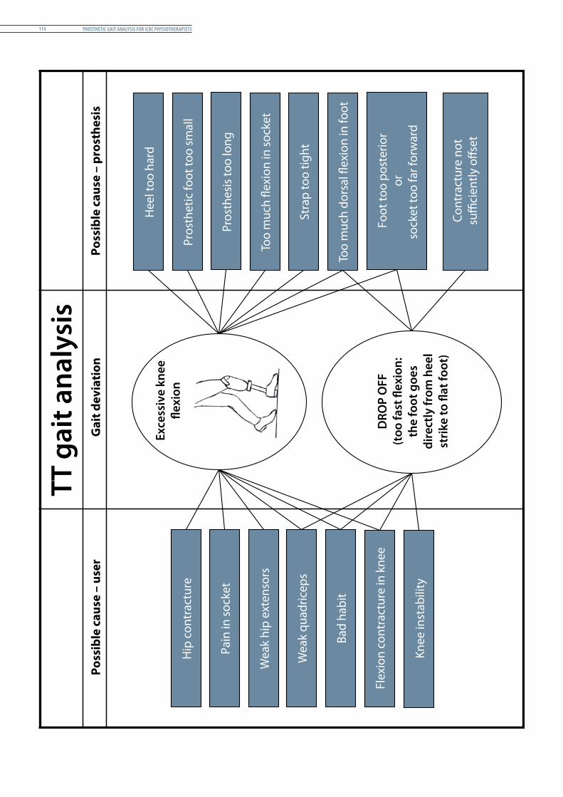
Prosthetic Gait analysis for icrc PhysiotheraPists114
Hip
con
trac
ture
Pain
in so
cket
Wea
k hi
p ex
tens
ors
Wea
k qu
adric
eps
Bad
habi
t
Flex
ion
cont
ract
ure
in k
nee
Knee
inst
abili
ty
Hee
l too
har
d
Pros
thet
ic fo
ot to
o sm
all
Pros
thes
is to
o lo
ng
Stra
p to
o tig
ht
Too
muc
h do
rsal
�ex
ion
in fo
ot
Too
muc
h �e
xion
in so
cket
Foot
too
post
erio
ror
sock
et to
o fa
r for
war
d
Cont
ract
ure
not
su�
cien
tly o
�set
DRO
P O
FF(t
oo fa
st �
exio
n:th
e fo
ot g
oes
dire
ctly
from
hee
lst
rike
to �
at fo
ot)
Exce
ssiv
e kn
ee�e
xion
TT g
ait a
naly
sis
Poss
ible
cau
se –
use
rG
ait d
evia
tion
Poss
ible
cau
se –
pro
sthe
sis
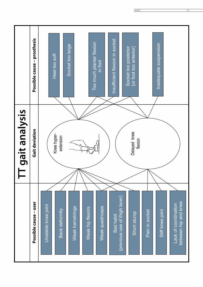
annexes 115
Unsta
ble
knee join
t
Weak q
uadriceps
Bad h
abit
(pre
vio
us u
se o
f th
igh lacer)
Pain
in s
ocket
Stiff k
nee join
t
Lack o
f coord
ination
betw
een h
ip a
nd k
nee
Heel to
o s
oft
Socket to
o larg
e
Too m
uch p
lanta
r flexio
n
in foot
Insuffic
ient flexio
n in s
ocket
Socket to
o p
oste
rior
(or
foot to
o a
nte
rior)
Inadequate
suspensio
n
Del
ayed
kne
efle
xion
Short
stu
mp
Knee
hyp
er-
exte
nsio
nB
ack d
efo
rmity
Weak h
am
str
ings
Weak h
ip fle
xors
TT g
ait a
naly
sis
Poss
ible
cau
se –
use
rG
ait d
evia
tion
Poss
ible
cau
se –
pro
sthe
sis
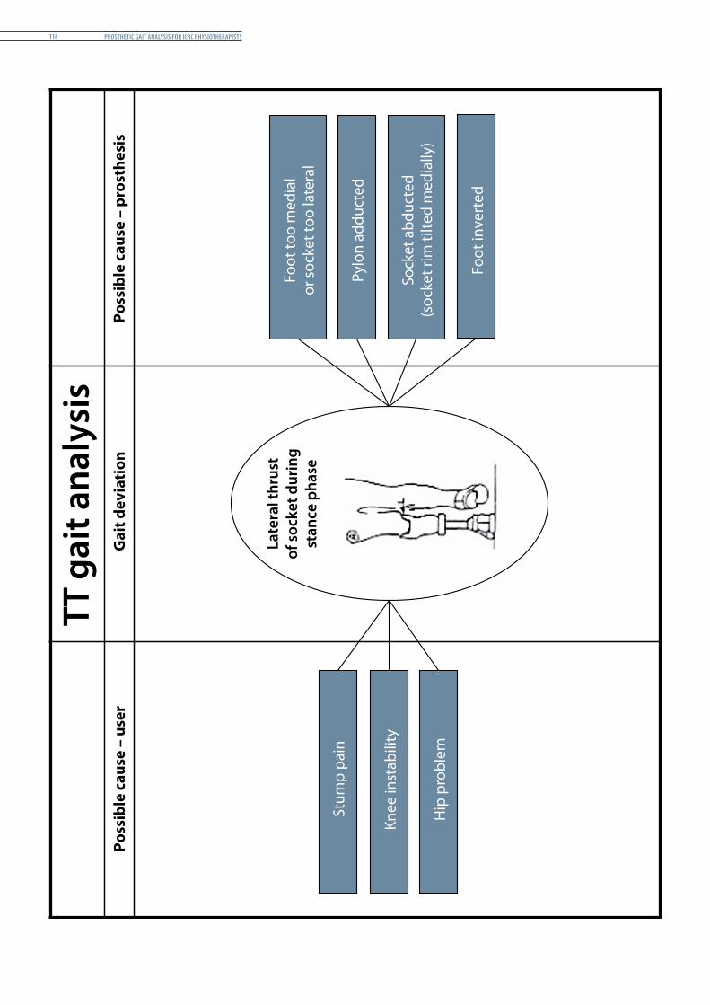
Prosthetic Gait analysis for icrc PhysiotheraPists116
Stum
p pa
in
Knee
inst
abili
ty
Foot
too
med
ial
or so
cket
too
late
ral
Pylo
n ad
duct
ed
Late
ral t
hrus
tof
soc
ket d
urin
gst
ance
pha
se
Hip
pro
blem
Foot
inve
rted
Sock
et a
bduc
ted
(soc
ket r
im ti
lted
med
ially
)
TT g
ait a
naly
sis
Poss
ible
cau
se –
use
rG
ait d
evia
tion
Poss
ible
cau
se –
pro
sthe
sis
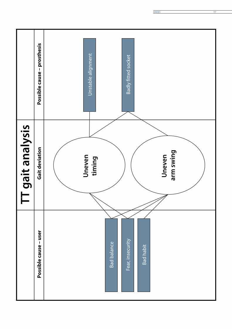
annexes 117
Une
ven
timin
g
Bad
bala
nce
Fear
, ins
ecur
ity
Bad
habi
t
Uns
tabl
e al
ignm
ent
Badl
y �t
ted
sock
et
Une
ven
arm
sw
ing
TT g
ait a
naly
sis
Poss
ible
cau
se –
use
rG
ait d
evia
tion
Poss
ible
cau
se –
pro
sthe
sis
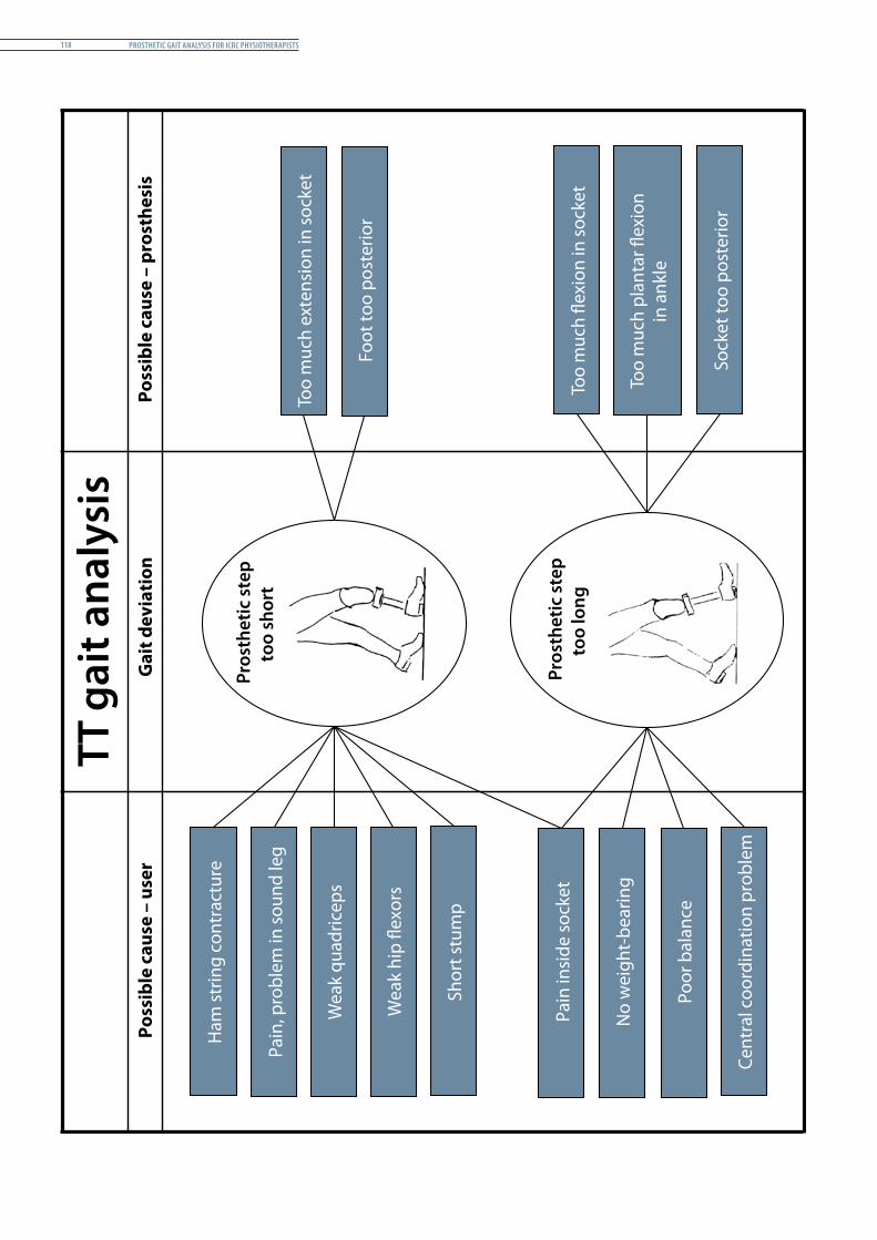
Prosthetic Gait analysis for icrc PhysiotheraPists118
Ham
strin
g co
ntra
ctur
e
Shor
t stu
mp
Pain
insi
de so
cket
No
wei
ght-
bear
ing
Poor
bal
ance
Too
muc
h ex
tens
ion
in so
cket
Foot
too
post
erio
r
Too
muc
h �e
xion
in so
cket
Too
muc
h pl
anta
r �ex
ion
in a
nkle
Sock
et to
o po
ster
ior
Pros
thet
ic s
tep
too
long
Cent
ral c
oord
inat
ion
prob
lem
Pain
, pro
blem
in so
und
leg
Wea
k hi
p �e
xors
Wea
k qu
adric
eps
TT g
ait a
naly
sis
Poss
ible
cau
se –
use
rG
ait d
evia
tion
Poss
ible
cau
se –
pro
sthe
sis
Pros
thet
ic s
tep
too
shor
t
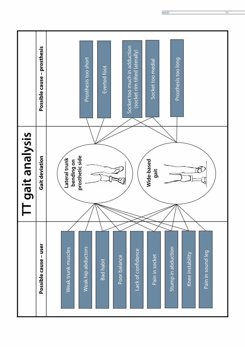
annexes 119
Wea
k tr
unk
mus
cles
Bad
habi
t
Poor
bal
ance
Pain
in so
cket
Stum
p in
abd
uctio
n
Knee
inst
abili
ty
Pros
thes
is to
o sh
ort
Sock
et to
o m
uch
in a
dduc
tion
(soc
ket r
im ti
lted
late
rally
)
Sock
et to
o m
edia
l
Pros
thes
is to
o lo
ng
Wea
k hi
p ab
duct
ors
Wid
e-ba
sed
gait
Late
ral t
runk
be
ndin
g on
pr
osth
etic
sid
e
Pain
in so
und
leg
Lack
of c
on�d
ence
Ever
ted
foot
TT g
ait a
naly
sis
Poss
ible
cau
se –
use
rG
ait d
evia
tion
Poss
ible
cau
se –
pro
sthe
sis
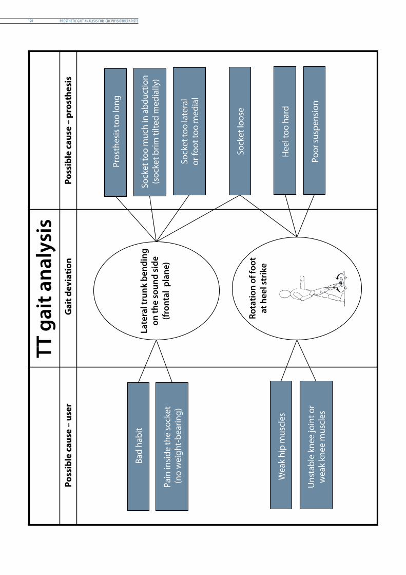
Prosthetic Gait analysis for icrc PhysiotheraPists120
Bad
habi
t
Pain
insi
de th
e so
cket
(n
o w
eigh
t-be
arin
g)
Wea
k hi
p m
uscl
es
Uns
tabl
e kn
ee jo
int o
r w
eak
knee
mus
cles
Pros
thes
is to
o lo
ng
Sock
et to
o m
uch
in a
bduc
tion
(soc
ket b
rim ti
lted
med
ially
)
Sock
et to
o la
tera
l or
foot
too
med
ial
Sock
et lo
ose
Hee
l too
har
d
Poor
susp
ensi
on
Late
ral t
runk
ben
ding
on th
e so
und
side
(fro
ntal
pla
ne)
Rota
tion
of fo
otat
hee
l str
ike
TT g
ait a
naly
sis
Poss
ible
cau
se –
use
rG
ait d
evia
tion
Poss
ible
cau
se –
pro
sthe
sis
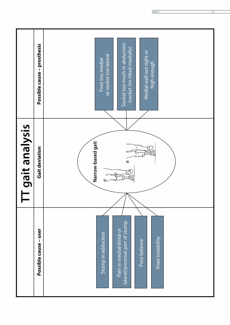
annexes 121
Stum
p in
add
uctio
n
Pain
in m
edia
l/dis
tal o
r la
tera
l/pro
xim
al p
art o
f stu
mp
Poor
bal
ance
Knee
inst
abili
ty
Foot
too
med
ial
or so
cket
too
late
ral
Sock
et to
o m
uch
in a
bduc
tion
(soc
ket r
im ti
lted
med
ially
)
Med
ial w
all n
ot ti
ght o
r hi
gh e
noug
h
Nar
row
-bas
ed g
ait
TT g
ait a
naly
sis
Poss
ible
cau
se –
use
rG
ait d
evia
tion
Poss
ible
cau
se –
pro
sthe
sis
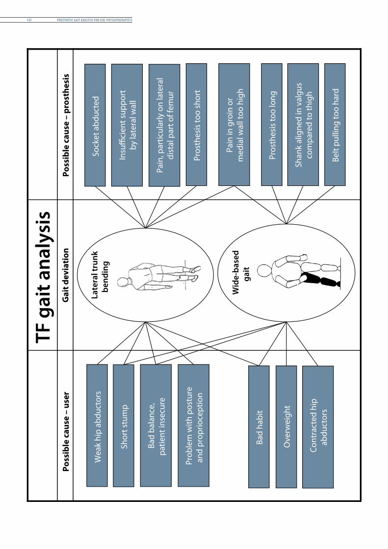
Prosthetic Gait analysis for icrc PhysiotheraPists122
TF g
ait a
naly
sis
Poss
ible
cau
se –
use
rPo
ssib
le c
ause
– p
rost
hesi
s
Wea
k hi
p ab
duct
ors
Shor
t stu
mp
Bad
bala
nce,
patie
nt in
secu
re
Bad
habi
t
Cont
ract
ed h
ipab
duct
ors
Sock
et a
bduc
ted
Insu
�ci
ent s
uppo
rtby
late
ral w
all
Pain
, par
ticul
arly
on
late
ral
dist
al p
art o
f fem
ur
Pros
thes
is to
o sh
ort
Pain
in g
roin
or
med
ial w
all t
oo h
igh
Pros
thes
is to
o lo
ng
Shan
k al
igne
d in
val
gus
com
pare
d to
thig
h
Belt
pulli
ng to
o ha
rd
Late
ral t
runk
bend
ing
Ove
rwei
ght
Prob
lem
with
pos
ture
and
prop
rioce
ptio
n
Gai
t dev
iati
on
Wid
e-ba
sed
gait
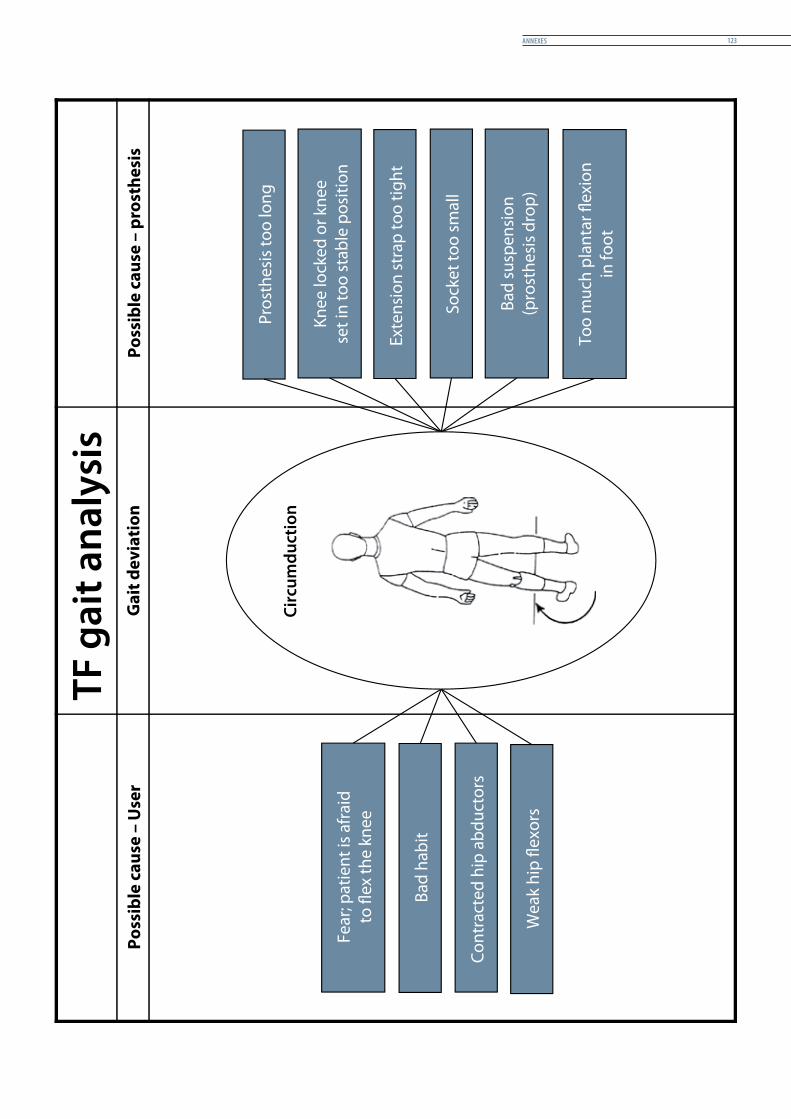
annexes 123
TF g
ait a
naly
sis
Poss
ible
cau
se –
Use
rPo
ssib
le c
ause
– p
rost
hesi
s
Fear
; pat
ient
is a
frai
dto
�ex
the
knee
Bad
habi
t
Cont
ract
ed h
ip a
bduc
tors
Pros
thes
is to
o lo
ng
Knee
lock
ed o
r kne
ese
t in
too
stab
le p
ositi
on
Bad
susp
ensi
on(p
rost
hesi
s dro
p)
Sock
et to
o sm
all
Too
muc
h pl
anta
r �ex
ion
in fo
ot
Exte
nsio
n st
rap
too
tight
Circ
umdu
ctio
n
Wea
k hi
p �e
xors
Gai
t dev
iati
on
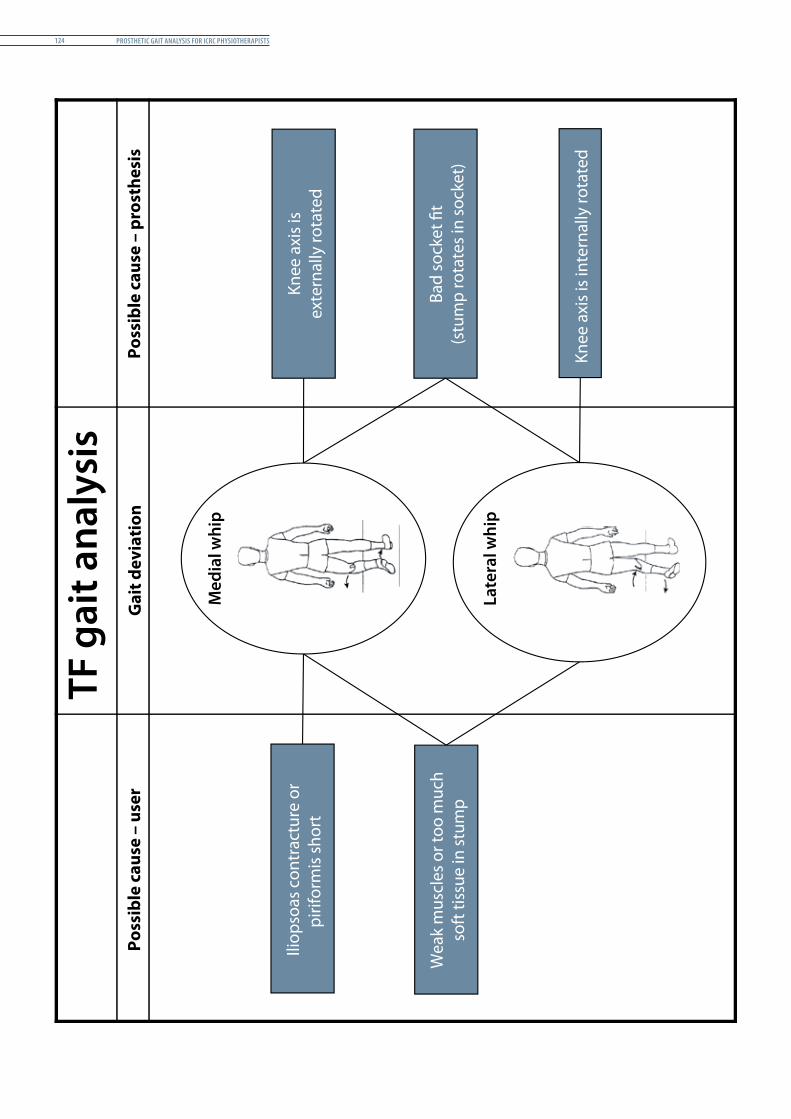
Prosthetic Gait analysis for icrc PhysiotheraPists124
TF g
ait a
naly
sis
Poss
ible
cau
se –
use
rPo
ssib
le c
ause
– p
rost
hesi
s
Wea
k m
uscl
es o
r too
muc
hso
ft ti
ssue
in st
ump
Knee
axi
s is
exte
rnal
ly ro
tate
d
Bad
sock
et �
t(s
tum
p ro
tate
s in
sock
et)
Knee
axi
s is i
nter
nally
rota
ted
Med
ial w
hip
Late
ral w
hip
Iliop
soas
con
trac
ture
or
pirif
orm
is sh
ort
Gai
t dev
iati
on
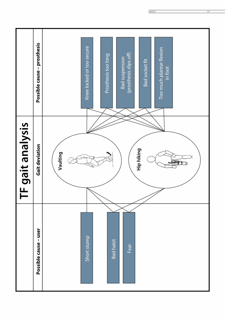
annexes 125
TF g
ait a
naly
sis
Poss
ible
cau
se –
use
rG
ait d
evia
tion
Fear
Shor
t stu
mp
Bad
habi
t Pr
osth
esis
too
long
Knee
lock
ed o
r too
secu
re
Bad
susp
ensi
on(p
rost
hesi
s slip
s o�)
Bad
sock
et �
t
Too
muc
h pl
anta
r �ex
ion
in fo
ot
Vaul
ting
Hip
hik
ing
Poss
ible
cau
se –
pro
sthe
sis
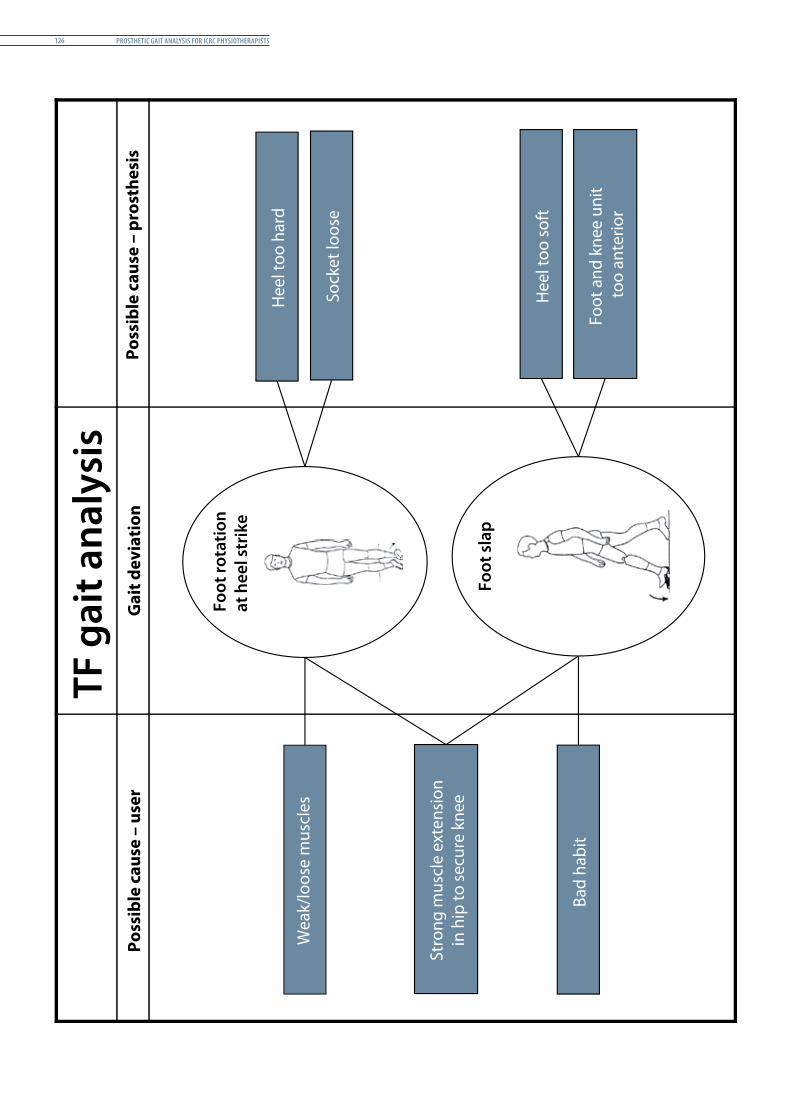
Prosthetic Gait analysis for icrc PhysiotheraPists126
TF g
ait a
naly
sis
Poss
ible
cau
se –
use
rG
ait d
evia
tion
Wea
k/lo
ose
mus
cles
Stro
ng m
uscl
e ex
tens
ion
in h
ip to
secu
re k
nee
Bad
habi
t
Hee
l too
har
d
Sock
et lo
ose
Hee
l too
soft
Foot
and
kne
e un
it to
o an
terio
r
Foot
rota
tion
at h
eel s
trik
e
Foot
sla
p
Poss
ible
cau
se –
pro
sthe
sis
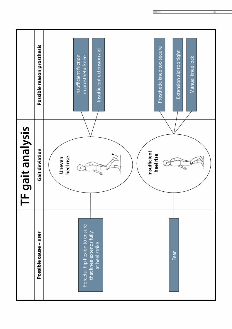
annexes 127
TF g
ait a
naly
sis
Poss
ible
cau
se –
use
rG
ait d
evia
tion
Forc
eful
hip
�ex
ion
to e
nsur
eth
at k
nee
exte
nds f
ully
at h
eel s
trik
e
Fear
Insu
ci
ent f
rictio
nin
pro
sthe
tic k
nee
Insu
ci
ent e
xten
sion
aid
Pros
thet
ic k
nee
too
secu
re
Exte
nsio
n ai
d to
o tig
ht
Man
ual k
nee
lock
Une
ven
heel
rise
Poss
ible
reas
on p
rost
hesi
s
Insu
�ci
ent
heel
rise
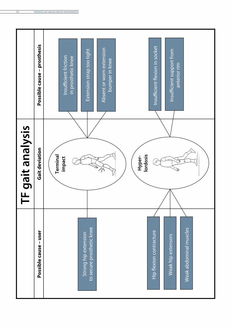
Prosthetic Gait analysis for icrc PhysiotheraPists128
TF g
ait a
naly
sis
Poss
ible
cau
se –
use
rG
ait d
evia
tion
Term
inal
impa
ct
Stro
ng h
ip e
xten
sion
to se
cure
pro
sthe
tic k
nee
Insu
�ci
ent f
rictio
nin
pro
sthe
tic k
nee
Exte
nsio
n st
rap
too
tight
Abs
ent o
r wor
n ex
tens
ion
bum
per i
n kn
ee
Hip
�ex
ion
cont
ract
ure
Wea
k hi
p ex
tens
ors
Wea
k ab
dom
inal
mus
cles
Insu
�ci
ent �
exio
n in
sock
et
Insu
�ci
ent s
uppo
rt fr
oman
terio
r rim
Hyp
er-
lord
osis
Poss
ible
cau
se –
pro
sthe
sis
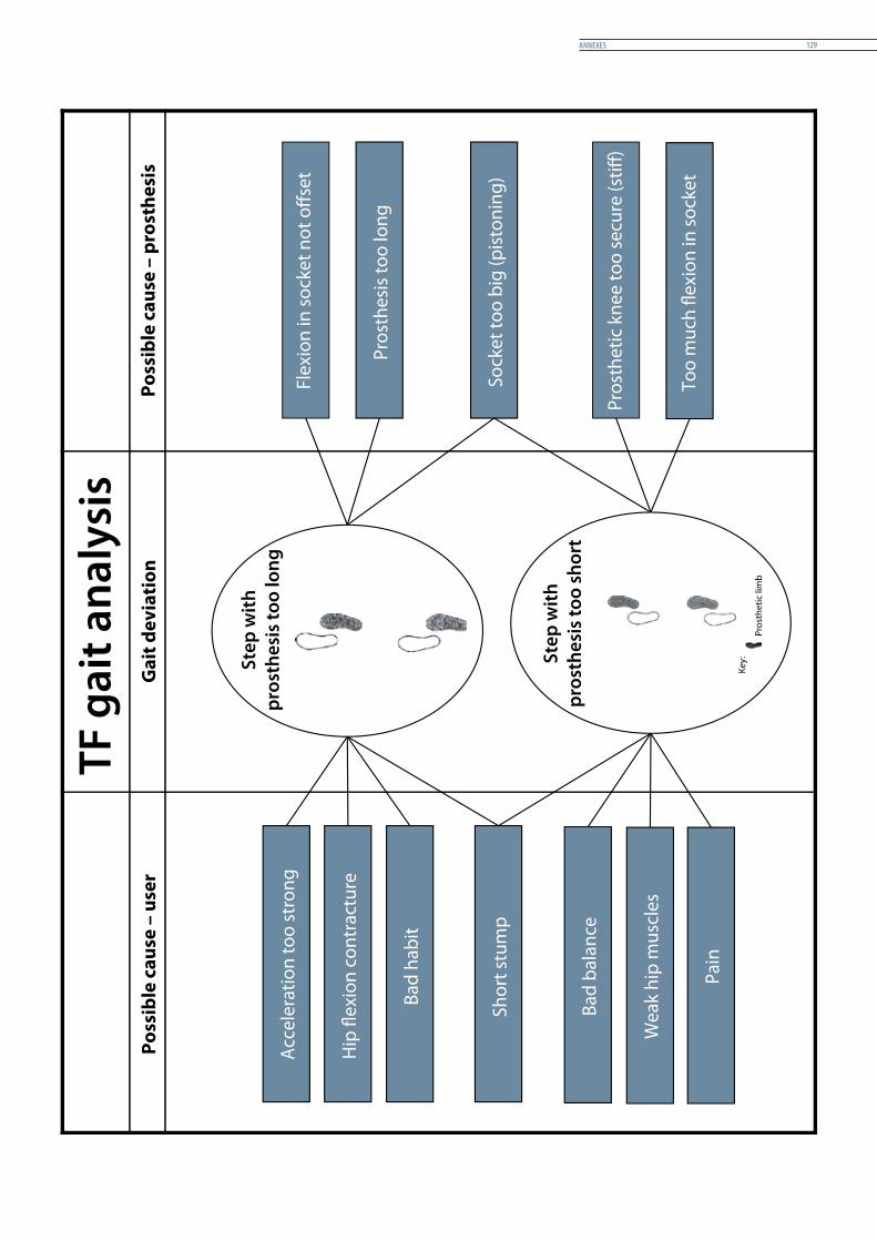
annexes 129
TF g
ait a
naly
sis
Poss
ible
cau
se –
use
rG
ait d
evia
tion
Hip
�ex
ion
cont
ract
ure
Bad
habi
t
Shor
t stu
mp
Bad
bala
nce
Wea
k hi
p m
uscl
es
Acc
eler
atio
n to
o st
rong
Pain
Flex
ion
in so
cket
not
o�s
et
Pros
thes
is to
o lo
ng
Sock
et to
o bi
g (p
isto
ning
)
Pros
thet
ic k
nee
too
secu
re (s
ti�)
Too
muc
h �e
xion
in so
cket
Step
with
pros
thes
is to
o lo
ng
Step
with
pros
thes
is to
o sh
ort
Poss
ible
cau
se –
pro
sthe
sis
Key:
Pros
thet
ic li
mb

MISSIONThe International Committee of the Red Cross (ICRC) is an impartial, neutral and independent organization whose exclusively humanitarian mission is to protect the lives and dignity of victims of armed conflict and other situations of violence and to provide them with assistance. The ICRC also endeavours to prevent suffering by promoting and strengthening humanitarian law and universal humanitarian principles. Established in 1863, the ICRC is at the origin of the Geneva Conventions and the International Red Cross and Red Crescent Movement. It directs and coordinates the international activities conducted by the Movement in armed conflicts and other situations of violence.

4030
;5/0
02 0
1/20
14


![Gait Rehabilitation - Brain Computer Interfaces€¦ · gait cycle and trigger an actuator to appropriately adjust one or more prosthetic or orthotic joint [8–12]. The second type](https://img.pdfslide.net/doc/110x75/5f3df2e94cd4b31384567c90/gait-rehabilitation-brain-computer-interfaces-gait-cycle-and-trigger-an-actuator.jpg)