Embed Size (px)
Citation preview
Protein Engineering
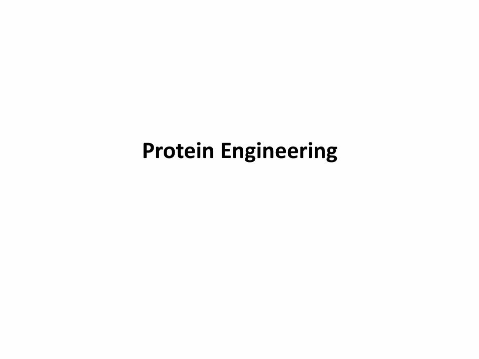
Proteins
• Consist of linear polymers built from series of up to 20 different L-amino acids
• Play central roles in biological events through interactions with cognate proteins / ligands : Cell signaling processes
• Enzymes catalyze the reactions with high specificity and efficiency in cellular metabolic pathways : major components
• Dysfunctions : Amyloidosis (formation of fibrils), cancers by mutations (p53,
Ras etc.), Sickle cell anemia, lysosomal storage disorders
• Therapeutic agents
- Many diseases caused by mutations and loss of their functions
- Cancers, lysosomal storage disorders, Alzheimer's disease etc.
• Industrial Biotechnology: Use of enzymes for bio-based processes
• Biomaterials: Biocompatible materials
Major functional molecules
Practical applications
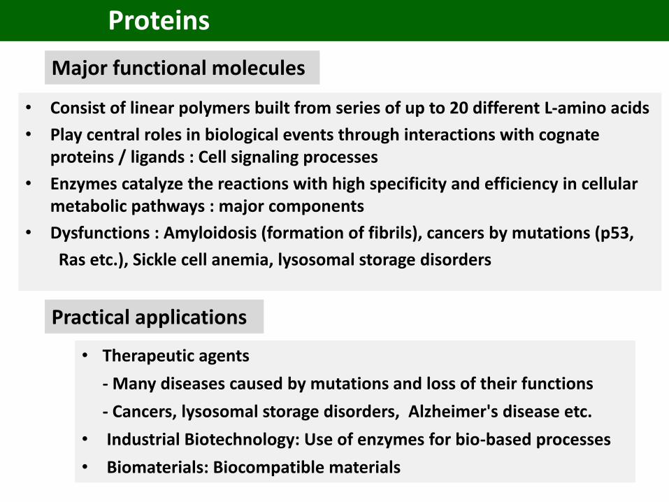
Protein-protein interaction network
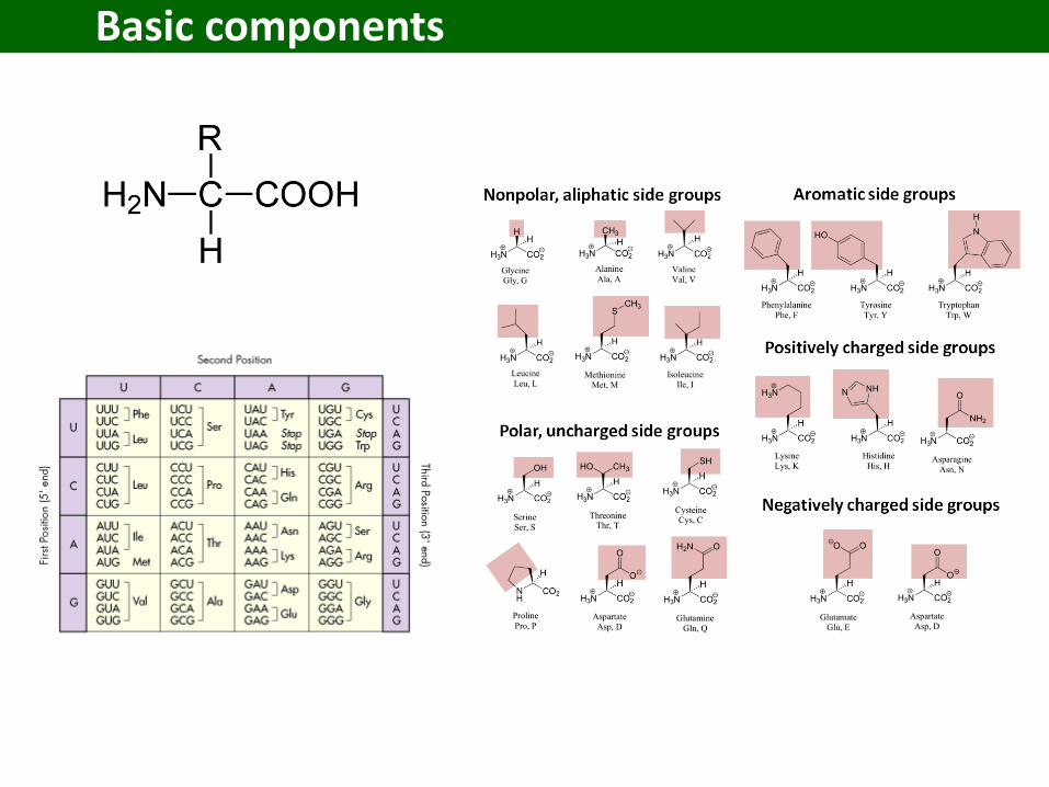
Basic components
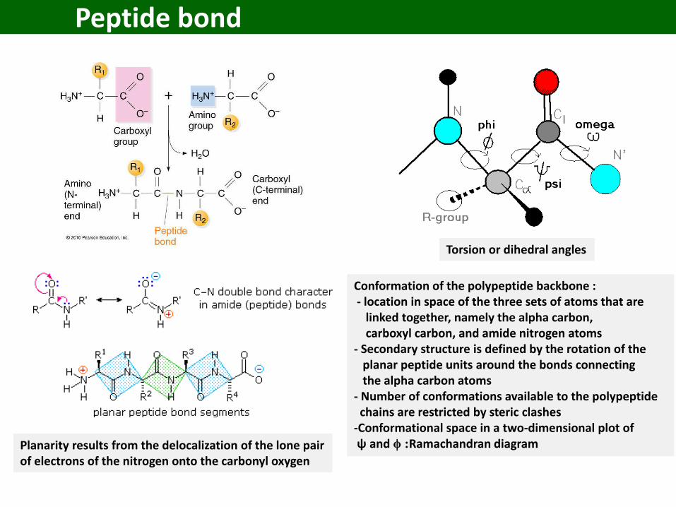
Peptide bond
Planarity results from the delocalization of the lone pair of electrons of the nitrogen onto the carbonyl oxygen
Torsion or dihedral angles
Conformation of the polypeptide backbone : - location in space of the three sets of atoms that are
linked together, namely the alpha carbon, carboxyl carbon, and amide nitrogen atoms
- Secondary structure is defined by the rotation of the planar peptide units around the bonds connecting the alpha carbon atoms
- Number of conformations available to the polypeptidechains are restricted by steric clashes
-Conformational space in a two-dimensional plot of ψ and ϕ :Ramachandran diagram
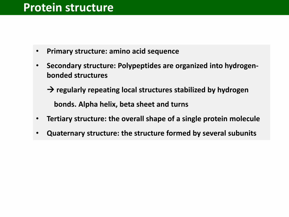
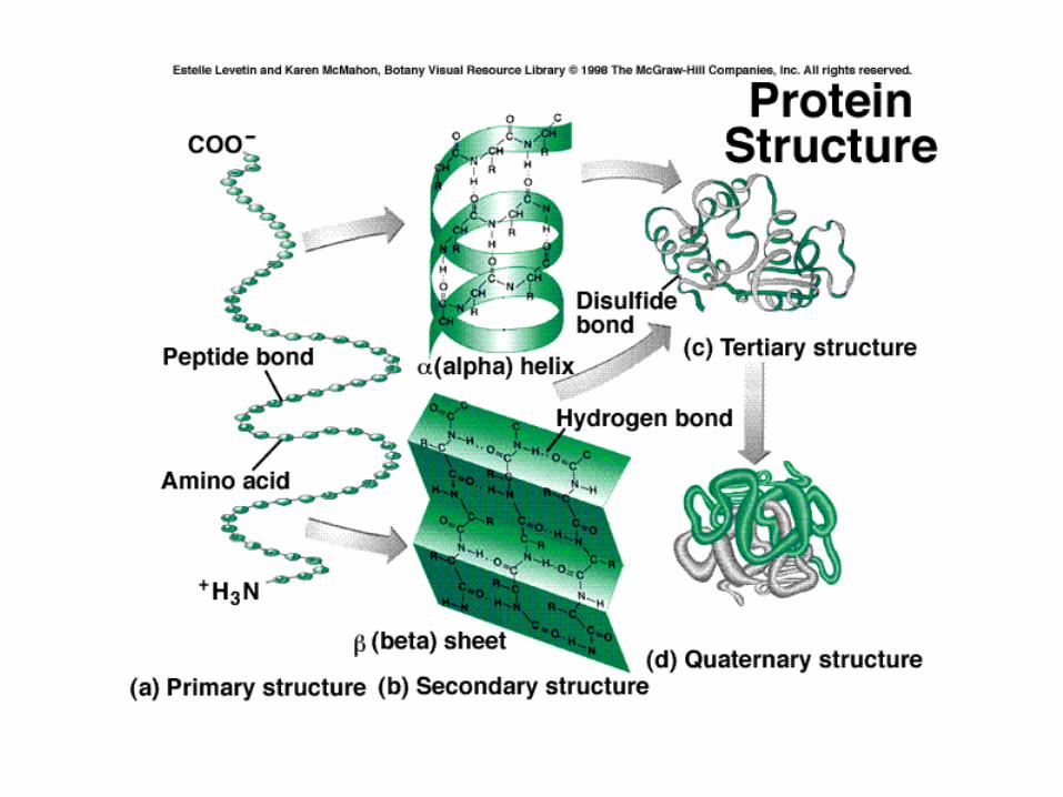
Protein structure
• Primary structure: amino acid sequence
• Secondary structure: Polypeptides are organized into hydrogen-bonded structures
regularly repeating local structures stabilized by hydrogen
bonds. Alpha helix, beta sheet and turns
• Tertiary structure: the overall shape of a single protein molecule
• Quaternary structure: the structure formed by several subunits
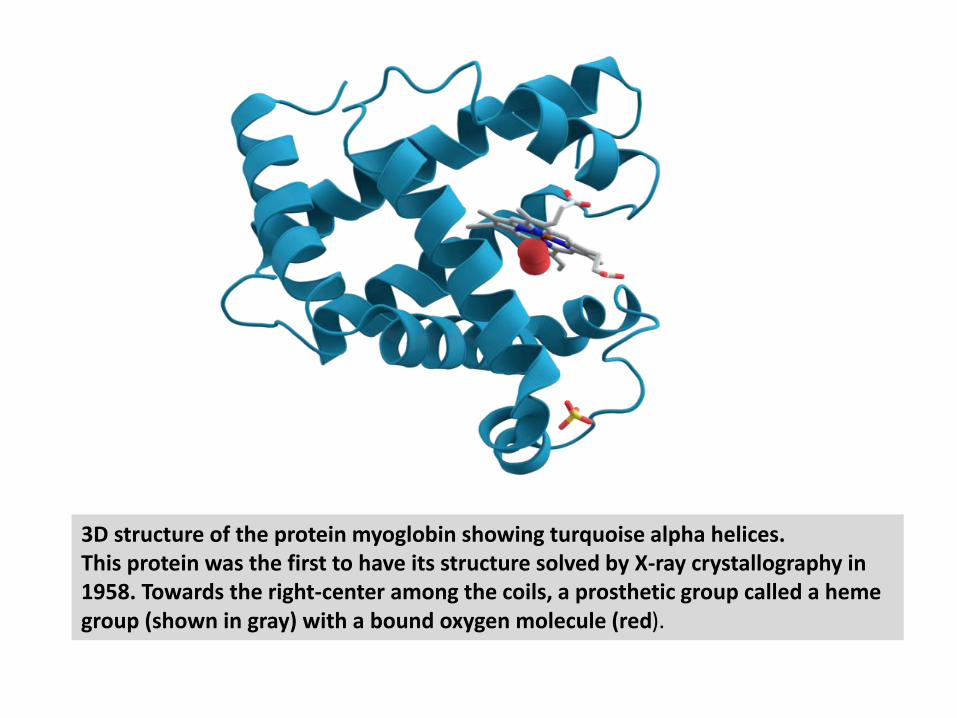
3D structure of the protein myoglobin showing turquoise alpha helices. This protein was the first to have its structure solved by X-ray crystallography in 1958. Towards the right-center among the coils, a prosthetic group called a hemegroup (shown in gray) with a bound oxygen molecule (red).
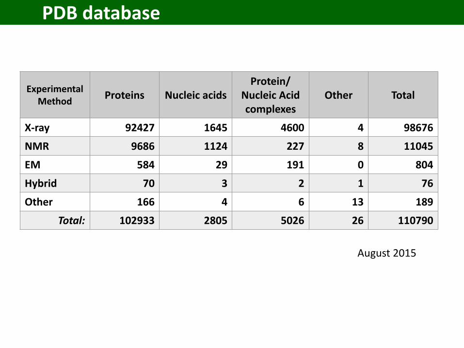
PDB database
ExperimentalMethod
Proteins Nucleic acidsProtein/
Nucleic Acidcomplexes
Other Total
X-ray 92427 1645 4600 4 98676
NMR 9686 1124 227 8 11045
EM 584 29 191 0 804
Hybrid 70 3 2 1 76
Other 166 4 6 13 189
Total: 102933 2805 5026 26 110790
August 2015
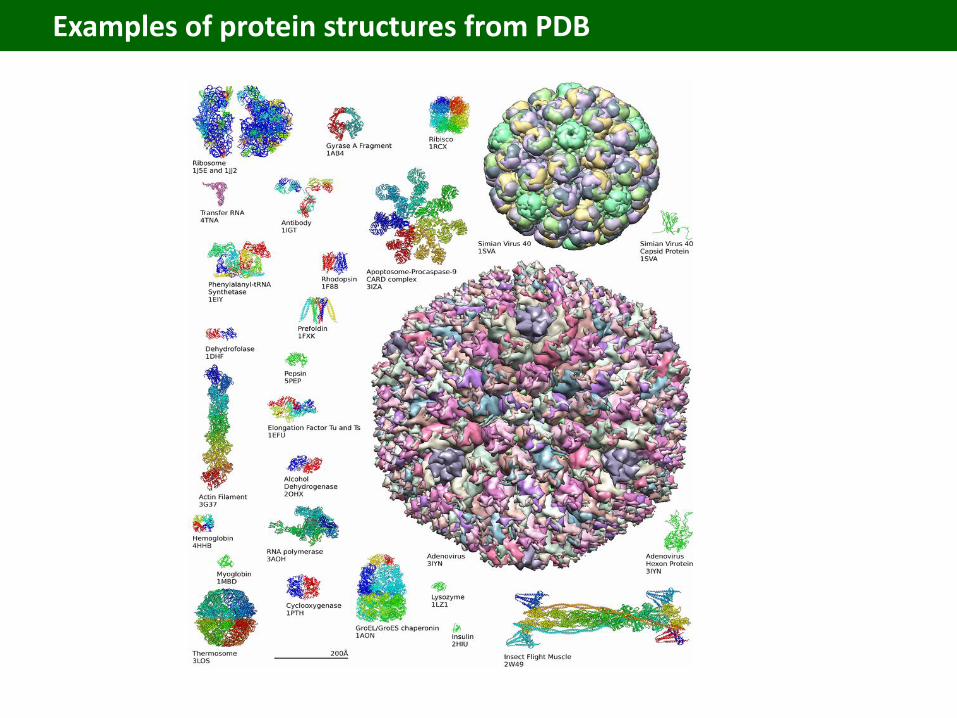
Examples of protein structures from PDB
Protein Engineering
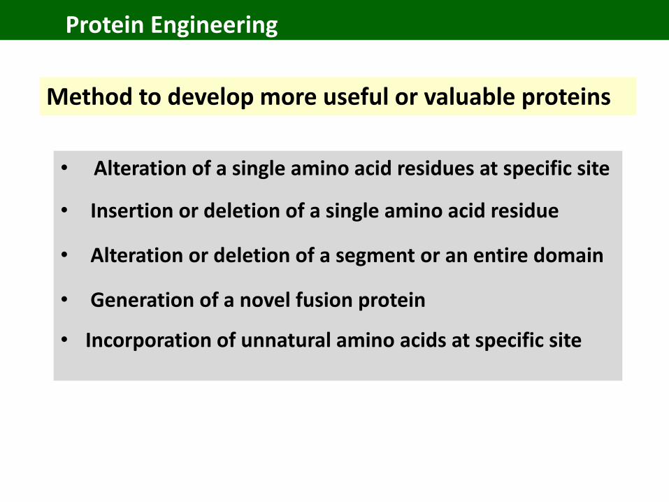
• Alteration of a single amino acid residues at specific site
• Insertion or deletion of a single amino acid residue
• Alteration or deletion of a segment or an entire domain
• Generation of a novel fusion protein
• Incorporation of unnatural amino acids at specific site
Method to develop more useful or valuable proteins
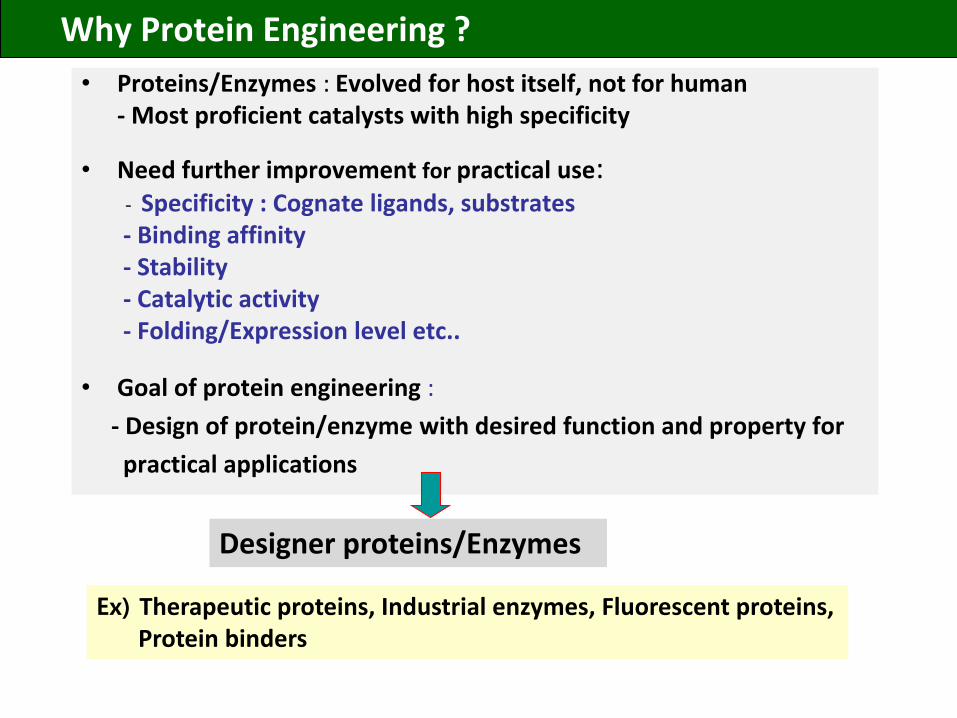
Why Protein Engineering ?
• Proteins/Enzymes : Evolved for host itself, not for human- Most proficient catalysts with high specificity
• Need further improvement for practical use:- Specificity : Cognate ligands, substrates- Binding affinity- Stability - Catalytic activity - Folding/Expression level etc..
• Goal of protein engineering :
- Design of protein/enzyme with desired function and property for
practical applications
Designer proteins/Enzymes
Ex) Therapeutic proteins, Industrial enzymes, Fluorescent proteins, Protein binders
Biomolecular Eng. Lab.
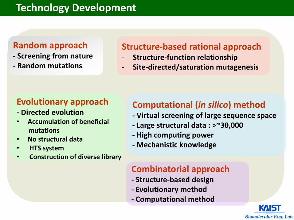
Random approach- Screening from nature- Random mutations
Structure-based rational approach- Structure-function relationship- Site-directed/saturation mutagenesis
Evolutionary approach- Directed evolution• Accumulation of beneficial
mutations• No structural data• HTS system• Construction of diverse library
Computational (in silico) method- Virtual screening of large sequence space- Large structural data : >~30,000- High computing power- Mechanistic knowledge
Combinatorial approach- Structure-based design- Evolutionary method- Computational method
Technology Development
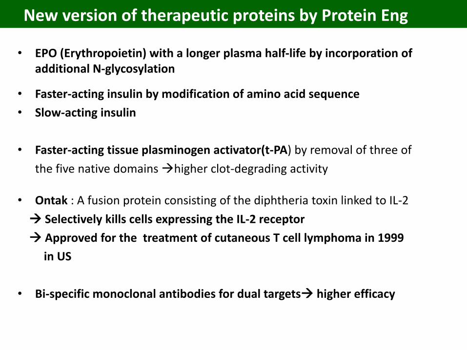
New version of therapeutic proteins by Protein Eng
• EPO (Erythropoietin) with a longer plasma half-life by incorporation of additional N-glycosylation
• Faster-acting insulin by modification of amino acid sequence
• Slow-acting insulin
• Faster-acting tissue plasminogen activator(t-PA) by removal of three of
the five native domains higher clot-degrading activity
• Ontak : A fusion protein consisting of the diphtheria toxin linked to IL-2
Selectively kills cells expressing the IL-2 receptor
Approved for the treatment of cutaneous T cell lymphoma in 1999
in US
• Bi-specific monoclonal antibodies for dual targets higher efficacy
Glycoprotein: Glycosylation, Glycobiology
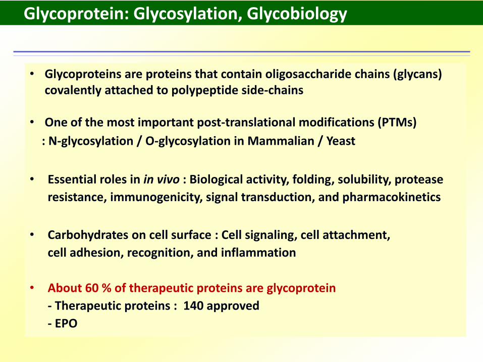
• Glycoproteins are proteins that contain oligosaccharide chains (glycans) covalently attached to polypeptide side-chains
• One of the most important post-translational modifications (PTMs)
: N-glycosylation / O-glycosylation in Mammalian / Yeast
• Essential roles in in vivo : Biological activity, folding, solubility, protease
resistance, immunogenicity, signal transduction, and pharmacokinetics
• Carbohydrates on cell surface : Cell signaling, cell attachment,
cell adhesion, recognition, and inflammation
• About 60 % of therapeutic proteins are glycoprotein
- Therapeutic proteins : 140 approved
- EPO
Glycan Profile of Glycoprotein
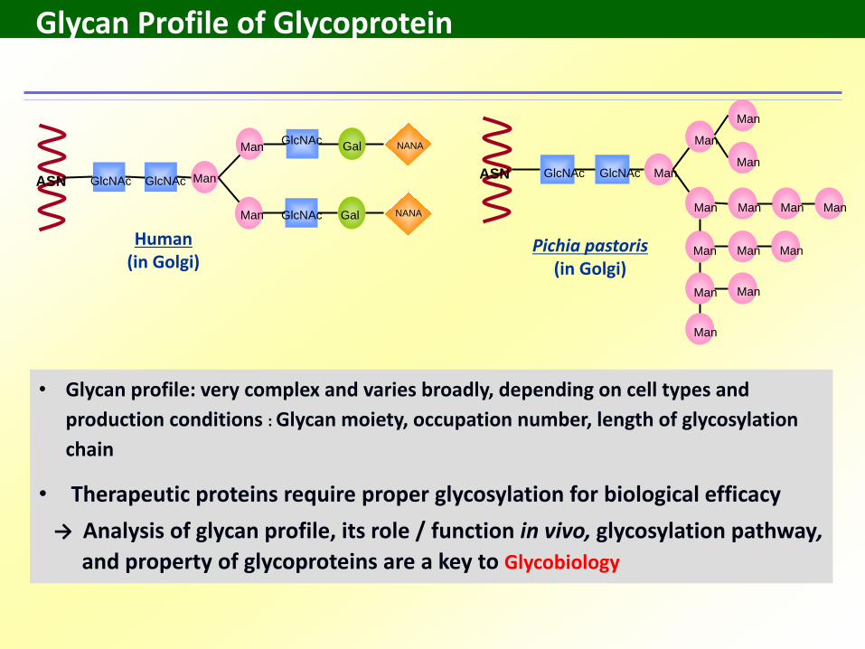
Human(in Golgi)
Pichia pastoris(in Golgi)
ASN GlcNAc GlcNAc Man
Gal
ManGlcNAc
GlcNAc
Gal NANA
NANAMan
• Glycan profile: very complex and varies broadly, depending on cell types and
production conditions : Glycan moiety, occupation number, length of glycosylation
chain
• Therapeutic proteins require proper glycosylation for biological efficacy
→ Analysis of glycan profile, its role / function in vivo, glycosylation pathway,
and property of glycoproteins are a key to Glycobiology
ASN GlcNAc GlcNAc Man
Man
Man
Man
Man
Man
Man
Man Man
Man Man
Man Man
Man
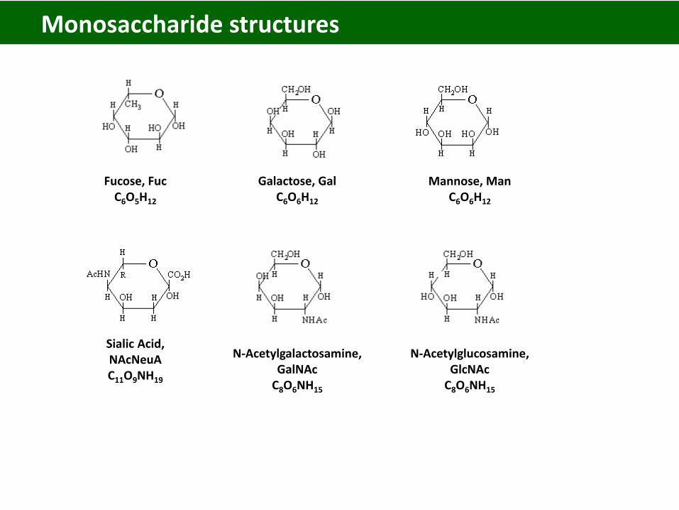
Fucose, FucC6O5H12
Galactose, GalC6O6H12
Mannose, ManC6O6H12
Sialic Acid,NAcNeuAC11O9NH19
N-Acetylgalactosamine,GalNAc
C8O6NH15
N-Acetylglucosamine,GlcNAc
C8O6NH15
Monosaccharide structures
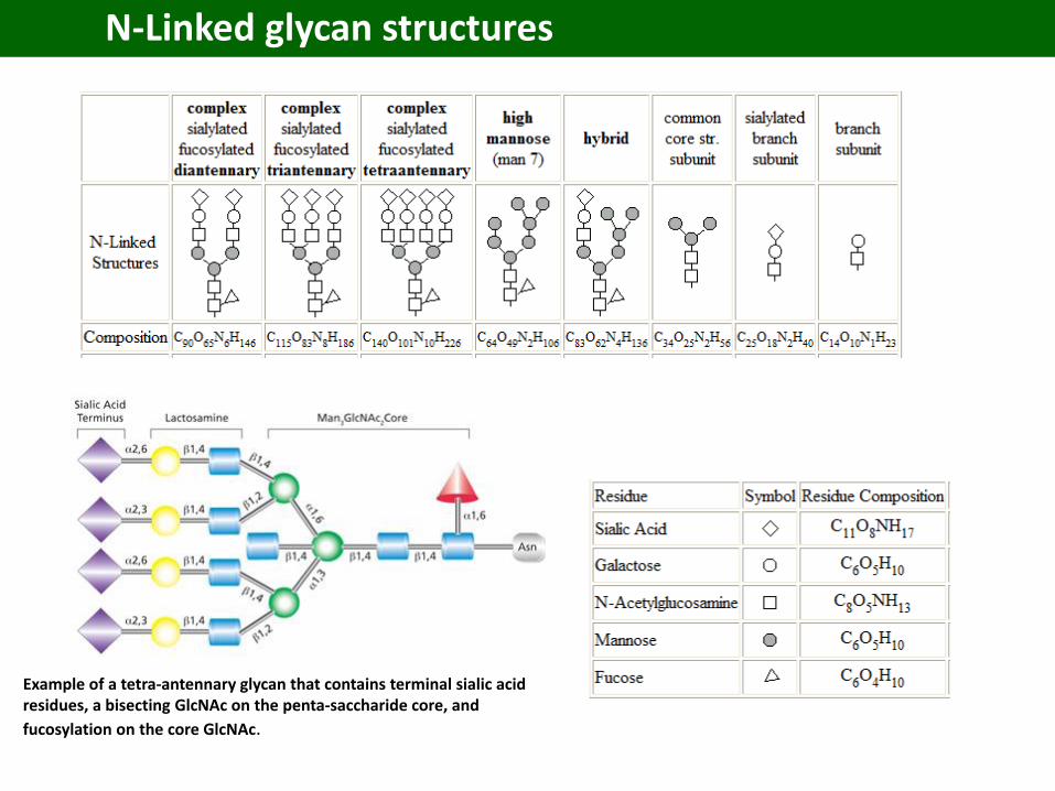
N-Linked glycan structures
Example of a tetra-antennary glycan that contains terminal sialic acid residues, a bisecting GlcNAc on the penta-saccharide core, and
fucosylation on the core GlcNAc.
Erythropoietin (EPO)
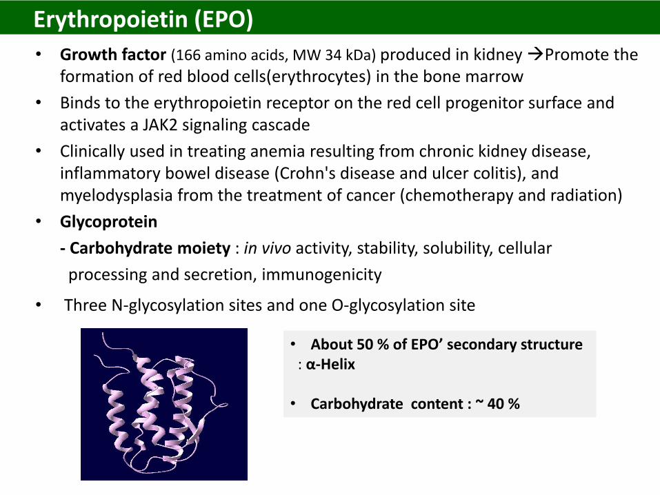
• Growth factor (166 amino acids, MW 34 kDa) produced in kidney Promote the formation of red blood cells(erythrocytes) in the bone marrow
• Binds to the erythropoietin receptor on the red cell progenitor surface and activates a JAK2 signaling cascade
• Clinically used in treating anemia resulting from chronic kidney disease, inflammatory bowel disease (Crohn's disease and ulcer colitis), and myelodysplasia from the treatment of cancer (chemotherapy and radiation)
• Glycoprotein
- Carbohydrate moiety : in vivo activity, stability, solubility, cellular
processing and secretion, immunogenicity
• Three N-glycosylation sites and one O-glycosylation site
• About 50 % of EPO’ secondary structure : α-Helix
• Carbohydrate content : ~ 40 %
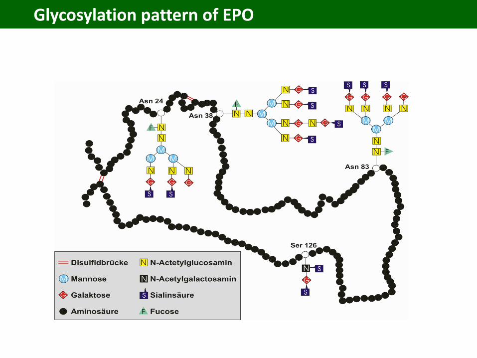
Glycosylation pattern of EPO
• 1971: First purified from the plasma of anemic sheep
• 1985 : Produced by recombinant DNA technology
• 1989: Approved by FDA for treatment of anemia resulting from chronic
kidney disease and cancer treatment (chemotherapy and radiation)
• Total sales : $ 11 billion (2010)
• Major EPO brands : Biosimilars
- Epogen by Amgen ($ 2.5 billion)
- Procrit by Ortho Biotech ($ 3.5 billion)
- Neorecormon by Boehringer-Mannheim ($ 1.5 billion)
History of the EPO development
• As the patent becomes expired, Amgen wanted to prolong the market share by developing a new version of EPO by protein engineering
• Recommended and usual therapy with EPO : two or three times per week by subcutaneous and or intravenous injection
• Aranesp : Introduction of two additional N-glycosylation sites
- Which site of EPO?
A prolonged serum half-life from 4-6 up to 21 hrs
- What benefit to patients?
Launched in 2001 Current sale : $ 3.5 billion
New version of EPO by protein engineering
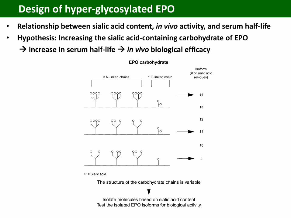
Design of hyper-glycosylated EPO
• Relationship between sialic acid content, in vivo activity, and serum half-life
• Hypothesis: Increasing the sialic acid-containing carbohydrate of EPO
increase in serum half-life in vivo biological efficacy
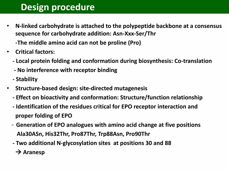
Design procedure
• N-linked carbohydrate is attached to the polypeptide backbone at a consensus sequence for carbohydrate addition: Asn-Xxx-Ser/Thr
-The middle amino acid can not be proline (Pro)
• Critical factors:
- Local protein folding and conformation during biosynthesis: Co-translation
- No interference with receptor binding
- Stability
• Structure-based design: site-directed mutagenesis
- Effect on bioactivity and conformation: Structure/function relationship
- Identification of the residues critical for EPO receptor interaction and
proper folding of EPO
- Generation of EPO analogues with amino acid change at five positions
Ala30ASn, His32Thr, Pro87Thr, Trp88Asn, Pro90Thr
- Two additional N-glycosylation sites at positions 30 and 88
Aranesp
Development of enzyme process: Atorvastatin (Lipitor)
• Lipitor : Brand name by Pfizer
• A competitive inhibitor of HMG -CoA reductase (3-hydroxy-3-methylglutaryl CoA reductase)
- HMG-CoA reductase catalyzes the reduction of 3-hydroxy-3-methylglutaryl-coenzymeA (HMG-CoA) to mevalonate, which is the rate-limiting step in hepatic cholesterol biosynthesis.
• Inhibition of the enzyme decreases de novo cholesterol synthesis, increasing expression of low-density lipoprotein receptors (L-receptors) on hepatocytes.
Increase in LDL uptake by the hepatocytes, decreasing the amount of LDL-cholesterol in the blood.
• Like other statins, atorvastatin also reduces blood levels of triglycerides and slightly increases the levels of HDL-cholesterol.
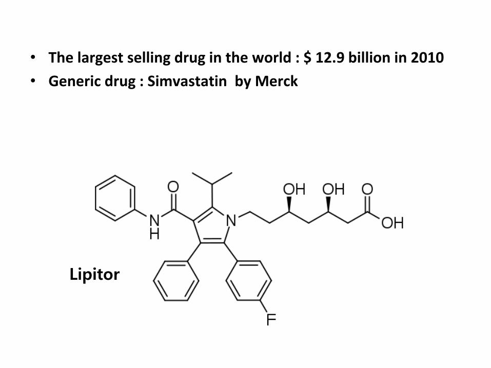
• The largest selling drug in the world : $ 12.9 billion in 2010
• Generic drug : Simvastatin by Merck
Lipitor
Pfizer fight against a simvastatin generic
• Doctors and patients began switching to a cheaper generic alternative drug called simvastatin from Merck.
• Pfizer launched a campaign including advertisements, lobbying efforts, and a paid speaking tour by Dr. Louis W. Sullivan, a former secretary of the federal
Depart. of Health and Human Services, to discourage the trend.
• Studies show that at commonly prescribed doses Lipitor and simvastatin are equally effective at reducing LDL cholesterol.
• Pfizer has begun promoting a study, conducted by Pfizer’s own researchers, concluding switching increased the rate of heart attacks among British patients.
Economic process using an enzyme with higher efficiency
Halohydrin dehalogenase (HHDH)
• Catalyze the nucleophilic displacement of a halogen by a vicinal hydroxyl
group in halohydrins, yielding an epoxide
• Interconverts halohydrins and epoxides
Manufacture of ethyl (R)-4-cyano-3-hydroxybutyrate (HN) from ethyl ethyl(S) -4 –chloro-3-hydroxybutyrate(ECHB)
• NH: Starting material for the production of the cholesterol-lowering drug : Atorvastatin (Lipitor)
• The specifications for the chemical and enantio-purity of HN are tightly controlled. The hydroxyl in HN is defined the second stereo-center in atorovastatin, and high chemical purity is essential for downstream chemistry
Essential for more economic process
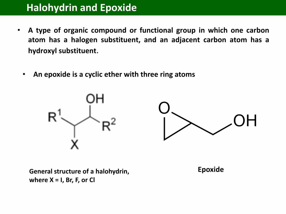
• A type of organic compound or functional group in which one carbonatom has a halogen substituent, and an adjacent carbon atom has a
hydroxyl substituent.
Halohydrin and Epoxide
General structure of a halohydrin, where X = I, Br, F, or Cl
• An epoxide is a cyclic ether with three ring atoms
Epoxide
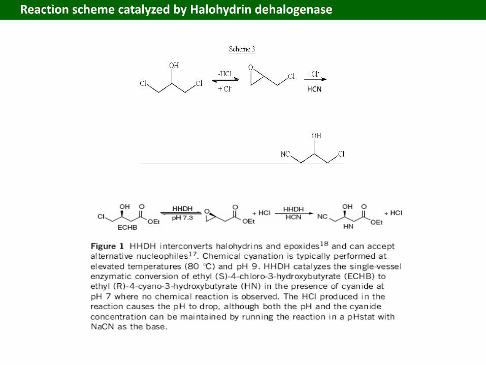
Reaction scheme catalyzed by Halohydrin dehalogenase
HCN
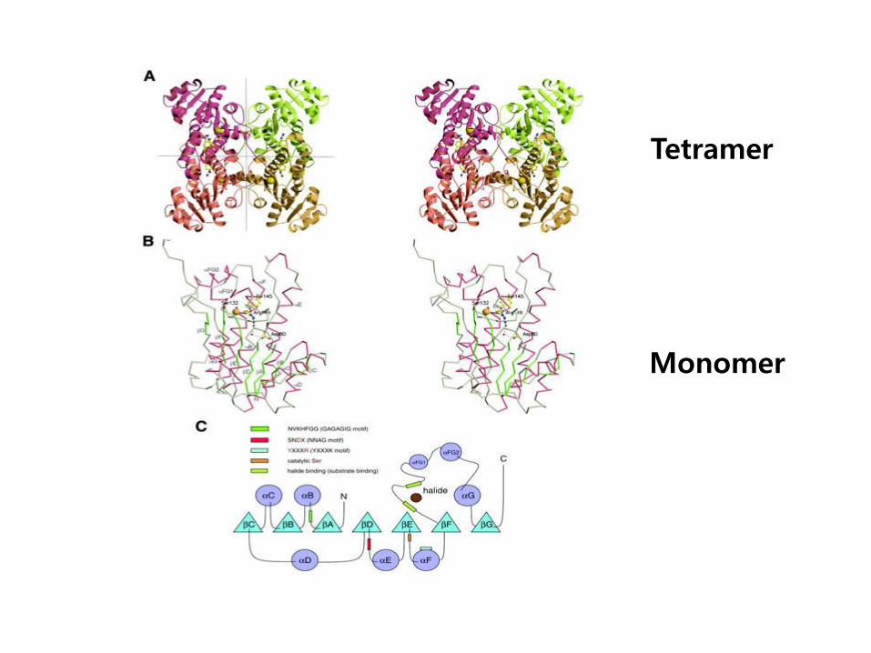
Tetramer
Monomer
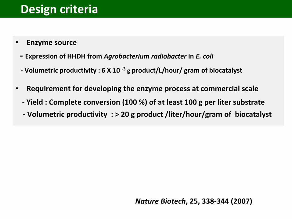
Design criteria
• Enzyme source
- Expression of HHDH from Agrobacterium radiobacter in E. coli
- Volumetric productivity : 6 X 10 -3 g product/L/hour/ gram of biocatalyst
• Requirement for developing the enzyme process at commercial scale
- Yield : Complete conversion (100 %) of at least 100 g per liter substrate
- Volumetric productivity : > 20 g product /liter/hour/gram of biocatalyst
Nature Biotech, 25, 338-344 (2007)
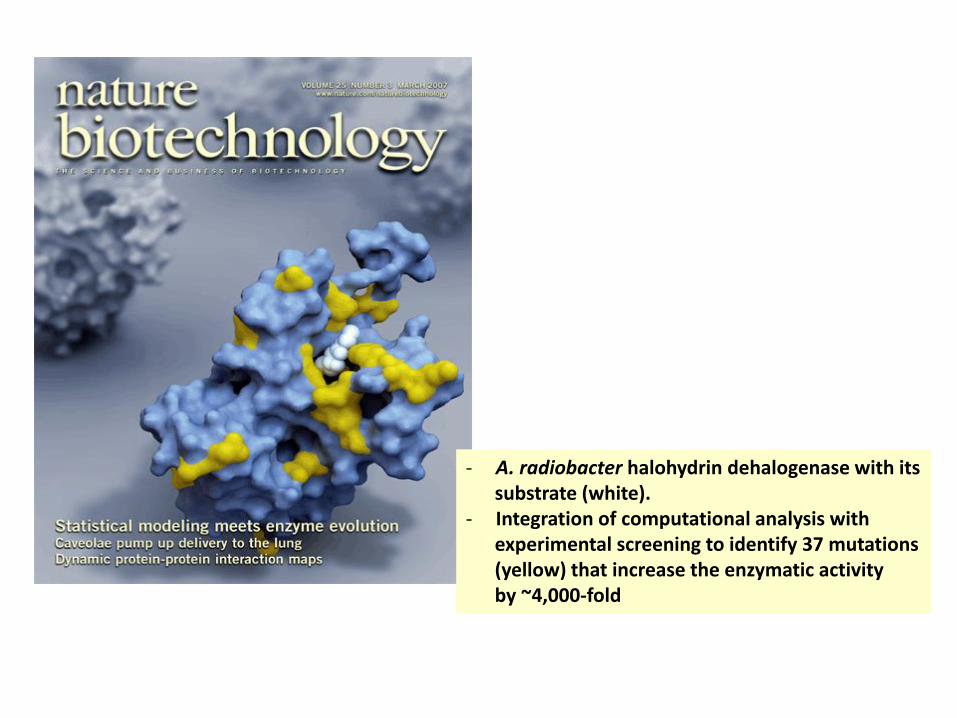
- A. radiobacter halohydrin dehalogenase with itssubstrate (white).
- Integration of computational analysis withexperimental screening to identify 37 mutations(yellow) that increase the enzymatic activityby ~4,000-fold
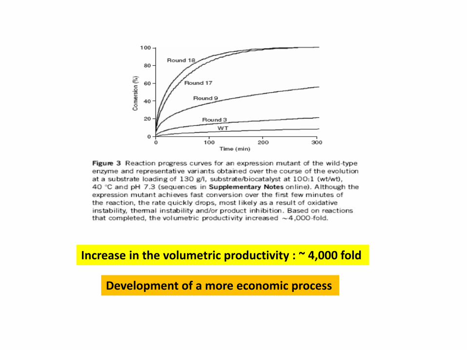
Increase in the volumetric productivity : ~ 4,000 fold
Development of a more economic process
1 2
33
2
1
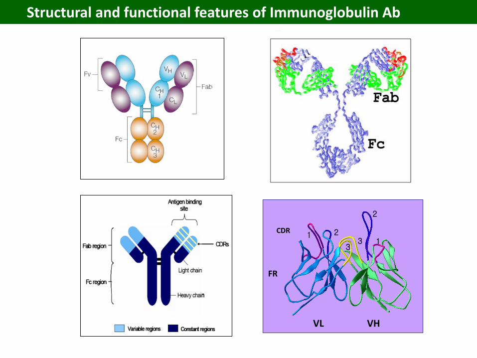
CDR
FR
VL VH
Structural and functional features of Immunoglobulin Ab
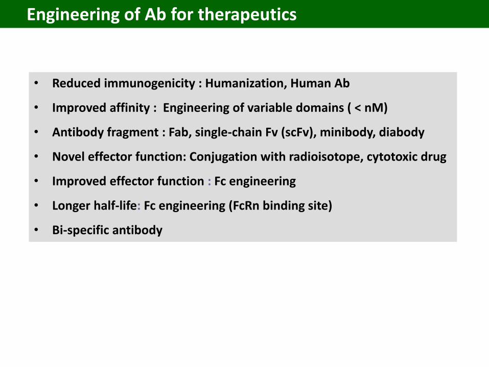
Engineering of Ab for therapeutics
• Reduced immunogenicity : Humanization, Human Ab
• Improved affinity : Engineering of variable domains ( < nM)
• Antibody fragment : Fab, single-chain Fv (scFv), minibody, diabody
• Novel effector function: Conjugation with radioisotope, cytotoxic drug
• Improved effector function : Fc engineering
• Longer half-life: Fc engineering (FcRn binding site)
• Bi-specific antibody
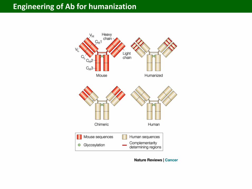
Engineering of Ab for humanization
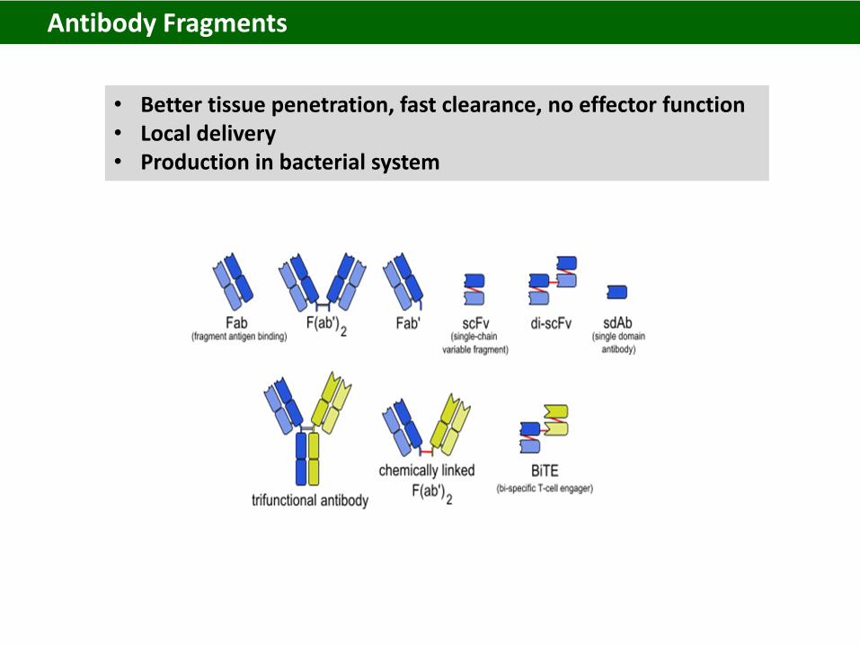
• Better tissue penetration, fast clearance, no effector function• Local delivery• Production in bacterial system
Antibody Fragments
Lucentis
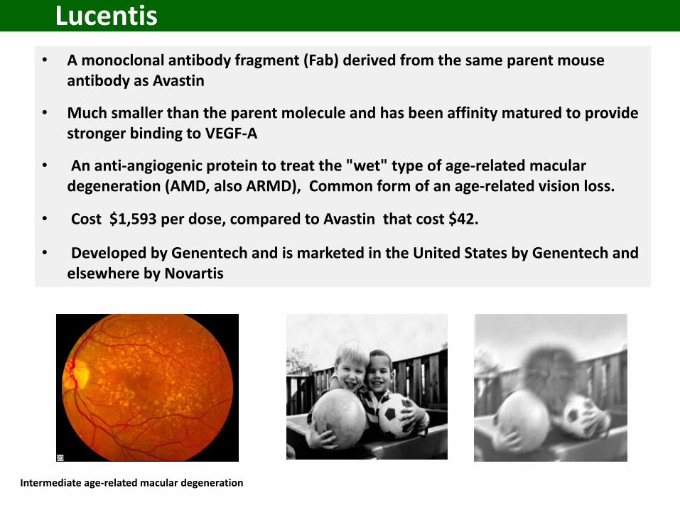
• A monoclonal antibody fragment (Fab) derived from the same parent mouse antibody as Avastin
• Much smaller than the parent molecule and has been affinity matured to provide stronger binding to VEGF-A
• An anti-angiogenic protein to treat the "wet" type of age-related macular degeneration (AMD, also ARMD), Common form of an age-related vision loss.
• Cost $1,593 per dose, compared to Avastin that cost $42.
• Developed by Genentech and is marketed in the United States by Genentech and elsewhere by Novartis
Intermediate age-related macular degeneration
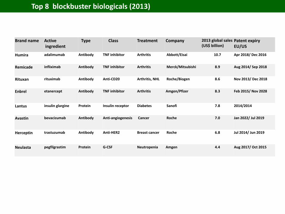
Brand name Activeingredient
Type Class Treatment Company 2013 global sales(US$ billion)
Patent expiryEU/US
Humira adalimumab Antibody TNF inhibitor Arthritis Abbott/Eisai 10.7 Apr 2018/ Dec 2016
Remicade infliximab Antibody TNF inhibitor Arthritis Merck/Mitsubishi 8.9 Aug 2014/ Sep 2018
Rituxan rituximab Antibody Anti-CD20 Arthritis, NHL Roche/Biogen 8.6 Nov 2013/ Dec 2018
Enbrel etanercept Antibody TNF inhibitor Arthritis Amgen/Pfizer 8.3 Feb 2015/ Nov 2028
Lantus insulin glargine Protein Insulin receptor Diabetes Sanofi 7.8 2014/2014
Avastin bevacizumab Antibody Anti-angiogenesis Cancer Roche 7.0 Jan 2022/ Jul 2019
Herceptin trastuzumab Antibody Anti-HER2 Breast cancer Roche 6.8 Jul 2014/ Jun 2019
Neulasta pegfilgrastim Protein G-CSF Neutropenia Amgen 4.4 Aug 2017/ Oct 2015
Top 8 blockbuster biologicals (2013)
Fluorescent proteins
History of Fluorescent Proteins
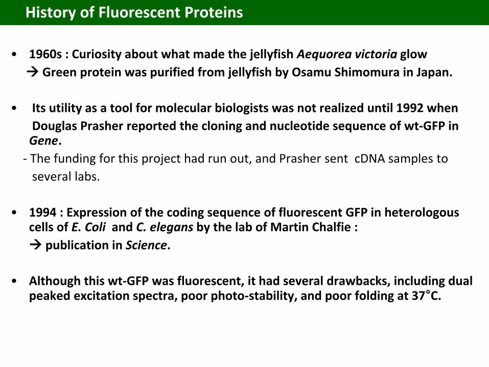
• 1960s : Curiosity about what made the jellyfish Aequorea victoria glow
Green protein was purified from jellyfish by Osamu Shimomura in Japan.
• Its utility as a tool for molecular biologists was not realized until 1992 when
Douglas Prasher reported the cloning and nucleotide sequence of wt-GFP in Gene.
- The funding for this project had run out, and Prasher sent cDNA samples to
several labs.
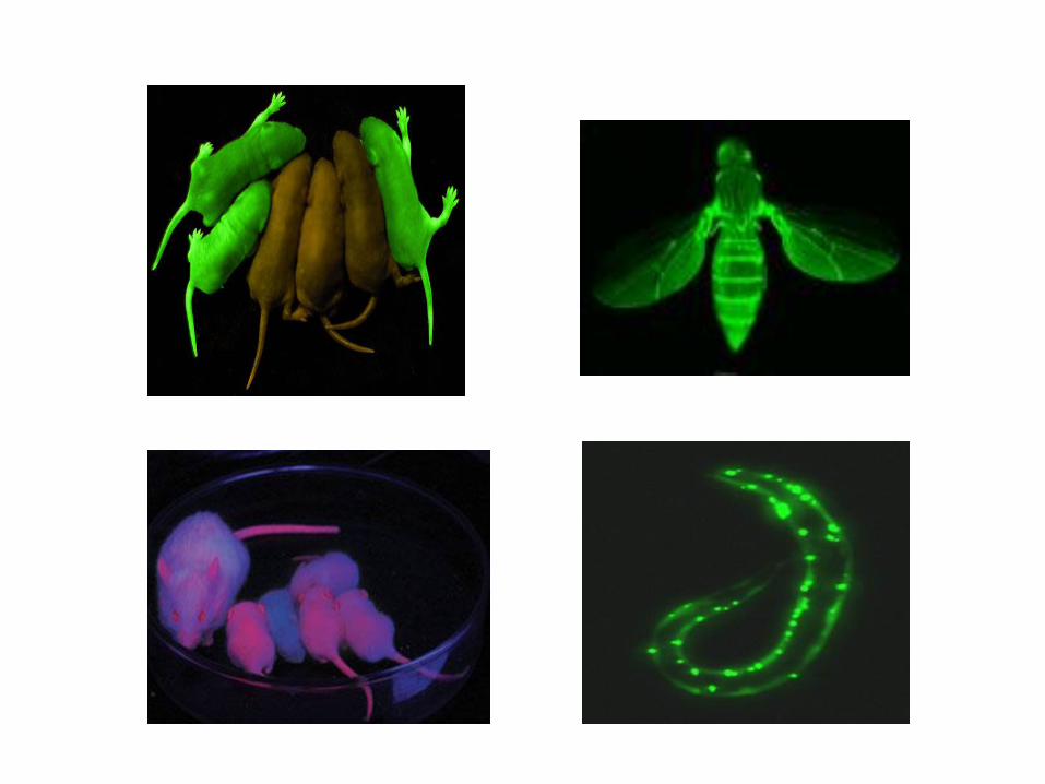
• 1994 : Expression of the coding sequence of fluorescent GFP in heterologous cells of E. Coli and C. elegans by the lab of Martin Chalfie :
publication in Science.
• Although this wt-GFP was fluorescent, it had several drawbacks, including dual peaked excitation spectra, poor photo-stability, and poor folding at 37°C.
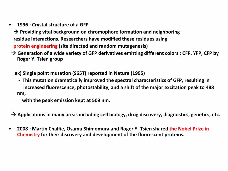
• 1996 : Crystal structure of a GFP
Providing vital background on chromophore formation and neighboring
residue interactions. Researchers have modified these residues using
protein engineering (site directed and random mutagenesis)
Generation of a wide variety of GFP derivatives emitting different colors ; CFP, YFP, CFP by Roger Y. Tsien group
ex) Single point mutation (S65T) reported in Nature (1995)
- This mutation dramatically improved the spectral characteristics of GFP, resulting in
increased fluorescence, photostability, and a shift of the major excitation peak to 488 nm,
with the peak emission kept at 509 nm.
Applications in many areas including cell biology, drug discovery, diagnostics, genetics, etc.
• 2008 : Martin Chalfie, Osamu Shimomura and Roger Y. Tsien shared the Nobel Prize in Chemistry for their discovery and development of the fluorescent proteins.
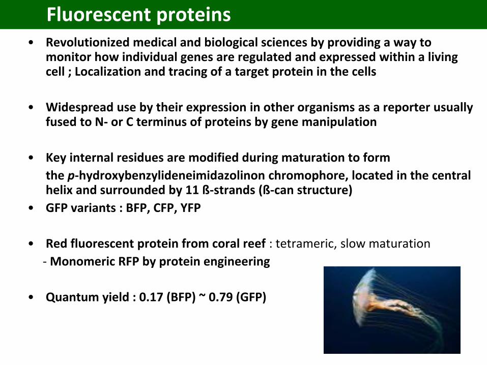
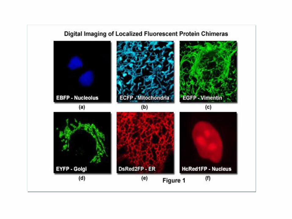
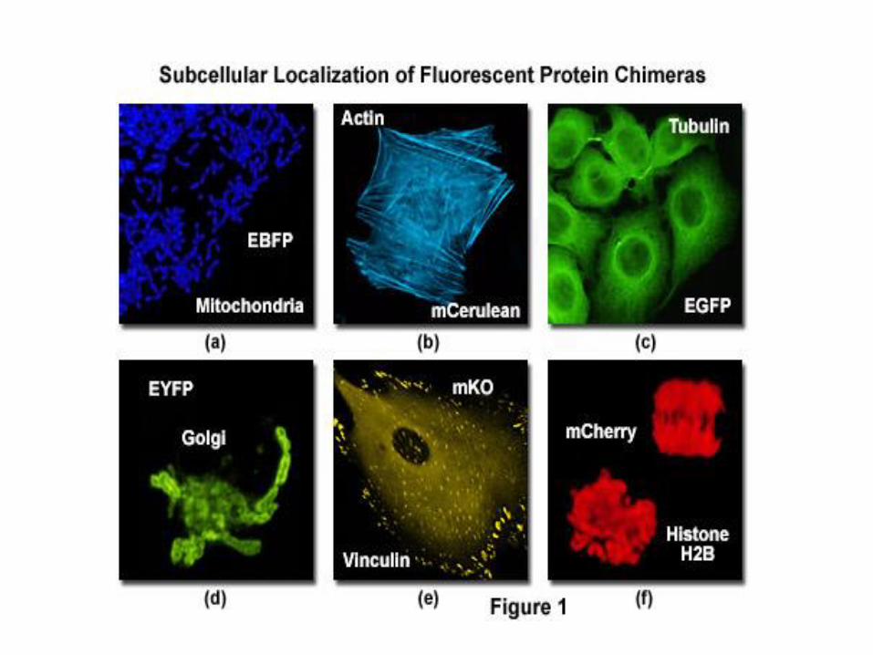
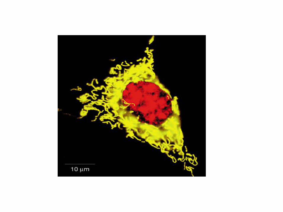
Fluorescent proteins• Revolutionized medical and biological sciences by providing a way to
monitor how individual genes are regulated and expressed within a living cell ; Localization and tracing of a target protein in the cells
• Widespread use by their expression in other organisms as a reporter usually fused to N- or C terminus of proteins by gene manipulation
• Key internal residues are modified during maturation to form
the p-hydroxybenzylideneimidazolinon chromophore, located in the central helix and surrounded by 11 ß-strands (ß-can structure)
• GFP variants : BFP, CFP, YFP
• Red fluorescent protein from coral reef : tetrameric, slow maturation
- Monomeric RFP by protein engineering
• Quantum yield : 0.17 (BFP) ~ 0.79 (GFP)

GFP (Green Fluorescent Protein)
• Jellyfish Aequorea victoria
• A tightly packed -can (11 -sheets) enclosing an -helix containing the chromophore
• 238 amino acids
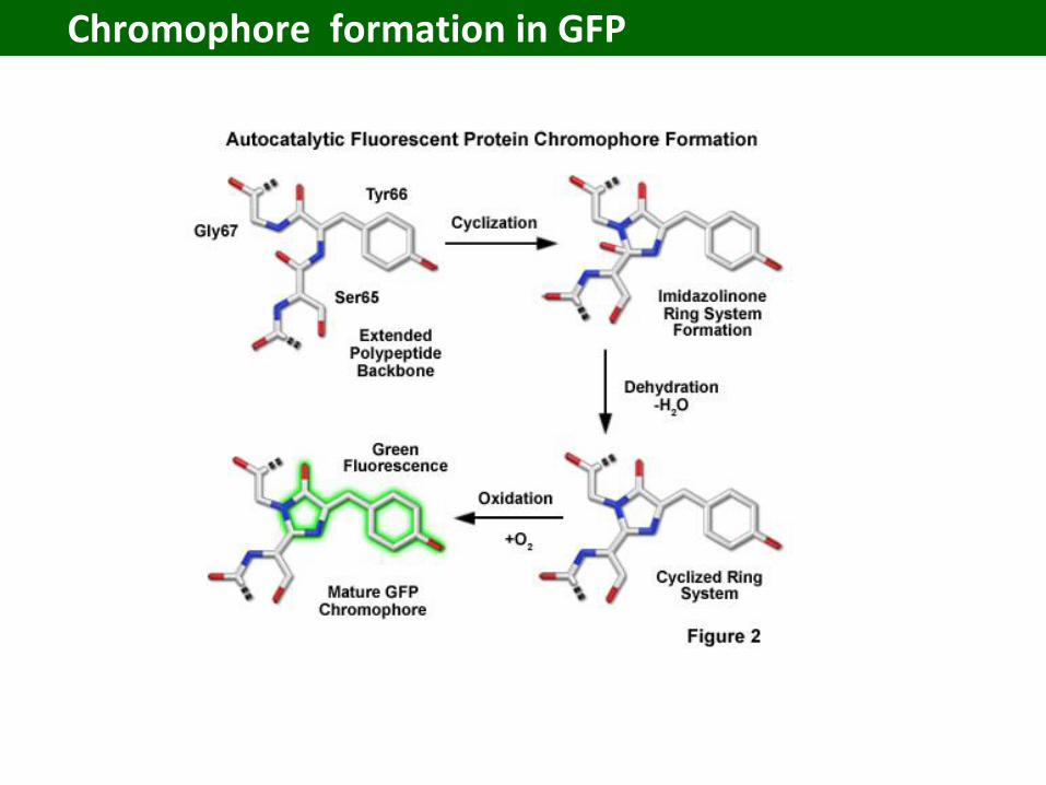
• Chromophore
– Cyclic tripeptide derived from
Ser(65)-Tyr(66)-Gly(67)
• Wt-GFP absorbs UV and blue light (395nm and 470nm) and emits green light (maximally at 509nm)
Chromophore formation in GFP

GFP and chromophore
- Covalently bonded chromophore : 4-(p-hydroxybenzylidene)imidazolidin-5-one (HBI).- HBI is nonfluorescent in the absence of the properly folded GFP scaffold and exists mainly in the
unionized phenol form in wt-GFP.- Maturation (post-translational modification) : Inward-facing side chains of the barrel induce
specific cyclization reactions in the tripeptide Ser65–Tyr66–Gly67 that induce ionization of HBI to the phenolate form and chromophore formation.
- The hydrogen-bonding network and electron-stacking interactions with these side chains influencethe color, intensity and photo-stability of GFP and its numerous derivatives
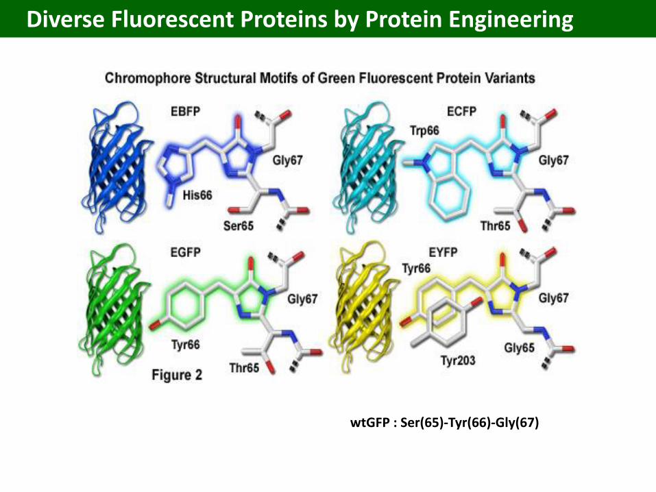
wtGFP : Ser(65)-Tyr(66)-Gly(67)
Diverse Fluorescent Proteins by Protein Engineering
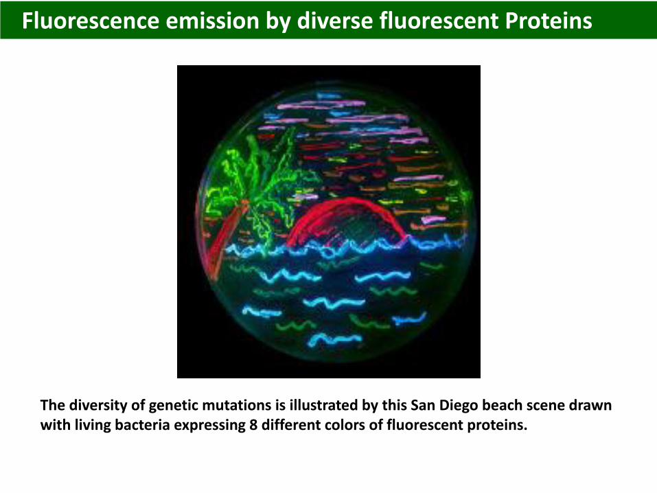
The diversity of genetic mutations is illustrated by this San Diego beach scene drawn with living bacteria expressing 8 different colors of fluorescent proteins.
Fluorescence emission by diverse fluorescent Proteins
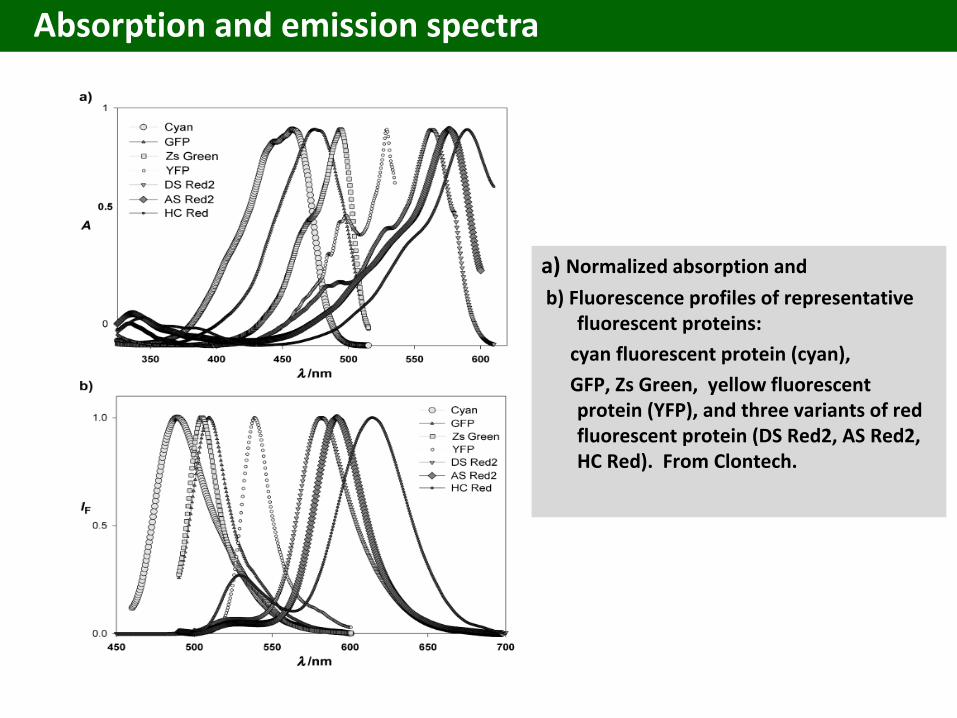
a) Normalized absorption and
b) Fluorescence profiles of representative fluorescent proteins:
cyan fluorescent protein (cyan),
GFP, Zs Green, yellow fluorescent protein (YFP), and three variants of red fluorescent protein (DS Red2, AS Red2, HC Red). From Clontech.
Absorption and emission spectra


![Analytica Chimica Acta - KAISTbel.kaist.ac.kr/extfiles/papers/20170804.pdf · plications in many areas [11]. To increase the sensitivity of immunoassays, chemical conjugation of either](https://img.pdfslide.net/doc/110x75/5c65ec8509d3f230488b5a6b/analytica-chimica-acta-plications-in-many-areas-11-to-increase-the-sensitivity.jpg)


![[PPT]Ch 3. The drug manufacturing process - KAISTbel.kaist.ac.kr/extfiles/lecture/2012spring/bs233... · Web viewTitle Ch 3. The drug manufacturing process Author Hak-Sung Kim Last](https://img.pdfslide.net/doc/110x75/5adb04dd7f8b9afc0f8d48f3/pptch-3-the-drug-manufacturing-process-viewtitle-ch-3-the-drug-manufacturing.jpg)