Embed Size (px)
Citation preview

REVIEW ARTICLE
Protein kinase C: perfectly balanced
Alexandra C. Newton
Department of Pharmacology, University of California at San Diego, La Jolla, CA, USA
ABSTRACTProtein kinase C (PKC) isozymes belong to a family of Ser/Thr kinases whose activity is governedby reversible release of an autoinhibitory pseudosubstrate. For conventional and novel isozymes,this is effected by binding the lipid second messenger, diacylglycerol, but for atypical PKC iso-zymes, this is effected by binding protein scaffolds. PKC shot into the limelight following the dis-covery in the 1980s that the diacylglycerol-sensitive isozymes are “receptors” for the potenttumor-promoting phorbol esters. This set in place a concept that PKC isozymes are oncoproteins.Yet three decades of cancer clinical trials targeting PKC with inhibitors failed and, in some cases,worsened patient outcome. Emerging evidence from cancer-associated mutations and proteinexpression levels provide a reason: PKC isozymes generally function as tumor suppressors andtheir activity should be restored, not inhibited, in cancer therapies. And whereas not enoughactivity is associated with cancer, variants with enhanced activity are associated with degenera-tive diseases such as Alzheimer’s disease. This review describes the tightly controlled mechanismsthat ensure PKC activity is perfectly balanced and what happens when these controls are deregu-lated. PKC isozymes serve as a paradigm for the wisdom of Confucius: “to go beyond is as wrongas to fall short.”
ARTICLE HISTORYReceived 2 January 2018Revised 13 February 2018Accepted 14 February 2018
KEYWORDSProtein kinase C;pseudosubstrate; phorbolesters; diacylglycerol;phosphorylation; tumorsuppressor; cancer;Alzheimer’s disease
Introduction
The discovery of protein kinase C (PKC) solved a 25-year long mystery of how agonists that promote lipidhydrolysis alter cell physiology (Figure 1). In a storythat involves a transatlantic boat crossing carryingtest tubes of 32P-labeled samples (Hokin 1987), Hokinand Hokin unearthed the first clue that cells use lip-ids to transduce information. Their finding that cholin-ergic stimulation of pancreatic slices promoted theincorporation of 32P from radiolabeled ATP into phos-pholipids (Hokin and Hokin 1953) initiated a flurry ofresearch into stimulus-dependent lipid turn-over. Itbecame quickly apparent that the phosphoinositideswere hydrolyzed in response to a plethora of extracel-lular signals to generate two critical second messen-gers, the lipid backbone diacylglycerol and the water-soluble headgroup inositol trisphosphate (IP3), whichwas eventually shown to directly elevate intracellularCa2þ (Michell 1975; Streb et al. 1983). However, theeffector enzyme coupled to this lipid signaling path-way remained elusive.
PKC family and regulation
The path from an unknown activity in brainfractions to a branch on the kinome
PKC was discovered by Nishizuka and colleagues atKobe University as a histone and protamine phos-phorylating activity in fractions of bovine and ratbrain. Because the only requirement for activity wasMg2þ, the new kinase was initially named protein kin-ase M (PKM) (Inoue et al. 1977; Takai et al. 1977).With the realization that PKM could be generated byCa2þ-dependent proteolysis, the race was on to iden-tify the pro-enzyme. Two years later, the Kobe groupdiscovered that the activity of the parent enzyme wasstimulated by extracts of brain phospholipids, namelyphosphatidylserine (Takai et al. 1979b), and a “traceimpurity” which was very quickly thereafter identifiedas diacylglycerol (Takai et al. 1979c). This conclusivelyidentified the new kinase as the missing link fromagonist-evoked lipid hydrolysis to alterations in cellphysiology (Takai et al. 1979c). The pro-enzyme wasnamed Ca2þ-phospholipid-dependent protein kinase,
CONTACT Alexandra C. Newton [email protected] Department of Pharmacology, University of California at San Diego, La Jolla, CA, USASupplemental data for this article can be accessed here.
� 2018 Informa UK Limited, trading as Taylor & Francis Group
CRITICAL REVIEWS IN BIOCHEMISTRY AND MOLECULAR BIOLOGY, 2018VOL. 53, NO. 2, 208–230https://doi.org/10.1080/10409238.2018.1442408

or protein kinase C (PKC) (Takai et al. 1979a, 1979b).Huang et al. (1986) then showed that this brain-iso-lated PKC resolved into three distinct species onhydroxylapatite columns, which they named Types I,II, and III, introducing the idea that PKC comprisedmultiple isozymes. In 1986, PKCa, b, and c (corre-sponding to Types III, II, and I, respectively) werecloned and shown to have a similar domain structure,consisting of conserved domains 1, 2, 3, and 4 (C1,C2, C3, and C4) interspersed with variable regions(Coussens et al. 1986; Knopf et al. 1986; Parker et al.1986); this domain structure defines the subfamily ofconventional PKC isozymes. Shortly thereafter, mem-bers of a class of isozymes with a slightly differentdomain composition were cloned: this class wasnamed the novel PKC isozymes (Ohno et al. 1988)and represents the Ca2þ-independent but diacylgly-cerol-dependent isozymes (d, e, g, h). At the sametime, PKC isozymes whose activity depended on nei-ther Ca2þ nor diacylglycerol, but was stimulated byphosphatidylserine (Nakanishi and Exton 1992), werecloned (Ono et al. 1988, 1989); this class was namedthe atypical PKC isozymes and comprises PKCf andPKCi/k (human/mouse) (Nishizuka 1992). By 1992,there were nine mammalian PKC genes (Nishizuka1992), which evolved from the single Pkc1 (corre-sponding not to PKC, but to the closely related PKNin mammals; Roelants et al. 2017) in Saccharomycescerevisiae (Levin et al. 1990; Watanabe et al. 1994).Determination of the human kinome established that
nine was, indeed, the total number of genes in thePKC family and verified that protein kinase D (PKD)(Valverde et al. 1994), which had also been namedPKCl (Johannes et al. 1994), was not part of the fam-ily (Manning et al. 2002). The PKC isozymes belong tothe AGC super family of eukaryotic protein kinases(Hanks and Hunter 1995) and are perched at the tipof a branch that contains Akt, S6 kinase, and PDK-1(Figure 2(A)). Splice variants, including the commonC-terminal splice variants, PKCbI and bII, and a brain-specific variant that encodes only the catalyticdomain of PKCf, PKMf, increase the number of iso-forms (Ono et al. 1987; Hernandez et al. 2003; Patelet al. 2006).
Domain composition of PKC isozymes
All PKC isozymes share a common architecture of an N-terminal regulatory moiety (approximately 35 kDa)linked by a hinge region to a C-terminal kinase domain(approximately 45 kDa) (Figure 2(B)). The regulatorymoiety of all PKC isozymes contains an autoinhibitorypseudosubstrate segment whose position in or out ofthe substrate-binding cavity is controlled by secondmessenger-binding or protein-binding modules specificto each PKC subclass.
Pseudosubstrate: The pseudosubstrate is a keymolecular switch in the regulation of PKC isozymes. Itcomprises a stretch of basic amino acids resembling theconsensus substrate sequence but with an Ala at the
Figure 1. Cartoon illustrating activation of conventional PKC. Receptor-mediated hydrolysis of phosphatidyl-inositol-4,5,-bisphos-phate (PIP2) generates inositol trisphosphate (IP3), which mobilizes intracellular Ca2þ and diacylglycerol, the allosteric activator ofconventional and novel PKC. Binding of these second messengers localizes PKC in an active conformation at the plasma mem-brane, where it phosphorylates diverse substrates, including membrane proteins such as transporters (e.g. drug transporters) andreceptors (e.g. receptor tyrosine kinases).
CRITICAL REVIEWS IN BIOCHEMISTRY AND MOLECULAR BIOLOGY 209

position of the phosphoacceptor site (House and Kemp1990). The affinity of isolated peptides based on thissequence for the kinase domain is relatively weak(0.1–1mM range; House and Kemp 1987), but they areeffective for autoinhibition in the context of the full-length protein because their interaction with the kinasedomain is intramolecular. Because of this relatively lowaffinity, pseudosubstrate peptides are not effectiveinhibitors of PKC in cells, even when myristoylated (Wu-Zhang et al. 2012). Additionally, there is little selectivity
amongst PKC isozymes for pseudosubstrate sequencesamong family members, further invalidating the use ofpseudosubstrate-based peptides for isozyme-specificstudies in cells (Kazanietz et al. 1993; Nishikawa et al.1997). This contrasts with PKI, the inhibitory peptide forprotein kinase A (PKA), which binds its cognate kinasewith high specificity and nM affinity and thus can beused to effectively inhibit PKA in cells (Scott et al. 1986).
C1 domains: All PKC isozymes contain either one ortwo C1 domains with no (atypical PKC isozymes), low
Figure 2. PKC isozymes are AGC kinases with N-terminal modules that control activity. (A) The AGC branch of the human kinome(reproduced from www.cellsignal.com/reference/kinase and courtesy of Gerard Manning) showing the position of the PKC iso-zymes. This branch includes Akt, p70S6 kinase, and PDK-1. Most closely related to the PKC isozymes are the PKN family membersthat diverge first from the branch, followed by the atypical PKC isozymes (purple), the novel PKC isozymes (orange), and finally,the conventional PKC isozymes (pink), which are at the tip of the branch. (B) Domain composition of PKC family members show-ing pseudosubstrate (red rectangle), C1 domain (orange rectangle; Y/W switch that dictates affinity for diacylglycerol-containingmembranes indicated by circle in C1B domain), C2 domain (yellow rectangle; basic patch that drives binding to PIP2 indicated byþþþ on domain), connecting hinge segment, kinase domain (cyan), and C-terminal tail (C tail, gray rectangle). Also shown arethe three priming phosphorylations: the activation loop in the kinase domain (magenta circle) and the turn motif (orange circle)and hydrophobic motif (green circle) in the C-terminal tail (note atypical PKC isozymes have Glu at phospho-acceptor position ofhydrophobic motif). Table shows dependence of PKC family members on second messengers (diacyglycerol (DG) and Ca2þ) andpharmacological tools to activate (phorbol esters) or inhibit (G€o 6983, G€o 6976, and PZ09) PKC; þ,þþ, and þþþ indicate rela-tive affinity for C1 domain ligands.
210 A. C. NEWTON

(conventional PKC isozymes), or high (novel PKC iso-zymes) affinity for diacylglycerol. Conventional andnovel PKC isozymes have tandem C1 domains(C1A–C1B). Whereas isolated domains both bind diac-ylglycerol, only one domain binds in the context ofthe full-length protein. This was established byScatchard plot analyses which revealed that the stoi-chiometry of binding of diacylglycerol, or its func-tional analogs, the phorbol esters, is one mole ligandper mole PKC (Kikkawa et al. 1983; K€onig et al. 1985).Additionally, bisfunctional phorbols are ineffective atengaging both domains, consistent with one C1domain binding ligand in a physiological context(Giorgione et al. 2003). For the conventional PKCbIIand the novel PKCd, mutagenesis studies haverevealed that the primary diacylglycerol sensor is theC1B domain (Szallasi et al. 1996; Pu et al. 2009; Antalet al. 2014). Of note, PKD also has tandem C1domains with the C1B domain being the relevantdiacylglycerol sensor in the context of the full-lengthprotein (Iglesias et al. 1998). The C1B domain of novelPKC isozymes binds diacylglycerol with two orders ofmagnitude higher affinity than the C1B of conven-tional PKC isozymes, allowing novel PKC isozymes torespond to agonist-evoked increases in diacylglycerolalone, whereas conventional PKC isozymes also needincreases in intracellular Ca2þ (Giorgione et al. 2006).This differential affinity is tuned by a single aminoacid in one of the ligand-binding loops of the C1Bdomain: when present as a Tyr (conventional iso-zymes), diacylglycerol affinity is low and when presentas a Trp (novel isozymes), diacylglycerol affinity ishigh (Dries et al. 2007). NMR studies have recentlyrevealed that Trp restricts the dynamics of the ligand-binding loops of the domain, with the more closedpocket favoring binding to diacylglycerol; Tyr permitsincreased movement and disfavors binding of thesmall diacylglycerol ligand (Stewart and Igumenova2017). The larger phorbol ester is not influenced asmuch by these loop dynamics. Note that a Trp is pre-sent in this affinity-toggling position of all PKC C1Adomains, the module that appears to be masked inthe full-length protein. Atypical PKC isozymes havejust one C1 domain and it is not a diacylglycerol sen-sor; a ring of basic residues surrounding the bindingcleft precludes ligand binding (Kazanietz et al. 1994;Pu et al. 2006). The C1 domain of atypical PKC iso-zymes also immediately follows the pseudosubstratesegment and functions as part of the autoinhibitorysegment (Graybill et al. 2012).
C2 domain: Conventional PKC isozymes have a Ca2þ-sensing C2 domain. Binding of Ca2þ to an Asp-linedbinding pocket localizes the domain to the plasma
membrane via (1) bridging of the C2-bound Ca2þ toanionic phospholipids and (2) interaction of a basic facein the C2 domain with phosphatidylinositol-4,5-bisphos-phate (PIP2), a lipid localized to plasma membrane(Nalefski et al. 2001; Kohout et al. 2002; Corbalan-Garciaet al. 2003; Sanchez-Bautista et al. 2006; Igumenova2015; Morales et al. 2016). Novel PKC isozymes have a“novel” C2 domain that does not serve as a Ca2þ orplasma membrane sensor: it lacks acidic residues thatcoordinate Ca2þ and basic residues that bind PIP2.Interestingly, novel C2 domains contain the sameb-strand fold as conventional C2 domains, but it is cir-cularly permutated, such that the N- and C- termini areat different positions in the fold (Nalefski and Falke1996). The C2 domain has also been identified to func-tion as a phospho-Tyr binding module in PKCd(Benes et al. 2005).
PB1 domain: Atypical PKC isozymes contain a PB1domain that mediates binding to protein scaffolds.Engagement to binding partners such as p62 and Par6serves the same function as diacylglycerol binding tothe C1 domain of conventional and novel PKC isozymes:the interaction disengages the pseudosubstrate fromthe substrate-binding cavity, resulting in kinase activa-tion. Thus, atypical PKC isozymes are constitutivelyactive when bound to protein scaffolds via PB1 domaininteractions (Graybill et al. 2012; Tsai et al. 2015; Tobiasand Newton 2016).
Kinase domain: The kinase domain of PKC isozymesis structurally similar to that of the archetypal kinase,protein kinase A (PKA), containing all the hallmarkmotifs, regulatory spines, and community networksthat tune the catalytic step (Taylor and Kornev 2011).The catalytic rate of PKCbII is a few reactions persecond (Johnson et al. 1997), comparable to the“average” of 10 reactions per second for enzymes(Bar-Even et al. 2011) and slightly lower than that ofPKA (20 reactions per second; Zhou and Adams1997); atypical PKC isozymes are exceptionally slowenzymes, catalyzing approximately 0.1 reactions persecond (Tobias et al. 2016). Compared with its closecousins PKA and Akt, PKC isozymes do not have astrong consensus phosphorylation motif beyond therequirement for basic residues. Studies using an ori-ented peptide library to determine optimal peptidesequences phosphorylated by PKC have confirmedthe importance of basic residues at either amino- orcarboxyl-terminal ends of the phosphoacceptor site,showing particular importance of a basic residue atthe P-3 position (Songyang et al. 1994). In addition, ahydrophobic residue at the Pþ 1 position markedlyenhances recognition by PKC. Nonetheless, the select-ivity for specific residues is modest and differences in
CRITICAL REVIEWS IN BIOCHEMISTRY AND MOLECULAR BIOLOGY 211
preferences between isozymes are generally subtle.Thus, as noted above under Pseudosubstrate, peptidesubstrates based on the pseudosubstrate segment ofone isozyme are generally effective substrates forother isozymes in vitro (Kazanietz et al. 1993;Songyang et al. 1994). Although the similarity inactive site architecture has made it difficult todevelop specific inhibitors, the bisindolylmaleimidesG€o6983, which inhibits conventional and novel PKCisozymes, and G€o6976, which inhibits conventionalPKC isozymes (but also PKD), are effective tools forcellular studies (Wu-Zhang and Newton 2013). Notethat PKC isozymes bound to protein scaffolds aregenerally refractory to ATP competitive inhibitors suchas G€o6983 and G€o6976, but are effectively inhibitedby BisIV, an uncompetitive inhibitor with respect tosubstrates (Hoshi et al. 2010). PZ09 is a small mol-ecule inhibitor that effectively inhibits atypical, butnot conventional or novel, PKC isozymes (Trujilloet al. 2009; Tobias and Newton 2016).
C-terminal tail: The C-terminal tail is a key regulatoryelement of AGC kinases, serving both as a tether tostructure determinants in the kinase domain and as adocking surface for regulatory proteins (Kannan et al.2007). It wraps from the bottom of the C-terminal lobeup and over the N-terminal lobe (see inset in Figure 3),serving to structure the catalytic core, facilitate ATPbinding, and promote substrate binding (Kannan et al.2007). One of the key structural features of the C-ter-minal tail is a conserved PXXP motif which interactswith a conserved Tyr in the aE-helix of the kinasedomain to create a binding surface for Hsp90 and itsco-chaperone Cdc37, an interaction that is required forPKC to undergo priming phosphorylations (Gould et al.2009). The C-terminal tail contains two highly conservedphosphorylation sites, the turn motif and hydrophobicmotif, whose phosphorylation is required for the stabil-ity of PKC (Bornancin and Parker 1997; Edwards andNewton 1997). In addition, it serves as the dockingsite for the upstream kinase PDK-1 (Gao et al. 2001), theprolyl isomerase Pin1 (Abrahamsen et al. 2012), theheat shock protein Hsp70 (Gao and Newton 2006), andthe mTORC2 component, Sin1 (Cameron et al. 2011).Deletion of the C-terminal tail prevents the generationof functional PKC (Yeong et al. 2006). Although theC-terminal tail is intrinsically disordered as an isolatedpeptide in solution, it has a propensity to adopt a hel-ical structure upon interaction with detergent:lipidmixed micelles, leading Igumenova et al. to proposethat the C-terminal tail serves as a membrane tetherduring the maturation process of PKC (Yang andIgumenova 2013).
Maturation of PKC
All PKC isozymes are processed by a series of orderedphosphorylations that occur shortly after biosynthesisto yield a stable, autoinhibited enzyme that is primedto respond to second messengers. Studies with theconventional PKCbII have established that newly-syn-thesized enzyme is in an open conformation in whichthe membrane-targeting modules are exposed andthe pseudosubstrate is not engaged in the kinasedomain (Figure 3). The molecular chaperone Hsp90binds this open PKC via a conserved PXXP motif inthe C-terminal tail (see above) to allow PKC tobecome phosphorylated (Gould et al. 2009). Thephosphoinositide-dependent kinase, PDK-1 binds theC-terminus of newly-synthesized and “open” PKC(Newton 2010) to phosphorylate a highly conservedThr (Thr500 in PKCbII) on the activation loop, a seg-ment near the entrance to the active site to positionkey determinants that control catalysis (Parker andParkinson 2001; Taylor and Kornev 2011). This phos-phorylation triggers two tightly coupled phosphoryla-tions in the C-terminal tail, the turn motif (containingthe segment LTP in conventional and atypical PKCisozymes and LS/TX in novel isozymes; Thr641 inPKCbII) and the hydrophobic motif (with the consen-sus motif FXXFSF/Y; Ser660 in PKCbII). Pulse chaseexperiments reveal that the C-terminal modifications,which result in distinct electrophoretic mobility shifts,occur with a half-time of approximately 15min afterbiosynthesis (Borner et al. 1989; Keranen et al. 1995;Sonnenburg et al. 2001). The C-terminal modificationsdepend on (1) the intrinsic catalytic activity of PKC,which in vitro, autophosphorylates by an intramolecu-lar reaction at the hydrophobic motif (Behn-Krappaand Newton 1999), and (2) the kinase complexmTORC2 (Sarbassov et al. 2004; Ikenoue et al. 2008).The fully-phosphorylated enzyme undergoes conform-ational transitions to adopt an autoinhibited conform-ation in which the pseudosubstrate is now tucked inthe substrate-binding cavity and the C1 and C2domains become masked. The crystal structure of full-length PKCbII (Leonard et al. 2011) revealed an add-itional function of the C2 domain in clamping overthe kinase domain of autoinhibited PKC, locking thepseudosubstrate segment in the substrate-bindingcavity and ensuring minimal signaling in the absenceof activators (Antal et al. 2015a). Effective autoinhibi-tion also depends on intact C1 domains, in particularthe C1A, which immediately follows the pseudosub-strate segment (Sommese et al. 2017). In this autoin-hibited conformation, the phosphorylation sites areprotected from dephosphorylation and the enzyme is
212 A. C. NEWTON

relatively resistant to degradation. Disruption of anyof the mechanisms that allow phosphorylation, suchas reducing the binding of Hsp90, inhibiting mTORC2,removing PDK-1, or preventing autophosphorylationby kinase-inactivating mutations in PKC, results in the
degradation of PKC; thus, enzyme does not accumu-late in cells. PKC matured by these phosphorylationevents is the species that accounts for almost all thePKC in the cell, and it is this species that is activatedby lipid hydrolysis to transduce signals.
Figure 3. Cartoon showing the life cycle of a conventional PKC. Following its biosynthesis, PKC is in an open and degradation-sensitive conformation in which all its regulatory modules are unmasked (species i). It is then processed by three orderedphosphorylations that depend on the binding of Hsp90 and Cdc37 (to a conserved PXXP motif in the kinase domain), the kinasecomplex mTORC2, and the activation loop kinase, PDK-1. Phosphorylation at these priming sites, the activation loop (magenta cir-cle), the turn motif (orange circle) and the hydrophobic motif (green circle), promote PKC to adopt an autoinhibited conformation.Specifically, the Ca2þ-sensing C2 domain (yellow) clamps the autoinhibitory pseudosubstrate segment (red) in the substrate-bind-ing cavity of the kinase domain (cyan), and the diacyglycerol-sensing C1 domains (orange) become masked (species ii). The mask-ing of the C1 domains, in particular, effectively prevents basal signaling in the absence of agonists. Agonists that bind Gq-coupledreceptors (R) cause phospholipase C (PLC)-catalyzed hydrolysis of PIP2 generating diacylglycerol and Ca2þ. This promotes Ca2þ-dependent recruitment of PKC to the plasma membrane via engagement of the Ca2þ-bound C2 domain (species iii), where PKCbinds its membrane-embedded ligand, diacylglycerol, via primarily the C1B domain, promoting release of the pseudosubstrate(species iv). This active PKC phosphorylates downstream substrates, with one function being to suppress oncogenic signaling viaits inactivating phosphorylation of proteins such as Ras and the EGF receptor. PKC returns to the autoinhibited conformation fol-lowing the decay of its second messengers (species ii). The kinetics of activation mirror those for the rise in intracellular Ca2þ andthe kinetics of inactivation mirror those for the decay in diacylglycerol. Note the membrane-bound conformation of PKC is sensi-tive to dephosphorylation, with the first dephosphorylation event on the hydrophobic motif catalyzed by PHLPP; subsequentdephosphorylation by PP2A produces a fully dephosphorylated PKC that is degraded via a proteasomal pathway (species v).However, binding of Hsp70 to the dephosphorylated turn motif allows PKC to become rephosphorylated to sustain the signalinglifetime of the enzyme. Phorbol esters bind the C1B domain with two-orders of magnitude higher affinity than diacylglycerol andare not readily metabolized, trapping PKC in the open, phosphatase-sensitive conformation and resulting in chronic loss, ordown-regulation, of PKC. Novel PKC isozymes are regulated by similar mechanisms except their C2 domain does not function as aCa2þ or plasma membrane sensor, resulting in the localization of novel PKC isozymes primarily to the more abundant and diacyl-glycerol-rich Golgi membranes. Atypical PKC isozymes are activated upon binding to specific protein scaffolds that tether thepseudosubstrate out of the substrate-binding cavity. Proteins indicated in gray are key regulators of the steady-state levels ofPKC: Hsp70, Hsp90, mTORC2, and PDK-1 function to increase the steady-state levels of PKC by permitting/catalyzing processingphosphorylations; Pin1 and the phosphatases PHLPP and PP2A function to decrease the steady-state levels of PKC by permitting/catalyzing the dephosphorylation of PKC. Targeting any of these proteins will disrupt the balance of PKC signaling. Inset showsthe structure of the autoinhibited kinase domain of PKCbII, showing the autoinhibitory pseudosubstrate locked in the substrate-binding cavity of the kinase domain (cyan) by the C2 domain (yellow); C-terminal tail is indicated in gray.
CRITICAL REVIEWS IN BIOCHEMISTRY AND MOLECULAR BIOLOGY 213

Novel PKC isozymes are also phosphorylated byPDK-1 at the activation loop (Cenni et al. 2002), butonly PKCe requires mTORC2 for processing phosphory-lations (Facchinetti et al. 2008; Ikenoue et al. 2008).Thus, PKCd, PKCg, and PKCh mature into fully-phos-phorylated and stable enzymes in cells lacking mTORC2,in striking contrast to conventional PKC isozymes andPKCe which do not become phosphorylated and thusare degraded (Facchinetti et al. 2008; Ikenoue et al.2008). PKCd is unusual in that it is the only PKCreported to be active when expressed in bacteria, albeitwith low activity (Stempka et al. 1997); whether this is aresult of not requiring mTORC2 for processing remainsto be determined.
Atypical PKC isozymes are also constitutively phos-phorylated, but their mechanism of phosphorylationdiffers in a few key aspects from that of the diacylgly-cerol-sensitive isozymes (Tobias et al. 2016). Similar towhat occurs for their close cousin Akt (Facchinetti et al.2008), atypical PKC isozymes are co-translationally phos-phorylated at the turn motif by ribosome-associatedmTORC2. This phosphorylation at the turn motif is fol-lowed by constitutive phosphorylation at the activationloop by PDK-1. These are the only known processingphosphorylations on atypical PKC isozymes as theyhave a Glu at the position of hydrophobic motif phos-phorylation site of the other PKC isozymes.
In addition to the priming phosphorylations at theactivation loop and C-terminal sites, it is clear fromunbiased mass spectrometric analyses that PKC has anabundance of other post-translational modifications(Freeley et al. 2011; Hornbeck et al. 2012). These includeTyr phosphorylation sites, first noted by Nishizuka et al.(Konishi et al. 1997), additional Ser/Thr phosphorylationsites, and acetylation and ubiquitination sites. Whetherany of these function as “priming” events remains to beestablished.
Reversible activation of PKC
Once processed by phosphorylation, PKC localizes inthe cytosol in an autoinhibited and stable conformationthat is poised to respond to second messengers.Importantly, masking of the C1 domains provides aneffective mechanism to suppress signaling in theabsence of agonists (Antal et al. 2014). PIP2 hydrolysisto generate Ca2þ and diacylglycerol results in a two-step activation for conventional PKC isozymes. First,binding of Ca2þ to the C2 domain recruits these iso-zymes primarily to the plasma membrane: Ca2þ bindsan Asp-lined mouth in the C2 domain such that upon adiffusion-controlled encounter with the plasma mem-brane, a bridge with anionic phospholipids is formed,
thus selecting a conformation in which the C2 is dis-placed from the kinase domain. This exposes a PIP2-binding basic face that is masked in the autoinhibitedconformation. The interaction of this basic face withPIP2, a lipid restricted to plasma membrane, localizesconventional PKC isozymes to plasma membrane.Second, the C1B domain then engages its membrane-embedded ligand, diacylglycerol, prompting a secondconformational change that expels the pseudosubstratefrom the substrate-binding cavity (Orr et al. 1992;Newton and Johnson 1998). This membrane transloca-tion is a hallmark of PKC (see video in SupplementalFigure 1), with the on-rate reflecting the kinetics of theCa2þ rise and the off-rate dictated by the kinetics ofdiacylglycerol decay (Gallegos et al. 2006). Pioneeringtechnologies by Tsien et al. led to the development offluorescence resonance energy transfer (FRET) reportersto measure the spatiotemporal dynamics of PKC signal-ing in live cells (Violin et al. 2003).
Novel PKC isozymes are activated by diacylglycerolalone, allowing them to be activated by phospholipaseC-catalyzed hydrolysis of lipids other than PIP2. Agoniststimulation causes their translocation to a variety ofintracellular locations, including plasma membrane,Golgi, mitochondria, and, in the case of PKCd, thenucleus. Because they lack a plasma membrane sensor,they favor binding to the more abundant Golgi mem-branes, with the kinetics of translocation mirroring thekinetics of diacylglycerol increases at this location. Thesustained elevation of diacylglycerol at Golgi results insustained signaling of novel PKC isozymes at this loca-tion (Gallegos et al. 2006).
Atypical PKC isozymes are regulated by neither Ca2þ
nor diacylglycerol. Rather, they are regulated by bindingof their PB1 domain to the PB1 domains of protein scaf-folds such as p62 and Par6. Scaffold binding not onlypositions these PKC isozymes near their substrates, butalso relieves autoinhibitory constraints by tethering thepseudosubstrate away from the substrate-binding cav-ity (Drummond and Prehoda 2016). In the case of theinteraction with p62, the pseudosubstrate of PKCf istethered to an acidic surface on the PB1 domain of thisscaffold, stabilizing the open and active conformationof the kinase (Tsai et al. 2015). Similarly, PKCf is main-tained in an open conformation when bound to Par6(Graybill et al. 2012; Tobias and Newton 2016).Colocalization of atypical PKC isozymes and substrateson protein scaffolds ensures efficient phosphorylationgiven the exceptionally slow catalytic rate of theseisozymes.
In addition to second messenger-dependent regula-tion, several post-translational modifications regulatethe function of PKC isozymes in an agonist-dependent
214 A. C. NEWTON

manner (Steinberg 2004; Reyland 2007, 2009). Notably,Src-dependent phosphorylation at positions in the regu-latory moiety, including the C2 domain, drives PKCdinto the nucleus, where it can be cleaved by caspasesto commit to an irreversible activation (Humphries et al.2008). Additionally, this isozyme can be converted to aconstitutively active enzyme following oxidative stress-induced phosphorylation of Tyr311 in the hinge region,forming a docking site for its C2 domain: interactionbetween the hinge and C2 not only disrupts autoinhibi-tory constraints, but promotes the dephosphorylationof Ser357 in the kinase domain, a residue whose phos-phorylation state tunes selectivity between Ser and Thr(Gong et al. 2016). Phosphorylation sites immediatelypreceding the pseudosubstrate of PKCd have also beenidentified and shown to enhance lipid-independentactivity (Gong et al. 2016). Additionally, PKCbII has beenshown to autophosphorylate at sites preceding its pseu-dosubstrate (Flint et al. 1990), suggesting a generalmechanism for PKC isozymes to sustain signaling. PKCesimilarly autophosphorylates at several sites in vitro, andinterestingly, conventional PKC isozymes have beenreported to control these modifications in cells(Sommese et al. 2017). In the invertebrate Aplysia, auto-phosphorylation of novel PKCs in the C2 domain isimportant for increased lipid binding and membranetranslocation (Pepio and Sossin 2001). This role of auto-phosphorylation in the C2 domain of novel PKCs maybe conserved through vertebrates as PKCg also hasautophosphorylation sites that regulate lipid interac-tions (Littler et al. 2006). Autophosphorylation sitesserve as a marker for PKC activation, and an autophos-phorylation site in the C2 domain of PKCa has beenused as a dynamic marker in human cancer tissues (Nget al. 1999). These sites contrast from the processingsites (see Maturation of PKC), which are constitutive andnot a marker of activity. Thus, in addition to reversibleactivation by binding second messengers, reversiblephosphorylation may fine tune the function of individ-ual isozymes.
Down-regulation of PKC
Given that PKC is only transiently activated followinglipid hydrolysis, it spends most of its life in a stable andautoinhibited conformation. Furthermore, at least in thecase of PKCa, even sustained stimulation by repeatedadditions of short-chained diacylglycerols to cells doesnot promote any significant turn-over of the protein(Lum et al. 2016). However, potent C1 domain ligands,such as phorbol esters or bryostatins (see next section),lock PKC in an open (active) conformation on the mem-brane. This results in their dephosphorylation and
subsequent degradation, a process referred to as down-regulation (Figure 4(C); Hansra et al. 1999).
Studies with the conventional PKCbII have revealedthat the first step in phorbol ester-induced down-regu-lation is dephosphorylation at the hydrophobic motif, areaction that is catalyzed by the PH domain Leucine-rich repeat Protein Phosphatase, PHLPP (Gao T et al.2008). This destabilizes the kinase domain, promotingsubsequent dephosphorylation at the turn motif andactivation loop by PP2A phosphatases, followed byubiquitination, and proteasomal degradation. For con-ventional PKC isozymes, a Pin1-catalyzed isomerizationof the phospho-Thr-Pro peptidyl bond of the turn motifconverts these isozymes into a species that can be read-ily dephosphorylated, ubiquitinated, and degraded(Abrahamsen et al. 2012). Sumoylation also controls thelifetime of PKC: mature (phosphorylated) PKCa issumoylated on a conserved Lys in the kinase domain(Lys65). Dephosphorylation at the priming sites reducessumoylation, which then promotes ubiquitination andthus down-regulation (Wang et al. 2016). Note that themolecular chaperone Hsp70 binds the dephosphory-lated turn motif, an event that stabilizes PKC and pro-motes its rephosphorylation and re-entry into the poolof signaling-competent enzymes. In contrast, atypicalPKC isozymes are neither down-regulated by phorbolesters (they do not have a ligand-binding C1 domain),nor are they dephosphorylated by PHLPP (they have aGlu at the hydrophobic motif phoshoacceptor site).
Downstream substrates of PKC
Diacylglycerol-sensitive PKC isozymes are activated atmembranes and it is thus not surprising that a largenumber of their substrates are membrane proteins. Amajor function of PKC-catalyzed phosphorylations oftransmembrane receptors is to suppress their steady-state levels. One of the earliest identified substrates ofPKC was the EGF receptor, whose inhibitory phosphoryl-ation on Thr654 was identified by Hunter and col-leagues over 30 years ago (Hunter et al. 1984). Thisphosphorylation reduces EGFR tyrosine kinase activity,decreases ligand-binding affinity, and promotes intern-alization of the receptor (Hunter et al. 1984; Livnehet al. 1987; Livneh et al. 1988; Santiskulvong andRozengurt 2007). PKC also phosphorylates and pro-motes the internalization of the proto-oncogene HER2(Ouyang et al. 1998).
PKC isozymes also phosphorylate and desensitize anabundance of G-protein coupled receptors such as theb-adrenergic (Pitcher et al. 1992), muscarinic (Hoseyet al. 1995), dopamine (Namkung and Sibley 2004), andhistamine (Fujimoto et al. 1999) receptors, among many
CRITICAL REVIEWS IN BIOCHEMISTRY AND MOLECULAR BIOLOGY 215

others. Linking their control of both receptor tyrosinekinases and GPCRs, PKCa has recently been shown tomediate a feedback inhibition of the EGF receptor trans-activation induced by Gq-coupled receptor agonists(Santiskulvong and Rozengurt 2007). PKC isozymes alsocontrol the levels of metabotropic glutamate receptors,with PKCe regulating the surface expression of mGluR5(Schwendt and Olive 2017).
In addition to receptors, a large number of transport-ers, including ABC transporters such as P-glycoproteinand solute-like carriers such as the organic cation trans-porter OCT1 have been reported to be phosphorylatedby PKC to regulate transport activity, substrate specifi-city, and/or plasma membrane localization. Acute phor-bol ester treatment induces the internalization of alarge number of these transporters, revealing a role incontrolling their steady-state levels (Mayati et al. 2017).Atypical PKC isozymes also regulate insulin-stimulated
glucose transport (Bandyopadhyay et al. 1997; Kotaniet al. 1998; Bandyopadhyay et al. 2000, 2002; Sajanet al. 2006; Farese et al. 2007).
In addition to suppressing signaling by receptor tyro-sine kinases, PKC phosphorylates a number of otheroncoproteins to suppress their activity. One such targetis K-Ras, one of the most frequently mutated genes incancer. PKC was first reported to phosphorylate K-Rason Ser181 in the basic segment of a farnesoyl-electro-static switch, causing the protein to disengage from theplasma membrane (Bivona et al. 2006). Although therole of this specific phosphorylation in tumors is unclear(Barcelo et al. 2014), McCormick et al. subsequentlyreported that activation of PKC suppresses K-Ras signal-ing in cancer (Wang et al. 2015). Strikingly, oral adminis-tration to mice of a phorbol ester of very weak potency(prostratin; Szallasi and Blumberg 1991) promoted K-Rasphosphorylation and repressed growth in orthotopic
Figure 4. Phorbol esters: potent tumor promoters. (A) Drawing of the plant Croton tiglium, whose milky white sap contains phor-bol esters (from Franz Eugen K€ohler, K€ohler's Medizinal-Pflanzen (1887)). Also shown is the structure of phorbol ester, whosepotency depends on the esterified group (R) at positions 12 and 13: phorbol-12-myristate-13-acetate (PMA) and phorbol-12, 13-dibutyrate (PDBu) are relatively water soluble phorbol esters used in cell biology. (B) Skin model of tumor promotion: painting asubthreshold amount of either a carcinogen (such as DMBA) or phorbol esters (such as PMA) alone on the skin of nude mousedoes not result in papilloma formation. However, painting a subthreshold amount of the carcinogen followed by repetitive treat-ment with phorbol esters causes papillomas to form, which eventually develop into carcinomas. (C) Treatment of NIH-3T3 fibro-blasts with either PMA or bryostatin 1 causes endogenous conventional and novel PKC redistribution from the cytosolic fraction(soluble; sol.) to the detergent-solubilized membrane fraction (membrane; mem.) and detergent-insoluble fraction (insoluble;insol.) within minutes, followed by down-regulation of the protein (apparent as loss of total (tot.) PKC). PKCa, d, and e have dif-ferent kinetics of down-regulation following PMA versus bryostatin 1 treatment, but all are effectively depleted following 48 h oftreatment. The atypical PKCf is not down-regulated by these C1 ligands. Reproduced courtesy of P. Blumberg from Szallasi et al.(1994).
216 A. C. NEWTON

models of human pancreatic cancer. PKCa catalyzes aninhibitory phosphorylation of another frequentlymutated oncoprotein, the catalytic subunit of phospha-tidylinositol-3-kinase (PI3K), thus suppressing signalingdownstream of the PI3K/Akt cell survival leg of growthfactor signaling (Sipeki et al. 2006). It also inactivatesAkt by inducing its PP2A-mediated dephosphorylation(Tanaka et al. 2003). Thus, a major function of PKC maybe to keep oncoproteins in check.
Atypical PKC isozymes phosphorylate a number ofsubstrates co-localized on protein scaffolds. Notably,their coordination on the signaling platform Par6 posi-tions them near substrates such as the polarity regula-tors Par3 and Lgl (Lin et al. 2000; Hirose et al. 2002;Nagai-Tamai et al. 2002; Betschinger et al. 2003; Plantet al. 2003; Yamanaka et al. 2003; Soriano et al. 2016).
Phorbol esters, tumor promotion, and down-regulation
Phorbol esters
The milky sap exuded by plants of the Euphorbiaceaefamily is a potent irritant that has been used over themillennia for diverse purposes, including poison arrows.The oil from one such species, the Croton tiglium plant(see Figure 4(A)), was used medicinally as a counter-irri-tant and cathartic (Hecker 1968). Classic studies startingin the 1940 s established that croton oil is a tumor pro-moter (Berenblum and Shubik 1947): painting a sub-threshold amount of a carcinogen such as 7,12-dime-thylbenz[a]anthracene (DMBA) on the skin of mice didnot result in papilloma formation; however, papillomasdeveloped if the carcinogen treatment was followed byrepetitive application of croton oil (reviewed in Grinerand Kazanietz 2007; Figure 4(B)). Indeed in humans, thecommon use of Croton flavens, one species ofEuphorbia, for “bush tea” in Curacao was proposed tobe causally related with the high incidence of esopha-geal cancer on this Caribbean island (Weber and Hecker1978). In the late 1960s, the active ingredient was iden-tified as a family of diesters of the tetracyclic diterpenephorbol, with varying acyl chains at the C-12 and C-13positions (Hecker et al. 1966; Hecker 1978; Figure 4(A)).The most potent compound was phorbol 12-myristate13-acetate (PMA, also referred to as TPA for 12–0-tetra-decanoyl phorbol 13-acetate). The highly lipophilicproperties of these molecules resulted in their non-spe-cific intercalation into cell membranes, impeding theidentification of their receptor. A breakthrough wasmade by Blumberg and colleagues, who reasoned thata more water-soluble molecule that still retained thepharmacophore would facilitate identification of the
receptor. Their synthesis of 3H-phorbol 12,13-dibutyrate(PDBu) revealed specific and saturable binding to cellmembranes (Driedger and Blumberg 1980; Blumberget al. 1984). Shortly thereafter, PKC was identified as amajor receptor for phorbol esters (Castagna et al. 1982).To this day, PDBu remains one of the most commonlyused tools to modulate signaling pathways in cells.
Phorbol esters bind diacylglycerol-sensitive C1domains in a competitive manner with respect to thephysiological ligand, diacylglycerol (Sharkey et al. 1984).Thus, they bind all conventional and novel, but notatypical, PKC isozymes, as well as PKD, Ras GRPs, andother C1 domain-containing proteins (Kazanietz 2000;Toker 2005). However, unlike diacylglycerol, phorbolesters are not readily metabolized and consequentlylock PKC in an open conformation that is sensitive todephosphorylation, ubiquitination, and degradation.Thus, while phorbol esters cause the acute activation ofPKC, this is followed by the chronic loss, or down-regu-lation, of all conventional and novel PKC isozymes(Figure 4(C); Jaken et al. 1981; Szallasi et al. 1994). As aresult, overnight treatment with phorbol esters was acommon and effective method to deplete cells of con-ventional and novel PKC isozymes in the era precedinggenetic knockdown.
Bryostatins
Bryostatins are macrocylic lactones found in marinebryozoans that compete with phorbol esters for bindingthe C1 domain and activating PKC (Kraft et al. 1986;Wender et al. 1988; de Vries et al. 1988). By locking PKCin the open conformation, they also cause the down-regulation of diacylglycerol-sensitive PKC isozymes (seeFigure 4). However, their biological effects are morevariable, and although they can effectively block someof the effects of phorbol esters, they do not alwaysmimic phorbol esters (Kraft et al. 1986).
Molecular dynamics simulations reveal that bryosta-tin 1 and PDBu differentially position the PKCd C1Bdomain in membranes: bryostatin 1 favors both a shal-low and deep penetration in the membrane, whereasPDBu only has one free energy minimum and that iswith the C1B domain embedded deeply in the mem-brane (Ryckbosch et al. 2017). The differential mem-brane interaction likely contributes to the differences inbiological effects between bryostatins and phorbolesters.
In the paradigm described above for carcinogen-induced tumor promotion, the repetitive application ofphorbol esters would be expected to cause a loss ofPKC. Indeed, prolonged infusion with bryostatin 1,resulted in a significant reduction in the levels of PKCa,
CRITICAL REVIEWS IN BIOCHEMISTRY AND MOLECULAR BIOLOGY 217

PKCe, and PKCg in peripheral blood monocytes ofpatients with advanced metastatic cancers (Marshallet al. 2002). This begs the question as to whether thetumor-promoting effects of TPA in the skin carcinogen-esis models could result from the chronic loss of PKC.Experiments with transgenic mice overexpressing spe-cific PKC isozymes suggest that loss of PKC could con-tribute to the tumor-promoting properties of phorbolesters. PKCd overexpression in the epidermis of miceprotects them from chemically induced development ofsquamous carcinomas (Reddig et al. 1999; Aziz et al.2006); PKCe overexpression in the epidermis reducedpapilloma burden by 95% compared with wild-typecontrols, but enhanced carcinomas (Reddig et al. 2000).Mice overexpressing PKCa in their epidermis werereported not to affect susceptibility to skin tumor pro-motion (Wang and Smart 1999). Such experiments sug-gest that in the skin tumor carcinogenesis models, theloss (rather than activation) of at least one isozyme,PKCd, promotes papilloma formation.
Further evidence that phorbol ester-dependent lossof PKC could be driving tumorigenesis came from stud-ies by Altman et al. that showed that long-term treat-ment of a metastatic fibrosarcoma cell line with PDBu,resulting in a four-fold reduction in PKC activity,increased lung tumor formation following intravenousinjection of the cells in mice (Isakov et al. 1991; Isakov2017). In contrast, a single short-term exposure toPDBu, shown to acutely activate PKC, decreased thecells’ ability to form lung metastasis.
PKC and cancer
The discovery that PKC is the major receptor for phor-bol esters was the genesis of the notion that PKC iso-zymes are oncogenes. At a time when more and morekinases were being shown to drive survival pathways,attributing tumor promotion to the hyperactivity of PKCwas unsurprising. Thus, inhibitors for PKC activityentered clinical trials. Three decades later, it becameapparent that not only were they not working as anti-cancer drugs, but in some cases, PKC inhibitors wereworsening patient outcome. Most strikingly, a meta-analysis of five clinical trials for non-small cell lung carci-nomas revealed worsened patient outcome when PKCinhibitors (enzastaurin, an ATP competitive inhibitor, oraprinocarsen, a PKCa antisense oligonucleotide) werecombined with chemotherapy compared with chemo-therapy alone (Zhang et al. 2015). Thus, clinical trialsunveiled a disconnect between the biology of phorbolesters and the presumed function of PKC.
LOF mutations
The first clue that PKC function may be lost in humancancers was the identification by Joubert and col-leagues, over 20 years ago, of an inactivating mutationin PKCa in human pituitary tumors (Prevostel et al.1995, 2000). This mutation, D294G, occurred in thehinge region separating the regulatory and catalyticmoieties and abolished the targeting of the enzyme tocell-cell contacts, effectively reducing its function(Vallentin et al. 2001). It took the advent of whole gen-ome sequencing of tumor and pair-matched controlsamples to realize that this loss-of-function (LOF) muta-tion in a human cancer was not the exception, butrather the rule. Yet numerous studies were consistentwith at least this isozyme being a tumor suppressor: asearly as the 1990s, Black and colleagues established arole for PKCa in suppressing cell growth (Saxon et al.1994; Frey et al. 1997).
There are now well over 1000 unique cancer-associ-ated somatic mutations in the PKC family annotated incBioPortal (Gao et al. 2013). They occur in all PKC iso-zymes and throughout their domain structure.Introduction of approximately 50 of these point muta-tions, throughout the entire PKC family, revealed thattwo thirds of these cancer-associated mutationsresulted in LOF of PKC, as assessed using live-cell imag-ing assays of PKC activity (Antal et al. 2015b). Strikingly,not a single activity-enhancing (i.e. gain-of-function(GOF)) mutation was identified (Antal et al. 2015b).Inactivation occurred by disabling regulatory inputsto process or activate PKC or by disabling catalyticmechanisms. Thus, one class of mutations preventedprocessing phosphorylations, resulting in decreasedsteady-state levels of the mutant PKC. Another classimpeded second messenger binding, either by muta-tions in the C1 domains or in the C2 domain. A thirdclass impaired catalysis, most commonly by mutation ofhighly conserved motifs required for catalytic activitysuch as the hallmark amino acid segments HRD, DFG, orAPE (Meharena et al. 2013). The recently developedsoftware KinView, which annotates cancer-associatedprotein kinase mutations, identifies many additionalLOF mutations in the kinase domain of PKC, one ofwhich was validated experimentally (McSkimming et al.2016). The peppering of mutations throughout thedomain structure is characteristic of tumor suppressors:in the case of PKC, there are abundant mechanisms totune its activity and thus an abundance of ways to dis-rupt function. This contrasts with mutations in onco-genes, which typically target one or two key residues tocause constitutive activity. It is noteworthy that muta-tions that render PKC constitutively active destroy the
218 A. C. NEWTON

protective advantage of autoinhibition, so such muta-tions are destabilizing and result in PKC degradationand LOF.
In addition to the LOF mutations, neomorphic muta-tions that disrupt normal signaling by redirecting PKCaway from physiological substrates, thus potentiallyengaging novel signaling pathways, may also contrib-ute to cancer. Indeed, the mislocalizing mutation identi-fied in PKCa may provide such a function (see above;Prevostel et al. 1995, 2000). Additionally, a mutation inPKCc that alters the substrate specificity of the kinasehas been identified in lung cancer (Creixell et al. 2015).Also of note, a number of fusion proteins in PKC havealso been identified in human cancers (Stransky et al.2014) and analysis of one such fusion in PKCe in a thy-roid cancer cell line reveals that its impaired functionprotects thyroid cells from apoptosis (Knauf et al. 1999).Lastly, numerous truncating mutations or indels (inser-tions or deletions) in each of the PKC isozymes havealso been identified. It is now clear that the identifica-tion in 1995 of a LOF mutation in a PKCa was generallyrepresentative of the mutational status of PKC isozymesin cancer.
PKCbII: haploinsufficient and dominant-negativemutations
Genome editing in a colon cancer cell line harboring amutant PKCb allele (A509T) revealed that this isozymesuppresses anchorage-independent growth in agar andtumor growth in a xenograft model, and is haploinsuffi-cient towards these functions. This mutation occurredin a mutational “warm spot” in the kinase domain, theAPE motif of the activation loop: the Glu in this motifforms a highly conserved salt bridge in the eukaryoticprotein kinase structure whose loss is associated with anumber of diseases (Torkamani et al. 2008; McClendonet al. 2014). Clonal cell lines made homozygous forwild-type PKCb had greatly reduced anchorage-inde-pendent growth, a hallmark of cancer, compared to theparental cell line, which was heterozygous for the PKCbA509T mutation. Furthermore, clonal cell lines madehemizygous for PKCb (by deletion of the A509T mutantallele) displayed increased anchorage-independentgrowth compared to the same cell line made homozy-gous for PKCb. Thus, one allele of wild-type PKC is con-siderably less effective than two alleles in suppressinganchorage-independent growth, revealing haploinsuffi-ciency; and the presence of a mutant allele is even lesseffective in suppressing growth, revealing the mutantPKCb is dominant negative. Most strikingly, correctionof the mutant allele to wild-type effectively suppressedtumor growth in a xenograft murine model (Antal et al.
2015b). This cell line also harbored an oncogenic muta-tion in K-Ras, underscoring the dominating tumor sup-pressive role of PKC, even in the context of one of themost potent driver oncogenes.
Many of the inactivating mutations characterized forPKC are dominant negative towards the global signalingoutput of other PKC isozymes (Antal et al. 2015b). Thisdominant negative effect on the activity of other PKCisozymes may result from the mutant PKC interferingwith the processing phosphorylation of other PKC iso-zymes, because their phosphorylation requires commontitratable components (see Figure 3). In support of this,expression of a mutant PKC isozyme that was not proc-essed by phosphorylation was shown to impede theaccumulation of other PKC isozymes both in overex-pression studies (Garcia-Paramio et al. 1998) and bycomparison of PKCa levels in cell lines harboring oneallele of PKCb A509T versus two wild-type alleles (Antalet al. 2015b). One potential candidate for this domin-ant-negative effect is PDK-1, required for the primingphosphorylations of all PKC isozymes and reported tobe present at 10 nM in HeLa cells, considerably belowthe sum concentration of all the PKC isozymes(>100 nM) (Hein et al. 2015). PDK-1 binds with highaffinity to the C-terminal tail of unprocessed PKC (Gaoet al. 2001), potentially sequestering it from its otherfunctions. Thus, LOF mutations may exert particularlybroad effects on suppressing oncogenic signalingbecause of global disruption of signaling by multiplePKC isozymes. The consensus emerging from analysis ofhuman cancers is that PKC acts as the brakes to onco-genic function: its levels not only tune its signaling out-put, but inactivating mutations are dominant-negativewith respect to other PKC isozymes.
Reduced PKC protein levels in human tumors
A general tumor-suppressive function of PKC begs thequestion of whether higher PKC levels may serve as apredictor for patient survival. Indeed, analysis of clinicaldata supports this possibility. Low levels of PKCaexpression (both mRNA and protein) have recentlybeen shown to predict poor outcome in T-cell AcuteLymphoblastic Leukemia (T-ALL) (Milani et al. 2014).Similarly, low levels of PKCbII protein predict poor out-come in colorectal cancer (Dowling et al. 2016). AndPKCbI, PKCbII, and PKCd protein levels have beenreported to be lower in high-grade and late-stage blad-der cancer compared with normal, low-grade, or early-stage tissue (Koren et al. 2000; Langzam et al. 2001;Varga et al. 2004). Low levels of PKCg in hepatocellularcarcinomas have also been shown to correlate withpoor survival in liver cancer (Lu et al. 2009). Recent
CRITICAL REVIEWS IN BIOCHEMISTRY AND MOLECULAR BIOLOGY 219

compilation of a pathology atlas of the human cancertranscriptome (www.proteinatlas.org/pathology), revealsthat high expression of PKC isozymes, in general, corre-lates with better survival in multiple cancers (Uhlenet al. 2017). Specifically, high expression levels of eachof the conventional PKC isozymes correlate withincreased survival in colon, breast, and prostate cancer;high-expression levels of PKCd correlate with high sur-vival in liver cancer, high levels of PKCe correlate withbetter survival in lung and renal cancer (but not endo-metrial), and high levels of PKCg are favorable in headand neck cancers. High-expression levels of both atyp-ical PKC isozymes track with better survival in renal can-cer (but high levels of PKCi are unfavorable inendometrial, pancreatic, and liver cancers). Thus, in gen-eral, high levels of either mRNA or protein expressionfor PKC isozymes serve as predictors of better survivalfor diverse cancers.
If a major function of PKC is to keep oncoproteinsin check (see Downstream Substrates of PKC), onco-genic mutations may not confer a significant survivaladvantage to cells unless PKC signaling is disabled. Inthe classic skin carcinogenesis studies, subthresholdamounts of a carcinogen may be ineffective becauseof the strong “brakes” applied by PKC; repetitivetreatment with phorbol esters would promote theloss of PKC and thus allow unchecked activity ofoncogenes such as K-Ras.
Could PKC function as an oncoprotein in certaincontexts?
The PRKCI gene is part of the 3q amplicon and consider-able evidence supports a role for PKCi as an oncopro-tein (Parker et al. 2014). Notably, Fields et al. haveidentified an unambiguous oncogenic role for PKCi inlung cancer: in lung squamous cell carcinomas, PKCiwas shown to phosphorylate SOX2, a master transcrip-tional regulator of stemness, thus allowing the expres-sion of Hedgehog acetyl transferase to permit growthin soft agar (Justilien et al. 2014; Ali et al. 2016).Furthermore, PKCi was shown to promote a tumor-ini-tiating phenotype in K-Ras-mediated lung adenocarci-nomas by phosphorylating ELF-3 to control Notchexpression (Ali et al. 2016). Glioblastoma may also be acancer in which atypical PKC isozymes function as onco-proteins: Ghosh and coworkers showed that high atyp-ical PKC immunoreactivity, primarily PKCi, correlatedwith poor disease prognosis in patients with glioblast-oma and that an atypical PKC inhibitor reduced tumorgrowth in a mouse model of glioblastoma (Kusne et al.2014). Nonetheless, no GOF mutations have yet beenidentified in atypical PKC isozymes. In contrast, LOF
mutations have been identified in PKCf and mutationsof the highly conserved APE motif have been identifiedin both PKCf (E421K in a breast cancer) and PKCi(E423D in lung cancer) (Galvez et al. 2009; Antal et al.2015b). Additionally, a frequently observed mutation inPKCi is neomorphic: mutation of R471C in PKCi changesits substrate specificity (Linch et al. 2013). As notedabove, low levels of PKCf correlate with poor patientoutcome in colon cancer, and functional studies inintestinal cells reveal that loss of PKCf promotes meta-bolic reprograming by two mechanisms – regulatingthe activity of a key metabolic enzyme, 3-phosphogly-cerate dehydrogenase, and regulating the nuclear trans-location of the transcription factors YAP and b-catenin(Ma et al. 2013; Llado et al. 2015). PKCi has also beenproposed to have a tumor-suppressive function in theintestine: this isozyme is lost in the intestinal epitheliumof patients with Crohn’s disease, a pathology associatedwith high risk of cancer, and mice lacking PKCi in theirintestinal epithelium have increased inflammation andtumorigenesis (Nakanishi et al. 2016). Taken together,atypical PKC isozymes have oncogenic functions in cer-tain contexts and tumor suppressive functions in othercontexts.
There is one cancer, Adult T-Cell Leukemia (ATL), inwhich recurrent activity-enhancing mutations in PKCbhave been identified. Whole genome and whole exomesequencing has revealed frequent (33% of patients)mutations in PKCb, with a hotspot at Asp427 in the kin-ase domain and a warm spot in the pseudosubstratesegment (Kataoka et al. 2015). Overexpression studiesindicate that mutation of Asp427 increases the activityof PKCb as assessed by several cellular readouts, includ-ing accelerated phorbol ester-dependent membranetranslocation and enhanced NF-jB transcription. Theseactivating effects are, however, so great that it raisesthe question as to whether this enhanced open con-formation of PKC may promote the degradation of themutants. Indeed, mutation of the pseudosubstrate todecrease autoinhibition destabilizes PKC. Analysis of thesteady-state levels of the mutant PKCb in these patientswill be important.
No GOF mutations in cancer have yet to be identifiedin novel PKC isozymes; however numerous reports sug-gest that they may function as oncoproteins in certaincontexts. Kazanietz et al. have recently shown that PKCeoverexpression in mice, which alone causes the devel-opment of preneoplastic lesions (Garg et al. 2014),cooperates with loss of PTEN in development of pros-tate cancer in a mouse model (Garg et al. 2017).Conversely, genetic ablation of PKCe in a transgenicmouse model of prostate adenocarcinoma inhibits pros-tate cancer development and metastasis (Hafeez et al.
220 A. C. NEWTON
2011). PKCd, which has roles both in survival and apop-totic pathways (Brodie and Blumberg 2003; Griner andKazanietz 2007; Reyland 2007; Basu and Pal 2010), hasbeen reported to promote tumor progression in pancre-atic cancer (Mauro et al. 2010), and mice deficient inthis isozyme have an increased incidence of lungtumors (Symonds et al. 2011). Reyland and colleagueshave shown that elevated PKCd mRNA levels negativelycorrelate with prognosis in Erb2-positive breast cancer,with mouse models suggesting that it is required forErbB2-driven mammary gland tumorigenesis (Allen-Petersen et al. 2014). Elevated PKCd mRNA has alsobeen reported to correlate with poor survival outcomein estrogen receptor-positive breast cancer (McKiernanet al. 2008; Gyorffy et al. 2010). Establishing whetherPKC isozymes may play oncogenic roles in specific con-texts in specific cancers, such as breast cancer, awaitsfunctional characterization of mutations in PKC iso-zymes in these cancers (Garg et al. 2014).
PKC LOF germline mutations in cancer
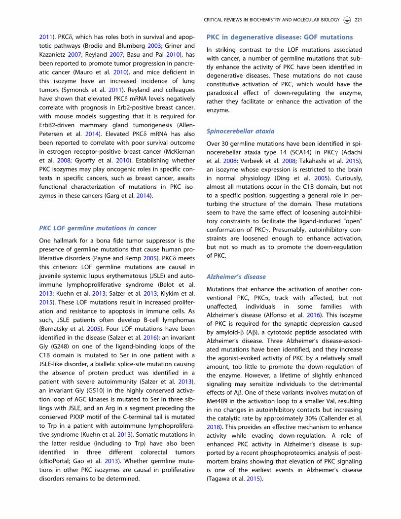
One hallmark for a bona fide tumor suppressor is thepresence of germline mutations that cause human pro-liferative disorders (Payne and Kemp 2005). PKCd meetsthis criterion: LOF germline mutations are causal injuvenile systemic lupus erythematosus (JSLE) and auto-immune lymphoproliferative syndrome (Belot et al.2013; Kuehn et al. 2013; Salzer et al. 2013; Kiykim et al.2015). These LOF mutations result in increased prolifer-ation and resistance to apoptosis in immune cells. Assuch, JSLE patients often develop B-cell lymphomas(Bernatsky et al. 2005). Four LOF mutations have beenidentified in the disease (Salzer et al. 2016): an invariantGly (G248) on one of the ligand-binding loops of theC1B domain is mutated to Ser in one patient with aJSLE-like disorder, a biallelic splice-site mutation causingthe absence of protein product was identified in apatient with severe autoimmunity (Salzer et al. 2013),an invariant Gly (G510) in the highly conserved activa-tion loop of AGC kinases is mutated to Ser in three sib-lings with JSLE, and an Arg in a segment preceding theconserved PXXP motif of the C-terminal tail is mutatedto Trp in a patient with autoimmune lymphoprolifera-tive syndrome (Kuehn et al. 2013). Somatic mutations inthe latter residue (including to Trp) have also beenidentified in three different colorectal tumors(cBioPortal; Gao et al. 2013). Whether germline muta-tions in other PKC isozymes are causal in proliferativedisorders remains to be determined.
PKC in degenerative disease: GOF mutations
In striking contrast to the LOF mutations associatedwith cancer, a number of germline mutations that sub-tly enhance the activity of PKC have been identified indegenerative diseases. These mutations do not causeconstitutive activation of PKC, which would have theparadoxical effect of down-regulating the enzyme,rather they facilitate or enhance the activation of theenzyme.
Spinocerebellar ataxia
Over 30 germline mutations have been identified in spi-nocerebellar ataxia type 14 (SCA14) in PKCc (Adachiet al. 2008; Verbeek et al. 2008; Takahashi et al. 2015),an isozyme whose expression is restricted to the brainin normal physiology (Ding et al. 2005). Curiously,almost all mutations occur in the C1B domain, but notto a specific position, suggesting a general role in per-turbing the structure of the domain. These mutationsseem to have the same effect of loosening autoinhibi-tory constraints to facilitate the ligand-induced “open”conformation of PKCc. Presumably, autoinhibitory con-straints are loosened enough to enhance activation,but not so much as to promote the down-regulationof PKC.
Alzheimer’s disease
Mutations that enhance the activation of another con-ventional PKC, PKCa, track with affected, but notunaffected, individuals in some families withAlzheimer’s disease (Alfonso et al. 2016). This isozymeof PKC is required for the synaptic depression causedby amyloid-b (Ab), a cytotoxic peptide associated withAlzheimer’s disease. Three Alzheimer’s disease-associ-ated mutations have been identified, and they increasethe agonist-evoked activity of PKC by a relatively smallamount, too little to promote the down-regulation ofthe enzyme. However, a lifetime of slightly enhancedsignaling may sensitize individuals to the detrimentaleffects of Ab. One of these variants involves mutation ofMet489 in the activation loop to a smaller Val, resultingin no changes in autoinhibitory contacts but increasingthe catalytic rate by approximately 30% (Callender et al.2018). This provides an effective mechanism to enhanceactivity while evading down-regulation. A role ofenhanced PKC activity in Alzheimer’s disease is sup-ported by a recent phosphoproteomics analysis of post-mortem brains showing that elevation of PKC signalingis one of the earliest events in Alzheimer’s disease(Tagawa et al. 2015).
CRITICAL REVIEWS IN BIOCHEMISTRY AND MOLECULAR BIOLOGY 221
Stroke
A polymorphism in the kinase domain of PKCg (V374I)is associated with increased risk for cerebral infarction(stroke) (Kubo et al. 2007), increased risk of arthritis(Takata et al. 2007), and severe gastric atrophy (Gotoet al. 2010). In vitro kinase assays reveal that the muta-tion enhances the catalytic activity of PKC, as assessedby increased autophosphorylation and substrate phos-phorylation (Kubo et al. 2007; Zurgil et al. 2014).Assuming autoinhibitory constraints are unchanged (asis the case for the activity-enhancing PKCa M489V vari-ant in Alzheimer’s disease), the enhanced catalytic activ-ity would serve as an effective mechanism to allowenhanced activity without compromising the stability ofthe mutant.
The increasing annotation of disease-associated sin-gle nucleotide polymorphisms (SNPs) will likely unveilan abundance of variants of PKC that predispose to spe-cific diseases, from heart disease to drug and alcoholaddiction (Olive and Messing 2004). Both conventional(PKCa and bII) and novel (PKCd and PKCe) have beenshown to affect cardiac function (Palaniyandi et al.2009), so variants that cause small changes in activitymay either protect or predispose to heart disease.Behavioral studies with knockout mice reveal that lackof PKCc causes decreased anxiety (Bowers et al. 2000)and a high ethanol drinking phenotype (Ron andMessing 2013), whereas mice lacking PKCe have a lowethanol drinking phenotype (Ron and Messing 2013).Diseases causally linked to PKC variants with enhancedPKC activity could thus benefit from inhibitors to adjustPKC signaling to lower levels.
Should PKC be inhibited in degenerative disease?
The enhanced signaling by PKC in degenerative dis-eases such as Alzheimer’s disease contrasts with thereduced signaling that is associated with cancer, sug-gesting opposing roles in survival versus degenerativediseases. Indeed, a recent meta-analysis of nine inde-pendent studies reveals that Alzheimer’s diseasepatients exhibit an overall 45% decreased risk of cancercompared with the general population (Shi et al. 2015),consistent with earlier reports that Alzheimer’s diseaseand cancer display an inverse association (Roe et al.2005; Driver et al. 2012). Perhaps repurposing PKCinhibitors for Alzheimer’s disease may temper theeffects of Ab on synapses and thereby mitigate loss ofcognitive function. In this regard, bryostatin 1, whichcauses the loss of PKC and failed in cancer therapies(Nezhat et al. 2004), is in phase II clinical trials forAlzheimer’s disease (Nelson et al. 2017), with mouse
studies showing promising results in improving learningdeficits in an Alzheimer’s disease mouse model (Russoet al. 2015; Schrott et al. 2015). It is noteworthy that tar-geting PKC in degenerative diseases may be particularlyfeasible as its activity would only need to be broughtdown from supraphysiological to physiological levels,essentially adjusting the balance to regain homeostasis.
Conclusion
The paradoxical acute activation, followed by chronicloss, of diacylglycerol-sensitive PKC isozymes by phorbolesters confounded understanding the role of PKC incancer. However, the recent availability of whole gen-ome sequencing of tumors and patient populations,coupled to growing databases analyzing survival curvesas a function of protein or mRNA expression for specificcancers, converge on a new understanding of the bio-logical function of PKC isozymes: they generally sup-press survival signaling, functioning as the brakes tooncogenic signaling. Furthermore, these isozymes fre-quently have LOF mutations, rather than gene dele-tions, allowing the defective PKC to exert dominant-negative effects more broadly on the signaling outputof other PKC isozymes. This new understanding of PKCfunction highlights the need to restore, rather thaninhibit PKC function for cancer therapies. Additionally,mechanistic insight into how PKC isozymes are regu-lated suggests caution in targeting pathways that con-trol levels of PKC. For example, the use of inhibitors forthe kinase mTOR (Don and Zheng 2011) or Hsp90(Neckers and Workman 2012), both currently in use inthe clinic, may have the detrimental effect of inhibitingPKC processing, in turn decreasing the steady-state lev-els of PKC (Guertin et al. 2006; Gould et al. 2009). Withbetter patient survival generally associated with higherPKC levels, careful consideration should be given toselecting therapies that do not remove PKC.
Acknowledgements
The author thanks Gerard Manning for providing a figure ofthe AGC branch of kinome, Gema Lorden for assistance indrafting, and members of the Newton lab for helpfulcomments.
Disclosure statement
No potential conflict of interest was reported by the author.
Funding
This work was supported by NIH [R35 GM122523] and theCure Alzheimer’s Fund.
222 A. C. NEWTON

References
Abrahamsen H, O'Neill AK, Kannan N, Kruse N, Taylor SS,Jennings PA, Newton AC. 2012. Peptidyl-prolyl isomerasePin1 controls down-regulation of conventional protein kin-ase C isozymes. J Biol Chem. 287:13262–13278.
Adachi N, Kobayashi T, Takahashi H, Kawasaki T, Shirai Y,Ueyama T, Matsuda T, Seki T, Sakai N, Saito N. 2008.Enzymological analysis of mutant protein kinase Cgammacausing spinocerebellar ataxia type 14 and dysfunction inCa2þ homeostasis. J Biol Chem. 283:19854–19863.
Alfonso SI, Callender JA, Hooli B, Antal CE, Mullin K, ShermanMA, Lesne SE, Leitges M, Newton AC, Tanzi RE, et al. 2016.Gain-of-function mutations in protein kinase Ca (PKCa)may promote synaptic defects in Alzheimer's disease. SciSignal. 9:ra47.
Ali SA, Justilien V, Jamieson L, Murray NR, Fields AP. 2016.Protein kinase Ci Drives a NOTCH3-dependent stem-likephenotype in mutant KRAS lung adenocarcinoma. CancerCell. 29:367–378.
Allen-Petersen BL, Carter CJ, Ohm AM, Reyland ME. 2014.Protein kinase Cd is required for ErbB2-driven mammarygland tumorigenesis and negatively correlates with prog-nosis in human breast cancer. Oncogene. 33:1306–1315.
Antal CE, Callender JA, Kornov AP, Taylor SS, Newton AC.2015a. Intramolecular C2 domain-mediated autoinhibitionof protein kinase CbII. Cell Reports. 12:1252–1260.
Antal CE, Hudson AM, Kang E, Zanca C, Wirth C, StephensonNL, Trotter EW, Gallegos LL, Miller CJ, Furnari FB, et al.2015b. Cancer-associated protein kinase C mutationsreveal kinase's role as tumor suppressor. Cell. 160:489–502.
Antal CE, Violin JD, Kunkel MT, Skovso S, Newton AC. 2014.Intramolecular conformational changes optimize proteinkinase C signaling. Chem Biol. 21:459–469.
Aziz MH, Wheeler DL, Bhamb B, Verma AK. 2006. Protein kin-ase C delta overexpressing transgenic mice are resistant tochemically but not to UV radiation-induced developmentof squamous cell carcinomas: a possible link to specificcytokines and cyclooxygenase-2. Cancer Res. 66:713–722.
Bandyopadhyay G, Kanoh Y, Sajan MP, Standaert ML, FareseRV. 2000. Effects of adenoviral gene transfer of wild-type,constitutively active, and kinase-defective protein kinase C-lambda on insulin-stimulated glucose transport in L6 myo-tubes. Endocrinology. 141:4120–4127.
Bandyopadhyay G, Sajan MP, Kanoh Y, Standaert ML, QuonMJ, Lea-Currie R, Sen A, Farese RV. 2002. PKC-zeta medi-ates insulin effects on glucose transport in cultured preadi-pocyte-derived human adipocytes. J Clin EndocrinolMetab. 87:716–723.
Bandyopadhyay G, Standaert ML, Zhao L, Yu B, Avignon A,Galloway L, Karnam P, Moscat J, Farese RV. 1997.Activation of protein kinase C (alpha, beta, and zeta) byinsulin in 3T3/L1 cells. Transfection studies suggest a rolefor PKC-zeta in glucose transport. J Biol Chem.272:2551–2558.
Bar-Even A, Noor E, Savir Y, Liebermeister W, Davidi D, TawfikDS, Milo R. 2011. The moderately efficient enzyme: evolu-tionary and physicochemical trends shaping enzymeparameters. Biochemistry. 50:4402–4410.
Barcelo C, Paco N, Morell M, Alvarez-Moya B, Bota-Rabassedas N, Jaumot M, Vilardell F, Capella G, Agell N.
2014. Phosphorylation at Ser-181 of oncogenic KRAS isrequired for tumor growth. Cancer Res. 74:1190–1199.
Basu A, Pal D. 2010. Two faces of protein kinase Cd: the con-trasting roles of PKCd in cell survival and cell death.Sci World J. 10:2272–2284.
Behn-Krappa A, Newton AC. 1999. The hydrophobicphosphorylation motif of conventional protein kinase C isregulated by autophosphorylation. Curr Biol. 9:728–737.
Belot A, Kasher PR, Trotter EW, Foray AP, Debaud AL, Rice GI,Szynkiewicz M, Zabot MT, Rouvet I, Bhaskar SS, et al. 2013.Protein kinase cd deficiency causes Mendelian systemiclupus erythematosus with B cell-defective apoptosis andhyperproliferation. Arthritis Rheum. 65:2161–2171.
Benes CH, Wu N, Elia AE, Dharia T, Cantley LC, Soltoff SP.2005. The C2 domain of PKCdelta is a phosphotyrosinebinding domain. Cell. 121:271–280.
Berenblum I, Shubik P. 1947. The role of croton oil applica-tions, associated with a single painting of a carcinogen,in tumour induction of the mouse's skin. Br J Cancer.1:379–382.
Bernatsky S, Boivin JF, Joseph L, Rajan R, Zoma A, Manzi S,Ginzler E, Urowitz M, Gladman D, Fortin PR, et al. 2005. Aninternational cohort study of cancer in systemic lupuserythematosus [Multicenter Study Research Support, N.I.H.,Extramural Research Support, Non-U.S. Gov't ResearchSupport, U.S. Gov't, P.H.S.]. Arthritis Rheum. 52:1481–1490.
Betschinger J, Mechtler K, Knoblich JA. 2003. The Par com-plex directs asymmetric cell division by phosphorylatingthe cytoskeletal protein Lgl. Nature. 422:326–330.
Bivona TG, Quatela SE, Bodemann BO, Ahearn IM, Soskis MJ,Mor A, Miura J, Wiener HH, Wright L, Saba SG, et al. 2006.PKC regulates a farnesyl-electrostatic switch on K-Ras thatpromotes its association with Bcl-XL on mitochondria andinduces apoptosis [Research Support, N.I.H., ExtramuralResearch Support, Non-U.S. Gov't]. Mol Cell. 21:481–493.
Blumberg PM, Jaken S, Konig B, Sharkey NA, Leach KL, JengAY, Yeh E. 1984. Mechanism of action of the phorbol estertumor promoters: specific receptors for lipophilic ligands.Biochem Pharmacol. 33:933–940.
Bornancin F, Parker PJ. 1997. Phosphorylation of protein kin-ase C-alpha on serine 657 controls the accumulation ofactive enzyme and contributes to its phosphatase- resist-ant state [published erratum appears in J Biol Chem.272(20):13458]. J Biol Chem. 272:3544–3549.
Borner C, Filipuzzi I, Wartmann M, Eppenberger U, Fabbro D.1989. Biosynthesis and posttranslational modifications ofprotein kinase C in human breast cancer cells. J BiolChem. 264:13902–13909.
Bowers BJ, Collins AC, Tritto T, Wehner JM. 2000. Mice lackingPKC gamma exhibit decreased anxiety. Behav Genet.30:111–121.
Brodie C, Blumberg PM. 2003. Regulation of cell apoptosis byprotein kinase c delta. Apoptosis. 8:19–27.
Callender JA, Yang Y, Stephenson N, Brognard J, Newton AC.2018. Protein kinase Ca (PKCa) gain-of-function variant inAlzheimer’s disease displays enhanced catalysis by mech-anism that evades down-regulation. FASEB J: Off PublFeder Am Soc Exp Biol. In press.
Cameron AJ, Linch MD, Saurin AT, Escribano C, Parker PJ.2011. mTORC2 targets AGC kinases through Sin1-depend-ent recruitment. Biochem J. 439:287–297.
CRITICAL REVIEWS IN BIOCHEMISTRY AND MOLECULAR BIOLOGY 223

Castagna M, Takai Y, Kaibuchi K, Sano K, Kikkawa U,Nishizuka Y. 1982. Direct activation of calcium-activated,phospholipid-dependent protein kinase by tumor-promot-ing Phorbol Esters. J Biol Chem. 257:7847–7851.
Cenni V, Doppler H, Sonnenburg ED, Maraldi N, Newton AC,Toker A. 2002. Regulation of novel protein kinase C epsi-lon by phosphorylation. Biochem J. 363:537–545.
Corbalan-Garcia S, Garcia-Garcia J, Rodriguez-Alfaro JA,Gomez-Fernandez JC. 2003. A new phosphatidylinositol4,5-bisphosphate-binding site located in the C2 domain ofprotein kinase Calpha [Research Support, Non-U.S. Gov't].J Biol Chem. 278:4972–4980.
Coussens L, Parker PJ, Rhee L, Yang-Feng TL, Chen E,Waterfield MD, Francke U, Ullrich A. 1986. Multiple, distinctforms of bovine and human protein kinase C suggestdiversity in cellular signaling pathways. Science.233:859–866.
Creixell P, Schoof EM, Simpson CD, Longden J, Miller CJ, LouHJ, Perryman L, Cox TR, Zivanovic N, Palmeri A, et al. 2015.Kinome-wide decoding of network-attacking mutationsrewiring cancer signaling. Cell. 163:202–217.
de Vries DJ, Herald CL, Pettit GR, Blumberg PM. 1988.Demonstration of sub-nanomolar affinity of bryostatin 1for the phorbol ester receptor in rat brain. BiochemPharmacol. 37:4069–4073.
Ding YQ, Xiang CX, Chen ZF. 2005. Generation and character-ization of the PKC gamma-Cre mouse line. Genesis.43:28–33.
Don AS, Zheng XF. 2011. Recent clinical trials of mTOR-tar-geted cancer therapies. Rev Recent Clin Trials. 6:24–35.
Dowling CM, Phelan J, Callender JA, Cathcart MC, Mehigan B,McCormick P, Dalton T, Coffey JC, Newton AC, O'Sullivan J,et al. 2016. Protein kinase C beta II suppresses colorectalcancer by regulating IGF-1 mediated cell survival.Oncotarget. 7:20919–20933.
Driedger PE, Blumberg PM. 1980. Specific binding of PhorbolEster tumor promoters. Proc Natl Acad Sci USA.77:567–571.
Dries DR, Gallegos LL, Newton AC. 2007. A single residue inthe C1 domain sensitizes novel protein kinase C isoformsto cellular diacylglycerol production. J Biol Chem.282:826–830.
Driver JA, Beiser A, Au R, Kreger BE, Splansky GL, Kurth T,Kiel DP, Lu KP, Seshadri S, Wolf PA. 2012. Inverse associ-ation between cancer and Alzheimer's disease: resultsfrom the Framingham Heart Study. BMJ. 344:e1442.
Drummond ML, Prehoda KE. 2016. Molecular control of atyp-ical protein kinase C: tipping the balance between self-renewal and differentiation. J Mol Biol. 428:1455–1464.
Edwards AS, Newton AC. 1997. Phosphorylation at conservedcarboxyl-terminal hydrophobic motif regulates the catalyticand regulatory domains of protein kinase C. J Biol Chem.272:18382–18390.
Facchinetti V, Ouyang W, Wei H, Soto N, Lazorchak A, GouldC, Lowry C, Newton AC, Mao Y, Miao RQ, et al. 2008. Themammalian target of rapamycin complex 2 controls fold-ing and stability of Akt and protein kinase C [ResearchSupport, N.I.H., Extramural Research Support, Non-U.S.Gov't]. EMBO J. 27:1932–1943.
Farese RV, Sajan MP, Yang H, Li P, Mastorides S, Gower WRJr, Nimal S, Choi CS, Kim S, Shulman GI, et al. 2007.Muscle-specific knockout of PKC-lambda impairs glucose
transport and induces metabolic and diabetic syndromes.J Clin Invest. 117:2289–2301.
Flint AJ, Paladini RD, Koshland DE Jr. 1990.Autophosphorylation of protein kinase C at three sepa-rated regions of its primary sequence. Science. 249:408–411.
Freeley M, Kelleher D, Long A. 2011. Regulation of ProteinKinase C function by phosphorylation on conserved andnon-conserved sites [Review]. Cell Signal. 23:753–762.
Frey MR, Saxon ML, Zhao X, Rollins A, Evans SS, Black JD.1997. Protein kinase C isozyme-mediated cell cycle arrestinvolves induction of p21(waf1/cip1) and p27(kip1) andhypophosphorylation of the retinoblastoma protein inintestinal epithelial cells. J Biol Chem. 272:9424–9435.
Fujimoto K, Ohta K, Kangawa K, Kikkawa U, Ogino S, Fukui H.1999. Identification of protein kinase C phosphorylationsites involved in phorbol ester-induced desensitization ofthe histamine H1 receptor. Mol Pharmacol. 55:735–742.
Gallegos LL, Kunkel MT, Newton AC. 2006. Targeting proteinkinase C activity reporter to discrete intracellular regionsreveals spatiotemporal differences in agonist-dependentsignaling. J Biol Chem. 281:30947–30956.
Galvez AS, Duran A, Linares JF, Pathrose P, Castilla EA, Abu-Baker S, Leitges M, Diaz-Meco MT, Moscat J. 2009. Proteinkinase Czeta represses the interleukin-6 promoter andimpairs tumorigenesis in vivo. Mol Cell Biol. 29:104–115.
Gao J, Aksoy BA, Dogrusoz U, Dresdner G, Gross B, SumerSO, Sun Y, Jacobsen A, Sinha R, Larsson E, et al. 2013.Integrative analysis of complex cancer genomics and clin-ical profiles using the cBioPortal. Sci Signal. 6:pl1.
Gao T, Brognard J, Newton AC. 2008. The phosphatasePHLPP controls the cellular levels of protein kinase C.J Biol Chem. 283:6300–6311.
Gao T, Newton AC. 2006. Invariant Leu preceding turn motifphosphorylation site controls the interaction of proteinkinase C with Hsp70. J Biol Chem. 281:32461–32468.
Gao T, Toker A, Newton AC. 2001. The carboxyl terminus ofprotein kinase c provides a switch to regulate its inter-action with the phosphoinositide-dependent kinase, pdk-1.J Biol Chem. 276:19588–19596.
Garcia-Paramio P, Cabrerizo Y, Bornancin F, Parker PJ. 1998.The broad specificity of dominant inhibitory protein kinaseC mutants infers a common step in phosphorylation.Biochem J. 333:631–636.
Garg R, Benedetti LG, Abera MB, Wang H, Abba M, KazanietzMG. 2014. Protein kinase C and cancer: what we know andwhat we do not. Oncogene. 33:5225–5237.
Garg R, Blando JM, Perez CJ, Abba MC, Benavides F,Kazanietz MG. 2017. Protein kinase C epsilon cooperateswith PTEN loss for prostate tumorigenesis through theCXCL13-CXCR5 pathway. Cell Rep. 19:375–388.
Giorgione J, Hysell M, Harvey DF, Newton AC. 2003.Contribution of the C1A and C1B domains to the mem-brane interaction of protein kinase C. Biochemistry.42:11194–11202.
Giorgione JR, Lin JH, McCammon JA, Newton AC. 2006.Increased membrane affinity of the C1 domain of proteinkinase Cdelta compensates for the lack of involvement ofits C2 domain in membrane recruitment. J Biol Chem.281:1660–1669.
Gong J, Holewinski RJ, Van Eyk JE, Steinberg SF. 2016. Anovel phosphorylation site at Ser130 adjacent to the
224 A. C. NEWTON

pseudosubstrate domain contributes to the activation ofprotein kinase C-d. Biochem J. 473:311–320.
Goto Y, Hishida A, Matsuo K, Tajima K, Morita E, Naito M,Wakai K, Hamajima N. 2010. PRKCH gene polymorphism isassociated with the risk of severe gastric atrophy. GastricCancer. 13:90–94.
Gould CM, Kannan N, Taylor SS, Newton AC. 2009. The chap-erones Hsp90 and Cdc37 mediate the maturation and sta-bilization of protein kinase C through a conserved PXXPmotif in the C-terminal tail. J Biol Chem. 284:4921–4935.
Graybill C, Wee B, Atwood SX, Prehoda KE. 2012. Partitioning-defective protein 6 (Par-6) activates atypical protein kinaseC (aPKC) by pseudosubstrate displacement. J Biol Chem.287:21003–21011.
Griner EM, Kazanietz MG. 2007. Protein kinase C and otherdiacylglycerol effectors in cancer. Nat Rev Cancer.7:281–294.
Guertin DA, Stevens DM, Thoreen CC, Burds AA, Kalaany NY,Moffat J, Brown M, Fitzgerald KJ, Sabatini DM. 2006.Ablation in mice of the mTORC components raptor, rictor,or mLST8 reveals that mTORC2 is required for signaling toAkt-FOXO and PKCalpha, but not S6K1. Dev Cell.11:859–871.
Gyorffy B, Lanczky A, Eklund AC, Denkert C, Budczies J, Li Q,Szallasi Z. 2010. An online survival analysis tool to rapidlyassess the effect of 22,277 genes on breast cancer progno-sis using microarray data of 1,809 patients. Breast CancerRes Treat. 123:725–731.
Hafeez BB, Zhong W, Weichert J, Dreckschmidt NE, Jamal MS,Verma AK. 2011. Genetic ablation of PKC epsilon inhibitsprostate cancer development and metastasis in transgenicmouse model of prostate adenocarcinoma. Cancer Res.71:2318–2327.
Hanks SK, Hunter T. 1995. Protein kinases 6. The eukaryoticprotein kinase superfamily: kinase (catalytic) domain struc-ture and classification. FASEB J. 9:576–596.
Hansra G, Garcia-Paramio P, Prevostel C, Whelan RD,Bornancin F, Parker PJ. 1999. Multisite dephosphorylationand desensitization of conventional protein kinase C iso-types [In Process Citation]. Biochem J. 342:337–344.
Hecker E. 1968. Cocarcinogenic principles from the seed oilof Croton tiglium and from other Euphorbiaceae. CancerRes. 28:2338–2349.
Hecker E. 1978. Co-carcinogens or modulators of carcinogen-esis. New aspects of the etiology of human tumorsand of the molecular mechanisms of carcinogenesis.Naturwissenschaften. 65:640–648.
Hecker E, Kubinyi H, Bresch H, von Szczepanski C. 1966.The chemical structure of a cocarcinogen and of phorbolisolated from croton oil. J Med Chem. 9:246–247.
Hein MY, Hubner NC, Poser I, Cox J, Nagaraj N, Toyoda Y,Gak IA, Weisswange I, Mansfeld J, Buchholz F, et al. 2015.A human interactome in three quantitative dimensionsorganized by stoichiometries and abundances. Cell.163:712–723.
Hernandez AI, Blace N, Crary JF, Serrano PA, Leitges M, LibienJM, Weinstein G, Tcherapanov A, Sacktor TC. 2003. ProteinKinase M zeta synthesis from a brain mRNA encoding anindependent protein kinase C zeta catalytic domain.Implications for the molecular mechanism of memory.J Biol Chem. 278:40305–40316.
Hirose T, Izumi Y, Nagashima Y, Tamai-Nagai Y, Kurihara H,Sakai T, Suzuki Y, Yamanaka T, Suzuki A, Mizuno K, et al.2002. Involvement of ASIP/PAR-3 in the promotion of epi-thelial tight junction formation. J Cell Sci. 115:2485–2495.
Hokin LE. 1987. The road to the phosphoinositide-generatedsecond messengers. Trends Pharmacol Sci. 8:53–56.
Hokin MR, Hokin LE. 1953. Enzyme secretion and the incorp-oration of P32 into phospholipides of pancreas slices.J Biol Chem. 203:967–977.
Hornbeck PV, Kornhauser JM, Tkachev S, Zhang B, SkrzypekE, Murray B, Latham V, Sullivan M. 2012. PhosphoSitePlus:a comprehensive resource for investigating the structureand function of experimentally determined post-transla-tional modifications in man and mouse [Research Support,N.I.H., Extramural]. Nucleic Acids Res. 40:D261–D270.
Hosey MM, Benovic JL, DebBurman SK, Richardson RM. 1995.Multiple mechanisms involving protein phosphorylationare linked to desensitization of muscarinic receptors. LifeSci. 56:951–955.
Hoshi N, Langeberg LK, Gould CM, Newton AC, Scott JD.2010. Interaction with AKAP79 modifies the cellularpharmacology of PKC. Mol Cell. 37:541–550.
House C, Kemp BE. 1987. Protein kinase C contains a pseudo-substrate prototope in its regulatory domain. Science.238:1726–1728.
House C, Kemp BE. 1990. Protein kinase C pseudosubstrateprototope: structure-function relationships. Cell Sig.2:187–190.
Huang K-P, Nakabayashi H, Huang FL. 1986. Isozymic formsof rat brain Ca2þ-activated and phospholipid-dependentprotein kinase. Proc Natl Acad Sci USA. 83:8535–8539.
Humphries MJ, Ohm AM, Schaack J, Adwan TS, Reyland ME.2008. Tyrosine phosphorylation regulates nuclear trans-location of PKCdelta. Oncogene. 27:3045–3053.
Hunter T, Ling N, Cooper JA. 1984. Protein kinase C phos-phorylation of the EGF receptor at a threonine residueclose to the cytoplasmic face of the plasma membrane.Nature. 311:480–483.
Iglesias T, Matthews S, Rozengurt E. 1998. Dissimilar phorbolester binding properties of the individual cysteine-richmotifs of protein kinase D. FEBS Lett. 437:19–23.
Igumenova TI. 2015. Dynamics and membrane interactions ofprotein kinase C. Biochemistry. 54:4953–4968.
Ikenoue T, Inoki K, Yang Q, Zhou X, Guan KL. 2008. Essentialfunction of TORC2 in PKC and Akt turn motif phosphoryl-ation, maturation and signalling [Research Support, N.I.H.,Extramural Research Support, U.S. Gov't, Non-P.H.S.]. EMBOJ. 27:1919–1931.
Inoue M, Kishimoto A, Takai Y, Nishizuka Y. 1977. Studies ona cyclic nucleotide-independent protein kinase and itsproenzyme in mammalian tissues. II. Proenzyme and itsactivation by calcium-dependent protease from rat brain.J Biol Chem. 252:7610–7616.
Isakov N. 2017. Protein kinase C (PKC) isoforms in cancer,tumor promotion and tumor suppression. Semin CancerBiol. 48:36–52.
Isakov N, Gopas J, Priel E, Segal S, Altman A. 1991. Effect ofprotein kinase C activating tumor promoters on metasta-ses formation by fibrosarcoma cells. Invasion Metastasis.11:14–24.
Jaken S, Tashjian AH Jr, Blumberg PM. 1981. Characterizationof phorbol ester receptors and their down-modulation in
CRITICAL REVIEWS IN BIOCHEMISTRY AND MOLECULAR BIOLOGY 225

GH4C1 rat pituitary cells [Research Support, U.S. Gov't,P.H.S.]. Cancer Res. 41:2175–2181.
Johannes F-J, Prestle J, Eis S, Oberhagemann P, PfizenmaierK. 1994. PKCu is a novel, atypical member of the proteinkinase C family. J Biol Chem. 269:6140–6148.
Johnson JE, Edwards AS, Newton AC. 1997. A putativephosphatidylserine binding motif is not involved in thelipid regulation of protein kinase C. J Biol Chem.272:30787–30792.
Justilien V, Walsh MP, Ali SA, Thompson EA, Murray NR,Fields AP. 2014. The PRKCI and SOX2 oncogenes are coam-plified and cooperate to activate Hedgehog signaling inlung squamous cell carcinoma. Cancer Cell. 25:139–151.
Kannan N, Haste N, Taylor SS, Neuwald AF. 2007. The hall-mark of AGC kinase functional divergence is its C-terminaltail, a cis-acting regulatory module. Proc Natl Acad SciUSA. 104:1272–1277.
Kataoka K, Nagata Y, Kitanaka A, Shiraishi Y, Shimamura T,Yasunaga J, Totoki Y, Chiba K, Sato-Otsubo A, Nagae G,et al. 2015. Integrated molecular analysis of adult T cellleukemia/lymphoma. Nat Genet. 47:1304–1315.
Kazanietz MG. 2000. Eyes wide shut: protein kinase C iso-zymes are not the only receptors for the phorbol estertumor promoters. Mol Carcinog. 28:5–11.
Kazanietz MG, Areces LB, Bahador A, Mischak H, Goodnight J,Mushinski JF, Blumberg PM. 1993. Characterization of lig-and and substrate specificity for the calcium-dependentand calcium-independent protein kinase C isozymes. MolPharmacol. 44:298–307.
Kazanietz MG, Bustelo XR, Barbacid M, Kolch W, Mischak H,Wong G, Pettit GR, Bruns JD, Blumberg PM. 1994. Zinc fin-ger domains and Phorbol Ester pharmacophore. Analysisof binding to mutated form of protein kinase C zeta andthe vav and c-raf proto-oncogene products. J Biol Chem.269:11590–11594.
Keranen LM, Dutil EM, Newton AC. 1995. Protein kinase C isregulated in vivo by three functionally distinct phosphory-lations. Curr Biol. 5:1394–1403.
Kikkawa U, Takai Y, Tanaka Y, Miyake R, Nishizuka Y. 1983.Protein kinase C as a possible receptor protein oftumor-promoting phorbol esters. J Biol Chem. 258:11442–11445.
Kiykim A, Ogulur I, Baris S, Salzer E, Karakoc-Aydiner E, OzenAO, Garncarz W, Hirschmugl T, Krolo A, Yucelten AD, et al.2015. Potentially beneficial effect of hydroxychloroquine ina patient with a novel mutation in protein kinase Cdeltadeficiency. J Clin Immunol. 35:523–526.
Knauf JA, Elisei R, Mochly-Rosen D, Liron T, Chen XN, GonskyR, Korenberg JR, Fagin JA. 1999. Involvement of proteinkinase Cepsilon (PKCepsilon) in thyroid cell death. A trun-cated chimeric PKCepsilon cloned from a thyroid cancercell line protects thyroid cells from apoptosis. J Biol Chem.274:23414–23425.
Knopf JL, Lee M-H, Sultzman LA, Kriz RW, Loomis CR, HewickRM, Bell RM. 1986. Cloning and expression of multiple pro-tein kinase C cDNAs. Cell. 46:491–502.
Kohout SC, Corbalan-Garcia S, Torrecillas A, Gomez-Fernandez JC, Falke JJ. 2002. C2 domains of protein kinaseC isoforms alpha, beta, and gamma: activation parametersand calcium stoichiometries of the membrane-boundstate. Biochemistry. 41:11411–11424.
K€onig B, DiNitto PA, Blumberg PM. 1985. Stoichiometric bind-ing of diacylglycerol to the Phorbol Ester Receptor. J CellBiochem. 29:37–44.
Konishi H, Tanaka M, Takemura Y, Matsuzaki H, Ono Y,Kikkawa U, Nishizuka Y. 1997. Activation of protein kinaseC by tyrosine phosphorylation in response to H2O2. ProcNatl Acad Sci USA. 94:11233–11237.
Koren R, Langzam L, Paz A, Livne PM, Gal R, Sampson SR.2000. Protein kinase C (PKC) isoenzymes immunohisto-chemistry in lymph node revealing solution-fixed, paraffin-embedded bladder tumors. Appl Immunohistochem MolMorphol: AIMM/Off Publ Soc Appl Immunohistochem.8:166–171.
Kotani K, Ogawa W, Matsumoto M, Kitamura T, Sakaue H,Hino Y, Miyake K, Sano W, Akimoto K, Ohno S, et al. 1998.Requirement of atypical protein kinase clambda for insulinstimulation of glucose uptake but not for Akt activation in3T3-L1 adipocytes. Mol Cell Biol. 18:6971–6982.
Kraft AS, Smith JB, Berkow RL. 1986. Bryostatin, an activatorof the calcium phospholipid-dependent protein kinase,blocks phorbol ester-induced differentiation of human pro-myelocytic leukemia cells HL-60. Proc Natl Acad Sci USA.83:1334–1338.
Kubo M, Hata J, Ninomiya T, Matsuda K, Yonemoto K,Nakano T, Matsushita T, Yamazaki K, Ohnishi Y, Saito S,et al. 2007. A nonsynonymous SNP in PRKCH (protein kin-ase C eta) increases the risk of cerebral infarction. NatGenet. 39:212–217.
Kuehn HS, Niemela JE, Rangel-Santos A, Zhang M, PittalugaS, Stoddard JL, Hussey AA, Evbuomwan MO, Priel DA,Kuhns DB, et al. 2013. Loss-of-function of the protein kin-ase C d (PKCd) causes a B-cell lymphoproliferative syn-drome in humans. Blood. 121:3117–3125.
Kusne Y, Carrera-Silva EA, Perry AS, Rushing EJ, Mandell EK,Dietrich JD, Errasti AE, Gibbs D, Berens ME, Loftus JC, et al.2014. Targeting aPKC disables oncogenic signaling byboth the EGFR and the proinflammatory cytokine TNFa inglioblastoma. Sci Signal. 7:ra75.
Langzam L, Koren R, Gal R, Kugel V, Paz A, Farkas A,Sampson SR. 2001. Patterns of protein kinase C isoenzymeexpression in transitional cell carcinoma of bladder.Relation to degree of malignancy [Comparative StudyResearch Support, Non-U.S. Gov't]. Am J Clin Pathol.116:377–385.
Leonard TA, Rozycki B, Saidi LF, Hummer G, Hurley JH. 2011.Crystal structure and allosteric activation of protein kinaseC bII. Cell. 144:55–66.
Levin DE, Fields FO, Kunisawa R, Bishop JM, Thorner J. 1990.A candidate protein kinase C gene, PKC1, is required forthe S. cerevisiae cell cycle. Cell. 62:213–224.
Lin D, Edwards AS, Fawcett JP, Mbamalu G, Scott JD, PawsonT. 2000. A mammalian PAR-3-PAR-6 complex implicated inCdc42/Rac1 and aPKC signalling and cell polarity. Nat CellBiol. 2:540–547.
Linch M, Sanz-Garcia M, Soriano E, Zhang Y, Riou P, Rosse C,Cameron A, Knowles P, Purkiss A, Kjaer S, et al. 2013. Acancer-associated mutation in atypical protein kinase Ciotaoccurs in a substrate-specific recruitment motif. Sci Signal.6:ra82.
Littler DR, Walker JR, She YM, Finerty PJ Jr, Newman EM,Dhe-Paganon S. 2006. Structure of human protein kinase Ceta (PKCeta) C2 domain and identification of
226 A. C. NEWTON
phosphorylation sites [Research Support, Non-U.S. Gov't].Biochem Biophys Res Commun. 349:1182–1189.
Livneh E, Dull TJ, Berent E, Prywes R, Ullrich A, Schlessinger J.1988. Release of a phorbol ester-induced mitogenic blockby mutation at Thr-654 of the epidermal growth factorreceptor. Mol Cell Biol. 8:2302–2308.
Livneh E, Reiss N, Berent E, Ullrich A, Schlessinger J. 1987. Aninsertional mutant of epidermal growth factor receptorallows dissection of diverse receptor functions. EMBO J.6:2669–2676.
Llado V, Nakanishi Y, Duran A, Reina-Campos M, Shelton PM,Linares JF, Yajima T, Campos A, Aza-Blanc P, Leitges M,et al. 2015. Repression of intestinal stem cell function andtumorigenesis through direct phosphorylation of beta-Catenin and Yap by PKCzeta. Cell Rep. [accessed 2015 Feb4]. https://doi.org/10.1016/j.celrep.2015.01.007
Lu HC, Chou FP, Yeh KT, Chang YS, Hsu NC, Chang JG. 2009.Analysing the expression of protein kinase C eta in humanhepatocellular carcinoma. Pathology. 41:626–629.
Lum MA, Barger CJ, Hsu AH, Leontieva OV, Black AR, BlackJD. 2016. Protein kinase calpha (PKCalpha) is resistant tolong term desensitization/down-regulation by prolongeddiacylglycerol stimulation. J Biol Chem. 291:6331–6346.
Ma L, Tao Y, Duran A, Llado V, Galvez A, Barger JF, CastillaEA, Chen J, Yajima T, Porollo A, et al. 2013. Control ofnutrient stress-induced metabolic reprogramming by PKCfin tumorigenesis. Cell. 152:599–611.
Manning G, Whyte DB, Martinez R, Hunter T, Sudarsanam S.2002. The protein kinase complement of the human gen-ome. Science. 298:1912–1934.
Marshall JL, Bangalore N, El-Ashry D, Fuxman Y, Johnson M,Norris B, Oberst M, Ness E, Wojtowicz-Praga S, Bhargava P,et al. 2002. Phase I study of prolonged infusion Bryostatin-1 in patients with advanced malignancies. Cancer BiolTher. 1:409–416.
Mauro LV, Grossoni VC, Urtreger AJ, Yang C, Colombo LL,Morandi A, Pallotta MG, Kazanietz MG, Bal de Kier JoffeED, Puricelli LL. 2010. PKC Delta (PKCdelta) promotestumoral progression of human ductal pancreatic cancer[Research Support, Non-U.S. Gov't]. Pancreas. 39:e31–e41.
Mayati A, Moreau A, Le Vee M, Stieger B, Denizot C,Parmentier Y, Fardel O. 2017. Protein kinases C-mediatedregulations of drug transporter activity, localization andexpression. IJMS. 18:764.
McClendon CL, Kornev AP, Gilson MK, Taylor SS. 2014.Dynamic architecture of a protein kinase. Proc Natl AcadSci USA. 111:E4623–46E4631.
McKiernan E, O'Brien K, Grebenchtchikov N, Geurts-MoespotA, Sieuwerts AM, Martens JW, Magdolen V, Evoy D,McDermott E, Crown J, et al. 2008. Protein kinase Cdeltaexpression in breast cancer as measured by real-time PCR,western blotting and ELISA. Br J Cancer. 99:1644–1650.
McSkimming DI, Dastgheib S, Baffi TR, Byrne DP, Ferries S,Scott ST, Newton AC, Eyers CE, Kochut KJ, Eyers PA, et al.2016. KinView: a visual comparative sequence analysis toolfor integrated kinome research. Mol Biosyst. 12:3651–3665.
Meharena HS, Chang P, Keshwani MM, Oruganty K, Nene AK,Kannan N, Taylor SS, Kornev AP. 2013. Deciphering thestructural basis of eukaryotic protein kinase regulation.PLoS Biol. 11:e1001680.
Michell RH. 1975. Inositol phospholipids and cell surfacereceptor function. Biochim Biophys Acta. 415:81–147.
Milani G, Rebora P, Accordi B, Galla L, Bresolin S,Cazzaniga G, Buldini B, Mura R, Ladogana S, Giraldi E, et al.2014. Low PKCa expression within the MRD-HR stratumdefines a new subgroup of childhood T-ALL with verypoor outcome. Oncotarget. 5:5234–5245.
Morales KA, Yang Y, Cole TR, Igumenova TI. 2016. Dynamicresponse of the C2 domain of protein kinase Calpha toCa(2þ) binding. Biophys J. 111:1655–1667.
Nagai-Tamai Y, Mizuno K, Hirose T, Suzuki A, Ohno S. 2002.Regulated protein-protein interaction between aPKC andPAR-3 plays an essential role in the polarization of epithe-lial cells. Genes Cells. 7:1161–1171.
Nakanishi H, Exton JH. 1992. Purification and characterizationof the zeta isoform of protein kinase C from bovine kid-ney. J Biol Chem. 267:16347–16354.
Nakanishi Y, Reina-Campos M, Nakanishi N, Llado V, Elmen L,Peterson S, Campos A, De SK, Leitges M, Ikeuchi H, et al.2016. Control of paneth cell fate, intestinal inflammation,and tumorigenesis by PKCk/i. Cell Rep. 16:3297–3310.
Nalefski EA, Falke JJ. 1996. The C2 domain calcium-bindingmotif: structural and functional diversity. Protein Sci.5:2375–2390.
Nalefski EA, Wisner MA, Chen JZ, Sprang SR, Fukuda M,Mikoshiba K, Falke JJ. 2001. C2 domains from differentCa2þ signaling pathways display functional and mechanis-tic diversity [Comparative Study Research Support, U.S.Gov't, P.H.S.]. Biochemistry. 40:3089–3100.
Namkung Y, Sibley DR. 2004. Protein kinase C mediates phos-phorylation, desensitization, and trafficking of the D2dopamine receptor. J Biol Chem. 279:49533–49541.
Neckers L, Workman P. 2012. Hsp90 molecular chaperoneinhibitors: are we there yet? Clin Cancer Res: Off J AmAssoc Cancer Res. 18:64–76.
Nelson TJ, Sun MK, Lim C, Sen A, Khan T, Chirila FV, AlkonDL. 2017. Bryostatin effects on cognitive function andPKCE in Alzheimer's disease Phase IIa and expanded accesstrials. J Alzheimers Dis. 58:521–535.
Newton AC. 2010. Protein kinase C: poised to signal. Am JPhysiol Endocrinol Metab. 298:E395–E402.
Newton AC, Johnson JE. 1998. Protein kinase C: a paradigmfor regulation of protein function by two membrane-tar-geting modules. Biochim Biophys Acta. 1376:155–172.
Nezhat F, Wadler S, Muggia F, Mandeli J, Goldberg G,Rahaman J, Runowicz C, Murgo AJ, Gardner GJ. 2004.Phase II trial of the combination of bryostatin-1 and cis-platin in advanced or recurrent carcinoma of the cervix: aNew York Gynecologic Oncology Group study. GynecolOncol. 93:144–148.
Ng T, Squire A, Hansra G, Bornancin F, Prevostel C, Hanby A,Harris W, Barnes D, Schmidt S, Mellor H, et al. 1999.Imaging protein kinase Calpha activation in cells. Science.283:2085–2089.
Nishikawa K, Toker A, Johannes FJ, Songyang Z, Cantley LC.1997. Determination of the specific substrate sequencemotifs of protein kinase C isozymes. J Biol Chem.272:952–960.
Nishizuka Y. 1992. Intracellular signaling by hydrolysis ofphospholipids and activation of protein kinase C. Science.258:607–614.
Ohno S, Akita Y, Konno Y, Imajoh S, Suzuki K. 1988. A novelPhorbol Ester receptor/protein kinase, nPKC, distantlyrelated to the protein kinase C family. Cell. 53:731–741.
CRITICAL REVIEWS IN BIOCHEMISTRY AND MOLECULAR BIOLOGY 227
Olive MF, Messing RO. 2004. Protein kinase C isozymes andaddiction. Mol Neurobiol. 29:139–154.
Ono Y, Fujii T, Ogita K, Kikkawa U, Igarashi K, Nishizuka Y.1988. The structure, expression, and properties of add-itional members of the protein kinase C family. J BiolChem. 263:6927–6932.
Ono Y, Fujii T, Ogita K, Kikkawa U, Igarashi K, Nishizuka Y.1989. Protein kinase C zeta subspecies from rat brain: itsstructure, expression, and properties. Proc Natl Acad SciUSA. 86:3099–3103.
Ono Y, Kikkawa U, Ogita K, Fujii T, Kurokawa T, Asaoka Y,Sekiguchi K, Ase K, Igarashi K, Nishizuka Y. 1987.Expression and properties of two types of protein kinaseC: alternative splicing from a single gene. Science.236:1116–1120.
Orr JW, Keranen LM, Newton AC. 1992. Reversible exposureof the pseudosubstrate domain of protein kinase C byphosphatidylserine and diacylglycerol. J Biol Chem.267:15263–15266.
Ouyang X, Gulliford T, Epstein RJ. 1998. The duration of phor-bol-inducible ErbB2 tyrosine dephosphorylation parallelsthat of receptor endocytosis rather than threonine-686phosphorylation: implications for the physiological role ofprotein kinase C in growth factor receptor signalling.Carcinogenesis. 19:2013–2019.
Palaniyandi SS, Sun L, Ferreira JC, Mochly-Rosen D. 2009.Protein kinase C in heart failure: a therapeutic target?Cardiovasc Res. 82:229–239.
Parker PJ, Coussens L, Totty N, Rhee L, Young S, Chen E,Stabel S, Waterfield MD, Ullrich A. 1986. The complete pri-mary structure of protein kinase C-the major Phorbol Esterreceptor. Science. 233:853–859.
Parker PJ, Justilien V, Riou P, Linch M, Fields AP. 2014.Atypical protein kinase Ci as a human oncogene andtherapeutic target. Biochem Pharmacol. 88:1–11.
Parker PJ, Parkinson SJ. 2001. AGC protein kinase phosphoryl-ation and protein kinase C. Biochem Soc Trans.29:860–863.
Patel NA, Song SS, Cooper DR. 2006. PKCdelta alternativelyspliced isoforms modulate cellular apoptosis in retinoicacid-induced differentiation of human NT2 cells andmouse embryonic stem cells. Gene Expr. 13:73–84.
Payne SR, Kemp CJ. 2005. Tumor suppressor genetics[Research Support, N.I.H., Extramural Research Support,Non-U.S. Gov't Review]. Carcinogenesis. 26:2031–2045.
Pepio AM, Sossin WS. 2001. Membrane translocation of novelprotein kinase Cs is regulated by phosphorylation of theC2 domain. J Biol Chem. 276:3846–3855.
Pitcher J, Lohse MJ, Codina J, Caron MG, Lefkowitz RJ. 1992.Desensitization of the isolated beta 2-adrenergic receptorby beta-adrenergic receptor kinase, cAMP-dependent pro-tein kinase, and protein kinase C occurs via distinctmolecular mechanisms. Biochemistry. 31:3193–3197.
Plant PJ, Fawcett JP, Lin DC, Holdorf AD, Binns K, Kulkarni S,Pawson T. 2003. A polarity complex of mPar-6 and atypicalPKC binds, phosphorylates and regulates mammalian Lgl.Nat Cell Biol. 5:301–308.
Prevostel C, Alice V, Joubert D, Parker PJ. 2000. Protein kinaseC(alpha) actively downregulates through caveolae-depend-ent traffic to an endosomal compartment. J Cell Sci.113:2575–2584.
Prevostel C, Alvaro V, de Boisvilliers F, Martin A, Jaffiol C,Joubert D. 1995. The natural protein kinase C alphamutant is present in human thyroid neoplasms. Oncogene.11:669–674.
Pu Y, Garfield SH, Kedei N, Blumberg PM. 2009.Characterization of the differential roles of the twin C1aand C1b domains of protein kinase C-delta. J Biol Chem.284:1302–1312.
Pu Y, Peach ML, Garfield SH, Wincovitch S, Marquez VE,Blumberg PM. 2006. Effects on ligand interaction andmembrane translocation of the positively charged arginineresidues situated along the C1 domain binding cleft in theatypical protein kinase C isoforms. J Biol Chem.281:33773–33788.
Reddig PJ, Dreckschmidt NE, Ahrens H, Simsiman R, TsengCP, Zou J, Oberley TD, Verma AK. 1999. Transgenic miceoverexpressing protein kinase Cdelta in the epidermis areresistant to skin tumor promotion by 12-O-tetradecanoyl-phorbol-13-acetate. Cancer Res. 59:5710–5718.
Reddig PJ, Dreckschmidt NE, Zou J, Bourguignon SE, OberleyTD, Verma AK. 2000. Transgenic mice overexpressing pro-tein kinase C epsilon in their epidermis exhibit reducedpapilloma burden but enhanced carcinoma formation aftertumor promotion. Cancer Res. 60:595–602.
Reyland ME. 2007. Protein kinase Cdelta and apoptosis.Biochem Soc Trans. 35:1001–1004.
Reyland ME. 2009. Protein kinase C isoforms: multi-functionalregulators of cell life and death [Review]. Front Biosci.2386–2399.
Roe CM, Behrens MI, Xiong C, Miller JP, Morris JC. 2005.Alzheimer disease and cancer. Neurology. 64:895–898.
Roelants FM, Leskoske KL, Martinez Marshall MN, Locke MN,Thorner J. 2017. The TORC2-dependent signaling networkin the yeast Saccharomyces cerevisiae. Biomolecules. 7.
Ron D, Messing RO. 2013. Signaling pathways mediatingalcohol effects. Curr Top Behav Neurosci. 13:87–126.
Russo P, Kisialiou A, Lamonaca P, Moroni R, Prinzi G, Fini M.2015. New drugs from marine organisms in Alzheimer'sdisease. Mar Drugs. 14.
Ryckbosch SM, Wender PA, Pande VS. 2017. Moleculardynamics simulations reveal ligand-controlled positioningof a peripheral protein complex in membranes. NatCommun. 8:6.
Sajan MP, Rivas J, Li P, Standaert ML, Farese RV. 2006.Repletion of atypical protein kinase C following RNA inter-ference-mediated depletion restores insulin-stimulated glu-cose transport. J Biol Chem. 281:17466–17473.
Salzer E, Santos-Valente E, Keller B, Warnatz K, Boztug K.2016. Protein kinase C d: a gatekeeper of immune homeo-stasis. J Clin Immunol. 36:631–640.
Salzer E, Santos-Valente E, Klaver S, Ban SA, Emminger W,Prengemann NK, Garncarz W, Mullauer L, Kain R, Boztug H,et al. 2013. B-cell deficiency and severe autoimmunitycaused by deficiency of protein kinase C d. Blood.121:3112–3116.
Sanchez-Bautista S, Marin-Vicente C, Gomez-Fernandez JC,Corbalan-Garcia S. 2006. The C2 domain of PKCalpha is aCa2þ -dependent PtdIns(4,5)P2 sensing domain: a newinsight into an old pathway [Research Support, Non-U.S.Gov't]. J Mol Biol. 362:901–914.
Santiskulvong C, Rozengurt E. 2007. Protein kinase Calphamediates feedback inhibition of EGF receptor
228 A. C. NEWTON

transactivation induced by Gq-coupled receptor agonists[Research Support, N.I.H., Extramural]. Cell Signal.19:1348–1357.
Sarbassov DD, Ali SM, Kim DH, Guertin DA, Latek RR,Erdjument-Bromage H, Tempst P, Sabatini DM. 2004.Rictor, a novel binding partner of mTOR, defines a rapamy-cin-insensitive and raptor-independent pathway that regu-lates the cytoskeleton. Curr Biol. 14:1296–1302.
Saxon ML, Zhao X, Black JD. 1994. Activation of protein kin-ase C isozymes is associated with post-mitotic events inintestinal epithelial cells in situ. J Cell Biol. 126:747–763.
Schrott LM, Jackson K, Yi P, Dietz F, Johnson GS, Basting TF,Purdum G, Tyler T, Rios JD, Castor TP, et al. 2015. Acuteoral Bryostatin-1 administration improves learning deficitsin the APP/PS1 transgenic mouse model of Alzheimer'sdisease. Curr Alzheimer Res. 12:22–31.
Schwendt M, Olive MF. 2017. Protein kinase CE activity regu-lates mGluR5 surface expression in the rat nucleus accum-bens. J Neurosci Res. 95:1079–1090.
Scott JD, Glaccum MB, Fischer EH, Krebs EG. 1986. Primary-structure requirements for inhibition by the heat-stableinhibitor of the cAMP-dependent protein kinase. Proc NatlAcad Sci USA. 83:1613–1616.
Sharkey NA, Leach KL, Blumberg PM. 1984. Competitiveinhibition by diacylglycerol of specific Phorbol Ester bind-ing. Proc Natl Acad Sci USA. 81:607–610.
Shi HB, Tang B, Liu YW, Wang XF, Chen GJ. 2015. Alzheimerdisease and cancer risk: a meta-analysis. J Cancer Res ClinOncol. 141:485–494.
Sipeki S, Bander E, Parker PJ, Farago A. 2006. PKCalphareduces the lipid kinase activity of the p110alpha/p85alphaPI3K through the phosphorylation of the catalytic subunit.Biochem Biophys Res Commun. 339:122–125.
Sommese RF, Ritt M, Swanson CJ, Sivaramakrishnan S. 2017.The role of regulatory domains in maintaining autoinhibi-tion in the multidomain kinase PKCa. J Biol Chem.292:2873–2880.
Songyang Z, Blechner S, Hoagland N, Hoekstra MF, Piwnica-Worms H, Cantley LC. 1994. Use of an oriented peptidelibrary to determine the optimal substrates of protein kin-ases. Curr Biol. 4:973–982.
Sonnenburg ED, Gao T, Newton AC. 2001. The phosphoinosi-tide-dependent kinase, PDK-1, phosphorylates conven-tional protein kinase C isozymes by a mechanism that isindependent of phosphoinositide 3-kinase. J Biol Chem.276:45289–45297.
Soriano EV, Ivanova ME, Fletcher G, Riou P, Knowles PP,Barnouin K, Purkiss A, Kostelecky B, Saiu P, Linch M, et al.2016. aPKC inhibition by Par3 CR3 flanking regions con-trols substrate access and underpins apical-junctionalpolarization. Dev Cell. 38:384–398.
Steinberg SF. 2004. Distinctive activation mechanisms andfunctions for protein kinase Cdelta. Biochem J. 384:449–459.
Stempka L, Girod A, M€uller H-J, Rincke G, Marks F,Gschwendt M, Bossemeyer D. 1997. Phosphorylation ofprotein kinase Cdelta (PKCdelta) at threonine 505 is not aprerequisite for enzymatic activity. Expression of ratPKCdelta and an alanine 505 mutant in bacteria in a func-tional form. J Biol Chem. 272:6805–6811.
Stewart MD, Igumenova TI. 2017. Toggling of diacylglycerolaffinity correlates with conformational plasticity in C1domains. Biochemistry. 56:2637–2640.
Stransky N, Cerami E, Schalm S, Kim JL, Lengauer C. 2014.The landscape of kinase fusions in cancer. Nat Commun.5:4846.
Streb H, Irvine RF, Berridge MJ, Schulz I. 1983. Release ofCa2þ from a nonmitochondrial intracellular store in pan-creatic acinar cells by inositol-1,4,5-trisphosphate. Nature.306:67–69.
Symonds JM, Ohm AM, Carter CJ, Heasley LE, Boyle TA,Franklin WA, Reyland ME. 2011. Protein kinase C d is adownstream effector of oncogenic K-ras in lung tumors.Cancer Res. 71:2087–2097.
Szallasi Z, Blumberg PM. 1991. Prostratin, a nonpromotingphorbol ester, inhibits induction by phorbol 12-myristate13-acetate of ornithine decarboxylase, edema, and hyper-plasia in CD-1 mouse skin. Cancer Res. 51:5355–5360.
Szallasi Z, Bogi K, Gohari S, Biro T, Acs P, Blumberg PM. 1996.Non-equivalent roles for the first and second zinc fingersof protein kinase Cdelta. Effect of their mutation on phor-bol ester-induced translocation in NIH 3T3 cells. J BiolChem. 271:18299–18301.
Szallasi Z, Smith CB, Pettit GR, Blumberg PM. 1994.Differential regulation of protein kinase C isozymes byBryostatin 1 and Phorbol 12-Myristate 13-Acetate in NIH3T3 fibroblasts. J Biol Chem. 269:2118–2124.
Tagawa K, Homma H, Saito A, Fujita K, Chen X, Imoto S, OkaT, Ito H, Motoki K, Yoshida C, et al. 2015. Comprehensivephosphoproteome analysis unravels the core signaling net-work that initiates the earliest synapse pathology in pre-clinical Alzheimer's disease brain. Hum Mol Genet.24:540–558.
Takahashi H, Adachi N, Shirafuji T, Danno S, Ueyama T,Vendruscolo M, Shuvaev AN, Sugimoto T, Seki T, HamadaD, et al. 2015. Identification and characterization of PKCc,a kinase associated with SCA14, as an amyloidogenic pro-tein. Hum Mol Genet. 24:525–539.
Takai Y, Kishimoto A, Inoue M, Nishizuka Y. 1977. Studies ona cyclic nucleotide-independent protein kinase and its pro-enzyme in mammalian tissues. I. Purification and charac-terization of an active enzyme from bovine cerebellum.J Biol Chem. 252:7603–7609.
Takai Y, Kishimoto A, Iwasa Y, Kawahara Y, Mori T, NishizukaY. 1979a. Calcium-dependent activation of a multifunc-tional protein kinase by membrane phospholipids. J BiolChem. 254:3692–3695.
Takai Y, Kishimoto A, Iwasa Y, Kawahara Y, Mori T, NishizukaY, Tamura A, Fujii T. 1979b. A role of membranes in theactivation of a new multifunctional protein kinase system.J Biochem. 86:575–578.
Takai Y, Kishimoto A, Kikkawa U, Mori T, Nishizuka Y. 1979c.Unsaturated diacylglycerol as a possible messenger for theactivation of calcium-activated, phospholipid-dependentprotein kinase system. Biochem Biophys Res Commun.91:1218–1224.
Takata Y, Hamada D, Miyatake K, Nakano S, Shinomiya F,Scafe CR, Reeve VM, Osabe D, Moritani M, Kunika K, et al.2007. Genetic association between the PRKCH geneencoding protein kinase Ceta isozyme and rheumatoidarthritis in the Japanese population. Arthritis Rheum.56:30–42.
Tanaka Y, Gavrielides MV, Mitsuuchi Y, Fujii T, Kazanietz MG.2003. Protein kinase C promotes apoptosis in LNCaP pros-tate cancer cells through activation of p38 MAPK and
CRITICAL REVIEWS IN BIOCHEMISTRY AND MOLECULAR BIOLOGY 229

inhibition of the Akt survival pathway. J Biol Chem.278:33753–33762.
Taylor SS, Kornev AP. 2011. Protein kinases: evolution ofdynamic regulatory proteins. Trends Biochem Sci.36:65–77.
Tobias IS, Kaulich M, Kim PK, Simon N, Jacinto E, Dowdy SF,King CC, Newton AC. 2016. Protein kinase Cf exhibitsconstitutive phosphorylation and phosphatidylinositol-3,4,5-triphosphate-independent regulation. Biochem J.473:509–523.
Tobias IS, Newton AC. 2016. Protein scaffolds control local-ized protein kinase C zeta Activity. J Biol Chem.291:13809–13822.
Toker A. 2005. The biology and biochemistry of diacylglycerolsignalling. Meeting on molecular advances in diacylgly-cerol signalling. EMBO Rep. 6:310–314.
Torkamani A, Kannan N, Taylor SS, Schork NJ. 2008.Congenital disease SNPs target lineage specific structuralelements in protein kinases. Proc Natl Acad Sci USA.105:9011–9016.
Trujillo JI, Kiefer JR, Huang W, Thorarensen A, Xing L, CaspersNL, Day JE, Mathis KJ, Kretzmer KK, Reitz BA, et al. 2009. 2-(6-Phenyl-1H-indazol-3-yl)-1H-benzo[d]imidazoles: designand synthesis of a potent and isoform selective PKC-zetainhibitor. Bioorg Med Chem Lett. 19:908–911.
Tsai LC, Xie L, Dore K, Xie L, Del Rio JC, King CC, Martinez-Ariza G, Hulme C, Malinow R, Bourne PE, et al. 2015. Zetainhibitory peptide disrupts electrostatic interactions thatmaintain atypical protein kinase C in its active conform-ation on the scaffold p62. J Biol Chem. 290:21845–21856.
Uhlen M, Zhang C, Lee S, Sjostedt E, Fagerberg L, Bidkhori G,Benfeitas R, Arif M, Liu Z, Edfors F, et al. 2017. A pathologyatlas of the human cancer transcriptome. Science. 357:660.
Vallentin A, Lo TC, Joubert D. 2001. A single point mutationin the V3 region affects protein kinase Calpha targetingand accumulation at cell-cell contacts. Mol Cell Biol.21:3351–3363.
Valverde AM, Sinnett-Smith J, Van Lint J, Rozengurt E. 1994.Molecular cloning and characterization of protein kinase D:a target for diacylglycerol and phorbol esters with a distinct-ive catalytic domain. Proc Natl Acad Sci USA. 91:8572–8576.
Varga A, Czifra G, Tallai B, Nemeth T, Kovacs I, Kovacs L, BiroT. 2004. Tumor grade-dependent alterations in the proteinkinase C isoform pattern in urinary bladder carcinomas.Eur Urol. 46:462–465.
Verbeek DS, Goedhart J, Bruinsma L, Sinke RJ, Reits EA. 2008.PKC gamma mutations in spinocerebellar ataxia type 14affect C1 domain accessibility and kinase activity leadingto aberrant MAPK signaling. J Cell Sci. 121:2339–2349.
Violin JD, Zhang J, Tsien RY, Newton AC. 2003. A geneticallyencoded fluorescent reporter reveals oscillatory phosphor-ylation by protein kinase C. J Cell Biol. 161:899–909.
Wang HQ, Smart RC. 1999. Overexpression of protein kinaseC-alpha in the epidermis of transgenic mice results in
striking alterations in phorbol ester-induced inflammationand COX-2, MIP-2 and TNF-alpha expression but not tumorpromotion. J Cell Sci. 112: 3497–3506.
Wang MT, Holderfield M, Galeas J, Delrosario R, To MD,Balmain A, McCormick F. 2015. K-Ras promotes tumorigen-icity through suppression of non-canonical Wnt signaling.Cell. 163:1237–1251.
Wang Y, Wang Y, Zhang H, Gao Y, Huang C, Zhou A, Zhou Y,Li Y. 2016. Sequential posttranslational modifications regu-late PKC degradation. Mol Biol Cell. 27:410–420.
Watanabe M, Chen C-Y, Levin DE. 1994. Saccharomyces cerevi-siae PKC1 encodes a protein kinase C (PKC) homolog witha substrate specificity similar to that of mammalian PKC.J Biol Chem. 269:16829–16836.
Weber J, Hecker E. 1978. Cocarcinogens of the diterpeneester type from Croton flavens L. and esophageal cancerin Curacao. Experientia. 34:679–682.
Wender PA, Cribbs CM, Koehler KF, Sharkey NA, Herald CL,Kamano Y, Pettit GR, Blumberg PM. 1988. Modeling of thebryostatins to the phorbol ester pharmacophore on pro-tein kinase C. Proc Natl Acad Sci U S A. 85:7197–7201.
Wu-Zhang AX, Newton AC. 2013. Protein kinase C pharmacol-ogy: refining the toolbox. Biochem J. 452:195–209.
Wu-Zhang AX, Schramm CL, Nabavi S, Malinow R, NewtonAC. 2012. Cellular pharmacology of Protein Kinase Mzeta(PKMzeta) contrasts with its in vitro profile: implicationsfor PKMzeta as a mediator of memory. J Biol Chem.287:12879–12885.
Yamanaka T, Horikoshi Y, Sugiyama Y, Ishiyama C, Suzuki A,Hirose T, Iwamatsu A, Shinohara A, Ohno S. 2003.Mammalian Lgl forms a protein complex with PAR-6 andaPKC independently of PAR-3 to regulate epithelial cellpolarity. Curr Biol. 13:734–743.
Yang Y, Igumenova TI. 2013. The C-terminal V5 domain ofprotein kinase Ca is intrinsically disordered, with propen-sity to associate with a membrane mimetic. PLoS One.8:e65699.
Yeong SS, Zhu Y, Smith D, Verma C, Lim WG, Tan BJ, Li QT,Cheung NS, Cai M, Zhu YZ, et al. 2006. The last 10 aminoacid residues beyond the hydrophobic motif are critical forthe catalytic competence and function of protein kinaseCalpha. J Biol Chem. 281:30768–30781.
Zhang LL, Cao FF, Wang Y, Meng FL, Zhang Y, Zhong DS,Zhou QH. 2015. The protein kinase C (PKC) inhibitors com-bined with chemotherapy in the treatment of advancednon-small cell lung cancer: meta-analysis of randomizedcontrolled trials. Clin Transl Oncol. 17:371–377.
Zhou J, Adams JA. 1997. Participation of ADP dissociation inthe rate-determining step in cAMP-dependent protein kin-ase. Biochemistry. 36:15733–15738.
Zurgil U, Ben-Ari A, Atias K, Isakov N, Apte R, Livneh E. 2014.PKCg promotes senescence induced by oxidative stressand chemotherapy. Cell Death Dis. 5:e1531.
230 A. C. NEWTON