Embed Size (px)
Citation preview
Proteoglycans
in primary human endothelial cells
and in the mouse kidney
Trine Marita Reine
Department of Nutrition
Institute of Basic Medical Sciences Faculty of Medicine University of Oslo
November 2012

© Trine Marita Reine, 2013 Series of dissertations submitted to the Faculty of Medicine, University of Oslo No. 1511 ISBN 978-82-8264-246-0 All rights reserved. No part of this publication may be reproduced or transmitted, in any form or by any means, without permission. Cover: Inger Sandved Anfinsen. Printed in Norway: AIT Oslo AS. Produced in co-operation with Akademika publishing. The thesis is produced by Akademika publishing merely in connection with the thesis defence. Kindly direct all inquiries regarding the thesis to the copyright holder or the unit which grants the doctorate.
III
Acknowledgements This work was carried out at the Department of Nutrition, Institute of Basic Medical
Sciences at the University of Oslo from 2008 to 2012 and was supported by The South Eastern Norway Regional Health Authority. The support from The Throne Holst foundation and The Norwegian Diabetes Association is also acknowledged.
I would like to express my gratitude to my supervisor Professor Svein O. Kolset. Not only for his guidance in the fields of both glycobiology and research, but also for his qualities as a remarkably understanding and wise man. Also, I would like to thank my co-supervisor Trond G. Jenssen for his support. And to the girls in the office - Ingrid, Astri and Ingunn - for always sharing the ups and downs both inside and outside the lab - how would I’ve survived without either one of you? Also, I thank both present and prior members of the glycobiology-group, especially Tram, Annicke and Van, and a special thank to Anne Kristin Aksaas for her efforts in the lab. Also, I greatly appreciate the contributions of our collaborators; Marion Kusche-Gullberg, Almir Feta, Inger Øynebråten, Anette Duelli, Katja Svennevig, Gunnar Pejler, Frøy Grøndahl, Elin Hadler-Olsen and Kristian Prydz.
Finally, many thanks to Simen, for remembering how it was, and to Marita for taking my mind off things, - and to my family and friends for being there for me.
Oslo, November 2012
Trine M Reine

IV
Table of contents
1
Table of contents
Acknowledgements…………………………………………………………………………...III
Abbreviations ……………………………………………………………………………….....2
Publications…………………………………………………………………………………….3
1 Introduction ........................................................................................................................ 4
1.1 Diabetes Mellitus (DM) ............................................................................................... 4
1.2 The endothelium .......................................................................................................... 7
1.2.1 Normal function of the endothelium .................................................................... 7
1.2.2 Endothelial dysfunction and atherosclerosis ........................................................ 9
1.2.3 Human umbilical vein endothelial cells - HUVEC ............................................ 11
1.3 Proteoglycans and glycosaminoglycans .................................................................... 12
1.3.1 Structure ............................................................................................................. 13
1.3.2 Biosynthesis ....................................................................................................... 18
1.3.3 Functions ............................................................................................................ 21
1.3.4 PGs in diabetic nephropathy .............................................................................. 24
2 Aims of study ................................................................................................................... 28
3 Summary of results ........................................................................................................... 29
4 General discussion ............................................................................................................ 32
5 Conlusions ........................................................................................................................ 38
6 References ........................................................................................................................ 39
Abbrevations
2
Abbrevations
AGE – Advanced Glycated End Products CAM – Cell adhesion molecule CS – Chondoritin sulfate CST - Chondroition sulfate transferase DM – Diabetes mellitus DS – Dermatan sulfate DST – Dermatan sulfate transferase ECM – Extra cellular matrix ER – Endoplasmatic reticulum ESRD – End stage renal disease EXT - Exostosin EXTL – EXT like gene product FGF – fibroblast growth factor FGFR – FGF receptor GAG – Glycosaminoglycan GalNAc – N-acetyl-galactosamine GalNAcT – GalNAc Transferase GFR – Glomerular filtration rate GlcA – Glucoronic acid GlcNAc – N-acetylated glucoronic acid GLUT – Glucose transporter HA - Hyaluronan HAS – Hyaluronan syntase HbA1c – Glycosylated heamoglobin A1c HexA – Hexuronic acid HS – Heparan sulfate HS4ST – HS-4-sulfotransferase HS6ST – HS-6-sulfotransferase
ICAM – Intracellular CAM IL – Interleukin KS – Keratan sulfate LDL – Low density lipoprotein LPL – Lipoprotein lipase MMP – Matrix metalloproteinase NDST – glucosaminyl N-deacetylase / sulfo transferase PAPS - phosphoadenosine 5’-phosphosulphate PCAM – Platelet CAM PDGF – Platelet derived growth factor PG – Proteoglycan PKC – Protein kinase C PSGL-1 – P-selectin-glycoprotein ligand-1 SLRP – Small leucine-rich PG Sulf – sulfatase T1DM – Type 1 DM T2DM – Type 2 DM TGFβ – Transforming growth factor β TLR –Toll like receptor tPA – Tissue plasminogen activator UDP – Uridine diphosphate VCAM – Vascular CAM VEGF –Vascular endothelial growth factor VEGFR – VEGF Receptor vWF – von Willebrant factor WPB – Weibel palade body
Publications
3
Publications
I.
Reine TM, Kusche-Gullberg M, Feta A, Jenssen T, Kolset SO. Heparan sulfate expression is affected by inflammatory stimuli in primary human endothelial cells. Glycoconj J. 2012 Jan;29(1):67-76. Epub 2011 Dec 22
II.
Meen AJ, Øynebråten I, Reine TM, Duelli A, Svennevig K, Pejler G, Jenssen T, Kolset SO. Serglycin is a major proteoglycan in polarized human endothelial cells and is implicated in the secretion of the chemokine GROalpha/CXCL1. J Biol Chem. 2011 Jan 28;286(4):2636-47. Epub 2010 Nov 12
III.
Reine TM, Vuong TT, Meen AJ, Jenssen T, Kolset SO. Serglycin expression is reduced in quiescent primary endothelial cells Submitted manuscript
IV.
Reine TM, Grøndahl F, Jenssen T, Hadler-Olsen E, Prydz K, Kolset SO. Reduced sulfation of chondroitin sulfate but not heparan sulfate in kidneys of diabetic db/db mice Manuscript
Introduction
4
1 Introduction During the past two decades, diabetes mellitus (DM) has emerged as a global
epidemic, and we are experiencing an explosive increase in the number of people with this diagnose worldwide (1). This alarming development is partly brought about by behavioral- and lifestyle-related changes during the last decades. Easier access to high-energy foods, reduced physical activity and an increased prevalence of obesity are all important risk factors for DM. The increase in this disease can also be seen in relation to the achievements in public health services during the 20th century, with longer life expectancy owing to elimination of many of the infectious diseases. Thus, non-communicable diseases including diabetes, cardiovascular diseases and cancers, have now become the main public health challenge for the 21st century (2). DM is related to a range of complications, which reduce both the life quality and life expectancy of the victims, and as a result has significant economic implications both to individuals and for the society as a whole.
1.1 Diabetes Mellitus (DM) DM is a group of chronic metabolic diseases characterized by hyperglycemia,
resulting from defects in insulin and glucagon secretion, and insulin action. Insulin is a metabolic hormone produced by the β-cells of the pancreas. It enables glucose uptake in the cells primarily in the liver, adipose tissue and muscles, resulting in a stable blood glucose level under normal conditions. Both carbohydrate, protein and fat metabolism is affected by this anabolic hormone (3,4). Glucagon, on the other hand, is secreted by the pancreatic α-cells in response to decreased insulin levels, increasing the blood sugar levels by stimulating the release of hepatic glucose (5). The combined actions of insulin and glucagon are normally balancing euglycemia, but in diabetes this balance is disrupted, resulting in abnormal glucagon secretion involved in both hypoglycemia and hyperglycemia in diabetes (6,7).
According to the World Health Organization (WHO) there was an estimated 153 million people worldwide suffering from diabetes in 1980 (8). This number has already increased to 366 million in 2011 (9) and is expected to reach 552 million in 2030 (4). Consequently, the mortality rate due to DM is projected to double from 1.2 million in 2005 to 2.4 million in 2030. The two main forms of DM are type 1 (T1DM) and type 2 (T2DM) (10). T2DM is the dominating and most rapidly increasing form of diabetes, accounting for 90 % of the cases, and resulting in both morbidity and mortality worldwide. The incidence of T1DM in children younger than 15 years was increasing up until 2005 (11), a trend which is now stopped (EASD 2012). Still, however, T1DM is the most common chronic disease of children in Europe. A large portion of the international diabetic epidemic is seen in developing countries. However, also in Norway as many as 135,000 people use diabetic medication today (12). In some women, pregnancy provokes hyperglycemia, and gestational diabetes is affecting 3-10 % of pregnancies (13). As with diabetes in pregnancy in general, these offspring are at risk for birth complications as well as increased risk of developing obesity
Introduction
5
and T2DM, and the mothers have increased risk of developing T2DM in the years following childbirth (14).
The current WHO diagnostic criteria for diabetes are fasting plasma glucose ≥ 7.0 mmol/l or 2 hours plasma glucose ≥ 11.1mmol/l (15). New WHO diagnostic criteria from January 2011 includes the use of glycosylated heamoglobin A1c (HbA1c) ≥ 6.5 %, reflecting the long-term blood glucose levels in diagnosing diabetes (16). These criteria were accepted also by the Norwegian Directorate of Health this fall.
Even though both T1DM and T2DM primarily are the results of impaired insulin action, these are two very different diseases. T1DM is an autoimmune disease, considered to be caused by a combination of genetic predisposition and environmental factors, such as viral infection (17). A recent study suggests a role for intestinal bacteria entering the pancreatic duct as a trigger for T1DM (18). Due to autoimmune destruction of the insulin-producing β-cells of the pancreas, the body is not able to produce insulin, which must be supplied by injection (10). In T2DM on the other hand, insulin is still produced, but either the insulin production is insufficient or the sensitivity for the hormone is too low, referred to as insulin resistance (10). Additionally, hyperglucaconemi is a result of this reduction in insulin secretion (6). The development of T2DM is closely related to lifestyle, such as intake of high-energy food rich in simple carbohydrates and saturated fat, as well as a sedentary lifestyle, culminating in excess body weight (1,2). T2DM can be managed with diet, physical activity, tablets increasing the sensitivity or secretion of insulin or reducing glucagon release, and in severe cases also insulin injections. Alarmingly, the T2DM patients are outnumbered by those experiencing the metabolic syndrome. This condition is recognized by the combination of risk factors such as insulin resistance, abdominal obesity and hypertension, associated with increased risk of developing T2DM and cardio-vascular disease (10). As with T2DM, the metabolic syndrome is a lifestyle related disease, associated with physical inactivity, inappropriate diet and overweight.
Initial symptoms of T1DM, are frequent urination, and increased thirst and hunger (4). In DM, the high blood glucose concentration results in excess urination, which in turn results in increased thirst. Hunger, and in some cases weight loss, is caused by the deprivation of glucose as an energy source, as it cannot enter the cells. More acute life-threatening consequences of DM include diabetic ketoacidosis and non-ketotic hyperosmolar syndrome (19). In the absence of insulin, fatty acids are released from the adipose tissue and used as an energy source. The resulting ketone bodies will reduce the pH of the blood, potentially resulting in ketoacidosis in T1DM. In T2DM hyperglycemia-induced osmotic diuresis could cause severe dehydration, resulting in the non-ketotic hyperosmolar syndrome. Finally, in the long term multiple vascular complications occur, resulting in organ and tissue damage in approximately one third to one half of diabetics (20). Microvascular complications include diabetic retinopathy and nephropathy, potentially resulting in blindness and kidney failure, as well as neuropathy which outcome depends on the nerves affected. Foot ulcers and amputations are often results of longstanding neuropathy in diabetes. Macrovascular complications include atherosclerosis and can lead to ischemic heart disease, peripheral vascular disease, and cerebrovascular disease (21).
Introduction
6
While T1DM is an autoimmune disease, T2DM is regarded as an autoinflammatory disease (22). Components of the immune system are altered in T2DM, suggesting a role of inflammation in the pathogenesis of T2DM. The obesity often observed in T2DM is linked to elevated levels of plasma free fatty acids (23), and excessive levels of glucose and free fatty acids will stress the pancreatic islets and insulin-sensitive tissues such as adipose tissue and muscle. This will lead to local production and release of cytokines and chemokines such as Interleukin (IL)-1β. These are also released to the circulation, affecting other tissues as well (22,24). This will in turn lead to endothelial dysfunction, inflammation and atherosclerosis, and ultimately resulting in the micro- and macro vascular complications mentioned above. Thus, prolonged hyperglycemia is a crucial factor in the development of the diabetic complications, especially microvascular complications (21,25-27), and therefore strict blood glucose control is essential in managing this disease by preventing both acute and long-term complications.
--
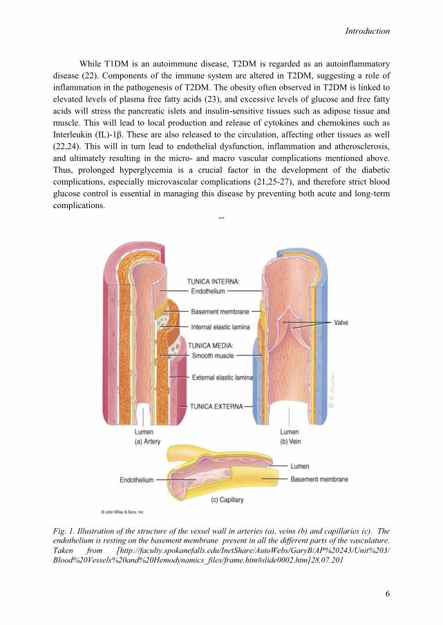
Fig. 1. Illustration of the structure of the vessel wall in arteries (a), veins (b) and capillaries (c). The endothelium is resting on the basement membrane present in all the different parts of the vasculature. Taken from [http://faculty.spokanefalls.edu/InetShare/AutoWebs/GaryB/AP%20243/Unit%203/ Blood%20Vessels%20and%20Hemodynamics_files/frame.htm#slide0002.htm]28.07.201
Introduction
7
1.2 The endothelium DM is hallmarked by vascular complications brought about by dysfunction of the
endothelium, vascular inflammation and atherosclerosis. The endothelium is forming the inner lining of the vasculature, and is the prime organ to be exposed to hyperglycemic conditions. Endothelial cells are highly specialized, multifunctional squamous epithelial cells, which predominately express the insulin-insensitive glucose transporter GLUT-1 (28,29). Consequently, intracellular glucose concentrations are reflected by the extracellular glucose levels, making endothelial cells especially sensitive to hyperglycemia.
1.2.1 Normal function of the endothelium
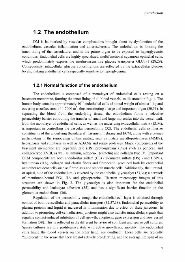
The endothelium is composed of a monolayer of endothelial cells resting on a basement membrane, forming the inner lining of all blood vessels, as illustrated in Fig. 1. The human body contains approximately 1013 endothelial cells of a total weight of almost 1 kg and covering a surface area of 4-7000 m2, thus constituting a large and important organ (30,31). In separating the blood from the underlying tissue, the endothelium forms a selective permeability barrier controlling the transfer of small and large molecules into the vessel wall. Both the monolayer of endothelial cells, as well as the underlying extracellular matrix (ECM), is important in controlling the vascular permeability (32). The endothelial cells synthesize constituents of the underlying (basolateral) basement mebrane and ECM, along with enzymes participating in the remodeling of this matrix, such as matrix metalloproteinases (MMPs), heparinases and sulfatases as well as ADAMs and serine proteases. Major components of the basement membrane are heparansulfate (HS) proteoglycans (PGs) such as perlecan and collagen type XVIII, as well as laminin, nidogen-1 (entactin) and collagen type IV. Important ECM components are both chondroitin sulfate (CS) / Dermatan sulfate (DS) - and HSPGs, hyaluronan (HA), collagen and elastin fibers and fibronectin, produced both by endothelial and other resident cells such as fibroblasts and smooth muscle cells. Additionally, the luminal, or apical, side of the endothelium is covered by the endothelial glycocalyx (33,34); a network of membrane-bound PGs, HA and glycoproteins. Electron microscopy images of this structure are shown in Fig. 2. The glycocalyx is also important for the endothelial permeability and leukocyte adhesion (35), and has a significant barrier function in the glomerular endothelium (36).
Regulation of the permeability trough the endothelial cell layer is obtained through control of both transcellular and paracellular transport (32,37,38). Endothelial permeability to plasma proteins and liquid is increased in inflammation due to effect on these junctions. In addition to promoting cell-cell adhesion, junctions might also transfer intracellular signals that regulate contact-induced inhibition of cell growth, apoptosis, gene expression and new vessel formation (39). This is reflected in the different behavior of confluent and sparse cell cultures. Sparse cultures are in a proliferative state with active growth and motility. The endothelial cells lining the blood vessels on the other hand, are confluent. These cells are typically “quiescent” in the sense that they are not actively proliferating, and the average life span of an
Introduction
8
endothelial cell is more than 1 year (40-42). The interendothelial junctions in the dense cell-layer contribute to this quiescent phenotype by transmitting signals within the cells, changing their gene expression. When they reach confluence and their junctions become more organized, they lose the ability to respond to growth factors, and they switch to a resting condition. Sparse cells, which lack cell-cell junctions, are unable to transduce such signals (39). This can be exemplified by the response of endothelial cells to stimuli with transforming growth factor (TGF) β1, which is dependent both on cell shape, proliferative state and the source of the cells (43,44). The contact-inhibition of dense cell-cultures will promote quiescence (45).
Fig. 2. Electron microscopy image of the endothelial glycocalyx in a coronary capillary, taken from (34).
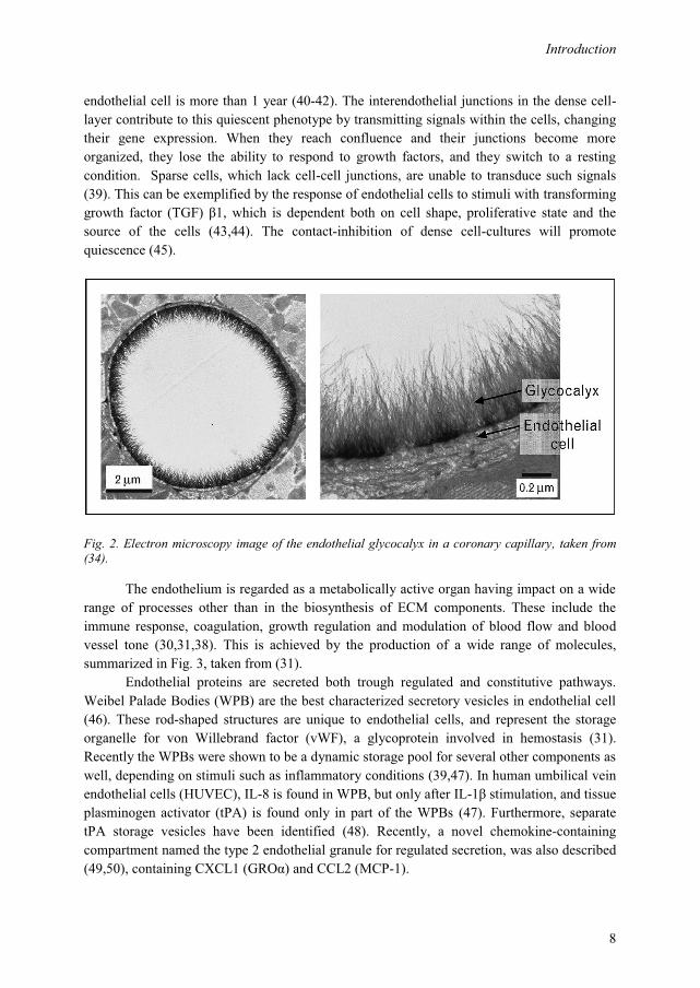
The endothelium is regarded as a metabolically active organ having impact on a wide range of processes other than in the biosynthesis of ECM components. These include the immune response, coagulation, growth regulation and modulation of blood flow and blood vessel tone (30,31,38). This is achieved by the production of a wide range of molecules, summarized in Fig. 3, taken from (31).
Endothelial proteins are secreted both trough regulated and constitutive pathways. Weibel Palade Bodies (WPB) are the best characterized secretory vesicles in endothelial cell (46). These rod-shaped structures are unique to endothelial cells, and represent the storage organelle for von Willebrand factor (vWF), a glycoprotein involved in hemostasis (31). Recently the WPBs were shown to be a dynamic storage pool for several other components as well, depending on stimuli such as inflammatory conditions (39,47). In human umbilical vein endothelial cells (HUVEC), IL-8 is found in WPB, but only after IL-1β stimulation, and tissue plasminogen activator (tPA) is found only in part of the WPBs (47). Furthermore, separate tPA storage vesicles have been identified (48). Recently, a novel chemokine-containing compartment named the type 2 endothelial granule for regulated secretion, was also described (49,50), containing CXCL1 (GROα) and CCL2 (MCP-1).
Introduction
9
Fig. 3. Illustration of the main classed and most important molecules the endothelial cell is capable of producing, taken from (31).
1.2.2 Endothelial dysfunction and atherosclerosis
Under normal conditions the endothelium participate in regulating blood clotting, assisting the body's immune response, controlling fluid volume and the amount of electrolytes that pass from the blood into the tissues, affecting dilation or constriction of blood vessels. However, in endothelial dysfunction, the ability to perform one or more of these functions is affected. In diabetes, dysfunction of the vascular endothelium is regarded an important factor in the pathogenesis of both diabetic micro- and macroangiopathy (51). Endothelial dysfunction plays a major role in the development of atherosclerosis, contributing to diabetic macrovascular complications.
The effect of hyperglycemia on the microvasculature is mediated through increased flux trough several metabolic pathways. These include the hexosamine-, polyol-, PKC- and the AGE (Advanced glycated end products) -pathways (52,53). High intracellular glucose concentrations will increase flux through the glycolysis and ultimately lead to an accumulation of the upstream glucose-metabolites, and increase the flux through all the above mentioned pathways. The glucose donors uridine diphosphate (UDP)-N-acetyl-glucosamine (GlcNAc) and UDP-N-acetyl-glucosamine (UDP-GalNAc) are intermediates of the hexosamine pathway. Such altered substrate-availability for glycosaminoglycan (GAG) synthesis has the potential to affect PG expression, as well as the modifications of other glycoproteins. In addition, increased flux trough the hexosamine pathway will increase the O-GlcNAc-modifications of a wide range of proteins. This is a process similar to phosphorylation, affecting gene expression and signaling, and contributing the glucose-toxicity of diabetes (54). In hyperglycemia, a series of proteins are also modified trough glycation leading to the generation of AGE and vascular damage. AGE levels are increased
Introduction
10
both in the circulation and the tissue of diabetics (55). Hyperglycemia will also increase diacylglycerol content and activate PKC, having a number of pathogenic consequences on the vasculature. Furthermore, increased flux through the polyol pathway is especially important in the development of diabetic cataract.
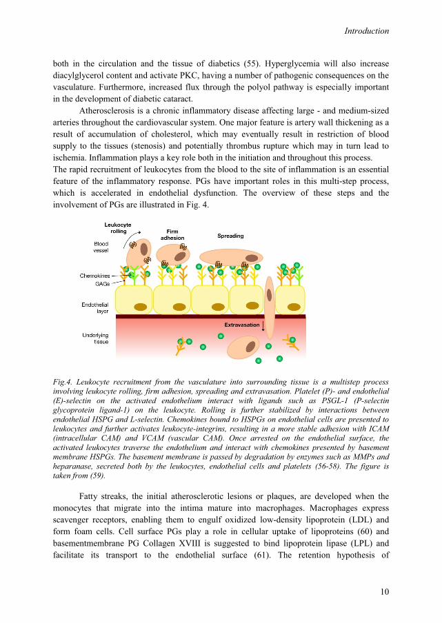
Atherosclerosis is a chronic inflammatory disease affecting large - and medium-sized arteries throughout the cardiovascular system. One major feature is artery wall thickening as a result of accumulation of cholesterol, which may eventually result in restriction of blood supply to the tissues (stenosis) and potentially thrombus rupture which may in turn lead to ischemia. Inflammation plays a key role both in the initiation and throughout this process. The rapid recruitment of leukocytes from the blood to the site of inflammation is an essential feature of the inflammatory response. PGs have important roles in this multi-step process, which is accelerated in endothelial dysfunction. The overview of these steps and the involvement of PGs are illustrated in Fig. 4.
Fig.4. Leukocyte recruitment from the vasculature into surrounding tissue is a multistep process involving leukocyte rolling, firm adhesion, spreading and extravasation. Platelet (P)- and endothelial (E)-selectin on the activated endothelium interact with ligands such as PSGL-1 (P-selectin glycoprotein ligand-1) on the leukocyte. Rolling is further stabilized by interactions between endothelial HSPG and L-selectin. Chemokines bound to HSPGs on endothelial cells are presented to leukocytes and further activates leukocyte-integrins, resulting in a more stable adhesion with ICAM (intracellular CAM) and VCAM (vascular CAM). Once arrested on the endothelial surface, the activated leukocytes traverse the endothelium and interact with chemokines presented by basement membrane HSPGs. The basement membrane is passed by degradation by enzymes such as MMPs and heparanase, secreted both by the leukocytes, endothelial cells and platelets (56-58). The figure is taken from (59).
Fatty streaks, the initial atherosclerotic lesions or plaques, are developed when the monocytes that migrate into the intima mature into macrophages. Macrophages express scavenger receptors, enabling them to engulf oxidized low-density lipoprotein (LDL) and form foam cells. Cell surface PGs play a role in cellular uptake of lipoproteins (60) and basementmembrane PG Collagen XVIII is suggested to bind lipoprotein lipase (LPL) and facilitate its transport to the endothelial surface (61). The retention hypothesis of
Introduction
11
atherosclerotic development first outlined in 1995 (62,63), focus on PGs mediating lipoprotein retention as a critical step in the initiation of atherosclerotic development. Deposited PGs such as biglycan in the intima may retain atherogenic lipoproteins such as LDL, contributing to fatty streak development. Formation of an advanced lesion or atherosclerotic plaque occurs as a necrotic core of leukocytes and lipids and a fibrous cap evolve in the fatty streaks, leading to the formation of the arterial plaque.
T2DM is regarded an inflammatory disease, with increased levels of pro-inflammatory cytokines including IL-6, TNFα, MCP-1 and IL-1β. Elevated IL-1β levels are predictive of T2DM (64) and IL-1 signals via IL-1 receptor type 1 (IL-1R1) and IL-1RAP /IL-1, also expressed by ECs. A number of inflammatory cytokines are also involved and elevated in T1DM (65), including IL-1 (66).
1.2.3 Human umbilical vein endothelial cells - HUVEC
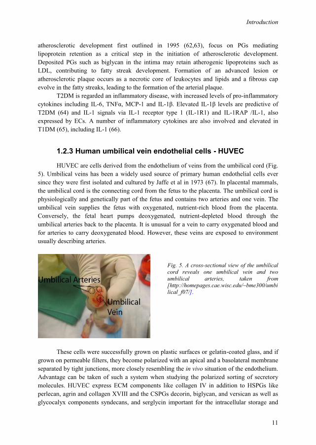
HUVEC are cells derived from the endothelium of veins from the umbilical cord (Fig. 5). Umbilical veins has been a widely used source of primary human endothelial cells ever since they were first isolated and cultured by Jaffe et al in 1973 (67). In placental mammals, the umbilical cord is the connecting cord from the fetus to the placenta. The umbilical cord is physiologically and genetically part of the fetus and contains two arteries and one vein. The umbilical vein supplies the fetus with oxygenated, nutrient-rich blood from the placenta. Conversely, the fetal heart pumps deoxygenated, nutrient-depleted blood through the umbilical arteries back to the placenta. It is unusual for a vein to carry oxygenated blood and for arteries to carry deoxygenated blood. However, these veins are exposed to environment usually describing arteries.
Fig. 5. A cross-sectional view of the umbilical cord reveals one umbilical vein and two umbilical arteries, taken from [http://homepages.cae.wisc.edu/~bme300/umbilical_f07/].
These cells were successfully grown on plastic surfaces or gelatin-coated glass, and if grown on permeable filters, they become polarized with an apical and a basolateral membrane separated by tight junctions, more closely resembling the in vivo situation of the endothelium. Advantage can be taken of such a system when studying the polarized sorting of secretory molecules. HUVEC express ECM components like collagen IV in addition to HSPGs like perlecan, agrin and collagen XVIII and the CSPGs decorin, biglycan, and versican as well as glycocalyx components syndecans, and serglycin important for the intracellular storage and
Introduction
12
sorting of cytokines. HUVEC are also able to express several proinflammatory cytokines such as IL-1β, IL-6, IL-8 and TNFα as well as Intracellular adhesion molecule-1 (ICAM-1), platelet-CAM-1 (PCAM-1), P-selectin, E-selectin, and VE-cadherin, corresponding to molecules expressed by endothelial cells in vivo (68).
1.3 Proteoglycans and glycosaminoglycans The endothelium is a main target of the diabetic complications (51), and PGs
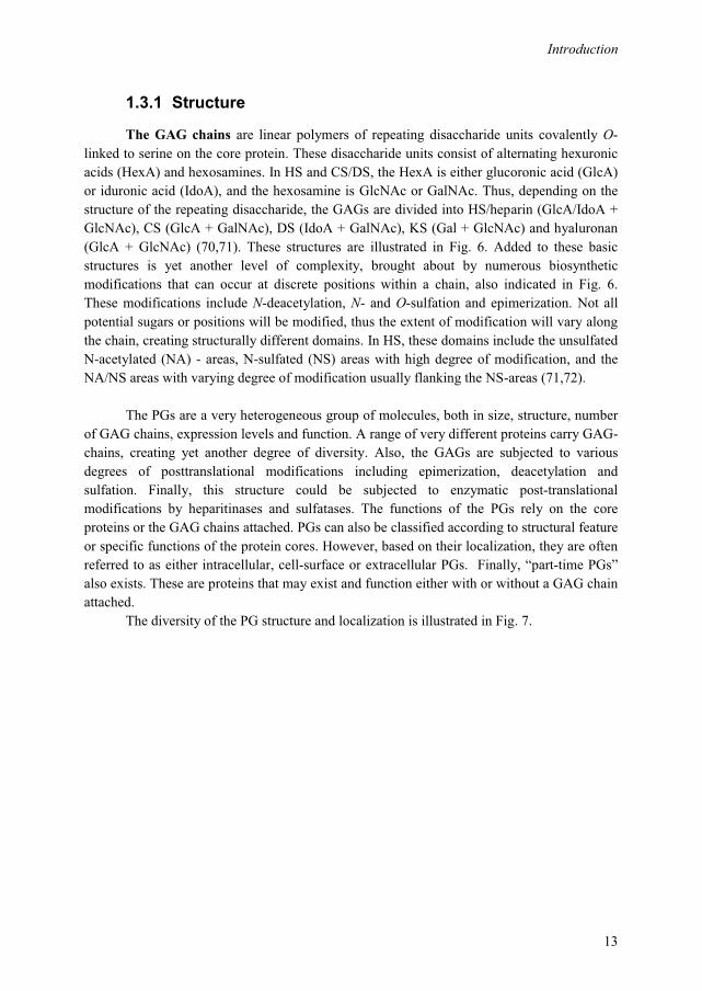
expressed by these cells have been implicated in the development of diabetic complications (56,69). PGs are molecules consisting of a protein core to which extended linear carbohydrate side chains are covalently attached. Based on the composition if these GAG chains, the PGs belong to the HS, CS, DS or keratan sulfates (KS) class. The anticoagulant heparin is a highly modified form of HS produced by mast cells and attached to the serglycin core protein. Hyaluronan on the other hand, is synthesized as a free GAG- chain with no attachment to a core protein. These GAG chains are highly polyanionic structures, providing the PGs with unique qualities.
Fig.6. The structure of the repeating disaccharide region of hyaluronan (HA), chondroitin sulfate (CS), dermatan sulfate (DS), heparan sulfate (HS)/heparin (Hep), and keratan sulfate (KS) is shown. Possible sulfation positions are given and an asterisk in the structure of HS/Hep indicates that the C5 position of the uronic acid residue may be epimerized. The picture is taken from [http://jcggdb.jp/GlycoPOD/protocolShow.action?nodeId=t15]28.07.2012.
Introduction
13
1.3.1 Structure
The GAG chains are linear polymers of repeating disaccharide units covalently O-linked to serine on the core protein. These disaccharide units consist of alternating hexuronic acids (HexA) and hexosamines. In HS and CS/DS, the HexA is either glucoronic acid (GlcA) or iduronic acid (IdoA), and the hexosamine is GlcNAc or GalNAc. Thus, depending on the structure of the repeating disaccharide, the GAGs are divided into HS/heparin (GlcA/IdoA + GlcNAc), CS (GlcA + GalNAc), DS (IdoA + GalNAc), KS (Gal + GlcNAc) and hyaluronan (GlcA + GlcNAc) (70,71). These structures are illustrated in Fig. 6. Added to these basic structures is yet another level of complexity, brought about by numerous biosynthetic modifications that can occur at discrete positions within a chain, also indicated in Fig. 6. These modifications include N-deacetylation, N- and O-sulfation and epimerization. Not all potential sugars or positions will be modified, thus the extent of modification will vary along the chain, creating structurally different domains. In HS, these domains include the unsulfated N-acetylated (NA) - areas, N-sulfated (NS) areas with high degree of modification, and the NA/NS areas with varying degree of modification usually flanking the NS-areas (71,72).
The PGs are a very heterogeneous group of molecules, both in size, structure, number of GAG chains, expression levels and function. A range of very different proteins carry GAG-chains, creating yet another degree of diversity. Also, the GAGs are subjected to various degrees of posttranslational modifications including epimerization, deacetylation and sulfation. Finally, this structure could be subjected to enzymatic post-translational modifications by heparitinases and sulfatases. The functions of the PGs rely on the core proteins or the GAG chains attached. PGs can also be classified according to structural feature or specific functions of the protein cores. However, based on their localization, they are often referred to as either intracellular, cell-surface or extracellular PGs. Finally, “part-time PGs” also exists. These are proteins that may exist and function either with or without a GAG chain attached.
The diversity of the PG structure and localization is illustrated in Fig. 7.
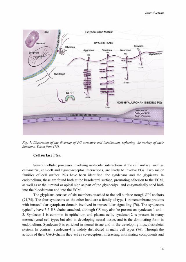
Introduction
14
Fig. 7. Illustration of the diversity of PG structure and localization, reflecting the variety of their functions. Taken from (73).
Cell surface PGs.
Several cellular processes involving molecular interactions at the cell surface, such as cell-matrix, cell-cell and ligand-receptor interactions, are likely to involve PGs. Two major families of cell surface PGs have been identified: the syndecans and the glypicans. In endothelium, these are found both at the basolateral surface, promoting adhesion to the ECM, as well as at the luminal or apical side as part of the glycocalyx, and enzymatically shed both into the bloodstream and into the ECM.
The glypicans consists of six members attached to the cell surface trough GPI-anchors (74,75). The four syndecans on the other hand are a family of type 1 transmembrane proteins with intracellular cytoplasm domain involved in intracellular signalling (76). The syndecans typically have 3-5 HS chains attached, although CS may also be present on syndecan-1 and -3. Syndecan-1 is common in epithelium and plasma cells, syndecan-2 is present in many mesenchymal cell types but also in developing neural tissue, and is the dominating form in endothelium. Syndecan-3 is enriched in neural tissue and in the developing musculoskeletal system. In contrast, syndecan-4 is widely distributed in many cell types (76). Through the actions of their GAG-chains they act as co-receptors, interacting with matrix components and
Introduction
15
growth factors which in turn promote intracellular signaling cascades (77). Also, syndecans may be proteolyticaly cleaved by enzymes such as MMPs, in a process known as shedding. This is an important regulatory mechanism, rapidly changing surface receptor dynamics and generating soluble ectodomains (78). Different MMPs has different affinities for different syndecans, and are inhibited by the actions of TIMPs. It has been reported that syndecan-1 in the circulation is increased in T1DM pasients with microalbuminuri compared to those without (79).
ECM PGs.
Connective tissue is generally composed of a network of proteins such as collagens,
glycoproteins such as fibronectin, HA and PGs. The properties and architecture of a tissue depend on which components are secreted locally to the matrix by the residing cells. The PG composition will vary between different tissues, depending on the functions of the PGs. Due to the striking polyanionicity of the GAG chains, PGs have the ability to form gels even at low concentrations, giving them important functions as shock absorbers, such as in cartilage. Importantly, although essential for structure and shock absorption, the PGs also have important roles in several other cellular functions. PGs are especially abundant in bone and cartilage, and matrix PGs are particularly well studied in cartilage where they constitute over 90 % of the dry weight of the tissue. The most common cartilage PG is aggrecan, named so because it forms large aggregates with HA. In vascular tissue, important matrix-PGs are CS/DSPG versican, small leucine rich PGs (SLRPs) decorin and biglycan along with the large basement membrane HSPGs perlecan, collagen XVIII and agrin.
Versican belongs to the family of the large aggregating CSPGs. It has multiple functions as it interact with many ECM components such as HA, tenascin, fibulins, fibrillin and fibronectin as well as cell surface proteins such as selectins, CD44, integrin and EGFR (epidermal growth factor receptor) and P-selectin-glycoprotein ligand-1 (PSGL-1). The multiple functions of versican can be explained by these varying binding partners and multiple isoforms of this PG (80).
The small leucine-rich PGs (SLRPs) are named for their small size (up to 42 kD) and the leucine-rich repeats of the protein core. CS/DS PGs Decorin and biglycan, belonging to the Class I of SLRPs, are the most extensively studied of these molecules. Decorin is substituted with one, and biglycan with two, CS/DS chains, as well as three and two N-linked oligosaccharides respectively. The leucine-rich repeats of the protein core, as well as the GAG side chain(s), allow for a wide range of matrix- matrix and matrix-cell interactions, and as ECM components they are important in matrix assembly (81). Illustrating this point, decorin is so named due to its ability to “decorate” collagen fibers in the ECM. However, other roles are emerging for soluble, no-matrix bound fragments of decorin and biglycan. They are secreted, or liberated from the matrix by the action of MMPs, and they have the ability to functions as receptor ligands influencing cell behavior (82,83). Thus, these PGs have functions related to structure, proliferation, differentiation, survival, adhesion, migration and the inflammatory response. Decorin has anti-fibrotic effects trough interaction with TGFβ
Introduction
16
signaling, and is involved in the pathogenesis of renal fibrosis (83). Decorin is also able to bind and inhibit CCN2 (connective tissue growth factor), essential for fibrosis (84). As for their roles in inflammation, decorin is inversely related to atherosclerosis (85), while soluble biglycan act as a pro-inflammatory signaling molecule (82).
Another small PG affected by inflammation is DSPG endocan. Endocan is synthesized in moderate amounts by endothelial cells, mainly in kidney and lung, but the expression is markedly increased in the presence of pro-inflammatory and pro-angiogenic molecules, and is regarded a marker of endothelial dysfunction (86,87) and is suggested to be a potential cancer marker (88).
The basement membrane is a specialized sheet like form of ECM that supports the endothelium, epithelium and several other cell types. It is composed of four major molecules; laminin, type IV collagen, nidogen and PGs. The subendothelial basement membrane has been shown to be dominated by HSPGs perlecan, collagen XVIII and agrin (89,90), although extensive studies on the CSPGs in this specialized ECM has not been performed. The HSPG composition of the basement membrane differs between various tissues; e.g. in glomerular basement membrane agrin is the dominating HSPG (91), while perlecan dominates in many other tissues.
Perlecan is named from the appearance of its protein core as a string of pearls. It is a ubiquitous macromolecule predominately expressed in basement membranes, as well as ECM in general. The ~470 kDa protein core consists of five domains labeled I-V. Adding the numerous O-linked oligosaccharides and as many as four HS chains, the molecular weight for intact perlecan can reach 800 kDa. The various modules of the perlecan protein core and its HS chains can take part in a large number of molecular interactions. The binding partners include proteins of the basement membrane and ECM, cell surface proteins, and growth factors such as fibroblastic growth factor (FGF), vascular endothelial growth factor (VEGF) and PDGF (platelet derived growth factor) (90,92). Perlecan is synthesized by both vascular endothelial and smooth muscle cells and deposited in the ECM, but can also be associated with the cell surface. Moreover, perlecan is also expressed in avascular tissues such as cartilage. Not surprisingly, perlecan is involved in a number of pathological processes, including atherosclerosis. Endorepellin is the 85 kDa C-terminal domain V of the perlecan protein core, which can be cleaved by the action of MMPs. Similar to the C-terminal of collagen XVIII, endostatin, and tumstatin of the part-time PG collagen IV, endorepellin is found to inhibit angiogenesis (93). These fragments of basment membrane PGs modulate endothelial cells trough interactions with integrins.
Also abundant in basement membranes, of e.g. blood vessels, is HSPG Collagen XVIII (94) which is expressed in three variants, of which the shorter one is found in most vascular and epithelial basement membrane structures. In mouse kidney, lack of the long isoforms affects kidney podocytes, whereas the short form is needed in the proximal tubular basement membrane (95). Lipoprotein lipase presentation on the luminal side of endothelium depends on collagen XVIII (61) implicating this molecule in triglyceride metabolism. The C-terminal fragment of collagen XVIII, endostatin, is an inhibitor of angiogenesis and tumor growth by restricting endothelial cell proliferation and migration (96).
Introduction
17
More abundant in the glomerular basement membrane is HSPG agrin, expressed in several isoforms in several tissues and originally known for its functions in the synaptic basement membrane (89,91). In the human glomerular basement membrane, agrin has important functions in the charge barrier (89,91,97).
Intracellular PGs.
Serglycin is the most prominent intracellular PG. Apart from some reports e.g. on the presence of syndecans in the cytoplasm and nucleus (98), serglycin is the PG with most documented intracellular functions. This molecule has its name from the extensive stretch of ser-gly-repeats present in the approximately 15 kDa protein core, representing the attachment sites for the GAG chains. As a result of the close proximity of these sites, serglycin is densely substituted with GAGs, providing its characteristic resistance to proteolytic cleavage (99,100).
Originally regarded as a PG of the hematopoietic cells, this molecule is most extensively studied in these cells (101). However, serglycin was later found to be expressed by several other cell types (102), including ECs (103,104), chondrocytes (105), smooth muscle cells (106) and in tumors (107).
Studies from connective tissue mast cells show that serglycin in these cells is substituted with the oversulfated form of HS, heparin. In mucosal mast cells however, as well as other hematopoietic cells, serglycin carries CS-chains, mostly of the CS-4S or CS-4,6S form, sulfated at the 4-O or 4 and 6-O position of GalNAc. Serglycin carrying heparin chains have so far only been detected in connective tissue mast cells. Serglycin containing HS has been identified (108), as well as hybrids carrying both heparin and CS (102). Finally, the size and number of GAG chains varies, not only between cell types, but also as a response to stimuli. All of these factors accentuates the dependence of serglycin structure on the tissue or cells type in which it is expressed.
Being most highly expressed by mast cells, the functions of serglycin is thoroughly studied in these cells. Lessons from the serglycin knockout mouse demonstrate that serglycin has a key role in the granular storage of several mast cell-specific proteases through electrostatic interactions to the sulfated GAG chains (109). Knockout of the serglycin protein results in several similar phenotypes as those in glucosaminyl N-deacetylase /N-sulfotransferase (NDST)-2 knockout (110), supporting the role of the GAG-chains (heparin) in this process. Trough use of the serglycin knock-out mouse it has been established that both leukocytes (109), platelets (111), macrophages (108) and cytolytic T-lyphocytes (112) are affected by the absence of this PG.
Thus, serglycin is important in granule formation, destined for storage or constitutive secretion, also induced by e.g. inflammatory stimuli. The functions of secreted serglycin are intriguing. Serglycin is implicated in the secretion of several compounds including proteases, but also cytokines and chemokines (113). Following secretion, the serglycin complexes can dissociate due to the increasing pH. Alternatively, depending on the strength of the interactions, the partner molecules may remain in complex with serglycin. This may have several functional consequences, including promotion of proteolytic protection, and
Introduction
18
facilitation of chemokine or cytokine transport and presentation or protease interaction with substrate. Serglycin is also a possible scavenger, sequestering inflammatory compounds such as GAG-binding chemokines (102,113,114). Thus, the exact function and structure of serglycin varies between these different cell types; hence the description of serglycin as “a structural and functional chameleon” (102).
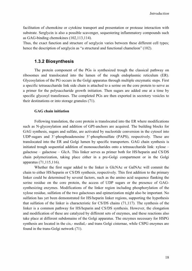
1.3.2 Biosynthesis
The protein component of the PGs is synthesized trough the classical pathway on ribosomes and translocated into the lumen of the rough endoplasmic reticulum (ER). Glycosylation of the PG occurs in the Golgi apparatus through multiple enzymatic steps. First a specific tetrasaccharide link side chain is attached to a serine on the core protein to serve as a primer for the polysaccharide growth initiation. Then sugars are added one at a time by specific glycosyl transferases. The completed PGs are then exported in secretory vesicles to their destinations or into storage granules (71).
GAG chain initiation
Following translation, the core protein is translocated into the ER where modifications such as N-glycosylation and addition of GPI-anchors are acquired. The building blocks for GAG synthesis, sugars and sulfate, are activated by nucleotide conversion in the cytosol into UDP-sugars and 3’-phosphoadenosine 5’-phosphosulfate (PAPS), respectively. These are translocated into the ER and Golgi lumen by specific transporters. GAG chain synthesis is initiated trough sequential addition of monosaccharides onto a tetrasaccharide link: xylose – galactose – galactose – GlcA. This linker serves as primer both for HS/heparin and CS/DS chain polymerization, taking place either in a pre-Golgi compartment or in the Golgi apparatus (71,115,116).
Whether the first sugar added to the linker is GlcNAc or GalNAc will commit the chain to either HS/heparin or CS/DS synthesis, respectively. This first addition to the primary linker could be determined by several factors, such as the amino acid sequence flanking the serine residue on the core protein, the access of UDP sugars or the presence of GAG-synthesizing enzymes. Modifications of the linker region including phosphorylation of the xylose residue, sulfation of the two galactoses and epimerization might also be important. No sulfation has yet been demonstrated for HS/heparin linker regions, supporting the hypothesis that sulfation of the linker is characteristic for CS/DS chains (71,117). The synthesis of the linker is a common pathway for HS/heparin and CS/DS synthesis. However, the elongation and modification of these are catalyzed by different sets of enzymes, and these reactions also take place at different subdomains of the Golgi apparatus. The enzymes necessary for HSPG synthesis are located in the cis,- medial,- and trans Golgi cisternae, while CSPG enzymes are found in the trans-Golgi network (71).
Introduction
19
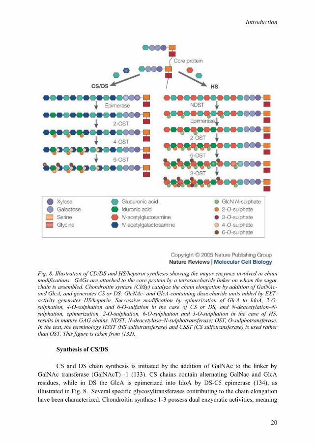
Synthesis of HS / heparin HS chain polymerization is initiated by the addition of GlcNAc by EXT -like gene
product 2 (EXTL2) or EXTL3, members of the exostosin (EXT) family (118). The HS chains are further elongated by the co-polymerase EXT1/2, a hetero-complex of EXT1 and EXT2 (119,120), both possessing dual GlcA/GlcNAc-transferase activity, but with higher activities when in complex (121). HS chains are elongated by the alternating addition of GlcA and GlcNAc residues to the non-reducing end of the chain. The growing chain is modified at various positions including deacetylation and N-sulfation of GlcNAc, epimerization of GlcA into IdoA and O-sulfation at various positions of the disaccharides (69).These steps are illustrated in Fig. 8.
The modification of HS are initiated by N-deacetylation and subsequent N-sulfation of selected GlcNAc residues, creating GlcNS. The two reactions are carried out by a bi-functional enzyme, the NDST. N-sulfation is unique to HS/heparin and do not occur in the other GAGs. In mammals four NDSTs have been identified; NDST1 and NDST2 are expressed in most tissues, while NDST3 and NDST4 are expressed during embryonic development (70). HS chains have a domain structure in which sulfated domains (NS-domains) alternate with unsulfated regions (NA-domains). The N-sulfation carried out by NDST is required for further modification of the HS chain. Thus, NDST will create the overall design of the HS chain. Following N-sulfation, C5-epimerization of GlcA into IdoA by C5 GlcA-epimerase occurs. Then HS chains are selectively sulfated at the 2-O position of the uronic acid and the 6-O and 3-O position of GlcNS residues. 2-O-sulfation is carried out by the HS 2-O-sulfortransferase (HS2ST) at the C2 position of GlcA or, preferably, IdoA. The transfer of sulfate from the sulfate donor PAPS to position 6 of GlcNAc and GlcS residues is catalyzed by three different HS 6-O-sulfotransferases (HS6ST). The 3-O-sulfation catalyzed by HS3ST of GlcNAc residues is the rarest modification, creating the characteristic modifications of the oversulfated heparin. NDST1, EXT1 and EXT2 is suggested to operate in large complexes called GAGosomes, possibly also in concert with sulfotransferases (122-124). Thus, GAGosomes of different compositions might result in different HS modification patterns (125).
After synthesis and extensive modification, the HSPG are transported from the Golgi to the ECM or the cell surface, where they might be subjected to additional modifications by the action of sulfatases (Sulfs), sheddases or heparanase. Sulf1 and Sulf2 are cell-surface associated sulfatases, able to remove 6-O-sulfate from GlcN-residues in HS (126). In several organisms, two different Sulfs has been identified, the human forms being designated hSulf1 and hSulf2. Both are believed to exert the same substrate specificity (127). However, recent findings indicate that Sulf1 and Sulf2 differentially contribute to the generation of organ-specific sulfation patterns of HS (128). Furthermore, HS structure could be significantly modulated by the action of heparanase, an endo-β(1,4)-glucoronidase, cleaving the HS chains at specific sites (129). Heparanase is upregulated in a number of inflammatory conditions (58), and increased in T2DM patients (130). Finally, sheddases such as MMPs cleaving the extracellular core protein, are important modulators of HS structure and function and ECM architecture (131).
Introduction
20
Fig. 8. Illustration of CD/DS and HS/heparin synthesis showing the major enzymes involved in chain modifications. GAGs are attached to the core protein by a tetrasaccharide linker on whom the sugar chain is assembled. Chondroitin syntase (ChSy) catalyze the chain elongation by addition of GalNAc- and GlcA, and generates CS or DS; GlcNAc- and GlcA-containing disaccharide units added by EXT-activity generates HS/heparin. Successive modification by epimerization of GlcA to IdoA, 2-O-sulphation, 4-O-sulphation and 6-O-sulfation in the case of CS or DS, and N-deacetylation–N-sulphation, epimerization, 2-O-sulphation, 6-O-sulphation and 3-O-sulphation in the case of HS, results in mature GAG chains. NDST, N-deacetylase–N-sulphotransferase; OST, O-sulphotransferase. In the text, the terminology HSST (HS sulfotransferase) and CSST (CS sulfotransferase) is used rather than OST. This figure is taken from (132).
Synthesis of CS/DS
CS and DS chain synthesis is initiated by the addition of GalNAc to the linker by GalNAc transferase (GalNAcT) -1 (133). CS chains contain alternating GalNac and GlcA residues, while in DS the GlcA is epimerized into IdoA by DS-C5 epimerase (134), as illustrated in Fig. 8. Several specific glycosyltransferases contributing to the chain elongation have been characterized. Chondroitin synthase 1-3 possess dual enzymatic activities, meaning
Introduction
21
both glucuronyltransferase and galactosaminyltransferase activity. Two other enzymes identified however, CSGlcA-T and CSGalNAc-T2, act by transferring GlcA or GalNAc, respectively (70). The sulfotransferases involved in the biosynthesis of CS/DS belong to three different families that transfer sulfate groups to C4 on GalNAc residues (C4ST), C6 on GalNAc residues (C6ST) or C2 on the hexuronic acid (70). Three C4STs has been characterized (135,136), in addition to a DS-spesific GalNAc 4-O-sulfotransferase (D4ST-1) (137). The latter is suggested to work immediately after epimerization preventing back-epimerization of the newly formed IdoA into GlcA. The sulfotransferases involved in the 6O-sulfation of GalNAc residues are C-6 sulfotransferase 1-2 (C6ST1-2) and GalNAc 4-sulfate -6-O-sulfotransferase (GalNAc4S-6ST) which sulfates C-6 on already 4-O sulfated GalNAc. The hexuronic acid can be sulfated in C-2 position by uronyl 2-O-sulfotransferase. CS/DS chains show a wide structural heterogeneity as a result of the variation in the repeating disaccharide units. The structure of CS depends on the animal species, tissue and physiological conditions (134). In contrast to HS however, few reports on post transcriptional modifications of CS are described.
Synthesis of HA
In contrast to HS/heparin and CS/DS, HA is not attached to a core protein and is
assembled at the cell surface. It is not sulfated or subjected to other modifications. HA is produced by three HA synthase isoenzymes (HAS1-3), which are integral plasma membrane proteins whose active sites are located at the intracellular face of the membrane (138). These enzymes polymerize HA by repeatedly adding GlcA and GlcNAc the nascent polysaccharide as it is extruded via ABC-transporter through the cell membrane into the extracellular space. However, recent studies indicate that HA is not restricted to the extracellular milieu, but is also present intracellularly, implicated in inflammatory processes (139).
Synthesis of KS
KS is found predominately in cornea and cartilage. In KS, synthesis is initiated as N-
or O-linked oligosaccharides, and is termed KS I-III depending on their oligosaccharide link to the core protein. KS is extended by alternating addition of Gal and GlcNAc and can be C-6 sulfated at GlcNAc or both Gal and GlcNAc (140) .
1.3.3 Functions
PGs exert their functions trough interactions with other molecules through their protein core or GAG chains. In both HS/heparin and CS/DS, interactions with the GAG chain can be based either on electrostatic interactions or on sequence specific interactions. The latter depend on the precise location of N- and O-sulfate modifications along the sugar chain. This sulfation pattern is important for the interactions with a range of GAG-binding proteins, such as several ECM components, growth factors, chemokines, cytokines (141), cell adhesion molecules, coagulation proteins and LPL (142).
Introduction
22
Heparin is the most well-known GAG, used in the clinic for more than 75 years as an anti-coagulant. Heparin is an extensively sulfated form of HS, and has the highest negative charge density of any known biological molecule. Similar to HS in general, heparin can interact with other molecules through relative non-specific electrostatic interactions, or alternatively, by sequence-specific interactions. The sequence of heparin interacting with antithrombin is the best characterized of the protein-binding HS domains. Antithrombin inhibits thrombin and several other serine proteases of the coagulation pathway, including factors IXa, Xa, XIa and XIIa, and antithrombin, thus inhibiting coagulation. The ability of antithrombin to inhibit thrombin is greatly accelerated in the presence of heparin. The binding is mediated by a specific pentasaccharide sequence, which is hallmarked by the essential 3-O-sulfated GlcNS unit (143):
GlcNAc6S/GlcNS6S – GlcA – GlcNS3S6S – IdoA2S – GlcNS6S This sequence induces conformational changes in antithrombin which increase its inhibiting activities. However, the full thrombin-inhibiting activity of heparin requires an oligosaccharide of 18 monosaccharide units (144). Notably, the anticoagulant effect of exogenous heparin does not seem to correspond with the biological functions of endogenous heparin. Attached to the serglycin core protein, heparin is exclusively expressed in mast cells, important for storage of components associated with allergic and inflammatory responses. Thus, heparin seems important in situations not necessarily associated with blood coagulation. Recently however it was showed that heparin initiates the production of bradykinin via factor XII of the blood coagulation system and thus having an important role in allergy and inflammatory reactions driven by mast cells via the coagulation system (145).
HSPGs other than heparin are expressed throughout the body, interacting with a range of different molecules and resulting in a multitude of biological activities. As with heparin, ligand-interaction specificity is determined by the positioning of sulfate groups on HS creating distinct binding motifs. HS – protein interactions vary with regard to specificity and may depend on charge density in addition to strict sequence motifs of HS. However, the extent of such specific interactions is questioned (146).
Growth factors are among those important molecules in which function depend on GAG-interactions (147). Several angiogenic growth factors, such as VEGF (148), FGF-2, and PDGF, depend on HS/heparin for their biological effects.
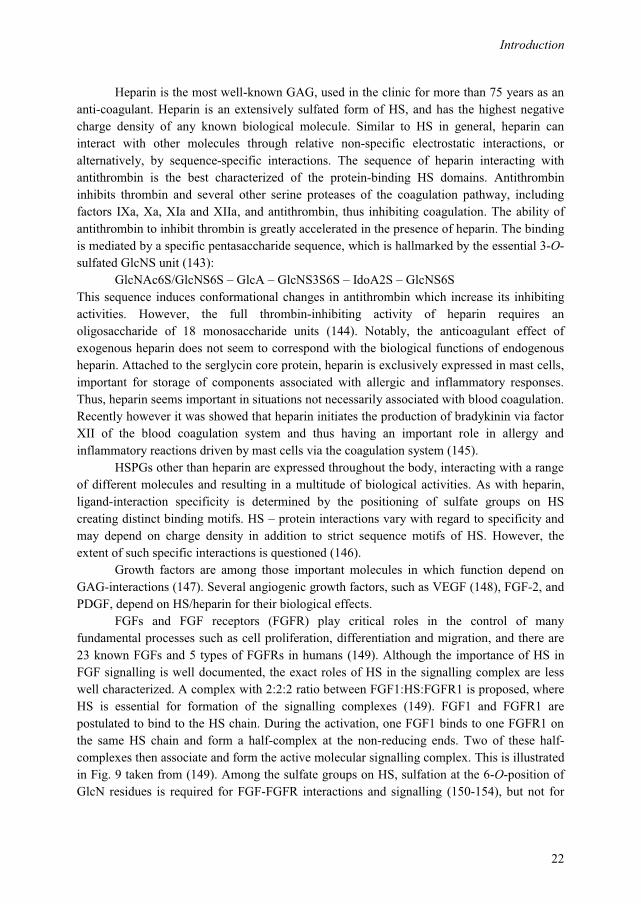
FGFs and FGF receptors (FGFR) play critical roles in the control of many fundamental processes such as cell proliferation, differentiation and migration, and there are 23 known FGFs and 5 types of FGFRs in humans (149). Although the importance of HS in FGF signalling is well documented, the exact roles of HS in the signalling complex are less well characterized. A complex with 2:2:2 ratio between FGF1:HS:FGFR1 is proposed, where HS is essential for formation of the signalling complexes (149). FGF1 and FGFR1 are postulated to bind to the HS chain. During the activation, one FGF1 binds to one FGFR1 on the same HS chain and form a half-complex at the non-reducing ends. Two of these half-complexes then associate and form the active molecular signalling complex. This is illustrated in Fig. 9 taken from (149). Among the sulfate groups on HS, sulfation at the 6-O-position of GlcN residues is required for FGF-FGFR interactions and signalling (150-154), but not for
Introduction
23
ternary complex formation (149). QSulf1 is a HS 6-O-endosulfatase, and QSulf1 mediated 6-O-desulfation reduce the formation of the FGF-HS-FGFR complex (151).
Fig.9. A proposed mechanism for the formation of the FGF1-FGFR1 signaling complex. FGF1 and FGFR1 bind to their binding sites on the HS chain. During the activation, FGF1 bind to FGFR on the same chain, forming a half-complex. Two of these half-complexes then associate to an active molecular signaling complex, taken from (149).
Changes in 6-O-sulfation can be on the level of biosynthesis, trough regulation of
HS6ST. In the adult mouse HS6ST-3 are rather ubiquitously expressed, while HS6ST-1 and HS6ST -2 are seen in specific organs. HS6ST-1 knockout mice exhibit deficient HS biosynthesis, abnormal placental and organ morphogenesis and late embryonic lethality (153,154). HS6ST-2, but not HS6ST-1, is mainly involved in the 6-O-sulfation of trisulfated units. HS6ST-2 KO mice survive and are fertile without apparent abnormal phenotypes, while HS6ST-1-/-/ HS6ST-2-/- mice die at slightly earlier age than HS6ST-1-/- mice. Alternatively, HS may be post-translationally modified through the action of two 6-O-endosulfatases, Sulf1 and 2 (151). Deficiency in human Sulf 1 and 2 affected VEGF-mediated signaling and kidney function (155).
Further emphasising the role of HS structure is the importance of 6-O-sulfation in HS interaction with L- and P-selectins. Selectins are a family of cell adhesion molecules important in inflammation. The anti-inflammatory effects of heparin in vivo is dependent primarily on P- and L-selectins, and the 6-O-sulfate group of GlcN units in heparin is critical for interaction with these selectins (156).
LPL, which is an important enzyme in lipid metabolism, binds to HSPGs. This interaction is crucial for several aspects of LPL function. Although LPL has high affinity to highly sulfated HS and heparin oligosaccharides, it is suggested that LPL binds to the more commonly expressed modestly sulfated sequences and can make use of general features of the HS chains with N-sulfated domains interrupted by low sulfated N-acetylated disaccharides (60,157).
Similarly to these HS sequences, various biological functions of CS/DS are thought to be attributed to particular domain structures with specific sulfation patterns (134,158). SLRPs
Introduction
24
like decorin and bilglycan are able to interact with their ligands both through their GAG chains or the leucine rich repeats (LLRs) characteristically of their protein core. They have the ability to bind a multitude of molecules including collagens, Toll-like receptor (TLR), EGF-R (epidermal growth factor receptor), IGF-IR (insulin-like growth factor receptor) and TGFβ (159). However, the role of CS and DS fine structure in ligand binding has not been subjected to extensive research.
1.3.4 PGs in diabetic nephropathy
Diabetes is a disease eventually resulting in both micro- and macro-vascular damages, caused by the complex effects of the diabetic milieu on a range of components. Alterations in both PG expression and structure are important pieces in this puzzle, affecting several organs. The kidney is one of the most severely affected organs, resulting in 10-20 % of people with diabetes dying of kidney failure (WHO).
Diabetes is the most common cause of chronic kidney disease, attacking 40 % of diabetics (160), and diabetic nephropathy is the cause of 44 % of all end-stage renal disease in the United States in 2009 (www.usrds.org). In Norway diabetic nephropathy is the cause of end-stage renal disease (ESRD) in 16-18 % of the patients entering renal replacement therapy, but another 15 % have diabetes as a covariant disease. Thus diabetes is part of the ESRD treatment in one third of the cases in Norway (Norwegian Renal Registry, www.nephro.no). The prevalence of T2DM is increasing both in Norway and worldwide. The relative prevalence of diabetic nephropathy is decreasing (161). Still, the diabetes epidemic leads to an increase in the absolute incidence of diabetic nephropathy. Understanding of the mechanisms behind the development of this disease is essential for the prevention of this prominent cause of end stage renal disease.
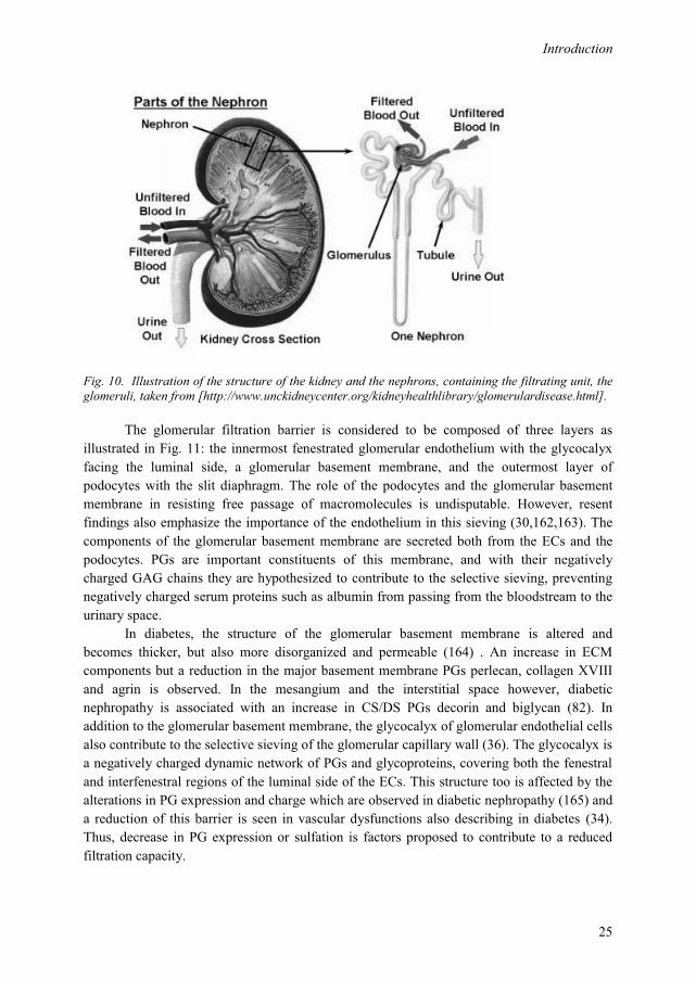
One of the main functions of the kidneys is filtration of the blood, as well as playing a crucial role in maintaining homeostasis, e.g. by regulation of elecrolytes and maintenance of the acid-bace balance. The filtrating entity of the nephron, the glomerulus, acts as the filtering unit and keeps normal proteins and cells in the bloodstream, allowing extra fluid and wastes to pass through. The architecture of the kidney is illustrated in Fig. 10.
Diabetic nephropathy is a progressive kidney disease caused by angiopathy of capillaries in the kidney glomeruli as a result of longstanding DM combined with poor blood-glucose control and genetic predispositions. An early detectable change in the course of diabetic nephropathy is the thickening of the glomerulus. At this stage microalbuminuria is experienced; namely excessive leakage of serum albumin in the urine. As diabetic nephropathy progresses, the glomerular filtration rate (GFR) is further reduced and urine albumin increases to the point that it may be detected by ordinary urinalysis techniques. At this stage, a kidney biopsy generally clearly shows diabetic nephropathy, which is an indication for dialysis and kidney transplantation in the western world.
Introduction
25
Fig. 10. Illustration of the structure of the kidney and the nephrons, containing the filtrating unit, the glomeruli, taken from [http://www.unckidneycenter.org/kidneyhealthlibrary/glomerulardisease.html].
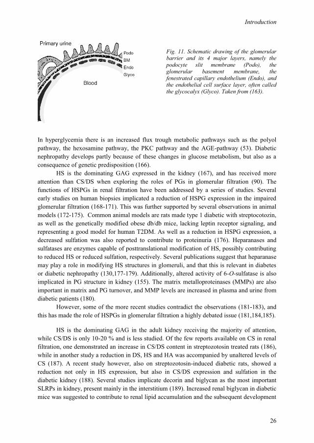
The glomerular filtration barrier is considered to be composed of three layers as
illustrated in Fig. 11: the innermost fenestrated glomerular endothelium with the glycocalyx facing the luminal side, a glomerular basement membrane, and the outermost layer of podocytes with the slit diaphragm. The role of the podocytes and the glomerular basement membrane in resisting free passage of macromolecules is undisputable. However, resent findings also emphasize the importance of the endothelium in this sieving (30,162,163). The components of the glomerular basement membrane are secreted both from the ECs and the podocytes. PGs are important constituents of this membrane, and with their negatively charged GAG chains they are hypothesized to contribute to the selective sieving, preventing negatively charged serum proteins such as albumin from passing from the bloodstream to the urinary space.
In diabetes, the structure of the glomerular basement membrane is altered and becomes thicker, but also more disorganized and permeable (164) . An increase in ECM components but a reduction in the major basement membrane PGs perlecan, collagen XVIII and agrin is observed. In the mesangium and the interstitial space however, diabetic nephropathy is associated with an increase in CS/DS PGs decorin and biglycan (82). In addition to the glomerular basement membrane, the glycocalyx of glomerular endothelial cells also contribute to the selective sieving of the glomerular capillary wall (36). The glycocalyx is a negatively charged dynamic network of PGs and glycoproteins, covering both the fenestral and interfenestral regions of the luminal side of the ECs. This structure too is affected by the alterations in PG expression and charge which are observed in diabetic nephropathy (165) and a reduction of this barrier is seen in vascular dysfunctions also describing in diabetes (34). Thus, decrease in PG expression or sulfation is factors proposed to contribute to a reduced filtration capacity.
Introduction
26
Fig. 11. Schematic drawing of the glomerular barrier and its 4 major layers, namely the podocyte slit membrane (Podo), the glomerular basement membrane, the fenestrated capillary endothelium (Endo), and the endothelial cell surface layer, often called the glycocalyx (Glyco). Taken from (163).
In hyperglycemia there is an increased flux trough metabolic pathways such as the polyol pathway, the hexosamine pathway, the PKC pathway and the AGE-pathway (53). Diabetic nephropathy develops partly because of these changes in glucose metabolism, but also as a consequence of genetic predisposition (166).
HS is the dominating GAG expressed in the kidney (167), and has received more attention than CS/DS when exploring the roles of PGs in glomerular filtration (90). The functions of HSPGs in renal filtration have been addressed by a series of studies. Several early studies on human biopsies implicated a reduction of HSPG expression in the impaired glomerular filtration (168-171). This was further supported by several observations in animal models (172-175). Common animal models are rats made type 1 diabetic with streptocotozin, as well as the genetically modified obese db/db mice, lacking leptin receptor signaling, and representing a good model for human T2DM. As well as a reduction in HSPG expression, a decreased sulfation was also reported to contribute to proteinuria (176). Heparanases and sulfatases are enzymes capable of posttranslational modification of HS, possibly contributing to reduced HS or reduced sulfation, respectively. Several publications suggest that heparanase may play a role in modifying HS structures in glomeruli, and that this is relevant in diabetes or diabetic nephropathy (130,177-179). Additionally, altered activity of 6-O-sulfatase is also implicated in PG structure in kidney (155). The matrix metalloproteinases (MMPs) are also important in matrix and PG turnover, and MMP levels are increased in plasma and urine from diabetic patients (180).
However, some of the more recent studies contradict the observations (181-183), and this has made the role of HSPGs in glomerular filtration a highly debated issue (181,184,185).
HS is the dominating GAG in the adult kidney receiving the majority of attention, while CS/DS is only 10-20 % and is less studied. Of the few reports available on CS in renal filtration, one demonstrated an increase in CS/DS content in streptozotosin treated rats (186), while in another study a reduction in DS, HS and HA was accompanied by unaltered levels of CS (187). A recent study however, also on streptozotosin-induced diabetic rats, showed a reduction not only in HS expression, but also in CS/DS expression and sulfation in the diabetic kidney (188). Several studies implicate decorin and biglycan as the most important SLRPs in kidney, present mainly in the interstitium (189). Increased renal biglycan in diabetic mice was suggested to contribute to renal lipid accumulation and the subsequent development
Introduction
27
of diabetic nephropathy (190). Their roles probably extend beyond those as structural matrix components, and include a role of soluble signaling molecules involved in kidney disease (82). However, their presence in the basement membrane has not been a focus of intensive study as the number of studies on the biology of CSPGs present in basement membranes has been low compared to their HSPG counterparts. The few studies on this topic include the description of CSPGs bamacan in basement membrane (191) and leprecan in glomerular basement membrane (192). As alterations in glomerular filtration are accompanied by changes in CSPGs, their role in the glomerular basement membrane deserves increased attention, particularly in light of the debated importance of HSPGs in the filtration barrier (181,184).
HA is also an important interstitial matrix component found in the renal papilla, but is scarcely expressed in the cortex. Due to its negative charge, HA has large water-binding capacity, and has important functions in renal water handling. Alterations in HA is observed in the diabetic kidney. Papillary HA was increased in diabetic rats (193) and increased glomerular HA synthesis has also been shown in glomerular core cultures from control and stre ptozotocin-diabetic rats (194), regulated trough HAS2 (195).
Aims of study
28
2 Aims of study
PGs are important matrix molecules with multiple functions, shown to be involved in several aspects of diabetic complications. Endothelial dysfunction, resulting from hyperglycemia and inflammation, is important in these processes. The overall aim was to obtain a better understanding of the expression and functions of the PGs in relation to endothelial dysfunction.
Specific aims were to:
� Study the effect of hyperglycemia and inflammation on PG structures in primary human endothelial cells.
� Gain further insight into the biosynthesis and secretion of PGs from polarized endothelial cells.
� Study the importance of PGs in relation to the endothelial proliferative status.
� To study the effects of diabetic conditions on GAG structure in one of the organs affected by diabetes, the kidney.

Summary of results
29
3 Summary of results
Paper I: Reine TM, Kusche-Gullberg M, Feta A, Jenssen T, Kolset SO. Heparan sulfate expression is affected by inflammatory stimuli in primary human endothelial cells. Glycoconj J. 2012;29:67-76.
In this study we investigated the effects of hyperglycemic and inflammatory
conditions on endothelial PGs, relevant for the diabetic setting. De novo secretion of 35S-PGs was studied in HUVEC exposed to hyperglycemia or the inflammatory mediators TNFα, IL-1α, IL-1β or TGFβ. The effect on total 35S-PG secretion and size distribution as well as HS and CS chain length, followed by HS polyanionicity and HS disaccharide composition, was examined.
We found no effect of hyperglycemia on neither of these parameters. In inflammatory conditions, no changes were observed in the overall 35S-PG size or in the 35S-CS chain length, indicating that CSPG expression was unaffected. The HSPGs however, were affected to various degrees. HS chain length was increased with TNFα and decreased with TGFβ, while both lead to a decrease in the overall 6-O-sulfation. IL-1α on the other hand, was related to an increase in 6-O-sulfation, but had no effect on chain length. IL-1β had no effect on these parameters. However, IL-1β was the only factor with stimulatory effects on 35S-PG secretion.
These results indicate that endothelial PG expression is unaffected by hyperglycemia, while different inflammatory stimuli had distinct, but different effects on endothelial HSPG expression.
Paper II: Meen AJ, Øynebråten I, Reine TM, Duelli A, Svennevig K, Pejler G, Jenssen T, Kolset SO. Serglycin is a major proteoglycan in polarized human endothelial cells and is implicated in the secretion of the chemokine GROalpha/CXCL1. J Biol Chem. 2011;286:2636-47
In this paper we studied the secretion of PGs from endothelial cells further. Culturing HUVEC on semipermeable filters allowed for the identification of serglycin as a major PG expressed by HUVEC, secreted predominantly to the apical side of these cells. Further, we studied the intracellular localization of serglycin in endothelial cells, as this has not been extensively studied. Serglycin could be detected in perinuclear regions corresponding to the
Summary of results
30
Golgi, as well as in two different types of secretory vesicles throughout the cytoplasm, identified by immunostaining and confocal microscopy. IL-1β stimulation increased the amount of serglycin-containing vesicles, in particular the smallest of the vesicles, corresponding to the type 2 granules. Also, following IL-1β stimulation, co-distribution with GROα/CXCL-1 was observed in a portion of the type 2 granula, suggesting interactions between these molecules. Further; abrogation of CS-chains with xyloside reduced the vesicular CXCL-1 expression, as well as the CXCL-1 secretion.
Together, these results indicate that serglycin is a major PG in endothelial cells, mainly secreted to the apical side and with possible roles in storage and secretion of inflammatory cytokines.
Paper III: Reine TM, Vuong TT, Meen AJ, Jenssen T, Kolset SO Serglycin expression is reduced in quiescent primary endothelial cells Submitted manuscript
As serglycin was found to be highly expressed in HUVEC, we aimed to investigate the functional implications of serglycin expression in endothelial cells further, focusing on proliferation. Sparse proliferating cell cultures are relevant for studies on angiogenesis and wound healing, while dense cultures are relevant for the quiescent endothelium lining the blood vessels.
De novo total 35S-PG secretion was reduced with 70 % in dense compared to sparse cell cultures. When investigating the secretion of individual PGs however, this was not a unifying pattern for all PGs. Reduced expression in dense cultures was evident for the highly expressed serglycin and biglycan, as well as decorin, but not for versican or collagen XVIII. Endocan and syndecan-4 levels on the other hand, were unaffected by cell density.
IL-1β stimulation revealed a more pronounced increase in 35S-PG secretion in dense cell cultures, which was also reflected in both protein and mRNA levels of the dominating PGs serglycin and perlecan. Versican, syndecan-4 and endocan on the other hand, increased irrespective of cell density, while decorin and biglycan secretion was not affected after this stimulus.
In sparse cultures, serglycin was localized to perinuclear regions corresponding to the Golgi, as well as in intracellular vesicles. In contrast, in dense cultures there was an obvious decrease in the Golgi staining, but extensive staining in intracellular vesicles. IL-1β stimulation increased the serglycin positive vesicles in both sparse and dense cultures, combined with a modest reduction in Golgi staining of the sparse cells.
These results indicate a relationship between serglycin expression and localization, with the proliferative status of the cells, as well as an effect of inflammatory stimuli on serglycin synthesis and secretion.
Summary of results
31
Paper IV: Reine TM, Grøndahl F, Jenssen T, Hadler-Olsen E, Prydz K, Kolset SO Reduced sulfation of chondroitin sulfate but not heparan sulfate in kidneys of diabetic db/db mice Manuscript
Changes in kidney function in relation to diabetes involve a series of factors, including PGs. The PGs are hypothesized to contribute to the filtration barrier trough the action of their highly polyanionic GAG chains, and we wanted to further investigate the roles of PGs in this context.
The kidney of diabetic db/db mice was compared to that of their non-diabetic db/+ controls. There was no observed difference in the distribution of important basement membrane components collagen IV and perlecan, as analyzed by immunohistochemistry. Furthermore, the cortex, rich in the filtrating glomeruli units, was isolated. GAGs were extracted from this material, and subjected to enzymatic treatment followed by analysis of the disaccharide composition. The GAG recovery was 74-75% HS, 18-20% CS/DS and 6 % HA, which was not significantly different between the two groups. The sulfation pattern of HS and CS/DS was compared, and no difference in HS composition was observed. However the sulfation of CS was significantly reduced in diabetic kidney cortex. In the healthy db/+ mice the 4-O- and 6-O-sulfated CS were 65.1 % and 10.8 % respectively, of the total sulfated CS. In the diabetic db/db mice these numbers were reduced to 40.2 % and 6.2 %. This is a reduction of 41 % of the total 4-O- and 6-O-sulfated in the db/db mice compared to db/+, with a corresponding increase in unsulfated sugars.
The role of PGs in kidney filtration and kidney disease is a highly debated issue, with focus on the role of the HSPGs. These findings showing no effect on HS structures, but rather a decrease in sulfation of CS in diabetic kidneys, should be taken into consideration when the importance of PG changes in this organ is addressed.

General discussion
32
4 General discussion
Diabetes is characterized by chronic hyperglycemic and inflammatory conditions. Endothelial cells are continuously exposed to and affected by these environmental factors, resulting in endothelial dysfunction. PGs are produced by endothelial cells as well as other tissue-specific cells, and are important structural and functional components of the vasculature. Endothelial dysfunction is related to several diabetic complications, including diabetic nephropathy. The roles of the PGs in these settings are intriguing, while still a rather unexplored field.
Endothelial PGs are important constituents of the glycocalyx, important for rolling and
homing of lymphocytes (34,59), as well as prominent components of the endothelial basement membranes and ECM (69). Thus, changes in PG biosynthesis and turnover may affect several aspects of endothelial functions. PGs interact with partner molecules trough their protein core, or more importantly, through their GAG chains. Some of these interactions rely solely on electrostatic interactions, while others are dependent on precise structural patterns of the GAG chain (142,144,146). Several studies emphasize the importance of changes in HS expression and structure in endothelial dysfunction (58,196,197).
In order to shed more light on the roles of PGs in diabetes, we used primary human endothelial cells as an experimental system to study the effects of diabetic conditions on PG structure and functions. HUVEC is a relevant cell system for studies on major human diseases such as diabetes and atherosclerosis (198). In contrast to permanent cell lines, these primary cell cultures mirror the genetic diversity of their donors, more relevant for the in vivo situation. In HUVEC, differences between donors are sees in cytokine, CAM, NO and endothelin secretion, while production of vWF and MMP-1 is reported to be stable (68).
The endothelial cells throughout the body are heterogeneous, with variations in features and functions in different organs. Both continuous, fenestrated and discontinuous endothelium exists (41), and there are phenotypic variations between ECs located at different portions of the vascular tree, and between arterial and venous cells (199). Accordingly, endothelial cells from different vascular beds show differences in e.g. the PG profile (200) and respond differently to identical stimulus (201). Thus, generalizations on endothelial cell functions from our findings in HUVEC should be done with caution, as these cells represent a unique and specialized part of the vasculature.
Normal endothelial cells can be regarded as quiescent as their turnover is approximately 1 year (41), being contact growth-inhibited and protected from apoptosis (39). By contrast, in proliferating endothelial cells, such as during angiogenesis or wound healing, their behavior is comparable to that of in vitro sparse cell cultures, which are sensitive to growth-factor stimulation (39). Thus, in cell culture experiments, different outcomes would be expected in cells cultured at different densities. Additionally, our studies were restricted to cells passaged no more than 4 times, as the cell culture properties could change with passage number (202) as seen for the expression of vWF (203).
General discussion
33
In paper I, we studied the impact of hyperglycemia as well as several inflammatory stimuli on the de novo secreted endothelial 35S-PGs. The effect on total 35S-PG secretion and size distribution as well as HS and CS chain length, followed by HS polyanionicity and HS fine structure, was examined. We could not detect any effect of hyperglycemia on neither of these parameters. Due to the profound effect of high glucose levels on endothelial dysfunction (204), this was a rather unexpected observation. A previous study on HUVEC showed a reduced PG secretion in hyperglycemia, but the experimental conditions were not the same as in this study (205). On the other hand, an increased GAG secretion was observed in PAEC (porcine aortic endothelial cells) (206) when the cells were cultured under hyperglycemic conditions. However, with the parameters used in this study HUVEC seem to be relatively robust when it comes to short term hyperglycemic conditions.
In contrast to hyperglycemia, we observed effects with inflammatory stimuli. CSPGs were not affected, while HSPGs were affected differently by the different inflammatory agents. HS chain length was increased with TNFα and decreased with TGFβ, while both induced a decrease in 6-O-sulfation. IL-1α on the other hand, was the only cytokine to give an increase in 6-O-sulfation, but had no effect on HS chain length. HS is shown to modulate inflammatory reactions by interactions with inflammatory cytokines (58), and 6-O-sulfation is important also in such interactions, exemplified by IL-8 interactions with HS (207). Interestingly, the different inflammatory cytokines in our study had different effects on PG expression and structure. IL-1β had no effect on chain length or sulfation pattern, but was the only factor with significant stimulatory effects on 35S-PG secretion. TNFα, TGFβ and IL-1α on the other hand affected HS chain length and/or sulfation pattern, notably in different directions.
HS fine structure is shown to be vital for interactions with partner molecules (208,209). The NDST-enzymes catalyzing the N-deacetylase / N-sulfotransferase modifications of HS come in four isoforms, where NDST-1 and -2 are found in most cells (210). The activities of NDSTs are a prerequisite for further modifications of HS, the importance of which is illustrated by the lethality of the NDST-1 knockout mice (209) and the effects on storage granules in mast cells of NDST-2 knockout mice (110,211). Hepatic NDST was suppressed 2 weeks after induction of T1DM in rats (212), and NDST mRNA was negatively correlated with blood glucose (213), illustrating a role for NDST and chain modifications in HS. However, in adipocytes, NDST expression and activity was not affected by hyperglycemia (214) and, in cultured fibroblast from diabetic patients with or without nephropathy, no difference in NDST expression was observed (215).
6-O-sulfation has proved to be particularly important for several HS interactions, including FGF-FGFR and VEGF:VEGF-receptor (VEGFR) interactions and signaling (149,197,216). 6-O-sulfation is catalyzed by HS6ST1 - 3 (70,217), having similar substrate specificity but differences in tissue expression. Altered expression of these enzymes will also affect the sulfation pattern of HS and affect HS functions. Furthermore, HS6ST1 -/- mice show defective HS synthesis, reduced embryonic survival, growth retardation and tissue abnormalities, illustrating the importance of this specific modification (153,154).
The modification of HS structure does not stop at the biosynthetic level however; the level of 6-O-sulfation is also regulated by the post-transcriptional action of the Sulfs (218).

General discussion
34
Accordingly, qSulf1 inhibited FGF signaling in mesoderm induction and angiogenesis (151). Some studies suggest a role for sulfatases in structural HSPG changes of diabetic kidney, such as the development of proteinuria in Sulf deficient mice (155). It has also been shown that the anti-inflammatory effect of heparin is due to binding to L- and P- selectin, blocking their interactions with cell surface HS. These interactions are dependent on 6-O-sulfate groups (156). The existence of a specific 6-O-sulfatase, without the apparent existence of other HS sulfatases, add another level of regulation and might suggest the 6-O-sulfation to be a particular important structural feature of the HS chain.
From the data presented in this study it is evident that even a short term exposure to inflammatory conditions is likely to affect HS structure. The biological relevance of these results could profit from further studies on interactions between chemokines or growth factors and HS with different degrees of 6-O-sulfation.
In paper II HUVEC were cultured on semipermeable filters to study polarized
secretion of PGs. This cell culture system is a useful supplement to conventional culturing techniques and gives the opportunity to obtain more extended data on endothelial cell biology. In this study we identified serglycin as a major PG expressed by these cells, secreted predominantly to the apical side. This apical secretion of serglycin is intriguing. PGs are important components of the basement membrane, and a basolateral secretion could be expected, as we observed for decorin and perlecan. Apical secretion on the other hand suggests roles for serglycin as part of the endothelial glycocalyx or in the circulation. The presence and importance of the endothelial glycocalyx is a field receiving increasing attention (33-36), although some questions the presence of this structure in in vitro endothelial cultures (219). Serglycin from HUVEC is substituted with CS-chains, and these data are in line with observations in polarized MDCK-cells, which suggest that CS is a signal for apical sorting (220,221).
The intracellular localization of serglycin in endothelial cells has not been extensively studied. Our observations of immunostained serglycin by confocal microscopy demonstrated the presence of serglycin in perinuclear regions corresponding to the Golgi compartment, as well as in two different secretory vesicles found throughout the cytoplasm. In paper I we found that PG expression was unaffected by hyperglycemia, but stimulated by exposure to IL-1β. This cytokine is an important mediator of inflammatory responses, and elevated levels are predictive of T2DM (64). Endothelial cells are both able to secrete and to respond to stimuli mediated by this cytokine, as they express IL-1R1 (31). This motivated for further investigations of the effects of IL-1β on PG expression in HUVEC. Thus, as we identified serglycin as a dominating PG, we investigated further the effects of IL-1β on this particular PG. Data obtained showed that IL-1β stimulation increased the amount of serglycin-containing vesicles in particular in the smallest of the vesicles, corresponding to the type 2 granules. These granules contain the chemokine CXCL-1 (GROα) as well as CCL-2 (MCP-1) (50), and when HUVEC is exposed to IL-1β, the secretion of CXCL-1 is stimulated (49). As serglycin is important for the storage and secretion of numerous components in hematopoietic cells (102,109), we wanted to investigate if serglycin had a similar role in endothelial cells. Following IL-1β stimulation, co-distribution with CXCL-1 was observed in a portion of the
General discussion
35
type 2 granula, suggesting interactions between these molecules. Further support for this notion was obtained by abrogating of CS attachment to core proteins with xyloside, which reduced the vesicular CXCL-1 expression as well as the CXCL-1 secretion.
These results are compatible with a role for serglycin in regulation of inflammatory responses of endothelial cells. Serglycin is important for the granule storage of proteases and histamine in mast cells (109), granzyme B storage in cytolytic T-cells (112), for chemokine and platelet derived growth factor storage in platelets (111) and TNFα-secretion in macrophages (108). Serglycin interacts with several potent and important biological molecules both in storage granules and secretory vesicles in several cell types. Accordingly, our results support a similar role for serglycin in endothelial cells, promoting storage and secretion of inflammatory cytokines in response to inflammatory stimuli.
Serglycin was found to be highly expressed in HUVEC. In paper III we aimed to
investigate the functional implications of serglycin expression in endothelial cells further, focusing on serglycin expression and proliferation. Dense monolayers of cultured endothelial cells resemble the quiescent endothelium coating the vasculature (45). These cells are contact-inhibited and non-proliferative. Sparse cell cultures in contrast, are proliferating and respond differently to stimuli, and this experimental system can be related to endothelial cells in wound healing or angiogenesis (39,42). Diabetes is recognized by a chronic low-grade inflammatory state, affecting the entire vasculature and promoting endothelial dysfunction (51,222,223). In vitro experiments in dense endothelial cell cultures are relevant for this situation. Furthermore, impaired wound healing is an important aspect of diabetes (224), and angiogenesis is an important issue in diabetic retinopathy (225,226). These aspects can be studied experimentally using sparse endothelial cell cultures, which was done in this study with focus on serglycin expression and intracellular localization.
De novo 35S-PG secretion was reduced 70 % in dense cultures. This was due to a decreased expression of serglycin and biglycan, whereas the expression of perlecan and versican increased in dense cultures. Accordingly, protein-levels and gene-expression of the dominating CS/DSPG serglycin was reduced in dense as compared to sparse cultures. IL-1β stimulation revealed that the response in 35S-PG secretion was more pronounced in dense cell cultures. This was observed for serglycin, as well as the highly expressed perlecan, but not the prominent SLRP biglycan. The increase in 35S-PG secretion could potentially be caused by increased sulfation or molecular size of the 35S-GAG chains. This is however contradicted by our findings in paper I, showing no effect of IL-1β on these parameters. It must be noted, however, that the experiments in paper I were performed in subconfluent cultures.
The striking difference in serglycin secretion in sparse and dense cell cultures was further investigated by immunocytochemistry. In accordance with the observations from paper II, sparse and subconfluent cultures contained serglycin in perinuclear regions corresponding to the Golgi apparatus, as well as in intracellular vesicles. In contrast, there was an obvious decrease in the Golgi staining in dense cultures, but extensive staining in intracellular vesicles. This might be interpreted as a decrease in serglycin synthesis and secretion, with an increase in vesicular serglycin for storage of secretory products. The data
General discussion
36
obtained clearly shows that serglycin expression and localization is related to the proliferative status of the cells.
Finally, after IL-1β stimulation, we observed an increase in serglycin positive vesicles in both sparse and dense cultures, with a modest reduction in Golgi staining of the sparse cells. These results are compatible with an increased serglycin synthesis and secretion following IL-1β stimulation. Although speculative, it can be suggested that in sparse HUVEC serglycin is involved in proliferation, while in quiescent cells it is more involved in inflammatory processes. Several PG binding chemokines are involved in both wound repair and angiogenesis (59,227,228), and some are known to interact with serglycin (102).
Further work on this issue should include identification of serglycin partner molecules in endothelial cells. Performing co-immunoprecipitation with serglycin and growth factor or chemokine antibodies could be a valuable experimental approach. Another interesting experiment would be to knock down serglycin expression of subconfluent and quiescent cells using si-RNA, and investigating the effects on proliferation and inflammatory responses. However, the most useful tool in the search of serglycin functions is the serglycin knockout mouse, providing in vivo information. These mice are viable and fertile, but aged knock-out animals have splenomegaly, which suggest that growth of certain tissues is affected by serglycin (229). It has also recently been demonstrated that human endothelium activated by UV-irradiation showed increased expression of serglycin (230). Serglycin has also been shown to be involved in the regulation of apoptosis in mast cells (231). These findings support the involvement of serglycin in inflammation and regulation of apoptosis and proliferation.
Diabetic nephropathy is one severe complication of diabetes. PGs are important structural components of the kidney, and have long been regarded as important filtration components of the glomerular basement membrane and in the endothelial glycocalyx. This is hypothesized to be due to the striking polyanionicity of these molecules, inhibiting the negatively charged serum proteins from passing from blood circulation to the urine (90). HSPGs are the dominating PG forms in the kidney, and have been regarded as the most important PGs in this organ. Both HSPG levels, and also degree of sulfation, are regarded as important factors in renal filtration, and have received more attention than CS/DS PGs. In early analyzes on human biopsies, a reduction in both HSPG expression and sulfation was reported in diabetes and kidney failure (168-171). This was supported by experiments on diabetic animal (172-175). However, in more recent publications, this relation has been questioned, which has made the role of HSPGs in glomerular filtration a highly debated issue (181-185).
We wanted to investigate further the role of PGs in kidney filtration in diabetes. In paper IV we analyzed kidneys from diabetic db/db mice, and compared these to their non-diabetic db/+ counterparts. The db/db mouse lack leptin receptor signaling, and is a much used animal model for T2DM (232). However, it is important to remember that lipid metabolism in mice and men are very different, manifested by the fact that mice do not develop atherosclerosis. Furthermore, streptozotosin-treated mice and rats are also common animal models when studying diabetes. One should keep in mind that such experimental

General discussion
37
systems are models for T1DM rather than T2DM. Additionally, there could be differences between mice and rats related to several aspects of diabetes pathology.
In addition to immunohistochemistry analysis of the kidneys, the cortex, rich in the filtrating glomeruli units, was isolated. GAGs were extracted from this material, and we found a composition of 74-75 % HS, 18-20 % CS/DS and 6 % HA. This is in accordance with a previous report on the GAG composition in adult kidney cortex (233). Also, this distribution was not different between the diabetic and the control animal. Further, the sulfation pattern of HS and CS/DS was compared between the two groups. In line with several of the more recent publications (181-183), no difference in HS composition was observed; further questioning the possible role of HS in diabetic nephropathy. Surprisingly, however, the sulfation of CS was significantly reduced in diabetic kidney cortex.
The structure of CS/DS has been shown to be important for partner molecule interactions (134). Several organs of the C6ST -/- mouse were affected (234), and structural alterations of CS/DS have been implicated in pathological conditions such as atherosclerosis, where the amount of 6-O-sulfated CS was increased (134). In murine diabetic kidney a decrease in CS/DS content accompanied with a decrease in disaccharides with 4- and 6- sulfated CS (D0a10 or CS-E) was observed (233). Also, changes in CS/DS PGs such as decorin and biglycan (82,235), as well as NG2 (236) have been reported in kidney. The reduction in CS sulfation patterns in the diabetic kidney, however, is novel information. The possible importance of changes in CS structure in diabetic kidneys has not been studied to any great extent. Our data suggest that such changes should be taken into consideration when the importance of PG in diabetic nephropathy is addressed. In future studies it will be of great interest to investigate whether the changes observed in this study in mice can be seen also in the human diabetic kidney.
Conclusions
38
5 Conclusions
Our studies have focused on the implications of HS and CS/DS PGs in diabetes, especially those derived from the endothelium. The field of PG biology in this regard has mainly focused on the HSPGs. We have confirmed that HUVEC indeed express several HSPGs which are altered by the diabetic conditions. However, we also identified the CS/DS PG serglycin to be a prominent PG expressed by endothelial cells. This is the major intracellular PG, and our work suggests a role for serglycin in the endothelial storage and secretion of different biologically active signaling molecules. This may have impact on regulation of endothelial cells in inflammatory reactions and regulation of angiogenesis.
Unraveling the different functions of both HSPGs and CSPGs in normal and pathological settings is a fascinating and demanding task. More knowledge of these ubiquitously expressed molecules is of importance for a range of processes, and will provide valuable knowledge in relation to e.g. diabetes, which has been the main focus of this study.
Further studies on serglycin should address its molecular partners under various conditions, using co-immunoprecipitation. Further information can be achieved by applying siRNA knockdown of serglycin, or furthermore, by more functional studies using the SRGN -/- mice.
The high expression and multiple possible roles of CS/DS PG serglycin emphasize the importance of CS/DS structural interactions in addition to the more studied HS. This notion is supported by our finding in diabetic kidney, showing an effect on CS structure only in the diabetic mice. Thus, more structural studied should be focused on the possible CS- changes in diabetes, preferably using human material such as kidney biopsies.
*
References
39
6 References
1. Zimmet, P., Alberti, K. G., and Shaw, J. (2001) Global and societal implications of the diabetes epidemic Nature 414, 782-787
2. Ezzati, M., and Riboli, E. (2012) Can noncommunicable diseases be prevented? Lessons from studies of populations and individuals Science (New York, N.Y.) 337, 1482-1487
3. Leto, D., and Saltiel, A. R. (2012) Regulation of glucose transport by insulin: traffic control of GLUT4 Nature reviews. Molecular cell biology 13, 383-396
4. Organization, W. H. Fact Sheet no 312. WHO, http://www.who.int/mediacentre/factsheets/fs312/en/
5. Freychet, L., Rizkalla, S. W., Desplanque, N., et al. (1988) Effect of intranasal glucagon on blood glucose levels in healthy subjects and hypoglycaemic patients with insulin-dependent diabetes Lancet 1, 1364-1366
6. Jiang, G., and Zhang, B. B. (2003) Glucagon and regulation of glucose metabolism Am J Physiol Endocrinol Metab 284, E671-678
7. Cryer, P. E. (2012) Minireview: Glucagon in the pathogenesis of hypoglycemia and hyperglycemia in diabetes Endocrinology 153, 1039-1048
8. Danaei, G., Finucane, M. M., Lu, Y., et al. (2011) National, regional, and global trends in fasting plasma glucose and diabetes prevalence since 1980: systematic analysis of health examination surveys and epidemiological studies with 370 country-years and 2.7 million participants Lancet 378, 31-40
9. Whiting, D. R., Guariguata, L., Weil, C., et al. (2011) IDF diabetes atlas: global estimates of the prevalence of diabetes for 2011 and 2030 Diabetes Res Clin Pract 94, 311-321
10. WorldHealthOrganization. (1999) Definition, Diagnosis and Classification of Diabetes mellitus and its Complications. Part 1: Diagnosis and Classification of Diabetes Mellitus. World Health Organization
11. Patterson, C. C., Dahlquist, G. G., Gyurus, E., et al. (2009) Incidence trends for childhood type 1 diabetes in Europe during 1989-2003 and predicted new cases 2005-20: a multicentre prospective registration study Lancet 373, 2027-2033
12. Folkehelseinstituttet. Diabetes - faktaark med helsestatistikk http://www.fhi.no/faktaark.
13. Dabelea, D., Snell-Bergeon, J. K., Hartsfield, C. L., et al. (2005) Increasing prevalence of gestational diabetes mellitus (GDM) over time and by birth cohort: Kaiser Permanente of Colorado GDM Screening Program Diabetes care 28, 579-584
14. Buchanan, T. A., Xiang, A. H., and Page, K. A. (2012) Gestational diabetes mellitus: risks and management during and after pregnancy Nature reviews. Endocrinology
15. Federation, I. W. H. O. I. I. D. (2006) Definition and diagnosis of diabetes mellitus and intermediate hyperglycemia : report of a WHO/IDF consultation
16. Organization, W. h. (2011) Use of Glycated Haemoglobin (HbA1c) in the Diagnosis of Diabetes Mellitus. Abbreviated Report of a WHO Consultation. (Organization, W. h. ed., WHO press, WHO Press, World Health Organization, 20 Avenue Appia, 1211 Geneva 27, Switzerland
References
40
17. Grieco, F. A., Sebastiani, G., Spagnuolo, I., et al. (2012) Immunology in the clinic review series; focus on type 1 diabetes and viruses: how viral infections modulate beta cell function Clinical and experimental immunology 168, 24-29
18. Korsgren, S., Molin, Y., Salmela, K., et al. (2012) On the etiology of type 1 diabetes: a new animal model signifying a decisive role for bacteria eliciting an adverse innate immunity response Am J Pathol 181, 1735-1748
19. Magee, M. F., and Bhatt, B. A. (2001) Management of decompensated diabetes. Diabetic ketoacidosis and hyperglycemic hyperosmolar syndrome Critical care clinics 17, 75-106
20. (1991) UK Prospective Diabetes Study (UKPDS). VIII. Study design, progress and performance Diabetologia 34, 877-890
21. Plutzky, J. (2011) Macrovascular effects and safety issues of therapies for type 2 diabetes The American journal of cardiology 108, 25B-32B
22. Donath, M. Y., and Shoelson, S. E. (2011) Type 2 diabetes as an inflammatory disease Nat Rev Immunol 11, 98-107
23. Boden, G., and Shulman, G. I. (2002) Free fatty acids in obesity and type 2 diabetes: defining their role in the development of insulin resistance and beta-cell dysfunction European journal of clinical investigation 32 Suppl 3, 14-23
24. Maedler, K., Sergeev, P., Ris, F., et al. (2002) Glucose-induced beta cell production of IL-1beta contributes to glucotoxicity in human pancreatic islets J Clin Invest 110, 851-860
25. (1998) Intensive blood-glucose control with sulphonylureas or insulin compared with conventional treatment and risk of complications in patients with type 2 diabetes (UKPDS 33). UK Prospective Diabetes Study (UKPDS) Group Lancet 352, 837-853
26. (1993) The effect of intensive treatment of diabetes on the development and progression of long-term complications in insulin-dependent diabetes mellitus. The Diabetes Control and Complications Trial Research Group N Engl J Med 329, 977-986
27. Stratton, I. M., Adler, A. I., Neil, H. A., et al. (2000) Association of glycaemia with macrovascular and microvascular complications of type 2 diabetes (UKPDS 35): prospective observational study BMJ 321, 405-412
28. Mann, G. E., Yudilevich, D. L., and Sobrevia, L. (2003) Regulation of amino acid and glucose transporters in endothelial and smooth muscle cells Physiological reviews 83, 183-252
29. Olson, A. L., and Pessin, J. E. (1996) Structure, function, and regulation of the mammalian facilitative glucose transporter gene family Annual review of nutrition 16, 235-256
30. Malyszko, J. (2010) Mechanism of endothelial dysfunction in chronic kidney disease Clin Chim Acta 411, 1412-1420
31. Sumpio, B. E., Riley, J. T., and Dardik, A. (2002) Cells in focus: endothelial cell Int J Biochem Cell Biol 34, 1508-1512
32. Mehta, D., and Malik, A. B. (2006) Signaling mechanisms regulating endothelial permeability Physiological reviews 86, 279-367
33. Reitsma, S., Slaaf, D. W., Vink, H., et al. (2007) The endothelial glycocalyx: composition, functions, and visualization Pflugers Arch 454, 345-359
34. Nieuwdorp, M., Meuwese, M. C., Vink, H., et al. (2005) The endothelial glycocalyx: a potential barrier between health and vascular disease Current opinion in lipidology 16, 507-511

References
41
35. Lipowsky, H. H. (2012) The endothelial glycocalyx as a barrier to leukocyte adhesion and its mediation by extracellular proteases Annals of biomedical engineering 40, 840-848
36. Obeidat, M., and Ballermann, B. J. (2012) Glomerular endothelium: a porous sieve and formidable barrier Experimental cell research 318, 964-972
37. Dvorak, A. M., and Feng, D. (2001) The vesiculo-vacuolar organelle (VVO). A new endothelial cell permeability organelle The journal of histochemistry and cytochemistry : official journal of the Histochemistry Society 49, 419-432
38. Feletou, M. (2011) The Endothelium: Part 1: Multiple Functions of the Endothelial Cells-Focus on Endothelium-Derived Vasoactive Mediators, 2011 by Morgan & Claypool Life Sciences Publishers., San Rafael CA
39. Dejana, E. (2004) Endothelial cell-cell junctions: happy together Nature reviews. Molecular cell biology 5, 261-270
40. Hobson, B., and Denekamp, J. (1984) Endothelial proliferation in tumours and normal tissues: continuous labelling studies British journal of cancer 49, 405-413
41. Aird, W. C. (2007) Phenotypic heterogeneity of the endothelium: I. Structure, function, and mechanisms Circulation research 100, 158-173
42. D'Amore, P. A. (1992) Mechanisms of endothelial growth control American journal of respiratory cell and molecular biology 6, 1-8
43. Sutton, A. B., Canfield, A. E., Schor, S. L., et al. (1991) The response of endothelial cells to TGF beta-1 is dependent upon cell shape, proliferative state and the nature of the substratum J Cell Sci 99 ( Pt 4), 777-787
44. Kaji, T., Yamada, A., Miyajima, S., et al. (2000) Cell density-dependent regulation of proteoglycan synthesis by transforming growth factor-beta(1) in cultured bovine aortic endothelial cells J Biol Chem 275, 1463-1470
45. Coller, H. A., Sang, L., and Roberts, J. M. (2006) A new description of cellular quiescence PLoS biology 4, e83
46. Weibel, E. R., and Palade, G. E. (1964) NEW CYTOPLASMIC COMPONENTS IN ARTERIAL ENDOTHELIA The Journal of cell biology 23, 101-112
47. Rondaij, M. G., Bierings, R., Kragt, A., et al. (2006) Dynamics and plasticity of Weibel-Palade bodies in endothelial cells Arteriosclerosis, thrombosis, and vascular biology 26, 1002-1007
48. Emeis, J. J., van den Eijnden-Schrauwen, Y., van den Hoogen, C. M., et al. (1997) An endothelial storage granule for tissue-type plasminogen activator The Journal of cell biology 139, 245-256
49. Oynebraten, I., Bakke, O., Brandtzaeg, P., et al. (2004) Rapid chemokine secretion from endothelial cells originates from 2 distinct compartments Blood 104, 314-320
50. Oynebraten, I., Barois, N., Hagelsteen, K., et al. (2005) Characterization of a novel chemokine-containing storage granule in endothelial cells: evidence for preferential exocytosis mediated by protein kinase A and diacylglycerol Journal of immunology (Baltimore, Md. : 1950) 175, 5358-5369
51. Schalkwijk, C. G., and Stehouwer, C. D. (2005) Vascular complications in diabetes mellitus: the role of endothelial dysfunction Clinical science (London, England : 1979) 109, 143-159
52. Brownlee, M. (2005) The pathobiology of diabetic complications: a unifying mechanism Diabetes 54, 1615-1625
53. Brownlee, M. (2001) Biochemistry and molecular cell biology of diabetic complications Nature 414, 813-820
References
42
54. Hart, G. W., Slawson, C., Ramirez-Correa, G., et al. (2011) Cross talk between O-GlcNAcylation and phosphorylation: roles in signaling, transcription, and chronic disease Annu Rev Biochem 80, 825-858
55. Brownlee, M. (2000) Negative consequences of glycation Metabolism: clinical and experimental 49, 9-13
56. Parish, C. R. (2006) The role of heparan sulphate in inflammation Nat Rev Immunol 6, 633-643
57. Zhang, J., Alcaide, P., Liu, L., et al. (2011) Regulation of endothelial cell adhesion molecule expression by mast cells, macrophages, and neutrophils PLoS One 6, e14525
58. Li, J. P., and Vlodavsky, I. (2009) Heparin, heparan sulfate and heparanase in inflammatory reactions Thromb Haemost 102, 823-828
59. Handel, T. M., Johnson, Z., Crown, S. E., et al. (2005) Regulation of protein function by glycosaminoglycans--as exemplified by chemokines Annu Rev Biochem 74, 385-410
60. Kolset, S. O., and Salmivirta, M. (1999) Cell surface heparan sulfate proteoglycans and lipoprotein metabolism Cell Mol Life Sci 56, 857-870
61. Bishop, J. R., Passos-Bueno, M. R., Fong, L., et al. (2010) Deletion of the basement membrane heparan sulfate proteoglycan type XVIII collagen causes hypertriglyceridemia in mice and humans PLoS One 5, e13919
62. Williams, K. J., and Tabas, I. (1995) The response-to-retention hypothesis of early atherogenesis Arteriosclerosis, thrombosis, and vascular biology 15, 551-561
63. Tannock, L. R., and King, V. L. (2008) Proteoglycan mediated lipoprotein retention: a mechanism of diabetic atherosclerosis Reviews in endocrine & metabolic disorders 9, 289-300
64. Spranger, J., Kroke, A., Mohlig, M., et al. (2003) Inflammatory cytokines and the risk to develop type 2 diabetes: results of the prospective population-based European Prospective Investigation into Cancer and Nutrition (EPIC)-Potsdam Study Diabetes 52, 812-817
65. Chatzigeorgiou, A., Harokopos, V., Mylona-Karagianni, C., et al. (2010) The pattern of inflammatory/anti-inflammatory cytokines and chemokines in type 1 diabetic patients over time Annals of medicine 42, 426-438
66. Goldbach-Mansky, R. (2012) Immunology in clinic review series; focus on autoinflammatory diseases: update on monogenic autoinflammatory diseases: the role of interleukin (IL)-1 and an emerging role for cytokines beyond IL-1 Clinical and experimental immunology 167, 391-404
67. Jaffe, E. A., Nachman, R. L., Becker, C. G., et al. (1973) Culture of human endothelial cells derived from umbilical veins. Identification by morphologic and immunologic criteria J Clin Invest 52, 2745-2756
68. Skliankina, N. N., Boldyreva, N. V., and Shcheglovitova, O. N. (2011) [Differences in functional activity of cultivated human vascular endothelium received from different donors] Tsitologiia 53, 341-346
69. Bishop, J. R., Schuksz, M., and Esko, J. D. (2007) Heparan sulphate proteoglycans fine-tune mammalian physiology Nature 446, 1030-1037
70. Kusche-Gullberg, M., and Kjellen, L. (2003) Sulfotransferases in glycosaminoglycan biosynthesis Curr Opin Struct Biol 13, 605-611
71. Prydz, K., and Dalen, K. T. (2000) Synthesis and sorting of proteoglycans J Cell Sci 113 Pt 2, 193-205
72. Sugahara, K., and Kitagawa, H. (2002) Heparin and heparan sulfate biosynthesis IUBMB life 54, 163-175
References
43
73. Schaefer, L., and Schaefer, R. M. (2010) Proteoglycans: from structural compounds to signaling molecules Cell Tissue Res 339, 237-246
74. Filmus, J. (2001) Glypicans in growth control and cancer Glycobiology 11, 19R-23R 75. Filmus, J., Capurro, M., and Rast, J. (2008) Glypicans Genome biology 9, 224 76. Couchman, J. R. (2010) Transmembrane signaling proteoglycans Annu Rev Cell Dev
Biol 26, 89-114 77. Multhaupt, H. A., Yoneda, A., Whiteford, J. R., et al. (2009) Syndecan signaling:
when, where and why? Journal of physiology and pharmacology : an official journal of the Polish Physiological Society 60 Suppl 4, 31-38
78. Manon-Jensen, T., Itoh, Y., and Couchman, J. R. (2010) Proteoglycans in health and disease: the multiple roles of syndecan shedding Febs J 277, 3876-3889
79. Svennevig, K., Kolset, S. O., and Bangstad, H. J. (2006) Increased syndecan-1 in serum is related to early nephropathy in type 1 diabetes mellitus patients Diabetologia 49, 2214-2216
80. Wu, Y. J., La Pierre, D. P., Wu, J., et al. (2005) The interaction of versican with its binding partners Cell research 15, 483-494
81. Iozzo, R. V., and Murdoch, A. D. (1996) Proteoglycans of the extracellular environment: clues from the gene and protein side offer novel perspectives in molecular diversity and function Faseb J 10, 598-614
82. Schaefer, L. (2011) Small leucine-rich proteoglycans in kidney disease J Am Soc Nephrol 22, 1200-1207
83. Iozzo, R. V., and Schaefer, L. (2010) Proteoglycans in health and disease: novel regulatory signaling mechanisms evoked by the small leucine-rich proteoglycans Febs J 277, 3864-3875
84. Vial, C., Gutierrez, J., Santander, C., et al. (2011) Decorin interacts with connective tissue growth factor (CTGF)/CCN2 by LRR12 inhibiting its biological activity J Biol Chem 286, 24242-24252
85. Singla, S., Hu, C., Mizeracki, A., et al. (2011) Decorin in atherosclerosis Therapeutic advances in cardiovascular disease 5, 305-314
86. Bechard, D., Gentina, T., Delehedde, M., et al. (2001) Endocan is a novel chondroitin sulfate/dermatan sulfate proteoglycan that promotes hepatocyte growth factor/scatter factor mitogenic activity J Biol Chem 276, 48341-48349
87. Sarrazin, S., Lyon, M., Deakin, J. A., et al. (2010) Characterization and binding activity of the chondroitin/dermatan sulfate chain from Endocan, a soluble endothelial proteoglycan Glycobiology 20, 1380-1388
88. Sarrazin, S., Adam, E., Lyon, M., et al. (2006) Endocan or endothelial cell specific molecule-1 (ESM-1): a potential novel endothelial cell marker and a new target for cancer therapy Biochim Biophys Acta 1765, 25-37
89. Iozzo, R. V., Zoeller, J. J., and Nystrom, A. (2009) Basement membrane proteoglycans: modulators Par Excellence of cancer growth and angiogenesis Molecules and cells 27, 503-513
90. Iozzo, R. V. (2005) Basement membrane proteoglycans: from cellar to ceiling Nature reviews. Molecular cell biology 6, 646-656
91. Miner, J. H. (2012) The glomerular basement membrane Experimental cell research 318, 973-978
92. Whitelock, J. M., Melrose, J., and Iozzo, R. V. (2008) Diverse cell signaling events modulated by perlecan Biochemistry 47, 11174-11183
93. Bix, G., and Iozzo, R. V. (2005) Matrix revolutions: "tails" of basement-membrane components with angiostatic functions Trends in cell biology 15, 52-60

References
44
94. Halfter, W., Dong, S., Schurer, B., et al. (1998) Collagen XVIII is a basement membrane heparan sulfate proteoglycan J Biol Chem 273, 25404-25412
95. Kinnunen, A. I., Sormunen, R., Elamaa, H., et al. (2011) Lack of collagen XVIII long isoforms affects kidney podocytes, whereas the short form is needed in the proximal tubular basement membrane J Biol Chem 286, 7755-7764
96. Seppinen, L., and Pihlajaniemi, T. (2011) The multiple functions of collagen XVIII in development and disease Matrix Biol 30, 83-92
97. Groffen, A. J., Ruegg, M. A., Dijkman, H., et al. (1998) Agrin is a major heparan sulfate proteoglycan in the human glomerular basement membrane The journal of histochemistry and cytochemistry : official journal of the Histochemistry Society 46, 19-27
98. Brockstedt, U., Dobra, K., Nurminen, M., et al. (2002) Immunoreactivity to cell surface syndecans in cytoplasm and nucleus: tubulin-dependent rearrangements Experimental cell research 274, 235-245
99. Stevens, R. L., Otsu, K., and Austen, K. F. (1985) Purification and analysis of the core protein of the protease-resistant intracellular chondroitin sulfate E proteoglycan from the interleukin 3-dependent mouse mast cell J Biol Chem 260, 14194-14200
100. Robinson, H. C., Horner, A. A., Hook, M., et al. (1978) A proteoglycan form of heparin and its degradation to single-chain molecules J Biol Chem 253, 6687-6693
101. Kolset, S. O., and Gallagher, J. T. (1990) Proteoglycans in haemopoietic cells Biochim Biophys Acta 1032, 191-211
102. Kolset, S. O., and Pejler, G. (2011) Serglycin: a structural and functional chameleon with wide impact on immune cells Journal of immunology (Baltimore, Md. : 1950) 187, 4927-4933
103. Meen, A. J., Oynebraten, I., Reine, T. M., et al. (2011) Serglycin is a major proteoglycan in polarized human endothelial cells and is implicated in the secretion of the chemokine GROalpha/CXCL1 J Biol Chem 286, 2636-2647
104. Schick, B. P., Gradowski, J. F., and San Antonio, J. D. (2001) Synthesis, secretion, and subcellular localization of serglycin proteoglycan in human endothelial cells Blood 97, 449-458
105. Zhang, X., Kidder, G. M., Zhang, C., et al. (1994) Expression of plasminogen activator genes and enzymatic activities in rat preimplantation embryos Journal of reproduction and fertility 101, 235-240
106. Lemire, J. M., Chan, C. K., Bressler, S., et al. (2007) Interleukin-1beta selectively decreases the synthesis of versican by arterial smooth muscle cells Journal of cellular biochemistry 101, 753-766
107. Li, X. J., Ong, C. K., Cao, Y., et al. (2011) Serglycin is a theranostic target in nasopharyngeal carcinoma that promotes metastasis Cancer Res 71, 3162-3172
108. Zernichow, L., Abrink, M., Hallgren, J., et al. (2006) Serglycin is the major secreted proteoglycan in macrophages and has a role in the regulation of macrophage tumor necrosis factor-alpha secretion in response to lipopolysaccharide J Biol Chem 281, 26792-26801
109. Abrink, M., Grujic, M., and Pejler, G. (2004) Serglycin is essential for maturation of mast cell secretory granule J Biol Chem 279, 40897-40905
110. Forsberg, E., Pejler, G., Ringvall, M., et al. (1999) Abnormal mast cells in mice deficient in a heparin-synthesizing enzyme Nature 400, 773-776
111. Woulfe, D. S., Lilliendahl, J. K., August, S., et al. (2008) Serglycin proteoglycan deletion induces defects in platelet aggregation and thrombus formation in mice Blood 111, 3458-3467
References
45
112. Grujic, M., Braga, T., Lukinius, A., et al. (2005) Serglycin-deficient cytotoxic T lymphocytes display defective secretory granule maturation and granzyme B storage J Biol Chem 280, 33411-33418
113. Kolset, S. O., and Tveit, H. (2008) Serglycin--structure and biology Cell Mol Life Sci 65, 1073-1085
114. Kolset, S. O., Prydz, K., and Pejler, G. (2004) Intracellular proteoglycans Biochem J 379, 217-227
115. Jonsson, M., Eklund, E., Fransson, L. A., et al. (2003) Initiation of the decorin glycosaminoglycan chain in the endoplasmic reticulum-Golgi intermediate compartment J Biol Chem 278, 21415-21420
116. Lohmander, L. S., Hascall, V. C., Yanagishita, M., et al. (1986) Post-translational events in proteoglycan synthesis: kinetics of synthesis of chondroitin sulfate and oligosaccharides on the core protein Archives of biochemistry and biophysics 250, 211-227
117. Gulberti, S., Jacquinet, J. C., Chabel, M., et al. (2012) Chondroitin sulfate N-acetylgalactosaminyltransferase-1 (CSGalNAcT-1) involved in chondroitin sulfate initiation: Impact of sulfation on activity and specificity Glycobiology 22, 561-571
118. Busse, M., Feta, A., Presto, J., et al. (2007) Contribution of EXT1, EXT2, and EXTL3 to heparan sulfate chain elongation J Biol Chem 282, 32802-32810
119. Busse, M., and Kusche-Gullberg, M. (2003) In vitro polymerization of heparan sulfate backbone by the EXT proteins J Biol Chem 278, 41333-41337
120. Zak, B. M., Crawford, B. E., and Esko, J. D. (2002) Hereditary multiple exostoses and heparan sulfate polymerization Biochim Biophys Acta 1573, 346-355
121. McCormick, C., Duncan, G., Goutsos, K. T., et al. (2000) The putative tumor suppressors EXT1 and EXT2 form a stable complex that accumulates in the Golgi apparatus and catalyzes the synthesis of heparan sulfate Proceedings of the National Academy of Sciences of the United States of America 97, 668-673
122. Dagalv, A., Holmborn, K., Kjellen, L., et al. (2011) Lowered expression of heparan sulfate/heparin biosynthesis enzyme N-deacetylase/n-sulfotransferase 1 results in increased sulfation of mast cell heparin J Biol Chem 286, 44433-44440
123. Esko, J. D., and Selleck, S. B. (2002) Order out of chaos: assembly of ligand binding sites in heparan sulfate Annu Rev Biochem 71, 435-471
124. Lindahl, U., and Li, J. P. (2009) Interactions between heparan sulfate and proteins-design and functional implications International review of cell and molecular biology 276, 105-159
125. Presto, J., Thuveson, M., Carlsson, P., et al. (2008) Heparan sulfate biosynthesis enzymes EXT1 and EXT2 affect NDST1 expression and heparan sulfate sulfation Proceedings of the National Academy of Sciences of the United States of America 105, 4751-4756
126. Holst, C. R., Bou-Reslan, H., Gore, B. B., et al. (2007) Secreted sulfatases Sulf1 and Sulf2 have overlapping yet essential roles in mouse neonatal survival PLoS One 2, e575
127. Ai, X., Do, A. T., Kusche-Gullberg, M., et al. (2006) Substrate specificity and domain functions of extracellular heparan sulfate 6-O-endosulfatases, QSulf1 and QSulf2 J Biol Chem 281, 4969-4976
128. Nagamine, S., Tamba, M., Ishimine, H., et al. (2012) Organ-specific sulfation patterns of heparan sulfate generated by extracellular sulfatases Sulf1 and Sulf2 in mice J Biol Chem 287, 9579-9590
References
46
129. Vlodavsky, I., Friedmann, Y., Elkin, M., et al. (1999) Mammalian heparanase: gene cloning, expression and function in tumor progression and metastasis Nature medicine 5, 793-802
130. Shafat, I., Ilan, N., Zoabi, S., et al. (2011) Heparanase levels are elevated in the urine and plasma of type 2 diabetes patients and associate with blood glucose levels PLoS One 6, e17312
131. Woessner, J. F., Jr. (1991) Matrix metalloproteinases and their inhibitors in connective tissue remodeling Faseb J 5, 2145-2154
132. Hacker, U., Nybakken, K., and Perrimon, N. (2005) Heparan sulphate proteoglycans: the sweet side of development Nature reviews. Molecular cell biology 6, 530-541
133. Sakai, K., Kimata, K., Sato, T., et al. (2007) Chondroitin sulfate N-acetylgalactosaminyltransferase-1 plays a critical role in chondroitin sulfate synthesis in cartilage J Biol Chem 282, 4152-4161
134. Malavaki, C., Mizumoto, S., Karamanos, N., et al. (2008) Recent advances in the structural study of functional chondroitin sulfate and dermatan sulfate in health and disease Connective tissue research 49, 133-139
135. Hiraoka, N., Nakagawa, H., Ong, E., et al. (2000) Molecular cloning and expression of two distinct human chondroitin 4-O-sulfotransferases that belong to the HNK-1 sulfotransferase gene family J Biol Chem 275, 20188-20196
136. Kang, H. G., Evers, M. R., Xia, G., et al. (2002) Molecular cloning and characterization of chondroitin-4-O-sulfotransferase-3. A novel member of the HNK-1 family of sulfotransferases J Biol Chem 277, 34766-34772
137. Evers, M. R., Xia, G., Kang, H. G., et al. (2001) Molecular cloning and characterization of a dermatan-specific N-acetylgalactosamine 4-O-sulfotransferase J Biol Chem 276, 36344-36353
138. Weigel, P. H., and DeAngelis, P. L. (2007) Hyaluronan synthases: a decade-plus of novel glycosyltransferases J Biol Chem 282, 36777-36781
139. Hascall, V. C., Majors, A. K., De La Motte, C. A., et al. (2004) Intracellular hyaluronan: a new frontier for inflammation? Biochim Biophys Acta 1673, 3-12
140. Funderburgh, J. L. (2002) Keratan sulfate biosynthesis IUBMB life 54, 187-194 141. Mulloy, B., and Rider, C. C. (2006) Cytokines and proteoglycans: an introductory
overview Biochemical Society transactions 34, 409-413 142. Zhang, L. (2010) Glycosaminoglycan (GAG) biosynthesis and GAG-binding proteins
Progress in molecular biology and translational science 93, 1-17 143. Bjork, I., and Lindahl, U. (1982) Mechanism of the anticoagulant action of heparin
Molecular and cellular biochemistry 48, 161-182 144. Gandhi, N. S., and Mancera, R. L. (2008) The structure of glycosaminoglycans and
their interactions with proteins Chemical biology & drug design 72, 455-482 145. Oschatz, C., Maas, C., Lecher, B., et al. (2011) Mast cells increase vascular
permeability by heparin-initiated bradykinin formation in vivo Immunity 34, 258-268 146. Kreuger, J., Spillmann, D., Li, J. P., et al. (2006) Interactions between heparan sulfate
and proteins: the concept of specificity The Journal of cell biology 174, 323-327 147. Smits, N. C., Kurup, S., Rops, A. L., et al. (2010) The heparan sulfate motif
(GlcNS6S-IdoA2S)3, common in heparin, has a strict topography and is involved in cell behavior and disease J Biol Chem 285, 41143-41151
148. Gitay-Goren, H., Soker, S., Vlodavsky, I., et al. (1992) The binding of vascular endothelial growth factor to its receptors is dependent on cell surface-associated heparin-like molecules J Biol Chem 267, 6093-6098
149. Wu, Z. L., Zhang, L., Yabe, T., et al. (2003) The involvement of heparan sulfate (HS) in FGF1/HS/FGFR1 signaling complex J Biol Chem 278, 17121-17129
References
47
150. Ornitz, D. M. (2000) FGFs, heparan sulfate and FGFRs: complex interactions essential for development BioEssays : news and reviews in molecular, cellular and developmental biology 22, 108-112
151. Wang, S., Ai, X., Freeman, S. D., et al. (2004) QSulf1, a heparan sulfate 6-O-endosulfatase, inhibits fibroblast growth factor signaling in mesoderm induction and angiogenesis Proceedings of the National Academy of Sciences of the United States of America 101, 4833-4838
152. Jemth, P., Kreuger, J., Kusche-Gullberg, M., et al. (2002) Biosynthetic oligosaccharide libraries for identification of protein-binding heparan sulfate motifs. Exploring the structural diversity by screening for fibroblast growth factor (FGF)1 and FGF2 binding J Biol Chem 277, 30567-30573
153. Habuchi, H., Nagai, N., Sugaya, N., et al. (2007) Mice deficient in heparan sulfate 6-O-sulfotransferase-1 exhibit defective heparan sulfate biosynthesis, abnormal placentation, and late embryonic lethality J Biol Chem 282, 15578-15588
154. Habuchi, H., and Kimata, K. (2010) Mice deficient in heparan sulfate 6-O-sulfotransferase-1 Progress in molecular biology and translational science 93, 79-111
155. Schumacher, V. A., Schlotzer-Schrehardt, U., Karumanchi, S. A., et al. (2011) WT1-Dependent Sulfatase Expression Maintains the Normal Glomerular Filtration Barrier J Am Soc Nephrol
156. Wang, L., Brown, J. R., Varki, A., et al. (2002) Heparin's anti-inflammatory effects require glucosamine 6-O-sulfation and are mediated by blockade of L- and P-selectins J Clin Invest 110, 127-136
157. Spillmann, D., Lookene, A., and Olivecrona, G. (2006) Isolation and characterization of low sulfated heparan sulfate sequences with affinity for lipoprotein lipase J Biol Chem 281, 23405-23413
158. Sugahara, K., Mikami, T., Uyama, T., et al. (2003) Recent advances in the structural biology of chondroitin sulfate and dermatan sulfate Curr Opin Struct Biol 13, 612-620
159. Merline, R., Schaefer, R. M., and Schaefer, L. (2009) The matricellular functions of small leucine-rich proteoglycans (SLRPs) J Cell Commun Signal 3, 323-335
160. Bakris, G. L. (2011) Recognition, pathogenesis, and treatment of different stages of nephropathy in patients with type 2 diabetes mellitus Mayo Clinic proceedings. Mayo Clinic 86, 444-456
161. Bojestig, M., Arnqvist, H. J., Hermansson, G., et al. (1994) Declining incidence of nephropathy in insulin-dependent diabetes mellitus N Engl J Med 330, 15-18
162. Advani, A., and Gilbert, R. E. (2012) The endothelium in diabetic nephropathy Seminars in nephrology 32, 199-207
163. Haraldsson, B., and Sorensson, J. (2004) Why do we not all have proteinuria? An update of our current understanding of the glomerular barrier News Physiol Sci 19, 7-10
164. Wahl, P., Deppermann, D., and Hasslacher, C. (1982) Biochemistry of glomerular basement membrane of the normal and diabetic human Kidney international 21, 744-749
165. Singh, A., Friden, V., Dasgupta, I., et al. (2011) High glucose causes dysfunction of the human glomerular endothelial glycocalyx Am J Physiol Renal Physiol 300, F40-48
166. Palmer, N. D., and Freedman, B. I. (2012) Insights into the genetic architecture of diabetic nephropathy Current diabetes reports 12, 423-431
167. Kanwar, Y. S., and Farquhar, M. G. (1979) Presence of heparan sulfate in the glomerular basement membrane Proceedings of the National Academy of Sciences of the United States of America 76, 1303-1307
References
48
168. Shimomura, H., and Spiro, R. G. (1987) Studies on macromolecular components of human glomerular basement membrane and alterations in diabetes. Decreased levels of heparan sulfate proteoglycan and laminin Diabetes 36, 374-381
169. Parthasarathy, N., and Spiro, R. G. (1982) Effect of diabetes on the glycosaminoglycan component of the human glomerular basement membrane Diabetes 31, 738-741
170. Vernier, R. L., Steffes, M. W., Sisson-Ross, S., et al. (1992) Heparan sulfate proteoglycan in the glomerular basement membrane in type 1 diabetes mellitus Kidney international 41, 1070-1080
171. Tamsma, J. T., van den Born, J., Bruijn, J. A., et al. (1994) Expression of glomerular extracellular matrix components in human diabetic nephropathy: decrease of heparan sulphate in the glomerular basement membrane Diabetologia 37, 313-320
172. van den Born, J., van den Heuvel, L. P., Bakker, M. A., et al. (1992) A monoclonal antibody against GBM heparan sulfate induces an acute selective proteinuria in rats Kidney international 41, 115-123
173. Utriainen, A., Sormunen, R., Kettunen, M., et al. (2004) Structurally altered basement membranes and hydrocephalus in a type XVIII collagen deficient mouse line Human molecular genetics 13, 2089-2099
174. Morita, H., Yoshimura, A., Inui, K., et al. (2005) Heparan sulfate of perlecan is involved in glomerular filtration J Am Soc Nephrol 16, 1703-1710
175. Yard, B. A., Kahlert, S., Engelleiter, R., et al. (2001) Decreased glomerular expression of agrin in diabetic nephropathy and podocytes, cultured in high glucose medium Experimental nephrology 9, 214-222
176. Kanwar, Y. S., Linker, A., and Farquhar, M. G. (1980) Increased permeability of the glomerular basement membrane to ferritin after removal of glycosaminoglycans (heparan sulfate) by enzyme digestion The Journal of cell biology 86, 688-693
177. Wijnhoven, T. J., Lensen, J. F., Rops, A. L., et al. (2006) Aberrant heparan sulfate profile in the human diabetic kidney offers new clues for therapeutic glycomimetics Am J Kidney Dis 48, 250-261
178. Wijnhoven, T. J., van den Hoven, M. J., Ding, H., et al. (2008) Heparanase induces a differential loss of heparan sulphate domains in overt diabetic nephropathy Diabetologia 51, 372-382
179. van den Hoven, M. J., Rops, A. L., Bakker, M. A., et al. (2006) Increased expression of heparanase in overt diabetic nephropathy Kidney international 70, 2100-2108
180. Tan, R. J., and Liu, Y. (2012) Matrix metalloproteinases in kidney homeostasis and diseases Am J Physiol Renal Physiol 302, F1351-1361
181. Harvey, S. J., Jarad, G., Cunningham, J., et al. (2007) Disruption of glomerular basement membrane charge through podocyte-specific mutation of agrin does not alter glomerular permselectivity Am J Pathol 171, 139-152
182. Chen, S., Wassenhove-McCarthy, D. J., Yamaguchi, Y., et al. (2008) Loss of heparan sulfate glycosaminoglycan assembly in podocytes does not lead to proteinuria Kidney international 74, 289-299
183. van den Born, J., Pisa, B., Bakker, M. A., et al. (2006) No change in glomerular heparan sulfate structure in early human and experimental diabetic nephropathy J Biol Chem 281, 29606-29613
184. Kanwar, Y. S., Danesh, F. R., and Chugh, S. S. (2007) Contribution of proteoglycans towards the integrated functions of renal glomerular capillaries: a historical perspective Am J Pathol 171, 9-13
References
49
185. Raats, C. J., Van Den Born, J., and Berden, J. H. (2000) Glomerular heparan sulfate alterations: mechanisms and relevance for proteinuria Kidney international 57, 385-400
186. Cadaval, R. A., Kohlman, O., and Michelacci, Y. M. (2000) Urinary excretion of glycosaminoglycans and albumin in experimental diabetes mellitus Glycobiology 10, 185-192
187. Saraswathi, S., and Vasan, N. S. (1983) Alterations in the rat renal glycosaminoglycans in streptozotocin-induced diabetes Biochim Biophys Acta 755, 237-243
188. Joladarashi, D., Salimath, P. V., and Chilkunda, N. D. (2011) Diabetes results in structural alteration of chondroitin sulfate / dermatan sulfate in rat kidney: Effects on the binding to extracellular matrix components Glycobiology
189. Schaefer, L. (2001) Small proteoglycans in human diabetic nephropathy: discrepancy between glomerular expression and protein accumulation of decorin, biglycan, lumican, and fibromodulin The FASEB Journal, Express version
190. Thompson, J., Wilson, P., Brandewie, K., et al. (2011) Renal accumulation of biglycan and lipid retention accelerates diabetic nephropathy Am J Pathol 179, 1179-1187
191. Wu, R. R., and Couchman, J. R. (1997) cDNA cloning of the basement membrane chondroitin sulfate proteoglycan core protein, bamacan: a five domain structure including coiled-coil motifs The Journal of cell biology 136, 433-444
192. Wassenhove-McCarthy, D. J., and McCarthy, K. J. (1999) Molecular characterization of a novel basement membrane-associated proteoglycan, leprecan J Biol Chem 274, 25004-25017
193. Rugheimer, L., Carlsson, C., Johnsson, C., et al. (2008) Renal hyaluronan content during experimental uncontrolled diabetes in rats Journal of physiology and pharmacology : an official journal of the Polish Physiological Society 59, 115-128
194. Mahadevan, P., Larkins, R. G., Fraser, J. R., et al. (1995) Increased hyaluronan production in the glomeruli from diabetic rats: a link between glucose-induced prostaglandin production and reduced sulphated proteoglycan Diabetologia 38, 298-305
195. Rugheimer, L., Olerud, J., Johnsson, C., et al. (2009) Hyaluronan synthases and hyaluronidases in the kidney during changes in hydration status Matrix Biol 28, 390-395
196. Wang, L., Fuster, M., Sriramarao, P., et al. (2005) Endothelial heparan sulfate deficiency impairs L-selectin- and chemokine-mediated neutrophil trafficking during inflammatory responses Nature immunology 6, 902-910
197. Ferreras, C., Rushton, G., Cole, C. L., et al. (2012) Endothelial heparan sulfate 6-o-sulfation levels regulate angiogenic responses of endothelial cells to fibroblast growth factor 2 and vascular endothelial growth factor J Biol Chem 287, 36132-36146
198. Onat, D., Brillon, D., Colombo, P. C., et al. (2011) Human vascular endothelial cells: a model system for studying vascular inflammation in diabetes and atherosclerosis Current diabetes reports 11, 193-202
199. Aird, W. C. (2007) Phenotypic heterogeneity of the endothelium: II. Representative vascular beds Circulation research 100, 174-190
200. Calabrese, G. C., Gazzaniga, S., Oberkersch, R., et al. (2011) Decorin and biglycan expression: its relation with endothelial heterogeneity Histology and histopathology 26, 481-490
201. Richardson, M. R., Lai, X., Witzmann, F. A., et al. (2010) Venous and arterial endothelial proteomics: mining for markers and mechanisms of endothelial diversity Expert review of proteomics 7, 823-831
References
50
202. Prasad Chennazhy, K., and Krishnan, L. K. (2005) Effect of passage number and matrix characteristics on differentiation of endothelial cells cultured for tissue engineering Biomaterials 26, 5658-5667
203. Howell, G. J., Herbert, S. P., Smith, J. M., et al. (2004) Endothelial cell confluence regulates Weibel-Palade body formation Molecular membrane biology 21, 413-421
204. Sheetz, M. J., and King, G. L. (2002) Molecular understanding of hyperglycemia's adverse effects for diabetic complications JAMA 288, 2579-2588
205. Gharagozlian, S., Borrebaek, J., Henriksen, T., et al. (2006) Effect of hyperglycemic condition on proteoglycan secretion in cultured human endothelial cells Eur J Nutr 45, 369-375
206. Han, J., Zhang, F., Xie, J., et al. (2009) Changes in cultured endothelial cell glycosaminoglycans under hyperglycemic conditions and the effect of insulin and heparin Cardiovasc Diabetol 8, 46
207. Spillmann, D., Witt, D., and Lindahl, U. (1998) Defining the interleukin-8-binding domain of heparan sulfate J Biol Chem 273, 15487-15493
208. Sasisekharan, R., and Venkataraman, G. (2000) Heparin and heparan sulfate: biosynthesis, structure and function Current opinion in chemical biology 4, 626-631
209. Forsberg, E., and Kjellen, L. (2001) Heparan sulfate: lessons from knockout mice J Clin Invest 108, 175-180
210. Kjellen, L. (2003) Glucosaminyl N-deacetylase/N-sulphotransferases in heparan sulphate biosynthesis and biology Biochemical Society transactions 31, 340-342
211. Humphries, D. E., Wong, G. W., Friend, D. S., et al. (1999) Heparin is essential for the storage of specific granule proteases in mast cells Nature 400, 769-772
212. Williams, K. J., Liu, M. L., Zhu, Y., et al. (2005) Loss of heparan N-sulfotransferase in diabetic liver: role of angiotensin II Diabetes 54, 1116-1122
213. Oturai, P., Deckert, M., Rolin, B., et al. (1999) Glucosaminyl N-deacetylase mRNA expression in diabetic rats Experimental and clinical endocrinology & diabetes : official journal, German Society of Endocrinology [and] German Diabetes Association 107, 453-456
214. Parthasarathy, N., Gotow, L. F., Bottoms, J. D., et al. (2000) Influence of glucose on production and N-sulfation of heparan sulfate in cultured adipocyte cells Molecular and cellular biochemistry 213, 1-9
215. Yard, B., Feng, Y., Keller, H., et al. (2002) Influence of high glucose concentrations on the expression of glycosaminoglycans and N-deacetylase/N-sulphotransferase mRNA in cultured skin fibroblasts from diabetic patients with or without nephropathy Nephrology, dialysis, transplantation : official publication of the European Dialysis and Transplant Association - European Renal Association 17, 386-391
216. Robinson, C. J., Mulloy, B., Gallagher, J. T., et al. (2006) VEGF165-binding sites within heparan sulfate encompass two highly sulfated domains and can be liberated by K5 lyase J Biol Chem 281, 1731-1740
217. Habuchi, H., Tanaka, M., Habuchi, O., et al. (2000) The occurrence of three isoforms of heparan sulfate 6-O-sulfotransferase having different specificities for hexuronic acid adjacent to the targeted N-sulfoglucosamine J Biol Chem 275, 2859-2868
218. Dhoot, G. K., Gustafsson, M. K., Ai, X., et al. (2001) Regulation of Wnt signaling and embryo patterning by an extracellular sulfatase Science (New York, N.Y.) 293, 1663-1666
219. Chappell, D., Jacob, M., Paul, O., et al. (2009) The glycocalyx of the human umbilical vein endothelial cell: an impressive structure ex vivo but not in culture Circulation research 104, 1313-1317
References
51
220. Vuong, T. T., Prydz, K., and Tveit, H. (2006) Differences in the apical and basolateral pathways for glycosaminoglycan biosynthesis in Madin-Darby canine kidney cells Glycobiology 16, 326-332
221. Tveit, H., Dick, G., Skibeli, V., et al. (2005) A proteoglycan undergoes different modifications en route to the apical and basolateral surfaces of Madin-Darby canine kidney cells J Biol Chem 280, 29596-29603
222. De Vriese, A. S., Verbeuren, T. J., Van de Voorde, J., et al. (2000) Endothelial dysfunction in diabetes British journal of pharmacology 130, 963-974
223. Hartge, M. M., Unger, T., and Kintscher, U. (2007) The endothelium and vascular inflammation in diabetes Diabetes & vascular disease research : official journal of the International Society of Diabetes and Vascular Disease 4, 84-88
224. Qutub, A. A., Mac Gabhann, F., Karagiannis, E. D., et al. (2009) Multiscale models of angiogenesis IEEE engineering in medicine and biology magazine : the quarterly magazine of the Engineering in Medicine & Biology Society 28, 14-31
225. Durham, J. T., and Herman, I. M. (2011) Microvascular modifications in diabetic retinopathy Current diabetes reports 11, 253-264
226. Wang, S., Park, J. K., and Duh, E. J. (2012) Novel targets against retinal angiogenesis in diabetic retinopathy Current diabetes reports 12, 355-363
227. Suffee, N., Hlawaty, H., Meddahi-Pelle, A., et al. (2012) RANTES/CCL5-induced pro-angiogenic effects depend on CCR1, CCR5 and glycosaminoglycans Angiogenesis
228. Hoh, B. L., Hosaka, K., Downes, D. P., et al. (2011) Monocyte chemotactic protein-1 promotes inflammatory vascular repair of murine carotid aneurysms via a macrophage inflammatory protein-1alpha and macrophage inflammatory protein-2-dependent pathway Circulation 124, 2243-2252
229. Wernersson, S., Braga, T., Sawesi, O., et al. (2009) Age-related enlargement of lymphoid tissue and altered leukocyte composition in serglycin-deficient mice Journal of leukocyte biology 85, 401-408
230. Werth, B. B., Bashir, M., Chang, L., et al. (2011) Ultraviolet irradiation induces the accumulation of chondroitin sulfate, but not other glycosaminoglycans, in human skin PLoS One 6, e14830
231. Melo, F. R., Waern, I., Ronnberg, E., et al. (2011) A role for serglycin proteoglycan in mast cell apoptosis induced by a secretory granule-mediated pathway J Biol Chem 286, 5423-5433
232. Allen, T. J., Cooper, M. E., and Lan, H. Y. (2004) Use of genetic mouse models in the study of diabetic nephropathy Current atherosclerosis reports 6, 197-202
233. Joladarashi, D., Salimath, P. V., and Chilkunda, N. D. (2011) Diabetes results in structural alteration of chondroitin sulfate/dermatan sulfate in the rat kidney: effects on the binding to extracellular matrix components Glycobiology 21, 960-972
234. Uchimura, K., Kadomatsu, K., Nishimura, H., et al. (2002) Functional analysis of the chondroitin 6-sulfotransferase gene in relation to lymphocyte subpopulations, brain development, and oversulfated chondroitin sulfates J Biol Chem 277, 1443-1450
235. Merline, R., Lazaroski, S., Babelova, A., et al. (2009) Decorin deficiency in diabetic mice: aggravation of nephropathy due to overexpression of profibrotic factors, enhanced apoptosis and mononuclear cell infiltration Journal of physiology and pharmacology : an official journal of the Polish Physiological Society 60 Suppl 4, 5-13
236. Xiong, J., Wang, Y., Zhu, Z., et al. (2007) NG2 proteoglycan increases mesangial cell proliferation and extracellular matrix production Biochemical and biophysical research communications 361, 960-967

References
52
Paper I
Reine TM, Kusche-Gullberg M, Feta A, Jenssen T, Kolset SO.
Heparan sulfate expression is affected by inflammatory stimuli in primary human endothelial cells.
Glycoconj J. 2012;29:67-76.

Paper II
Meen AJ, Øynebråten I, Reine TM, Duelli A, Svennevig K, Pejler G, Jenssen T, Kolset SO.
Serglycin is a major proteoglycan in polarized human endothelial cells and is implicated in the secretion of the chemokine GROalpha/CXCL1
J Biol Chem. 2011;286:2636-47


Paper III
Reine TM, Vuong TT, Meen AJ, Jenssen T, Kolset SO
Serglycin expression is reduced in quiescent primary endothelial cells
Submitted manuscript

Paper IV
Reine TM, Grøndahl F, Jenssen T, Hadler-Olsen E, Prydz K, Kolset SO
Reduced sulfation of chondroitin sulfate but not heparan sulfate in kidneys of diabetic db/db mice
Manuscript


Chondroitin�sulfate�in�diabetic�kidney�
Reduced Sulfation of Chondroitin Sulfate but Not Heparan Sulfate in Kidneys of Diabetic db/db Mice*
Trine M Reine.1,6, Frøy Grøndahl.2,6, Trond G. Jenssen3,4, Elin Hadler-Olsen5,, Kristian Prydz2 and Svein O. Kolset1,7
1Department of Nutrition, Institute of Basic Medical Sciences, University of Oslo, Box 1046, Blindern, 0316 Oslo, Norway 2Department of Molecular Biosciences, University of Oslo, Box 1041, Blindern, 0316 Oslo, Norway 3Section of Renal Diseases, Department of Organ Transplantation, Oslo University Hospital – Rikshospitalet, 0424 Oslo, Norway. 4Institute of Clinical Medicine, University of Tromsø, 9037 Tromsø, Norway. 5Institute of Medical Biology, Faculty of Health Sciences, University of Tromsø 6These authors contributed equally to this work 7Author for correspondence: Svein O. Kolset, Department of Nutrition, Institute of Basic Medical Sciences, University of Oslo, Box 1046, Blindern, 0316 Oslo, Norway, Tel.: +47 22851383, Email: [email protected]�*Running title: Chondroitin sulfate in diabetic kidney Keywords: Proteoglycan, chondroitin sulfate, heparan sulfate, hyaluronan, diabetes, kidney, nephropathy, db/db-mice
Background: The implications of proteoglycans in kidney filtration are debated.Results: The sulfation of chondroitin sulfate, but not heparan sulfate, was reduced in diabetic mouse kidney.Conclusion: Chondroitin sulfate structure is relevant for studies on kidney function.Significance: These results are relevant for understanding the functions of heparan sulfate and chondroitin sulfate proteoglycans in diabetic nephropathy. SUMMARY
Changes in kidney function in relation to diabetes involve a series of factors, including proteoglycans (PGs). These highly polyanionic molecules have been hypothesized to contribute to the filtration barrier in kidney glomerulus and possibly also in the glycocalyx of endothelial cells. To investigate potential PG changes in the diabetic kidney we isolated glycosaminoglycans (GAGs) from the kidney cortex of the diabetic db/db mouse strain and its corresponding control, the db/+ mouse. Tissue sections were analyzed using antibodies against one of the major basement membrane PGs, perlecan. No difference in staining for this PG was observed between kidney sections from the two mouse strains, nor was there any difference in the staining of collagen IV, the major collagen in basement membranes. On a molar basis there was no difference in the amount of GAGs, nor in the ratio of hyaluronan (HA) / heparan sulphate (HS) / chondroitin sulfate (CS). Disaccharide analyses of HS did not reveal any differences between material from db/db and db/+ mice when analyzed by HPLC. Likewise, no difference was observed for the amount of HA in kidneys from the two mouse
Chondroitin�sulfate�in�diabetic�kidney�
strains. In contrast there was a decrease in the sulfation of CS in kidneys from the db/db mice compared to corresponding material from db/+ mice. The proportion of 4-O-sulfated disaccharides (D0a4) was reduced from 65 % to 40 %, while 6-O-sulfated disaccharides (D0a6) were reduced from 11 % to 6 %. A corresponding increase in unsulfated disaccharides was observed. The data presented show a decrease in sulfation of CS in diabetic kidneys, which should be taken into consideration when the importance of PG changes in this organ is addressed.
INTRODUCTION
Kidney filtration of blood and the formation of urine is a complex process. Both cell surface components on endothelial cells and podocytes, as well as extracellular matrix (ECM) components in basement membranes (BMs), play important roles in glomeruli, tubules and the tubulointerstitial space (1). Early animal experiments demonstrated that the glomerular BM is an important filtration barrier (2) and later it was shown that heparan sulfate (HS) proteoglycans (PGs) are part of this BM (3). Studies showed that native ferritin reached the urinary space in perfused kidney after removal of HSPGs with heparinase treatment (4), suggesting that HSPGs have functions related to kidney filtration. The hypothesis investigated, based on these and similar findings, has been that HSPGs contribute to the dense distribution of negative charges in kidney BMs preventing serum proteins such as albumin from passing from the circulation into the urinary space. Several studies have addressed this hypothesis. Injecting a monoclonal antibody against HS epitopes resulted in proteinuria in rats (5). Further, collagen XVIII deficient mice had expanded glomerular basement membranes and increased serum creatinine, suggesting that kidney filtration was affected (6). Also, when the attachment region for HS chains in domain 1 was deleted in perlecan, mice with this deleted perlecan gene developed proteinuria (7). In human studies, biopsies from patients with diabetic nephropathy showed decreased staining of agrin and HS epitopes (8).
The importance of HSPGs for kidney filtration has however been questioned in several studies. Knock-out of agrin in podocytes did not result in proteinuria (9). Also, knocking out the HS polymerase system (exostosin-1, EXT1) in podocytes affected cellular morphology but was not accompanied by albuminuria (10).
The complexity of this issue has been reviewed (11,12) and debated (13) and concerns several factors that contribute to kidney filtration, such as the nephrin slits and glycocalyx on endothelial cells. Changes in HSPGs in the tubulointerstitial space also contribute to proteinuria (14). It is of interest to note that mutating an enzyme important for sialylation of podocalyxin, resulted in hyposialylation and proteinuria (15), highlighting the importance of other negatively charged components than PGs in kidney filtration.
The major part of glycosaminoglycans (GAGs) in adult kidney is HS, but in the embryonic kidney the amount of chondroitin sulfate (CS) can be as high as 75 % (16), demonstrating the potential for GAG changes in the kidney, depending on external signals. It has also been demonstrated that the BM contains CSPGs (17) which can be important for the filtering process (18).
Some studies suggest that changes in the sulfation patterns of kidney HSPGs in diabetes can be due to increased local levels of heparanase (19), or changes in the regulation of sulfatases (20). Increased levels of heparanase has been demonstrated in the blood of patients with diabetes, which might contribute to the development of kidney complications (21).
Comparison of HS structures in kidney or other tissues from diabetic and control animals have been made in some studies. Early studies showed that the sulfation of HSPGs
Chondroitin�sulfate�in�diabetic�kidney�
from livers and cartilage explants from diabetic rats were less sulfated than control material (22,23). In contrast, no changes in HS structure were observed in a recent study using livers from diabetic mice (24). Also, glomerular HS structure in diabetic rats was not affected (25). However, another study focusing on HS in kidneys, showed that the larger glomeruli in kidneys from diabetic rats contained HS with a lower degree of N-sulfation compared to controls (26).
Few studies have focused on CS/DS (dermatan sulfate) changes in the diabetic kidney. In diabetic rats a decrease in CS/DS content of kidneys was observed, accompanied by a decrease in the content of disaccharides of the D0a10 (CS-E) type (27).
To investigate further the possible relationship between diabetes and structural changes in HS and CS/DS in the kidney we isolated these GAGs from cortex of kidneys taken from the diabetic mouse strain db/db and their non-diabetic controls, db/+ mice. Results presented show that HS structures were not different in kidneys from the two mouse strains, but a relative decrease in the sulfation of CS could be demonstrated in kidneys from db/db mice when compared to db/+ controls. These findings suggest that kidney CS undergoes significant changes that should be taken into consideration when appraising the possible importance of PG changes in diabetic kidney.
EXPERIMENTAL PROCEDURES
Tissue extraction of GAGs and preparation of disaccharides - Kidneys were obtained from four db/db diabetic mice and four db/+ non-diabetic heterozygote littermates. The mean body weight was 49.2 g and 27.9 g, respectively, and the animals were 10-12 weeks old when sacrificed. The isolation of GAG chains was performed as described by Ledin et al (28). Mouse kidney cortexes were dissected from mouse kidneys and external fat was removed. The cortexes were freeze homogenized in a Bio-Pulverizer from Research Products International Corp and then freeze dried. The freeze dried kidney cortexes differed in mass from 20 to 36 mg dry weight and all results were normalized to 30 mg dry weight.
4 parallels (cortex from ½ kidney) from each group of mice (db/db and db/+) were degraded with Pronase (0.8 mg/ml; Pronase 165921, Roche Diagnostic) in 0.5 ml of Pronase buffer (50 mM Tris-HCl, pH 8.0, 1 mM CaCl2 and 1% (w/w) Triton X-100) at 55 �C overnight with shaking. Subsequently, 0.4 mg of Pronase E from Sigma (P5147) was added and the samples were incubated for three more hours. After heat inactivation and adjustment to 2 mM MgCl2, 12 mU endonuclease (Benzonase E8263; Sigma) was added, followed by additional two hours incubation at 37 °C, heat inactivation, and adjustment to 0.1 M NaCl.
GAG chains were isolated by ion-exchange chromatography on 0.3 ml DEAE-Sephacel (GE Healthcare) columns as described in Grøndahl et al (29). Columns were primed by washing with 2 M NH4HCO3, followed by loading buffer (50 mM Tris-HCl, pH 8.0, 0.1 M NaCl, 0.1 % (w/w) Triton X-100). Samples were applied, and the columns were washed successively with loading buffer, pH 8.0, washing buffer, pH 4.0 (50 mM sodium acetate, pH 4.0, 0.1 M NaCl, 0.1 % (w/w) Triton X-100) and 0.2 M NH4HCO3. GAG chains were eluted with 3 x 0.3 ml 2 M NH4HCO3, and collected in micro-centrifuge tubes and repeatedly freeze-dried until the pH of the samples was close to 7. The samples were dissolved in 50 μl (final volume) hyaluronidase (HAdase) buffer (0.05 M Na-phosphate buffer, pH 6.2), added 2.5 mU HAdase (100741, Seikagaku) to a final concentration of 0.1 mU/μl HAdase buffer, incubated at 37 �C for 2.5 hours and inactivated. 10 % of the sample volume was subjected to disaccharide analysis. After freeze drying, the samples were dissolved in 50 μl (final volume) cABC (chondroitinase ABC) buffer, pH 8.0, and the whole volume was incubated with 0.2 mU cABC (100330, Seikagaku)/μl cABC buffer (33 mM Tris–HCl and 33 mM sodium acetate, pH 8.0) for 2.5 hours at 37 °C and inactivated. Yet another 10 % of the original
Chondroitin�sulfate�in�diabetic�kidney�
sample volume was subjected to disaccharide analysis. The remaining part of the samples were centrifuged with Microcon UM-3 centrifuge filters from Millipore to remove hyaluronan (HA) and CS disaccharides before they were divided in two equal parts; one half was subjected to disaccharide analysis. The other half was dissolved in 20 μl (final volume) of HS-degrading buffer (5 mM HEPES, 2 mM CaCl2, 50 mM NaCl, 0.01 % BSA, pH 7.5) added 0.6 mU of each of heparinase I, II and III (GE0001, GE0002, GE0003, Grampian Enzymes) to a final concentration of 0.03 mU/μl of each of the three heparinases, incubated at 28 °C for 16 hours, inactivated and subjected to disaccharide analysis.
HPLC disaccharide analysis - Quantitative analysis of disaccharides was performed by reverse phase ion pair (RPIP)-HPLC (30) on a Luna 5� C18 reversed phase column (4.6 × 150 mm, Phenomenex) in acetonitrile (8.5 %) and tetra-n-butylammonium hydrogen sulfate (1.2 mM; 86853, Fluka) by applying a stepwise gradient of 0.2 M NaCl from 1 to 62 %. The HPLC system (altogether purchased from Dionex) was run at a flow rate of 1.1 ml/minute, and the fluorescent labelling reaction was performed by post column addition of 2-cyanoacetamide (0.25 %; 10.844-8, Sigma) and NaOH (0.25 %) at a flow rate of 0.35 ml/min by a PC10 post-column pneumatic delivery package (Dionex). Signals were quantified by comparison with known amounts of standard disaccharides (Sigma and Grampian Enzymes) analyzed in parallel runs. The chromatography software used was Chromeleon from Dionex.
Statistical analysis - The mean number of moles of different disaccharides in diabetic and control mice was analyzed for possible statistical differences using an unpaired t-test. P-values < 0.05 were considered to be statistical significant. P < 0.05 was denoted with * and p < 0.005 was denoted with **.
Immunohistochemistry - Immunohistochemistry was performed on 4μm thick sections of ZBF-fixed, paraffin embedded kidney tissue from nine db/+ mice and nine db/db mice. Sections were deparaffinized in xylene and rehydrated in graded alcohol, followed by 10 minutes incubation in 3 % hydrogen peroxide to block endogenous peroxidase activity. To reduce unspecific staining, sections were incubated with 1.5 % normal goat serum (Dako) in PBS for 20 minutes, and antibodies were diluted in the same solution. Sections were then incubated with primary monoclonal antibodies against perlecan (Millipore MAB1948P, clone A7L6, diluted 1:50) for 60 minutes at room temperature, or with primary polyclonal antibodies against collagen IV (Millipore, AB756P, diluted 1:200) for 30 minutes at room temperature. Sections stained for perlecan were then incubated with a rabbit anti rat linking antibody (Dako, E0468, diluted 1:400) for 30 minutes at room temperature. For all sections horseradish peroxidase (HRP)-labeled goat anti rabbit secondary antibodies and diaminobenzidine substrate was used for visualization (Dako, EnVision+ system-HRP for rabbit primary antibodies) according to product manuals, before counter-staining in Harris Heamatoxylin (Chemi - Teknik). All antibody incubation steps were followed by rinsing in PBS 3 x 5 minutes. Sections in which the primary antibody was replaced by 1.5 % normal goat serum were used as negative controls.
RESULTS
In a previous study on parallel kidney tissue sections from the same db/db mice, electron microscopy analyses and morphometric analyses showed that BMs were thicker and glomeruli surface areas were expanded compared to corresponding tissue in db/+ mice (31). Here, new sections were analyzed for possible difference in HS distribution by using an antibody against perlecan, a classical PG in BM. From Fig. 1 upper panel it is evident that there is no difference in perlecan staining between kidney sections from db/db and db/+ mice. Furthermore, staining with an antibody against the important collagen in BM, collagen IV, did not reveal any difference in staining patterns nor intensity between the two tissues examined,
Chondroitin�sulfate�in�diabetic�kidney�
neither in glomeruli nor in the tubules (Fig. 1, lower panel). In essence, although the kidneys in the db/db mice were shown to be affected using morphometry (31), no differences using immunohistochemistry against two prominent BM components could be demonstrated.
For more detailed analyses of possible differences between kidneys from db/db and db/+ mice involving PGs in the kidneys, GAGs were isolated from the glomeruli-rich cortex from both animal groups. Based on susceptibility to enzymes degrading either HA, CS/DS or HS, the ratio between the different GAGs was not different in material from db/db and db/+ mice, as can be seen in Fig. 2. The dominating GAG is HS, representing 74-75 % of the total GAG in both preparations, whereas the CS/DS content was 18-20 % and HA approximately 6 %. Notably, the recovery of total GAGs was similar from both types of kidneys.
The disaccharide composition of HS was further analyzed by RPIP-HPLC after heparinase digestions. The elution of the disaccharides obtained was compared to those of defined disaccharide standards. No differences between HS disaccharides from kidneys of db/db and db/+ could be detected, which is evident in Fig. 3, showing mean number of moles adjusted to 30 mg tissue. From these data it can be calculated that the N-acetylated regions in both HS species represent approximately 40 % of the total and the N-sulfated regions approximately 60 %, which is typical for several HSPGs, including those in kidney (32). The amount of the di- and tri-sulfated disaccharide was similar in both types of material. Disaccharides with free glucosamine (GlcN) units could also be detected, but the variation between the different samples was so high that no conclusion on possible differences in amount of this particular structure could be made. The amount (mean ± SEM) of sulfate per disaccharide unit was 0.90 ± 0.03 for HS from db/db mice and 0.79 ± 0.10 for HS from db/+ mice, demonstrating that there is no significant difference in sulfation of HS in material from diabetic db/db mice compared to db/+ control mice.
Further analyses were performed to determine the CS/DS structure in the kidney material from the two mouse strains. The disaccharides obtained after cABC treatment were also subjected to HPLC analyses. From Fig. 4 it is evident that the major monosulfated disaccharide was the 4-O-sulfated species in material from both db/+ and db/db mice. The ratio between the 4-O-sulfated and 6-O-sulfated disaccharides was similar in the two groups; 6.1 in db/+ material and 6.8 in corresponding db/db material. However, the total amount of the two monosulfated disaccharide types was significantly lower in kidney material from the diabetic db/db mice, with p = 0.0118 for the 4-O-sulfated D0a4 disaccharide, and p = 0.0022 for the 6-O-sulfated D0a6 disaccharide. On a molar basis the amount of 4-O-sulfated and 6-O-sulfated disaccharides in db/db mice was reduced 41 % compared to the db/+ control mice. Correspondingly, the amount of unsulfated D0a0 disaccharides was 2.4 times higher in kidney cortex from db/db mice compared to db/+ mice. The amount of more highly sulfated disaccharides was low in both preparations. The level of disaccharides with 4-and 6-O sulfated GalNAc (D0a10) was somewhat higher than disaccharides with sulfate groups in position 4 on the GalNAc and position 2 on the uronic acid part (D2a4). However, there was no difference in the amount of these disaccharides in kidney extracts from the two different mouse strains. The percent distribution of the different disaccharides is shown in Table1. From these analyses on CS in the kidney extracts it can be concluded that kidneys from the diabetic db/db mice contain CS with a lower degree of sulfation than db/+ control mice. The difference in sulfation is summarized in Fig. 5 and clearly demonstrates that the sulfation of CS, but not HS, is significantly reduced in kidney PGs from db/db mice compared to db/+ mice (p = 0.0036).
Chondroitin�sulfate�in�diabetic�kidney�
DISCUSSION
The db/db mouse is a well-suited animal model for type 2 diabetes with renal diseases, and can be used to study diabetic nephropathy (33). In the present study we have used kidneys from these animals and corresponding db/+ controls for immunohistochemical and GAG structure analyses. The immunohistochemical analyses did not reveal any differences in neither perlecan nor collagen IV staining between tissues from the two animal groups. In another study on parallel tissues to those used here increased BM thickness and expanded glomerular surface areas in kidneys of db/db mice compared to db/+ mice was demonstrated (31), clearly showing that the kidneys of the db/db mice used here are affected. The two latter analyses were done using electron microscopy giving more detailed data than immunohistochemistry used in this study.
In the present study, we found no difference in the total GAG amount between the normal and the diabetic kidney. Also, the data we present on the structures of kidney HS did not reveal any differences in disaccharide compositions between preparations from db/db and db/+ mice. The major fraction of the disaccharides contained GlcN units that were either N-sulfated or N-acetylated. Our HS disaccharide analyses are in line with those presented recently for liver HS in mice made diabetic by streptozotocin injection (24). Although our analyses concerned a different tissue, it is interesting to note that HS changes were not detected in neither liver nor kidney, two organs affected in insulin dependent diabetic mice (type 1 diabetes) and mice used as a model for type 2 diabetes. Also, in rats made diabetic with streptozotocin, glomerular HS structure was not altered (25), while in a similar study, kidney HS from large glomeruli was less N-sulfated than HS from controls (26). Also, an early study on livers from diabetic rats showed decreased sulfation of HS (22). Whether there is a difference in the changes observed for HS in diabetic rats compared to diabetic mice has, however, not been reported. Such comparative analysis would be interesting, as it is not a trivial issue which animal model is best suited for studies on diabetes, both for studies on its development and pathology and treatment and search for early disease and tissue complication markers.
The major results in the present study are the changes we observe in kidney CS structures. The major disaccharide components of CS GAGs in control kidney preparations were D0a4 (CS-4) constituting 65 % and D0a0 (C-0S) which amounted to 21.5 % of the total CS disaccharides. In kidneys of db/db mice D0a4 disaccharides was reduced to 40 % and the non-sulfated species increased to 51 % of the total of disaccharides. To the best of our knowledge, such changes in CS structures in kidneys of diabetic mice have not been reported before. One recent report, did demonstrate structural changes in CS/DS in rat kidney, but these were due to a decrease in the percentage of disaccharides of the oversulfated D0a10 type. The degree of CS/DS sulfation was only slightly reduced (27).
The functional implications of the CS structural changes in mouse diabetic kidney reported here remains to be established. It is interesting to note that the biological functions of kidney HS linked to filtration are still a matter of debate (11-13). There has been little focus so far on the possible importance of kidney CS/DS for filtration or other kidney functions. One probable reason for this is that CS/DS represent a much smaller part of the total kidney GAGs than HS and has therefore been regarded as less important. In the kidneys studied here, HS and CS represented approximately 75 and 17 %, respectively of total kidney GAG. However, the large decrease we found in CS sulfation could potentially affect the functions of kidney PGs carrying CS chains.
In the diabetic kidney, changes in CS/DS PGs like decorin and biglycan have been reported (34). Furthermore, both mRNA and protein expression of the transmembrane CSPG
Chondroitin�sulfate�in�diabetic�kidney�
NG2 increased in kidneys of diabetic rats (35). Decorin, biglycan and NG2 functions can potentially be changed if the sulfation of their CS/DS chains is decreased, as demonstrated here for the total kidney CS. Furthermore, immunoelectron microscopy with an antibody against a CSPG core protein showed increased staining of BM of diabetic rat kidneys, in particularly in areas where BM thickening was evident (36). If such a BM CSPG is important for filtration in the kidneys, the findings we report here may have relevance to changes in this function in diabetes.
CS/DS in the endothelial glycocalyx may also be relevant to the structural changes we report, as the removal of these GAGs affects glomerular filtration (18). A lowered charge in the glycocalyx could affect filtration functions. Accordingly, there are several CSPGs that have important kidney functions, and changes in the sulfation of their respective GAG chains could possibly affect such functions.
CS/DS PGs in the kidneys are located both in the BM, the mesangium, the tubulointerstitial space and the glycocalyx (1). The results presented show that the sulfation of CS in kidneys is reduced in db/db diabetic mice. It has been documented that this is a good animal model for type 2 diabetes in humans. Our study raises new questions on the functions of PGs in the kidneys and for CSPGs in diabetes in particular. In future studies it will be of interest to investigate whether the reported CS/DS changes in mice are also evident in kidney biopsies in persons with type 1 and type 2 diabetes.
REFERENCES
1.� Kolset, S. O., Reinholt, F. P., and Jenssen, T. (2012) Diabetic Nephropathy and Extracellular Matrix Journal of Histochemistry & Cytochemistry
2.� Farquhar, M. G., Wissig, S. L., and Palade, G. E. (1961) Glomerular permeability. I. Ferritin transfer across the normal glomerular capillary wall The Journal of experimental medicine 113, 47-66
3.� Kanwar, Y. S., and Farquhar, M. G. (1979) Presence of heparan sulfate in the glomerular basement membrane Proceedings of the National Academy of Sciences of the United States of America 76, 1303-1307
4.� Kanwar, Y. S., Linker, A., and Farquhar, M. G. (1980) Increased permeability of the glomerular basement membrane to ferritin after removal of glycosaminoglycans (heparan sulfate) by enzyme digestion The Journal of cell biology 86, 688-693
5.� van den Born, J., van den Heuvel, L. P., Bakker, M. A., et al. (1992) A monoclonal antibody against GBM heparan sulfate induces an acute selective proteinuria in rats Kidney international 41, 115-123
6.� Utriainen, A., Sormunen, R., Kettunen, M., et al. (2004) Structurally altered basement membranes and hydrocephalus in a type XVIII collagen deficient mouse line Humanmolecular genetics 13, 2089-2099
7.� Morita, H., Yoshimura, A., Inui, K., et al. (2005) Heparan sulfate of perlecan is involved in glomerular filtration J Am Soc Nephrol 16, 1703-1710
8.� Yard, B. A., Kahlert, S., Engelleiter, R., et al. (2001) Decreased glomerular expression of agrin in diabetic nephropathy and podocytes, cultured in high glucose medium Experimental nephrology 9, 214-222
9.� Harvey, S. J., Jarad, G., Cunningham, J., et al. (2007) Disruption of glomerular basement membrane charge through podocyte-specific mutation of agrin does not alter glomerular permselectivity Am J Pathol 171, 139-152
Chondroitin�sulfate�in�diabetic�kidney�
10.� Chen, S., Wassenhove-McCarthy, D. J., Yamaguchi, Y., et al. (2008) Loss of heparan sulfate glycosaminoglycan assembly in podocytes does not lead to proteinuria Kidneyinternational 74, 289-299
11.� Kanwar, Y. S., Danesh, F. R., and Chugh, S. S. (2007) Contribution of proteoglycans towards the integrated functions of renal glomerular capillaries: a historical perspective Am J Pathol 171, 9-13
12.� Raats, C. J., Van Den Born, J., and Berden, J. H. (2000) Glomerular heparan sulfate alterations: mechanisms and relevance for proteinuria Kidney international 57, 385-400
13.� Morita, H., Yoshimura, A., and Kimata, K. (2008) The role of heparan sulfate in the glomerular basement membrane Kidney international 73, 247-248
14.� Celie, J. W., Reijmers, R. M., Slot, E. M., et al. (2008) Tubulointerstitial heparan sulfate proteoglycan changes in human renal diseases correlate with leukocyte influx and proteinuria Am J Physiol Renal Physiol 294, F253-263
15.� Galeano, B., Klootwijk, R., Manoli, I., et al. (2007) Mutation in the key enzyme of sialic acid biosynthesis causes severe glomerular proteinuria and is rescued by N-acetylmannosamine J Clin Invest 117, 1585-1594
16.� Steer, D. L., Shah, M. M., Bush, K. T., et al. (2004) Regulation of ureteric bud branching morphogenesis by sulfated proteoglycans in the developing kidney Developmental biology 272, 310-327
17.� McCarthy, K. J., Bynum, K., St John, P. L., et al. (1993) Basement membrane proteoglycans in glomerular morphogenesis: chondroitin sulfate proteoglycan is temporally and spatially restricted during development The journal of histochemistry and cytochemistry : official journal of the Histochemistry Society 41, 401-414
18.� Jeansson, M., and Haraldsson, B. (2003) Glomerular size and charge selectivity in the mouse after exposure to glucosaminoglycan-degrading enzymes J Am Soc Nephrol 14, 1756-1765
19.� Wijnhoven, T. J., van den Hoven, M. J., Ding, H., et al. (2008) Heparanase induces a differential loss of heparan sulphate domains in overt diabetic nephropathy Diabetologia 51, 372-382
20.� Schumacher, V. A., Schlotzer-Schrehardt, U., Karumanchi, S. A., et al. (2011) WT1-Dependent Sulfatase Expression Maintains the Normal Glomerular Filtration Barrier JAm Soc Nephrol
21.� Shafat, I., Ilan, N., Zoabi, S., et al. (2011) Heparanase levels are elevated in the urine and plasma of type 2 diabetes patients and associate with blood glucose levels PLoSOne 6, e17312
22.� Kjellen, L., Bielefeld, D., and Hook, M. (1983) Reduced sulfation of liver heparan sulfate in experimentally diabetic rats Diabetes 32, 337-342
23.� Unger, E., Pettersson, I., Eriksson, U. J., et al. (1991) Decreased activity of the heparan sulfate-modifying enzyme glucosaminyl N-deacetylase in hepatocytes from streptozotocin-diabetic rats J Biol Chem 266, 8671-8674
24.� Bishop, J. R., Foley, E., Lawrence, R., et al. (2010) Insulin-dependent diabetes mellitus in mice does not alter liver heparan sulfate J Biol Chem 285, 14658-14662
25.� van den Born, J., Pisa, B., Bakker, M. A., et al. (2006) No change in glomerular heparan sulfate structure in early human and experimental diabetic nephropathy J Biol Chem 281, 29606-29613
26.� Lauer, M. E., Hascall, V. C., and Wang, A. (2007) Heparan sulfate analysis from diabetic rat glomeruli J Biol Chem 282, 843-852

Chondroitin�sulfate�in�diabetic�kidney�
27.� Joladarashi, D., Salimath, P. V., and Chilkunda, N. D. (2011) Diabetes results in structural alteration of chondroitin sulfate/dermatan sulfate in the rat kidney: effects on the binding to extracellular matrix components Glycobiology 21, 960-972
28.� Ledin, J., Ringvall, M., Thuveson, M., et al. (2006) Enzymatically active N-deacetylase/N-sulfotransferase-2 is present in liver but does not contribute to heparan sulfate N-sulfation J Biol Chem 281, 35727-35734
29.� Grondahl, F., Tveit, H., and Prydz, K. (2009) Neutralization of endomembrane compartments in epithelial MDCK cells affects proteoglycan synthesis in the apical secretory pathway Biochem J 418, 517-528
30.� Staatz, W. D., Toyoda, H., Kinoshita-Toyoda, A., et al. (2001) Analysis of proteoglycans and glycosaminoglycans from Drosophila Methods in molecular biology (Clifton, N.J.) 171, 41-52
31.� Hadler-Olsen, E., Winberg, J. O., Reinholt, F. P., et al. (2011) Proteases in Plasma and Kidney of db/db Mice as Markers of Diabetes-Induced Nephropathy ISRN endocrinology 2011, 832642
32.� Maccarana, M., Sakura, Y., Tawada, A., et al. (1996) Domain structure of heparan sulfates from bovine organs J Biol Chem 271, 17804-17810
33.� Allen, T. J., Cooper, M. E., and Lan, H. Y. (2004) Use of genetic mouse models in the study of diabetic nephropathy Current atherosclerosis reports 6, 197-202
34.� Schaefer, L. (2001) Small proteoglycans in human diabetic nephropathy: discrepancy between glomerular expression and protein accumulation of decorin, biglycan, lumican, and fibromodulin The FASEB Journal, Express version
35.� Xiong, J., Wang, Y., Zhu, Z., et al. (2007) NG2 proteoglycan increases mesangial cell proliferation and extracellular matrix production Biochemical and biophysical research communications 361, 960-967
36.� McCarthy, K. J., Abrahamson, D. R., Bynum, K. R., et al. (1994) Basement membrane-specific chondroitin sulfate proteoglycan is abnormally associated with the glomerular capillary basement membrane of diabetic rats The journal of histochemistry and cytochemistry : official journal of the Histochemistry Society 42, 473-484
37.� Lawrence, R., Lu, H., Rosenberg, R. D., et al. (2008) Disaccharide structure code for the easy representation of constituent oligosaccharides from glycosaminoglycans Nature methods 5, 291-292
�
FOOTNOTES
* This work was supported by grants from The South Eastern Norway Regional Health Authority, The Throne Holst Foundation, The Nansen Foundation, and The Norwegian Diabetes Association 4Author for correspondence: Svein Olav Kolset, Department of Nutrition, Institute of Basic Medical Sciences, University of Oslo, Box 1046, Blindern, 0316 Oslo, Norway, Telephone: + 47 22 85 13 83, E-mail: [email protected] Abbreviations used: BM, basement membrane; cABC, chondroitinase ABC; CS, chondroitin sulfate; DS, dermatan sulfate; ECM, extracellular matrix; EXT, extososin, GalNAc, N-acetyl-galactosamine; GlcN, Glucosamine; HA, hyaluronan; HRP, horseradish peroxidase; HS, heparan sulfate; PG, proteoglycan; RPIP HPLC, Reverse Phase Ion Pair High Pressure Liquid Chromatography.
Chondroitin�sulfate�in�diabetic�kidney�
Disaccharide notations used for HS: D0H0 = �UA-GlcNH2, D0A0 = �UA-GlcNAc, D0S0 = �UA-GlcNS, D0A6 = �UA-GlcNAc6S, D0S6 = �UA-GlcNS6S, D2S0 = �UA2S-GlcNS, D2A6 = �UA2S-GlcNAc6S and D2S6 = �UA2S-GlcNS6S. Disaccharide notations used for CS: D0a0 = �UA-GalNAc, D0a4 = �UA-GalNAc4S, D0a6 = �UA-GalNAc6S, D0a10 = �UA-GalNAc4S6S, D2a4 = �UA2S-GalNAc4S FIGURE LEGENDS FIGURE 1. Immunohistochemistry of diabetic and control mouse kidney. Kidney sections from diabetic (db/db) mice and control (db/+) mice were subjected to immunohistochemistry using antibodies against perlecan (upper panels) and collagen IV (lower panels). Magnification is 400x, and the results are representative of analysis on nine mice from each group.
FIGURE 2. Relative distribution of HA, CS/DS and HS in kidney from diabetic and control mice. The disaccharide composition of GAGs from four diabetic (db/db) and four control (db/+) mice were analyzed after subjected to enzymatic degradation of HA, CS/DS or HS respectively. The results are presented as mean percent of total moles, with standard error indicated by vertical bars.
FIGURE 3. HS disaccharide structure in diabetic and control mouse kidney. HS disaccharide structures were analyzed by RPIP HPLC following heparinase digestion. The elution time of the disaccharides was compared to those of defined disaccharide standards. The notation is as follows (37): D0H0 = �UA-GlcNH2, D0A0 = �UA-GlcNAc, D0S0 = �UA-GlcNS, D0A6 = �UA-GlcNAc6S, D0S6 = �UA-GlcNS6S, D2S0 = �UA2S-GlcNS, D2A6 = �UA2S-GlcNAc6S and D2S6 = �UA2S-GlcNS6S. The results are presented as mean moles adjusted to 30 mg tissue in each sample, with standard error indicated by vertical bars. Statistical significance of differences between four db/+ and four db/db mice were tested using the students t-test, and no significant differences were found.
FIGURE 4. CS disaccharide structure in diabetic and control mouse kidney. CS/DS disaccharide structures were analyzed by RPIP HPLC following cABC digestion. The elution time of the disaccharides was compared to those of defined disaccharide standards. The notation is as follows (37): D0a0 = �UA-GalNAc, D0a4 = �UA-GalNAc4S, D0a6 = �UA-GalNAc6S, D0a10 = �UA-GalNAc4S6S, D2a4 = �UA2S-GalNAc4S. The results are presented as mean moles, adjusted to 30 mg tissue in each sample, with standard error indicated by vertical bars. Statistical significance of differences between the four db/+ and four db/db mice were tested using the students t-test, * denotes p < 0.05 and ** denotes p < 0.005.
FIGURE 5. Degree of sulfation of CS/DS and HS in diabetic and control mouse kidney. The number of sulfates per disaccharide was determined for both HS and CS/DS and presented as mean and SEM. The statistical significance of the difference between four db/db mice and four db/+ mice was tested using the students t-test, a p value < 0.005 is denoted by **.
TABLES
TABLE 1 Mean % distribution of the different CS/DS disaccharides with SEM D0a0 D0a4 D0a6 D0a10 D2a4
db/+ 21.52 ± 2.47 65.06 ± 1.72 10.77 ± 0.67 2.06 ± 0.21 0.60 ± 0.08
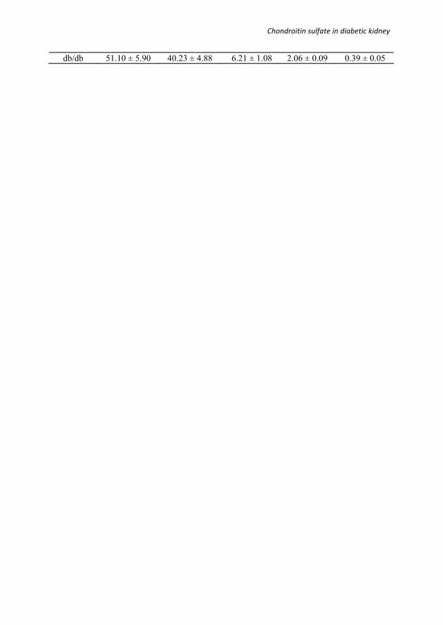
Chondroitin�sulfate�in�diabetic�kidney�
db/db 51.10 ± 5.90 40.23 ± 4.88 6.21 ± 1.08 2.06 ± 0.09 0.39 ± 0.05
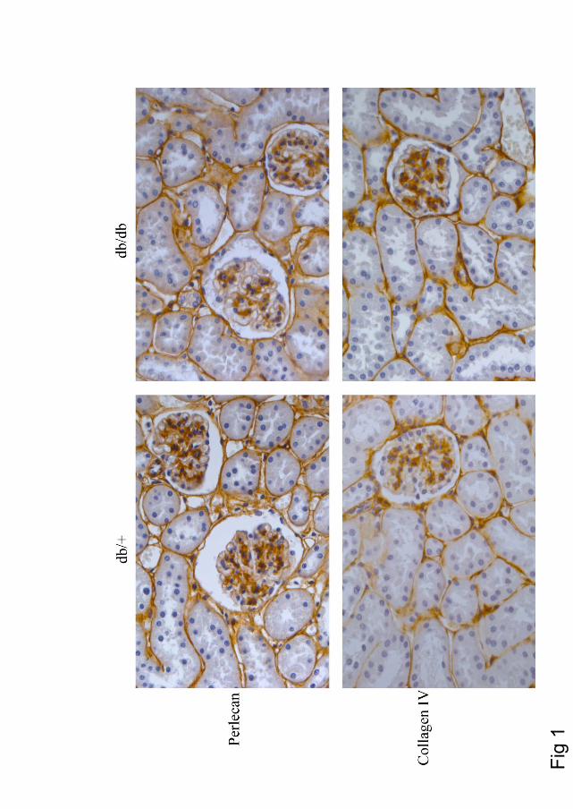
Fig
1
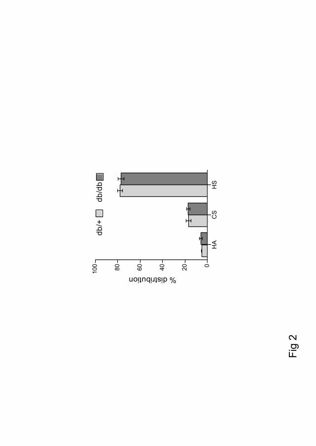
HA
CS
HS
020406080100
db/+
db/d
b
% distribution
Fig
2

D0H
0D
0A0
D0S
0D
0A6
D0A
2D
0S6
D2S
0D
2S6
0510152025db
/+db
/db
Moles x 10-10
Fig
3
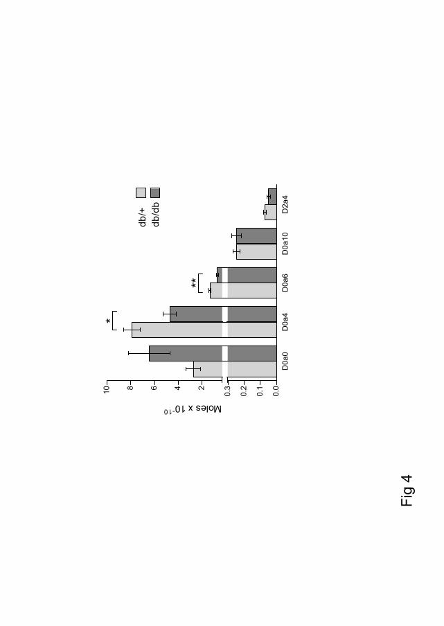
D0a
0D
0a4
D0a
6D
0a10
D2a
40.
0
0.1
0.2
0.3246810
db/+
db/d
b
*
**Moles x 10-10
Fig
4

CS
H
S0.
0
0.2
0.4
0.6
0.8
1.0
db/+
db/d
b
**
No of sulfate / disaccharide
Fig
5
Trine M Reine PhD thesis / Errata 05 feb 2013 Errata Please note the following corrections to the thesis: Paper IV Footnotes Disaccharide notations used for HS: “….D0A0 = ∆UA-GlcNAc, D0S0 = ……..” should read “….D0A0 = ∆UA-GlcNAc, D0A2 = ∆UA2S-GlcNAc, D0S0 = ……..” Figure legends FIGURE 3 “….D0A0 = ∆UA-GlcNAc, D0S0 = ……..” should read “….D0A0 = ∆UA-GlcNAc, D0A2 = ∆UA2S-GlcNAc, D0S0 = ……..” Fig 3 The notation “D0A2” is miss-spelled; it should read “D2A0”