Embed Size (px)
Citation preview
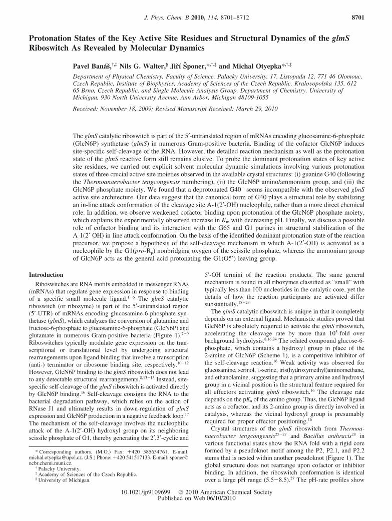
Protonation States of the Key Active Site Residues and Structural Dynamics of the glmSRiboswitch As Revealed by Molecular Dynamics
Pavel Banas,†,‡ Nils G. Walter,§ Jirı Sponer,*,†,‡ and Michal Otyepka*,†,‡
Department of Physical Chemistry, Faculty of Science, Palacky UniVersity, 17. Listopadu 12, 771 46 Olomouc,Czech Republic, Institute of Biophysics, Academy of Sciences of the Czech Republic, KraloVopolska 135, 61265 Brno, Czech Republic, and Single Molecule Analysis Group, Department of Chemistry, UniVersity ofMichigan, 930 North UniVersity AVenue, Ann Arbor, Michigan 48109-1055
ReceiVed: NoVember 18, 2009; ReVised Manuscript ReceiVed: March 29, 2010
The glmS catalytic riboswitch is part of the 5′-untranslated region of mRNAs encoding glucosamine-6-phosphate(GlcN6P) synthetase (glmS) in numerous Gram-positive bacteria. Binding of the cofactor GlcN6P inducessite-specific self-cleavage of the RNA. However, the detailed reaction mechanism as well as the protonationstate of the glmS reactive form still remains elusive. To probe the dominant protonation states of key activesite residues, we carried out explicit solvent molecular dynamic simulations involving various protonationstates of three crucial active site moieties observed in the available crystal structures: (i) guanine G40 (followingthe Thermoanaerobacter tengcongensis numbering), (ii) the GlcN6P amino/ammonium group, and (iii) theGlcN6P phosphate moiety. We found that a deprotonated G40- seems incompatible with the observed glmSactive site architecture. Our data suggest that the canonical form of G40 plays a structural role by stabilizingan in-line attack conformation of the cleavage site A-1(2′-OH) nucleophile, rather than a more direct chemicalrole. In addition, we observe weakened cofactor binding upon protonation of the GlcN6P phosphate moiety,which explains the experimentally observed increase in Km with decreasing pH. Finally, we discuss a possiblerole of cofactor binding and its interaction with the G65 and G1 purines in structural stabilization of theA-1(2′-OH) in-line attack conformation. On the basis of the identified dominant protonation state of the reactionprecursor, we propose a hypothesis of the self-cleavage mechanism in which A-1(2′-OH) is activated as anucleophile by the G1(pro-Rp) nonbridging oxygen of the scissile phosphate, whereas the ammonium groupof GlcN6P acts as the general acid protonating the G1(O5′) leaving group.
Introduction
Riboswitches are RNA motifs embedded in messenger RNAs(mRNAs) that regulate gene expression in response to bindingof a specific small molecule ligand.1-6 The glmS catalyticriboswitch (or ribozyme) is part of the 5′-untranslated region(5′-UTR) of mRNAs encoding glucosamine-6-phosphate syn-thetase (glmS), which catalyzes the conversion of glutamine andfructose-6-phosphate to glucosamine-6-phosphate (GlcN6P) andglutamate in numerous Gram-positive bacteria (Figure 1).7-9
Riboswitches typically modulate gene expression on the tran-scriptional or translational level by undergoing structuralrearrangements upon ligand binding that involve a transcription(anti-) terminator or ribosome binding site, respectively.10-12
However, GlcN6P binding to the glmS riboswitch does not leadto any detectable structural rearrangements.8,13-15 Instead, site-specific self-cleavage of the glmS riboswitch is activated directlyby GlcN6P binding.16 Self-cleavage consigns the RNA to thebacterial degradation pathway, which relies on the action ofRNase J1 and ultimately results in down-regulation of glmSexpression and GlcN6P production in a negative feedback loop.17
The mechanism of the self-cleavage involves the nucleophilicattack of the A-1(2′-OH) hydroxyl group on its neighboringscissile phosphate of G1, thereby generating the 2′,3′-cyclic and
5′-OH termini of the reaction products. The same generalmechanism is found in all ribozymes classified as “small” withtypically less than 100 nucleotides in the catalytic core, yet thedetails of how the reaction participants are activated differsubstantially.18-23
The glmS catalytic riboswitch is unique in that it completelydepends on an external ligand. Mechanistic studies proved thatGlcN6P is absolutely required to activate the glmS riboswitch,accelerating the cleavage rate by more than 105-fold overbackground hydrolysis.8,16,24 The related compound glucose-6-phosphate, which contains a hydroxyl group in place of the2-amine of GlcN6P (Scheme 1), is a competitive inhibitor ofthe self-cleavage reaction.16 Weak activity was observed forglucosamine, serinol, L-serine, tris(hydroxymethyl)aminomethane,and ethanolamine, suggesting that a primary amine and hydroxylgroup in a vicinal position is the structural feature required forall effectors activating glmS riboswitch.16 The cleavage ratedepends on the pKa of the amino group. Thus, the GlcN6P ligandacts as a cofactor, and its 2-amino group is directly involved incatalysis, whereas the vicinal hydroxyl group is presumablyrequired for proper effector positioning.16
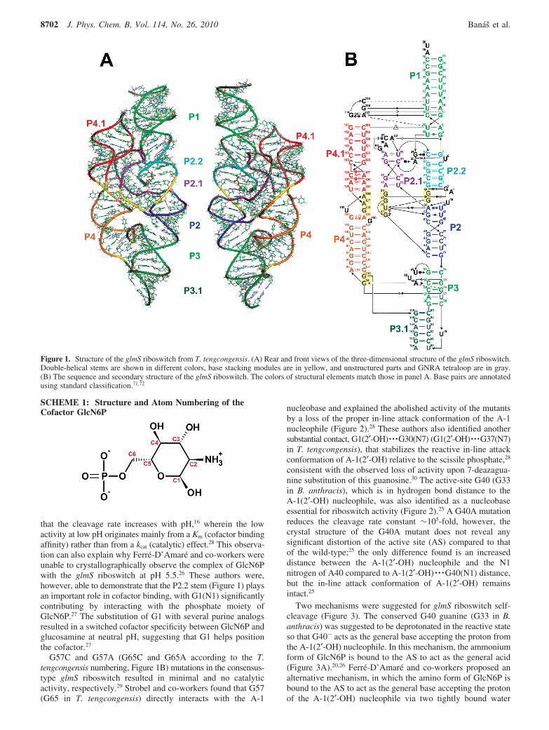
Crystal structures of the glmS riboswitch from Thermoa-naerobacter tengcongensis25-27 and Bacillus anthracis28 invarious functional states show the RNA fold with a rigid coreformed by a pseudoknot motif among the P2, P2.1, and P2.2stems that is nested within another pseudoknot (Figure 1). Theglobal structure does not rearrange upon cofactor or inhibitorbinding. In addition, the riboswitch conformation is identicalover a large pH range (5.5-8.5).27 The pH-rate profiles show
* Corresponding authors. (M.O.) Fax: +420 585634761. E-mail:[email protected]. (J.S.) Phone: +420 541517133. E-mail: [email protected].
† Palacky University.‡ Academy of Sciences of the Czech Republic.§ University of Michigan.
J. Phys. Chem. B 2010, 114, 8701–8712 8701
10.1021/jp9109699 2010 American Chemical SocietyPublished on Web 06/10/2010
that the cleavage rate increases with pH,16 wherein the lowactivity at low pH originates mainly from a Km (cofactor bindingaffinity) rather than from a kcat (catalytic) effect.28 This observa-tion can also explain why Ferre-D’Amare and co-workers wereunable to crystallographically observe the complex of GlcN6Pwith the glmS riboswitch at pH 5.5.26 These authors were,however, able to demonstrate that the P2.2 stem (Figure 1) playsan important role in cofactor binding, with G1(N1) significantlycontributing by interacting with the phosphate moiety ofGlcN6P.27 The substitution of G1 with several purine analogsresulted in a switched cofactor specificity between GlcN6P andglucosamine at neutral pH, suggesting that G1 helps positionthe cofactor.27
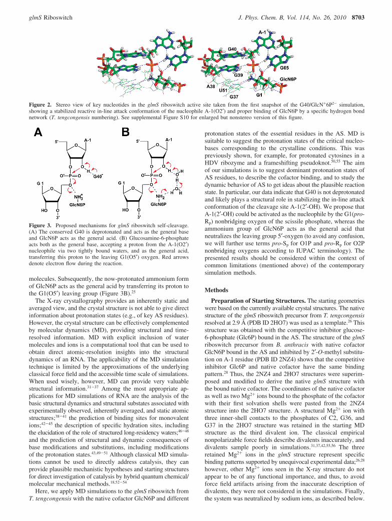
G57C and G57A (G65C and G65A according to the T.tengcongensis numbering, Figure 1B) mutations in the consensus-type glmS riboswitch resulted in minimal and no catalyticactivity, respectively.29 Strobel and co-workers found that G57(G65 in T. tengcongensis) directly interacts with the A-1
nucleobase and explained the abolished activity of the mutantsby a loss of the proper in-line attack conformation of the A-1nucleophile (Figure 2).28 These authors also identified anothersubstantial contact, G1(2′-OH) · · ·G30(N7) (G1(2′-OH) · · ·G37(N7)in T. tengcongensis), that stabilizes the reactive in-line attackconformation of A-1(2′-OH) relative to the scissile phosphate,28
consistent with the observed loss of activity upon 7-deazagua-nine substitution of this guanosine.30 The active-site G40 (G33in B. anthracis), which is in hydrogen bond distance to theA-1(2′-OH) nucleophile, was also identified as a nucleobaseessential for riboswitch activity (Figure 2).25 A G40A mutationreduces the cleavage rate constant ∼105-fold, however, thecrystal structure of the G40A mutant does not reveal anysignificant distortion of the active site (AS) compared to thatof the wild-type;25 the only difference found is an increaseddistance between the A-1(2′-OH) nucleophile and the N1nitrogen of A40 compared to A-1(2′-OH) · · ·G40(N1) distance,but the in-line attack conformation of A-1(2′-OH) remainsintact.25
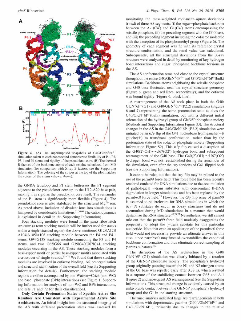
Two mechanisms were suggested for glmS riboswitch self-cleavage (Figure 3). The conserved G40 guanine (G33 in B.anthracis) was suggested to be deprotonated in the reactive stateso that G40- acts as the general base accepting the proton fromthe A-1(2′-OH) nucleophile. In this mechanism, the ammoniumform of GlcN6P is bound to the AS to act as the general acid(Figure 3A).20,26 Ferre-D’Amare and co-workers proposed analternative mechanism, in which the amino form of GlcN6P isbound to the AS to act as the general base accepting the protonof the A-1(2′-OH) nucleophile via two tightly bound water
Figure 1. Structure of the glmS riboswitch from T. tengcongensis. (A) Rear and front views of the three-dimensional structure of the glmS riboswitch.Double-helical stems are shown in different colors, base stacking modules are in yellow, and unstructured parts and GNRA tetraloop are in gray.(B) The sequence and secondary structure of the glmS riboswitch. The colors of structural elements match those in panel A. Base pairs are annotatedusing standard classification.71,72
SCHEME 1: Structure and Atom Numbering of theCofactor GlcN6P
8702 J. Phys. Chem. B, Vol. 114, No. 26, 2010 Banas et al.
molecules. Subsequently, the now-protonated ammonium formof GlcN6P acts as the general acid by transferring its proton tothe G1(O5′) leaving group (Figure 3B).25
The X-ray crystallography provides an inherently static andaveraged view, and the crystal structure is not able to give directinformation about protonation states (e.g., of key AS residues).However, the crystal structure can be effectively complementedby molecular dynamics (MD), providing structural and time-resolved information. MD with explicit inclusion of watermolecules and ions is a computational tool that can be used toobtain direct atomic-resolution insights into the structuraldynamics of an RNA. The applicability of the MD simulationtechnique is limited by the approximations of the underlyingclassical force field and the accessible time scale of simulations.When used wisely, however, MD can provide very valuablestructural information.31-37 Among the most appropriate ap-plications for MD simulations of RNA are the analysis of thebasic structural dynamics and structural substates associated withexperimentally observed, inherently averaged, and static atomicstructures;38-41 the prediction of binding sites for monovalentions;42-45 the description of specific hydration sites, includingthe elucidation of the role of structured long-residency waters;46-48
and the prediction of structural and dynamic consequences ofbase modifications and substitutions, including modificationsof the protonation states.43,49-51 Although classical MD simula-tions cannot be used to directly address catalysis, they canprovide plausible mechanistic hypotheses and starting structuresfor direct investigation of catalysis by hybrid quantum chemical/molecular mechanical methods.18,52-54
Here, we apply MD simulations to the glmS riboswitch fromT. tengcongensis with the native cofactor GlcN6P and different
protonation states of the essential residues in the AS. MD issuitable to suggest the protonation states of the critical nucleo-bases corresponding to the crystalline conditions. This waspreviously shown, for example, for protonated cytosines in aHDV ribozyme and a frameshifting pseudoknot.50,55 The aimof our simulations is to suggest dominant protonation states ofAS residues, to describe the cofactor binding, and to study thedynamic behavior of AS to get ideas about the plausible reactionstate. In particular, our data indicate that G40 is not deprotonatedand likely plays a structural role in stabilizing the in-line attackconformation of the cleavage site A-1(2′-OH). We propose thatA-1(2′-OH) could be activated as the nucleophile by the G1(pro-Rp) nonbridging oxygen of the scissile phosphate, whereas theammonium group of GlcN6P acts as the general acid thatneutralizes the leaving group 5′-oxygen (to avoid any confusion,we will further use terms pro-Sp for O1P and pro-Rp for O2Pnonbridging oxygens according to IUPAC terminology). Thepresented results should be considered within the context ofcommon limitations (mentioned above) of the contemporarysimulation methods.
Methods
Preparation of Starting Structures. The starting geometrieswere based on the currently available crystal structures. The nativestructure of the glmS riboswitch precursor from T. tengcongensisresolved at 2.9 Å (PDB ID 2HO7) was used as a template.26 Thisstructure was obtained with the competitive inhibitor glucose-6-phosphate (Glc6P) bound in the AS. The structure of the glmSriboswitch precursor from B. anthracis with native cofactorGlcN6P bound in the AS and inhibited by 2′-O-methyl substitu-tion on A-1 residue (PDB ID 2NZ4) shows that the competitiveinhibitor Glc6P and native cofactor have the same bindingpattern.28 Thus, the 2NZ4 and 2HO7 structures were superim-posed and modified to derive the native glmS structure withthe bound native cofactor. The coordinates of the native cofactoras well as two Mg2+ ions bound to the phosphate of the cofactorwith their first solvation shells were pasted from the 2NZ4structure into the 2HO7 structure. A structural Mg2+ ion withthree inner-shell contacts to the phosphates of C2, G36, andG37 in the 2HO7 structure was retained in the starting MDstructure as the third divalent ion. The classical empiricalnonpolarizable force fields describe divalents inaccurately, anddivalents sample poorly in simulations.31,37,42,55,56 The threeretained Mg2+ ions in the glmS structure represent specificbinding patterns supported by unequivocal experimental data;26,28
however, other Mg2+ ions seen in the X-ray structure do notappear to be of any functional importance, and thus, to avoidforce field artifacts arising from the inaccurate description ofdivalents, they were not considered in the simulations. Finally,the system was neutralized by sodium ions, as described below.
Figure 2. Stereo view of key nucleotides in the glmS riboswitch active site taken from the first snapshot of the G40/GlcN+6P2- simulation,showing a stabilized reactive in-line attack conformation of the nucleophile A-1(O2′) and proper binding of GlcN6P by a specific hydrogen bondnetwork (T. tengcongensis numbering). See supplemental Figure S10 for enlarged but nonstereo version of this figure.
Figure 3. Proposed mechanisms for glmS riboswitch self-cleavage.(A) The conserved G40 is deprotonated and acts as the general baseand GlcN6P acts as the general acid. (B) Glucosamine-6-phosphateacts both as the general base, accepting a proton from the A-1(O2′)nucleophile via two tightly bound waters, and as the general acid,transferring this proton to the leaving G1(O5′) oxygen. Red arrowsdenote electron flow during the reaction.
glmS Riboswitch J. Phys. Chem. B, Vol. 114, No. 26, 2010 8703

Seven starting structures differing in protonation of the im-portant acid-base groups in the AS were prepared on the basisof this structure. These include systems with both the aminoand ammonium form in combination with both the singlyprotonated monocharged phosphate and deprotonated double-charged phosphate of GlcN6P. The abbreviation GlcN06P2-
denotes the amino form of GlcN6P with deprotonated double-charged phosphate, and GlcN+6P2- and GlcN+6P- stand forthe ammonium form of GlcN6P with deprotonated double-charged and singly protonated monocharged phosphate, respec-tively. The abbreviation GlcN6P generally represents theglucosamine-6-phosphate without specification of its protonationstates. Two simulations were carried out with deprotonatedguanine G40- in the AS, and the remaining five were executedwith a canonical G40. Thus, the following simulations wereprepared (Table 1): G40-/GlcN+6P2-, G40-/GlcN+6P-, G40/GlcN06P2-, G40/GlcN+6P2-, and three independent simulationsof G40/GlcN+6P- differing in the starting orientation of thehydroxyl group at phosphate moiety of the GlcN6P cofactor.The simulation in which the hydroxyl group was initiallyoriented toward the G1 nucleobase is labeled as G40/GlcN+6P-(G1). G40/GlcN+6P-(P2.2) represents the simulationwith the hydroxyl group oriented toward the P2.2 stem, whereasthe hydroxyl group of simulation G40/GlcN+6P-(bulk) wasinitially exposed to the bulk solvent (Supporting Information,Figure S3). In addition, one reference structure was preparedwith a ligand-free AS and a canonical G40 (G40/free). Thestructural triple inner-shell bound Mg2+ ion was retained, andthe other two Mg2+ ions neighboring the cofactor were deleted.
Molecular Dynamics Simulations. All structures wereneutralized with Na+ counterions (Na+ radius 1.868 Å and welldepth 0.0028 kcal/mol) that were iteratively placed into theminima of the electrostatic potential calculated on a grid withspacing 1 Å using the program Leap of AMBER 9.0.57 Thestructures with a ligand in the AS contained three Mg2+ ionsand 136-138 Na+ ions, depending on the charge of the ASresidues. The reference structure contained one structural Mg2+
ion and 140 Na+ ions. All structures were immersed in arectangular box with at least a 10-Å-thick layer of TIP3P watermolecules around the solute. The size of the boxes was ∼130× 80 × 70 Å3. The complete structures contained ∼55 000atoms, including ∼6500 water molecules. The overall concen-tration of monovalent ions was ∼0.33 mol/L, which is entirely
sufficient to provide stable RNA simulation trajectories withrealistic local counterion accumulation around the solutemolecule.
The whole RNA-solvent system was minimized prior to thesimulations as follows. Minimization of the riboswitch hydrogenatoms was followed by minimization of counterions and watermolecules. Subsequently, the riboswitch was constrained, andsolvent molecules with ions were allowed to move during a 10ps long MD run. The nucleobases were allowed to relax inseveral minimization runs with decreasing force constantsapplied to the backbone phosphate atoms. After full relaxation,each system was slowly heated to 298.15 K over 100 ps using2-fs time steps and the NpT conditions. The simulations wereevolved under periodic boundary conditions in the NpTensemble (298.15 K, 1 atm) with 2-fs time steps.
The particle-mesh Ewald method was used to calculateelectrostatic interactions, and a 9.0-Å cutoff was applied forLennard-Jones interactions. The SHAKE algorithm was appliedto all bonds containing hydrogen atoms. The PMEMD moduleof AMBER 9.057 with the Cornell et al. force field parm9958,59
was used for all simulations. The length of each simulation was20 ns, except for the G40/GlcN+6P2- and G40/free simulations,which were expanded to 50 ns. The parameters of all nonstand-ard residues were determined by the RESP procedure of Cornellet al.60 The ab initio calculations required for the parametrizationof GlcN6P in various protonation states and deprotonatedguanine were carried out using Gaussian03 (see the SupportingInformation for details and parameters).61,62
Results
We carried out MD simulations of the glmS riboswitch tocharacterize cofactor binding and to study the dynamic behaviorof the glmS riboswitch as a whole. Nonetheless, the main aimwas to elucidate the dominant protonation states of the key ASresidues and to obtain insights about plausible reaction mech-anisms. Three different groups in the AS were identified as ofan uncertain protonation state: (i) the N1 nitrogen of guanineG40 that was suggested to be deprotonated in a precursor stateto act as the general base,26,28 (ii) the amino group of GlcN6Pthat was suggested to act as either a general base26 or generalacid28 during cleavage, and (iii) the phosphate moiety of GlcN6P(see Methods and Table 1 for details).
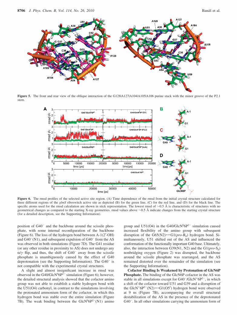
Basic Structural Dynamics Shows Extremely RigidPseudoknot Core of glmS Riboswitch. The structural dynamicsand flexibility of the glmS riboswitch were monitored asB-factors of the glmS nucleotides. The pseudoknot core formedby the P2, P2.1, and P2.2 stems and including the AS wasextremely rigid in all simulations. This is in agreement withthe experimentally observed low B-factors in the crystalstructures.26,28 By contrast, the P1, P3, P3.1, and P4 stemsrepresent more flexible regions of the RNA fold with cor-respondingly high B-factors (Figure 4). The rigidity of thepseudoknot core likely originates from a structural stabilizationof the core arrangement by the coaxial P4 and P4.1 stems. Theystabilize the pseudoknot fold by two interactions: (i) a ribosezipper motif63 formed between the GNRA tetraloop64 closingP4.1 and the P1 stem, which contains a type I A-minorinteraction65 between A117 and the C10dG31 base pair, and(ii) the oblique interaction of the G128|A127|A104|A105|A106 purine stack with the minor groove of P2.1(Figure 5).26 This salient tertiary interaction is entirely stableand very rigid in all simulations and might represent the keystabilizing interaction of the P2.1 stem fold. The flexibility ofthe P1 stem is nonuniform. The ribose zipper motif between
TABLE 1: Overview of the MD Simulations PerformedHerea
simulationname cofactor G40
GlcN6Paminogroup
GlcN6Pphosphate
simulationlength(ns)
G40-/GlcN+6P2- yes -1 +1 -2 20G40-/GlcN+6P- yes -1 +1 -1 20G40/GlcN06P2- yes 0 0 -2 20G40/GlcN+6P2- yes 0 +1 -2 50G40/GlcN+6P-
(G1)byes 0 +1 -1 20
G40/GlcN+6P-
(P2.2)byes 0 +1 -1 20
G40/GlcN+6P-
(bulk)byes 0 +1 -1 20
G40/free no 0 50
a The presence of ligand in the active site and the charge of theacid-base groups differing in the protonation state are indicated.b The labels “G1”, “P2.2”, or “bulk” in parentheses indicate initialorientation of the hydroxyl of the GlcN+6P- phosphate toward G1,P2.2 stem, and bulk solvent, respectively.
8704 J. Phys. Chem. B, Vol. 114, No. 26, 2010 Banas et al.
the GNRA tetraloop and P1 stem buttresses the P1 segmentadjacent to the pseudoknot core up to the U12-A29 base pair,making it as rigid as the pseudoknot core itself. The remainderof the P1 stem is significantly more flexible (Figure 4). Thepseudoknot core is also stabilized by the structural Mg2+ ion.As noted above, inclusion of divalent ions into simulations ishampered by considerable limitations.31,56,66 The cation dynamicsis explained in detail in the Supporting Information).
Four stacking modules were found in the glmS riboswitchstructure (a term stacking module will be further used for stackswithin a single-stranded region): the above-mentioned G128|A127|A104|A105|A106 stacking module between the P4 and P4.1stems, G94|G138 stacking module connecting the P3 and P4stems, and two G65|G66 and G39|G40|U67|G41 stackingmodules occurring in the AS. These stacking modules form abase-zipper motif or partial base-zipper motifs associated witha crossover of single strands.67-70 We found that these stackingmodules are involved in cofactor binding, AS preorganizationand structural stabilization of the overall fold (see the SupportingInformation for details). Furthermore, the stacking moduleregions are often accompanied by non-Watson-Crick (non-WC)and base-phosphate (BPh) interactions (see Figure 1, Support-ing Information for analysis of non-WC and BPh interactions,and refs 71 and 72 for their classification).
Only Certain Protonation States of Specific Active SiteResidues Are Consistent with Experimental Active SiteArchitecture. An initial insight into the structural integrity ofthe AS with different protonation states was assessed by
monitoring the mass-weighted root-mean-square deviations(rmsd) of three AS segments: (i) the sugar-phosphate backbonebetween the A-1(C4′) and G1(C4′) atoms encompassing thescissile phosphate, (ii) the preceding segment with the G40 base,and (iii) the preceding segment including the cofactor moleculewith the exception of its phosphomethyl group (Figure 6). Thegeometry of each segment was fit with its reference crystalstructure conformation, and the rmsd value was calculated.Subsequently, all the structural deviations from the X-raystructure were analyzed in detail by monitoring of key hydrogenbond interactions and sugar-phosphate backbone torsions inthe AS.
The AS conformation remained close to the crystal structurethroughout the entire G40/GlcN+6P2- and G40/GlcN+6P-(bulk)simulations. Backbone atoms neighboring the scissile phosphateand G40 base fluctuated near the crystal structure geometry(Figure 6, green and red lines, respectively), and the cofactorwas bound tightly (Figure 6, black line).
A rearrangement of the AS took place in both the G40/GlcN+6P-(G1) and G40/GlcN+6P-(P2.2) simulations (Figures6 and 7) representing the same protonation state as does theG40/GlcN+6P-(bulk) simulation, but with a different initialorientation of the hydroxyl group of GlcN6P phosphate moiety(Methods and Supporting Information Figure S3). The structuralchanges in the AS in the G40/GlcN+6P-(P2.2) simulation wereinitiated by an R/γ flip of the G41 nucleobase from gauche(-)/gauche(+) to trans/trans conformation, rather than by theprotonation state of the cofactor phosphate moiety (SupportingInformation Figure S2). This R/γ flip caused a disruption ofthe G40(2′-OH) · · ·U67(O2′) hydrogen bond and subsequentrearrangement of the G40 base. The G40(2′-OH) · · ·U67(O2′)hydrogen bond was not reestablished during the remainder ofthe simulation, even after the R/γ torsions of G41 flipped back(see the Supporting Information).
It cannot be ruled out that the R/γ flip may be related to theuse of the parm99 force field. This force field has been recentlyrendered outdated for DNA simulations due to the accumulationof pathological γ-trans substates with concomitant B-DNAdegradation in longer simulations and has been replaced by theparmbsc0 force field.73 However, the R/γ flip issue of parm99is assumed to be irrelevant for RNA simulations in which theR/γ t/t substates do occur in X-ray structures and do notaccumulate during MD simulations in a manner that woulddestabilize the RNA structure.42,74,75 Nevertheless, we still cannotrule out that the parm99 force field modestly exaggerates thepropensity to adopt the γ-trans substate for this particularnucleotide. Note that even an application of the parmbsc0 forcefield would not necessarily provide an ultimate answer in thiscase, since parmbsc0 may instead overstabilize the canonicalbackbone conformation and thus eliminate correct sampling ofγ-trans substates.76
The disruption of the AS architecture in the G40/GlcN+6P-(G1) simulation was clearly initiated by a rotationof the GlcN6P phosphate moiety. The phosphate’s hydroxylgroup originally pointing toward the N1 and N2 nitrogen atomsof the G1 base was repelled early after 0.38 ns, which resultedin a rupture of the stabilizing contact between G65 and A-1(Figure 2) and subsequent AS rearrangement (see the SupportingInformation). This structural change is evidently caused by anunfavorable contact between the GlcN6P phosphate’s hydroxylgroup and the G1 in the starting structure.
The rmsd analysis indicated large AS rearrangements in bothsimulations with deprotonated guanine (G40-/GlcN+6P2- andG40-/GlcN+6P-), primarily due to changes in the relative
Figure 4. (A) The superimposed snapshots of G40/GlcN+6P2-
simulation taken at each nanosecond demonstrate flexibility of P1, P3,P3.1 and P4 stems and rigidity of the pseudoknot core. (B) The thermalB-factors of the backbone atoms of each residue calculated from MDsimulation (for comparison with X-ray B-factors, see the SupportingInformation). The coloring of the stripes at the top of the plot matchesthe colors of the stems (shown above).
glmS Riboswitch J. Phys. Chem. B, Vol. 114, No. 26, 2010 8705
position of G40- and the backbone around the scissile phos-phate, with some internal reconfiguration of the backbone(Figure 6). The loss of the hydrogen bond between A-1(2′-OH)and G40-(N1), and subsequent expulsion of G40- from the ASwas observed in both simulations (Figure 7D). The G41 residue(or any other residue in proximity to AS) does not undergo anyR/γ flip, and thus, the shift of G40- away from the scissilephosphate is unambiguously caused by the effect of G40deprotonation (see the Supporting Information). The G40- isnot compatible with the experimental crystal structures.
A slight and almost insignificant increase in rmsd wasobserved in the G40/GlcN06P2- simulation (Figure 6); however,the detailed structural analysis showed that the cofactor aminogroup was not able to establish a stable hydrogen bond withthe U51(O4) carbonyl, in contrast to the simulations involvingthe protonated ammonium form of the cofactor, in which thishydrogen bond was stable over the entire simulation (Figure7B). The weak binding between the GlcN06P2-(N1) amino
group and U51(O4) in the G40/GlcN06P2- simulation causedincreased flexibility of the amino group with subsequentdisruption of the G65(N2) · · ·G1(pro-Rp) hydrogen bond. Si-multaneously, U51 shifted out of the AS and influenced theconformation of the functionally important G40 base. Ultimately,also, the interaction between G39(N1, N2) and the G1(pro-Sp)nonbridging oxygen (Figure 2) was disrupted, the backbonearound the scissile phosphate was rearranged, and the ASremained distorted over the remainder of the simulation (seethe Supporting Information).
Cofactor Binding Is Weakened by Protonation of GlcN6PPhosphate. The binding of the GlcN6P cofactor in the AS wasstable in all simulations except for G40-/GlcN+6P2-, in whicha shift of the cofactor toward U51 and G39 and a disruption ofthe GlcN+6P2-(N2) · · ·G1(O5′) hydrogen bond were observedat 9 ns (Figure 7B), accompanying the overall structuraldestabilization of the AS in the presence of the deprotonatedG40-. In all other simulations carrying the ammonium form of
Figure 5. The front and rear view of the oblique interaction of the G128|A127|A104|A105|A106 purine stack with the minor groove of the P2.1stem.
Figure 6. The rmsd profiles of the selected active site region. (A) Time dependence of the rmsd from the initial crystal structure calculated forthree different regions of the glmS riboswitch active site as depicted (B) for the green line, (C) for the red line, and (D) for the black line. Thespecific atoms used for the rmsd calculation are shown in stick representation. The lowest rmsd of ∼0.5 Å is characteristic of structures with nogeometrical changes as compared to the starting X-ray geometries. rmsd values above ∼0.5 Å indicate changes from the starting crystal structure(for a detailed description, see the Supporting Information).
8706 J. Phys. Chem. B, Vol. 114, No. 26, 2010 Banas et al.
GlcN6P, the cofactor remained in the conformation correspond-ing to its crystallographic binding pattern with a stable hydrogenbond between the ammonium group of the cofactor and theG1(O′5) backbone oxygen. This hydrogen bond is assumed tobe important in self-cleavage of the glmS riboswitch.16 Inaddition, the GlcN6P ammonium group was stabilized in itsposition by a hydrogen bond to the U51(O4) carbonyl (Figure7B), whereas the third hydrogen of the ammonium group wasbound to a water molecule.
The stabilizing interaction between U51(O4) and the cofactorwas absent in the G40/GlcN06P2- simulation (Figure 7B), wherethe cofactor bore the unprotonated amino group incapable ofsuch a hydrogen bond, causing a shift of the amino group towardthe nonbridging oxygens of the scissile phosphate and subse-quent AS rearrangement.
A firm and stable hydrogen bond was observed between theGlcN6P C1-OH hydroxyl group and the G1(pro-Rp) oxygenin all simulations. This interaction was supported by two
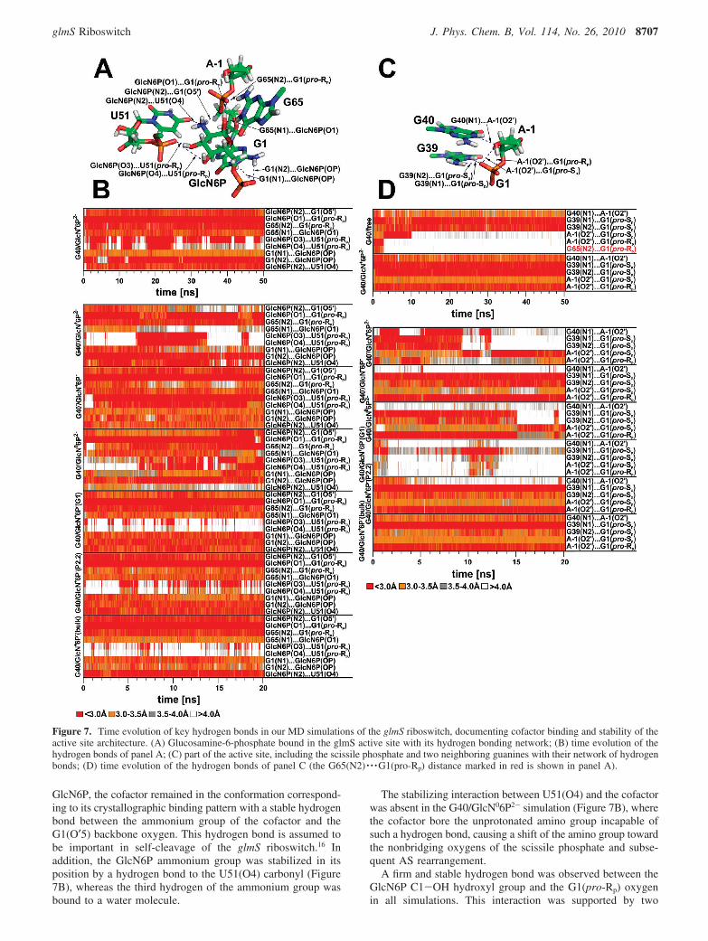
Figure 7. Time evolution of key hydrogen bonds in our MD simulations of the glmS riboswitch, documenting cofactor binding and stability of theactive site architecture. (A) Glucosamine-6-phosphate bound in the glmS active site with its hydrogen bonding network; (B) time evolution of thehydrogen bonds of panel A; (C) part of the active site, including the scissile phosphate and two neighboring guanines with their network of hydrogenbonds; (D) time evolution of the hydrogen bonds of panel C (the G65(N2) · · ·G1(pro-Rp) distance marked in red is shown in panel A).
glmS Riboswitch J. Phys. Chem. B, Vol. 114, No. 26, 2010 8707
additional G65(N1) · · ·GlcN6P(C1-OH) and G63(N2) · · ·G1(pro-Rp) hydrogen bonds (Figure 7B) which, however, were com-pletely lost in simulation G40-/GlcN+6P2- and partially insimulations G40-/GlcN+6P- and G40/GlcN06P2- accompaniedby a rearrangement of the AS. The local interaction of G65,the scissile phosphate, and the C1-OH hydroxyl group ofGlcN6P represents a compact binding pattern, which stabilizesthe contact between the cofactor ammonium group and G1(O5′).
A strong bifurcated hydrogen bond between G1(N1, N2) andthe cofactor phosphate group was observed in all simulationswith a deprotonated double-charged phosphate. The sameinteraction was observed in simulations with a singly protonatedmonocharged GlcN6P phosphate (one with deprotonated G40-
and three with canonical G40). However, an early reorientationof the cofactor phosphate group was observed in the G40-/GlcN+6P- and G40/GlcN+6P-(G1) simulations that started witha phosphate hydroxyl group oriented toward the G1 base. Thishydroxyl group was exposed to the bulk solvent during the firstnanosecond of the respective simulations; that is, the hydroxylgroup switched to the conformation corresponding to the startingstructure of the G40/GlcN+6P-(bulk) simulation. G40/GlcN+-6P-(P2.2) was the only simulation, in which the cofactorphosphate hydroxyl group was not oriented toward the bulksolvent but, rather, interacted directly with the G64 or G65 bases,whereas in other simulations, the GlcN6P phosphate interactedwith guanines G64 and G65 via a solvated Mg2+ ion.
Partially occupied hydrogen bonds were observed betweenthe cofactor hydroxyl groups C3-OH and C4-OH and the U51(pro-Rp) nonbridging oxygen. These hydrogen bonds arefluctuating and do not appear to be essential for GlcN6P binding(Figure 7).
In summary, both a deprotonated G40- and an unprotonatedamino form of cofactor (GlcN06P instead of GlcN+6P) resultin significant destabilization of cofactor binding. Further,protonation of the cofactor phosphate moiety (GlcN6P- insteadof GlcN6P2-) likely negatively affects the stability of theinteraction between the cofactor phosphate and the G1 nucleo-base. More specifically, we note that the GlcN+6P- with thephosphate hydroxyl group oriented toward G1 represents anunstable binding motif. By contrast, the cofactor was firmlybound in the active site when the GlcN+6P- phosphate hydroxylgroup was oriented toward the bulk solvent. Thus, protonationof the GlcN6P phosphate moiety does not prevent cofactorbinding for an appropriate geometry but restricts a conforma-tional variability of the cofactor, which should be accompaniedby an entropic penalty for cofactor binding.
Structural Dynamics of the glmS Riboswitch in theAbsence of Cofactor Show That A-1(2′-OH) In-Line AttackConformation Requires Cofactor Binding. Reference 50-ns-long simulation of the ligand-free glmS riboswitch (withoutGlcN6P) was started from the crystal structure geometry (i.e.,the structure with bound GlcN6P) and revealed profoundchanges of the AS. This indicates that cofactor binding is neededfor properly structuring the AS. The two-step rearrangement ofthe AS is described in the Supporting Information.
In contrast to the AS occupied by the cofactor, the stableligand-free AS arrangement lacks the G65(N2) · · ·G1(pro-Rp)hydrogenbondand the in-lineattackconformationofA-1(2′-OH)(Supporting Information Figure S5 and Table 2). On the otherhand, the base-phosphate interaction between G39(N1) and theG1(pro-Sp) nonbridging oxygen of the scissile phosphate as wellas the G65(N2) · · ·A-1(N3) and G65(2′-OH) · · ·A-1(N1) hydro-gen bonds were preserved (Supporting Information Figure S4).It seems that the position of the G1 base controls the conforma-
tion of the backbone between A-1 and G1. We suggest thatcofactor binding can affect the position of G1 and, thus, is vitalfor a proper local conformation of the scissile phosphate.Furthermore, cofactor binding can help to establish the interac-tion between the scissile phosphate and G65 and thus stabilizesthe A-1 nucleotide in a way that supports the A-1(2′-OH) in-line conformation.
Discussion
We have carried out a set of explicit solvent MD simulationsof the glmS riboswitch with the aim to elucidate its overallstructural dynamics, the arrangement of its AS in the reactivestate, and its major protonation state.
Because the X-ray crystallography is not able to identifyproton positions, the MD simulation can be employed to assigna protonation state, which corresponds to the state reflected inthe X-ray structure. Here, such identification is based onstructural deviations of AS residues from the X-ray structurein MD simulation applying various protonation states of ASresidues. It can be expected that the protonation state corre-sponding to that observed in the X-ray structure (here resolvedat pHs ranging from 5.5 to 8.5) would exhibit the smalleststructural deviation in MD. This represents an indirect way howto assign a protonation state that likely corresponds to theexperimentally observed crystal structure.
A direct theoretical method to address the protonation stateand estimate the pKa of a given titrable group is a constant-pHmolecular dynamics.77-79 However, the constant-pH MD meth-ods that are used and widely tested for proteins77-80 typicallyutilize implicit solvent models, which reasonably limits theirapplicability in the case of nucleic acids. We used the constant-pH MD as implemented in AMBER57,77 to estimate pKa valuesof three discussed titrable groups of glmS riboswitch (see theSupporting Information). Unfortunately, we observed a rapidand massive degradation of the glmS riboswitch global foldduring the first ∼10 ps of the thermalization phase in simulationsusing the implicit solvent (see the Supporting Information).Thus, we restrained the structure of the glmS riboswitch to itscrystal-like geometry and estimated the pKa using constant-pHMD. The sign of the observed pKa shifts was in agreement withthe conclusions presented in this work; however, the absolutevalue of pKa shifts was severely overestimated (see SupportingInformation). The overestimated pKa shifts together with therapid degradation of RNA structure in implicit solvent showthat the implicit solvent methods (at least in current implemen-tation) are not able to efficiently screen out the electrostatics ofcomplex folded RNA molecules. It seems that the RNAmolecule remains a challenge for MD simulations in combina-tion with implicit solvent methods.
Overall Dynamics and Stability. Our simulations highlighta significant rigidity of the pseudoknot core of the glmS
TABLE 2: The Mean Values of A-1(2′-O) · · ·G1(P)-G1(O5′)In-Line Attack Angle (IAA) in Degrees with StandardDeviations in All Presented Simulations
simulation IAA simulation IAA
G40-/GlcN+6P2- a,b 155 ( 10 G40/GlcN+6P- (G1)a,c 135 ( 10G40-/GlcN+6P- a 155 ( 10 G40/GlcN+6P- (P2.2)a 165 ( 5G40/GlcN06P2- a,b 160 ( 10 G40/GlcN+6P- (bulk) 170 ( 5G40/GlcN+6P2- 170 ( 5 G40/freed 130 ( 10
a Poor or missing interaction between G40 and A-1(2′-OH).b Poor or missing interaction between G39 and G1(pro-Sp).c Disruption of G65(N2) · · ·A-1(N3) hydrogen bond. d Missingcofactor binding.
8708 J. Phys. Chem. B, Vol. 114, No. 26, 2010 Banas et al.
riboswitch, formed by the P2, P2.1, and P2.2 stems, which iscontrasted by the flexible outer parts, including the top of theP1 stem and the pseudoknot formed by stems P3 and P3.1. Thecore appears to be made rigid by interactions with coaxial P4and P4.1 stems; namely, oblique interaction of the G128|A127|A104|A105|A106 stacking module with the minor groove ofthe P2.1 stem26 and the interaction of the GNRA tetraloop atthe tip of the P4.1 stem with the minor groove of the P1 stemthat forms a ribose-zipper motif with a class I A-minorinteraction. Our simulations further suggest that the stackingmodules made by stacked single strands or base-zipper motifsplay an important role in structural stabilization, AS preorga-nization, and cofactor binding. There are several base-phosphateinteractions that are stable in MD simulations. These interactionsare often connected with the noncanonical segments of the glmSriboswitch, especially the stacking modules, and likely play animportant role in the structural stabilization of its noncanonicalparts. All interactions above are very stable in MD simulations.
Protonation State and Proposed Functional Role of G40.Simulations of the glmS riboswitch were used to identify thelikely dominant protonation state of three critical moieties inthe AS. G40 was the first moiety of the uncertain protonationstate, which was previously identified by biochemical data toplay an essential role in glmS self-cleavage.24,27 It was furthersuggested that the deprotonated G40- acts as the general baseduring self-cleavage;25,26,28,81 however, expulsion of the G40-
base from the AS and subsequent AS distortion were observedin both simulations with a deprotonated G40-. The deprotonatedG40- appears to be incompatible with the glmS AS architecturefound in all available crystallographic studies. The same hasbeen observed in simulations with the deprotonated form of G8,which was suggested to be involved in the hairpin ribozymeself-cleavage.89 A structural rather than catalytic role of G8 intransition state stabilizing the self-cleavage reaction in thehairpin ribozyme was suggested by York et al. who proposedthat the tautomeric form of the active site guanine G8 is notlikely involved in the reaction chemistry of hairpin ribozyme.52,82
We found that G40 structurally stabilizes the A-1(2′-OH) in-line attack conformation relative to the scissile phosphate, andthus, we suggest that G40 is important for structural andelectrostatic stabilization of the reactive state rather than directlyacting in the catalytic reaction as the general base.
Still, it is possible that the deprotonated form of G40- maybe involved in catalysis; however, in such a case, it would likelycorrespond to a low-populated, transient, reactive state. In the20+ ns scale simulations, the AS of systems with G40- isentirely unstable and rearranges swiftly to very differentgeometries. Assuming that the contact between A-1(O2′) andG40- was disrupted at the beginning of the simulation and wasnot reestablished for even one single 2-ps-long snapshot, a roughestimate would yield an occupation of the deprotonated G40-
in the reactive state of less than 0.01%. The corresponding ∆Gcorrection for the low-populated reactive state would be higherthan 6 kcal/mol. In addition, the unperturbed pKa of guanine of9.283 suggests a free energy correction for formation of a guanineanion of 3 kcal/mol at pH 7. Thus, the reaction mechanism withG40- acting as the general base would have to be chemicallyfavored in the transition state stabilization by at least 9 kcal/mol over other alternative pathways (with significantly populatedreactive state). We cannot ultimately rule out that longersimulations may rearrange the AS into a suitable, morepopulated geometry involving G40- that would be separatedfrom the presently sampled geometries by an energy barrier andthus not immediately accessible. However, we have no indica-
tion what type of geometry that would be and consider thispossibility as less likely. In addition, it is common in thehydrolysis of the sugar-phosphate backbone that the departureof the O5′-alcoholate from the pentahedral intermediate is therate-limiting step, rather than the 2′-OH nucleophilic attack.84,85
Thus, it appears unlikely that the increased basicity of G40-
affects the reaction rate sufficiently to counteract the 9 kcal/mol penalty in ∆G arising from a low-populated G40- reactivestate and rare protonation state of G40-.
Mechanistic and structural experiments have previously foundthat a G40A mutation abrogates most of the catalytic activityof the glmS riboswitch but does not extensively affect thearrangement of the AS in both T. tengcongensis and B.anthracis.25,81 The only difference between G40A and wild-type AS conformation is a slight increase in the distance betweenthe N1 of residue 40 and the A-1(O2′) nucleophile, whereasthe in-line conformation remains intact.25,81 One explanation isthat G40 is deprotonated and acts as the general base.25,26,28,81
Alternatively, donation of the hydrogen bond by canonical G40to A-1(O2′) could be critical for the precise positioning of thenucleophile to a reactive in-line attack conformation or forelectrostatic stabilization of the attacking A-1(2′-OH), whereasan in-line like position in G40A mutant is nonproductive.Preliminary MD simulations of the G40A mutant (data notshown) suggest that the mutation, consistent with the crystalstructures, causes only slight structural changes in the AS. TheA40 remains locked in the same position as G40 of the wildtype, but A-1(O2′) does not establish any hydrogen bond withthe A40(N1). Furthermore, the lack of a stabilizing G40(N1) · · ·A-1(O2′) hydrogen bond allows tighter binding of A-1(O2′) tothe G1(pro-Rp) nonbridging oxygen as compared with the wildtype MD simulations.
On the basis of the above-mentioned arguments, we suggestthat G40 is not directly involved in the chemical reaction but,rather, plays a role in electrostatic stabilization of the attackingA-1(2′-OH) nucleophile (i.e., in electrostatic transition statestabilization). The same role has also been proposed for the G8involved in the hairpin ribozyme self-cleavage;49,52,82 however,further clarification of the inhibition effect of G40A mutationis still required.
GlcN6P: Amino or Ammonium Group? The secondchemical group in the AS with an uncertain protonation stateis the amino/ammonium group of GlcN6P. The hydrogen bondbetween the cofactor ammonium group and U51(O4) was stablein all simulations with the ammonium form of GlcN6P;however, this hydrogen bond was missing in the simulation inwhich the cofactor contained the (uncharged) amino group,consistently with the reduced proton donor capability of theamino as compared with the ammonium group. Because thehydrogen bond contact between U43(O4) (U51(O4) in T.tengcongensis) and GlcN6P nitrogen was observed in crystalstructures of B. anthracis with native GlcN6P cofactor,28 theloss of this hydrogen bond detected in our MD simulations withthe amino form of GlcN6P shows that the amino form ofGlcN6P is not consistent with X-ray structures. Since the crystalstructures of B. anthracis were obtained at pH 6.828 and theunperturbated pKa of the GlcN6P equals 8.2,29 our data do notsupport a significant pKa shift of this amino/ammonium groupin the active site. The simulations suggest that the ammoniumform of the cofactor is preferentially bound to the AS atphysiological pH and is bound more tightly as compared withthe amino form. Thus, the ammonium form of the cofactor iscapable of tightly binding G1(O5′) and is in a suitable positionto act as the general acid of the reaction.
glmS Riboswitch J. Phys. Chem. B, Vol. 114, No. 26, 2010 8709
The Protonation of GlcN6P Phosphate Weakened Cofac-tor Binding. The third moiety of the uncertain protonation stateis the phosphate of GlcN6P. From the presented simulations, itwas not possible to clearly suggest whether the phosphate groupof the cofactor bound to the AS is deprotonated (double-charged)or singly protonated (monocharged). Both protonation formsappear to remain bound to the AS, yet tighter binding of thecofactor phosphate moiety with G1 base was observed in thedeprotonated double-charged, as compared with the singlyprotonated monocharged, form. In addition, the binding of asingly protonated phosphate may be disadvantageous becausethe phosphate hydroxyl group cannot bind to the G1 base andinstead prefers the orientation to the bulk solvent. This confor-mational restriction is likely associated with an additionalentropic penalty for binding the GlcN6P with the protonatedphosphate moiety. The increase in Km with decreasing pH28
supports our observation that GlcN6P with a protonatedphosphate binds more weakly as compared with the double-charged deprotonated phosphate of GlcN6P. A recent kineticanalysis shows that the pH dependence of cofactor binding isconsistent with the pKa of the GlcN6P phosphate moiety,suggesting that the protonation of the phosphate moiety ofGlcN6P may inhibit cofactor binding.81 This experimentalfinding, together with the MD data, lends support to the notionthat GlcN6P with a fully deprotonated phosphate binds mostefficiently to the glmS riboswitch.
Possible Role of G65. Biochemical analysis has suggestedan essential role for the G65 base (or equivalent G57 in B.anthracis) in catalysis.28,29 G65 was suggested to stabilize aproper conformation of the A-1 base and to neutralize thenegative charge on the G1(pro-Rp) nonbridging oxygen of thescissile phosphate. We found that both these interactions are,indeed, required for stabilization of the AS in a reactive A-1(2′-OH) in-line attack conformation. In addition, we suggest that afurther crucial role of G65 is to stabilize the hydrogen bondbetween the C1-OH group of GlcN6P and the G1(pro-Rp)oxygen that in turn helps maintain the hydrogen bond betweenthe cofactor ammonium group and G1(O5′). The GlcN6P(C1-OH) · · ·G1(pro-Rp) contact is the most rigid hydrogen bond inthe AS of the glmS riboswitch. The G65 base stabilizes thishydrogen bond and forms the core of the GlcN6P binding motif.Our findings are consistent with the observation that ethanol-amine is the minimal motif binding to the AS of the glmSriboswitch to activate self-cleavage.16 In addition, on the basisof the simulation with ligand-free AS, we suggest that cofactorbinding is crucial for establishing the interactions among thescissile phosphate, the cofactor, and G65 that structurally supportthe A-1(2′-OH) in-line attack conformation. In other words, wesuggest that there are two roles of the GlcN6P cofactor: (i) theA-1(2′-OH) in-line attack conformation is induced by cofactorbinding, and (ii) the ammonium group of the cofactor acts asthe general acid in the self-cleavage reaction.
Hypothesis of the Reaction Mechanism Based on theSuggested Active Site Protonation State. Taken together,presented MD simulations suggest a plausible AS conformationof the reactive state, including the protonation states of key ASresidues that are compatible with X-ray structures. On the basisof this reactive conformation, we can obtain insight into thepossible mechanism of the self-cleavage reaction. Ribozymesare thought to employ four strategies to achieve catalysis:86,87
(i) they can stabilize the in-line attack conformation of thenucleophile toward the scissile phosphate; (ii) they can activatethe nucleophile by deprotonation of the cleavage site 2′-hydroxyleither prior to or simultaneously with the nucleophile attack;
(iii) they can neutralize the increased electron density of thescissile phosphate during catalysis, making it more susceptibleto nucleophilic attack; and (iv) they can help to protonate theleaving 5′-O- alcoholate group. Here, we argue that canonicalG40 plays the role in the structural and electrostatic stabilizationof the transition state rather than acting as the general base inthe deprotonated form (G40-). We consequently suggest thatthe proton of the A-1(2′-OH) group could be transferred to theG1(pro-Rp) nonbridging oxygen of the scissile phosphate (Figure8). It is worth noting that the nonperturbed pKa of thenonbridging oxygen is equal to ∼1;83 however, the pKa of thenonbridging oxygen of the phosphorane intermediate is equalto ∼6.584 so that the basicity of the nonbridging oxygens of thescissile phosphate is increasing during the course of theadvancing nucleophilic attack. Thus, the nonbridging oxygenis capable of acting as the general base during the reaction andaccepting the proton from the 2′-OH nucleophile.
A similar reaction scenario has been proposed for the hairpinribozyme.52,88 Simultaneously with the transfer of the proton tothe G1(pro-Rp) oxygen, the electron density located on theoxygen atom would be polarized toward the incoming hydrogen,making the scissile phosphate even more susceptible to nucleo-philic attack. In addition to this effect, the 5BPh interaction ofG39 with the scissile phosphate and strong hydrogen bondpattern between C1-OH hydroxyl of GlcN6P, G65, and theG1(pro-Rp) nonbridging oxygen further draws the electrondensity from the scissile phosphate. Finally, the ammoniumgroup of the GlcN6P cofactor is perfectly positioned to act asa general acid, protonating the leaving G1(O5′) group (Figure8).
Conclusions
MD simulations suggest that a deprotonated G40- is incom-patible with the active site architecture observed in all glmSriboswitch crystal structures. We therefore propose that canoni-cal G40 stabilizes the A-1(2′-OH) in-line conformation, playsthe key role in electrostatic stabilization of transition state ratherthan directly participating in reaction chemistry, or both. Wesuggest a possibility that the A-1(2′-OH) nucleophile is activatedand deprotonated by the G1(pro-Rp) nonbridging oxygen of thescissile phosphate.
The simulations reveal that the protonated ammonium formof the cofactor is bound in the AS more tightly and is moreconsistent with crystal structures than its uncharged amino form.
Figure 8. Proposed mechanism for glmS riboswitch self-cleavage basedon presented MD simulations. Simultaneously with the nucleophilicattack, the G1(pro-Rp) nonbridging oxygen acts as the general base,accepting a proton from the A-1(O2′) nucleophile, and the GlcN6Pacts as the general acid to donate its proton to the leaving oxygenG1(O5′). Red arrows denote electron flow during the reaction.
8710 J. Phys. Chem. B, Vol. 114, No. 26, 2010 Banas et al.
In addition, the ammonium group is in a suitable position toact as the general acid.
GlcN+6P- with a singly protonated phosphate binds to theG1 nucleobase more weakly as compared with the double-charged, deprotonated phosphate of GlcN+6P2-, which isconsistent with experimental data.28,81
We suggest that alongside a role in structural stabilization ofthe A-1(2′-OH) in-line attack conformation, G65 might play acrucial role in cofactor binding by stabilizing the hydrogen bondbetween the C1-OH hydroxyl of GlcN6P and the G1(pro-Rp)oxygen that, in turn, helps maintain the hydrogen bond betweenthe cofactor ammonium group and G1(O5′). Thus, two roles ofthe GlcN6P cofactor in self-cleavage are suggested: (i) theA-1(2′-OH) in-line attack conformation is induced by thecofactor binding, and (ii) the ammonium group of the cofactoracts as the general acid in the reaction.
Acknowledgment. This study was supported by GrantsLC512, LC06030, and MSM6198959216 from the Ministry ofEducation of the Czech Republic, Grants 203/09/1476 and 203/09/H046 from the Grant Agency of the Czech Republic, GrantsIAA400040802 and 1QS500040581 from the Grant Agency ofthe Academy of Sciences of the Czech Republic, GrantsAV0Z50040507 and AV0Z50040702 from the Academy ofSciences of the Czech Republic, and NIH Grant GM62357 (toN.G.W.). We thank S. R. Das for helpful comments anddiscussions.
Supporting Information Available: The content of theSupporting Information includes force field parameters ofnonstandard residues, a detailed analysis of the structuraldynamic of the glmS riboswitch active site, details of thestructural dynamics of the glmS riboswitch without cofactor,analysis of constant-pH MD simulations using implicit solventmethods, and some other material. This material is availablefree of charge via the Internet at http://pubs.acs.org.
References and Notes
(1) Mandal, M.; Boese, B.; Barrick, J. E.; Winkler, W. C.; Breaker,R. R. Cell 2003, 113, 577.
(2) Tucker, B. J.; Breaker, R. R. Curr. Opin. Struct. Biol. 2005, 15,342.
(3) Winkler, W. C.; Breaker, R. R. Annu. ReV. Microbiol. 2005, 59,487.
(4) Henkin, T. M. Genes DeV. 2008, 22, 3383.(5) Coppins, R. L.; Hall, K. B.; Groisman, E. A. Curr. Opin. Microbiol.
2007, 10, 176.(6) Winkler, W. C. Curr. Opin. Chem. Biol. 2005, 9, 594.(7) Barrick, J. E.; Corbino, K. A.; Winkler, W. C.; Nahvi, A.; Mandal,
M.; et al. Proc. Natl. Acad. Sci. 2004, 101, 6421.(8) Winkler, W. C.; Nahvi, A.; Roth, A.; Collins, J. A.; Breaker, R. R.
Nature 2004, 428, 281.(9) Milewski, S. Biochim. Biophys. Acta 2002, 1597, 173.
(10) Batey, R. T.; Gilbert, S. D.; Montange, R. K. Nature 2004, 432,411.
(11) Corbino, K. A.; Barrick, J. E.; Lim, J.; Welz, R.; Tucker, B. J.,Genome Biol. 2005, 6, R70.
(12) Grundy, F. J.; Henkin, T. M. Crit. ReV. Biochem. 2006, 41, 329.(13) Hampel, K. J.; Tinsley, M. M. Biochemistry 2006, 45, 7861.(14) Fedor, M. J. Annu. ReV. Biophys. 2009, 38, 271.(15) Tinsley, R. A.; Furchak, J. R. W.; Walter, N. G. RNA 2007, 13,
468.(16) McCarthy, T. J.; Plog, M. A.; Floy, S. A.; Jansen, J. A.; Soukup,
J. K.; et al. Chem. Biol. 2005, 12, 1221.(17) Collins, J. A.; Irnov, I.; Baker, S.; Winkler, W. C. Genes DeV. 2007,
21, 3356.(18) Banas, P.; Rulisek, L.; Hanosova, V.; Svozil, D.; Walter, N. G.;
Sponer, J.; Otyepka, M. J. Phys. Chem. B 2008, 112, 11177.(19) Bevilacqua, P. C.; Yajima, R. Curr. Opin. Chem. Biol. 2006, 10,
455.(20) Cochrane, J. C.; Strobel, S. A. Acc. Chem. Res. 2008, 41, 1027.
(21) Strobel, S. A.; Cochrane, J. C. Curr. Opin. Chem. Biol. 2007, 11,636.
(22) Lilley, D. M. J.; Eckstein, F. Ribozymes and RNA Catalysis; TheRoyal Society of Chemistry: Cambridge, 2008.
(23) Walter, N. G. Mol. Cell 2007, 28, 923.(24) Roth, A.; Nahvi, A.; Lee, M.; Jona, I.; Breaker, R. R. RNA 2006,
12, 607.(25) Klein, D. J.; Been, M. D.; Ferre-D’Amare, A. R. J. Am. Chem.
Soc. 2007, 129, 14858.(26) Klein, D. J.; Ferre-D’Amare, A. R. Science 2006, 313, 1752.(27) Klein, D. J.; Wilkinson, S. R.; Been, M. D.; Ferre-D’Amare, A. R.
J. Mol. Biol. 2007, 373, 178.(28) Cochrane, J. C.; Lipchock, S. V.; Strobel, S. A. Chem. Biol. 2007,
14, 95.(29) Soukup, G. A. Nucleic Acids Res. 2006, 34, 968.(30) Jansen, J. A.; McCarthy, T. J.; Soukup, G. A.; Soukup, J. K. Nat.
Struct, Mol. Biol. 2006, 13, 517.(31) Banas, P.; Jurecka, P.; Walter, N. G.; Sponer, J.; Otyepka, M.
Methods 2009, 49, 202.(32) McDowell, S. E.; Spackova, N.; Sponer, J.; Walter, N. G.
Biopolymers 2007, 85, 169.(33) Auffinger, P.; Hashem, Y. Curr. Opin. Struct. Biol. 2007, 17, 325.(34) Hall, K. B. Curr. Opin. Chem. Biol. 2008, 12, 612.(35) Sponer, J.; Lankas, F. Computational Studies of RNA and DNA;
Springer: Dordrecht, The Netherlands, 2006.(36) Cheatham, T. E. Curr. Opin. Struct. Biol. 2004, 14, 360.(37) Ditzler, M. A.; Otyepka, M.; Sponer, J.; Walter, N. G. Acc. Chem.
Res. 2010, 42, 40.(38) Razga, F.; Koca, J.; Mokdad, A.; Sponer, J. Nucleic Acids Res.
2007, 35, 4007.(39) Almlof, M.; Ander, M.; Aqvist, J. Biochemistry 2007, 46, 200.(40) Villa, A.; Wohnert, J.; Stock, G. Nucleic Acids Res. 2009, 37, 4774.(41) Lee, T. S.; Lopez, C. S.; Giambasu, G. M.; Martick, M.; Scott,
W. G.; York, D. M. J. Am. Chem. Soc. 2008, 130, 3053.(42) Krasovska, M. V.; Sefcikova, J.; Reblova, K.; Schneider, B.; Walter,
N. G.; et al. Biophys. J. 2006, 91, 626.(43) Reblova, K.; Spackova, N.; Stefl, R.; Csaszar, K.; Koca, J.; et al.
Biophys. J. 2003, 84, 3564.(44) Auffinger, P.; Bielecki, L.; Westhof, E. J. Mol. Biol. 2004, 335,
555.(45) Lee, T. S.; Giambasu, G. M.; Sosa, C. P.; Martick, M.; Scott, W. G.;
et al. J. Mol. Biol. 2009, 388, 195.(46) Martick, M.; Lee, T. S.; York, D. M.; Scott, W. G. Chem. Biol.
2008, 15, 332.(47) Rhodes, M. M.; Reblova, K.; Sponer, J.; Walter, N. G. Proc. Natl.
Acad. Sci. 2006, 103, 13380.(48) Razga, F.; Koca, J.; Sponer, J.; Leontis, N. B. Biophys. J. 2005,
88, 3466.(49) Ditzler, M. A.; Sponer, J.; Walter, N. G. RNA 2009, 15, 560.(50) Csaszar, K.; Spackova, N.; Stefl, R.; Sponer, J.; Leontis, N. B. J.
Mol. Biol. 2001, 313, 1073.(51) Lee, T. S.; York, D. M. J. Am. Chem. Soc. 2008, 130, 7168.(52) Nam, K. H.; Gao, J. L.; York, D. M. J. Am. Chem. Soc. 2008, 130,
4680.(53) Trobro, S.; Aqvist, J. Proc. Natl. Acad. Sci. 2005, 102, 12395.(54) Trobro, S.; Aqvist, J. Mol. Cell 2007, 27, 758.(55) Krasovska, M. V.; Sefcikova, J.; Spackova, N.; Sponer, J.; Walter,
N. G. J. Mol. Biol. 2005, 351, 731.(56) Gresh, N.; Sponer, J. E.; Spackova, N.; Leszczynski, J.; Sponer, J.
J. Phys. Chem. B 2003, 107, 8669.(57) Case, D. A.; Darden, T. A.; Cheatham, I.; Simmerling, C. L.; Wang,
J., AMBER 9; University of California: San Francisco, 2006.(58) Cornell, W. D.; Cieplak, P.; Bayly, C. I.; Gould, I. R.; Merz, K. M.;
Ferguson, D. M.; Spellmeyer, D. C.; Fox, T.; Caldwell, J. W.; Kollman,P.A. J. Am. Chem. Soc. 1995, 117, 5179.
(59) Wang, J. M.; Cieplak, P.; Kollman, P. A. J. Comput. Chem. 2000,21, 1049.
(60) Cornell, W. D.; Cieplak, P.; Bayly, C. I.; Kollman, P. A. J. Am.Chem. Soc. 1993, 115, 9620.
(61) Dennington, R. I.; Keith, T.; Millam, J.; Eppinnett, K.; Hovell,W. L., GaussView, Version 3.0; Semichem, Inc.: Shawnee Mission, KS,2003.
(62) Frisch, M. J.; Trucks, G. W.; Schlegel, H. B.; Scuseria, G. E.; Robb,M. A.; Cheeseman, J. R.; Montgomery, J. A., Jr.; Vreven, T.; Kudin, K. N.;Burant, J. C.; Millam, J. M.; Iyengar, S. S.; Tomasi, J.; Barone, V.;Mennucci, B.; Cossi, M.; Scalmani, G.; Rega, N.; Petersson, G. A.;Nakatsuji, H.; Hada, M.; Ehara, M.; Toyota, K.; Fukuda, R.; Hasegawa, J.;Ishida, M.; Nakajima, T.; Honda, Y.; Kitao, O.; Nakai, H.; Klene, M.; Li,X.; Knox, J. E.; Hratchian, H. P.; Cross, J. B.; Bakken, V.; Adamo, C.;Jaramillo, J.; Gomperts, R.; Stratmann, R. E.; Yazyev, O.; Austin, A. J.;Cammi, R.; Pomelli, C.; Ochterski, J. W.; Ayala, P. Y.; Morokuma, K.;Voth, G. A.; Salvador, P.; Dannenberg, J. J.; Zakrzewski, V. G.; Dapprich,S.; Daniels, A. D.; Strain, M. C.; Farkas, O.; Malick, D. K.; Rabuck, A. D.;
glmS Riboswitch J. Phys. Chem. B, Vol. 114, No. 26, 2010 8711
Raghavachari, K.; Foresman, J. B.; Ortiz, J. V.; Cui, Q.; Baboul, A. G.;Clifford, S.; Cioslowski, J.; Stefanov, B. B.; Liu, G.; Liashenko, A.; Piskorz,P.; Komaromi, I.; Martin, R. L.; Fox, D. J.; Keith, T.; Al-Laham, M. A.;Peng, C. Y.; Nanayakkara, A.; Challacombe, M.; Gill, P. M. W.; Johnson,B.; Chen, W.; Wong, M. W.; Gonzalez, C.; Pople, J. A. Gaussian 03,ReVision C.02; Gaussian, Inc.: Wallingford, CT, 2004.
(63) Tamura, M.; Holbrook, S. R. J. Mol. Biol. 2002, 320, 455.(64) Hsiao, C.; Mohan, S.; Hershkovitz, E.; Tannenbaum, A.; Williams,
L. D. Nucleic Acids Res. 2006, 34, 1481.(65) Nissen, P.; Ippolito, J. A.; Ban, N.; Moore, P. B.; Steitz, T. A.
Proc. Natl. Acad. Sci. 2001, 98, 4899.(66) Sponer, J.; Sabat, M.; Gorb, L.; Leszczynski, J.; Lippert, B.; Hobza,
P. J. Phys. Chem. B 2000, 104, 7535.(67) Chou, S. H.; Zhu, L. M.; Reid, B. R. J. Mol. Biol. 1994, 244, 259.(68) Shepard, W.; Cruse, W. B. T.; Fourme, R.; de la Fortelle, E.; Prange,
T. Structure 1998, 6, 849.(69) Spackova, N.; Berger, I.; Sponer, J. J. Am. Chem. Soc. 2000, 122,
7564.(70) Zimmermann, G. R.; Jenison, R. D.; Wick, C. L.; Simorre, J. P.;
Pardi, A. Nat. Struct. Biol. 1997, 4, 644.(71) Leontis, N. B.; Stombaugh, J.; Westhof, E. Nucleic Acids Res. 2002,
30, 3497.(72) Zirbel, C. L.; Sponer, J. E.; Sponer, J.; Stombaugh, J.; Leontis,
N. B. J. Biol. Struct. Dyn. 2009, 26, 819.(73) Perez, A.; Marchan, I.; Svozil, D.; Sponer, J.; Cheatham, T. E.; et
al. Biophys. J. 2007, 92, 3817.(74) Reblova, K.; Lankas, F.; Razga, F.; Krasovska, M. V.; Koca, J.; et
al. Biopolymers 2006, 82, 504.
(75) Besseova, I.; Otyepka, M.; Reblova, K.; Sponer, J. Phys. Chem.Chem. Phys. 2009, 11, 10701.
(76) Fadrna, E.; Spackova, N.; Sarzynska, J.; Koca, J.; Orozco, M.;Cheatham, T. E., III; Kulinski, T.; Sponer, J. J. Chem. Theory Comput.2009, 5, 2514.
(77) Mongan, J.; Case, D. A.; McCammon, J. A. J. Comput. Chem. 2004,25, 2038.
(78) Khandogin, J.; Brooks, C. L. Biophys. J. 2005, 89, 141.(79) Lee, M. S.; Salsbury, F. R.; Brooks, C. L. Proteins: Struct. Funct.
Bioinf. 2004, 56, 738.(80) Meng Y.; Roitberg, A. E. J. Chem. Theory Comput. 2010, 6, 1401-
1412.(81) Cochrane, J. C.; Lipchock, S. V.; Smith, K. D.; Strobel, S. A.
Biochemistry 2009, 48, 3239.(82) Nam, K.; Gao, J. L.; York, D. M. RNA 2008, 14, 1501.(83) Bevilacqua, P. C.; Brown, T. S.; Nakano, S.; Yajima, R. Biopoly-
mers 2004, 73, 90.(84) Perreault, D. M.; Anslyn, E. V. Angew. Chem., Int. Ed. 1997, 36,
432.(85) Zhou, D. M.; Taira, K. Chem. ReV. 1998, 98, 991.(86) Emilsson, G. M.; Nakamura, S.; Roth, A.; Breaker, R. R. RNA 2003,
9, 907.(87) Breaker, R. R.; Emilsson, G. M.; Lazarev, D.; Nakamura, S.;
Puskarz, I. J.; et al. RNA 2003, 9, 949.(88) Liu, L.; Cottrell, J. W.; Scott, L. G.; Fedor, M. J. Nat. Chem. Biol.
2009, 5, 351.(89) Mlynsky, V.; Banas, P.; Hollas, D.; Reblova, K.; Walter, N. G.;
Sponer, J.; Otyepka, M. J. Phys. Chem. B 2010, 114, 6642–6652.
JP9109699
8712 J. Phys. Chem. B, Vol. 114, No. 26, 2010 Banas et al.


![Protonation and Muoniation Regiochemistry of …Protonation and Muoniation Regiochemistry of [FeFe]-Hydrogenase Subsite Analogues Jamie N.T. Peck , Joseph A. Wright, Stephen Cottrell,](https://img.pdfslide.net/doc/110x75/5e32c9cbd76e9f08de66e1cf/protonation-and-muoniation-regiochemistry-of-protonation-and-muoniation-regiochemistry.jpg)

![Protonation and solvent effects on a resorcin[4]arene](https://img.pdfslide.net/doc/110x75/625e5da6d862740eeb16be8d/protonation-and-solvent-effects-on-a-resorcin4arene-.jpg)