Embed Size (px)
Citation preview
Protozoan parasites(Cryptosporidium, Giardia, Cyclospora)
With C.R. Fricker1 and H.V. Smith2
2Water Quality Centre, Thames Water UtilitiesReading, United Kingdom
3Scottish Parasite Diagnostic Laboratory, Stobhill Hospital, NHS TrustGlasgow, United Kingdom
Chapter 1
10
The authors wish to thank Morteza Abbaszadegan, Jamie Bartram, Phil Berger,Joe Cotruvo, David Cunliffe, dr. Feuerpfeil, Arie Havelaar, Henk Ketelaars, MarkLeChevallier, Yasumoto Magara, Ynez Ortega, E. Pozio, Stig Regli, Joan Rose,Steven Schaub, Ciska Schets, Susan Shaw and all the participants of themeeting on the Rolling Revision of the WHO Guidelines for Drinking-waterquality in Medmenham, 17 to 21 March 1998 for critical reading of themanuscript. Their useful comments have improved the comprehensiveness ofthe description of the current knowledge of these parasites and of the means tocontrol waterborne transmission.
This paper will be published as addendum to the Guidelines for Drinking-waterQuality of the World Health Organisation, Geneva, Switzerland.
Protozoan parasites
11
WATERBORNE PROTOZOAN PARASITES
Several species of parasitic protozoa are transmitted through water. Of these,Giardia intestinalis and Entamoeba histolytica/dispar are long recognised as themost common intestinal parasites throughout the world. Morbidity and, inparticular for E.histolytica/dispar, mortality rates are high, especially in non-industrialised countries. More information on Entamoeba can be found in volume2 of the WHO Guidelines for Drinking Water Quality and in the informalconsultation document on enteric protozoa (1994).A wide variety of free-living amoebae is capable of multiplication in (drinking)water, but only few species have been identified as pathogenic for man. Theseare Naegleria fowleri and Acanthamoeba spp. Naegleria fowleri can be presentin thermally polluted waters and sporadically causes lethal primary amoebicmeningoencephalitis. Only one outbreak has been related to a drinking watersupply system (Marshall et al.,1997). Acanthamoeba can be found in the entireaquatic environment. It sporadically causes keratitis in contact lens wearersafter exposure to contaminated recreational water and contact lens cleaningfluids (Marshall et al., 1997). Drinking water taps were identified as the sourceof contamination of home-made lens cleaning solutions containingAcanthamoeba (Seal et al., 1995). Acanthamoeba has also been suggested as avehicle for environmental transmission of Legionella bacteria (Campbell et al.,1995).The increasing population of severely immunocompromised people, due to theAIDS epidemic, cancer chemotherapy and organ transplants, has increased theprevalence of opportunistic infections and has led to the recognition of thedisease causing potential of other intestinal protozoan parasites, such asCryptosporidium parvum, Cyclospora sp. and Microsporidia as humanpathogens.
The first human cases of cryptosporidiosis were reported in 1976 (Meisel et al.,1976; Nime et al., 1976) and Cryptosporidium was first thought to be anopportunistic pathogen of immunocompromised persons. The recognition offrequent cases in immunocompetent individuals and a number of waterborneoutbreaks have changed this image. C. parvum is now recognised as one of themost commonly identified intestinal pathogens throughout the world. It’srelative occurrence is dependent on factors such as age and other demographiccharacteristics of the study population and season. In children at the age of 1-5with diarrhoea, it can be the most frequently found pathogen (Palmer, 1990).Cyclospora sp. has recently been recognised as a waterborne pathogen. Initially,it was referred to as cyanobacterium-like bodies but is now classified asCyclospora sp. (Bendall et al., 1993; Ortega et al., 1993). It has beenassociated with several waterborne outbreaks world-wide.Microsporidia are a large group (almost 1000) of species and are widelydistributed in nature (Casemore, 1996). Although they were recognised aspathogens in fish, birds and some mammals, several species have recently beenidentified as cause of disease in severely immunocompromised humans. Thesespecies have primarily been associated with infections of the intestinal tract, but
Chapter 1
12
dissemination to the biliary, urinary and respiratory tract may occur. Somespecies have been implicated in ocular infections in immunocompetent persons.The mode of transmission is still unclear, but a faecal-oral route is likely.Waterborne transmission has not been demonstrated, but their persistence inwater, resistance to disinfection and small size (some as small as 1-2 µm)suggest that this must be considered possible, especially forimmunocompromised individuals.Toxoplasma gondii is a coccidian parasite that has long been recognised ashuman pathogen. It is an intracellular parasite with felines as the definitive host.These are infected primarily by the consumption of infected mammals and birds,that act as secondary hosts. In these secondary hosts, the parasite settles itselfas tissue cysts in muscle and brain tissue. Only felines carry the parasite in theirintestinal tract and shed oocysts that sporulate in the environment. The oocystsare 10-12 µm and can survive in water and moist soils for long periods of time.Consumption of undercooked meats and raw milk and contact with feline faeces(cat litter, sand boxes) are the primary sources of Toxoplasma infections inhumans (Guy, 1996). Two waterborne outbreaks have been reported. Both werebelieved to have been derived from contamination of water by cat faeces.
This review focuses on Cryptosporidium parvum, Giardia intestinalis andCyclospora sp., since these are the parasites of primary concern to drinkingwater supply and a large amount of information on waterborne transmission hasaccumulated from recent research.
Significance of Cryptosporidium and Giardia as waterborne pathogensOocysts of Cryptosporidium and cysts of Giardia are ubiquitously present in theaquatic environment. They have been found in most surface waters, theirconcentration being related to the level of faecal pollution or human use of thesurface water (Hansen & Ongerth, 1991; LeChevallier et al., 1991).Theenvironmentally robust (oo)cysts are very persistent in water (DeReignier et al.,1989; Robertson et al., 1992; Chauret et al., 1995) and are extremely resistantto the disinfectants commonly used in drinking water treatment (Hibler et al.,1987; Korich et al., 1990; Finch et al., 1993a.b). These characteristics, coupledwith the low numbers of (oo)cysts required for an infection (Rendtorff, 1954;Dupont et al., 1995; Okhuysen et al., 1998) make these organisms the mostcritical pathogens for the production of safe drinking water from surface waterwith disinfection and for post-treatment contamination. Well protectedgroundwaters, that are not under the influence of surface water or othersources of contamination, are free of these (and other) enteropathogens. Ifabstraction, treatment and distribution of these waters are properly designedand operated, the risk of faecal contamination is very low and they will not be asource of waterborne transmission of parasitic protozoa. Groundwaters that areunder the influence of surface water or other contamination sources (surfacerun-off) can be contaminated with, low levels of, Cryptosporidium and Giardia(Hancock et al., 1997) and cause waterborne illness (Craun et al., 1998).Treatment of these waters with disinfection alone offers no protection against
Protozoan parasites
13
Cryptosporidium and only limited protection against Giardia. Hence, filtration ofthese waters is necessary to produce safe drinking water.Many waterborne outbreaks of giardiasis and cryptosporidiosis have beenreported in industrialised countries (Craun, 1990; MacKenzie et al., 1994; Craunet al., 1998). In these outbreaks, (oo)cysts have entered the drinking waterbecause of surface water treatment failure, (increased) contamination of thesource water and leakage into the distribution system.In a significant number of these outbreaks, the drinking water that wasimplicated as the cause of the outbreak complied with the WHO-guidelines forEscherichia coli levels and turbidity (Craun 1990; Craun et al., 1998). In mostoutbreaks, deviations from normal raw water quality or treatment operationcould be identified. However, in a drinking waterborne outbreak in Las Vegas,no abnormalities in operation or water quality (raw or treated) were detected(Goldstein et al., 1996).The occurrence of outbreaks in the absence of a warning signal from the routinewater quality monitoring for coliforms that the water may be contaminated is asevere shortcoming of the coliforms as indicator for microbiologically safedrinking water, which calls for additional means to safeguard drinking water.
THE PARASITES AND THE DISEASE
Cryptosporidium parvum
TaxonomyMembers of the genus Cryptosporidium (Apicomplexa, Cryptosporidiidae) aresmall coccidian protozoan parasites that infect the microvillous region ofepithelial cells in the digestive and respiratory tract of vertebrates. Severalspecies of Cryptosporidium have been described. These species appear to bespecific for a class of vertebrates: C. parvum, C. muris, C. felis and C. wrairiinfect mammals, C. baileyi and C. meleagridis infect birds, C. serpentis infectsreptiles and C. nasorum tropical fish. Infections in humans are almostexclusively caused by C. parvum. This species is also frequently found ininfections of cattle and sheep and causes infections in a wide range of othermammal species.
Life-cycleInfected hosts shed oocysts, the environmentally resistant transmission stage ofthe parasite, with their faeces (Fayer & Ungar,1986, Fayer et al., 1997). Theseoocysts are immediately infectious and may remain in the environment for verylong periods without losing their infectivity, due to a very robust oocyst wallthat protects the four sporozoites against physical and chemical damage. Whenthe oocyst is ingested by a new host, the suture in the oocyst wall opens(excystation), triggered by the body temperature and the interaction withstomach acid and bile salts. Four motile sporozoites are released that infect theepithelial cells of the small intestine, mainly in the jejunum and ileum. Theparasite infects the apex of the epithelial cells and resides beneath the cell
Chapter 1
14
membrane of the epithelial cells but outside of the cytoplasm. The sporozoitestransform into several life stages in an asexual (merogony) and a sexualreproduction cycle (gametogony). The oocysts are the result of the sexualreproduction cycle. Oocysts of C. parvum are spherical with a diameter of 4-6µm. Thick- and thin-walled oocysts are formed. The thin-walled oocysts mayexcyst within the same host and start a new life cycle (autoinfection). This maylead to a heavily infected epithelium of the small intestine, resulting inmalabsorptive or secretory diarrhoea. The thick-walled oocyst is excreted withthe faeces and is environmentally robust.
PathogenicityInfection studies with healthy human volunteers show a very good relationbetween probability of infection and the ingested oocyst dose of a bovine C.parvum strain (Dupont et al., 1995). At the lowest dose (30 oocysts) theprobability of infection was 20%. This probability increased to 100% at 1000oocysts. When the dose-response data are fitted with an exponential model, theprobability of infection (Pi) is described by:Pi = 1-e-r x dose , where r, the dose-response parameter, is 0.004005 (95% CI0.00205 - 0.00723) for this C. parvum strain (Teunis et al., 1996). Thisapproach assumes that ingestion of even a single oocyst results in a distinctprobability of infection (of 0.5%). Although there was a clear dose-responserelation for infection, occurrence of symptoms of intestinal illness in thevolunteers was not dose related. Recent studies indicate that the relationbetween oocyst dose and probability of infection and illness varies between C.parvum strains (Chappell, pers. comm)
The diseaseThe average incubation period is around 7 days, but shows a strong variation(Ungar, 1990; Dupont et al., 1995). Watery diarrhoea is the most prominentsymptom of an intestinal infection with C. parvum (Fayer & Ungar, 1986;Ungar, 1990). Frequent and voluminous bowel movements can causedehydration and weight loss (Arrowood, 1997). Other symptoms are nausea,abdominal cramps, vomiting and mild fever. MacKenzie et al. (1994) comparedclinical data from cases detected by (passive) laboratory surveillance with casesdetected by (active) telephone surveys during the 1993 Milwaukee waterborneoutbreak, which involved 400 000 patients. Patients who submitted a stoolsample for laboratory diagnosis suffered more serious disease, as manifest fromthe higher prevalence of the following complaints in these patients: fatigue, lossof appetite, nausea, fever, chills and sweats, and vomiting.In immunocompetent persons, the infection is limited by the immune responsethat eventually clears the host of the parasite. The occurrence of persistent andheavy infections in patients with deficiencies in the cellular (AIDS,chemotherapy, congenital) or humoral (congenital hypogammaglobulinaemia)immune responses suggests that both types of immune response are needed tolimit and clear the infection. Several animal studies suggest that the immuneresponse results in protection against re-infection (Zu et al., 1992). Protectiveimmunity in humans is suggested by the high rates of asymptomatic carriage in
Protozoan parasites
15
countries with a high prevalence of cryptosporidiosis. Also, infected volunteersthat were rechallenged with the same strain one year after the initial infectionwere significantly less sensitive to (re)infection (Okhuysen et al., 1998).However, the rates of diarrhoea were similar in both exposures, but the illnesswas less intense in the re-infected volunteers, which indicates some degree ofprotective immunity.
The duration of the infection is generally 7-14 days for the immunocompetent,but also 23-32 days have been reported as median duration of the infection (vanAsperen et al., 1996). The peak intensity of oocysts shedding, with an averageconcentration of 106/g, coincides with the peak intensity of clinical symptoms.Oocyst shedding lasts for at least 2 weeks in 82% of the infected persons, 42%shed oocysts for at least 3 weeks and 21% for at least 4 weeks (Baxby et al.,1985). Again, there is a difference between cases from laboratory surveillance(duration 2-4 weeks) and cases in the general population (duration typically 3-6days). Relapses of diarrhoea are commonly seen, both population based(outbreak) studies and in volunteer experiments report 1-5 additional episodes in40-70% of patients. This phenomenon considerably increases the mean durationof disease and its variability.The mortality in immunocompetent patients is generally low. In immunodeficientpersons however, the infection can be persistent and severe (Ungar, 1990)resulting in very profuse diarrhoea that leads to severe dehydration. Severeinfections have been reported in patients with concurrent infections (AIDS, butalso measles, chicken pox), persons with congenital immune deficiencies,persons receiving immunosuppresive drugs (for cancer therapy, transplants orskin lesions) and malnourished persons (Fayer et al., 1997). Also, pregnancymay predispose to Cryptosporidium infection (Ungar, 1990). The prevalence ofcryptosporidiosis in AIDS patients in industrialised countries is around 10-20%(Current & Garcia, 1991). In the absence of an effective immune response, theinfection may spread throughout the entire intestinal tract and to other parts ofthe body (gall bladder, pancreas, respiratory tract). Despite extensive effort, noconsistently effective therapeutic agent has been found (Blagburn & Soave,1997). Immunotherapy with monoclonal antibodies or hyperimmune bovinecolostrum have been reported to resolve diarrhoea in AIDS patients at leasttemporarily (Riggs, 1997). Similar findings were reported for severalchemotherapeutic agents (azithromycin, paromomycin) (Blagburn & Soave,1997).
The severe dehydration, the spread of the infection and the lack of an effectivetherapy lead to a high mortality in immunodeficient patients. Accurate data arelacking. In one study in the UK, 19% of the AIDS patients with cryptosporidiosiswere suspected to have died from the infection (Connolly et al., 1988). Acompilation of case reports of cryptosporidiosis resulted in a mortality rate of46% in AIDS patients and 29% in patients with other immunodeficiencies (Fayer& Ungar, 1986).
Prevalence
Chapter 1
16
In stool surveys of patients with gastro-enteritis, the reported prevalence ofCryptosporidium is 1-4% in Europe and North America and 3-20% in Africa,Asia, Australia, South and Central America (Current & Garcia, 1991). Peaks inthe prevalence in developed countries are observed in the late summer (vanAsperen et al., 1996) and in spring (Casemore, 1990).Asymptomatic carriage, as determined by stool surveys, generally occurs atvery low rates in industrialised countries (<1%) (Current & Garcia, 1991),although in day care centres higher rates have been reported (Lacroix et al.,1987; Crawford & Vermund, 1988; Garcia-Rodriguez et al., 1989). Routine bileendoscopy suggests a higher asymptomatic prevalence: 13% of non-diarrhoeicpatients were shown to carry Cryptosporidium oocysts (Roberts et al., 1989).High rates of asymptomatic carriage (10-30%) are common in non-industrialisedcountries (Current & Garcia, 1991). Seroprevalence rates are generally higherthan faecal carriage rates, from 25-35% in industrialised countries up to 95% inSouth America (Casemore et al, 1997). Seroprevalence rates increase withincreasing age (Zu et al., 1992; Kuhls et al., 1994) and are relatively high indairy farmers (Lengerich et al., 1993) and day care centre attendants (Kuhls etal., 1994).
Routes of transmissionA major route of exposure is person-to-person transmission, as illustrated byoutbreaks in day-care centres (Fayer & Ungar, 1986; Casemore, 1990; Cordell& Addiss, 1994) and the spread of these outbreaks in the households of theattending children. Also sexual practices that imply oro-anal contact yield a highrisk for exposure to Cryptosporidium. Transmission from animals (mammals) toman occurs, especially from newborn animals. Many infections have beenderived from contact with infected calves and lambs (Casemore, 1990). Alsopet animals can be infected with oocysts, but appear to be no important sourceof human infection (Casemore et al., 1997; Glaser et al., 1998). Indirect person-to-person or zoonotic transmission may occur by contamination of water usedfor recreation (swimming pools) or drinking or by food (raw milk and meat, farm-made apple cider)(Casemore et al., 1997). Waterborne outbreaks of cryptosporidiosis have been attributed to contaminateddrinking water, both from surface and ground water sources (Craun, 1990;Mackenzie et al., 1994; de Jong & Andersson, 1997), and to recreational waterand swimming pools (Joce et al., 1991; MacKenzie et al., 1995; van Asperen etal., 1996; Anon, 1998; Kramer et al., 1998).Drinking-waterborne outbreaks have been caused by contamination of thesource water due to heavy rainfall or melting snow (Richardson et al., 1991;Pett et al., 1993; MacKenzie et al., 1994) or to sewage contamination of wells(d’Antonio et al., 1985; Kramer et al., 1996), inadequate treatment practices(Richardson et al., 1991; Craun et al., 1998) or treatment deficiencies (Anon.,1990; Leland et al., 1993; Craun et al., 1998) or combinations of these factors(MacKenzie et al., 1994). Also, leakage or cross-connections in the distributionsystem have caused outbreaks of cryptosporidiosis (Craun, 1990; de Jong &Andersson, 1997; Craun et al., 1998). The number of people affected by a
Protozoan parasites
17
cryptosporidiosis outbreak through drinking water ranges from several up to400.000.During several of these outbreaks, oocysts were detected in the drinking waterin a wide range of concentrations (Haas & Rose, 1995). Examination of drinkingwater during outbreaks is usually too late to determine the concentrations thattriggered the outbreak. To obtain ‘historical’ data on the occurrence of oocystsin drinking water, researchers have attempted to detect oocysts in ice(MacKenzie et al., 1994), in in-line filters (van Asperen et al., 1996) and insediments of water storage tanks (Pozio et. al., 1997). The detectedconcentrations are probably an underestimation of the concentrations that led tothe outbreak, although Haas & Rose (1994) showed for the Milwaukee outbreakthat, with some assumptions, the measured concentration in drinking water wasclose to the predicted concentration on the basis of the attack rate, waterconsumption and dose-response relation.Low oocyst concentrations in drinking water have also been found in situationswhere no evidence for the occurrence an outbreak was present (LeChevallier etal., 1991; Karanis & Seitz, 1996; Rose et al., 1997; McClellan, 1998). Currentdetection methods do not allow the determination of pathogenicity of oocysts inwater, which makes it difficult to determine the significance of low oocyst levelsin drinking water. Given this uncertainty, detection of oocysts in treated watershould always lead to the use of additional tests to confirm the presence of(viable) C. parvum oocysts and a thorough examination of other water qualityparameters that may indicate a faecal contamination event. If these additionaltests indicate the presence of C. parvum oocysts, this should lead to anepidemiological study to determine if significant waterborne transmission occursand careful examination of the source of the contamination and the installationof control measures (improved source protection and/or water treatment).
Giardia intestinalis
TaxonomyGiardia is a flagellated protozoan. The taxonomy and host specificity of thisorganism have been and still are matter of much debate. Giardia has been foundin more than 40 animal species (Meyer, 1994). Nowadays, five species ofGiardia are established in the scientific literature: including the three speciesproposed by Filice (1952): G. muris in rodents, birds and reptiles, G. intestinalis(syn: duodenalis, syn: lamblia) in mammals (including man), rodents, reptiles andpossibly in birds, G. agilis in amphibians, G. ardae in the Great Blue Heron(Erlandsen et al., 1990) and G. psittaci in the budgerigar (Erlandsen & Bemrick,1987). Recently, a morphologically distinct Giardia was isolated from the straw-necked ibis (Forshaw et al., 1992), that was later suggested to be a distinctstrain of G. ardae (McRoberts et al., 1996).
Giardia is thought to be predominantly asexual, which makes the speciesconcept difficult to apply.
Chapter 1
18
A high degree of genetic heterogeneity is found in human and animal isolates(Nash et al., 1985; Andrews et al., 1989; Meloni et al., 1989; Morgan et al.,1994) which makes speciation uncertain and suggests that it is a clonal parasite(Tibayrenic, 1994). G. intestinalis can be subdivided by several techniques intotwo groups (Homan et al., 1992, 1994). It is still uncertain if and how thisheterogeneity is related to host specificity and pathogenicity of Giardia.
Life-cycleGiardia has a simple life cycle (Feely et al., 1990; Meyer, 1994). As withCryptosporidium, the parasite is shed with the faeces as environmentally robustcyst, that is transmitted to a new host. In the duodenum of the new host, thetrophozoite emerges from the cysts and completes a mitotic division to producetwo trophozoites that attach to the epithelial cells by their adhesive disc andfeed on the epithelial cell. The trophozoites detach from the epithelial cells,probably because these cells have a rapid turnover (72 hours) and undergomitotic division in the intestinal lumen. During periods of diarrhoea, thesetrophozoites may be transported with the intestinal contents and are excreted.The trophozoites do not survive long outside the host. During the passagethrough the intestine, part of the trophozoites begin to encyst and leave thehost with the faeces as cysts. Cysts are more often encountered in formedstools. Giardia intestinalis cysts are elliptical and 8-12 µm long and 7-10 µmwide. The cyst wall is 0.3-0.5 µm thick and has a fibrillous structure. In thecyst, two to four nuclei can be found together with axonemes of the flagella ofthe trophozoite.
PathogenicityHuman feeding studies with G. intestinalis cysts produced a dose responserelation between the probability of infection and the ingested cyst dose(Rendtorff, 1954). No data on the viability of the ingested cysts were provided.A dose of 10 cysts resulted in an infection in 100% (2/2) of the volunteers. Thedose-infection relation could be described with an exponential model (Rose etal., 1991b): Pi = 1-e-0.0199*dose (95% CI of r: 0.0044-0.0566). Although overall53% of the volunteers became infected in this feeding study, and changes inbowel motions were observed, none of the volunteers developed symptoms ofgiardiasis. The infection-to-illness ratio varies between different isolates, asshown by the different response to two different isolates from symptomatichuman infections in the volunteer study of Nash et al. (1987). Also host factors(age, nutritional status, predisposing illness, and previous exposure) determinethe outcome of an infection (Flannagan, 1992). Asymptomatic carriage appearsto be the most common form of infection with Giardia (Farthing, 1994), rangingfrom 16-86% of the infected individuals. The mechanism by which Giardiacauses diarrhoea and malabsorption is still unclear. Giardia could act as physicalbarrier, but the area covered by trophozoites is probably too small for affectingthe absorption of nutrients. No evidence has been found for the production oftoxins (Buret, 1994). Giardia infections appears to affect gut enzyme (lactase,disaccharidase) activities and damage the mucosal surface (shortening of crypts
Protozoan parasites
19
and villi) and give rise to overgrowth of the small intestine by bacteria (Tomkinset al., 1978) or yeasts (Naik et al., 1978).
The diseaseThe time between infection and the occurrence of Giardia cysts in the stool is12 to19 days (Jokipii et al, 1985). The incubation period for the occurrence ofsymptoms varies between 1-75 days, but is generally between 6-15 days, andcoincides with the occurrence of Giardia in stool (Rendtorff, 1954; Brodsky etal., 1974). The most prominent symptoms are diarrhoea (fatty, yellowish)weakness, weight loss and abdominal pain and to a lesser extent nausea,vomiting, flatulence and fever. In the majority of cases, the infection is acuteand self-limiting within 2-4 weeks. A significant proportion of the infectedpopulation will go on to have chronic infection with intermittent diarrhoea(estimated at 30-50%)(Farthing, 1994). Weight loss can be profound (10-20%)in this group. The ability of Giardia to change the surface epitopes of thetrophozoites during infection (Nash, 1992), may play a role in the occurrence ofchronic infections. There is evidence that infection with Giardia results in ‘failureto thrive’ in children, by impairment of the uptake of nutrients (especially fatsand vitamin A and B12) (Farthing, 1994; Hall, 1994). Excretion of cysts variesbetween 106-108 per gram of stool, as determined in positive stool samples(Tsuchiya, 1931), but a significant proportion of the stool samples does notshow Giardia in detectable levels. Excretion patterns vary between hosts andisolates.
PrevalenceGiardia infections are very common in children in developing countries (Rabbani& Islam, 1994; Farthing, 1994). Also in developed countries, the prevalencepeaks at the age of 1-4 years (Flannagan, 1992); a second peak is observed atthe 20-40 age group, partly due to the care for the young children and partlydue to travelling. In developing countries, the prevalence of giardiasis in patientswith diarrhoea lies around 20%, ranging from 5-43% (Islam 1990). In developedcountries, this prevalence varies from 3% (Hoogenboom-Verdegaal et al, 1989;Adam, 1991; Farthing, 1994; Kortbeek et al., 1994) to 7% (Quinn, 1971).As a reaction to infection with Giardia, both a humoral and cellular immuneresponse is generated by the host. Secretory IgA and IgM appear to play a rolein clearance of the intestinal infection, by reducing the mobility of trophozoitesand preventing their adhesion to the mucosa (Farthing and Goka, 1987). Theimmune response can also be seen in the serum antibodies. The immuneresponse can give some degree of protection against reinfection, as indicated bylower attack rates in chronically exposed populations (Istre et al., 1984; Rabbani& Islam, 1994). This protection is limited however, recurrence of symptomaticinfections, even after several infections, is common (Gilman et al., 1988; Wolfe1992; Hall, 1994), which may be related to the antigenic variation shown byGiardia (Nash, 1992).Giardiasis can be treated with nitroimidazoles, quinacrine and furazolidone(Boreham, 1994). For patients with persistent giardiasis several approaches can
Chapter 1
20
be taken, such as increasing duration and dose of drug admission, administeringan alternate drug or a combination of drugs.
Routes of transmissionPerson-to-person faecal-oral transfer of Giardia cysts is the major route oftransmission of giardiasis, as indicated by the high prevalence in situations withpoor hygienic conditions in developing countries, in day-care centres andnurseries (Black et al., 1977; Pickering & Engelkirk, 1990; van de Bosch, 1991)and secondary spread to the house-hold in day care centre outbreaks (Black etal., 1977). Foodborne outbreaks have been the result of contamination of foodby infected workers or household members (Osterholm et al., 1981; Islam,1990; Thompson et al., 1990).The role of animals in the transmission of human giardiasis is still controversial.Although Giardia commonly occurs in pet, farm and wild mammals, there is nounequivocal evidence that these Giardia have caused infections in humans(Erlandsen, 1994). Giardia intestinalis isolates from animals and man may bemorphologically indistinguishable (Flannagan, 1992) and this has led to manyreports on animal sources of human giardiasis, including waterborne casescaused by Giardia cysts from beavers and muskrats (Moore et al., 1969; Dykeset al., 1980). However, the genetic diversity within and between human andanimal isolates (Thompson et al., 1988) is too high to draw definite conclusionsregarding host specificity. Cross-transmission studies have not been wellcontrolled and the results have been contradicting (Davies & Hibler, 1979;Hewlett et al., 1982; Belosevic et al., 1984; Kirkpatrick & Green, 1985; Woo &Patterson, 1986).Waterborne outbreaks of giardiasis have been reported for almost 30 years(Moore et al., 1969; Brodsky et al., 1974; Craun, 1990). In the US, Giardia isthe most commonly identified pathogen with more than 100 waterborneoutbreaks, based on epidemiological evidence (Craun, 1990). Waterborneoutbreaks have also been reported from Canada, Australia, New Zealand, UnitedKingdom and Sweden. These outbreaks have been linked to consumption ofuntreated surface water that was contaminated by human sewage (Craun,1990) or by wild rodents (Moore et al., 1969; Dykes et al., 1980), to groundwater that was contaminated by human sewage or contaminated surface water,to surface water systems receiving only disinfection (Craun, 1984; Kent et al.,1988) or ineffective filtration (Dykes et al., 1980; Craun, 1990) and by cross-connections or damage in the distribution system (Craun, 1986).
Cyclospora sp.
TaxonomyCyclospora was first described by Eimer in 1870 from the intestines of moles, andis related taxonomically to other protozoan parasites such as Cryptosporidium andToxoplasma. The first likely observation of this parasite as a pathogen for humanbeings was by Ashford (1979). Confirmation of the coccidian identity and genuswas made in 1993 (Ashford et al., 1993; Ortega et al., 1993). Cyclospora is a
Protozoan parasites
21
member of the subphylum Apicomplexa, class Sporozoasida, subclass Coccidiasina,family Eimeriidae. Organisms of the genus Cyclospora have an oocyst with twosporocysts, each of which contains two sporozoites (Levine, 1973) and havebeen found in snakes, insectivores and rodents. Molecular phylogenetic analysissuggests that the genus Cyclospora is closely related to the genus Eimeria (Relmanet al., 1996).
Life-cycleMany of the details of the life cycle of Cyclospora in human beings are not yetknown. Cyclospora completes its life cycle within one host (monoxenous). Ortegaet al (1993) proposed that Cyclospora that are infective to human beings should bedesignated Cyclospora cayetanensis on the basis of the development of the oocystin vitro. However, Ashford et al., (1993) question the species name and Bendall etal (1993) preferred the use of the term CLB (denoting Cyclospora-like body) untilfurther information is forthcoming regarding the biology of this coccidian parasite. Inthis review, Cyclospora sp. will be the nomenclature used to describe thoseorganisms infective to man.The endogenous stages of Cyclospora sp. are intra-cytoplasmic and containedwithin a vacuole (Bendall et al., 1993), and the transmissive stage, the oocyst, isexcreted in the stool. The life cycle of Cyclospora sp. may complete withinenterocytes (Sun et al., 1996). Cyclospora sp. oocysts are spherical, measuring 8-10 µm in diameter. They are excreted unsporulated in the stool and sporulate toinfectivity in the environment. Unsporulated oocysts contain a central morula-likestructure consisting of a variable number of inclusions whilst sporulated oocystscontain two ovoid sporocysts. Within each sporocyst reside two sporozoites.Each sporozoite measures 1.2 x 9 µm.
PathogenicityCyclospora sp. infects enterocytes of the small bowel and can produce disease(Bendall et al., 1993). Both symptomatic and asymptomatic states have beendescribed. A moderate to marked erythema of the distal duodenum can occurwith varying degrees of villous atrophy and crypt hyperplasia (Connor et al.,1993). However, little is known of the pathogenic mechanisms. As yet, novirulence factors have been described for Cyclospora sp. No animal or humanfeeding studies have been undertaken. As for Giardia and Cryptosporidium, it isassumed that the organisms are highly infectious, and that doses less than 100sporulated oocysts may lead to a high probability of infection.
The diseaseSymptoms include watery diarrhoea, fatigue, abdominal cramping, anorexia,weight loss, vomiting, low-grade fever and nausea which can last several weekswith bouts of remittance and relapse. The incubation period is between 2 and11 days (Soave, 1996) with moderate numbers of unsporulated oocysts beingexcreted for up to 60 days or more. Illness may last for weeks and episodes ofwatery diarrhoea may alternate with constipation (Soave, 1996). Inimmunocompetent individuals the symptoms are self-limiting and oocyst
Chapter 1
22
excretion is associated with clinical illness (Shlim et al., 1991), whereas inimmunocompromised individuals diarrhoea may be prolonged.
PrevalenceCyclospora sp. oocysts have been isolated from the stools of children,immunocompetent and immunocompromised adults. Oocysts have been describedin the stools of residents in, and travellers returning from, developing nations, andin association with diarrhoeal illness in individuals from North, Central and SouthAmerica, the Caribbean, the Indian sub-continent, Southeast Asia, Australia andEurope. Outbreaks of cyclosporiasis have been reported from Nepal and North andSouth America. In north America and Europe cyclosporiasis is associated withoverseas travel and travellers’ diarrhoea. Point source outbreaks have been reportedin the USA and Nepal. In 1996, a total of 1465 cases were reported in the USA andCanada, with around half of them occurring following events at which raspberrieshad been served (Anon., 1996; Herwaldt et al., 1997). Most cases occurredduring spring and summer. Sporadic cases of cyclosporiasis have been reportedfrom many countries and Cyclospora sp. oocysts are increasingly being identified instools from immunocompetent individuals without foreign travel histories. Studies inNepal, Peru and Tanzania seek to address Cyclospora sp. epidemiology, life cycleand pathology.Cyclospora sp. oocysts were detected in faecal samples from 11% of Haitianswith chronic diarrhoea who were seropositive for human immunodeficiency virus(HIV) (Pape et al., 1994). Apart from HIV, Cyclospora sp. oocysts were the solepathogen identified in many of these patients. Whilst clinical disease can resolvewithout treatment, trimethoprim-sulphamethoxazole (TMP-SMZ) is the drug ofchoice.
Routes of transmissionEpidemiology indicates that Cyclospora sp. is transmitted by water and food(Hoge et al., 1993; Anon., 1996; Herwaldt et al., 1997). An outbreak amongsthouse staff and employees in a hospital dormitory in Chicago occurred followingthe failure of the dormitory’s water pump. Illness was associated with theingestion of water in the 24 hours after the pump failure and Cyclospora sp.oocysts were detected in the stools of 11 of 21 persons who developeddiarrhoea (Anon, 1991; Wurtz, 1994).An outbreak occurred amongst British soldiers and dependants stationed in asmall military detachment in Nepal and 12 of 14 persons developed diarrhoea.Cyclospora sp. oocysts were detected in stool samples from 6 of 8 patients.Oocysts were also detected microscopically in a concentrate from a 2 litre watersample. Drinking water for the camp consisted of a mixture of river water andchlorinated municipal water. Chlorine residuals of 0.3 to 0.8 ppm weremeasured before and during the outbreak. No coliforms were detected in thedrinking water (Rabold et al., 1994).
DETECTION METHODS
Protozoan parasites
23
Cryptosporidium and GiardiaThe methodology required for the detection of Cryptosporidium oocysts andGiardia cysts in water is completely different from that traditionally used in thewater industry. The methods that are currently available are at best tentativebecause of their low and variable recovery and the inability to differentiateviable oocysts of strains that are infectious to humans. The overall procedureconsists of several stages, namely: sample collection and concentration,separation of (oo)cysts from contaminating debris and detection of (oo)cysts.Many factors, such as water quality and age of the (oo)cysts, can havesignificant effect on the overall recovery efficiency and thus it is almostimpossible to compare the effectiveness of two methods that have beenperformed in different laboratories unless these factors are standardised.Furthermore, there is considerable interest in determining if (oo)cysts are viableand potentially infectious. Thus methods have been and are currently beingdeveloped to assess the viability of (oo)cysts in the environment.
Quality assuranceMicroscope countsCare must be taken to ensure that the particles being counted are (oo)cysts,whether or not they contain sporozoites, and that algae and yeast cells areexcluded from any counts that are made. The criteria used for determining thata particle is in fact a Cryptosporidium oocyst or Giardia cyst vary betweenlaboratories. Some workers use only the fact that (oo)cysts fluoresce whenlabelled with a fluorescein isothiocyanate tagged anti-Cryptosporidium or anti-Giardia monoclonal antibody and that it is in the proper size range that a particleis a cyst or oocyst, whilst others will additionally use differential interferencecontrast microscopy or nucleic acid stains to determine if the particles that arecounted are indeed (oo)cysts. This more detailed analysis allows theconfirmation of the counted particles as presumptive (oo)cysts.Many factors influence the microscope counts: the amount of backgrounddebris and background fluorescence, experience and alertness of the countingtechnician, fluorescence intensity after staining with the monoclonal antibodyand the quality of the microscope. QA protocols should define how thesefactors are addressed.
Recovery efficiencyGiven the low and variable recovery efficiency of the methods that are used forenvironmental monitoring for Cryptosporidium and Giardia, it is essential thatlaboratories collect their own data on the recovery efficiency of their method inthe different water types they monitor. This can be achieved by seeding asecond water sample with a known number of cysts and oocysts and determinewhich percentage of these (oo)cysts is recovered by the total protocol forsampling, processing and counting of environmental samples.This assay is influenced by the number, age and storage conditions of the(oo)cysts used for seeding. These have to be standardised (at least within alaboratory) to collect meaningful recovery data. The recovery efficiency shouldbe assessed sufficiently frequent to be able to determine how the variation in
Chapter 1
24
the recovery efficiency influences the uncertainty of the monitoring data. This isessential for the interpretation of environmental monitoring.
Concentration techniquesCartridge filtrationThe initial methodology to detect Giardia and Cryptosporidium in water usedpolypropylene cartridge filters with a nominal pore size of 1 µm, through whichlarge volumes of water (100-1000 litres) are passed at a flow rate of 1-5 litresper minute. Trapped material is then eluted by cutting the filter open andwashing either by hand or by stomaching using a dilute detergent solution. Theresulting washings from these cartridges sometimes totals three or four litresand they must then be further concentrated by centrifugation. The ability torecover Cryptosporidium oocysts by this technique was originally reported to bein the range of 14-44% (Musial et al., 1987) although lower recoveryefficiencies (<1-30%) have often been reported since (Ongerth & Stibbs, 1987;Clancy et al., 1994; Shepherd & Wyn-Jones, 1996). Differences in reportedrecovery rates may be due to a number of factors including water quality,laboratory efficiency and oocyst age.
Membrane filtrationA method described by Ongerth & Stibbs (1987) utilised large (142 or 293 mmdiameter) 2 µm absolute, flat bed membranes for the concentration of oocystsfrom water samples and many workers have now adopted this procedure. Wateris pumped through the membranes and the concentrated materials are recoveredby ‘scraping’ the surface of the membrane together with washing with dilutedetergent followed by further concentration using centrifugation. However,whilst with low turbidity water, it is relatively easy to filter 10-40 litres, withsome high turbidity waters, it is only possible to filter 1-2 litres. As withcartridge filtration, a range of recovery efficiencies has been reported for flatbed membranes. Nieminski et al. (1995) reported an average recovery of 9% forCryptosporidium and 49% for Giardia. In a study of the efficiencies of severaldifferent membranes for recovering both Cryptosporidium oocysts and Giardiacysts, Shepherd & Wyn-Jones (1996) suggested that 1.2 µm cellulose acetatemembranes gave higher recovery (30-40% and 50-67% respectively) than the 2µm polycarbonate membranes (22-36% and 41-49% respectively) preferred byOngerth & Stibbs (1987).
FlocculationAnother established procedure for concentrating (oo)cysts is the calciumcarbonate flocculation procedure developed by Vesey et al. (1993b). A fineprecipitate of calcium carbonate (CaCO3) is formed in a water sample by theaddition of calcium chloride and sodium bicarbonate, followed by adjusting thepH to 10.0 with sodium hydroxide. After allowing the precipitate to settle, thesupernatant fluid is aspirated off and the sedimented material resuspended afterdissolving the calcium carbonate with sulphamic acid. Recovery efficienciesusing this method have been reported to be as high as 70% for bothCryptosporidium and Giardia (Campbell et al., 1994; Vesey et al., 1993b; Vesey
Protozoan parasites
25
et al., 1994; Shepherd & Wyn-Jones, 1996). More recent work hasdemonstrated that this is the upper limit of the detection efficiency and thatlower recoveries are usually encountered. Use of aged oocysts for seedingexperiments together with leaving the oocysts in contact with water for a fewdays prior to analysis showed that recovery rates of 30-40% were morenormally seen. The viability of the oocysts is affected by this concentration(Campbell et al., 1995). Flocculation with aluminium sulphate (Al2(SO4)3) did notaffect the viability of oocysts, while the recovery efficiency was comparable tothe CaCO3 flocculation (Schwartzbrod, pers. comm.).
New methodsThe search for new methods for concentrating water samples to detect thepresence of protozoan parasites continues and many methods have beenevaluated, including cross-flow filtration, continuous flow centrifugation andvortex flow filtration (Whitmore, 1994). Methods which are currently receivingattention include vortex flow filtration (Fricker et al., 1997), the Gelmanenvirochek filters (Clancy et al., 1997) and the Genera filter system (Sartory,pers. comm.), amongst others.
There continues to be much debate over which method is most appropriate.Realistically there is no one single method which is most suitable for allsituations. The choice of method should be made with due regard to a numberof factors, including the purpose of sampling, the water quality and the facilitiesin the laboratory which will perform the analysis. Ideally, the method chosenshould efficiently concentrate as large a sample as possible and yield aconcentrate which can be examined easily. Many workers prefer to concentrateonly a small volume of water initially and to examine the entire concentrate,whilst others take large samples and examine only a fraction of the finalconcentrate. Either approach is defensible, but the methods used to concentratesmall volumes (e.g. 10-20 l) tend to be easier to perform and generally have ahigher recovery efficiency and so it is often preferable to take a large number oflow volume samples and to examine all of the concentrate. Other factors whichmay affect the choice of concentration method include the site of samplecollection and the distance which samples must be transported.
Separation techniquesSince the concentration of Cryptosporidium oocysts and Giardia cysts is basedalmost exclusively on particle size, the techniques are not specific and a largeamount of extraneous material is concentrated as well. This material mayinterfere with the successful detection of (oo)cysts, either by increasing thetotal volume which needs to be examined, or by obscuring or mimicking(oo)cysts during examination. Some form of separation technology is thereforenormally required to reduce the time taken to examine a sample and to prevent(oo)cysts being missed.
Chapter 1
26
Density centrifugationDensity centrifugation is used by many workers to separate (oo)cysts frombackground debris and thus reduce the amount of material to be examined.Several workers use sucrose density centrifugation to separate parasites fromfaecal material in clinical samples. This basic technique has been adopted foruse with environmental samples, although some workers prefer to use Percoll-sucrose or Percoll-percoll gradients. Whatever flotation method is used, severalgroups have demonstrated that this is an inefficient procedure when trying todetect protozoan parasites in water concentrates. Of particular interest was thefinding of Bukhari & Smith (1996) that sucrose density centrifugation selectivelyconcentrated viable, intact Cryptosporidium oocysts. Fricker (1995)demonstrated that the recovery of oocysts from water samples could beaffected by the length of time that they were in contact with the waterconcentrate but that this was only the case when sucrose flotation wasperformed. Spiked samples which are examined directly without densitycentrifugation gave similar recovery efficiencies, irrespective of whether theywere examined immediately after seeding or after 48 hrs contact with theconcentrate. However, when sucrose flotation was used, the recovery of(oo)cysts in raw water fell from a mean of 55% to 18% after the same period ofcontact. This reduction in recovery efficiency was also seen with concentratesof reservoir water (67 to 23%) and fully treated water (80 to 52%).
Immunomagnetic separationAutofluorescing algae, which may not be completely removed by the densitygradient centrifugation, can cause severe problems when examining slides forprotozoa by epifluorescence microscopy. More efficient methods for separationof (oo)cysts from other particulates have been sought. Many workers haveattempted the use of immunomagnetic separation (IMS). The principles behindthis technology involve the attachment of specific antibodies to magnetisableparticles and efficient mixing of the particles in the sample. The (oo)cysts attachto the magnetisable particles and are isolated from this debris with a strongmagnet. The technique is very simple, but there are several sources of failure.An important source is the quality and specificity data of the monoclonalantibodies which are available. Most of the commercially available monoclonalantibodies to Cryptosporidium or Giardia are of the IgM type, and are thereforeof low affinity since they have not undergone affinity maturation or isotypeswitching. When IMS is used and beads are mixed with water concentrates, theimmunoglobulin-(oo)cyst-bonds are subjected to shear forces and therefore thestronger the bond, the more likely the bead is to remain in contact with the(oo)cyst. The way in which the antibody is attached to the bead may also havean effect on recovery efficiency, since if the attachment between the bead andthe antibody is not strong, the antibody may detach and the oocyst will not berecovered. The turbidity of the water concentrate appears to be the most criticalfactor associated with the recovery efficiency of IMS. Oocysts seeded intorelatively clean suspensions are recovered efficiently, with recoveries of over90% being reported (Campbell et al., 1997a,b). However, the real benefit of agood separation technique is with samples which have yielded a highly turbid
Protozoan parasites
27
concentrate and it is in these samples that IMS does not appear to perform asefficiently. The use of antibodies of higher affinity may serve to improve therecovery efficiency of oocysts from high turbidity samples. Although thistechnique is also able to separate Giardia cysts, not much effort has been putinto testing the recovery efficiency of these cysts by IMS.
Flow cytometryWorkers in the United Kingdom attempted to use flow cytometry withenvironmental samples in order to detect Cryptosporidium oocysts, but foundthat the sensitivity of these instruments was not high enough to distinguishoocysts from background noise (Vesey et al., 1991). Incorporation of a cellsorting facility onto flow cytometers enabled oocysts to be sorted efficientlyfrom background material (Vesey et al., 1993a). This technique is shown towork equally efficient for Giardia cysts (Vesey et al., 1994; Medema et al.,1998a). Water concentrates are stained in suspension with FITC-labelledantibodies and passed through the fluorescence activated cell sorter (FACS).Particles with the fluorescence and light scatter characteristics of (oo)cysts aresorted from the sample stream and collected on a microscope slide or membranefilter, that is examined by epifluorescence microscopy to confirm the presenceof (oo)cysts. The FACS procedure is not specific and sensitive enough to enablethe count of sorted particles as a definitive number of (oo)cysts present, sinceother organisms/particles of similar size may cross-react with the monoclonalantibody and have similar fluorescence characteristics. In addition, some watersamples contain high numbers of autofluorescent algae which may also mimic(oo)cysts and therefore lead to incorrect conclusions if the FACS is used directlyto produce (oo)cyst counts. However, the confirmation by epifluorescencemicroscopy can be performed much easier and more reliably than directmicroscopy of non-sorted samples. Several researchers from the United States,France and the Netherlands have confirmed the benefits of FACS whenexamining water samples for the presence of (oo)cysts (Danielson et al., 1995;Compagnon et al., 1997; Medema et al., 1998a). FACS is widely used in theUnited Kingdom for water analysis and is becoming more and more adopted inother parts of Europe, in Australia and in South-Africa.
DetectionImmunofluorsecence microscopyRoutine detection of Cryptosporidium oocysts and Giardia cysts relies on theuse of epifluorescence microscopy which may be applied to examine materialdeposited on multiwell slides or membrane filters. The (oo)cysts are specificallystained with monoclonal antibodies which have been labelled directly with FITCor are labelled during staining with an FITC-labelled anti-mouse antibody. Therehave been no definitive studies to compare the efficiency of these procedures,but the tendency now is towards staining with a directly labelled antibody. Thistends to give less non-specific binding and can make preparations easier toexamine. Several anti-Cryptosporidium antibodies and anti-Giardia antibodies arecommercially available and whilst most workers have their preferences, theredoes not appear to be a single antibody which is preferred for all purposes. One
Chapter 1
28
specific failing of the commercially available antibodies is that they all apparentlycross-react with other members of the genera and therefore cannot be used tospecifically identify C. parvum or G. intestinalis.
A number of other detection techniques have been tried by various workers inorder to improve the ease of identification of both Cryptosporidium oocysts andGiardia cysts.
FISHFluorescence In-Situ Hybridisation (FISH) has been suggested as a tool for thespecific detection of Cryptosporidium parvum (Vesey et al., 1997; Lindquist,1997). Vesey et al. (1997) also showed that the stainability of oocysts with theFISH-method correlated with excystation. This FISH method could be combinedwith the IFA method. However, the intensity of the FISH-fluorescence signal isrelatively weak, which makes microscopic interpretation difficult.
PCRPerhaps one of the most extensively tested procedures is the use of thepolymerase chain reaction (PCR) to detect specific sequences of nucleic acidswhich may be species or genus specific. Clearly, the ability to distinguishbetween C. parvum and other morphologically similar members of the genus isuseful and nucleic acid based techniques may prove useful for this.However, despite the exquisite specificity and sensitivity which PCR can offer,difficulties have been experienced with the application of PCR to waterconcentrates. This has largely been due to inhibition of the DNA amplificationprocess. PCR is sensitive to the concentration of many compounds within thereaction mixture and those of particular concern to researchers working withwater concentrates are divalent cations and humic and fulvic acids, which arecompounds frequently found in water and which can cause a high degree ofinhibition. Nonetheless many workers have described protocols for the detectionof Cryptosporidium oocysts by PCR and a wide variety of primers have beendescribed. These primers have been designed from various regions of thegenome and some which have apparent specificity include those from regionscoding for the 18 S rRNA (Johnson et al., 1995), or mRNA coding for theCryptosporidium heat shock protein Hsp70 (Stinear et al., 1996, Kaucner &Stinear, 1998), in combination with cell culture (Rochelle et al., 1996, 1997).Abbaszadegan et al. (1997) first reported the application of PCR primers fromgene sequences coding for inducible heat shock proteins to specifically detectGiardia cysts. The sensitivity of the standard PCR was reported to be one cystin water samples. They also reported that amplification of heat shock-inducedmRNA utilising the same HSP primers was indicative of viable Giardia cysts.The use of PCR for the detection of cysts and oocysts in water concentratesoffers some advantages over that of direct microscopical examination, since theprocess can largely be automated and thus several samples can be processedsimultaneously. Furthermore, the technique is theoretically sensitive down to alevel of a single (oo)cyst and recent developments have suggested that it maybe possible to distinguish viable from non-viable (oo)cysts. Some workers claim
Protozoan parasites
29
to be able to detect a single oocyst in a water concentrate by using a procedureinvolving reverse transcription (RT) PCR where the target sequence codes forthe Cryptosporidium heat shock protein Hsp 70 (Stinear et al., 1997). The datapresented showed that a single viable oocyst could be detected by thisprocedure, even in the presence of PCR inhibitors. Such a method would be ofconsiderable value to the water industry, facilitating rapid screening of samplesalthough as yet it is not quantitative and thus may be of limited value in somecircumstances.The use of RT-PCR against induced mRNA, a nucleic acid with a short half-life,overcomes the concern that “false positive” results could be obtained eitherfrom non-viable oocysts or from free DNA. Many researchers still favour aholistic approach, where the intact organism can be viewed directly. Acombined approach may be used whereby molecular techniques are used as ascreening tool on a portion of a water concentrate and that where positiveresults are generated, other approaches which involve microscopicalexamination are used.
Methods for determining oocyst viabilityThe significance of finding oocysts in treated and to a lesser extent raw watersis not always clear, since some of the organisms which are detected may benon-viable and thus pose no threat to public health. Therefore, there has beenconsiderable interest in developing in vitro methods which can determine oocystviability.
ExcystationThe most widely accepted in vitro procedure for determining oocyst viability,excystation, has not been used in combination with the IFA method, becauseexcystation is difficult to incorporate in the IFA protocol. Excystation has beenused in combination with PCR to detect the presence of viable Cryptosporidiumoocysts (Filkorn et al., 1994; Wiedenmann et al., 1997). The sensitivity of thismethod in environmental samples needs further research. Excystation has beenused in survival and disinfection studies. In the latter, this technique appears toyield a lower inactivation rate than the neonatal mouse infectivity assay (Finchet al., 1993a; Clancy et al., 1998).
Vital dyesThe ability of Giardia cysts to stain with the vital exclusion dye propidium iodide(PI) has been shown by various workers to correlate with the inability to excystor infect animals (Schupp & Erlandsen, 1987; Smith & Smith, 1989). PI cantherefore be used as indicator of cell death for Giardia cysts.Campbell et al. (1992) developed a procedure based on the exclusion of PI forCryptosporidium oocysts, using 4'6-diamidino-2-phenyl indole (DAPI) assupporting stain, which gave a good correlation with in vitro excystation. Fourclasses of oocysts can be identified using the assay: those which are viable andinclude DAPI but exclude PI, those which are non-viable and include both DAPIand PI and two classes which include neither DAPI or PI, those with internalcontents (sporozoites) and therefore potentially viable, and those without and
Chapter 1
30
therefore non-viable, as determined by DIC. microscopy. The DAPI/PI procedureis simple to perform and whilst some workers have expressed some reservationsover its' applicability, it can be used for routine environmental work. Theincorporation of DAPI into the nucleic acid acts as a further criterion fordetermining if a particle is an oocyst or not.An alternative to the DAPI/PI approach to determine viability has beensuggested by Belosevic and Finch (1997) who have used new nucleic acidstains to differentiate between viable and non-viable oocysts. Two new stainshave been identified, SYTO9 which stains non-viable oocysts green or brightyellow, while viable oocysts have a green halo surrounding the cell whilst theinterior remains unstained and MPR71059 which stains non-viable oocysts redwhilst viable oocysts remain unstained. These approaches have not been widelytested although Belosevic and Finch (1997) have demonstrated that the resultsobtained with these dyes, correlate well with mouse infectivity using an outbredCD-1 neonatal mouse model. Since these vital stain-assays are apparentlysimple and quick to perform, they may be suitable for incorporation into themethods for the detection of oocysts in water samples, but this has yet to beproven.
Cell cultureAttempts have been made to develop in vitro models of infectivity using tissueculture (Upton et al, 1994, Rochelle et al, 1996; Slifko et al., 1997). For theseassays, water samples are concentrated by normal procedures and bacteria maybe removed by exposure of the concentrate to concentrations of chlorine whichare lethal to bacterial cells but which are thought not to effect oocysts. Theconcentrates are then inoculated onto the tissue culture monolayer, left incontact for a period to allow potentially infectious oocysts to infect cells beforethe remaining debris is washed away. The monolayer is then left for 24-48hours before being examined for the presence of intracellular parasite antigen ornucleic acid. Immunofluorescent techniques have been used to identify cellswhich have become infected. This offers a way in which infection maypotentially be quantified. However, it is not clear if the presence of a singleinfectious oocyst will lead to one or more infected cells. In theory one mightexpect that an oocyst which excysts successfully would produce four infectedtissue culture cells, but initial results have not demonstrated that this can beconsistently achieved. Other workers (Rochelle et al., 1996) have adopted asomewhat different approach whereby they detect the presence ofCryptosporidium nucleic acids using PCR. Whilst the cell culture method cannotbe used to directly enumerate the oocysts present in any given sample, it canbe applied in a "most probable number" format to give an estimation of thenumber of oocysts present in a water concentrate.
Molecular methodsThe RT-PCR methods that amplify induced mRNA that codes for heat shockproteins also indicate viability of Giardia cysts (Abbaszadegan et al., 1997) andCryptosporidium oocysts (Stinear et al., 1997; Kaucner & Stinear, 1998). In
Protozoan parasites
31
combination with the reported sensitivity and specificity (see Detection), thesemethods may prove to be very valuable for the water industry.
Typing methodsWith the current detection techniques, it is not possible to identify the origin of(oo)cysts in a water sample. Several typing methods are available for bothCryptosporidium and Giardia and these are able to discriminate between humanand animal C. parvum strains (Ogunkadale et al., 1993; Bonnin et al., 1996;Deng & Cliver, 1998), but these are not yet applicable to surface watersamples.
Cyclospora
Detection methods for stool samplesNo methods have been developed for the detection of Cyclospora inenvironmental samples. Therefore, the information on detection of this parasitein stool samples is given as guidance.Identification of Cyclospora in stool samples is based upon the appearance ofthe oocyst either in direct or concentrated wet films. Concentration either by theformalin-ether (formalin-ethyl acetate) method or sucrose flotation is effective.Oocysts have also been reported from jejunal aspirates (Bendall et al., 1993).Organisms seen in stool samples are normally the unsporulated oocysts ofCyclospora sp. In wet mounts, oocyst walls appear as well-defined non-refractile spheres measuring 8-10 µm in diameter by bright field microscopy, andwithin an oocyst is a central morula-like structure containing a variable numberof inclusions. At higher (x 400) magnification, the inclusions appear refractile,exhibiting a greenish tinge. Oocysts are remarkably uniform in size (Ashford,1979; Long et al., 1991). Occasionally, oocysts which either have collapsedinto crescents or are empty are encountered. Under UV illumination (330-380nm) the oocyst wall autofluoresces causing the organisms to appear as bluecircles. Organisms do not stain with Lugol’s iodine. Staining of air-dried faecalsmears with acid fast stains can aid identification, and, according to Wurtz(1994), the rapid dimethyl sulphoxide-modified acid fast staining method is moreeffective than either the Kinyouin or the modified Ziehl-Neelsen method. Oocystsstain variably with acid fast stains ranging from deep red to unstained. Amodified safranin method (microwaving followed by safranin staining) stainsoocysts a brilliant reddish orange (Visvesvara et al., 1997).Sporulated oocysts contain two sporocysts and each sporocyst contains twocrescentic sporozoites. In instances where excystation in vitro have beensuccessful, exposure of oocysts/sporocysts to an excystation medium at 37°Cfor up to 40 minutes causes the emergence of two crescentic sporozoites fromeach sporocyst.
Concentration techniques for environmental samplesAs mentioned earlier, no method has been developed specifically for thedetection of Cyclospora sp. in environmental samples, but because Cyclospora
Chapter 1
32
sp. oocysts are larger than C. parvum oocysts and smaller that G. intestinaliscysts, it is assumed that methods developed for Cryptosporidium and Giardiawill prove effective for sampling and recovering Cyclospora sp. oocysts fromwater concentrates.
Detection techniques for use in environmental samplesThere are no in vitro culture methods for increasing the numbers of Cyclosporasp. oocysts nor have any in vivo amplification models been described. Aproportion of oocysts stored in faeces, water or 2.5% potassium dichromate attemperatures between 22°C and 37°C for up to 14 days in the laboratory willsporulate (Ortega et al., 1993; Smith et al., 1997). No commercially availablepolyclonal or monoclonal antibody with specificity to exposed epitopes onCyclospora sp. oocysts is available currently. Therefore, the autofluorescentproperties of the oocyst wall under UV illumination have been used in an attempt todetect oocysts in a variety of food and water concentrates. The primers identifiedby Relman et al. (1996), which amplify the small subunit rRNA coding region, havebeen used to amplify the Cyclospora-specific sequence from nucleic acid liberatedfrom berries (strawberries and raspberries) implicated in a series of outbreaks in theUSA in 1996. However, to date, no positive results have been reported.
CONTROL OF WATERBORNE TRANSMISSION
Cryptosporidium and Giardia are ubiquitous in surface waters throughout theworld. Reported concentrations generally range from 0.01-100 per litre. Theseconcentration data are not corrected for the (low) recovery of the detectionmethod, so the actual concentrations may be more than tenfold higher. Higherconcentrations are found in urbanised or agricultural waters than in pristinewaters (LeChevallier et al., 1991; Rose et al., 1991a).Sources of surface water contamination are the discharge of untreated andtreated sewage, run-off of manure and wildlife. The relative significance ofthese sources may differ between watersheds. Large rivers and lakes oftenreceive both agricultural run-off and treated and untreated domestic wastewaterand their relative contribution has not been quantified.Wildlife may be an important contamination source in pristine watersheds andhas been implicated as the source of waterborne giardiasis, although this is stilla matter of much controversy.Oocysts and cysts can survive for months in surface water (DeReignier et al.,1989; Robertson et al., 1992; Chauret et al., 1995; Medema et al., 1997a).Under natural conditions, the die-off rate of Cryptosporidium oocysts in water is0.005-0.037 10log-units per day. For Giardia, the die-off rate is higher and(more) temperature dependant: from 0.015 10log units per day at 1°C to 0.2810log-units per day at 23°C (DeReignier et al., 1989).Although the state in which (oo)cysts occur in water (suspended or attached toparticles) is relevant for water treatment (sedimentation, filtration), and cystsand oocysts readily attach to particles (Medema et al., 1998b), little information
Protozoan parasites
33
is available as yet on the significance of these factors in the environmentalecology of (oo)cysts.Recent information shows that overall 12% of groundwater supplies in the USwere contaminated with Cryptosporidium and/or Giardia (Hancock et al., 1997),mostly in infiltration galleries and horizontal wells. No data on the level ofprotection and travel time and distance of these groundwater sources weregiven.
Prevention of the transmission of protozoan parasites through drinking waterrequires a multiple barrier approach: protection of watersheds used for drinkingwater production to contamination with protozoa and the installation ofadequate treatment coupled with verification that the treatment workseffectively by monitoring of water quality and operational parameters.
Watershed protectionThe major sources of surface water contamination with Cryptosporidium andGiardia are discharges of treated or untreated sewage (stormwater overflows),run-off or discharges of manure from agricultural lands and, in more pristinewaters, wildlife. One of the most important aspects of watershed protection isthe recognition of the local sources of contamination with Cryptosporidium andGiardia and to control the contamination as much as possible, by diversion ortreatment of discharges, reduction of direct input of faeces, especially inotherwise pristine waters, by man, farm animals, wildlife or manure.Treatment of sewage in activated sludge systems or waste stabilisation ponds isan important barrier against environmental transmission. Both types ofprocesses remove 90-99.7% of the cysts and oocysts (Sykora et al., 1991;Grimason et al., 1992).Treatment of agricultural wastes before land application also reduces thenumber and viability of Cryptosporidium oocysts: aerobic treatment of cattleslurry at increased temperatures and ammonia concentrations rapidly inactivatesoocyst (Svoboda et al., 1997) and also composting of bedding reduces theviability of oocysts.Storm runoff and snowmelt from unprotected watersheds have been implicatedas source of peak contamination of source water (Stewart et al., 1997;Atherholt et al., 1998), and may result in a treatment overload and thecontamination of drinking water with (oo)cysts. Knowledge of thecharacteristics of the plume of contamination from watershed sources can beused to locate and design abstraction points. The importance of this isillustrated by the fact that the intake of the southern plant of Milwaukee in LakeMichigan proved to be exactly in the plume of the Milwaukee river. The turbidityin the raw water peaked and this coincided with treatment failure resulting inthe breakthrough of turbidity and oocysts in the Milwaukee drinking waterleading to the massive outbreak (MacKenzie et al., 1994).Installation of pretreatment storage reservoirs flattens peak contaminations(Ketelaars et al., 1995) and, because of the storage capacity, it is possible tostop the intake of surface water temporarily during high contamination events.
Chapter 1
34
Since the protozoa are typically related to faecal contamination of surfacewater, several studies have tried to determine the use of indicator bacteria topredict high protozoa levels. No consistent relation is observed, however,between indicator bacteria (thermotolerant coliform) levels and concentration ofGiardia or Cryptosporidium. The low and varying recovery of the protozoadetection methods may be an important confounder in detecting theserelationships. As (oo)cysts are much more persistent than coliforms andenterococci in water, it is likely that these bacteria are not valid indicators,especially if the contamination source is distant. More persistent bacteria(spores of Clostridium perfringens) may prove useful indicators for thesepersistent protozoa (Payment & Franco,1993; Hijnen et al., 1997). Since no valid surrogates are available, watershedmonitoring to determine local sources of contamination and to define theamount of treatment necessary should therefore include monitoring forprotozoa.Development of transport and fate models for predicting the (oo)cystconcentrations based on data on the sources may help identify importantsources or environmental events that determine protozoa levels at abstractionpoints (Medema et al., 1997b).
Currently, neither the number of species of Cyclospora infective to human beings isknown nor is it known whether human-derived oocysts are infectious to non-humanhosts. However, the primary sources of pollution will be human faecescontaminated with oocysts. As Cyclospora sp. oocysts are larger than C. parvumoocysts but smaller than G. intestinalis cysts, it is likely that they will bedischarged with final effluents from waste stabilisation ponds and sewagetreatment works. Oocysts take up to 14 days to mature (sporulate) in thelaboratory, sporulating more rapidly at higher (up to 37°C) temperatures.Sporulation time in the environment will depend upon ambient temperature andsporulated oocysts may be found distant from the pollution source in the aquaticenvironment. Sources of pollution with unsporulated oocysts are likely to beeffluent discharges from sewage treatment and waste stabilisation ponds withdetention times of less than 1 week.Like C. parvum oocysts and G. intestinalis cysts, oocysts of Cyclospora sp. arelikely to survive longer at lower temperatures when suspended in water.Cyclospora sp. oocysts stored 4°C do not appear to sporulate (Smith et al.,1997). A proportion of oocysts stored at 4°C for up to 2 months will sporulatewhen subsequently incubated at temperatures between 22°C and 37°C. No dataare available regarding survival and transport in soil.
Adequate treatmentFiltrationThe principal barrier for these resistant protozoa is physical removal by filtration.The smaller size of Cryptosporidium oocysts makes them more difficult toremove than Giardia cysts. Rapid sand filtration is a common treatment processused to remove particles and when operating efficiently is theoretically capableof 3 log removal of Cryptosporidium oocysts (Ives, 1990). Other investigations
Protozoan parasites
35
have given a range of removal rates including 91% (Rose et al., 1986) andgreater than 99.999% (Hall et al., 1994b) with the higher removal rates beingachievable when coagulant dosing has been applied to the water prior tofiltration.Diatomaceous earth filtration has been reported to achieve >99% removal ofGiardia (Jakubowski, 1990) and even up to 4-6 log-units for Cryptosporidiumunder laboratory conditions (Ongerth & Hutton, 1997).Conventional treatment (coagulation, sedimentation, filtration), direct filtration(with chemical pretreatment) and high-rate filtration can remove 99% of the(oo)cysts, when properly designed and operated (LeChevallier et al., 1991;Nieminski, 1994; West et al., 1994). Typically the chemicals used are ferric oraluminium salts and there appears to be no real difference in the effectivenessof aluminium sulphate, polyaluminium chloride, ferric sulphate and ferric chloridein removing oocysts and similarly sized particles (Ives, 1990).If filters are backwashed, the backwash water may contain high levels of(oo)cysts (Richardson et al., 1991). If this backwash water is recycled,treatment with coagulation and sedimentation or microfiltration will reduce re-contamination of the water with (oo)cysts. If this is not feasible, it isrecommended that the recycled water is returned at a constant, low rate (Roseet al., 1997).Slow sand filtration can efficiently remove (oo)cysts, but the efficiency reducesat lower temperatures. No data are available for removal of oocysts in full scaleplants but a number of pilot scale studies have been completed where theremoval efficiencies were generally good. Hall et al. (1994) demonstratedremovals of greater than 99.95%. In another study using surface water, heat-inactivated oocysts were added at a concentration of 4000 per litre and nooocysts were found in the filtrate. At the end of the study, intact oocysts werefound only in the upper 2.5 cm of the sand filter (Timms et al., 1995).Micro- and ultrafiltration can remove over 99.99% (Jacangelo et al., 1991;Adham et al., 1994; Drozd & Schwartzbrod, 1997) as long as the integrity ofthe system is maintained.
Soil passageSoil passage, used in bank filtration and infiltration, is probably an effectivephysical barrier against (oo)cysts. It’s effectiveness depends on travel time anddistance and composition of the soil (Mawdsley et al., 1996).
Pretreatment reservoirsStorage in reservoirs with a residence time of 5 months can reduce the (oo)cystconcentration by 99% (Ketelaars et al., 1995). Experimental evidence suggeststhat sedimentation of Cryptosporidium oocysts and Giardia cysts is unlikely tohave a significant effect on their removal from a body of water unless they areattached to other particles (Medema et al., 1998b).
DisinfectionDisinfection with chlorine has always been an important barrier for waterbornepathogens. The high resistance of especially Cryptosporidium oocysts against
Chapter 1
36
chlorine disinfection (Korich et al., 1990; Smith et al., 1990; Ransome et al.,1993) renders this process ineffective for oocyst inactivation in drinking watertreatment. Chlorine dioxide is slightly more effective, but still requires a high CTproduct (measure of disinfectant dose: (residual) concentration of disinfectant Cx contact time T) of 78 mg.min l-1 for 90% inactivation of oocysts (Korich et al.,1990). Giardia is less resistant against chlorine: 99.99% reduction can beachieved with a CT of 180-530, depending on temperature and pH of the water(Hibler et al., 1987). Chlorine dioxide gives 99% reduction at CT values of 4.7-28 (Leahy et al., 1987; Rubin, 1988).Ozone is the most potent (oo)cysticide: at 20°C, the CT for 99% inactivation ofC. parvum oocysts is 3.5 mg.min.l-1 (Finch et al., 1993a) and for G. intestinaliscysts 0.6 mg.min.l-1 (Finch et al., 1993b). The effectiveness of ozone reduces atlower temperatures. Peeters et al.(1989) found that 0.4 mg.l-1 residual ozone forsix minutes was sufficient to kill 10 000 oocysts ml-1 whilst Korich et al. (1990)demonstrated that 1 mg.l-1 for ten minutes at 25°C would result in a reductionin viability of 99%. Parker et. al. (1993) found that 3 mg.l-1 for ten minutes wasrequired to kill all oocysts and similar high figures were quoted by Ransome etal. (1993). Hence, the CT values required for inactivation of cysts and oocystsare high. CT values are limited, however, since high CT’s can give rise toformation of high concentrations of (geno)toxic by-products. Exposure ofCryptosporidium oocysts to multiple disinfectants has been shown to be moreeffective than was to be expected from both disinfectants alone (Finch et al.,1994; Liyanage et al., 1997) and synergism between environmental stressduring sand filtration has also been observed (Parker et al., 1993). The multiplestresses that (oo)cysts encounter in the environment and during treatment mightlimit the infectivity of (oo)cysts.Conventional UV systems have a limited effect on Cryptosporidium and Giardiaviability. UV doses of 110-120 mJ/cm2 result in 99% inactivation of C. parvumoocysts (Ransome et al., 1993), as assayed with in vitro viability methods and97% of G. intestinalis cysts (Rice & Hoff, 1981). A recent study of Clancy et al.(1998), using animal infectivity, showed that pulsed and advanced UV are muchmore effective against Cryptosporidium; they obtained 99.98% inactivation atUV-doses as low as 19 mJ/cm2. The results of laboratory disinfectionexperiments should be translated with caution to the full scale treatment ofenvironmental (oo)cysts. In surface water treatment, (oo)cysts may be protectedagainst the disinfectant because they are attached to colloids. On the otherhand, (oo)cysts that have been exposed to environmental stressors may bemore susceptible to disinfectants (Parker et al., 1993). Moreover, the designand operation of full-scale treatment systems will, in general, be less optimal forinactivation than the laboratory setting.The removal of Cryptosporidium oocysts and Giardia cysts by well designed,maintained and operated treatment processes are summarised in Table 1.
There is little information available regarding the ability of water treatmentprocesses to remove or inactivate Cyclospora sp. oocysts. As Cyclospora sp.oocysts (8-10 µm diameter) are larger than C. parvum oocysts but smaller thanG. intestinalis cysts, it is likely that physical removal will be similar to that
Protozoan parasites
37
obtained with Giardia and Cryptosporidium. In an outbreak in Nepal, filtrationand chlorination did not affect the integrity of the oocysts (Rabold et al., 1994).Although chlorine residuals remained at acceptable levels (0.3 - 0.8 ppm) andno coliform indicator bacteria were detected, Cyclospora sp. oocysts werefound in the drinking water supply. Little is known about survival of the oocystsin different environments or what treatment can effectively inactivate theoocysts.
Risk assessment to design adequate treatmentOne of the key issues in treatment is to determine what level of treatment isadequate. This requires maximum acceptable concentrations in drinking water.In analogy to toxic compounds, these could be derived from a maximumacceptable risk and the dose response relation of these parasites. An infectionrisk of 10-4
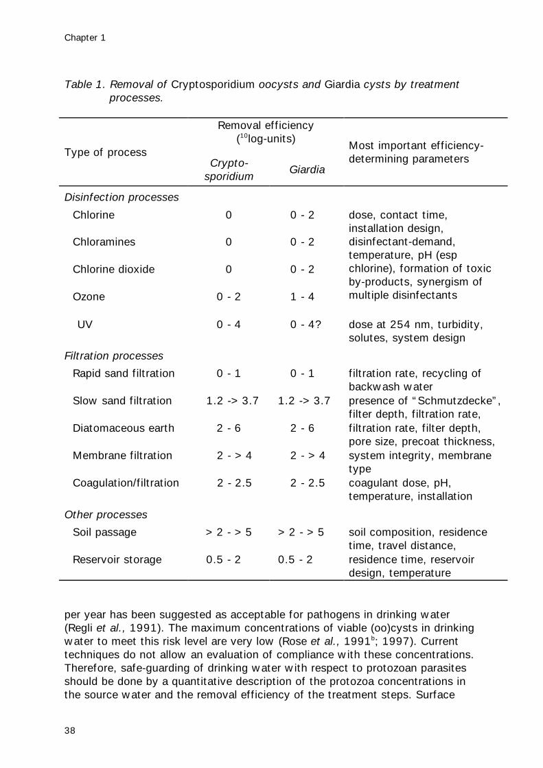
Chapter 1
38
Table 1. Removal of Cryptosporidium oocysts and Giardia cysts by treatmentprocesses.
Removal efficiency(10log-units)
Type of processCrypto-
sporidiumGiardia
Most important efficiency-determining parameters
Disinfection processes
Chlorine 0 0 - 2
Chloramines 0 0 - 2
Chlorine dioxide 0 0 - 2
Ozone 0 - 2 1 - 4
dose, contact time,installation design,disinfectant-demand,temperature, pH (espchlorine), formation of toxicby-products, synergism ofmultiple disinfectants
UV 0 - 4 0 - 4? dose at 254 nm, turbidity,solutes, system design
Filtration processes
Rapid sand filtration 0 - 1 0 - 1 filtration rate, recycling ofbackwash water
Slow sand filtration 1.2 ->3.7 1.2 ->3.7 presence of “Schmutzdecke”,filter depth, filtration rate,temperatureDiatomaceous earth 2 - 6 2 - 6 filtration rate, filter depth,pore size, precoat thickness,filter integrityMembrane filtration 2 - >4 2 - >4 system integrity, membranetype
Coagulation/filtration 2 - 2.5 2 - 2.5 coagulant dose, pH,temperature, installationdesign, addition of polymers,Other processes
Soil passage >2 - >5 >2 - >5 soil composition, residencetime, travel distance,presence of sedimentReservoir storage 0.5 - 2 0.5 - 2 residence time, reservoirdesign, temperature
per year has been suggested as acceptable for pathogens in drinking water(Regli et al., 1991). The maximum concentrations of viable (oo)cysts in drinkingwater to meet this risk level are very low (Rose et al., 1991b; 1997). Currenttechniques do not allow an evaluation of compliance with these concentrations.Therefore, safe-guarding of drinking water with respect to protozoan parasitesshould be done by a quantitative description of the protozoa concentrations inthe source water and the removal efficiency of the treatment steps. Surface
Protozoan parasites
39
water utilities and groundwater utilities that may be influenced by surface wateror other sources of contamination should monitor their source water forprotozoa and determine the rate of protozoa removal and inactivation achievedin the treatment plant, in order to determine if acceptable concentrations ofprotozoa in drinking water have been achieved.
Verification of efficiency of parasite removalFor routine monitoring, water quality and process parameters are required toverify treatment performance. Several parameters have been suggested as asurrogate for (oo)cyst removal by filtration processes: turbidity, particle counts(LeChevallier & Norton., 1992; Hall & Croll, 1997), clostridial spores (Payment &Franco, 1993; Hijnen et al, 1997) or aerobic spores (Nieminski, 1997) andparticulate matter (Anon., 1997). Although turbidity or particle counts of filteredwater depend on both the levels in raw water and filter performance, in general,a turbidity of 0.1 to 0.5 NTU or counts of particles >3 µm below 50 per ml areindicative of good quality water. On-line monitoring of turbidity or particlecounts gives direct and continuous information on (individual) filter performanceand are very valuable tools for optimising treatment efficiency for (oo)cystremoval.Critical moments in the filter cycle are just after backwash or, in case of slowsand filtration, scraping of the clogged top-layer from the filterbed. A slowincrease in filtration rate or filtering-to-waste minimises the risk of (oo)cystbreakthrough.For disinfection processes, disinfectant dose, contact time, residual disinfectantconcentration after this contact time, pH and temperature are commonly used tomonitor the disinfection performance. The most critical conditions fordisinfection processes are low temperatures and high turbidity of the water thatis to be disinfected.
CONCLUSIONS AND RECOMMENDATIONS
Health risk assessmentThe abundance and size of drinking waterborne outbreaks in developedcountries show that transmission of Giardia and Cryptosporidium by drinkingwater is a significant risk. In the case of Cryptosporidium, the absence of anadequate cure for immunocompromised patients increases the problem.Although the outbreaks receive most attention, low-level transmission of theseprotozoa through drinking water is very likely to occur in developed countriesand in developing countries alike (Fraser & Cooke, 1991; Isaac-Renton et al.,1996). Cysts and oocysts are regularly found in drinking water (Isaac-Renton etal., 1996; Karanis & Seitz, 1996; Rose et al, 1997), although only a smallproportion may be viable and infectious to man. A major drawback for thedetermination of the health significance of (oo)cysts in (drinking) water is thatmethods for a sensitive and specific detection of infectious (oo)cysts, with aconsistently high recovery are not available.
Chapter 1
40
Risk managementCryptosporidium poses a serious health risk to immunocompromised persons,especially AIDS patients. An important step forward in the reduction of theconsequences of waterborne outbreaks and other cases of cryptosporidiosiswould be the definition of an adequate therapy for the immunocompromisedpatients. Currently, prevention of exposure to (potentially) contaminated wateris a means to reduce the risk. Especially the immunocompromised populationshould be informed about the risk and means to prevent exposure. Boiling of tapwater, use of mineral or bottled water, not swimming in surface water or poolsare some of the options. Local considerations play a major role and public healthauthorities are encouraged to provide guidance on the safety of drinking waterfor the immunocompromised and on applicable means to reduce exposure(Anon., 1995a.b; Juranek, 1995). In outbreak situations, rapid investigation ofsize and source of the outbreak and installation of control measures to preventfurther transmission is required. Useful guidance on management of waterborneoutbreaks can be found in the report of the UK group of experts (Anon., 1990)and the CDC Guidance Manual (Juranek, 1995) and a workshop report (Anon.,1995).The protozoa, and to a lesser extent the viruses, have changed the philosophyin the developed countries towards safe-guarding of drinking water frommonitoring of the ‘end-product’ drinking water to monitoring raw water and theefficiency of the treatment. Furthermore, the extreme resistance of theseorganisms implies that a “zero-risk” is no longer achievable. Treatments shouldbe designed to reduce the (oo)cyst concentrations in the raw water as far aspossible and preferably include filtration step(s). This implies that information onthe parasite concentrations in the raw water is necessary, as well as informationon the removal efficiency of the treatment. Quantitative risk assessmentprovides a tool for the combination of information on raw water quality(concentrations detected, recovery of the detection method, viability) andtreatment efficiency (removal by different steps in the treatment) (Teunis et al.,1997). The current detection methods are generally sensitive enough todetermine the concentrations of Cryptosporidium and Giardia in surface water,but are in many cases not sensitive enough for an accurate description ofremoval efficiency. For the latter description, data from laboratory studies and(seeded) pilot plant studies may provide additional information. Anotherapproach is to determine if an adequate surrogate parameter can be found forthe description of removal efficiency for Cryptosporidium (as Giardia is easier toeliminate both with disinfection and filtration, the description of treatmentefficiency should be targeted on Cryptosporidium). Several parameters havebeen evaluated on a limited scale as surrogates for protozoa removal: aerobicspores, clostridial spores, particles and algae, but a broader evaluation isnecessary to determine the value of these parameters.The definition of maximum acceptable concentrations of pathogens in drinkingwater based on a maximum acceptable (infection) risk level has become possibleby the availability of volunteer data and dose-response models (Haas, 1983;Dupont et al., 1995; Teunis et al., 1996).
Protozoan parasites
41
An annual infection risk level of 10-4, as proposed by the US EPA, is currentlyused in the US (Rose et al., 1997), Canada (Wallis et al., 1995) and theNetherlands (Medema et al., 1995) as the basis to determine the appropriateremoval efficiency of surface water treatment systems. Although there are stillquestions about the significance of (oo)cyst occurrence in drinking water, this isthe way forward. The implementation of guideline levels is still hampered,however, by the difficulty to determine source water quality and treatmentefficiency accurately.
REFERENCES
Abbaszadegan M, Huber MS, Gerba CP, Pepper IL (1997). Detection of viable Giardia cysts byamplification of heat shock-induced mRNA. Appl. Environ. Microbiol. 63:324-328.
Adam RD (1991). The biology of Giardia spp. Microbiol Rev, 55:706-732.Adham SS, Jacangelo JG, Laine J (1994). Effect of membrane type on the removal of
Cryptosporidium parvum, Giardia muris and MS2 virus. Proc. Am. Water Works Assoc.Annual Conf., New York, June 1994, Am. Water Works Assoc., Denver, USA.
Andrews RH, Adams M, Boreham PFL, Mayrhofer G, Meloni BP (1989). Giardia intestinalis:electrophoretic evidence for a species complex. Int. J. Parasitol. 19:183-190.
Anonymous (1990). Cryptosporidium in water supplies, London: HMSO, London, UK.Anonymous (1991). Centres for Disease Control. Outbreaks of diarrhoeal illness associated with
cyanobacteria (blue-green algae)-like bodies - Chicago and Nepal, 1989 and 1990. MMWR40:325-327.
Anonymous (1995). Assessing the public health threat associated with waterbornecryptosporidiosis: report of a workshop. MMWR 44(RR-6):1-19.
Anonymous (1996). Centers for Disease Control and Prevention. Update: outbreaks ofCyclospora cayetanensis infection - United States and Canada, 1996. MMWR 45:611-612.
Anonymous (1997). Microscopic particulate analysis (MPA) correlations with Giardia andCryptosporidium occurrence in ground water under the direct influence of surface watersources. Contract 68-C6-0059, OGWDW, USEPA, Washington DC, USA.
Anonymous (1998). Swimming pools implicated in Australia’s largest cryptosporidiosis outbreakever reported. Cryptosporidium Capsule, 3(5): 1-2.
Arrowood MJ (1997). Diagnosis. In: Fayer R (ed). Cryptosporidium and cryptosporidiosis. CRCPress, Boca Raton, USA. pp. 43-64.
Ashford RW (1979). Occurrence of an undescribed coccidian in man in Papua New Guinea. Ann.Trop. Med.Parasitol. 73:497-500.
Ashford RW, Warhurst DC, Reid GDF (1993). Human infection with cyanobacterium-like bodies.Lancet 341:1034.
Atherholt TB, LeChevallier MW, Norton WD, Rosen JS (1998). Effect of rainfall on Giardia andCryptosporidium. J. Am. Water Works Assoc., 90:66-80.
Baxby D, Hart CA, Blundell N (1985). Shedding of oocysts by immunocompetent individuals withcryptosporidiosis. J. Hyg. Camb. 95:703.
Belosevic GM, Faubert GM, MacLean JD (1984). Observations on natural and experimentalinfections with Giardia isolated from cats. Can. J. Comp. Med. 48:241-244.
Belosevic GM, Finch GF (1997). Presentation at the Int. Symp on Waterborne Cryptosporidium,March 1997, Newport Beach Ca, USA.
Bendall RP, Lucas S, Moody A, Tovey G, Chiodini PL (1993). Diarrhoea associated withcyanobacterium-like bodies: a new coccidian enteritis of man. Lancet 341:590-592.
Black RE, Dykes AC, Sinclair SP, Wells JG (1977). Giardiasis in day-care centers: evidence ofperson-to-person transmission. Pediatrics 60:486-491.
Blagburn BL, Soave R (1997). Prophylaxis and chemotherapy: human and animal. In: Fayer R(ed). Cryptosporidium and cryptosporidiosis. CRC Press, Boca Raton, USA. pp. 111-128.
Chapter 1
42
Bonnin A, Fourmaix MN, Dubremetz JF et al. (1996). Genotyping human and bovine isolates ofCryptosporidium parvum by polymerase chain reaction-restriction fragment lengthpolymorphism analysis of a repetitive DNA sequence. FEMS Microbiol. Lett. 137:207-211.
Boreham PFL (1994). The current status of chemotherapy for giardiasis. In: Thompson RCA,Reynoldson JA, Lymbery AJ (eds.). Giardia from molecules to disease, CAB Int., Wallingford,UK. pp. 317-328.
Brodsky RE, Spencer HC, Schultz MG (1974). Giardiasis in American travellers to the SovietUnion. J. Inf. Dis.130:319-323.
Bukhari Z, Smith HV (1996). Effect of three concentration techniques on viability ofCryptosporidium parvum oocysts removed from bovine faeces. J. Clin. Microbiol. 33:2592-2595.
Buret A (1994). Pathogenesis – how does Giardia cause disease? In: Thompson RCA,Reynoldson JA, Lymbery AJ (eds.). Giardia from molecules to disease, CAB Int., Wallingford,UK. pp. 293-315.
Campbell AT, Robertson LJ, Smith HV (1992). Viability of Cryptospordium parvum oocysts -correlation of in vitro excystation with inclusion or exclusion of fluorogenic vital dyes. Appl.Environ. Microbiol. 58(11):3488-3493.
Campbell AT, Robertson LJ, Smith HV, Girdwood RWA (1994). Viability of Cryptosporidiumparvum oocysts concentrated by calcium carbonate flocculation. J. Appl. Bacteriol. 76:638-639.
Campbell I, Tennant W, Seal DV, Hay J, McLuckie I, Paterson W (1995). UK National study ofLegionella contamination of hospital transplant unit water supplies: relevance of free-livingamoebae. In: Betts WB, Casemore D, Fricker C et al. (eds). Protozoan parasites and water.Royal Society of Chemistry, Cambridge, UK, pp.30-32.
Campbell AT, Smith HV (1997a). Immunomagnetic separation of Cryptosporidium oocysts: roundrobin comparison of techniques. Wat. Sci. Tech. In press.
Campbell AT, Gron B, Johnsen SE (1997b). Immunomagnetic separation of Cryptosporidiumoocysts from high turbidity water sample concentrates. Proc. Int. Symp. on WaterborneCryptosporidium, March 1997, Newport Beach Ca, USA, pp.91-96..
Casemore DP (1990). Epidemiological aspects of human cryptosporidiosis. Epidemiol.Infect.104:1-28.
Casemore DP (1996). In: Report from the Expert Workshop on Emerging Waterborne Pathogensand their Public Health Significance. 6-7 June 1996, Beckenridge, Co, USA. Am. WaterWorks Res. Found., Denver, Co, USA.
Casemore DP, Wright SE, Coop RL.(1997). Cryptosporidiosis – Human and animal epidemiology.In: Fayer R (ed). Cryptosporidium and cryptosporidiosis. CRC Press, Boca Raton, USA. pp.65-92.
Chauret C, Chen P, Springthorpe S, Sattar S (1995). Effect of environmental stressors on thesurvival of Cryptosporidium oocysts. Proc. Am. Water Works Assoc. Water Quality Technol.Conf., November 1995, New Orleans, Am. Water Works Assoc., Denver, USA.
Clancy JL, Gollnitz WD, Tabib Z (1994). Commercial labs: how accurate are they? J. Am. WaterWorks Assoc. 86:89.
Clancy JL, Hargy TM, Schaub S (1997). Improved sampling methods for the receiving of Giardiaand Cryptosporidium from source and treated water. Proc. Int. Symp. on WaterborneCryptosporidium, March 1997, Newport Beach Ca, USA, pp.79-86.
Clancy JL, Hargy TM, Marshall MM, Dyksen JE (1998). UV light inactivation of Cryptosporidiumoocysts. J. Am. Water Works Assoc. 90(9):92-102.
Compagnon B, Robert C, Mennecart V, Roubin MR de, Cervantes P, Joret JC (1997). Improveddetection of Giardia cysts and Cryptosporidium oocysts in water by flow cytometry. ProcWater Quality Technol Conf, November 1997, USA.
Connolly GM, Dryden MS, Shanson DC, Gazzard BG (1988). Cryptosporidial diarrhoea in AIDSand its treatment. Gut 129:593.
Connor BA, Shlim DR, Scholes JV, Rayburn JL, Reidy J, Rajah R (1993). Pathological changes inthe small bowel in nine patients with diarrhea associated with a coccidia-like body. Ann.Intern. Med. 119:377-382.
Cordell RL, Addiss DG (1994). Cryptosporidiosis in child care settings: a review of the literatureand recommendations for prevention and control. Pediatr. Inf. Dis. J. 13:310.
Protozoan parasites
43
Craun GF (1984). Waterborne outbreaks of giardiasis: current status. In: Erlandsen SL, Meyer EA(eds). Giardia and giardiasis. Plenum Press, New York, USA, pp. 243-261.
Craun GF (1986). Waterborne giardiasis in the United States 1965-84. Lancet 1986; ii:513-514.Craun GF (1990). Waterborne giardiasis. In: Meyer EA (ed). Human Parasitic Diseases, Vol. 3,
Giardiasis. Elsevier Science Publ., Amsterdam, The Netherlands, pp. 267-293.Craun GF, Hubbs SA, Frost F, Calderon RL, Via SH (1998). Waterborne outbreaks of
cryptosporidiosis. J. Am. Water Works Assoc. 90:81-91.Crawford FG, Vermund SH (1988). Human cryptosporidiosis. Crit. Rev. Microbiol. 16:113-159.Current WL, Garcia LS (1991). Cryptosporidiosis. Clin. Microbiol. Rev. 4(3):325-258.Danielson RE, Cooper RC, Riggs JL (1995). Giardia and Cryptosporidium analysis: a comparison
of microscopic and flow cytometric techniques. Proc.Water Quality Technol. Conf. November1995, New Orleans, USA, pp. 1673-1685.
d’Antonio RG, Winn E, Taylor JP et al (1985). A waterborne outbreak of cryptosporidiosis innormal hosts. Ann. Intern. Med. 103:886-888.
Davies RR, Hibler CP (1979). Animal reservoirs and cross-species transmission of Giardia. In:Jakubowski W, Hoff JC (eds.). Waterborne transmission of giardiasis. Cincinnati OH: USEPA, 1979, pp.104-126.
de Jong B, Andersson Y (1997) Waterborne cryptosporidiosis – a single outbreak in Sweden.Proc. Int. Symp. on Waterborne Cryptosporidium. March 1997, Newport Beach CA, USA.pp. 367-368.
Deng MQ, Cliver DO (1998). Differentiation of Cryptosporidium parvum isolates by a simplifiedrandomly amplified polymorphic DNA technique. Appl. Environ. Microbiol. 64:1954-1957.
DeReignier DP, Cole L, Schupp DG, Erlandsen SL (1989). Viability of Giardia cysts suspended inlake, river and tap water. Appl. Environ. Microbiol.55:1223-1229..
Drozd C, Schwartzbrod J (1997). Removal of Cryptosporidium from river water by crossflowmicrofiltration. A pilot scale study. Wat. Sci. Tech. 35(11-12):391-395.
Dupont HL, Chappell CL, Sterling CR, Okhuysen PC, Rose JB, Jakubowski W (1995). Theinfectivity of Cryptosporidium parvum in healthy volunteers. N. Engl. J. Med. 332(13):855-859.
Dykes AC, Juranek DD, Lorenz RA, Sinclair S, Jakubowski W, Davies RB (1980). Municipalwaterborne giardiasis: an epidemiological investigation. Ann. Int. Med. 92:165-170.
Erlandsen SL (1994). Biotic transmission – Is giardiasis a zoonosis?. In: Thompson RCA,Reynoldson JA, Lymbery AJ (eds.). Giardia from molecules to disease, CAB Int., Wallingford,UK. pp. 83-97.
Erlandsen SL, Bemrick WJ (1987). SEM evidence for a new species: Giardia psittaci. J. Parasitol.73:623-629.
Erlandsen SL, Wells CL, Feely LK, Campbell SR, Keulen H van, Jarroll EL (1990). Axenic cultureand characterization of Giardia ardae from the great blue heron (Ardae herodias). J. Parasitol.76:717-724.
Farthing MJG (1994). Giardiasis as a disease. In: Thompson RCA, Reynoldson JA, Lymbery AJ(eds.). Giardia from molecules to disease, CAB Int., Wallingford, UK. pp. 15-37.
Farthing MJG, Goka AJ (1987). Immunology of giardiasis. Ballieres Clin. Gastroenterol. 1:589-603.
Fayer R, Ungar BLP (1986). Cryptosporidium spp. and cryptosporidiosis. Microbiol. Rev.50(4):458-483.
Fayer R, Speer CA, Dubey JP (1997). The general biology of Cryptosporidium. In: Fayer R (ed).Cryptosporidium and cryptosporidiosis. CRC Press, Boca Raton, USA. pp. 1-42.
Feely DE, Holberton DV, Erlandsen SL (1990). The biology of Giardia. In: Meyer EA (ed). HumanParasitic Diseases, Vol. 3, Giardiasis. Amsterdam: Elsevier Science Publ., pp. 11-50.
Filice FP (1952). Studies on the cytology and life history of a Giardia from the laboratory rat.Univ. Calif. Publ. Zool. 57:53-146.
Filkorn R, Wiedenmann A, Botzenhart K (1994). Selective detection of viable Cryptosporidiumoocysts by PCR. Zbl. Hyg. 195:484-494.
Finch GR, Black EK, Gyurek L, Belosevic M (1993a). Ozone inactivation of Cryptosporidiumparvum in demand-free phosphate buffer determined by in vitro excystation and animalinfectivity. Appl. Environ. Microbiol. 59(12):4203-4210.
Chapter 1
44
Finch GR, Black EK, Labatiuk CW, Gyürek L, Belosevic M (1993b). Comparison of Giardia lambliaand Giardia muris cyst inactivation by ozone. Appl. Environ. Microbiol. 59:3674-3680.
Finch GF, Kathleen B, Gyurek LL (1994). Ozone and chlorine inactivation of CryptosporidiumProc. Am. Water Works Assoc. Water Quality Technol. Conf., November 1994, SanFrancisco, Am. Water Works Assoc., Denver, USA.
Flannagan PA (1992). Giardia - Diagnosis, clinical course and epidemiology - A review. Epidemiol.Infect. 109(1):1-22.
Forshaw D, Palmer DG, Halse SA, Hopkins RM, Thompson RCA (1992). Giardia in Straw-NeckedIbis (Threskiornis spinicollis) in Western-Australia. Vet. Rec. 131(12):267-8.
Fraser GG, Cooke KR (1991). Endemic giardiasis and municipal water supply. Am. J. PublicHealth, 81:760-762.
Fricker CR (1995). Detection of Cryptosporidium and Giardia in water. In: Betts W.B. et al. (eds).Protozoan parasites and water. Royal Society of Chemistry, Cambridge, UK, pp.91-96..
Fricker CR, Jonas A, Crabb J, Turner N, Smith HV (1997). The concentration and separation ofCryptosporidium oocysts and Giardia cysts using vortex flow filtration and immunomagneticseparation. Proc Int Symp on Waterborne Cryptosporidium, March 1997, Newport Beach Ca,USA, pp.1-8.
Garcia-Rodriguez JA, Martin Sanchez AM, Canut Blasco A, Cedeno Montano J, Heras de PedroMI (1989). The incidence of cryptosporidiosis in children: a one year prospective survey in ageneral hospital in Spain. Eur. J. Epidemiol. 5:70-73.
Gilman RH, Marquis GS, Miranda E, Vestegui M, Martinez H (1988) Rapid reinfection by Giardialamblia after treatment in a hyperendemic world community. Lancet 1988: 343-345.
Glaser CA et al. (1998). Association between Cryptosporidium infection and animal exposure inHIV-infected individuals. J. AIDS Retrovirol. 17:79-82.
Goldstein ST, Juranek DD, Ravenholt O, Hightower AW, Martin GD, Mesnik JL, Griffiths SD,Bryant AJ, Reich BA en Herwaldt BL (1996). Cryptosporidiosis: an outbreak associated withdrinking water despite state-of-the-art treatment. Ann. Intern. Med. 124:459-468.
Grimason AM, Smith HV, Thitai WN, Smith PG, Jackson MH, Girdwoos RWA (1992). Theoccurrence and removal of Cryptosporidium spp. oocysts and Giardia spp. cysts in Kenyanwaste stabilisation ponds. Wat. Sci. Tech. 27:97-104.
Guy E (1996). In: Report from the expert workshop on emerging waterborne pathogens and theirpublic health significance. 6-7 June 1996, Beckenridge, Co, USA. A. Water Works Res.Found., Denver, Co, USA.
Haas CN (1983). Estimation of risk due to low doses of microorganisms: a comparison ofalternative methodologies. Am. J. Epidemiol. 118(4):573-582.
Haas C, Rose JB (1994). Reconciliation of microbial risk models and outbreak epidemiology: thecase of the Milwaukee outbreak. Proc. AWWA Conf. June 1994, New York, USA.
Haas CN, Rose JB (1995). Development of an action level for Cryptosporidium. Am. WaterWorks Assoc. J. 87:9:91.
Hall A (1994). Giardia infections: epidemiology and nutritional consequences. In: ThompsonRCA, Reynoldson JA, Lymbery AJ (eds.). Giardia from molecules to disease, CAB Int.,Wallingford, UK. pp. 251-279.
Hall T, Pressdee J, Carrington EC (1994). Removal of Cryptosporidium oocysts by watertreatment processes. Foundation for Water Research, Bucks, UK.
Hall T, Croll B (1997). Particle counters as tools for managing Cryptosporidium risk in watertreatment. Wat. Sci. Tech. 36(4):143-149.
Hancock CM, Rose JB, Callahan M (1997). The prevalence of Cryptosporidium in USgroundwaters. Proc. Int. Symp. on Waterborne Cryptosporidium. March 1997, NewportBeach, CA, USA. pp. 147-152.
Hansen JS, Ongerth JE (1991). Effects of time and watershed characteristics on theconcentration of Cryptosporidium oocysts in river water. Appl. Environ. Microbiol. 57:2790-2795.
Herwaldt BL, Ackers M. and the Cyclospora working group (1997). An outbreak of cyclosporiasisassociated with imported raspberries. N. Engl. J. Med. 336: 1548-1556.
Hewlett EL, Andrews JS, Ruffier J, Schaefer FW (1982) Experimental infection of mongrel dogswith Giardia intestinalis cysts and cultured trophozoites. J. Inf. Dis. 152:1166-1171.
Protozoan parasites
45
Hibler CP, Hancock CM, Perger LM, Wegrzyn JG, Swabby KD (1987). Inactivation of Giardiacysts with chlorine at 0.5°C and 5°C. AWWARF, Denver, USA.
Hoge CW, Shlim DR, Rajah R, Triplett J, Shear M, Rabold JG, Echeverria P (1993). Epidemiologyof diarrhoeal illness associated with coccidian-like organism among travellers and foreignresidents in Nepal. Lancet 341:1175-1179.
Homan WL, Vanenckevort FHJ, Limper L, van Eys GJJM, Schoone GJ, Kasprzak W, MajewskaAC, van Knapen F (1992). Comparison of Giardia isolates from different laboratories byisoenzyme analysis and recombinant DNA probes. Parasitol Res. 78:313-323.
Homan WL, Limper L, van Enckevort FHJ, van Knapen F (1994). Characterization of Giardiaisolates. In: Thompson RCA, Reynoldson JA, Lymbery AJ (eds.). Giardia from molecules todisease, CAB Int., Wallingford, UK. pp. 54.
Hoogenboom-Verdegaal AMM, During M, Leentvaar-Kuypers A et al (1989). Epidemiologisch enmicrobiologisch onderzoek met betrekking tot gastro-enteritis bij de mens in de regio’sAmsterdam en Helmond, in 1987. Bilthoven: RIVM, Report no. 148612001.
Hijnen WAM, Houtepen FAP, van der Speld WMN van der Kooij D (1997). Spores of sulphitereducing clostridia: a surrogate parameter for assessing the effects of water treatmentprocesses on protozoan (oo)cysts? Proc. Int. Symp. on waterborne Cryptosporidium. March1997, Newport Beach CA, USA. pp. 115-126.
Isaac-Renton J, Moorehead W, Ross A (1996). Longitudinal studies of Giardia contamination intwo community drinking water supplies: cyst levels, parasite viability and health impact.Appl. Environ. Microbiol. 62:47-54.
Islam A (1990). Giardiasis in developing countries. In: Meyer EA (ed). Human Parasitic Diseases,Vol. 3, Giardiasis. Elsevier Science Publ., Amsterdam, The Netherlands, pp. 235-266.
Istre GR, Dunlop TS, Gaspard B, Hopkins RS (1984). Waterborne giardiasis at a mountain resort;evidence for acquired immunity. Am. J. Public Health 74:602-604.
Ives K (1990). Cryptosporidium and water supplies: treatment processes and oocyst removal. In:Badenoch, J. Cryptosporidium in water supplies. HMSO. London.
Jacangelo JG, Laîné JM, Carns KE, Cummings EW, Mallevialle J (1991). Low-pressuremembrane filtration for removing Giardia and microbial indicators. J. Am. Water WorksAssoc. 83: 97-106.
Jakubowski, W (1990). The control of Giardia in water supplies. In: Meyer EA (ed). HumanParasitic Diseases, Vol. 3, Giardiasis. Elsevier Science Publ., Amsterdam, the Netherlands,pp. 335-354.
Johnson DW, Pieniazek NJ, Griffin DW, Misener L, Rose JB (1995). Development of a PCRprotocol for sensitive detection of Cryptosporidium oocysts in water samples. Appl. Environ.Microbiol. 61(11):3849-3855.
Joce RE, Bruce J, Kiely D et al (1991). An outbreak of cryptosporidiosis associated with aswimming pool. Epidemiol. Infect.107(3):497-508.
Jokipii AMM, Hemila M, Jokipii L (1985). Prospective study of aquisition of Cryptosporidium,Giardia lamblia, and gastrointestinal illness. Lancet 1985; 487-489.
Juranek D (1995). Cryptosporidosis: sources of infection and guidelines for prevention. Clin. Inf.Dis. 21(suppl1):s57-61.
Karanis P, Seitz HM (1996). Vorkommen und Verbreitung von Giardia und Cryptosporidium imRoh und Trinkwasser von Oberflächenwasserwerken. Wasser – Abwasser GWF.137:94-100.
Kaucner C, Stinear T (1998). Sensitive and rapid detection of viable Giardia cysts andCryptosporidium parvum oocysts in large-volume water samples with wound fiberglasscartridge filters and reverse transcription-PCR. Appl. Environ. Microbiol. 64:1743-1749.
Kent GP, Greenspan JR, Herndon JL et al. (1988). Epidemic giardiasis caused by a contaminatedwater supply. Am. J. Public Health 78:139-143.
Ketelaars HAM, Medema GJ, Breemen LWCA van, Kooij D van der, Nobel PJ, Nuhn P (1995).Occurrence of Cryptosporidium oocysts and Giardia cysts in the river Meuse and removal inthe Biesbosch reservoirs. J. Water SRT – Aqua 44(suppl. 1):108-111.
Kirkpatrick CE, Green B (1985). Susceptibility of domestic cats to infections with Giardiaintestinalis cysts and trophozoites from human sources. J. Clin. Microbiol. 21:671-680.
Korich DG, Mead JR, Madore MS, Sinclair NA, Sterling CR (1990). Effects of ozone, chlorinedioxide, chlorine, and monochloramine on Cryptosporidium parvum oocyst viability. Appl.Environ. Microbiol. 56(5):1423-1428.
Chapter 1
46
Kortbeek LM, Deursen D van, Hoogenboom-Verdegaal AMM (1994). Giardia lamblia and otherintestinal parasites: incidence in gastroenteritis patients and a healthy population in theNetherlands. In: Thompson RCA, Reynoldson JA, Lymbery AJ (eds.). Giardia from moleculesto disease. CAB Int., Wallingford, UK. pp. 361-362.
Kramer MH, Herwaldt BL, Craun GF, Calderon RL, Juranek DD (1996). Waterborne disease:1993-1994. J. Am. Water Works Assoc. 88(3):66-80.
Kramer MH et al. (1998). First reported outbreak in the United States of cryptosporidiosisassociated with a recreational lake. Clin. Inf. Dis. 26:27-33.
Kuhls TL, Mosier DA, Crawford DL, Griffis J (1994). Seroprevalence of cryptosporidial antibodiesduring infancy, childhood, and adolescence. Clin. Infect. Dis.18(5):731-735.
Lacroix C, Berthier M, Agius G, Bonneau D, Pallu B, Jaquemin JL (1987) Cryptosporidiumoocysts in immunocompetent children: epidemiological investigations in the day care centresof Poitiers, France. Eur. J. Epidemiol. 3:381-385.
Leahy, J.G., Rubin, A.J. en Sproul, O.J (1987). Inactivation of Giardia muris cysts by freechlorine. Appl. Environ. Microbiol. 53:1448-1453.
LeChevallier MW, Norton WD, and Lee RG (1991). Occurrence of Giardia and Cryptosporidiumspp. in surface water supplies. Appl. Environ. Microbiol. 56:2610-2616.
LeChevallier MW, Norton WD (1992). Examining relationships between particle counts andGiardia, Cryptosporidium and turbidity. J. Am. Water Works Assoc. 84:54-60.
Leland D, McAnulty J, Keene W, Stevens G (1993). A cryptosporidiosis outbreak in a filteredwater supply. J. Am. Water Works Assoc. 85:34.
Lengerich EJ, Addiss DG, Marx JJ, Ungar BLP, Juranek DD (1993). Increased exposure tocryptosporidia among dairy farmers in Wisconsin. J. Infect. Dis. 167(5):1252-1255.
Levine ND (1973). Introduction, history and taxonomy, p. 1-22. In Hammond, D.M. Long, P.L(eds.) The coccidia: Eimeria, Isospora, Toxoplasma and related genera. University Park Press,Baltimore, Maryland, USA.
Lindquist AHD (1997). Probes for the specific detection of Cryptosporidium parvum. Water Res.31:2668-2671.
Liyanage LRJ et al. (1997). Sequential disinfection of Cryptosporidium parvum by ozone andchlorine dioxide. Ozone Sci. Engin. 19:409-423.
Long EG, White EH, Carmichael WW, Quinlisk PM, Raja R, Swisher BL, Daugharty H, Cohen MT(1991). Morphologic and staining characteristics of a cyanobacterium-like organismassociated with diarrhea. J. Inf. Dis. 164:199-202.
MacKenzie WR, Kazmierczak JJ, Davis JP (1995). An outbreak of cryptosporidiosis associatedwith a resort swimming pool. Epidemiol. Infect. 1995; 115(3):545-553.
MacKenzie WR, Hoxie NJ, Proctor ME et al (1994). A massive outbreak in Milwaukee ofCryptosporidium infection transmitted through the public water supply. N. Engl. J. Med.331(3):161-167.
Marshall MM, Naumovitz D, Ortega Y, Sterling CR (1997). Waterborne protozoan pathogens.Clin. Microbiol. Rev. 10:67-85.
Mawdsley JL, Brooks AE, Merry RJ (1996). Movement of the protozoan pathogenCryptosporidium parvum through three contrasting soil types. Biology and Fertility of Soils21(1-2):30-36.
McClellan P (1998). Sydney Water Inquiry. Fifth Report. Premier’s Department, New SouthWales, Australia.
McRoberts KM, Meloni BP, Morgan UM, Marano R, Binz N, Erlandsen SL, Halse SA, ThompsonRCA (1996). Morphological and molecular characterization of Giardia isolated from the straw-necked ibis (Threskiornis spinicollis) in Western Australia. J. Parasitol. 82:711-718.
Medema GJ, Teunis PFM, Gornik V, Havelaar AH, Exner M (1995). Estimation of theCryptosporidium infection risk via drinking water. In: Betts WB, Casemore D, Fricker C et al.(eds). Protozoan parasites and water. Royal Society of Chemistry, Cambridge, UK, pp.53-56.
Medema GJ, Bahar M, Schets FM (1997a). Survival of Cryptosporidium parvum, Escherichia coli,faecal enterococci and Clostridium perfringens in river water: Influence of temperature andautochthonous microorganisms. Wat. Sci. Tech. 35(11-12):249-252.
Medema GJ, Schijven JF, de Nijs ACM, Elzenga JG (1997b). Modelling the discharge ofCryptosporidium and Giardia by domestic sewage and their dispersion in surface water. Proc.
Protozoan parasites
47
Int. Symp. on Waterborne Cryptosporidium. March 1997, Newport Beach CA, USA. pp. 177-182.
Medema GJ, Schets FM, Ketelaars HAM, Boschman G (1998a). Improved detection and vitalstaining of Cryptosporidium and Giardia with flow cytometry. Wat. Sci. Tech. 38(12):61-66.
Medema GJ, Schets FM, Teunis PFM, Havelaar H (1998b) Sedimentation of free and attachedCryptosporidium oocysts and Giardia cysts in water. Appl. Environ. Microbiol. 64(11): 4460-4466.
Meisel JL, Perera DR, Meligro C, Rubin CE (1976). Overwhelming watery diarrhea associatedwith a Cryptosporidium in an immunosuppressed patient. Gastroenterology 70:1156.
Meloni BP, Lymbery AJ, Thompson RCA (1989). Characterization of Giardia isolates using a non-radiolabelled DNA probe, and correlation with the results of isozyme analysis. Am. J. Trop.Med. Hyg. 40:629-637.
Meyer EA (1994). Giardia as an organism. In: Thompson RCA, Reynoldson JA, Lymbery AJ(eds.). Giardia from molecules to disease, CAB Int., Wallingford, UK. 1994. pp.3-14.
Moore GT, Cross WM, McGuire D, Molahan CS, Gleason NN, Nealy GR, Newton LH (1969).Epidemic giardiasis in a ski resort. N. Engl. J. Med. 281:402-407.
Morgan Um, Constantine CC, Greene WK, Thompson RCA. RAPD (random amplified polymorphicDNA) analysis of Giardia DNA and correlation with isoenzyme data. In: Thompson RCA,Reynoldson JA, Lymbery AJ (eds.). Giardia from molecules to disease, CAB Int., Wallingford,UK. 1994. pp. 61-63.
Musial CE, Arrowood MJ, Sterling CR, Gerba CP (1987). Detection of Cryptosporidium in waterby using polypropylene cartridge filters. Appl. Environ. Microbiol. 53(4):687-92.
Naik SR, Rao NR, Vinayak, Narayanna VA, Zunzurwade S, Hegal SC, Talwar P (1978). Presenceof Candida albicans in normal and Girdia lamblia infected human jejunum. Ann. Trop. Med.Parasitol. 72:493-494.
Nash T (1992). Surface antigen variability and variation in Giardia intestinalis. Parasitol. Today,8:229-234.
Nash TE, McCutchan T, Keister D, Dame JB, Conrad JD, Gillin FD (1985). Restrictionendonuclease analysis of DNA from 15 Giardia isolates obtained from humans and animals.J. Inf. Dis. 152:64-73.
Nash TE, Herrington DA, Losonsky GA, Levine MM (1987). Experimental human infections withGiardia intestinalis. J. Inf. Dis. 156:974-984.
Nieminski EC (1994). Giardia and Cryptosporidium cysts removal through direct filtration andconventional treatment performance. Proc. Am. Water Works Assoc. Annual Conf., NewYork, June 1994, Am. Water Works Assoc., Denver, USA.
Nieminski EC, Schaeffer III FW, Ongerth JE (1995). Comparison of two methods for detection ofGiardia cysts and Cryptosporidium oocysts in water. Appl. Environ. Microbiol. 61:1714-1719.
Nieminski EC (1997). From target pathogens to surrogate indicators. Proc. Int. Symp. onwaterborne Cryptosporidium. March 1997, Newport Beach CA, USA. pp. 243-252.
Nime FA, Burck JD, Page DL, Holscher MA, Yardley JH (1976). Acute enterocolitis in a humanbeing infected with the protozoan Cryptosporidium. Gastroenterology 70:526.
Ogunkodale BW, Robinson HA, McDonald V, Webster K, Evans DA (1993). Isoenzyme variationwithin the genus Cryptosporidium. Parasitol. Res. 79:385-388.
Okhuysen PC, Chappell CL, Sterling CR, Jakubowski W, DuPont HL (1998). Susceptibility andserological response of healthy adults to reinfection with Cryptosporidium parvum. Infect.Immun. 66:441-443.
Ongerth JE, Stibbs HH (1987). Identification of Cryptosporidium oocysts in river water. Appl.Environ. Microbiol. 53:672-676.
Ongerth JE, Hutton PE (1997). DE filtration to remove Cryptosporidium. J. Am. Water WorksAssoc. 89:39-46.
Ortega YR, Sterling CR, Gilman RH, Cama VA, Diaz F (1993). Cyclospora species: a newprotozoan pathogen of humans. New Engl. J. Med. 328:1308-1312.
Osterholm MT, Forgang JC, Ristinen TL et al (1981). An outbreak of foodborne giardiasis. N.Engl. J. Med. 304:24-28.
Palmer SR (1990). Epidemiological methods in the investigation of food poisoning outbreaks.Lett. Appl. Microbiol. 11(3):109-115.
Chapter 1
48
Pape JW, Verdier RI, Boncy J, Johnson WD Jr (1994). Cyclospora infection in adults infected withHIV. Clinical manifestations, treatment, and prophylaxis. Ann. Intern. Med. 121:654-657.
Parker JFW, Greaves GF, Smith HV (1993). The effect of ozone on the viability ofCryptosporidium parvum oocysts and a comparison of experimental methods. Wat. Sci. Tech.27:93-96.
Payment, P, Franco, E (1993). Clostridium perfringens and somatic coliphages as indicators ofthe efficiency of drinking water treatment for viruses and protozoan cysts. Appl. Environ.Microbiol, 59:2418-2424.
Peeters JE, Mazas EA, Masschelein WJ, Martinez de Maturana IV, Debacker E (1989). Effect ofdisinfection of drinking water with ozone or chlorine dioxide on survival of Cryptosporidiumparvum oocysts. Appl. Environ. Microbiol. 55:1519-1522.
Pett B, Smith F, Stendahl D (1993). Cryptosporidiosis outbreak from an operations poit of view –Kitchener – Waterloo, Ontario, Spring 1993. Proc. Water Quality Technol. Conf., Am. WaterWorks Assoc., Miami Fl, USA.
Pickering LK, Engelkirk PG (1990). Giardia among children in day care. In: Meyer EA (ed). HumanParasitic Diseases, Vol. 3, Giardiasis. Elsevier Science Publ., Amsterdam, The Netherlands,pp. 294-303.
Pozio E, Rezza G, Boschini A, Pezzotti P, Tamburrini A, Rossi P, DiFine M, Smacchia C, SchiesariA, Gattei E, Zucconi R, Ballarini P (1997). Clinical crytposporidiosis and HIV-inducedimmunosuppresion: findings from a longitudinal study of HIV-positive and negative formerdrug users. J. Inf. Dis. 176:969-975.
Quinn RW (1971). The epidemiology of intestinal parasites of inportance in the United States.South Med. Bull. 59:29-30.
Rabbani GH, Islam A (1994) Giardiasis in humans: populations most at risk and prospects forcontrol. In: Thompson RCA, Reynoldson JA, Lymbery AJ (eds.). Giardia from molecules todisease, CAB Int., Wallingford, UK. pp. 217-249.
Rabold JG, Hoge CW, Shlim DR, Kefford C, Rajah R, Echeverria P (1994). Cyclospora outbreakassociated with chlorinated drinking water. Lancet 344:1360-1361.
Ransome ME, Whtmore TN, Carrington EG (1993). Effect of disinfectants on the viability ofCryptosporidium parvum oocysts. Water Supply 11:75-89.
Regli S, Rose JB, Haas CN, Gerba CP. (1991). Modeling the risk from Giardiaand viruses in drinking water. Am. Water Works Assoc. J. 83:76-84.
Relman DA, Schmidt TA, Gajadhar A, Sogin M, Cross J, Yoder K, Sethabutr O, Echeverria P(1996). Molecular phylogenetic analysis of Cyclospora, the human intestinal pathogen,suggests that it is closely related to Eimeria species. J. Inf. Dis.173:440-445.
Rendtorff RC (1954). The experimental transmission of human instestinal protozoan parasites:Giardia intestinalis cysts given in capsules. Am. J. Hyg. 59:209-220.
Rice EW, Hoff JC (1981). Inactivation of Giardia lamblia cysts by ultraviolet radiation. Appl.Environ. Microbiol. 42:546-547.
Richardson AJ, Frankenberg RA, Buck AC et al (1991). An outbreak of waterbornecryptosporidiosis in Swindon and Oxfordshire. Epidemiol. Infect. 107(3):485-495.
Riggs MW (1997). Immunology: host response and development of passive immunotherapy andvaccines. In: Fayer R (ed). Cryptosporidium and cryptosporidiosis. CRC Press, Boca Raton,USA. pp. 129-162.
Roberts WG, Green PH, MAJ, Carr M, Ginsberg AM (1989). Prevalence of cryptosporidiosis inpatients undergoing endoscopy: evidence for an asymptomatic carrier state. Am. J. Med.87:537-539.
Robertson LJ, Campbell AT, Smith HV (1992). Survival of Cryptosoridium parvum oocysts undervarious environmental pressures. Appl. Environ. Microbiol. 58:3494-3500.
Rochelle PA, Ferguson DM, Handojo TJ, DeLeon R, Stewart MH, Wolfe RL (1996). Developmentof a rapid detection procedure for Cryptosporidium, using in vitro cell culture and PCR. J.Eukaryotic Microbiol. 43:725.
Rochelle PA, DeLeon R, Ferguson DM, Stewart MH, Wolfe RL (1997). Optimization of aninfectivity assay, combining cell culture and PCR, for waterborne Cryptosporidium parvum.Proc. Int. Symp. on Waterborne Cryptosporidium, March 1997, Newport Beach Ca, USA,pp.31-40.
Protozoan parasites
49
Rose JB, Cifrino A, Madore MS, Gerba CP, Sterling CR, and Arrowood (1986). Detection ofCryptosporidium from wastewater and freshwater environments. Wat. Sci. Tech.18(10):233-239.
Rose JB, Gerba CP, Jakubowski W (1991a). Survey of potable water supplies forCryptosporidium and Giardia. Environ. Sci. Technol. 25:1393-1400.
Rose JB, Haas CN, Regli S (1991b). Risk assessment and control of waterborne giardiasis. Am. J.Publ. Health 81:709-713.
Rose JB, Lisle JT, LeChevallier MW (1997). Waterborne cryptosporidiosis: incidence, outbreaksand treatment strategies. In: Fayer R (ed). Cryptosporidium and cryptosporidiosis. CRC Press,Boca Raton, USA. pp. 93-110.
Rubin, A.J (1988). Factors affecting the inactivation of Giardia cysts by monochloramine andcomparison with other disinfectants. Proceedings Conference on Current Research in DrinkingWater Treatment, US EPA rapport EPA/600/9-88/004, Cincinnati, pp. 224-229.
Schupp DG, Erlandsen LS (1987) A new method to determine Giardia cysts viability: correlationof fluorescein diacetate and propidium iodide staining with animal infectivity. Appl. Environ.Microbiol. 53:704-707
Seal DV, Hay J, Kirkness CM (1995). Acanthamoeba keratitis: a water-borne disease. In: BettsWB, Casemore D, Fricker C et al. (eds). Protozoan parasites and water. Royal Society ofChemistry, Cambridge, UK, pp. 223-225.
Shepherd KM, Wyn-Jones AP (1996). An evaluation of methods for the simultaneous detectionof Cryptosporidium oocysts and Giardia cysts in water. Appl. Environ. Microbiol. 62:1317-1322.
Shlim DR, Cohen MT, Eaton M, Rajah R, Long EG, Ungar BLP (1991). An alga-like organismassociated with an outbreak of prolonged diarrhea among foreigners in Nepal. Am. J. Trop.Med. Hyg. 45:383-389.
Slifko TR, Freidman D, Rose JB, Jakubowski W (1997). An in vitro method for detectinginfectious Cryptosporidium oocysts with cell culture. Appl. Environ. Microbiol. 63:3669-3675.
Smith AL, Smith HV (1989) A comparison of fluorescein diacetate and propidium iodide stainingand in vitro excystation for determining Giardia intestinalis cyst viability. Parasitol, 99:329-331.
Smith HV, Smith AL, Girdwood RWA, Carrington EC (1990). The effect of free chlorine on theviability of Cryptosporidium sp. oocysts isolated from human faeces. In: Badenoch J (ed).Cryptosporidium in water supplies. HMSO, London, UK.
Smith HV, Paton CA, Girdwood RWA, Mtambo MMA (1997). Sporulation of Cyclospora sp.oocysts. Appl. Environ. Microbiol. 63:1631-1632.
Soave R, Dubey JP, Ramos LJ, Tummings M (1986). A new intestinal pathogen? (Abstract 533).In: Clinical Research Vol. 34.
Soave R (1996). Cyclospora: an overview. Clin. Inf. Dis. 23:429-437.Stinear T, Matusan A, Hines K, Sandery M (1997). Detection of a single viable Cryptosporidium
parvum oocyst in environmental water concentrates by reverse transcription PCR. Appl.Environ. Microbiol. 63(2):815.
Stewart MH, Ferguson DM, DeLeon R, Taylor WD (1997). Monitoring program to determinepathogen occurrence in relationship to storm events and watershed conditions. Proc. WQTC1997, Am. Water Works Assoc., Denver, USA.
Sun T, Ilardi CF, Asnis D, Bresciani AR, Goldenberg S, Roberts B, Teichberg S (1996). Light andelectron microscopic identification of Cyclospora species in the small intestine. Evidence ofthe presence of asexual life cycle in the human host. Am. J. Clin. Pathol. 105:216-220.
Svoboda I, Kemp JS, Wright SE, Coop RL, Mawdsley JL, Merry RJ, Theodoru MK, Pain BF,Bukhari Z, Smith HV (1997). Cryptosporidium on cattle farms. Proc. CIWEM symposium“Cryptosporidium in water – the challenge to policy makers and water managers.”, Glasgow, 4December 1997, CIWEM, London, UK.
Sykora JL, Sorber CA, Jakubwski W, Casson LW, Gavaghan PD, Shapiro MA, Schott MJ (1991).Distribution of Giardia cysts in waste water. Wat. Sci. Tech. 24:187-192.
Teunis PFM, Medema GJ, Kruidenier L, Havelaar AH (1997). Assessment of the risk of infectionby Cryptosporidium or Giardia in drinking water from a surface water source. Water Res.31(6):1333-1346.
Chapter 1
50
Teunis PFM, van der Heijden OG, van der Giessen JWB, Havelaar AH (1996). The dose-responserelation in human volunteers for gastro-intestinal pathogens. Bilthoven: RIVM report no.28455002.
Thompson RCA, Meloni BP, Lymbery AJ (1988). Humans and cats have genetically identicalforms of Giardia: evidence of a zoonotic relationship. Med. J. Australia 148:207-209.
Thompson RCA, Lymbery AJ, Meloni BP (1990). Genetic variation in Giardia, Kunstler 1882:taxonomic and epidemiological significance. Protozool. Abs. 14:1-28.
Tibayrenic M (1994). How many species of Giardia are there? In: Thompson RCA, ReynoldsonJA, Lymbery AJ (eds.). Giardia from molecules to disease, CAB Int., Wallingford, UK. 1994.pp.41-48.
Timms S, Slade JS, Fricker CR (1995). Removal of Cryptosporidium by slow sand filtration. Wat.Sci. Tech. 31(5-6):81-84.
Tomkins AM, Drasar BS, Bradley AK, Williamson W (1978). Bacterial colonisation of jejunalmucosa in giardiasis. Trans. R. Soc. Trop. Med. Hyg. 72:33-36.
Tsuchiya H (1931). A study on the variability in dimensions and number of discharged Giardialamblia (Stiles, 1915) from day to day under normal conditions. Am. J. Hyg. 13:544-567.
Ungar BLP (1990). Cryptosporidiosis in humans (Homo sapiens). In: J.P. Dubey, C.A. Speer enR. Fayer (eds). Cryptosporidiosis of man and animals. CRC Press. Boca Raton: pp. 59-82.
Upton SJ, Tilley M, Brillhart DB (1994). Comparative development of Cryptosporidium parvum(Apicomplexa) in 11 continuous host cell lines. FEMS Microbiol. Lett. 118(3):233-236.
van Asperen A, Mank T, Medema GJ, Stijnen C, de Boer AS, Groot JF, ten Ham P, Sluiters JF,Borgdorff MW (1996). An outbreak of cryptosporidiosis in the Netherlands. Eur. Comm. Dis.Bull. 1(2):11-12.
van de Bosch DA (1991). Prevalentie van Giardia lamblia in peuterspeelzalen enkinderdagverblijven in Tilburg. Infectieziektenbulletin 2(8):2-6.
Vesey G, Slade JS, and Fricker CR (1991). Taking the eye strain out of environmentalcryptosporidium analysis. Lett. Appl. Microbiol. 13:62-65.
Vesey G, Slade JS, Byrne M, Shepherd K, Dennis PJ, Fricker CR (1993a). Routine monitoring ofCryptosporidium oocysts in water using flow cytometry. J Appl. Bacteriol. 75:87-90.
Vesey G, Slade JS, Byrne M, Shepherd K, Fricker CR (1993b). A new method for theconcentration of Cryptosporidium oocysts from water. J. Appl. Bacteriol. 75(1):82-86.
Vesey G, Hutton P, Champion A et al (1994). Application of flow cytometric methods for theroutine detection of Cryptosporidium and Giardia in water. Cytometry 16(1):1-6.
Vesey G, Deere D, Dorsch M, Veal D, Wiliams K, Ashbolt N (1997). Fluorescent in-situ labelingof viable Cryptosporidium parvum in water samples. Proc. Int. Symp. on WaterborneCryptosporidium, March 1997, Newport Beach Ca, USA, pp.21-30.
Visvesvara GS, Moura H, Kovacs-Nace E, Wallace S, Eberhard ML (1997). Uniform staining ofCyclospora oocysts in fecal smears by a modified saffranin technique with microwaveheating. J. Clin. Microbiol. 35:730-733.
Wallis PM, Isaac-Renton J, Erlandsen SL, Olson ME (1995). Risk assessment for waterbornegiardiasis and cryptosporidiosis in Canada. Report project k221366-1, Hyperion Research,Medicine Hat, Ab, Canada.
West T, Daniel P, Meyerhofer P, DeGraca A, Leonard S, Gerba C (1994). Evaluation ofCryptosporidium removal through high-rate filtration. Proc. Am. Water Works Assoc. AnnualConf., New York, June 1994, Am. Water Works Assoc., Denver, USA.
Whitmore TN (1994). Rapid techniques for the recovery of Cryptosporidium. In: Betts W.B. et al.(eds). Protozoan parasites and water. Royal Society of Chemistry, Cambridge, UK, pp.139-142.
Wiedenmann A, Steuer S, Kruger P, Botzenhart K (1997). A simple procedure for an exactevaluation of the sensitivity of the selective detection of viable Cryptosporidium oocysts by invitro excystation and PCR. Proc. Int. Symp. on Waterborne Cryptosporidium, March 1997,Newport Beach Ca, USA, pp.109-114.
Wolfe MS (1992). Giardiasis. Clin. Microbiol. Rev. 5(1):93-100.Woo PTK, Patterson WB (1986). Giardia intestinalis in children in day-care centres in southern
Ontario, Canada, and susceptibility of animals to G. intestinalis. Trans. R. Soc. Trop. Med.Hyg. 80:56-59.
Protozoan parasites
51
Wurtz RM (1994). Cyclospora: a newly identified intestinal pathogen of humans. Clin. Inf. Dis.18:620-623.
Zu SX, Fang GD, Fayer R, Guerrant RL (1992). Cryptosporidiosis - Pathogenesis andImmunology. Parasitol. Today 8(1):24-27.