Embed Size (px)
Citation preview
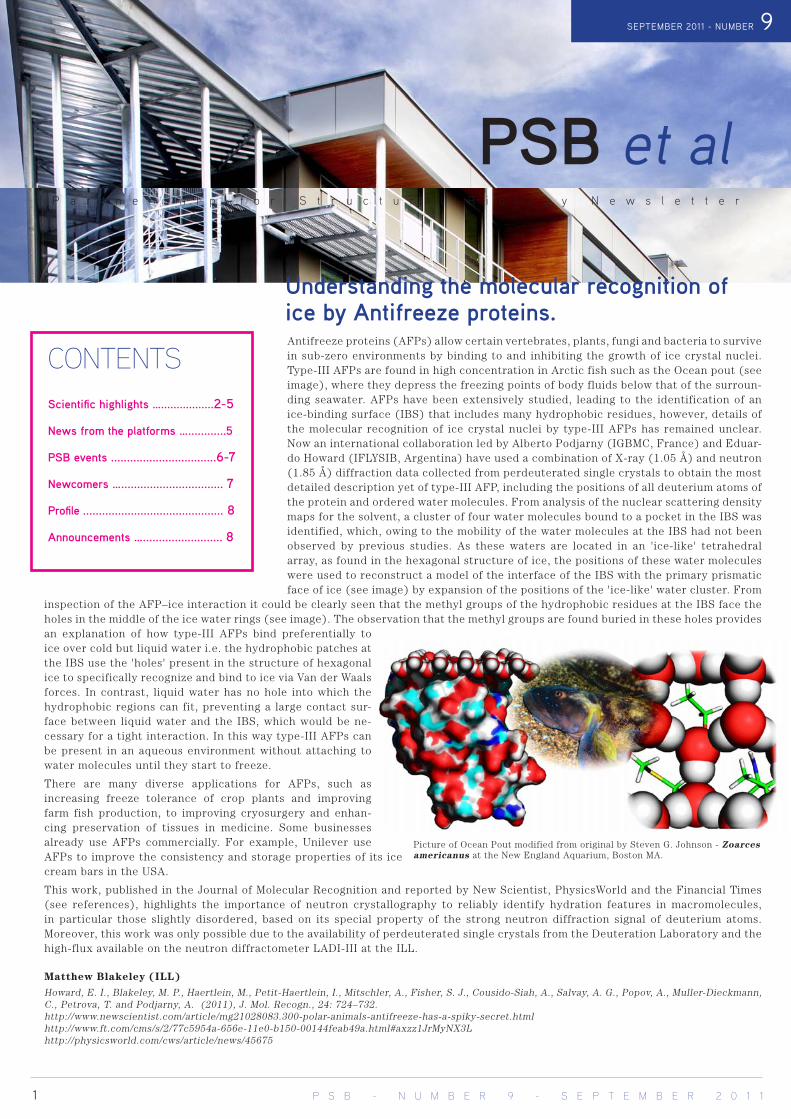
Antifreeze proteins (AFPs) allow certain vertebrates, plants, fungi and bacteria to survive in sub-zero environments by binding to and inhibiting the growth of ice crystal nuclei. Type-III AFPs are found in high concentration in Arctic fish such as the Ocean pout (see image), where they depress the freezing points of body fluids below that of the surroun-ding seawater. AFPs have been extensively studied, leading to the identification of an ice-binding surface (IBS) that includes many hydrophobic residues, however, details of the molecular recognition of ice crystal nuclei by type-III AFPs has remained unclear. Now an international collaboration led by Alberto Podjarny (IGBMC, France) and Eduar-do Howard (IFLYSIB, Argentina) have used a combination of X-ray (1.05 Å) and neutron (1.85 Å) diffraction data collected from perdeuterated single crystals to obtain the most detailed description yet of type-III AFP, including the positions of all deuterium atoms of the protein and ordered water molecules. From analysis of the nuclear scattering density maps for the solvent, a cluster of four water molecules bound to a pocket in the IBS was identified, which, owing to the mobility of the water molecules at the IBS had not been observed by previous studies. As these waters are located in an 'ice-like' tetrahedral array, as found in the hexagonal structure of ice, the positions of these water molecules were used to reconstruct a model of the interface of the IBS with the primary prismatic face of ice (see image) by expansion of the positions of the 'ice-like' water cluster. From
inspection of the AFP–ice interaction it could be clearly seen that the methyl groups of the hydrophobic residues at the IBS face the holes in the middle of the ice water rings (see image). The observation that the methyl groups are found buried in these holes provides an explanation of how type-III AFPs bind preferentially to ice over cold but liquid water i.e. the hydrophobic patches at the IBS use the 'holes' present in the structure of hexagonal ice to specifically recognize and bind to ice via Van der Waals forces. In contrast, liquid water has no hole into which the hydrophobic regions can fit, preventing a large contact sur-face between liquid water and the IBS, which would be ne-cessary for a tight interaction. In this way type-III AFPs can be present in an aqueous environment without attaching to water molecules until they start to freeze.
There are many diverse applications for AFPs, such as increasing freeze tolerance of crop plants and improving farm fish production, to improving cryosurgery and enhan-cing preservation of tissues in medicine. Some businesses already use AFPs commercially. For example, Unilever use AFPs to improve the consistency and storage properties of its ice cream bars in the USA.
This work, published in the Journal of Molecular Recognition and reported by New Scientist, PhysicsWorld and the Financial Times (see references), highlights the importance of neutron crystallography to reliably identify hydration features in macromolecules, in particular those slightly disordered, based on its special property of the strong neutron diffraction signal of deuterium atoms. Moreover, this work was only possible due to the availability of perdeuterated single crystals from the Deuteration Laboratory and the high-flux available on the neutron diffractometer LADI-III at the ILL.
Matthew Blakeley (ILL)
Howard, E. I., Blakeley, M. P., Haertlein, M., Petit-Haertlein, I., Mitschler, A., Fisher, S. J., Cousido-Siah, A., Salvay, A. G., Popov, A., Muller-Dieckmann, C., Petrova, T. and Podjarny, A. (2011), J. Mol. Recogn., 24: 724–732.http://www.newscientist.com/article/mg21028083.300-polar-animals-antifreeze-has-a-spiky-secret.htmlhttp://www.ft.com/cms/s/2/77c5954a-656e-11e0-b150-00144feab49a.html#axzz1JrMyNX3Lhttp://physicsworld.com/cws/article/news/45675
P a r t n e r s h i p f o r S t r u c t u r a l B i o l o g y N e w s l e t t e r
SEPTEMBER 2011 - NUMBER 9
CONTENTSScientific highlights ….................2-5
News from the platforms …............5
PSB events .................................6-7
Newcomers …................................ 7
Profile ............................................ 8
Announcements …......................... 8
Understanding the molecular recognition of ice by Antifreeze proteins.
1 P S B - N U M B E R 9 - S E P T E M B E R 2 0 1 1
PSB et al
Picture of Ocean Pout modified from original by Steven G. Johnson - Zoarces americanus at the New England Aquarium, Boston MA.

2 P S B - N U M B E R 9 - S E P T E M B E R 2 0 1 1
Scientific highlights
The solution of the so-called “phase problem” has always been one of the main bottlenecks in the determination of new protein structures by X-ray crystallography. Experimental X-ray diffrac-tion datasets lack information about the phase of the diffracted X-rays. This missing information is critical for calculating the electron density maps used to elucidate molecular structures. Consequently, several different techniques have been developed to solve the “phase problem” and determine initial phases.In recent years the use of UV radiation to induce specific damage in the protein crystal has been gaining interest in the protein crystallography community as an alternative approach for solving the phase problem. The technique, named UV-RIP (for UV radiation damage induced phasing) consists of collecting an initial complete dataset, then exposing the crystal to UV radia-tion for few minutes before finally collecting a second dataset. The differences between the two datasets correspond to the sites in the protein damaged by UV radiation. These sites can be used to calculate initial phases in much the same way as an isomorphous replacement experiment, where two different crys-tals are needed (one of which has heavy atoms incorporated). For a few months now (see PSB newsletter #7) the setup to perform this experiment has been available to the users of ID23-1 through the mxCuBE GUI, the interface for macromolecular data collec-tion at the ESRF.
To date a handful of protein structures have been solved by applying UV-RIP to parts of proteins that are sensitive to damage by UV radiation, like disulphide groups or photosensitive com-pounds. Recently, we revealed that the UV-RIP technique can also be applied to seleno-labelled proteins, which contain se-lenomethionine in the place of methionine. Seleno-labelling is the most common way to produce crystal derivatives for deter-mining protein structures. We have shown that, quite surprisin-gly, selenomethionine strongly absorbs UV radiation and can be damaged after a few minutes of exposure. The damage induces sufficient differences between the two diffraction datasets to al-low the the 3D structure to be solved using the UV-RIP phasing protocol. The use of a small-size X-ray beam (tens of micron) per-mits the two datasets to be collected from different regions of the same crystal. In this way the damage produced while collecting the second dataset is limited. The quality of the phases and of the calculated map is sufficient to allow most of the protein structure to be built. The evident advantage of this technique, compared to the well-established protocol that benefits from the anomalous signal measured from selenium atoms, is that there is no need to tune the beam wavelength. Consequently, it is possible to use the UV-RIP protocol on seleno-labelled proteins with in-house X-ray sources such as standard rotating anodes. The UV-RIP data can
also be combined with anomalous scattering data. In these cases both anomalous scattering and UV damage can be used to deter-mine the position of selenium atoms, in combination (so-called UV-RIPAS) or independently. The latter approach can be particu-larly successful in cases where there are too few selenomethio-nines to obtain good phases using anomalous phasing protocols alone (SAD/MAD), or where the anomalous signal is not sufficient to unambiguously determine the positions of the selenium atoms
Daniele de Sanctis (ESRF).de Sanctis, D.; Tucker, PA.; Panjikar, S.; J Synchrotron Radiat. (2011) 18:374-80.Panjikar, S.; Mayerhofer, H.; Tucker, PA.; Mueller-Dieckmann, J.; de Sanc-tis, D.; (2011) Acta Crystallogr. D67:32-44.
UV-induced phasing of Seleno-labelled proteins
Details of seleno-labelled protein solved by UV-RIP. Electron density difference map calculated between the two datasets. Green mesh re-presents positive difference, showing the UV damage ef-fect, red mesh negative dif-ference. The two closely lo-cated meshes show that the selenomethionine has flipped from one position (green) to another (red) after exposure to UV. Below: absorption of some amino acids in solution. Selenomethionine is repre-sented in purple.
Bacteria have a complicated set of stress responses that give them the ability to thrive in harsh conditions. Acid resistance in enteric bacteria such as Escherichia coli or Salmonella is one important example of such an adaptation. In the infection process, enteric bacteria are inevitably exposed to the gastric acid in the stomach (pH 2.5), which serves as a natural antibiotic barrier. Therefore, understanding the regulatory me-chanisms and pathways of the bacterial acid stress response is crucial to developing new strategies against enteric bacterial infections.
Irina Gutsche and her collaborator Walid Houry (University of Toronto,
Canada) are investigating the acid stress response system based on the inducible lysine decarboxylase named LdcI, which plays a role in main-taining the internal pH of the bacteria and counteracts the acidity of the surrounding medium. They discovered that LdcI tightly binds to alar-mone, an intracellular signalling molecule produced in response to harsh environmental conditions (Kanjee et al., 2011). While high levels of LdcI are required in response to acid stress, cells also need to keep the intra-cellular pool of lysines constant, and to rapidly shut down LdcI activity when the pH gets back to neutral. Thus, alarmone which allosterically
Understanding enteric bacteria acid stress response
P S B - N U M B E R 9 - S E P T E M B E R 2 0 1 1 3
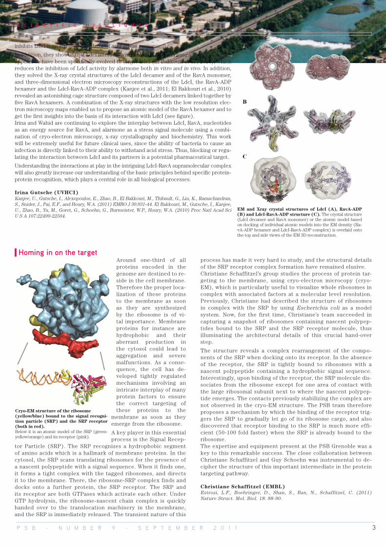
EM and Xray crystal structures of LdcI (A), RavA-ADP (B) and LdcI-RavA-ADP structure (C). The crystal structure (LdcI decamer and RavA monomer) or the atomic model based on docking of individual atomic models into the EM density (Ra-vA-ADP hexamer and LdcI-RavA-ADP complex) is overlaid onto the top and side views of the EM 3D reconstruction.
S C I E N T I F I C H I G H L I G H T S
Around one-third of all proteins encoded in the genome are destined to re-side in the cell membrane. Therefore the proper loca-lization of these proteins to the membrane as soon as they are synthesized by the ribosome is of vi-tal importance. Membrane proteins for instance are hydrophobic and their aberrant production in the cytosol could lead to aggregation and severe malfunctions. As a conse-quence, the cell has de-veloped tightly regulated mechanisms involving an intricate interplay of many protein factors to ensure the correct targeting of these proteins to the
membrane as soon as they emerge from the ribosome.
A key player in this essential process is the Signal Recep-
tor Particle (SRP). The SRP recognizes a hydrophobic segment of amino acids which is a hallmark of membrane proteins. In the cytosol, the SRP scans translating ribosomes for the presence of a nascent polypeptide with a signal sequence. When it finds one, it forms a tight complex with the tagged ribosomes, and directs it to the membrane. There, the ribosome-SRP complex finds and docks onto a further protein, the SRP receptor. The SRP and its receptor are both GTPases which activate each other. Under GTP hydrolysis, the ribosome-nascent chain complex is quickly handed over to the translocation machinery in the membrane, and the SRP is immediately released. The transient nature of this
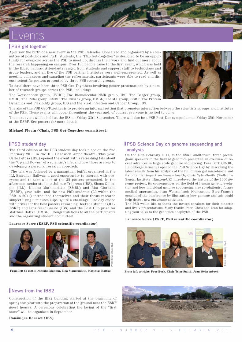
process has made it very hard to study, and the structural details of the SRP receptor complex formation have remained elusive. Christiane Schaffitzel’s group studies the process of protein tar-geting to the membrane, using cryo-electron microcopy (cryo-EM), which is particularly useful to visualize whole ribosomes in complex with associated factors at a molecular level resolution. Previously, Christiane had described the structure of ribosomes in complex with the SRP by using Escherichia coli as a model system. Now, for the first time, Christiane’s team succeeded in capturing a snapshot of ribosomes containing nascent polypep-tides bound to the SRP and the SRP receptor molecule, thus illuminating the architectural details of this crucial hand-over step.
The structure reveals a complex rearrangement of the compo-nents of the SRP when docking onto its receptor. In the absence of the receptor, the SRP is tightly bound to ribosomes with a nascent polypeptide containing a hydrophobic signal sequence. Interestingly, upon binding of the receptor, the SRP molecule dis-sociates from the ribosome except for one area of contact with the large ribosomal subunit next to where the nascent polypep-tide emerges. The contacts previously stabilizing the complex are not observed in the cryo-EM structure. The PSB team therefore proposes a mechanism by which the binding of the receptor trig-gers the SRP to gradually let go of its ribosome cargo, and also discovered that receptor binding to the SRP is much more effi-cient (50-100 fold faster) when the SRP is already bound to the ribosome. The expertise and equipment present at the PSB Grenoble was a key to this remarkable success. The close collaboration between Christiane Schaffitzel and Guy Schoehn was instrumental to de-cipher the structure of this important intermediate in the protein targeting pathway.
Christiane Schaffitzel (EMBL)Estrozi, L.F., Boehringer, D., Shan, S., Ban, N., Schaffitzel, C. (2011) Nature Struct. Mol. Biol. 18: 88-90.
Homing in on the target
Cryo-EM structure of the ribosome (yellow/blue) bound to the signal recogni-tion particle (SRP) and the SRP receptor (both in red). Below it is an atomic model of the SRP (green-yellow/orange) and its receptor (pink).
inhibits LdcI is an effective regulator of acid stress response.
In addition, they showed that LdcI interacts with a novel AAA+ ATPase called RavA, which appears to have been specifically evolved to target LdcI (El Bakkouri et al., 2010), which reduces the inhibition of LdcI activity by alarmone both in vitro and in vivo. In addition, they solved the X-ray crystal structures of the LdcI decamer and of the RavA monomer, and three-dimensional electron microscopy reconstructions of the LdcI, the RavA-ADP hexamer and the LdcI-RavA-ADP complex (Kanjee et al., 2011; El Bakkouri et al., 2010)revealed an astonishing cage structure composed of two LdcI decamers linked together by five RavA hexamers. A combination of the X-ray structures with the low resolution elec-tron microscopy maps enabled us to propose an atomic model of the RavA hexamer and to get the first insights into the basis of its interaction with LdcI (see figure). Irina and Wahid are continuing to explore the interplay between LdcI, RavA, nucleotides as an energy source for RavA, and alarmone as a stress signal molecule using a combi-nation of cryo-electron microscopy, x-ray crystallography and biochemistry. This work will be extremely useful for future clinical uses, since the ability of bacteria to cause an infection is directly linked to their ability to withstand acid stress. Thus, blocking or regu-lating the interaction between LdcI and its partners is a potential pharmaceutical target.
Understanding the interactions at play in the intriguing LdcI-RavA supramolecular complex will also greatly increase our understanding of the basic principles behind specific protein-protein recognition, which plays a central role in all biological processes.
Irina Gutsche (UVHCI)Kanjee, U., Gutsche, I., Alexopoulos, E., Zhao, B., El Bakkouri, M., Thibault, G., Liu, K., Ramachandran, S., Snider, J., Pai, E.F., and Houry, W.A. (2011) EMBO J 30:931-44. El Bakkouri, M., Gutsche, I., Kanjee, U., Zhao, B., Yu, M., Goret, G., Schoehn, G., Burmeister, W.P., Houry, W.A. (2010) Proc Natl Acad Sci U S A 107:22499-22504.

4 P S B - N U M B E R 9 - S E P T E M B E R 2 0 1 1
S C I E N T I F I C H I G H L I G H T S
Numerous cancers have their origins in abnormal activation of the protein kinase signaling pathway, making kinases privileged targets for “signal transduction therapies” and for developing new treatments against cancer.
Among these kinases, protein kinase-2 (named CK2) is able to regulate a wide range of signaling pathways controlling the balance between cell proliferation and apoptosis. A high expres-sion and a high enzymatic activity of CK2 are also observed in the case of many cancers such as prostate, breast cancer and leukemia and several classes of CK2 inhibitors are exhibiting in vitro efficacy and specificity. However, so far only one molecule, CX-4945, has been reported exhibiting an in vivo effect, under-lining the need to generate new active molecules based on such inhibitors.Scientists representing a large spectrum of expertise, such as molecular and cellular biology, biochemistry, chemistry or crys-tallography, from well-known French research institutes (Institut Curie in Paris, Institut Parisien de Chimie Moléculaire and IBS in Grenoble), were brought together by Claude Cochet from the iRTSV-CEA-Grenoble to develop a multidisciplinary strategy to - 1) screen large libraries of small compounds using high through-put technologies - 2) design and synthetize derivatives from the most promising compounds - 3) characterize in vitro, in cellulo, in vivo and at the atomic level, the potency of those newly desi-gned inhibitors. This fruitful collaboration led to identify several families of new nanomolar inhibitors of CK2 after screening more than 11500 different compounds.
Difurandicarboxylic Acid is one of these new inhibitors. Its selective profiling shows an exceptional targeting for CK2 and Pim kinases, both enzymes involved in similar but not redundant pathophysiological functions. By a structure-function approach, Jean-Baptiste Reiser (IBS) and his collaborators described the binding mode of these inhibitors on both kinases and identified essential structural determinants for their activity (Figure, Panel A). Although, these compounds cannot enter the cell and there-fore have no anti-tumor effects, they provide an excellent basis to develop novel inhibitors for targeted cancer therapies (1).
In addition to Difurandicarboxylic Acid, this work also identified molecules derived from Ellipticine, a berrywood tree poison used in ovarian and breast cancer treatments, as CK2 kinase inhibi-tors. Some of these analogs are 10 to 50 times more powerful than the original Ellipticine mainly because of a new binding mode in the catalytic pocket of CK2 (Figure, Panel B). Remarkably, these latter inhibitors are able to reduce cancerous cell proliferation in vitro and one of them shows anti-tumor effects in a mouse xeno-graft model of human cancer cells without any noticeable side effects (2).
These results, along with the identification of others inhibitor fa-milies targeting exosites by Claude Cochet’s group, are leading to promising new perspectives for the design of original drugs and future therapies against cancers.
Jean-Baptiste Reiser (IBS)
(1) M. Lopez-Ramos, R. Prudent, V. Moucadel, C. F. Sautel,_C. Barette, L. Lafanechere, L. Mouawad, D. Grierson, F. Schmidt, J.-C. Florent, P. Filippakopoulos, A.N. Bullock, S. Knapp, J.-B. Reiser & C. Cochet. (2010) FASEB J. 9: 3171-3185.(2) R. Prudent, V. Moucadel, C.-H. Nguyen, C. Barette, F. Schmidt, J.-C. Florent, L. Lafanechere, C. F Sautel, E. Duchemin-Pelletier, E. Spreux, O. Filhol, J.-B. Reiser & C. Cochet. (2010) Cancer Res. 23 : 9865-9874.
Zoom on catalytic site of the crystal structure of CK2 in complex with (A) a Difuranedicarboxylate Acid analog and (B) an Ellipticine analog (in yellow)
New leads for future anti-cancer drugs
Nuclear hormone receptors (NHR) regulate key functions in the body and are implicated in many major diseases such as diabetes, obesity, dyslipidemia, inflammation and osteoporosis. About 50 different NHRs have been identified that are assumed to be of relevance for humans. Around 10% of today's medicines act through nuclear receptors that are activated naturally by the hor-monal system.
Hormones are transported by the blood stream to one or more target organs, where they bind to a protein receptor. Nuclear receptors function as a switch for the transcription of genes in a
specific cell and therefore control what proteins the cell contains. Some nuclear receptors are activated or inhibited by hormones, others by vitamins, fatty acids or bile acids, or other ligands. By designing drugs that, like natural ligands, fit into binding pockets at the nuclear receptors, gene expression can be used in treating many diseases and medical conditions. NHRs act as ligand-indu-cible transcription factors which regulate gene expression and share a common modular organization with a variable N-terminal domain, a conserved DNA-binding domain and the ligand-binding domain that represses or activates gene transcription upon ligand
A
B
Insight into nuclear receptors – regulators of cellular key functions

P S B - N U M B E R 9 - S E P T E M B E R 2 0 1 1 5
S C I E N T I F I C H I G H L I G H T S
ESPRIT random library construct screening. ESPRIT uses genomics robotics to screen libraries of tens of thousands of random fragments of a single challenging protein to identify solubly expressing constructs for structural studies.
Academic structural biologists often work on proteins lacking accurate domain annotations. When the full-length protein cannot be expressed and a domain-focused approach is necessary, problems arise because it is unclear how to design high yielding, soluble expression constructs.
Some proteins have little or no sequence similarity to others and this prevents domain identification using multiple sequence alignments. Even when a soluble construct is obtained, poorly defined structural domains may confound crystallisation attempts. We are all familiar with these situations; in many cases they are what keep our proteins “hot” and out of the PDB.
The ESPRIT technology has been developed in the Hart lab at EMBL to express proteins whose domain boundaries are difficult to predict. It does not aim to replace classic PCR cloning based upon careful inspection of the protein sequence, but provides a rescue strategy when this fails. ESPRIT, which stands for “expression of soluble proteins by random incremental truncation”, is a directed evolution-type process combining random deletion mutagenesis with high throughput solubility screening.
We use exonucleases to truncate the ends of the target gene sequence for varying amounts of time, thereby generating all possible construct ends. Following ligation and transformation, up to 28,000 constructs per gene are isolated using colony picking robots to form high density colony arrays for protein expression testing.
To detect soluble constructs, the efficiency of in vivo biotinylation of a fused C-ter peptide is measured using fluorescent probes. Recent developments of the ESPRIT technology include a co-expression system adapted to protein complexes (An et al., 2011a) and genetic selection to eliminate out-of-frame constructs from the library, a step that greatly enhances the screening power of ESPRIT and its application to more challenging systems (An et al., 2011b).
Over the last seven years, the ESPRIT platform has been visited by European scientists who have brought their problematic targets for screening. This has now been formalised by an EU grant, P-CUBE, which
funds their travel and experimentation costs. About half our users have returned home with soluble, purified proteins, after typically one to two years of failed attempts. A recent highlight was the identification and so-luble expression of the terminase domain from HCMV that resulted in its crystal structure (Nadal et al., 2010). Applications beyond structural bio-logy have included vaccination of animals with domains from pathogen proteins and using soluble constructs to raise monoclonal antibodies.
Darren Hart (EMBL)An, Y., Meresse, P., Mas, P. J., and Hart, D. J. (2011a) PLoS ONE 6, e16261.An, Y., Yumerefendi, H., Mas, P. J., Chesneau, A., and Hart, D. J. (2011b) J. Struct. Biol. April 16 [Epub ahead of print].Nadal, M., Mas, P. J., Blanco, A. G., Arnan, C., Solà, M., Hart, D. J., and Coll, M. (2010) Proc. Natl. Acad. Sci. U.S.A 107, 16078-16083.
A quasi-atomic model of the NHR heterodimer (RXR, cyan; VDR, orange) in complex with DNA (red). The length of the whole complex is 140 Å. The hinge region that connects the DNA-binding and Li-gand-binding domains of the NHR subunits is indicated.
News from the platformsNew developments from the ESPRIT platform
binding and/or cofactor positioning. The ligand binding domain possesses nume-rous interaction surfaces for NHRs but also many other partners, co-repressors, coactivators and other bridging factors that participate in signal transduction toward the basal transcriptional machinery.
A recently published paper in Nature Structural & Molecular Biology, by Rochel and coworkers (Dino Moras team, IGBMC, Illkirch, France), provides a structural basis for understanding the role of DNA in the spatial organization of NHR hetero-dimers in complexes with coactivators. Complementary multi-resolution structural biology approaches were used to address the solution structures of three heterodi-mers formed between the human retinoid X receptor (RXR) and either the human retinoic acid receptor (RARα), the human vitamin D nuclear receptor (VDR) or the human peroxisome proliferator–activated receptor. A quasi-atomic model refined as a rigid-body in the ab initio envelope is shown in the image for one of the hete-rodimers.
Small angle X-ray and neutron scattering combined with isotope labeling (deu-teration carried out in the ILL-EMBL deuteration platform) and Fluorescence Resonance Energy Transfer were used to study the NHR interacting domains of coactivators bound to their cognate direct repeat elements. Neutron scattering and “match-out-labeling” were used to validate SAXS derived ab initio models for the complexes. The structures of the complexes show an extended asymmetric shape and point to the important role played by the hinge domains in establishing and maintaining the integrity of the structures. The results reveal two additional features: the conserved position of the ligand-binding domains at the 5' ends of the target DNAs and the binding of only one coactivator molecule per heterodimer.
Michael Haertlein & Phil Callow (ILL)Rochel, N.; Ciesielski, F.; Godet, J.; Moman, E.; Roessle, M.; Peluso-Iltis, C.; Moulin, M.; Haertlein, M.; Callow, P.; Mély, Y.; Svergun, DI.; Moras, D.;(2011). Nature. Struct. Mol. Biol. 18:564-70.
6 P S B - N U M B E R 9 - S E P T E M B E R 2 0 1 1
PSB get togetherApril saw the birth of a new event in the PSB Calendar. Conceived and organised by a com-mittee of post-docs and Ph.D. students, the "PSB Get-Together" is designed to be an oppor-tunity for everyone across the PSB to meet up, discuss their work and find out more about the research happening on campus. Over 130 people came to the first event, which was held in the ILL20 hallway. Attendants ranged from students and support staff to technicians and group leaders, and all five of the PSB partner Institutes were well-represented. As well as meeting colleagues and sampling the refreshments, participants were able to read and dis-cuss scientific posters presented by three PSB research groups.
To date there have been three PSB Get-Togethers involving poster presentations by a num-ber of research groups across the PSB, including:
The Weissenhorn group, UVHCI; The Biomolecular NMR group, IBS; The Berger group, EMBL; The Pillai group, EMBL; The Cusack group, EMBL; The MX group, ESRF; The Protein Dynamics and Flexibility group, IBS and the Viral Infection and Cancer Group, IBS.
The aim of the PSB Get-Together is to provide an informal setting that promotes interaction between the scientists, groups and institutes of the PSB. These events will occur throughout the year and, of course, everyone is invited to come.
The next event will be held at the IBS on Friday 23rd September. There will also be a PSB Post-Doc symposium on Friday 25th November at the ESRF. See posters for more details.
Michael Plevin (Chair, PSB Get-Together committee).
Events
Pho
to: S
erge
Cla
isse
(IL
L)
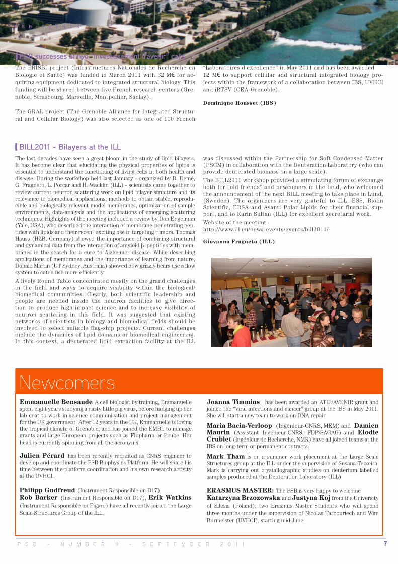
From left to right: Peer Bork, Chris Tyler-Smith, Jean Weissenbach
The third edition of the PSB student day took place on the 2nd February 2011 in the ILL Chadwick Amphitheatre. This year, Carlo Petosa (IBS) opened the event with a refreshing talk about the “Up and Downs” of a scientist’s life, and how those are key to developing a personal research approach.
The talk was followed by a gargantuan buffet organized in the ILL Entrance Hallway, a good opportunity to interact with eve-ryone and to take a look at the 25 posters presented. In the afternoon, senior students Juliette Trépreau (IBS), Shona Gilles-pie (ILL), Nikolas Mathioudakis (EMBL) and Rita Giordano (ESRF), gave talks, and the new PhD students (20 within the PSB in 2011) introduced themselves and their thesis research subject using 2 minutes clips. Quite a challenge! The day ended with prizes for the best posters rewarding Deeksha Munnur (ILL/ESRF) and Lina Siauciunaite (IBS) and the Best Clip prize for Matthias Haffke (EMBL). Congratulations to all the participants and the organizing student committee!
Laurence Serre (ESRF, PSB scientific coordinator)
PSB student day
News from the IBS2
Pho
to: S
erge
Cla
isse
(IL
L)
From left to right: Deeshka Munnur, Lina Siauciunaite, Matthias Haffke
On the 18th February 2011, at the ESRF Auditorium, three presti-gious speakers in the field of genomics presented an overview of re-cent advances in large scale genome sequencing. Peer Bork (EMBL, Heidelberg-Germany) opened the PSB Science Day by describing the latest results from his analysis of the full human gut microbiome and its potential impact on human health; Chris Tyler-Smith (Wellcome Sanger Institute, Hinxton-UK) introduced the history of the 1000 ge-nome project, its consequences on the field of human genetic evolu-tion and how individual genome sequencing may revolutionise future medical approaches. Jean Weissenbach (Genoscope, Evry-France) concluded the conference by illustrating how genome analysis could help detect new enzymatic activities. The PSB would like to thank the invited speakers for their didactic and lively presentations. Many thanks Peer, Chris and Jean for adap-ting your talks to the genomics neophytes of the PSB.
Laurence Serre (ESRF, PSB scientific coordinator)
PSB Science Day on genome sequencing and analysis
Construction of the IBS2 building started at the beginning of spring this year with the preparation of the ground near the ESRF guest houses. A ceremony celebrating the laying of the “first stone” will be organized in September.
Dominique Housset (IBS)
P S B - N U M B E R 9 - S E P T E M B E R 2 0 1 1 7
The last decades have seen a great bloom in the study of lipid bilayers. It has become clear that elucidating the physical properties of lipids is essential to understand the functioning of living cells in both health and disease. During the workshop held last January - organized by B. Demé, G. Fragneto, L. Porcar and H. Wacklin (ILL) - scientists came together to review current neutron scattering work on lipid bilayer structure and its relevance to biomedical applications, methods to obtain stable, reprodu-cible and biologically relevant model membranes, optimization of sample environments, data-analysis and the applications of emerging scattering techniques. Highlights of the meeting included a review by Don Engelman (Yale, USA), who described the interaction of membrane-penetrating pep-tides with lipids and their recent exciting use in targeting tumors. Thomas Hauss (HZB, Germany) showed the importance of combining structural and dynamical data from the interaction of amyloid β peptides with mem-branes in the search for a cure to Alzheimer disease. While describing applications of membranes and the importance of learning from nature, Donald Martin (UT Sydney, Australia) showed how grizzly bears use a flow system to catch fish more efficiently.
A lively Round Table concentrated mostly on the grand challenges in the field and ways to acquire visibility within the biological/biomedical communities. Clearly, both scientific leadership and people are needed inside the neutron facilities to give direc-tion to produce high-impact science and to increase visibility of neutron scattering in this field. It was suggested that existing networks of scientists in biology and biomedical fields should be involved to select suitable flag-ship projects. Current challenges include the dynamics of lipid domains or biomedical engineering. In this context, a deuterated lipid extraction facility at the ILL
was discussed within the Partnership for Soft Condensed Matter (PSCM) in collaboration with the Deuteration Laboratory (who can provide deuterated biomass on a large scale).
The BILL2011 workshop provided a stimulating forum of exchange both for “old friends” and newcomers in the field, who welcomed the announcement of the next BILL meeting to take place in Lund, (Sweden). The organizers are very grateful to ILL, ESS, Biolin Scientific, EBSA and Avanti Polar Lipids for their financial sup-port, and to Karin Sultan (ILL) for excellent secretarial work.
Website of the meeting - http://www.ill.eu/news-events/events/bill2011/
Giovanna Fragneto (ILL)
BILL2011 - Bilayers at the ILL
PSB successes at two “Investissement d'avenir” calls
P S B E V E N T S
NewcomersEmmanuelle Bensaude A cell biologist by training, Emmanuelle spent eight years studying a nasty little pig virus, before hanging up her lab coat to work in science communication and project management for the UK government. After 12 years in the UK, Emmanuelle is loving the tropical climate of Grenoble, and has joined the EMBL to manage grants and large European projects such as Flupharm or Pcube. Her head is currently spinning from all the acronyms.
Julien Pérard has been recently recruited as CNRS engineer to develop and coordinate the PSB Biophysics Platform. He will share his time between the platform coordination and his own research activity at the UVHCI.
Philipp Gudfreud (Instrument Responsible on D17), Rob Barker (Instrument Responsible on D17), Erik Watkins (Instrument Responsible on Figaro) have all recently joined the Large Scale Structures Group of the ILL.
Joanna Timmins has been awarded an ATIP/AVENIR grant and joined the "Viral infections and cancer" group at the IBS in May 2011. She will start a new team to work on DNA repair.
Maria Bacia-Verloop (Ingénieur-CNRS, MEM) and Damien Maurin (Assistant Ingénieur-CNRS, FDP/SAGAG) and Elodie Crublet (Ingénieur de Recherche, NMR) have all joined teams at the IBS on long-term or permanent contracts.
Mark Tham is on a summer work placement at the Large Scale Structures group at the ILL under the supervision of Susana Teixeira. Mark is carrying out crystallographic studies on deuterium labelled samples produced at the Deuteration Laboratory (ILL).
ERASMUS MASTER: The PSB is very happy to welcome Katarzyna Brzozowska and Justyna Koj from the University of Silesia (Poland), two Erasmus Master Students who will spend three months under the supervision of Nicolas Tarbouriech and Wim Burmeister (UVHCI), starting mid June.
The FRISBI project (Infrastructures Nationales de Recherche en Biologie et Santé) was funded in March 2011 with 32 M€ for ac-quiring equipment dedicated to integrated structural biology. This funding will be shared between five French research centers (Gre-noble, Strasbourg, Marseille, Montpellier, Saclay).
The GRAL project (The Grenoble Alliance for Integrated Structu-ral and Cellular Biology) was also selected as one of 100 French
“Laboratoires d'excellence” in May 2011 and has been awarded 12 M€ to support cellular and structural integrated biology pro-jects within the framework of a collaboration between IBS, UVHCI and iRTSV (CEA-Grenoble).
Dominique Housset (IBS)

The Partnership for Structural Biology (PSB) is a collaboration between a number of prestigious European and French scientific laboratories in Grenoble which has received support of from the EU FP6 programme. The PSB is unique in combining world leading user facilities for synchrotron X-ray and neutron scattering with NMR, electron microscopy, molecular biology and high throughput techniques on a single site together with strong projects in a broad range of structural biology, notably host-pathogen interactions.
ESRF
ContactsEditors: Emmanuelle Bensaude (EMBL), Dominique Housset (IBS), Ricardo Ferraz Leal (ESRF), Michael Plevin (IBS), Laurence Serre (ESRF), Susana Teixeira (ILL, Keele University).Layout: Virginie GuerardEmail: [email protected]
Announcements9th September 2011 - Dinshaw Patel (Sloan-Kettering Institute, NYC, USA) has been invited by the PSB and will present his latest results on the structural biology of RNA silencing and the histone/epigenetics code.
19-20th October: - “Neutrons in Biology and Biotechno-logy meeting” (http://www.ill.fr/nib2011). Whereas in recent decades there has been a major emphasis on high throughput techniques and high resolution information, it is becoming evident that there is now a far greater need for interdiscipli-nary approaches. This workshop focuses on the areas where neutron techniques go beyond traditional limitations by making use other methods such as X-ray diffraction, electron microscopy, NMR, SPR, ITC, etc.
21-23rd October - “Neutrons in Biology 2011” (http://www.ill.eu/news-events/events/nib2011/satellite-school-21-23rd-oct/). The NIB 2011 intensive School will provide extensive trai-ning on the application of neutrons and X-rays in crystallo-graphy, small-angle scattering, and reflection studies with an emphasis on the use of state-of-the-art data analysis and modelling software appropriate to each technique.
ProfileChloe ZubietaShe is lovely and full of smiles, despite being kept up all night by a teething child. Meet Chloe Zubieta, the recently appointed ESRF molecular biology lab manager.
So, Chloe, what does your new job involve?I supervise all the postdocs, PhD students and technicians working in the lab, make sure I give them wha-tever support they need to be successful, be it my expertise, some chemicals, or collaborations I can facilitate. I also have my own research projects, including new research on plant enzymes and families of transcription factors. And we work on a bug called Deinococcus radio-durans, which is incredibly resistant to both ionizing and ultra-violet radiation. Irradiation is a way to preserve food because it will cause the production of free radicals, which damage microbial and viral proteins and nucleic acids. This bacteria is a survivor, and it looks like it has a combination of anti-radiation protections, such as a lot of manganese (which mops up free radicals), carotenoids (which basically act like a sunscreen), and a protective surface layer.
How did you end up at ESRF?I’m an inorganic chemist, but I got interested in the structural biology of some plant enzymes during my PhD at the Salk Institute in La Jolla,
California. This is also where I met my husband, who is now working at EMBL.
Aha, so chemists don’t reproduce by budding then?No, this is what happens when you share lab space!I came to Grenoble to use the synchrotron during my PhD, and at the time, I carried my crystals onto the airplane in a mini-cooler, a bit like a lunchbox! I really liked it here, so decided to learn French and to come back to France for a post-doc. I did a three year postdoc in Stephen Cusack’s lab at EMBL, working on the adenovirus penton base, then went back to California for a while. But we were really missing France, and eventually came back to Grenoble in 2009 for another postdoc, and now this post at ESRF.
Wow, you seem to love France, so what’s your favourite thing about living here? It’d have to be the boulangerie. I live across the street from a great boulangerie, and every morning I get bread, except when I get pains au chocolat. If I time it well, I can get bread fresh out of the oven. How much better can it get?
Interviewed by Emmanuelle Bensaude (EMBL)
Pho
to: S
erge
Cla
isse
(IL
L)
8 P S B - N U M B E R 9 - S E P T E M B E R 2 0 1 1
![(RON) 14AUG 0.95 1.01 1.05 1.05 1.05 1.04 2.3% PSB Industries [FR] 11.6x 1.3x … · 2017-08-14 · APS (RON) 0.95 1.01 1.05 1.05 1.05 1.04 2.3% 11.6x 1.3x ROaE À 8.2% 0.7% 0.8%](https://img.pdfslide.net/doc/110x75/5f7f1b4bb4b94c6a071b1181/ron-14aug-095-101-105-105-105-104-23-psb-industries-fr-116x-13x-2017-08-14.jpg)


![Uso del teclado[1] eduar](https://img.pdfslide.net/doc/110x75/55b47177bb61ebac148b4663/uso-del-teclado1-eduar.jpg)