Embed Size (px)
Citation preview
PSEUDOMONAS
Pseudomonas aeruginosa has become an important cause of infection, especially in patients
with compromised host defense mechanisms. It is the most common pathogen isolated from
patients who have been hospitalized longer than 1 week, and it is a frequent cause of
nosocomial infections. Pseudomonal infections are complicated and can be life-threatening.
Signs and symptoms
Pseudomonal infections can involve the following parts of the body, with corresponding
symptoms and signs:
Respiratory tract (eg, pneumonia)
Bloodstream (bacteremia)
Heart (endocarditis)
CNS (eg, meningitis, brain abscess)
Ear (eg, otitis externa and media)
Eye (eg, bacterial keratitis, endophthalmitis)
Bones and joints (eg, osteomyelitis)
GI tract (eg, diarrhea, enteritis, enterocolitis)
Urinary tract
Skin
Physical findings depend on the site and nature of the infection, as follows:
Endocarditis: Fever, murmur, and positive blood culture findings; peripheral stigmata
such as Roth spots, Janeway lesions, Osler nodes, splinter hemorrhages, and
splenomegaly
Pneumonia: Rales, rhonchi, fever, cyanosis, retractions, and hypoxia; occasionally
shock; with cystic fibrosis, clubbing, increased anteroposterior (AP) diameter, and
malnutrition
GI tract: Fever, signs of dehydration, abdominal distention, and signs of peritonitis;
physical findings of Shanghai fever
Skin and soft tissue infections: Hemorrhagic and necrotic lesions, with surrounding
erythema; subcutaneous nodules, deep abscesses, cellulitis, and fasciitis; in burns,
black or violaceous discoloration or eschar
Skeletal infections: Local tenderness and a decreased range of motion; neurologic
deficits
Eye infections: Lid edema, conjunctival erythema and chemosis, and severe
mucopurulent discharge
Malignant otitis externa: Erythematous, swollen, and inflamed external auditory canal;
local lymphadenopathy
Bacteremia: Fever, tachypnea, and tachycardia; hypotension and shock; jaundice
Diagnosis
Laboratory studies that may be helpful include the following:
Complete blood count (CBC)
Blood cultures
In urinary tract infection (UTI), urinalysis
In pneumonia, culture of sputum and respiratory secretions, as well as blood gas
analysis
Wound and burn cultures and cultures from other body fluids and secretions according
to the clinical scenario
Gram stain and culture of CSF if meningitis is suspected
Pseudomonas aeruginosa is member of the Gamma Proteobacteria class of Bacteria. It is
aerobic bacterium belonging to the bacterial family Pseudomonadaceae.
GRAM stain
Pseudomonas aeruginosa is a Gram-negative rod measuring 0.5 to 0.8 µm by 1.5 to 3.0 µm.
Almost all strains are motile by means of a single polar flagellum.
Gram stain of Pseudomonas aeruginosa.
Cultivation on Blood agar
Pseudomonas aeruginosa has very simple nutritional requirements. It is often observed
"growing in distilled water", which is evidence of its minimal nutritional needs. In the
laboratory, the simplest medium for growth of Pseudomonas aeruginosa consists of acetate as
a source of carbon and ammonium sulfate as a source of nitrogen.
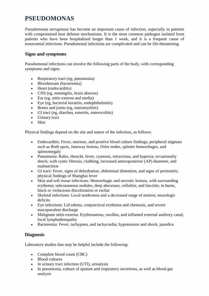
Pseudomonas aeruginosa on Blood Agar (typical metallic sheen).
P. aeruginosa isolates may produce three colony types. Natural isolates from soil or water
typically produce a small, rough colony. Clinical samples, in general, yield one or another of
two smooth colony types. One type has a fried-egg appearance which is large, smooth, with
flat edges and an elevated appearance. Another type, frequently obtained from respiratory and
urinary tract secretions, has a mucoid appearance, which is attributed to the production of
alginate slime. The smooth and mucoid colonies are presumed to play a role in colonization
and virulence.
Cultivation on Deoxycholate Citrate Agar
P. aeruginosa strains produce soluble pigments, the fluorescent pigment pyoverdin and the
blue pigment pyocyanin. The latter is produced abundantly in media of low-iron content and
functions in iron metabolism in the bacterium. Pyocyanin (from "pyocyaneus") refers to "blue
pus", which is a characteristic of suppurative infections caused by Pseudomonas aeruginosa.
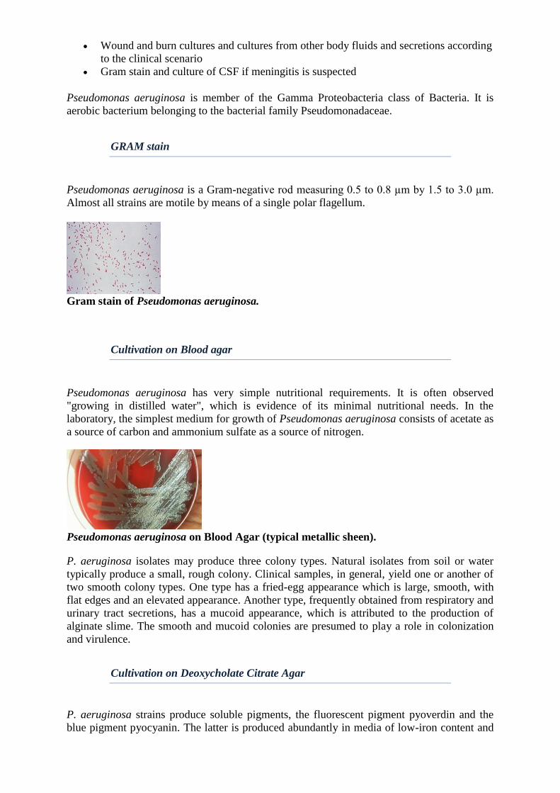
Pseudomonas aeruginosa on Deoxycholate Citrate Agar (lactose negative, H2S negative).
The green pigment pyoverdin and blue pigment pyocyanin are produced by many, but
not all, strains of Pseudomonas aeruginosa.
Cultivation on TSI (Hajn) Agar
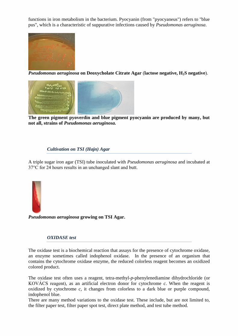
A triple sugar iron agar (TSI) tube inoculated with Pseudomonas aeruginosa and incubated at
37°C for 24 hours results in an unchanged slant and butt.
Pseudomonas aeruginosa growing on TSI Agar.
OXIDASE test
The oxidase test is a biochemical reaction that assays for the presence of cytochrome oxidase,
an enzyme sometimes called indophenol oxidase. In the presence of an organism that
contains the cytochrome oxidase enzyme, the reduced colorless reagent becomes an oxidized
colored product.
The oxidase test often uses a reagent, tetra-methyl-p-phenylenediamine dihydrochloride (or
KOVÁCS reagent), as an artificial electron donor for cytochrome c. When the reagent is
oxidized by cytochrome c, it changes from colorless to a dark blue or purple compound,
indophenol blue.
There are many method variations to the oxidase test. These include, but are not limited to,
the filter paper test, filter paper spot test, direct plate method, and test tube method.
Filter Paper Test Method 1. Soak a small piece of filter paper in 1% Kovács oxidase reagent and let dry.
2. Use a loop and pick a well-isolated colony from a fresh (18- to 24-hour culture) bacterial
plate and rub onto treated filter paper (please see Comments and Tips section for notes on
recommended media and loops).
3. Observe for color changes.
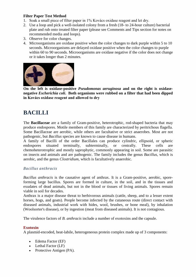
4. Microorganisms are oxidase positive when the color changes to dark purple within 5 to 10
seconds. Microorganisms are delayed oxidase positive when the color changes to purple
within 60 to 90 seconds. Microorganisms are oxidase negative if the color does not change
or it takes longer than 2 minutes.
On the left is oxidase-positive Pseudomonas aeruginosa and on the right is oxidase-
negative Escherichia coli. Both organisms were rubbed on a filter that had been dipped
in Kovács oxidase reagent and allowed to dry
BACILLI
The Bacillaceae are a family of Gram-positive, heterotrophic, rod-shaped bacteria that may
produce endospores. Motile members of this family are characterized by peritrichous flagella.
Some Bacillaceae are aerobic, while others are facultative or strict anaerobes. Most are not
pathogenic, but Bacillus species are known to cause disease in humans.
A family of Bacilli of the order Bacillales can produce cylindric, ellipsoid, or spheric
endospores situated terminally, subterminally, or centrally. These cells are
chemoheterotrophic and mostly saprophytic, commonly appearing in soil. Some are parasitic
on insects and animals and are pathogenic. The family includes the genus Bacillus, which is
aerobic, and the genus Clostridium, which is facultatively anaerobic.
Bacillus anthracis
Bacillus anthracis is the causative agent of anthrax. It is a Gram-positive, aerobic, spore-
forming large bacillus. Spores are formed in culture, in the soil, and in the tissues and
exudates of dead animals, but not in the blood or tissues of living animals. Spores remain
viable in soil for decades.
Anthrax is a major disease threat to herbivorous animals (cattle, sheep, and to a lesser extent
horses, hogs, and goats). People become infected by the cutaneous route (direct contact with
diseased animals, industrial work with hides, wool, brushes, or bone meal), by inhalation
(Woolsorter's disease), or by ingestion (meat from diseased animals). It is not contagious.
The virulence factors of B. anthracis include a number of exotoxins and the capsule.
Exotoxin A plasmid-encoded, heat-labile, heterogeneous protein complex made up of 3 components:
Edema Factor (EF)
Lethal Factor (LF)
Protective Antigen (PA).
In vivo, these three factors act synergistically (for toxic effects). The protective antigen binds
to surface receptors on eucaryotic cells and is subsequently cleaved by a cellular protease. The
larger C-terminal piece of PA remains bound to the receptor and then binds either EF or LF,
which enters the cell by endocytosis. Edema Factor, when inside the cells binds calmodulin-
dependent and acts as adenylate cyclase. Lethal factor's mechanism of action involves
activation of macrophages and production of cytokines which cause necrosis, fever, shock and
death. Individually, the three proteins have no known toxic activity. Antibodies to protective
antigens prevent PA binding to cells stop EF and LF entry.
Capsule The capsule consists of a polypeptide of D-glutamic acid which is encoded by a plasmid and
is anti-phagocytic. It is not a good immunogen and, even if any antibodies produced, they are
not protective against the disease.
Clinical diagnosis of anthrax can be confirmed by direct examination or culture. Fresh smears
of vesicular fluid, fluid from under the eschar, blood, or spleen or lymph node aspirates are
stained with polychrome methylene blue and examined for the characteristic blunt ended,
blue-black rods with a pink capsule. In case of a negative finding, the specimen can be
cultured on blood agar plates. Cultured organisms stain as Gram-positive long thin rods.
Bacillus cereus Bacillus cereus is a type of bacteria that produces toxins. These toxins can cause two types of
illness: one type characterized by diarrhea and the other, called emetic toxin, by nausea and
vomiting. These bacteria are present in foods and can multiply quickly at room temperature.
Most B. cereus are motile.
The symptoms of B. cereus diarrheal type food poisoning mimic those of Clostridium
perfringens food poisoning. The onset of watery diarrhea, abdominal cramps, and pain occurs
6-15 hours after consumption of contaminated food. Nausea may accompany diarrhea, but
vomiting (emesis) rarely occurs. Symptoms persist for 24 hours in most instances.
The emetic type of food poisoning is characterized by nausea and vomiting within 0.5 to 6 h
after consumption of contaminated foods. Occasionally, abdominal cramps and/or diarrhea
may also occur. Duration of symptoms is generally less than 24 h. The symptoms of this type
of food poisoning parallel those caused by Staphylococcus aureus foodborne intoxication.
Confirmation of B. cereus as the etiologic agent in a foodborne outbreak requires either (1)
isolation of strains of the same serotype from the suspect food and feces or vomitus of the
patient, (2) isolation of large numbers of a B. cereus serotype known to cause foodborne
illness from the suspect food or from the feces or vomitus of the patient, or (3) isolation of B.
cereus from suspect foods and determining their enterotoxigenicity by serological (diarrheal
toxin) or biological (diarrheal and emetic) tests. The rapid onset time to symptoms in the
emetic form of disease, coupled with some food evidence, is often sufficient to diagnose this
type of food poisoning.
Examination of Foods for B. cereus
1. Sampling
If the quantity of food to be examined is large, take representative samples of 50 g
each from different parts of the suspect food because contamination may be unevenly
distributed. If the food is a powder or consists of small discrete particles, then it should
be thoroughly mixed before taking samples.
2. Transporting and storage of samples
Transport samples promptly in insulated shipping containers with enough gel-type
refrigerant to maintain them at 6°C or below. Upon receipt in the laboratory, store the
samples at 4°C and analyze as soon as possible. If analysis cannot be started within 4
days after collection, freeze samples promptly and store at -20°C until examined.
Thaw at room temperature and proceed with analysis as usual. Maintain frozen
samples at -20°C until examined. Ship on dry ice to avoid thawing. Dehydrated foods
may be stored at room temperature and shipped without refrigeration.
GRAM stain
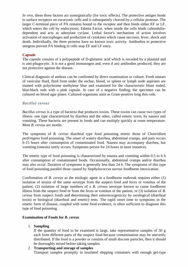
Bacillus anthracis
Bacillus anthracis is very large, Gram-positive, sporeforming rod (1 - 1.2µm in width x 3 -
5µm in length) arranged in chains. Genotypically and phenotypically it is very similar to
Bacillus cereus, the species have the same cellular size and morphology.
Bacillus anthracis Bacillus cereus
ENDOSPORE staining
Bacilli form oval, central to subterminal spores that do not swell the bacterial cell. Spores are
not present in clinical specimens. Endospores produced by Bacillus do not stain
easily. Endospores are stained by Wirtz-Conklin method where malachite green is used for
staining and heat is used to penetrate stain. The rest of the cell is then counterstained a light
pink-red with safranin.
Wirtz-Conklin’s method
1. Prepare a smear and heat gently to fix.
2. Flood the slide with 5 - 10% malachite green solution.
3. Leave the slide to stain for 45min or alternatively, the slide can be
heated gently to steaming for 3 - 6min, reapplying stain if it begins
to dry out.
4. Rinse under running tap water.
5. Counterstain with 0.5% safranin for 30sec.
6. Rinse and dry.
7. View slide under oil immersion with a light microscop.
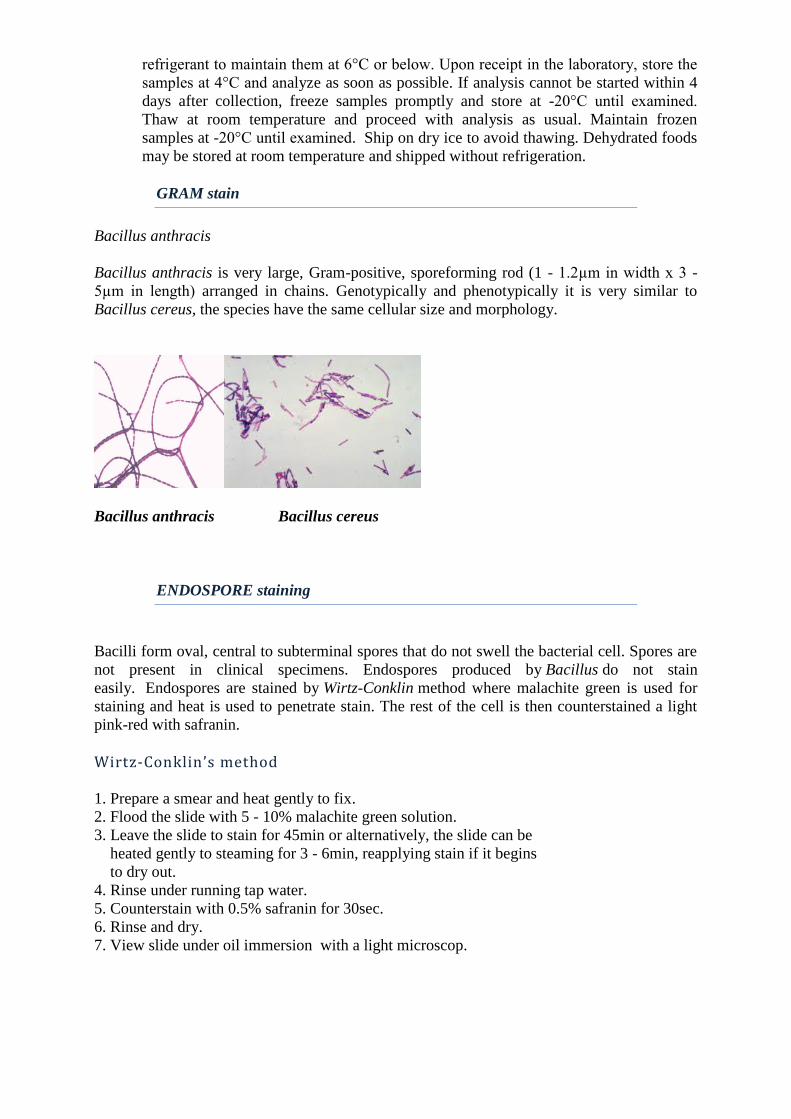
Bacillus anthracis -Wirtz-Conklin stain - bacterial spores stain green, vegetative cells
stain red.
Endospores
Some bacteria have the ability to enter a state of suspended animation when conditions are
unfavorable. An endospore is an extremely resistant dormant cell structure produced by some
bacterial species (term endospore: 'endo-' means 'inside' and '-spore' refers to the 'dormant
structure,') so the endospore is a structure formed inside the cell. There are many examples of
endospore-forming bacteria. The two most common are Clostridium and Bacillus. In
favorable conditions, these bacteria are actively growing and dividing cells. If a nutrient, such
as carbon or nitrogen, becomes scarce or if the population becomes too dense, the bacteria can
become stressed. They will enter a stasis phase, which is their equivalent of survival mode.
The bacteria can survive in stasis until better growth conditions return.
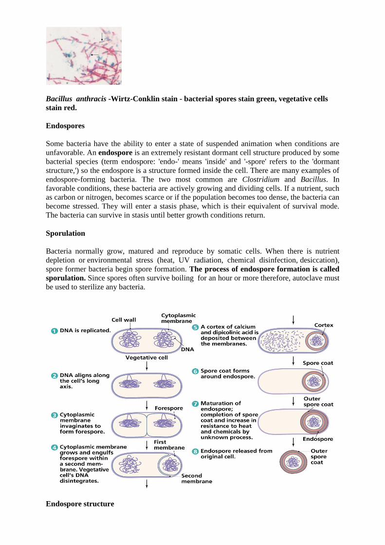
Sporulation
Bacteria normally grow, matured and reproduce by somatic cells. When there is nutrient
depletion or environmental stress (heat, UV radiation, chemical disinfection, desiccation),
spore former bacteria begin spore formation. The process of endospore formation is called
sporulation. Since spores often survive boiling for an hour or more therefore, autoclave must
be used to sterilize any bacteria.
Endospore structure
Properties of Endospores:
1. Core: The core is the spore protoplast. It contains a complete nucleus (chromosome), all of
the components of the protein-synthesizing apparatus, and an energy-generating system based
on glycolysis. A number of unique enzymes are formed (eg, dipicolinic acid synthetase).
Spores contain no ATP. The energy for germination is stored as 3-phosphoglycerate rather
than as ATP. The heat resistance of spores is due in part to their dehydrated state and in part
to the presence in the core of large amounts of calcium dipicolinate.
2. Spore Wall: The innermost layer surrounding the inner spore membrane is called the spore
wall. It contains normal peptidoglycan and becomes the cell wall of the germinating
vegetative cell.
3. Cortex: The cortex is the thickest layer of the spore envelope. Cortex peptidoglycan is
extremely sensitive to lysozyme, and its autolysis plays a role in spore germination.
4. Coat: The coat is composed of a keratin-like protein. The impermeability of this layer
confers on spores their relative resistance to antibacterial chemical agents.
5. Exosporium: The exosporium is a lipoprotein membrane containing some carbohydrate.
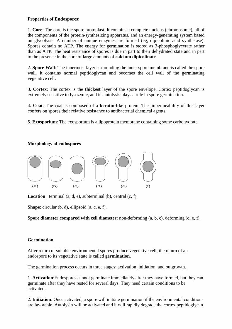
Morphology of endospores
Location: terminal (a, d, e), subterminal (b), central (c, f).
Shape: circular (b, d), ellipsoid (a, c, e, f).
Spore diameter compared with cell diameter: non-deforming (a, b, c), deforming (d, e, f).
Germination
After return of suitable environmental spores produce vegetative cell, the return of an
endospore to its vegetative state is called germination.
The germination process occurs in three stages: activation, initiation, and outgrowth.
1. Activation:Endospores cannot germinate immediately after they have formed, but they can
germinate after they have rested for several days. They need certain conditions to be
activated.
2. Initiation: Once activated, a spore will initiate germination if the environmental conditions
are favorable. Autolysin will be activated and it will rapidly degrade the cortex peptidoglycan.
Water is taken up, calcium dipicolinate is released, and a variety of spore constituents are
degraded by hydrolytic enzymes.
3. Outgrowth: Degradation of the cortex and outer layers results in the emergence of a new
vegetative cell consisting of the spore protoplast with its surrounding wall. Now, using
nutrients around, the cell can multiply again.
Cultivation on Blood agar
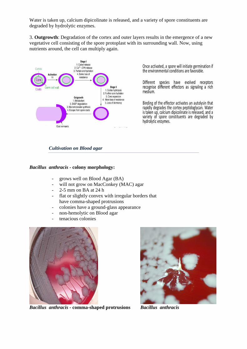
Bacillus anthracis - colony morphology:
- grows well on Blood Agar (BA)
- will not grow on MacConkey (MAC) agar
- 2-5 mm on BA at 24 h
- flat or slightly convex with irregular borders that
have comma-shaped protrusions
- colonies have a ground-glass appearance
- non-hemolytic on Blood agar
- tenacious colonies
Bacillus anthracis - comma-shaped protrusions Bacillus anthracis
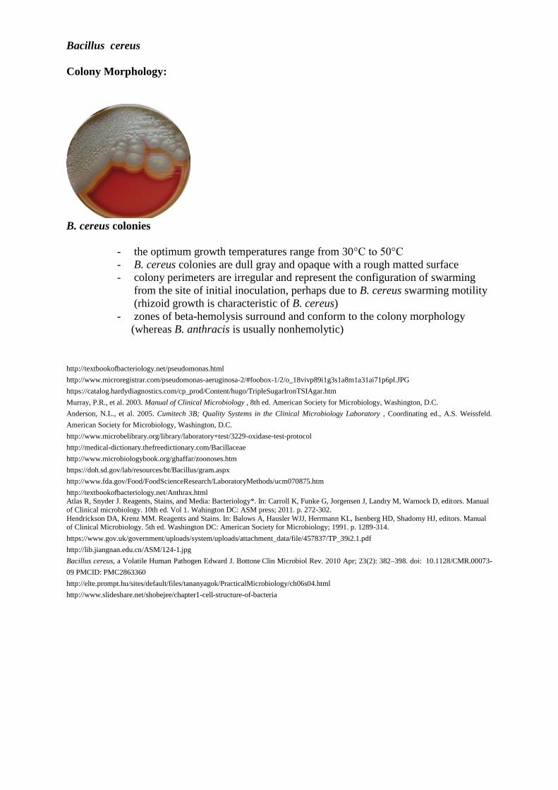
Bacillus cereus
Colony Morphology:
B. cereus colonies
- the optimum growth temperatures range from 30°C to 50°C
- B. cereus colonies are dull gray and opaque with a rough matted surface
- colony perimeters are irregular and represent the configuration of swarming
from the site of initial inoculation, perhaps due to B. cereus swarming motility
(rhizoid growth is characteristic of B. cereus)
- zones of beta-hemolysis surround and conform to the colony morphology
(whereas B. anthracis is usually nonhemolytic)
http://textbookofbacteriology.net/pseudomonas.html
http://www.microregistrar.com/pseudomonas-aeruginosa-2/#foobox-1/2/o_18vivp89i1g3s1a8m1a31ai71p6pl.JPG
https://catalog.hardydiagnostics.com/cp_prod/Content/hugo/TripleSugarIronTSIAgar.htm
Murray, P.R., et al. 2003. Manual of Clinical Microbiology , 8th ed. American Society for Microbiology, Washington, D.C.
Anderson, N.L., et al. 2005. Cumitech 3B; Quality Systems in the Clinical Microbiology Laboratory , Coordinating ed., A.S. Weissfeld.
American Society for Microbiology, Washington, D.C.
http://www.microbelibrary.org/library/laboratory+test/3229-oxidase-test-protocol
http://medical-dictionary.thefreedictionary.com/Bacillaceae
http://www.microbiologybook.org/ghaffar/zoonoses.htm
https://doh.sd.gov/lab/resources/bt/Bacillus/gram.aspx
http://www.fda.gov/Food/FoodScienceResearch/LaboratoryMethods/ucm070875.htm
http://textbookofbacteriology.net/Anthrax.html Atlas R, Snyder J. Reagents, Stains, and Media: Bacteriology*. In: Carroll K, Funke G, Jorgensen J, Landry M, Warnock D, editors. Manual
of Clinical microbiology. 10th ed. Vol 1. Wahington DC: ASM press; 2011. p. 272-302.
Hendrickson DA, Krenz MM. Reagents and Stains. In: Balows A, Hausler WJJ, Herrmann KL, Isenberg HD, Shadomy HJ, editors. Manual of Clinical Microbiology. 5th ed. Washington DC: American Society for Microbiology; 1991. p. 1289-314.
https://www.gov.uk/government/uploads/system/uploads/attachment_data/file/457837/TP_39i2.1.pdf
http://lib.jiangnan.edu.cn/ASM/124-1.jpg
Bacillus cereus, a Volatile Human Pathogen Edward J. Bottone Clin Microbiol Rev. 2010 Apr; 23(2): 382–398. doi: 10.1128/CMR.00073-
09 PMCID: PMC2863360
http://elte.prompt.hu/sites/default/files/tananyagok/PracticalMicrobiology/ch06s04.html
http://www.slideshare.net/shobejee/chapter1-cell-structure-of-bacteria
LISTERIA
Listeria monocytogenes is a Gram-positive rod-shaped bacterium. L. monocytogenes can be
isolated in soil, wood, and decaying matter in the natural environment. The principal route of
acquisition of Listeria is through the ingestion of contaminated food products. Listeria has
been isolated from prepared meat (eg, hot dogs, deli meat), dairy products, unwashed raw
vegetables, and seafood. Soft cheeses and unpasteurized milk have been the most frequently
incriminated dairy products. Invasive infection by L. monocytogenes causes the disease
listeriosis. When the infection is not invasive, any illness as a consequence of infection is
termed febrile gastroenteritis. Listeriosis is relatively rare and occurs primarily in newborn
infants, elderly patients, and patients who are immunocompromised. The two main clinical
manifestations are sepsis and meningitis. Meningitis is often complicated by encephalitis, a
pathology that is unusual for bacterial infections. The manifestations of listeriosis include
septicemia, meningitis or meningoencephalitis), encephalitis, corneal ulcer, pneumonia, and
intrauterine or cervical infections in pregnant women, which may result in spontaneous
abortion (second to third trimester) or stillbirth. Surviving neonates may suffer
granulomatosis infantiseptica (pyogenic granulomas distributed over the whole body) and
physical retardation.
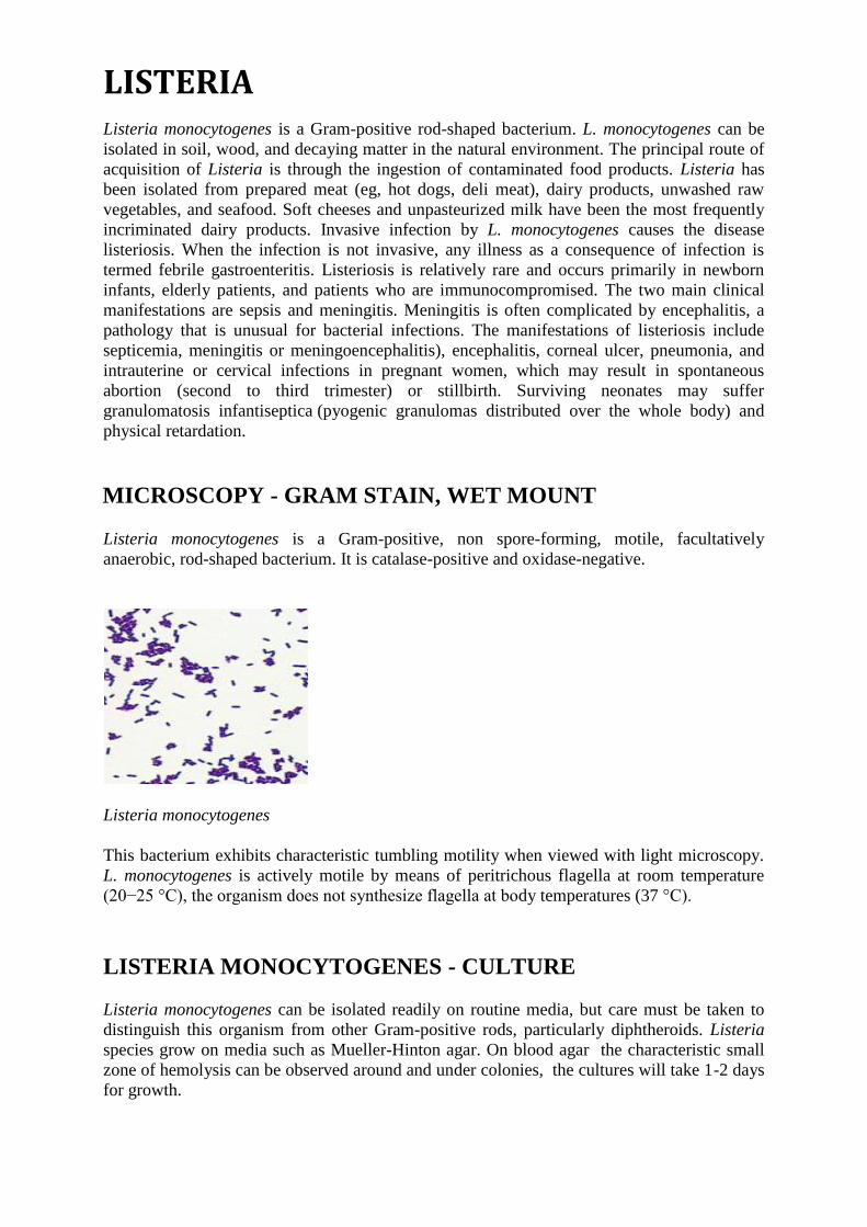
MICROSCOPY - GRAM STAIN, WET MOUNT
Listeria monocytogenes is a Gram-positive, non spore-forming, motile, facultatively
anaerobic, rod-shaped bacterium. It is catalase-positive and oxidase-negative.
Listeria monocytogenes
This bacterium exhibits characteristic tumbling motility when viewed with light microscopy.
L. monocytogenes is actively motile by means of peritrichous flagella at room temperature
(20−25 °C), the organism does not synthesize flagella at body temperatures (37 °C).
LISTERIA MONOCYTOGENES - CULTURE
Listeria monocytogenes can be isolated readily on routine media, but care must be taken to
distinguish this organism from other Gram-positive rods, particularly diphtheroids. Listeria
species grow on media such as Mueller-Hinton agar. On blood agar the characteristic small
zone of hemolysis can be observed around and under colonies, the cultures will take 1-2 days
for growth.
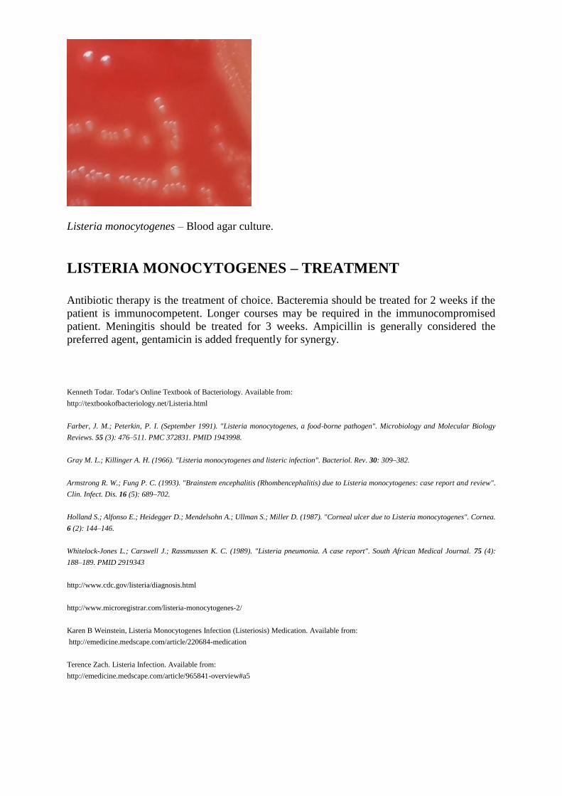
Listeria monocytogenes – Blood agar culture.
LISTERIA MONOCYTOGENES – TREATMENT
Antibiotic therapy is the treatment of choice. Bacteremia should be treated for 2 weeks if the
patient is immunocompetent. Longer courses may be required in the immunocompromised
patient. Meningitis should be treated for 3 weeks. Ampicillin is generally considered the
preferred agent, gentamicin is added frequently for synergy.
Kenneth Todar. Todar's Online Textbook of Bacteriology. Available from:
http://textbookofbacteriology.net/Listeria.html
Farber, J. M.; Peterkin, P. I. (September 1991). "Listeria monocytogenes, a food-borne pathogen". Microbiology and Molecular Biology
Reviews. 55 (3): 476–511. PMC 372831. PMID 1943998.
Gray M. L.; Killinger A. H. (1966). "Listeria monocytogenes and listeric infection". Bacteriol. Rev. 30: 309–382.
Armstrong R. W.; Fung P. C. (1993). "Brainstem encephalitis (Rhombencephalitis) due to Listeria monocytogenes: case report and review".
Clin. Infect. Dis. 16 (5): 689–702.
Holland S.; Alfonso E.; Heidegger D.; Mendelsohn A.; Ullman S.; Miller D. (1987). "Corneal ulcer due to Listeria monocytogenes". Cornea.
6 (2): 144–146.
Whitelock-Jones L.; Carswell J.; Rassmussen K. C. (1989). "Listeria pneumonia. A case report". South African Medical Journal. 75 (4):
188–189. PMID 2919343
http://www.cdc.gov/listeria/diagnosis.html
http://www.microregistrar.com/listeria-monocytogenes-2/
Karen B Weinstein, Listeria Monocytogenes Infection (Listeriosis) Medication. Available from:
http://emedicine.medscape.com/article/220684-medication
Terence Zach. Listeria Infection. Available from:
http://emedicine.medscape.com/article/965841-overview#a5