Embed Size (px)
Citation preview
Psoas hitch and Boari flapureteroneocystostomyRaimund Stein, Peter Rubenwolf, Christopher Ziesel, Mohamed M. Kamal* andJoachim W. Thüroff*Division of Paediatric Urology and *Department of Urology, University Medical Center, Johannes GutenbergUniversity, Mainz, Germany
ILLUSTRATIONS by STEPHAN SPITZER, www.spitzer-illustration.com
Keywordsureteroneocystostomy, ureteric reimplantation, Boari flap, Psoas hitch
IntroductionIn the middle of the last century, Dolff [1], Paquin [2]and Zimmermann et al. [3] developed principles forureteroneocystostomy after gynaecological ureter injuries.Turner-Warwick and Worth [4] adopted these techniques,named it the ‘Psoas Bladder-Hitch Procedure’ and appliedthis technique of ureteroneocystostomy for the treatmentof distal ureteric obstruction, ureteric fistulas and ‘distendedduplication’ of the upper urinary tract.
This technique includes three major advantages for acomplex ureteroneocystostomy: i) Mobilisation of thebladder with fixation above the iliac vessels to guarantee atension-free ureteric anastomosis; ii) Formation of anadequate submucosal tunnel to prevent VUR; iii)Implantation of the ureter into an immobilised part of thebladder to prevent kinking during filling and emptying ofthe bladder [5, 6].
In contrast to intravesical techniques for uretericreimplantation, the psoas hitch procedure is also wellsuited for children with megaureters, as it can becombined with mobilisation of the ureter up to the kidney,so that secondary kinking of the ureter can be resolved.Tapering of a megaureter is rarely required, given that thediameters of the middle and proximal thirds of the ureterare usually smaller than that of the distal part, permittingcreation of a submucosal tunnel for anti-refluxing uretericimplantation.
The psoas (bladder) hitch technique is an almost universalapproach for ureteric reimplantation whatever the problemof the distal ureter. If this technique is not possible, anadditional Boari flap may be useful.
At the end of the nineteenth century, Boari described abladder flap substitution of the distal ureter [7]. Thesurgical technique was developed to bridge lesions of thedistal ureter arising from surgical interventions (mainlygynaecological complications) or tuberculosis [8–11]. Amodification by Übelhör is a deviation from the formal flaptechnique.
Planning and Preparation
Indications• High-grade reflux with dilated ureter not suitable for an
extravesical reimplantation.• Obstructive and/or refluxive megaureter.• Double system not suitable for extravesical
reimplantation.• Distal ureteric defects up to 5–8 cm above the ureteric
orifice (for higher defects an additional Boari-flap may beused).
• Ureteric trauma, ureteric fistula, obstruction of the distalureter or distal (solitary, low grade) urothelial cancer ofthe ureter.
Specific Instruments and Materials• Optical loupes (2.5–3.5, 50-cm focal length).• Small Langenbeck retractors.• 6/0 or 7/0 glyconate monofilament absorbable sutures for
ureteric reimplantation (Monosyn® or Monocryl®).• 4/0 and 5/0 poly-p-dioxanone monofilament absorbable
sutures (e.g. Monoplus® or PDS®) for bladder closure.
© 2013 BJU International | 112, 137–155 | doi:10.1111/bju.12103 137
Surgical Education
• 4/0 or 5/0 glyconate or polyglytone monofilament rapidlyabsorbable sutures for fixation of stents, cystostomy (e.g.Monosyn Quick® or Caprosyn®).
• 3/0 poly-p-dioxanone monofilament absorbable sutures(e.g. Monoplus® or PDS®) for bladder fixation at thepsoas muscle.
• 4, 6 or 8 F polyurethane/polypropylene ureteric stents.• 10 F pigtail cystostomy catheter.
Patient PreparationConfirmation of adequate bladder capacity should beobtained preoperatively to allow a tension-freeanastomosis.
In patients with severe ureteric pathology, which mightextend towards the middle or upper third of the ureter,bowel preparation is advisable in the event of intestinalureter substitution becoming necessary.
Acute UTI must be treated preoperatively.
Patient PositioningThe patient is placed supine on the table with ª15 °overextension. A Foley transurethral catheter is inserted,which must provide intraoperative access for filling thebladder during the operation.
Stein et al.
138 © 2013 BJU International
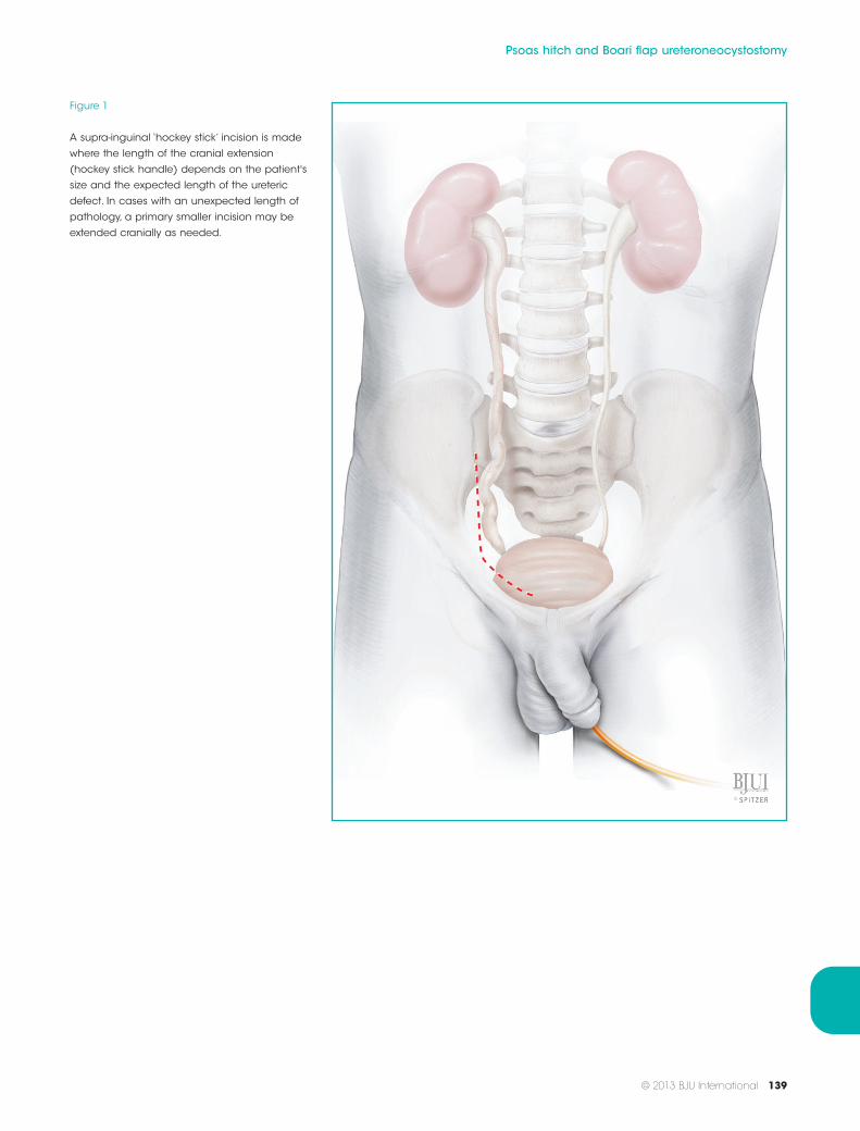
Figure 1
A supra-inguinal ‘hockey stick’ incision is made
where the length of the cranial extension
(hockey stick handle) depends on the patient's
size and the expected length of the ureteric
defect. In cases with an unexpected length of
pathology, a primary smaller incision may be
extended cranially as needed.
Psoas hitch and Boari flap ureteroneocystostomy
© 2013 BJU International 139
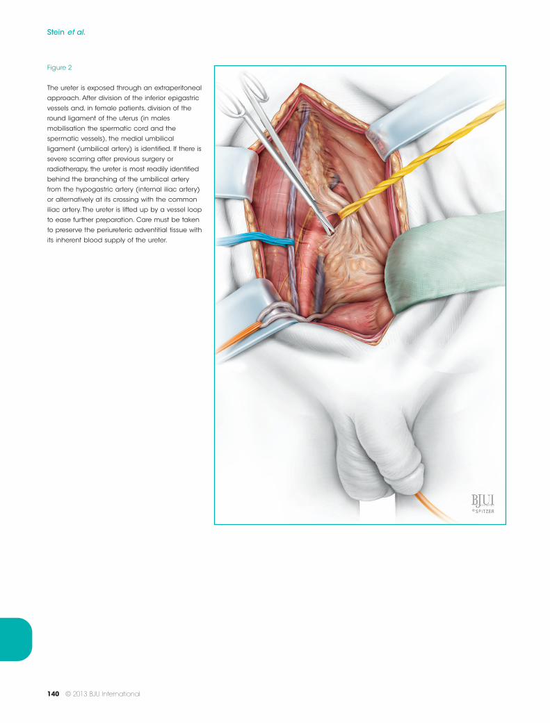
Figure 2
The ureter is exposed through an extraperitoneal
approach. After division of the inferior epigastric
vessels and, in female patients, division of the
round ligament of the uterus (in males
mobilisation the spermatic cord and the
spermatic vessels), the medial umbilical
ligament (umbilical artery) is identified. If there is
severe scarring after previous surgery or
radiotherapy, the ureter is most readily identified
behind the branching of the umbilical artery
from the hypogastric artery (internal iliac artery)
or alternatively at its crossing with the common
iliac artery. The ureter is lifted up by a vessel loop
to ease further preparation. Care must be taken
to preserve the periureteric adventitial tissue with
its inherent blood supply of the ureter.
Stein et al.
140 © 2013 BJU International
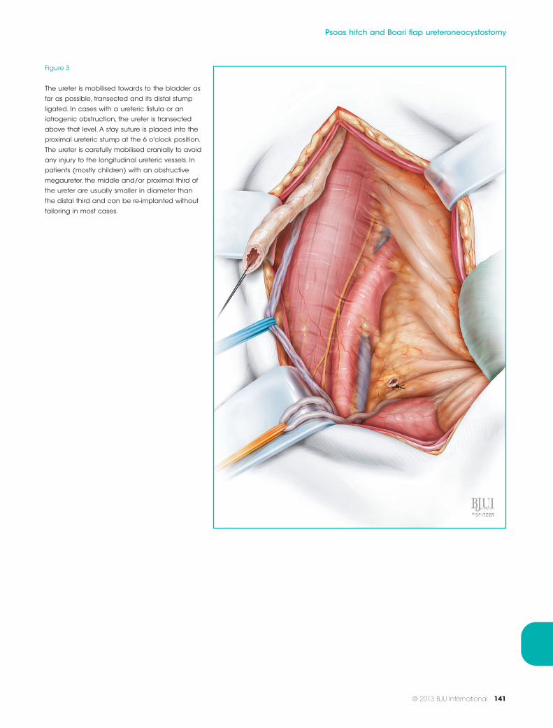
Figure 3
The ureter is mobilised towards to the bladder as
far as possible, transected and its distal stump
ligated. In cases with a ureteric fistula or an
iatrogenic obstruction, the ureter is transected
above that level. A stay suture is placed into the
proximal ureteric stump at the 6 o'clock position.
The ureter is carefully mobilised cranially to avoid
any injury to the longitudinal ureteric vessels. In
patients (mostly children) with an obstructive
megaureter, the middle and/or proximal third of
the ureter are usually smaller in diameter than
the distal third and can be re-implanted without
tailoring in most cases.
Psoas hitch and Boari flap ureteroneocystostomy
© 2013 BJU International 141
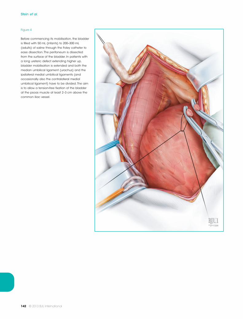
Figure 4
Before commencing its mobilisation, the bladder
is filled with 50 mL (infants) to 200–300 mL
(adults) of saline through the Foley catheter to
ease dissection. The peritoneum is dissected
from the surface of the bladder. In patients with
a long ureteric defect extending higher up,
bladder mobilisation is extended and both the
median umbilical ligament (urachus) and the
ipsilateral medial umbilical ligaments (and
occasionally also the contralateral medial
umbilical ligament) have to be divided.The aim
is to allow a tension-free fixation of the bladder
at the psoas muscle at least 2–3 cm above the
common iliac vessel.
Stein et al.
142 © 2013 BJU International
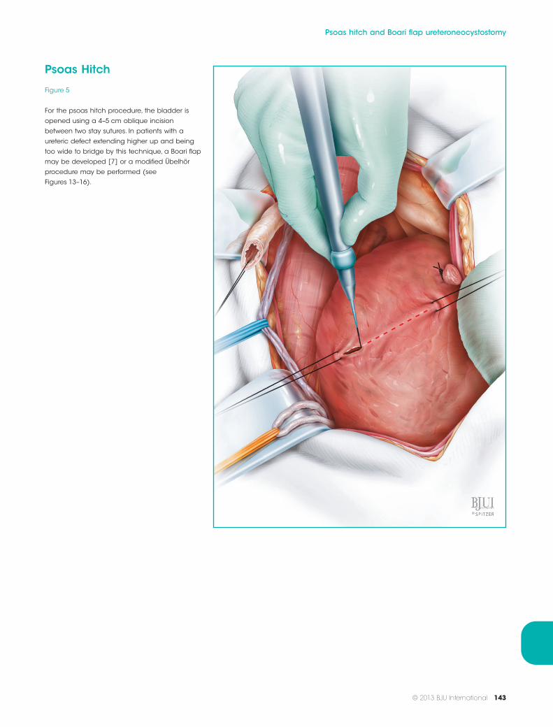
Psoas Hitch
Figure 5
For the psoas hitch procedure, the bladder is
opened using a 4–5 cm oblique incision
between two stay sutures. In patients with a
ureteric defect extending higher up and being
too wide to bridge by this technique, a Boari flap
may be developed [7] or a modified Übelhör
procedure may be performed (see
Figures 13–16).
Psoas hitch and Boari flap ureteroneocystostomy
© 2013 BJU International 143
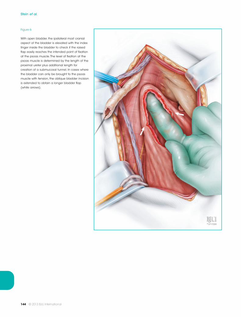
Figure 6
With open bladder, the ipsilateral most cranial
aspect of the bladder is elevated with the index
finger inside the bladder to check if the raised
flap easily reaches the intended point of fixation
at the psoas muscle. The level of fixation at the
psoas muscle is determined by the length of the
proximal ureter plus additional length for
creation of a submucosal tunnel. In cases where
the bladder can only be brought to the psoas
muscle with tension, the oblique bladder incision
is extended to obtain a longer bladder flap
(white arrows).
Stein et al.
144 © 2013 BJU International
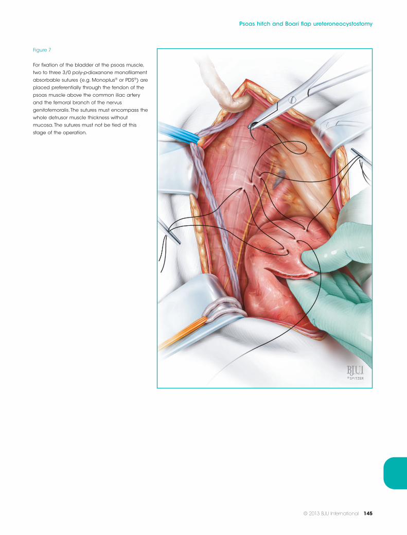
Figure 7
For fixation of the bladder at the psoas muscle,
two to three 3/0 poly-p-dioxanone monofilament
absorbable sutures (e.g. Monoplus® or PDS®) are
placed preferentially through the tendon of the
psoas muscle above the common iliac artery
and the femoral branch of the nervus
genitofemoralis. The sutures must encompass the
whole detrusor muscle thickness without
mucosa.The sutures must not be tied at this
stage of the operation.
Psoas hitch and Boari flap ureteroneocystostomy
© 2013 BJU International 145
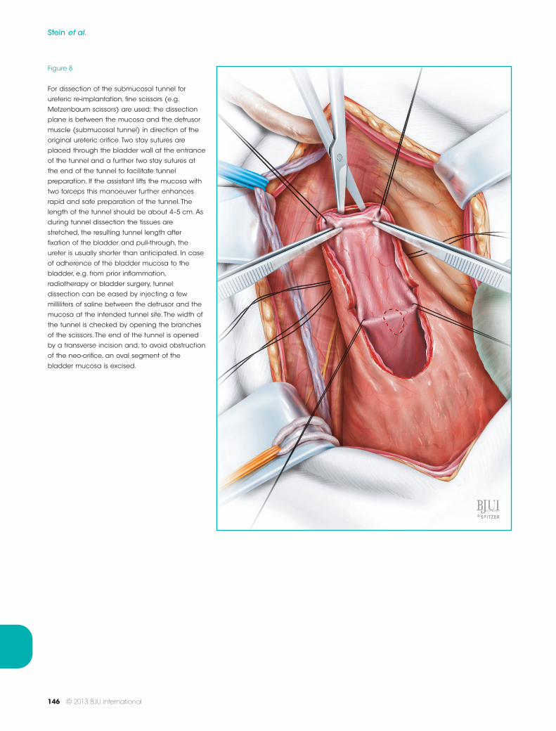
Figure 8
For dissection of the submucosal tunnel for
ureteric re-implantation, fine scissors (e.g.
Metzenbaum scissors) are used; the dissection
plane is between the mucosa and the detrusor
muscle (submucosal tunnel) in direction of the
original ureteric orifice. Two stay sutures are
placed through the bladder wall at the entrance
of the tunnel and a further two stay sutures at
the end of the tunnel to facilitate tunnel
preparation. If the assistant lifts the mucosa with
two forceps this manoeuver further enhances
rapid and safe preparation of the tunnel. The
length of the tunnel should be about 4–5 cm. As
during tunnel dissection the tissues are
stretched, the resulting tunnel length after
fixation of the bladder and pull-through, the
ureter is usually shorter than anticipated. In case
of adherence of the bladder mucosa to the
bladder, e.g. from prior inflammation,
radiotherapy or bladder surgery, tunnel
dissection can be eased by injecting a few
milliliters of saline between the detrusor and the
mucosa at the intended tunnel site. The width of
the tunnel is checked by opening the branches
of the scissors. The end of the tunnel is opened
by a transverse incision and, to avoid obstruction
of the neo-orifice, an oval segment of the
bladder mucosa is excised.
Stein et al.
146 © 2013 BJU International
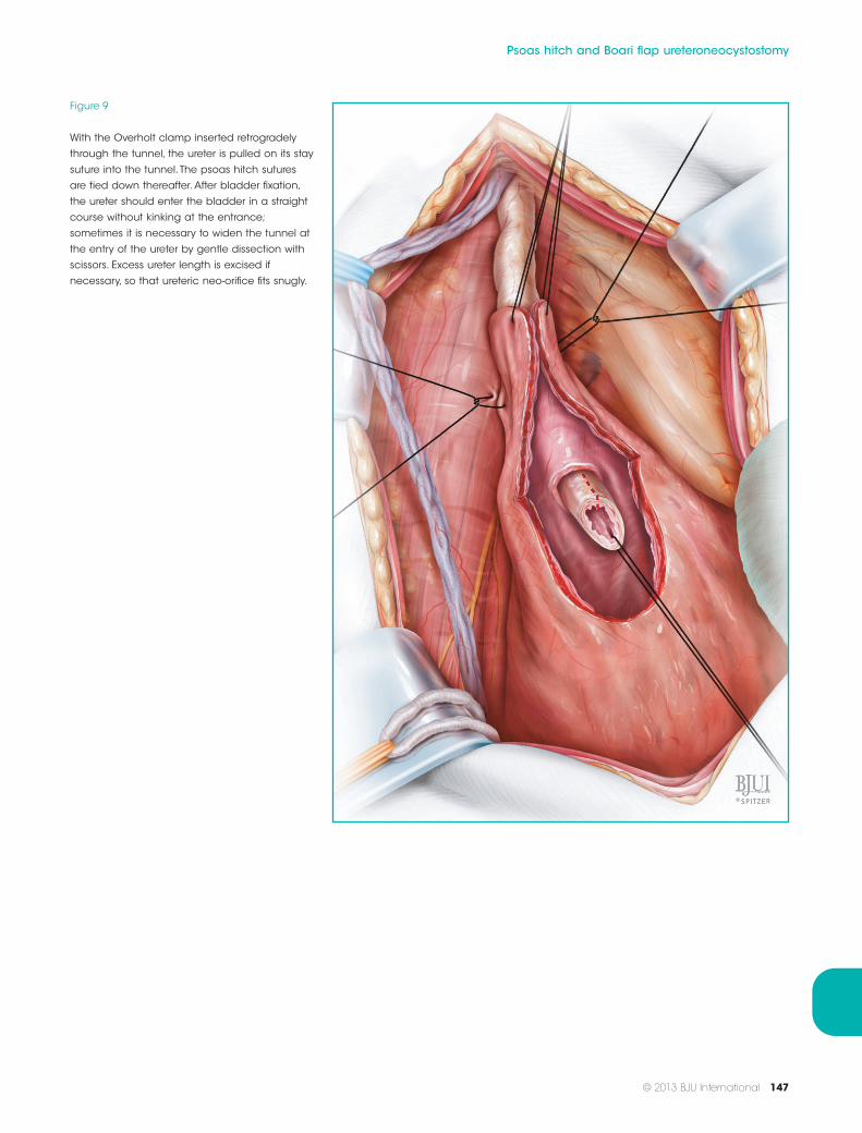
Figure 9
With the Overholt clamp inserted retrogradely
through the tunnel, the ureter is pulled on its stay
suture into the tunnel. The psoas hitch sutures
are tied down thereafter. After bladder fixation,
the ureter should enter the bladder in a straight
course without kinking at the entrance;
sometimes it is necessary to widen the tunnel at
the entry of the ureter by gentle dissection with
scissors. Excess ureter length is excised if
necessary, so that ureteric neo-orifice fits snugly.
Psoas hitch and Boari flap ureteroneocystostomy
© 2013 BJU International 147
a
b
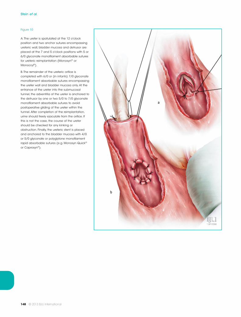
Figure 10
A.The ureter is spatulated at the 12 o'clock
position and two anchor sutures encompassing
ureteric wall, bladder mucosa and detrusor are
placed at the 7 and 5 o'clock positions with 5 or
6/0 glyconate monofilament absorbable sutures
for ureteric reimplantation (Monosyn® or
Monocryl®).
B. The remainder of the ureteric orifice is
completed with 6/0 or (in infants) 7/0 glyconate
monofilament absorbable sutures encompassing
the ureter wall and bladder mucosa only. At the
entrance of the ureter into the submucosal
tunnel, the adventitia of the ureter is anchored to
the detrusor by one or two 5/0 to 7/0 glyconate
monofilament absorbable sutures to avoid
postoperative gliding of the ureter within the
tunnel. After completion of the reimplantation,
urine should freely ejaculate from the orifice. If
this is not the case, the course of the ureter
should be checked for any kinking or
obstruction. Finally, the ureteric stent is placed
and anchored to the bladder mucosa with 4/0
or 5/0 glyconate or polyglytone monofilament
rapid absorbable sutures (e.g. Monosyn Quick®
or Caprosyn®).
Stein et al.
148 © 2013 BJU International
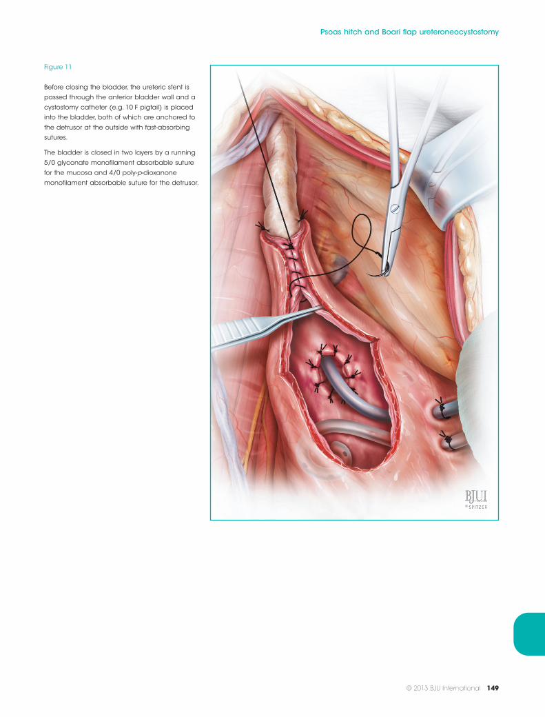
Figure 11
Before closing the bladder, the ureteric stent is
passed through the anterior bladder wall and a
cystostomy catheter (e.g. 10 F pigtail) is placed
into the bladder, both of which are anchored to
the detrusor at the outside with fast-absorbing
sutures.
The bladder is closed in two layers by a running
5/0 glyconate monofilament absorbable suture
for the mucosa and 4/0 poly-p-dioxanone
monofilament absorbable suture for the detrusor.
Psoas hitch and Boari flap ureteroneocystostomy
© 2013 BJU International 149
a
b
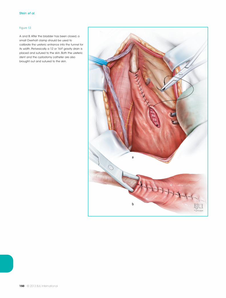
Figure 12
A and B. After the bladder has been closed, a
small Overholt clamp should be used to
calibrate the ureteric entrance into the tunnel for
its width. Perivesically a 12 or 16-F gravity drain is
placed and sutured to the skin. Both the ureteric
stent and the cystostomy catheter are also
brought out and sutured to the skin.
Stein et al.
150 © 2013 BJU International
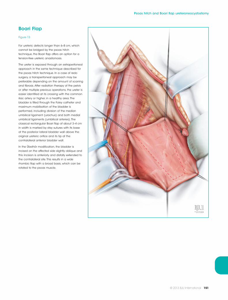
Boari Flap
Figure 13
For ureteric defects longer than 6–8 cm, which
cannot be bridged by the psoas hitch
technique, the Boari flap offers an option for a
tension-free ureteric anastomosis.
The ureter is exposed through an extraperitoneal
approach in the same technique described for
the psoas hitch technique. In a case of redo
surgery, a transperitoneal approach may be
preferable depending on the amount of scarring
and fibrosis. After radiation therapy of the pelvis
or after multiple previous operations, the ureter is
easier identified at its crossing with the common
iliac artery or higher, in a healthy area.The
bladder is filled through the Foley catheter and
maximum mobilisation of the bladder is
performed, including division of the median
umbilical ligament (urachus) and both medial
umbilical ligaments (umbilical arteries). The
classical rectangular Boari flap of about 3–4 cm
in width is marked by stay sutures with its base
at the posterior lateral bladder wall above the
original ureteric orifice and its tip at the
contralateral anterior bladder wall.
In the Übelhör modification, the bladder is
incised on the affected side slightly oblique and
this incision is anteriorly and distally extended to
the contralateral site. This results in a wide
rhombic flap with a broad basis, which can be
rotated to the psoas muscle.
Psoas hitch and Boari flap ureteroneocystostomy
© 2013 BJU International 151
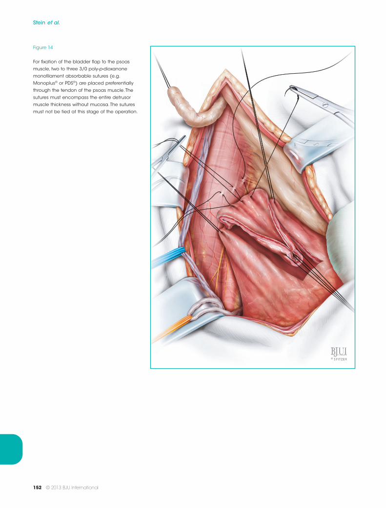
Figure 14
For fixation of the bladder flap to the psoas
muscle, two to three 3/0 poly-p-dioxanone
monofilament absorbable sutures (e.g.
Monoplus® or PDS®) are placed preferentially
through the tendon of the psoas muscle. The
sutures must encompass the entire detrusor
muscle thickness without mucosa.The sutures
must not be tied at this stage of the operation.
Stein et al.
152 © 2013 BJU International

Figure 15
After creation of the submucosal tunnel an
Overholt clamp is inserted retrogradely into the
tunnel and the ureter is pulled on its stay suture
into the tunnel. Thereafter the fixation sutures to
the psoas muscle are tied. After bladder fixation,
the ureter should enter the Boari flap in a
straight course without kinking at the entrance.
The ureter is re-implanted into the bladder flap
using the same technique as for the psoas
hitch.
Psoas hitch and Boari flap ureteroneocystostomy
© 2013 BJU International 153
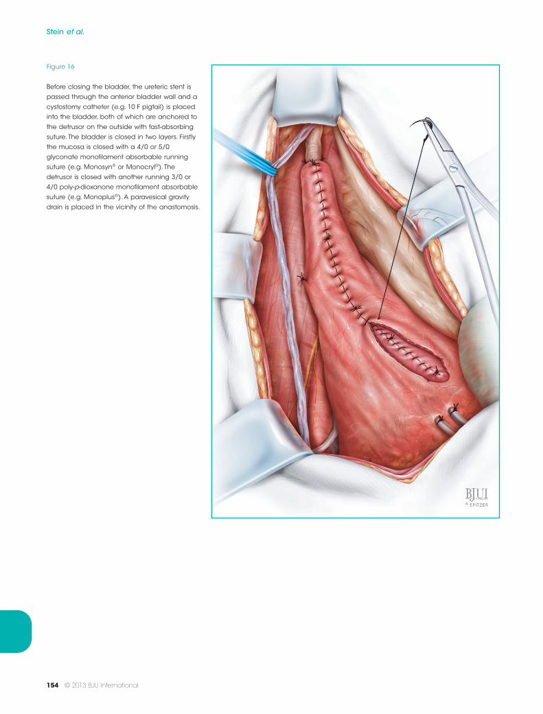
Figure 16
Before closing the bladder, the ureteric stent is
passed through the anterior bladder wall and a
cystostomy catheter (e.g. 10 F pigtail) is placed
into the bladder, both of which are anchored to
the detrusor on the outside with fast-absorbing
suture. The bladder is closed in two layers. Firstly
the mucosa is closed with a 4/0 or 5/0
glyconate monofilament absorbable running
suture (e.g. Monosyn® or Monocryl®). The
detrusor is closed with another running 3/0 or
4/0 poly-p-dioxanone monofilament absorbable
suture (e.g. Monoplus®). A paravesical gravity
drain is placed in the vicinity of the anastomosis.
Stein et al.
154 © 2013 BJU International
Postoperative Management
MedicationAntibiotics (i.e. cephalosporines) are started at the time ofsurgery and are continued as long as the stent is in situ.
The perivesical drain is removed on day 1 or 2 and as soonas the urine is almost clear, the indwelling Foley catheter isremoved.
Depending on the difficulty of the procedure, the uretericstent is removed between day 7 and 10 (in complicatedcases or after Boari flap on day 10). After removal of thestent, the cystostomy is clamped and the patient starts tovoid. The upper tract is checked by ultrasonography on thefollowing day (in adults upper tract imaging is achieved byIVU). The cystostomy is removed after bladder emptyingwithout residual urine has been achieved.
Surgeon to SurgeonIn some patients, dilatation of the upper tract persistspostoperatively. This usually resolves within 3–6 months. Ifthere is any concern about persisting obstruction, a MAG3clearance with furosemide should be obtained. In childrenantibiotic prophylaxis should be continued as long as thereis increased dilatation of the upper tract as compared withpreoperatively.
In patients with a symptomatic postoperative uretericobstruction a JJ catheter is placed antegradely through apercutaneous nephrostomy tract and left for 3 months. Incase of persistent obstruction from implantation stenosis,redo-surgery may be performed not earlier than after 3months.
If the ureter cannot be re-implanted into the Boari flapwithout tension, an additional 3–5 cm of ureteral lengthcan be gained. If ureteric reimplantation remainsimpossible by bladder bridging techniques, intestinal uretersubstitution or kidney autotransplantation remain anoption. Both procedures can be performed through thesame access, should psoas hitch or Boari flap uretericreimplantation unexpectedly be infeasible.
References1 Dolff C. [Improved results of ureterocystanastomosis
by flexible bladder fixation]. Zentralbl Gynakol 1952;74: 1777–87
2 Paquin AJ Jr. Ureterovesical anastomosis: thedescription and evaluation of a technique. J Urol 1959;82: 573–83
3 Zimmerman IJ, Precourt WE, Thompson CC. Directuretero-cysto-neostomy with the short ureter in thecure of ureterovaginal fistula. J Urol 1960; 83: 113–5.
4 Warwick RT, Worth PH. The psoas bladder-hitchprocedure for the replacement of the lower third of theureter. Br J Urol 1969; 41: 701–9
5 Gross M, Peng B, Waterhouse K. Use of the mobilizedbladder to replace the pelvic ureter. J Urol 1969; 101:40–4
6 Riedmiller H, Becht E, Hertle L et al. Psoas-hitchureteroneocystostomy: experience with 181 cases. EurUrol 1984; 10: 145–50
7 Boari A. L’urétéro-cystonostomie. Étude clinique etexpérimentale. Ann Mal Org Gen Urin 1899; 14:1059–88 141–170
8 Bergmeyer M. [The Boari-Casati Technic of FlapPlastic Surgery in the Treatment of Passage Disordersof the Caudal Ureter]. Urologe 1964; 14: 118–27
9 Gil Vernet JM. [Ureterovesicoplasty under mucousmembrane. (Modifications of Boari’s technic)]. J UrolMedicale Chir 1959; 65: 504–8
10 Pytel’ IuA. [Reconstruction of the lower third of theureter from a bladder flap according to Boari]. VestnKhir Im I I Grek 1959; 83: 80–5
11 Kimchi D, Wiesenfeld A. Injuries to the lower third ofureter treated by bladder flap plasty: Boari-Kusstechnique; report of two cases. J Urol 1963; 89:800–3
Correspondence: Raimund Stein, Division of PaediatricUrology, Department of Urology, University MedicalCenter, Johannes Gutenberg University, Langenbeckstrasse1, 55131 Mainz, Germany.
e-mail: [email protected]
Psoas hitch and Boari flap ureteroneocystostomy
© 2013 BJU International 155


![Psoas Compartment Blockade in a Laterally Herniated Disc Compressing the Psoas Muscle · 2017. 3. 23. · lesser trochanter [1]. The psoas muscle plays a most im-portant role when](https://img.pdfslide.net/doc/110x75/60d87b6e5f782c7e9868c0e0/psoas-compartment-blockade-in-a-laterally-herniated-disc-compressing-the-psoas-muscle.jpg)