Embed Size (px)
Citation preview
QCB301: FALL2010
D. Botstein; M. McClean; M. Noyes; T. Hansen; S. McIsaac 9/15/2010
Contents People Involved in QCB301 ........................................................................................................................... 6
Introduction .................................................................................................................................................. 7
Subject Matter .............................................................................................................................................. 8
Course Schedule .......................................................................................................................................... 10
Weekly Schedule ..................................................................................................................................... 10
Important Due Dates .............................................................................................................................. 11
Assignments and Grading ........................................................................................................................... 12
Projects and Project Schedules ................................................................................................................... 13
Project 0: Laboratory Basics .................................................................................................................... 13
Project 1: Zinc-finger Project .................................................................................................................. 13
Project 2: Microscopy and Microfluidics ................................................................................................ 15
Project 3: Transformation of Yeast ......................................................................................................... 15
Project 4: Dissection of S. cerevisiae tetrads .......................................................................................... 15
Project 5: Analysis of Gene Expression (Microarray Assignment) ......................................................... 16
Reading List ................................................................................................................................................. 18
Laboratory Sessions (Day by Day) ............................................................................................................... 20
September 16 (Thursday): ...................................................................................................................... 20
Materials ............................................................................................................................................. 20
Streaking Microorganisms for Single Colonies.................................................................................... 20
Pipetting Exercise ................................................................................................................................ 21
Quantification of Microorganisms ...................................................................................................... 23
PUMA Registration .............................................................................................................................. 26
September 20 (Monday): ........................................................................................................................ 27
Materials ............................................................................................................................................. 27
Bacterial Transformation .................................................................................................................... 27
Recovery in minimal media ................................................................................................................. 28

Wash out Histidine .............................................................................................................................. 28
Titration and plating cells .................................................................................................................... 28
September 21 (Tuesday): ........................................................................................................................ 30
Materials ............................................................................................................................................. 30
Heat Shock of Yeast ............................................................................................................................ 31
-Estradiol induction of MSN2 .............................................................................................................. 31
Harvesting Cells By Filtering ................................................................................................................ 31
RNA Harvesting from Yeast for Array Analysis.................................................................................... 33
Count colonies/Plate cells ................................................................................................................... 34
September 22 (Wednesday): .................................................................................................................. 34
Materials ............................................................................................................................................. 34
RNA precipitation ................................................................................................................................ 35
Dissolving RNA pellet .......................................................................................................................... 35
RNEasy Cleanup of total RNA for array labeling ................................................................................. 35
Labeling of RNA by Reverse Transcription .......................................................................................... 37
September 23 (Thursday) ....................................................................................................................... 39
Materials ............................................................................................................................................. 39
RNA degradation ................................................................................................................................. 39
Cleanup using Zymo Clean and Concentrator Kit ............................................................................... 39
Check Yield of labeled cDNA ............................................................................................................... 40
Count Colonies .................................................................................................................................... 40
Select clones for sequencing ............................................................................................................... 40
September 27 (Monday): ........................................................................................................................ 41
Materials ............................................................................................................................................. 41
Ligation of candidate fingers into expression vectors ........................................................................ 41
Transformation ................................................................................................................................... 42
Hybridization of cDNA to Microarrays ................................................................................................ 42
RNA Hybridization ............................................................................................................................... 43
September 28 (Tuesday): ........................................................................................................................ 47
Verify successful ligations by PCR ....................................................................................................... 47
Array Washing ............................................................................................................................................. 47
Array Scanning Using an Axon GenePix 4000B Scanner ..................................................................... 48
Start overnight cultures ...................................................................................................................... 51
September 29 (Wednesday): .................................................................................................................. 53
Materials ............................................................................................................................................. 53
Bacterial Transformation .................................................................................................................... 53
Recovery in minimal media ................................................................................................................. 54
Wash out Histidine .............................................................................................................................. 54
Titration and plating cells .................................................................................................................... 54
September 30 (Thursday): ...................................................................................................................... 56
Materials ............................................................................................................................................. 56
Count colonies/Plate cells ................................................................................................................... 56
Yeast Transformation .......................................................................................................................... 56
Microarray Gridding ............................................................................................................................ 57
October 4 (Monday): .............................................................................................................................. 61
Materials ............................................................................................................................................. 61
Count Colonies .................................................................................................................................... 61
PDMS Chip Construction ..................................................................................................................... 61
Tetrad dissection ................................................................................................................................. 63
October 5 (Tuesday):............................................................................................................................... 68
Materials ............................................................................................................................................. 68
Analyze sequences and create motif .................................................................................................. 68
Characterizing your Microfluidic Chip ................................................................................................. 68
October 6 (Wednesday): ......................................................................................................................... 70
Materials ............................................................................................................................................. 70
Characterizing STRE response in the Gradient-Maker Chip ................................................................ 70
Start overnight yeast cultures ............................................................................................................. 71
October 7 (Thursday) .............................................................................................................................. 72
Induce activation of zinc finger – activation domain (9AM) ............................................................... 72
Appendix ..................................................................................................................................................... 73
Assignment Guidelines ............................................................................................................................ 73
Mini Project Proposal (Due 10/14) ..................................................................................................... 73
Draft Project Proposal (Due 10/21) .................................................................................................... 73
Journal Club Presentations (10/11-10/28) .......................................................................................... 75
Written Progress Reports and Oral Project Updates (11/7-12/17) .................................................... 76
Final Project Poster (Due 1/17) ........................................................................................................... 77
Final Project Presentation (1/6 and 1/7) ............................................................................................ 77
Final Project Paper (Due 1/17) ............................................................................................................ 77
Lab Notebook Guidelines ........................................................................................................................ 84
Additional Protocols ................................................................................................................................ 86
Preservation of Strains ........................................................................................................................ 86
Sonicator Use ...................................................................................................................................... 87
Microscope Use ................................................................................................................................... 88
Media and Reagents used in QCB301 (Partial List) ................................................................................. 89
SOB medium ....................................................................................................................................... 89
SOC medium........................................................................................................................................ 89
NM medium ........................................................................................................................................ 89
2xYT medium agar plates .................................................................................................................... 89
NM selective plates, 10 and 25mM 3AT ............................................................................................. 89
β-Estradiol ........................................................................................................................................... 89
Microarray blocking reagent, 500 mL ................................................................................................. 90
RNA Lysis Buffer (100ml) ..................................................................................................................... 90
50X dNTP mix ...................................................................................................................................... 90
Media Color Codes .............................................................................................................................. 90
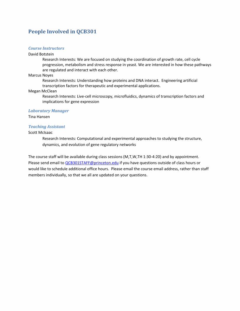
People Involved in QCB301
Course Instructors
David Botstein Research Interests: We are focused on studying the coordination of growth rate, cell cycle progression, metabolism and stress response in yeast. We are interested in how these pathways are regulated and interact with each other. Marcus Noyes
Research Interests: Understanding how proteins and DNA interact. Engineering artificial transcription factors for therapeutic and experimental applications.
Megan McClean Research Interests: Live-cell microscopy, microfluidics, dynamics of transcription factors and implications for gene expression
Laboratory Manager
Tina Hansen
Teaching Assistant
Scott McIsaac
Research Interests: Computational and experimental approaches to studying the structure,
dynamics, and evolution of gene regulatory networks
The course staff will be available during class sessions (M,T,W,TH 1:30-4:20) and by appointment.
Please send email to [email protected] if you have questions outside of class hours or
would like to schedule additional office hours. Please email the course email address, rather than staff
members individually, so that we all are updated on your questions.
Introduction The purpose of QCB301 is to allow students to integrate the elements that must be combined to
do successful experimental science. For most of you this will be the first opportunity to control all the
steps of the process. We will introduce you, in the first few weeks, to a number of leading-edge
technologies in quantitative biology that have only recently been invented and reduced to practice, and
thus offer opportunities for learning new things nobody knows yet, even about intensively studied areas
like regulation of gene expression. We will also introduce you to some basic ideas and experimental
features of budding yeast, Saccharomyces cerevisiae, the leading experimental system for study of gene
expression at the cellular and molecular level.
After the introductory phase, it will be up to you to:
• conceive and develop some ideas for experiments that you believe might lead to new
discoveries and/or understanding
• reduce these ideas to a practical experimental plan that can be communicated, in the form of
a proposal, to others.
• execute the experimental plan, repeating or amending procedures, until you obtain useful
data.
• analyze your data, defining the strengths and weaknesses in design and execution, especially
pointing out any issues that might have to be addressed to make the experiments conclusive enough to
publish.
• interpret your results in the context of what was known before you began and suggesting
what one might want to do to verify or follow up your findings.
• communicate your work to others, as an oral presentation, a poster, and a formal paper such
as one might submit to a journal.
In all of this, the role of the teaching staff will be to serve as advisors and consultants. We will
not tell you what to do, but we may well point out issues (often ones of practicality) that you will want
to consider. We also require lots of advance notice for unusual or oddball materials, strains, etc.
In the end, your efforts will be judged by the quality of your intellectual and operational effort,
not necessarily the technical success of your experiments. We are all painfully aware of the fact that the
majority of our own experiments don’t work out as we expect on the first or second pass, and often they
never work out. That does not mean that they were bad efforts.
Subject Matter This year the area of science that we will concentrate on in QCB301 is the regulation of transcription in
budding yeast, Saccharomyces cerevisiae. The general outlines of this process are very well known: all of
you have studied this process in considerable depth. Briefly, genes are expressed when RNA polymerase
II is guided to sites (called promoters) where the enzyme can begin a transcript. This guiding is done by
proteins (called transcription factors) that bind to particular DNA sequences within the promoter
region. It is the combined specificity of the transcription factors and the RNA polymerase itself that are
the major reasons particular genes are transcribed under particular circumstances. There are other
factors (for example, the positions of nucleosomes) that are also important, but for the systems we will
study, the transcription factor binding is thought to be the main determinant of specificity.
There are hundreds of transcription factors encoded by the yeast genome. These are not all expressed
all the time; indeed the regulation of their expression and activity is the major way in which cells
respond to changing conditions. The paper by Harbison et al., 2004 is a good introduction to the issues
around transcription factor activity in yeast. Once expressed and activated, the transcription factors
bind, presumably to every instance of the short DNA sequences they recognize, provided only that those
sequences are accessible (e.g. not buried completely in nucleosomes). Generally, genes that respond
strongly to particular transcription factors will contain more than one copy of the cognate binding
sequence in their promoter regions. Often the effectiveness of binding is modulated by the presence or
absence of other transcription factors at the same promoter, a phenomenon often referred to as
combinatorial regulation. Some transcription factors function as repressors of transcription, others as
activators of transcription, and some appear to be able to do both. The ability of the same factor to
either repress or activate is not well understood, and may depend on subtle promoter sequence
differences or context (presence or absence of other factors).
We will focus on transcription factors associated with stress response, because we can easily manipulate
the yeast cultures so that these factors will become active by providing one or another type of stress
(heat shock, osmotic shock, radiation, etc.). Specifically, we will focus on the product of the MSN2 gene
(also referred to as Msn2p) that binds to a sequence called the stress response element STRE. STRE
elements contain a “core” sequence AAGGG, of which there are more than 1,000 in the genome. You
will learn how to measure stress response using DNA microarrays (to this end you will want to study
carefully the paper by Gasch et al., 2000). In this paper you will see that some genes controlled by STRE
elements respond to some stresses and not others. We will undertake to study the reasons for this by
doing some protein engineering to try to understand what is required. We will begin with a completely
artificial DNA-binding protein based on an expandable zinc-finger motif that binds the core STRE
sequence element and modify it in order to achieve more specific binding. In order to understand the
issues around protein binding motifs and specificity you will want to study the paper by Stormo and
Fields (1998).
The basic plan is to make variant artificial STRE-binding proteins and test them against natural STRE
element-containing promoters that drive transcription of a fusion gene that results in expression of a
fluorescent marker protein whose expression we can detect. We will also test the activity of the

endogenous transcription factors by overexpressing them in yeast WITHOUT exposing the yeast to any
stress, and following gene expression with DNA microarrays. This will give us a comparison point for our
artificially engineered transcription factors. We will employ microfluidic technology to study the
response of cells to particular stresses. Under laminar flow conditions, we can add something that
causes a particular stress by changing the medium in the microfluidic device. We can then follow
expression of fluorescent fusion genes in a fluorescence microscope.
In the early part of the course, we will introduce all of this state-of-the-art technology to you. As you
learn the ideas and techniques, you should be thinking about what kind of use you will want to make of
this technology in your independent projects. We will not tell you what to do here, but we are ready to
discuss any and all ideas that you have. These ideas should, in the best case, take advantage of the novel
technological elements and aim to discover something about what are the determinants of specificity
not just in DNA binding, but in causing expression of subsets of STRE-element containing genes in an
intact yeast genome.
Course Schedule A detailed schedule is given in the following pages. Class sessions are M,T,W,TH from 1:30-4:20 in CIL 005. Laboratory exercises are time intensive, and can occasionally run overtime. Students are encouraged to avoid scheduling commitments directly after class. Monday and Wednesday class sessions will occasionally begin in CIL 280 for the purpose of having journal clubs and lab meetings. This will be noted on the course schedule.
Weekly Schedule Week 1(9/16-9/23): Introduction to the course Laboratory exercises Reading Week 2 (9/23-9/30): Continue lab exercises Reading Week 3 (9/30-10/7) Continue lab exercises Reading Week 4 (10/7-10/14) Journal Club Presentation I (10/11 1:30-2:30 CIL 280)
**Mini draft project proposal DUE (10/14) Week 5 (10/14-10/21): Journal Club Presentation II (10/18 1:30-2:30 CIL 280)
**Revised project proposal DUE (10/21) Week 6 (10/21-10/28): Journal Club Presentation III (10/25 1:30-2:30 CIL 280) Prepare for fall break (Freeze down strains, tidy your work space, etc) Fall Recess (10/29-11/7): RELAX! Week 7 (11/8-11/15): Project Updates (11/8 1:30-2:30 CIL 280) **Progress Report Due (11/8) Week 8 (11/15-11/22): Project Updates (11/15 1:30-2:30 CIL 280) **Progress Report Due (11/15) Week 9 (11/22-11/29): Project Updates (11/22 1:30-2:30 CIL 280) **Progress Report Due (11/22) Thanksgiving Recess (11/24-11/29) Week 10 (11/29-12/6):
Project Updates (11/29 1:30-2:30 CIL 280) **Progress Report Due (11/29) Week 11 (12/6-12/13):
Project Updates (12/6 1:30-2:30 CIL 280) **Progress Report Due (12/6) Week 12 (12/13-12/17):
Project Updates (12/13 1:30-2:30 CIL 280)
**Progress Report Due (12/13) Winter Recess (12/17-1/2): Reading Period (1/3-1/11): **Final Presentations (1/6, 1/7) Exam Period (1/12-1/22): **Final paper and poster due (1/17)
Important Due Dates Reprinted here so that you can’t miss them!
9/20-10/7 – DO the assigned reading and READ PROTOCOLS before class. EVERY DAY! 10/14—Mini-project proposal due 10/21—Revised draft of project proposal due. This is the version that will be graded. 10/11-10/28—Journal club presentations in class 11/7-12/17—Weekly written project updates and presentations in class Mondays and Wednesdays (if necessary). Each group should present briefly ever week. Written progress reports will be graded. 1/6, 1/7—Final Presentations 1/17—Final papers and posters due.
Assignments and Grading Detailed explanations of assignments and detailed grading rubrics are listed in the appendices. Deadlines for these projects are indicated in the course schedule with asterisks (**). Grading of the course is based on written work, oral presentations, and our assessment of your effort and participation. The actual outcome of your experiments is not a major determinant, although the soundness of your experimental design and the quality of your work in execution of this design will be taken into account. Specifically the grading will be as follows: Written work (65%)
Final paper (30%)
project proposal (20%)
project progress reports (10%)
poster (5%) Oral Presentations (20%)
final presentation (10%)
journal club presentation (5%)
project updates (5%) Course Effort and Participation (15%) This category includes an overall assessment of:
Preparation: Do you come to class prepared having READ the protocols BEFORE class? This is very important especially during the first three weeks and will determine your success on any given day. Printing out a protocol and taking notes before class starts is a good idea.
Intellectual effort: Some of you will go well beyond the minimum required in reading, designing new experiments, and analyzing them.
General effort: Some of you inevitably work harder than others.
Participation in class discussions
Lab notebook: Is it well enough organized that you can find all the important information when questions come up about what was done and how, several weeks after the fact?
Cleanliness in the laboratory: Is your space clean? Are you considerate in your use of common equipment and reagents?
Projects and Project Schedules During the first three weeks of class we will complete 5 projects. These projects are designed to give
you the tools to design and complete your own research projects in weeks 4-10. Due to the realities of
experimental science (i.e. organism growth rates, incubation times, etc) you will be working on several
projects at once. On any given day you will need to do at least one thing towards several of the
projects. There also might be some lab work to do over the weekend. Things can get confusing! The
overview will help you keep track of the different projects. READ protocols and assignment details
BEFORE you come to class each day. We will take your preparedness for each laboratory session into
account when we calculate the course participation grade.
Project 0: Laboratory Basics This project is designed to refresh your basic laboratory techniques. You will complete a pipetting
exercise to hone and refresh your pipetting skills. You will also measure the density (cells/ml) of a
culture of yeast cells using both the Klett colorimeter and counting chambers with the microscope to
provide you with the basic tools for working with yeast as an experimental organism. Finally you will
practice streaking cells for single colonies.
Pipetting Exercise
September 16th (Thursday)
o Dilute a solution of fluorescein and measure your pipetting accuracy using a plate
reader to measure the fluorescence of your dilutions
Quantification of Microorganisms
September 16st (Thursday)
o Take a culture of S. cerevisiae and prepare a series of four 1:10 dilutions of this culture
in the growth medium
o Determine the Klett reading of your dilutions
o Determine the cell density using the chamber slides
o Create a plot of cell density vs. Klett
Streaking for Singles
September 16th (Thursday)
o Streak for single colonies on a fresh YPD plate from frozen stock.
Project 1: Zinc-finger Project To demonstrate that we are able to tightly regulate a specific gene with engineered, artificial
transcription factors we will mimic the Msn2 transcription factor in yeast. This factor binds to the stress
response element (STRE) with the core sequence AAGGGG and regulates neighboring genes in response
to stress. To regulate a specific gene, we will engineer DNA-binding domains that mimic Msn2
specificity but with extended specificity to include the adjacent bases to a specific STRE. Below are the 4
target sequences we will attempt to regulate. We will utilize a bacterial one-hybrid system to engineer a
Cys2His2 zinc finger (ZF) that will extend the specificity to include the adjacent triplet (color coded
sequence). Zinc fingers that specific the STRE element itself have already be engineered by Marcus
Noyes. Therefore, the goal of this project is to engineer a zinc finger that will extend the artificial
transcription factor specificity from the core STRE element to the specific target you wish to regulate.
These extended factors will be expressed in vivo to regulate fluorescent proteins installed downstream
of the target STREs specified below. This will allow us to quantify just how tightly your artificial factors
are able to regulate one gene versus another.
TTR1 - YDR513W – AAGGGGATAt
CTT1 - YGR088W - AAGGGGCCAa
HXK1 - YFR053C – AAGGGGTTGa
This project will proceed as follows:
September 20th (Monday)
o 1st day of zinc finger selection
o The technique requires: Bacteria transform, recovery, wash, titration (4+ hours)
September 21st (Tuesday)
o 2nd day of zinc finger selection
o Plate on selection plates
September 23rd (Thursday)
o Count colonies and calculate fold over background
o Select colonies for sequencing
September 26th (Sunday)
o Analyze sequences, looking for amino acid enrichment
o Determine which “candidate” clones represent the selected amino acid profile
September 27th (Monday)
o Ligate candidate clones into expression vectors
o Transform ligations into bacteria
September 28th (Tuesday)
o Confirm insert by PCR
o Start O/N cultures
September 29th (Wednesday)
o 1st day of binding site selection
o The technique requires: Bacteria transform, recovery, wash, titration (4+ hours)
September 30th (Thursday)
o 2nd day of binding site selection
o Plate on selection plates
o Yeast transformation
October 2nd (Saturday)
o Take plates out
October 4th (Monday)
o Count colonies
October 5th (Tuesday)
o Analyze/Create motif
October 6th (Wednesday)
o Start O/N Yeast culture with DBD-AD transformants in raffinose.
October 7th (Thursday)
o Induce activation of the DBD-AD with galactose (9AM)
o Cultures to FACS (3PM)
Project 2: Microscopy and Microfluidics The purpose of this project is to introduce you to some basic techniques for using the microscopes and
building microfluidic devices to manipulate the environment of single cells. You will learn how to put
together a PDMS microfluidic-chip for growing cells and observing induction of STRE-responsive genes
real-time under the microscope.
October 4th (Monday)
o Cut PDMS chips from molds, bond chips to glass coverslips
October 5th (Tuesday)
o Measure fluorescein gradient in constructed PDMS chips
October 6th (Wednesday)
o Measure induction from STRE-responsive promoter in gradient microfluidic device
Project 3: Transformation of Yeast Saccharomyces cerevisiae is easily transformed with DNA which is one of the reasons it is such a
powerful experimental organism. In this exercise, you will transform a yeast strain with a fluorescently
tagged STRE-responsive promoter with your candidate AD-ZF expression plasmids. Once transformed,
you will attempt to artificially induce expression from the STRE-promoter by inducing expression of your
engineered AD-ZF construct.
September 29th (Wednesday)
o Start O/N culture of the yeast strain compatible with your ZF target.
September 30th (Thursday)
o Transform each candidate AD-ZF expression vectors into compatible yeast strain.
Project 4: Dissection of S. cerevisiae tetrads As part of the life cycle of Saccharomyces cerevisiae, diploid cells can be induced to sporulate (undergo meiosis to produce haploid spores). They do so when any one of a number of essential elements becomes growth rate limiting. This process, like meiosis in higher eukaryotes, generates 4 meiotic products (ascospores) or haploid yeast cells. The 4 ascospores derived from a single meiotic event are contained in an ascus or sac. In this project you will learn how to digest the ascus and dissect the remaining tetrad of spores to separate the four products of meiosis. The ability to grow all 4 haploid products of yeast meiotic events greatly facilitates our ability to do genetic analyses with this organism. Learning tetrad dissection can be a frustrating process, but if you stick with it and master the technique you will have at your disposal one of the most powerful tools in the geneticist’s toolbox.

October 3rd (Sunday) o Watch the tetrad dissection video before class on Monday
October 4th (Monday)
o Digest tetrads (cultures will already be sporulated by the course staff) during class and
begin dissecting tetrads
October 4th (Monday)-October 14th (Thursday)
o Dissect at least 10 yeast tetrads. Test the drug resistance and mating type of your
spores. Take your tetrads to a staff member for scoring and to be checked off for this
assignment.
Project 5: Analysis of Gene Expression (Microarray Assignment) In this experiment you will induce overexpression of the transcription factor Msn2 and its homologue
Msn4 and identify their downstream targets via genome-wide transcriptional analysis with custom cDNA
microarrays. Overexpression of Msn2 and Msn4 will be accomplished by using engineered yeast strains
that contain MSN2 (MSN4) expression under the control of the PGAL1 promoter. Expression from this
promoter is activated through the GEVp transcription factor, which in turn is activated by exposing the
engineered cells to β-estradiol. You will also measure yeast gene expression in response to heat shock.
This experiment will serve as a control, since Msn2 is known to be important for activating gene
expression in response to heat shock.
o September 16th (Thursday)
o Sign up for PUMA
o September 21th (Tuesday)
o Sample cultures from heat shock and Msn2, Msn4 β-estradiol induction experiments.
o Prep RNA from these samples and ethanol precipitate. Store sample at -20C.
o September 22nd (Wednesday)
o Wash/dissolve RNA in water
o RNEasy RNA cleanup
o Ethanol precipitate RNA to concentration
o Check RNA quality on gel
o Setup reverse transcription, after stopping the reaction at 95°C for 5 minutes, store at -
20° C
o September 23rd (Thursday)
o RNA degradation, leaving labeled cDNA
o Cleanup with Zymo Clean and Concentrator
o September 26th (Monday)
o Hybridize arrays
o September 27th (Tuesday)
o Wash and scan arrays
o September 29th (Thursday)
o Begin gridding, all gridding must be finished by Monday 10/9. The lab will be open
during the weekend, hours TBD

Reading List In order to complement the experiments you are doing during the first three weeks and to provide
background/motivation you will be required to read original research papers before certain classroom
sessions. We will discuss these papers at the beginning of class before starting experiments. The
required reading is denoted with an asterisk (*) and must be completed before the designated class
session. Readings with a (+) are highly recommended.
DNA Binding
These articles need to be read before class on September 27th.
* Harbison, CT, Gordon, DB, Lee, TI, Rinaldi NJ, Macisaac, KD, Danford, TW, et al. Transcriptional
regulatory code of a eukaryotic genome. Nature 431 99-104 (2004)
*Stormo, GD and Fields, DS. Specificity, free energy and information content in protein-DNA
interactions. Trends Biochem Sci 23 109-113. (1998)
+Schneider, TD and Stephens, RM. Sequence logos: a new way to display consensus sequences. Nucleic
Acids Res. 18 6097-6100 (1990)
Two-Hybrid Assays
These articles need to be read before class on September 20th.
*Chien, C, Bartel, PL, Sternglanz, R, and Fields, S. The two-hybrid system: A method to identify and clone
genes for proteins that interact with a protein of interest. Proc. Nat. Acad. Sci USA 88 9578-82 (1998)
+ Fields, S and Song, O. A novel genetic system to detect protein-protein interactions Nature 340 (6230)
245-6 (1989)
Microfluidic Techniques and Low Reynolds Number Flows
These articles need to be completed before class on October 4th.
*Dertinger, SK, Chiu, DT, Jeon, NL and Whitesides, GM. Generation of gradients having complex shapes
using microfluidic networks Anal. Chem. (2001) 1240-1246
+Beebe, DJ, Mensing, GA and Walker, GM Physics and applications of microfluidics in biology Annu Rev Biomed Eng (2002) 261-86 +Purcell EM Life at low Reynolds number Amer J Physics (1977) 3-11
Microarray Analysis
These articles need to be read before class on September 22nd.
*Eisen, MB., Spellman, PT., Brown, PO., Botstein, D. Cluster analysis and display of genome-wide
expression patterns. Proc. Nat. Acad. Sci USA 95 (25) 14863-8 (1998)

*Gash, AP., Spellman, PT., Kao, CM., Carmel-Harel, O., Eisen, MB., Storz, G., Botstein, D., Brown, PO.,
Genomics expression programs in the response of yeast cells to environmental changes. Mol. Biol. Cell
11(12) 4241-57 (2000)
Sporulation and Tetrad Analysis
These articles need to be read before class on October 4th.
*Read Experiment III (p 21) in the Methods in Yeast Genetics Cold Spring Harbor course manual.
Laboratory Sessions (Day by Day)
September 16 (Thursday): This laboratory session with have four components
o Pipetting exercise
o Quantification of microorganisms
o Streaking for single colonies
o Sign up for PUMA (Princeton University MicroArray database)
For the laboratory portion of the day, please do experiments in the following order:
1. Streaking for singles
2. Pipetting exercise
3. Quantification of microorganisms
Please sign up for PUMA ONLY after all laboratory exercises have been completed. PUMA sign-up can
be viewed as a weekend homework assignment if you do not have time during the class period.
Materials
10µM fluorescein solution (2 ml per student)
Costar 96-well flat bottom plate (1 per student)
Log-phase yeast culture (1 per group)
Klett tubes (4 per group)
Blank Klett tube with YPD (1 for entire class)
Glycerol stocks of yeast strain for streaking (2 strains per group)
Eppendorf tubes
Double-sided tape
Microscope slides and coverslips
Razor blades
Sterile streaking instruments (either platinum loops or toothpicks)
yMM421 (S. cerevisiae MATa ade2 his6 met1 ura1 can1 cyh1 rme sst1-3)
yMM422 (S. cerevisiae MATα leu- ura- ade- sst2)
Streaking Microorganisms for Single Colonies
1. Divide the plate into two sections
2. Label the plate on the bottom (not on the lid, why?) with your initials, date, and the strain.
3. Using a sterile streaking instrument, take a small amount of the first strain from the frozen stock
on your bench
4. Dab it on the plate and then streak across the plate.
5. After sterilizing your instrument (or replacing it with a sterile one) streak it through the streak
you’ve already made.
6. Repeat step 5 and continue streaking over the surface of the place.
7. Repeat the above steps for your other strain on the other half of the plate
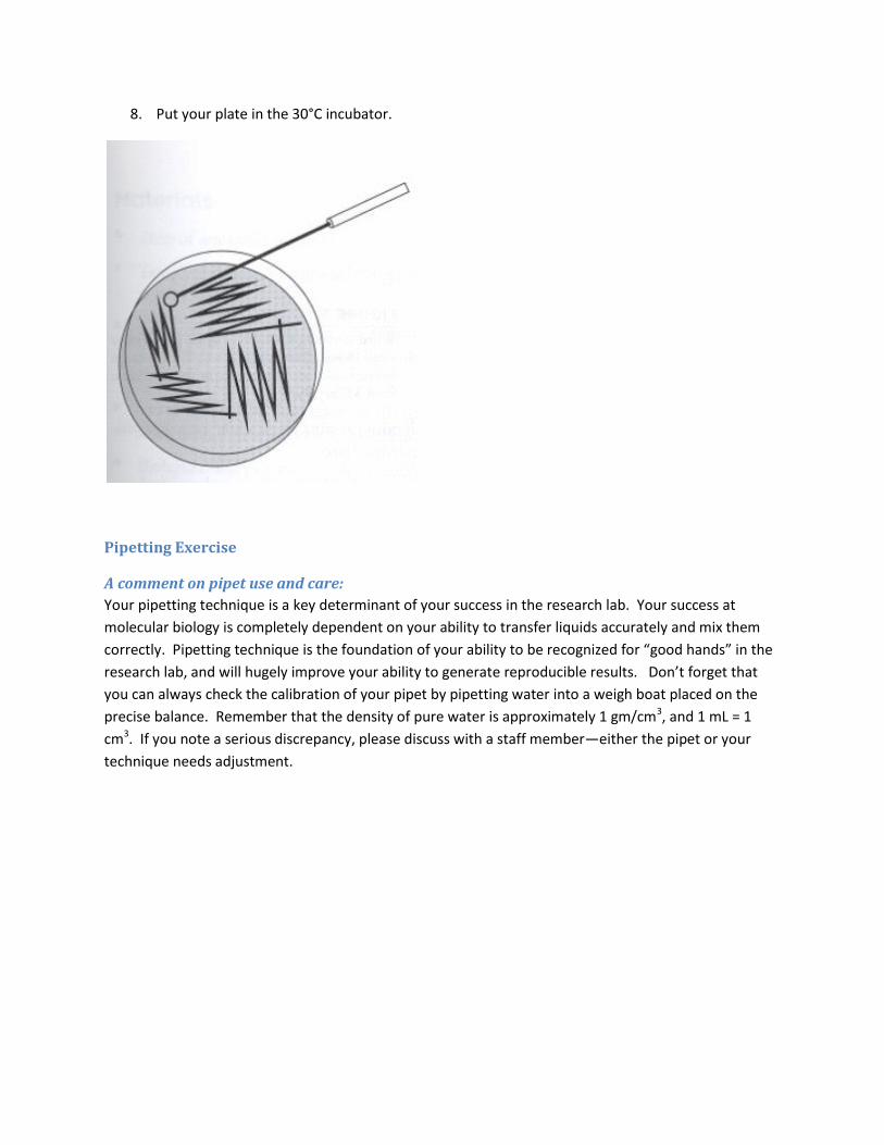
8. Put your plate in the 30°C incubator.
Pipetting Exercise
A comment on pipet use and care:
Your pipetting technique is a key determinant of your success in the research lab. Your success at
molecular biology is completely dependent on your ability to transfer liquids accurately and mix them
correctly. Pipetting technique is the foundation of your ability to be recognized for “good hands” in the
research lab, and will hugely improve your ability to generate reproducible results. Don’t forget that
you can always check the calibration of your pipet by pipetting water into a weigh boat placed on the
precise balance. Remember that the density of pure water is approximately 1 gm/cm3, and 1 mL = 1
cm3. If you note a serious discrepancy, please discuss with a staff member—either the pipet or your
technique needs adjustment.
We will use fluorescein, a fluorescent dye, to measure your pipetting technique. You will make various
dilutions and put them into a 96 well microplate, as indicated below. (More specific instructions follow
the diagram.)
Small volume pipetting (rows A-C)
Fill three rows of wells with 100 ul water. Add the indicated amount of undiluted 10 µM fluorescein (see
figure.)
Serial dilutions (rows D-H)
When a large dilution is required, an accurate dilution cannot be performed in a single dilution step and
serial dilutions are necessary. The difference between the concentration of the original solution and the
concentration of the desired solution (the dilution factor) will determine how many dilutions are
required. The most common examples deal with concentration of cells or the concentration of a solute.
You will use such dilutions on an almost daily basis.
Key techniques for accurate serial dilutions:
- Mix the sample very well by vortexing after each addition - Use a new pipet tip for each step of the dilution. Can you guess why this is important? - 1:10 doesn’t mean 100µl of sample and 1000µl of diluent….that will give you a 11-1 dilution, not a
10-1, and those errors add up fast as you make serial dilutions! For a 10-1 dilution you want 100µl of sample and 900µl of diluent.
- Always make your calculations (trivial as they are) in advance. Write them in your notebook as, for example, “100 µl sample + 900 µl diluents = 1000 µl” NOT “1/10 dilution”. Check each line off as you actually execute the dilutions. This is good practice and even very experienced experimentalists make these notes in advance and check them off during the procedure. During a complicated protocol it is very easy to skip a step or screw up a dilution if all you have in front of you is a protocol.
1:10 dilutions.
Fill 5 microfuge tubes with 900µl water. Beginning with your concentrated fluorescein stock, make a
1:10 dilution (i.e. 10-1) by adding 100µl fluorescein to the water. Vortex well to mix, then transfer 100µl
from this tube to your next tube, for a 10-2 dilution. Continue as described to 10-5.
Place 100 µl in each well of your 96 well microplate as indicated.
1:2 dilution
Fill 5 microfuge tubes with 700 ul water. Beginning with your concentrated fluorescein stock, make a
series of 1:2 dilutions, continuing to 2-5.
Place 100 ul in each well of your 96 well microplate as indicated.
Give to a staff member to take your microplate for counting on the Tecan.
Plot your data using your favorite program. Did you achieve the dilution curve you expected? What is
the appropriate way to plot your data given the dilution series that you did? If your correlation is less
than 0.95, make a new set of dilutions and try again. If you are running short on time, please move on
to the “Quantification of Microorganisms” experiment and plot your data at home (and show to a
member of the staff tomorrow).
Quantification of Microorganisms
This portion of the laboratory assignment will have two parts. You will begin with a culture of yeast.
You will then make a 1:10 dilution series of this culture at 100, 10-1, 10-2, 10-3 in conical tubes. First, you
will quantify the density of your dilutions using the Klett colorimeter. Secondly, you will count the
number of cells in the culture using a counting chamber and a light microscope. You will plot the
relationship between the number of cells/ml in the culture and the Klett reading. Keep this plot. It will
be useful to you in your future research projects. As you look at cells under the microscope, take careful
note of what they look like. You should see cells in various stages of budding. Yeast cells DO NOT look
like bacterial cells. If there is any doubt, always ask a staff member to look at the cells with you.
Counting yeast cultures using the Klett
The Klett (Klett-Summerson photoelectric colorimeter) is an excellent tool for measuring culture density.
Important Notes for Operating the Klett:
Make sure tubes are clean and dry. Wipe outside with a Kimwipe.
Regular test tubes DO NOT work with the Klett. You must use the Klett tubes.
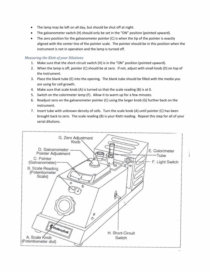
The lamp may be left on all day, but should be shut off at night.
The galvanometer switch (H) should only be set in the “ON” position (pointed upward).
The zero position for the galvanometer pointer (C) is when the tip of the pointer is exactly
aligned with the center line of the pointer scale. The pointer should be in this position when the
instrument is not in operation and the lamp is turned off.
Measuring the Klett of your Dilutions:
1. Make sure that the short-circuit switch (H) is in the “ON” position (pointed upward).
2. When the lamp is off, pointer (C) should be at zero. If not, adjust with small knob (D) on top of
the instrument.
3. Place the blank tube (E) into the opening. The blank tube should be filled with the media you
are using for cell growth.
4. Make sure that scale knob (A) is turned so that the scale reading (B) is at 0.
5. Switch on the colorimeter lamp (F). Allow it to warm up for a few minutes.
6. Readjust zero on the galvanometer pointer (C) using the larger knob (G) further back on the
instrument.
7. Insert tube with unknown density of cells. Turn the scale knob (A) until pointer (C) has been
brought back to zero. The scale reading (B) is your Klett reading. Repeat this step for all of your
serial dilutions.
Counting yeast cultures using a counting chamber
S. cerevisiae cells often remain associated after division. Be sure to count individual cells. In the future you might use a sonicator (please see the sonication protocol in the appendix) to break apart cells. However, for the purposes of time, please count the cells without sonication.
Construct a counting chamber
1. Take a microscope slide and put two parallel pieces of double stick tape on the slide about 1 cm apart.
2. Place a cover slip across the double stick tape and push the slide down onto the tape creating a seal (use your fingernail or a toothpick).
3. Remove any excess tape from the edge of the slide with a razor blade. The chamber is now ready for the sample.
Imaging the sample
4. Take about 40μL of the culture and pipette it into the chamber. (If you wanted to observe the cells for a long period of time you could seal the two sides of the chamber with grease. Note that this cuts off the oxygen supply to the cells. For the purpose of time, please leave your chamber unsealed).
5. Label the slide with the dilution information (1:10, 1:100, etc) 6. Move the slide to a random position on the microscope. 7. Using the 40x objective, focus the microscope close to the bottom surface of the
counting chamber. 8. Switch the image to the Firewire camera (slide the lever on the trinocular tube). 9. Using the VI SnapShot in Labview (see protocols in the appendix), take a picture of the
bottom of the slide and save the image with a filename that reflects the sample. Adjust exposure using the Gain control (higher is brighter) and the Shutter controls (higher is brighter).
10. Move the slide to a RANDOM new location and take a new image. 11. Take at least a total of 5 images. For low concentration cultures, you may need to take
additional images to get a statistically significant cell count (> 100 counts).
Counting the cells
12. Now you can open the images in ImageJ and count the number of cells in the field. When there are few cells, you can count them by eye. Record the number in your notebook.
Calculating culture density
13. The volume of the field of view is 1.2 x 10⁻⁶ mL, xy area calculated using a micrometer,
multiplied by the known thickness of the tape, validated with a hemocytometer;
14. Multiply your count by 8.3x10⁵ to calculate the cells per mL of culture.
PUMA Registration
The Princeton University MicroArray database (PUMAdb) stores data from microarray experiments.
PUMA serves as a resource for data retrieval, analysis and visualization. You will be able to use this
fantastic resource for your own microarray experiments.
1. Register for PUMA at: http://puma.princeton.edu/cgi-bin/tools/display/registration.pl
a. For lab phone, but 8-9085
b. For fax, put 8-8020
c. For laboratory in which you work/collaborate, select QCB301 project
2. If you are already signed up for PUMA because you have worked in a lab at Princeton, email
the curators at [email protected] and ask to be added to the QCB301 project
3. NOTE: When you finish the QCB301 course, please email [email protected] and
let them know that you no longer need access to this resource. If you join a lab on campus,
and would like PUMAdb access through that lab, please email [email protected]
to let them know your change in status.