Embed Size (px)
Citation preview

The copyright of this thesis vests in the author. No quotation from it or information derived from it is to be published without full acknowledgement of the source. The thesis is to be used for private study or non-commercial research purposes only.
Published by the University of Cape Town (UCT) in terms of the non-exclusive license granted to UCT by the author.
Univers
ity of
Cap
e Tow
n
Univers
ity of
Cap
e Tow
n
1
Locking plates for distal femur fractures: Does an increased working length improve healing?
By
DR IAN M KOLLER
KLLIAN003
SUBMITTED TO THE UNIVERSITY OF CAPE TOWN In partial fulfilment of the requirements for the degree
Master of Medicine in Orthopaedic Surgery
Faculty of Health Sciences
UNIVERSITY OF CAPE TOWN
Date of Submission 18 February 2013
Supervisors Dr S Maqungo, Prof. J Walters
Department of Orthopaedic Surgery,
University of Cape Town

Univers
ity of
Cap
e Tow
n
2
DECLARATION
I, Dr Ian Koller hereby declare that the work on which this dissertation/thesis is based is my original work (except where acknowledgements indicate otherwise) and that neither the whole work nor any part of it has been, is being, or is to be submitted for another degree in this or any other university.
I empower the university to reproduce for the purpose of research either the whole or any portion of the contents in any manner whatsoever.
Signature: …………………………………
Date: …………………………………….

Univers
ity of
Cap
e Tow
n
3
ACKNOWLEDGEMENTS
1. I wish to thank Dr Sithombo Maqungo and Prof. Johan Walters for their guidance and support during the preparation of this dissertation.
2. I would like to thank the AO Foundation for permission to use their images.
3. This dissertation is presented in a standard monograph format but it is also my intention to submit the research to a peer reviewed orthopaedic journal for publication.

Univers
ity of
Cap
e Tow
n
4
TABLE OF CONTENTS Declaration……………………………………………………………………………2
Acknowledgements………………………………………………………...................3
Index of graphs, tables and figures…………………………………………………...5
Glossary of abbreviations…………………………………………………………….6
Abstract……………………………………………………………………………….7
PART A
Literature review…………………………………………………………...................8
Objectives……………………………………………………………………..8
Literature search strategy……………………………………………………...8
Evolution of distal femur fracture management………………………………9
Epidemiology of distal femur fractures……………………………………...12
Principles of fracture healing as they relate to surgical fixation of distal femur
fractures…………………………………………………………………...…13
Distal femur locking plate design and surgical technique ………………….14
Early outcome sand complications……………………...…………………..15
Working length……………………………………………………………...17
Is there a problem with fracture healing?.......................................................19
Summary…………………………………………………………………….20
PART B
Locking plates for distal femur fractures: does an increased working length improve
healing?.......................................................................................................................22
Materials and methods ……………………………………………………...22
Results…………………………………………………………………….....25
Discussion…………………………………………………………………...31
Strengths and weakness…...………………………………………………...38
Conclusion…………………………………………………………………..38

Univers
ity of
Cap
e Tow
n
5
Appendices …………………………………………………………………………39
Distal femur fracture study proforma……………………………….............39
Smith and Nephew Perilock Plate template………..………………….........40
Titanium plate templates…………………………………………………….41
References……………………………………………………..................................42
INDEX OF GRAPHS, TABLES AND FIGURES
GRAPHS
Graph 1. Case frequency as per mech. of injury …………………………….27
Graph 2. Case frequency as per AO classification ………………………….27
Graph 3. Healing outcomes…………………………………………………..29
Graph 4. Histogram of age at time of injury………………………………....32
Graph 5. Mean union time by fracture pattern …………..…………………..32
Graph 6. Mean union by AO classification…………………………………..33
Graph 7. Scatter gram of working length vs date of surgery………………...34
Graph 8. Histogram of union times…………………………………………..37
TABLES
Table 1. Details of surgical technique: Mean values ………………………28
Table 2. Healing Outcomes………………………………………………....29

Univers
ity of
Cap
e Tow
n
6
Table 3. Outcome of regression analysis……………………………………31
FIGURES
Fig.1 AO classification-groups……………………………………………..12
Fig. 2 The “box “ defining the supracondylar region……………………....13
Fig 3. Anatomy of a distal femur locking plate construct…………………..21
GLOSSARY OF ABBREVIATIONS AO Foundation: Arbeitsgemeinschaft für Osteosynthesefragen (Association for
the Study of Internal Fixation)
ABP: angular blade plate
CBP: condylar buttress plate
DCS: dynamic condylar screw
IM: intramedullary
GSH nail: Green-Seligson-Henry retrograde intramedullary nail
GSW: gunshot wound
GPa: gigapascal
LCP: locked compression plate
LISS: Less Invasive Stabilization System
MI(P)PO: minimally invasive (percutaneous) plate osteosynthesis
MVA (P): motor vehicle accident (pedestrian)
OTA: Orthopaedic Trauma Association
SDR: screw density ratio
TKR: total knee replacement
UCT: University of Cape Town
W/L: working length
Univers
ity of
Cap
e Tow
n
7
ABSTRACT Purpose of study
Distal femur locking plates have become a very popular means of internal fixation
because of their ability to provide stable distal periarticular fixation. In spite of this
enthusiasm however several studies have reported significant problems with healing.
In the distal femur it is recognized that locking plate fixation may be too rigid if used
in certain configurations that limit the essential micro movement required for
biological healing. Implant failure may arise from rigid configurations that cause
excessive hardware stress concentrations. In an attempt to address these problems
longer plates and an increased working length have been proposed to reduce construct
rigidity. The purpose of our study is to investigate whether an increased working
length translates into improved healing.
Description of method
We undertook a retrospective review of 64 suitable cases performed at our institution
from April 2007 to February 2012. Case notes and X-rays were reviewed. Working
length, plate to fracture zone ratios and working length to fractures zone ratios were
calculated. Union was assessed radiographically. Multiple regression analysis was
utilized with time to union as the dependent variable. The independent variables
smoking, age, fracture severity and soft tissue injury as well as working length were
included.
Summary of results
Mean time to union was 18.03 weeks (±6.19). 14 delayed unions (21.8%.), 3
nonunions (4.7%) and 1 implant failure were recorded. Multiple regression analysis
identified an open fracture as the only significant risk factor P=0.002. Smoking
showed a strong trend P=0.07 but working length did not show any significant
influence over union (P=0.341) in this series.
Conclusion
Biomechanical studies have demonstrated increased flexibility of longer plates with an
increased working length. In this study however, working length did not show any
Univers
ity of
Cap
e Tow
n
8
correlation to union. In contrast, the risk factors known to prolong healing were the
only significant independent variables that correlated with union times.
LITERATURE REVIEW
Objectives of the literature review
The objectives of this literature review are to gain an insight into the challenges
present during the management of distal femur fractures. Visiting the earliest literature
on the subject will allow me to track the evolution of management and the
progression of surgical implant design that resulted in improved outcomes. The review
will provide insight into the current management, potential complications and finally
identify the most recent research pertaining to working length and its relationship to
distal femur fracture healing.
Literature search strategy
Internet based search engines using numerous electronic databases, including Pubmed
and Google Scholar were used to scan the English literature. The scope of the search
included the English medical literature extending back to 1940. Article bibliographies
were manually reviewed to identify relevant articles. Relevant veterinary studies were
manually identified. Follow up searches of authors’ names were performed to identify
special interest threads. In two instances authors were contacted via email for direct
commentary on their articles.
All articles were obtained in full and downloaded via the UCT library institutional
access. Print articles were scanned and stored electronically.

Univers
ity of
Cap
e Tow
n
9
Evolution of distal femur fracture management
Distal femur fractures are recognized as being difficult injuries to manage
successfully. The short distal segment often with fracture extension into the knee joint,
poor bone quality and large potential deforming forces from muscle action and weight
bearing pose mechanical and surgical challenges.
The earliest reports on treatment noted varying results with conservative management
consisting of traction followed by cast bracing or spica application. 1 Local
complications of mal union, knee stiffness, and nonunion together with the systemic
complications of prolonged bed rest led surgeons to explore operative means of
treatment. Initial attempts at surgical management showed inferior results compared
to conservative techniques. In 1967 Neer et al reviewed 110 fractures treated with
either internal fixation by various means or conservatively and noted only 52%
satisfactory results compared with 84% respectively.2 They concluded that no category
of distal femur fracture appeared amenable to internal fixation. Stewart et al arrived at
a similar conclusion after comparing surgical and conservative methods of treatment3.
Closer inspection of these studies however revealed significant bias. The more severe
grades of fractures were treated surgically while minimally displaced fractures were
managed conservatively. Further, it appeared that numerous different surgical
techniques were employed from Blount angled blade plates to various pin or screw
constructs. The X-rays presented in those studies show that most of the constructs
were inferior biomechanically and demonstrated poor surgical technique.
The early challenges identified during open procedures included achieving rigid
fixation, especially in the short distal metaphyseal fragment, dealing with intra
articular involvement and multi fragmentary stabilization. During the 1960’s and
1970’s improvements in fixation devices and surgical technique began to swing the
pendulum back in favour of open techniques. Schatzker et al in 1974 and Schatzker
and Lambert in 1979 published results demonstrating the superiority of the AO
principles of open reduction and internal fixation over both conservative treatment and
other forms of internal fixation. 4,5 It was also noted that age and bone quality has a
significant effect on outcome. Healey and Brooker reported 81% vs 35% good
functional outcome in favour of surgical management.6 Complications were 3 times
higher and hospital stays were also significantly longer in the non surgical groups.6.

Univers
ity of
Cap
e Tow
n
10
Specific implants were designed in order to address some of these challenges. The
arrival of the 95° angular blade plate (ABP) provided rigid fixation but was
unforgiving with regards to mal alignment. Incorrect alignment of the seating chisel in
the condyles was not correctable and no adjustment was possible after plate
insertion.5,7 Another weakness was its difficulty in dealing with an intercondylar split
as it required an intact condylar block for rigid fixation. The recommendation was to
restore the block with an interfragmentary screw prior to blade insertion. With sound
surgical technique good results were achievable however and the 95° angular blade
plate remains in use today.
The dynamic condylar screw (DCS) introduced in 1980 was a more forgiving fixed
angle device than the ABP. It still required intact or provisionally reduced condyles for
insertion but was able to provide some compression via the lag screw. Its main
weakness was instability in the sagittal plane as rotation could occur around the lag
screw with knee motion resulting in loosening. 6
In an attempt to deal with multi fragmentary fracture patterns, the condylar buttress
plate (CBP) was developed. It was contoured to match the distal femur and could
accept multiple lag screws through its flared distal end in order to compress
comminuted condylar fragments. In order to achieve this versatility the CBP sacrificed
stability. Its main weakness was that it was not a fixed angle device like the ABP or
DCS and varus collapse was a problem.8 To overcome this problem Saunders et al
proposed dual plating of both the medial and lateral sides to provide additional
stability in comminuted fracture patterns.9 This required extensive exposure and
periosteal stripping in a region of already compromised vascularity.
The advantages of intramedullary (IM) nailing techniques were recognized early on in
the management of distal femur fractures. Insertion was through a limited incision
without having to expose the fracture site. This avoided further disruption of the soft
tissue envelope, affecting healing and potentially reduced the risk of sepsis. The
earliest attempts at closed surgical management involved the use of Rush rods, Enders
or Zickel nails. All these nails were too flexible to provide adequate stability and were
not able to deal with comminution. Additional bracing or casting was required thus
negating the advantages of surgical fixation. Many nails were also supplemented with
Univers
ity of
Cap
e Tow
n
11
other forms of internal fixation. Although nonunion was low (0-2%), malunion and
knee stiffness were a problem.10
As IM nailing of femur fractures became established, the technique was extended to
include distal metaphyseal and even intercondylar fractures. The introduction of the
interlocking nail by Grosse Kempf was a major step forward in controlling stability of
the distal fragment especially in the rotational plane.11 Initially only anterograde
nailing was performed. Technical problems encountered were the distance of the
locking screws from the end of the nail preventing secure fixation of very distal
fractures. Yeung et al reported good results by modifying the nails, cutting off the
distal 15mm, allowing very distal purchase.12 The modern technique of retrograde
nailing was first introduced in 1988 by Green with the introduction of the Green-
Seligson-Henry (GSH) supracondylar intramedullary nail. Limb length equality, axial
alignment and mechanical axis restoration were challenges reported.13 Union rates
were very good however. Short nails, which terminated in the diaphysis, were thought
to be adequate but stress risers with fracture at the proximal tip have led to the
abandonment of short retrograde nail constructs. Salem et al reported in a 2006 series
of 47 patients that there were no differences between anterograde and retrograde
outcomes with regard to alignment.14 The use of blocking or Poller screws in the distal
fragment aids alignment and increases construct stiffness. 15 The weakness of
intramedullary nailing is again dealing with comminution of the metaphysis and or
intercondylar region. Intercondylar splits require prior fixation with lag screws, which
can obstruct nail passage. The most recent retrograde nail designs have increased the
number of interlocking screw options in order to provide maximum stability in a
variety of fracture configurations.
By the mid 1990’s surgical management of distal femur fractures was well established
and consistently provided superior outcomes to conservative treatment. There were
however certain fracture patterns that still presented significant challenges. Highly
comminuted fractures with intra articular involvement frequently produced worse
results. Delayed union and nonunion was highest in this group as was loss of
reduction. Elderly patients with osteoporotic bone faired the worst of all.16,17 With the
increasing popularity of total knee replacements (TKR) especially in older patients
periprosthetic fractures became more common. A very short, osteoporotic distal
fragment containing the femoral prosthesis provides very limited room for fixation.18
Univers
ity of
Cap
e Tow
n
12
Some implants depending on design can accommodate a retrograde nail but many such
as the posterior cruciate substituting designs may not allow the passage of a nail due to
the presence of the central cam.19 These fractures require internal fixation by other
means such as a distal femur locking plate.
Epidemiology and classification of distal femur fractures
The definition of what constitutes a distal femur fracture varies in the literature.
Variations arise due to the differing distances from the knee joint described.
Qualitatively the fracture must involve the supracondylar region of the femur or
“box”. The height of the box equals the width of the condyles. Fracture lines may
extend proximally into the diaphysis or distally to involve the articular surface.
The largest epidemiological study was under taken by the AO Foundation published in
2000. Distal femur fractures were noted to follow a bimodal pattern with one group of
high energy fractures mostly from road traffics accidents occurring in young males
and a second group of older patients, mostly female with osteoporotic fractures from
low energy falls.20
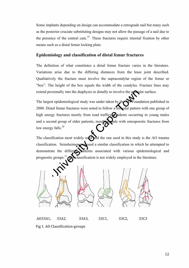
The classification most widely used and the one used in this study is the AO trauma
classification. Seinsheimer proposed a similar classification in which he attempted to
demonstrate the differing patterns associated with various epidemiological and
prognostic groups.21 His classification is not widely employed in the literature.
AO33A1, 33A2, 33A3, 33C1, 33C2, 33C3
Fig 1. AO Classification-‐groups

Univers
ity of
Cap
e Tow
n
13
Fig. 2 The “box “ defining the supracondylar region
Principles of fracture healing as they relate to surgical fixation of
distal femur fractures
The physiological process of fracture healing requires some motion at the fracture site
to allow mechanical induction of external callus formation. It also requires an adequate
local blood supply to optimize the metabolic processes of bone formation. This is
considered secondary or indirect healing and is how fractures heal with conservative
management and with intramedullary nailing. In the 1960’s the principles of open
reduction and internal fixation were formalized. Rigid, anatomical surgical
stabilization of fractures was considered essential for early joint motion to prevent
stiffness and muscle wasting. Anatomical reduction of fracture fragments especially
intra articular involvement was desired in order to minimize the late development of
arthrosis. To achieve this, large dissections were performed. Interfragmentary
compression was achieved via lag screws and neutralization plates were fixed with
screws as close to the fracture as possible to maximize rigidity.22 This construct
results in direct or primary bone healing. Due to the compression and absolute rigidity
no micromotion is present for the mechanical induction of callus formation. The
healing process skips the intermediate steps of tissue differentiation and resorption.
The cutting cones and osteones cross the fracture and proceed directly to the internal
remodeling of the Haversian system. The disadvantage of this rigid construct is that if
anatomical reduction and compression are not achieved then there is a high risk of
Univers
ity of
Cap
e Tow
n
14
delayed union. This is because of the high strain generated across the fracture gap. The
strain theory states that a tissue cannot be produced under conditions that exceed the
elongation at rupture.23,24 In the case of fracture healing, it refers to the osteoblasts
involved in the remodeling process being disrupted.
In the distal femur especially in comminuted fractures it is often not possible or
desirable to achieve primary bone healing, as it would require large dissections and
further disruption of local blood supply. Technically it may be impossible to
anatomically reduce all fracture fragments without completely devitalizing them. The
current aims of distal femur fracture management are to bridge the fracture site
restoring overall alignment while minimizing soft tissue disruption. This is achieved
with an IM nail if the fracture pattern allows or with a distal femur locking plate.
Distal femur locking plate design and surgical technique.
In the late 1990’s the plate and screw construct, now known as a locking plate, was
developed that attempted to address the limitations of standard compression plating.
Toggling between the screw and plate resulted in screw loosening and loss of
fixation.24 Other limitations were disruption of fracture zone blood supply and weak
fixation in osteoporotic and metaphyseal bone. Locking plates or angular stable plates
have a threaded screw plate interface providing an angular stable construct in the same
manner as the ABP and DCS. They do not rely on compression between the plate and
bone for stability.25 This in turn allows preservation of the periosteal blood supply.
Purchase in osteoporotic bone is superior to standard compression plating constructs
as the plate and screws function as a single unit to resist pullout. They have been
referred to as “internal external fixators” upon which their biomechanical principles
are based.
The classic indications for locking plate use are with fractures that are periarticular and
or involve osteoporotic bone.26 In fractures of the distal femur these advantages of
locking plates became immediately attractive. They offer superior distal fragment
fixation via multiple angular stable screws arranged in a periarticular cluster. Intra
articular fracture extension can be separately fixed or held through the plate as
required. This is their biggest advantage over the retrograde intra medullary nail,

Univers
ity of
Cap
e Tow
n
15
which is the other surgical modality currently in use for distal femur fracture
management.
Recognition of the importance of blood supply in fracture healing led to the
emergence of minimally invasive techniques known as minimally invasive
percutaneous plate osteosynthesis (MIPPO).27,28 These procedures incorporate the
concept of ‘biological osteosynthesis’ whereby the fracture zone is bridged,
minimizing disruption to the fracture zone. The surgical incision is not made directly
over the fracture but in the periarticular region proximally or distally. It is important
not to confuse minimally invasive techniques with locking plate surgical technique. A
locking plate can be applied using an open technique and an example of this is in distal
radius fractures where anatomical reduction is desirable. Minimally invasive
techniques have been applied to distal femur locking plate constructs to combine the
advantages of both concepts to maximize healing potential. The first such system
available was the Less Invasive Stabilization System(LISS) plate by Synthes (USA,
Paoli, PA) followed by others such as the Perilock plate by Smith and Nephew. 29,30
These plates are pre contoured to the lateral distal femur and are inserted with the aid
of a targeting jig. Intra articular fracture extension is anatomically reduced under direct
vision via arthrotomy and held with lag screws. The plate is then inserted in a
retrograde fashion along the femoral shaft following a submuscular plane, bridging the
fracture zone. The most important step is correct alignment of the plate to the lateral
condyle as the precontoured plate is then used as a template to guide fracture reduction
via indirect methods. Reduction is not required to be anatomic, the goal being
restoration of alignment. Flouroscopic guidance and percutaneous reduction tools
assist with reduction. Finally proximal screws are inserted percutaneously via the
targeter.
Early outcomes and complications
Early results of the use of locking plates were favorable. 29,30,31,32 Nonunion ranged
from 0-9%, delayed union 0-7%.33,34 Of particular interest was the incidence of varus
collapse in the mechanically unstable comminuted fracture patterns. Syed et al
reported only 1 patient (4%).32 Biomechanical studies quickly emerged comparing
locking plate stability to IM nailing, the ABP and DCS. Results showed that locking
Univers
ity of
Cap
e Tow
n
16
plates are superior in resisting deformity under axial loads and are only slightly
weaker than IM nails under torsional loads.35,36
On the back of these favorable early results locking plates became the implant of
choice for many surgeons. Amongst the successes there were some clear failures.
Kregor in his 2001 study noted 5% implant failure in 103 cases.29 Sommer in 2004
reported on 4 cases with broken plates and Vallier in 2006 presented 6 cases of
implant failure.37,38 What was not clearly understood were some of the mechanical
factors regarding locking plate constructs that optimize fracture healing.
Two patterns of implant failure have emerged from the literature. Early failure,
occurring in the first few weeks to months post op, is considered to be related to
technical factors such as poor patient compliance with restricted weight bearing or
inferior surgical technique. Late failure, occurring after what would be considered the
expected time to union is related to the healing process. As with all orthopaedic
constructs used to treat fractures, implant integrity is a race against time. If the bone
does not unite the implant will eventually fail. The rate at which this occurs depends
upon the forces acting on the construct and the construct’s ability to withstand the
repetitive loading over time.
Biological or secondary bone healing requires micro motion for callus formation.
Motion at the fracture site results from reversible deformation of the bridging plate
under cyclic load. Biomechanical analysis of locking plates noted that their stiffness is
an order of magnitude greater than external fixators and comparable to conventional
compression plating under axial load.39 Stiffness of this magnitude is considered to be
too great to allow adequate micro motion required for callus generation (0.2-1mm).40
There are many factors that regulate the stiffness of the locking plate construct. Plate
factors include dimensions of the plate and material used. Stainless steel (SS) and
titanium are the materials currently in use. Titanium has a Young’s modulus of
elasticity of +/-110GPa and stainless steel 200 GPa. In contrast bone ranges from 10
GPa for trabecular bone up to 30 GPa for cortical bone. Clearly both metals are more
rigid than bone but titanium is closer to bone in its mechanical properties than stainless
steel. Titanium also has a higher yield stress than SS and has superior fatigue
resistance.41 Gaines reviewed this in 2008 and found a significant difference in non
union between the titanium 7% and stainless steel 23% (P=0.05) groups.42

Univers
ity of
Cap
e Tow
n
17
Working length
The working length of the plate refers to the unsecured section of the plate that bridges
the fracture. The margins are defined by the most distal screw in the proximal
fragment and the most proximal screw in the distal fragment. The location of the
screws regulates the working length. Stoffel in 2003 in biomechanical testing of the
locked compression plate (LCP, Synthes) demonstrated that the omission of one screw
on either side of the fracture site almost doubled the flexibility. 43 Increased flexibility
results in greater motion at the fracture site for the same load. The increased working
length also distributes stress over a greater area thus decreasing stress concentrations
in the plate and minimizing the chance of fatigue failure with cyclic loading43,44.
A difficult question to answer is what the appropriate working length for a given
fracture should be. The introduction of minimally invasive plate osteosynthesis
(MIPO) has allowed longer plates to be used without greatly increasing the surgical
insult. Gautier in his paper divided the plate into three zones; the proximal zone, the
middle fracture zone and the distal zone.. He empirically proposed that the overall
plate length or “plate span width “should be three times the fracture zone length for
comminuted fractures and 8-10 times in simple transverse fractures.44 The 4th zone to
consider is the working length. Gautier did not state how long the working length
should be in relation to the fracture zone.. The fracture zone and the working length
are not necessarily the same. The fracture zone may be short in simple transverse
fractures but may also be extensive in severely comminuted fractures. The working
length is always equal to or greater than the fracture zone. The only way the working
length can be shorter than the fracture zone is if compression screws are placed within
the fracture zone which is against biological healing principles.
The recommendations for plate length and technique have mostly been made on
models of diaphyseal fractures with proximal and distal zones that are unconstrained
in length.43 The distal femur however presents a different scenario. The distal zone is
constrained by the knee joint distally and the fracture proximally. The fracture zone is
mostly metaphyseal. The proximal zone is unconstrained and can be altered by plate
length and screw location. The significance of this is that the working length usually

Univers
ity of
Cap
e Tow
n
18
cannot be extended distally past the edge of the fracture zone and any manipulation of
working length must occur by omitting screws proximal to the fracture zone.
The second recommendation that Gautier made concerned the number of screws used
for fixation. The “screw density ratio” (SDR) is calculated by dividing the total
number of screw holes available in the plate by the number of screws used.
He proposed that SDR be used as a proxy for working length. Again he empirically
recommended ratios of between 0.4-0.5. while this SDR provides a guideline for
overall screw use it is less useful in guiding screw placement. Mechanical
characteristics of the construct can be significantly altered by varying screw location
while maintaining the same screw density ratio. Where the screws are placed is more
important than how many are used. In distal femur fractures especially those involving
the joint the priority is to obtain anatomic reduction of any intra articular extension
and adequate distal purchase to prevent varus collapse. As many screws as are
required to achieve this are used.
Returning to the working length the two most important factors to consider are how
long the working length relative to the fracture zone should be and how long should
the proximal zone be. The single most important screw is the first screw proximal to
the fracture zone. This screw determines the working length. Placing the screw just
proximal to the fracture zone will result in a short working length. In comminuted
fractures the fracture zone may be large and placing the first screw close to the
proximal margin of the fracture zone ie making the working length equal to the
fracture zone may provide sufficient flexibility.
In the clinical setting there are two different construct scenarios. The first is a simple
transverse or short oblique fracture with a small fracture zone bridged by the plate.
Making the working length equal to the short fracture zone will result in a very stiff
construct.39,43,44 This may result in implant failure due to high stress concentrations or
poor healing due to lack of fracture site micro motion. Here it is proposed that making
the working length longer than the fracture zone (large working length to fracture zone
ratio) minimizes these complications.
The second clinical scenario is a comminuted fracture with a large fracture zone. In
such circumstances smaller working length to fracture zone ratios may be more

Univers
ity of
Cap
e Tow
n
19
appropriate. Employing the same ratios that are used in the first scenario may not be
necessary or physically possible due to femur or plate length. The possibility of
making a construct too flexible and therefore unstable has not to date been reviewed in
the literature. Stoffel noted that in large fracture gaps there was no further reduction in
plate stress beyond a working length of four holes.43
The proximal zone length is governed by the location of the first screw that divides the
working length zone and the proximal zone for any given plate length. The strength of
fixation in the diaphyseal bone is determined by bone quality, type of screw (locked or
unlocked), number of screws and length of the proximal zone. The cantilever effect of
lengthening the proximal zone reduces the pullout load on the most proximal screw
thus reducing the risk of proximal zone failure.44 Again it becomes more important
where you place the screws rather than how many are used. A proximal zone length of
6 holes with 3 screws filling alternate holes has superior pullout out resistance to a 3
hole length filled with 3 screws. In healthy cortical bone there is no clinically
significant difference in pullout resistance between locked and unlocked screws but
using unlocked screws may make the construct stiffer45. There is no added mechanical
advantage beyond four screws.42
Is there a problem with fracture healing?
A comprehensive review of distal femur locking plates and potential problems with
healing was performed by Henderson recently.52 The systematic review noted an
incidence of problem healing was as high as 32%. Nonunion rates were from 0% to
19%. Henderson included delayed union, nonunion and implant failure under the
collective term of problem healing.
There are many biomechanical studies and review papers discussing locking plate use
but there are very few clinical distal femur studies comparing working length and
healing outcomes. In an abstract of the OTA annual meeting 2009, Ricci presented the
findings of a large multi center study of 305 patients looking at risk factors for
complications including nonunion and implant failure. The only independent risk
factor for nonunion was diabetes. Technical risk factors for implant failure were
analyzed and on those findings he proposed recommendations for plate length, screw

Univers
ity of
Cap
e Tow
n
20
density ratio.46 He recommended a combined fracture zone and proximal zone length
of no fewer than 10 holes with 5 or more proximal screws and a screw density ratio of
less than 60%. To date the findings have not been published in a peer reviewed
journal.
Lugan in 2010 performed volumetric callus mapping as a means of measuring healing
in locking plate fixation of distal femur fractures47. Periosteal callus formation is a
marker of secondary bone healing. He noted that lateral locked plates produce
asymmetrical and inconsistent callus formation. He also compared the effect of
working length and plate material on callus formation. There was a very weak
correlation between medial callus at 6 weeks and working length. Follow up at 12 and
24 weeks showed no correlation. Further he noted that 40% of the cases produced very
little (< 20mm2) callus even at 6 months. Titanium produced significantly more callus
than stainless steel constructs.
Bottlang also in a 2010 review of 70 patientts noted a 19% non union rate and no
significant difference in working length between the non union and union groups. 39
These last 3 studies represent the current clinical evidence for working length and its
affect on healing. The findings are conflicting and no significant conclusion can be
drawn from these studies. Of caution is the observation that the studies of Lugan and
Bottlang appear to be very similar in materials and method and they are both co-
authors of each others papers. Possibly there is some overlap of the data samples.
Summary
The literature shows that distal femur fracture management has progressed a long way
from the early days of conservative management. It also shows however that these
fractures remain challenging in spite of the recent advances in orthopaedic techniques
and implants. The recent literature has identified concerns about prolonged healing
and its proposed relationship to plate working length. The theory that increasing the
working length will improve the healing rate has not been widely tested in the
literature to date and this represents an area of active research. Other interventions that
potentially improve healing besides working length represent areas of future research
potential.
Univers
ity of
Cap
e Tow
n
21
The aim of this study is to identify whether there is a relationship between working
length and fracture healing. The null hypothesis to be tested is that greater working
lengths are not able to improve distal femur fracture healing.
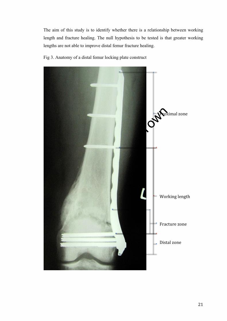
Fig 3. Anatomy of a distal femur locking plate construct
Proximal zone
Working length
Fracture zone
Distal zone

Univers
ity of
Cap
e Tow
n
22
LOCKING PLATES FOR DISTAL FEMUR FRACTURES: DOES AN INCREASED WORKING LENGTH IMPROVE HEALING? Materials and methods
A retrospective review of 126 consecutive distal femur fractures treated by locking
plate between April 2007 and February 2012 were identified for inclusion in the study.
The indication for locking plate fixation is a distal femur fracture not amenable to IM
nail fixation specifically those fractures that are too comminuted or are too distal to
obtain adequate stability. Cases were identified through the theatre case records, and
cross referenced with company implant invoice data bases to ensure a complete series.
Inclusion criteria:
All skeletally mature patients with distal femur fractures who underwent locking plate
fixation at our institution between April 2007 and February 2012.
Exclusion criteria:
• Partial articular fractures, AO 33 type B: these plates have been used in a buttress
or anti glide fashion and there is no working length to consider.
• Pathological fractures: The healing of pathological bone is unpredictable and is
dependent on successful treatment of the underlying condition.
• Incomplete or missing notes or X-rays preventing adequate data acquisition
• Inadequate follow up: minimum 6 months
• Skeletally immature patients
Ethics committee approval was obtained (270/2011), case notes and X-rays were
reviewed. Of 126 cases identified, 64 were suitable for analysis. A proforma was
drawn up to capture relevant data (see appendix A).

Univers
ity of
Cap
e Tow
n
23
Parameters measured were the following:
Demographic data
• Age
• Gender
Medical history
• Presence of diabetes
• History of smoking
Fracture characteristics
• Mechanism of injury
• Classification (AO)
• Fracture pattern (descriptive)
• Soft tissue injury - open or closed fracture
Details of surgery
• Details of implant
• Manufacturer
• Metal type
• Length (holes and millimeters)
• Details of surgical technique
• Distal zone: length (mm), holes available, holes filled
• Fracture zone: length (mm), holes available, holes filled
• Proximal zone: length (mm), holes available, holes filled
• Working length: length (mm), holes available, holes filled
• Zone distal to working length: length (mm), holes available, holes filled
• Zone proximal to working length : length (mm), holes available, holes
filled
• Reduction: alignment in coronal and sagittal plane

Univers
ity of
Cap
e Tow
n
24
Outcome
• Duration of follow up
• Time to radiological union
Complications
• Sepsis
• Loss of alignment
• Delayed union
• Non union
• Implant failure
Parameters recorded were based on factors known to influence bony union such as
smoking, severity of injury and diabetes.
In order to account for magnification error when measuring the X-rays the locking
plate template provided by the implant manufacturer was used. The length of the
plates and the distance between each screw hole was shown on the template in
millimeters. Knowing the distances between the screw holes from the template and
measuring the fracture lines in screw hole increments on the post op X-ray, the precise
length of each zone could be calculated.
Fractures that had intra articular extension fixed with interfragmentary screws to
restore the “box” were treated as their extra articular equivalents with regard to
fracture zones. Once restored, the distal zone did not contribute to the fracture zone or
working length.
The standard follow up care consisted of visits conducted at 2, 6, 12 and then every 6
weeks until union. Complications required deviation from this schedule. In order to
assess progression to union and identify complications serial X-rays were assessed
from each out patients follow up. Images were reviewed by the author and callus
bridging 3 cortices on 2 views was used to confirm union.48,49 Nonunion was defined
as no evidence of progressive healing for 3 consecutive months after a minimum 6
months. Delayed union was defined as union that occurred after 20 months. Malunion
Univers
ity of
Cap
e Tow
n
25
was defined as malalignment of the anatomical axis of the femur of more than 10
degrees in the coronal plane or 15 degrees in the sagittal plane.
Data analysis
All data was captured on an excel spreadsheet (©Microsoft Corporation). Descriptive
analysis was performed on the data using embedded statistical functions within excel.
Additional analyses utilized StatPlus: mac LE. 2009 (© 2010 AnalystSoft Inc.)
Bone healing is a complex process with many influencing factors. This is the reason
for the choice of multiple regression as the analysis tool. Time to union was used as
the dependent variable (DV). The following were considered as independent variables
(IV) on the X axis:
• Screw density ratio,
• Working length,
• Working length to fracture zone ratio
• Plate to fracture zone ratio
• Age,
• Diabetes,
• Fracture severity (AO classification)
• Sepsis
• Smoking
• Soft tissue Injury (open or closed fracture)
Results
126 cases were identified for inclusion in the study
• 12 AO type B fractures excluded
• 1 pathological fracture (TB osteitis)
• 20 cases had incomplete data available (inadequate notes/missing X-rays)
• 29 did not meet minimum follow up period of 6 months(6 deaths, 23 lost to
follow up)

Univers
ity of
Cap
e Tow
n
26
64 cases remained that were suitable for analysis
Demographic data:
Males 40 (62.5%)
Females 24 (37.5%)
Mean age 44,3yrs range 67 (15,8 – 83yrs)
Relevant medical co morbidities:
• Smokers 26 (40.6%)
• Non smokers 26 (40.6%)
• Not recorded 12 (18.8%)
• Diabetics 4 (6.3%)
• Non diabetics 60 (93.7%)
Fracture characteristics:
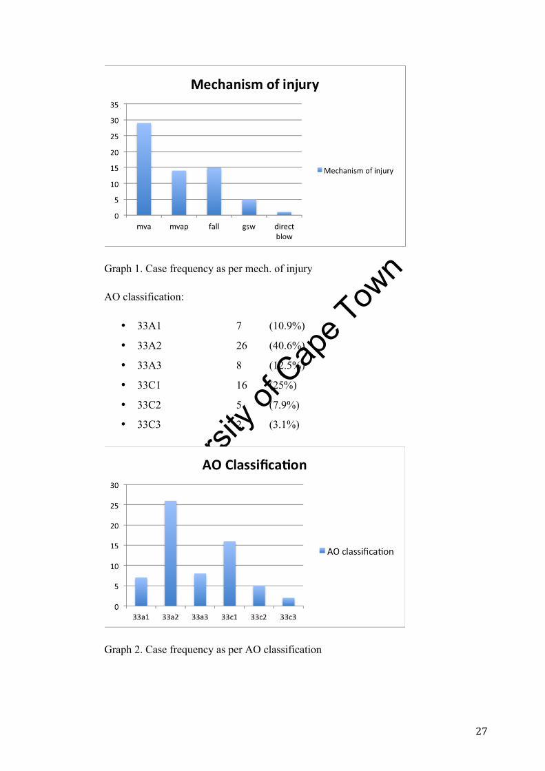
Mechanism of injury
• MVA 29 (45.3%)
• MVAP 14 (21.8%)
• Fall 15 (23.5%)
• GSW 5 (7.8%)
• Direct blow 1 (1.6%)
Univers
ity of
Cap
e Tow
n
27
Graph 1. Case frequency as per mech. of injury
AO classification:
• 33A1 7 (10.9%)
• 33A2 26 (40.6%)
• 33A3 8 (12.5%)
• 33C1 16 (25%)
• 33C2 5 (7.9%)
• 33C3 2 (3.1%)
Graph 2. Case frequency as per AO classification

Univers
ity of
Cap
e Tow
n
28
Fracture pattern descriptive classification:
• Transverse 12 (18.6%)
• Oblique 26 (40.6%)
• Spiral 5 (7.9%)
• Comminuted 21 (32.9%)
Soft tissue injury:
• Closed 59 (92.2%)
• Open 5 (7.8%)
Details of surgery
• Details of plate:
• Metal type
o Stainless steel 54 (81.5%)
o Titanium 10 (18.5%)
• Mean length 204mm +/- 49.11, 8.7 holes
Table 1. Details of surgical technique: (Mean values )
Zone Length (mm) +/- std dev Holes
available Holes filled
distal zone 31.5 +/- 13.26 5.3 4.6
Fracture zone 58.5 +/- 37.42 2.4 0
Proximal zone 116 +/- 33.0 5.8 3.6
Distal to W/L * 31.5 +/- 13.26 5.3 4.6
Working length
(W/L) 87.5 +/- 45.87 3.8 0
Proximal to W/L 86.5 +/- 23.47 4.6 3.7
* The distal zone and distal to W/L zone are the same
Univers
ity of
Cap
e Tow
n
29
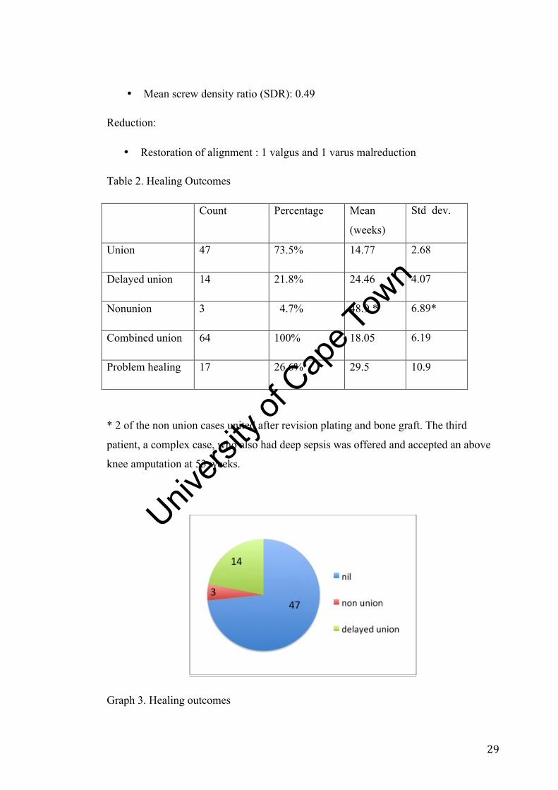
• Mean screw density ratio (SDR): 0.49
Reduction:
• Restoration of alignment : 1 valgus and 1 varus malreduction
Table 2. Healing Outcomes
Count Percentage Mean
(weeks)
Std dev.
Union 47 73.5% 14.77 2.68
Delayed union 14 21.8% 24.46 4.07
Nonunion 3 4.7% 48.9 * 6.89*
Combined union 64 100% 18.05 6.19
Problem healing 17 26.6% 29.5 10.9
* 2 of the non union cases united after revision plating and bone graft. The third
patient, a complex case, who also had deep sepsis was offered and accepted an above
knee amputation at 53 weeks.
Graph 3. Healing outcomes
Univers
ity of
Cap
e Tow
n
30
Other complications:
• Sepsis: 3 (4.7%)
• Late loss of alignment 0 (see below)
• Implant failure: 1 ( 20° varus due to bent plate)
• Mean follow up time 30.6 weeks ( std dev. 19.6)
Analysis of results
The decision as to which covariates to include in the analysis was based upon
literature demonstrating an influence over fracture healing.43 These were prioritized
based on whether they were felt to be major or minor prognostic contributors.
Non modifiable prognostic factors
Major contributors:
• Fracture severity
• Smoking
• Soft tissue injury (open/ closed fractures)
• Diabetes
• Sepsis
Minor contributors:
• Age
Modifiable prognostic factors
Potential modifiable factors were considered to be the surgical technique with which
the fracture was treated. There is very little literature examining this aspect of
management and many recommendations are empirical or extrapolated from other
regions.
• Working length
• Plate material (stainless steel vs titanium)
• Plate length to fracture zone ratio
• Working length to fracture zone ratio
• Screw density ratio
Univers
ity of
Cap
e Tow
n
31
Multiple linear regression analysis was used to run various combinations of variables
against time to union. The data count of 64 meant that only 5 variables could be used.
Scatter plots were used to screen for simple linearity prior to inclusion in the analysis.
SDR and working length demonstrated significant multi-collinearity (r20.69) and
therefore SDR was removed from the analysis (essentially duplication). Sepsis (3,
4.6%) and diabetes (4, 6.25%) were rare events and did not show any linearity in our
series therefore they were not included. None of the modifiable factors demonstrated
any simple linearity against time to union.
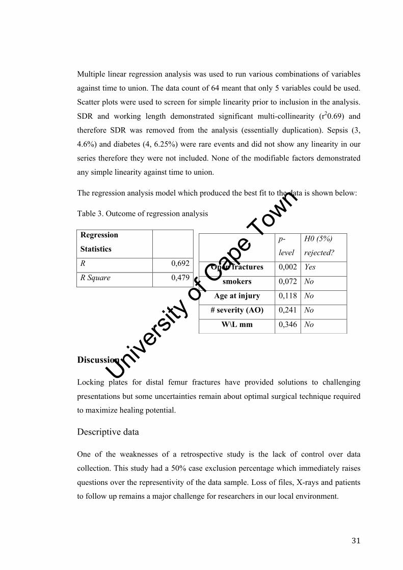
The regression analysis model which produced the best fit to the data is shown below:
Table 3. Outcome of regression analysis
Regression
Statistics
R 0,692
R Square 0,479
Discussion
Locking plates for distal femur fractures have provided solutions to challenging
presentations but some uncertainties remain about optimal surgical technique required
to maximize healing potential.
Descriptive data
One of the weaknesses of a retrospective study is the lack of control over data
collection. This study had a 50% case exclusion percentage which immediately raises
questions over the representivity of the data sample. Loss of files, X-rays and patients
to follow up remains a major challenge for researchers in our local environment.
p-
level
H0 (5%)
rejected?
Open fractures 0,002 Yes
smokers 0,072 No
Age at injury 0,118 No
# severity (AO) 0,241 No
W\L mm 0,346 No
Univers
ity of
Cap
e Tow
n
32
The histogram of age at time of injury is consistent with the bimodal distribution
pattern reported in the literature.
Graph 4. Histogram of age at time of injury
Fracture severity is considered to be a major contributor to delayed union. The mean
union times of the descriptive fracture patterns supports this. The AO classification
system in general has a progression of fracture severity through the classification
system. Again this is supported by the mean union times of the data set. On the basis
of this simple linear correlation, the AO classification was used as a surrogate for
fracture severity in the regression analysis but It was not significant however
(p=0.241). Ricci found The 33A3 pattern to be a significant independent risk factor for
implant failure. The only implant failure in this study was also in a 33a3 pattern when
the plate bent in the fracture zone (working length 110mm). The femur still went on to
unite. It was recorded that the particular patient was not compliant with restricted
weight bearing, a recommendation that Button made in 2004 to reduce implant
failure.50
Graph 5. Mean union time by fracture pattern
Univers
ity of
Cap
e Tow
n
33
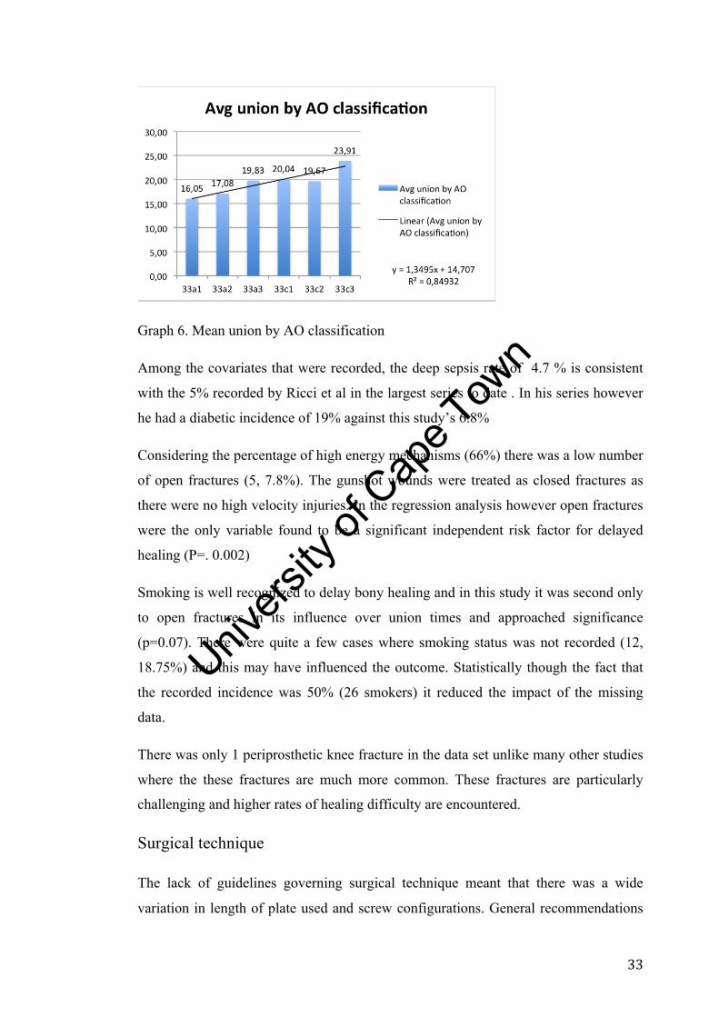
Graph 6. Mean union by AO classification
Among the covariates that were recorded, the deep sepsis rate of 4.7 % is consistent
with the 5% recorded by Ricci et al in the largest series to date . In his series however
he had a diabetic incidence of 19% against this study’s 6.8%
Considering the percentage of high energy mechanisms (66%) there was a low number
of open fractures (5, 7.8%). The gunshot wounds were treated as closed fractures as
there were no high velocity injuries. In the regression analysis however open fractures
were the only variable found to be a significant independent risk factor for delayed
healing (P=. 0.002)
Smoking is well recognized to delay bony healing and in this study it was second only
to open fractures in its influence over union times and approached significance
(p=0.07). There were quite a few cases where smoking status was not recorded (12,
18.75%) and this may have influenced the outcome. Statistically though the fact that
the recorded incidence was 50% (26 smokers) it reduced the impact of the missing
data.
There was only 1 periprosthetic knee fracture in the data set unlike many other studies
where the these fractures are much more common. These fractures are particularly
challenging and higher rates of healing difficulty are encountered.
Surgical technique
The lack of guidelines governing surgical technique meant that there was a wide
variation in length of plate used and screw configurations. General recommendations

Univers
ity of
Cap
e Tow
n
34
have been made for the use of longer plates and longer working lengths in response to
the healing complications experienced. The mean working length in this study was 3.8
holes (87mm). The literature has not reported clearly on mean working lengths used
but the case series’ discussing implant failure have shown images of very short
working lengths either 0 or 1 hole. This study did not have reveal any implant
breakages suggesting that the working lengths were long enough to reduce cyclic
loading stresses or the plates were strong enough to withstand them until union.
The scatter plot of date of injury vs working length has shown a mild but significant
linear correlation r =0.337 r2=0.13 (p=0.007) suggesting that as the surgeons at our
institution became more familiar with these relatively new implants they were
modifying their surgical technique. As the analysis has shown however this has not
translated into any difference in union times in this study. This is in agreement with
Bottlang’s findings in 2010 where he used volumetric callus mapping as a surrogate
for healing measured against bridging span (working length) r=0.04 P=0.4.39
Graph 7. Scattergram of working length vs date of surgery
Working length as an absolute value measured against union has not demonstrated any
significant correlation. Working length is not independent of the nature and magnitude
of the fracture zone. A large fracture zone will always result in a large working length.
The nature of the fracture zone may also differ even though it has the same length. A
severely comminuted fracture zone will have no load sharing ability under all
Univers
ity of
Cap
e Tow
n
35
conditions of plate flexion until sufficient callus forms. A long spiral fracture bridged
with the same working length may allow some load sharing if the plate flexes
sufficiently and the fracture edges appose each other. The relevance of this is that there
will be differing plate stresses for the same working length. In the comminuted
fracture the stress will continue increasing whereas it will decrease upon fracture gap
closure in the spiral fracture40.
Screw density ratio has been proposed as a surrogate for working length and an
empirical value of 0.4-0.5 reported by Gautier as desirable. Our SDR average of 0.49
appears consistent with recommendations. The linear correlation coefficient r= -0.83
with r2 0.69 obtained from the data set confirms this strong correlation. When
measured against union it consequently does not show any significant independent
relationship (r2 0.0068)
Titanium vs stainless steel
Bottlang did however note that the titanium plates produced a significantly increased
volume of callus (68%) over the stainless steel plates.39 In this study only 10 titanium
plates were used and most of them within the last year because of consignment
requirements. There was no difference in healing. This is an area for future research as
the majority of distal femur plates now being inserted at our institution are titanium
and a study with greater numbers may be able to demonstrate significant differences.
Mean healing time
In our study the mean healing time of 18.05 weeks appears to be longer than most
other studies. We postulate that it may have been affected by our sampling frequency.
Our standard follow up was at 2, 6, 12, 18 weeks with X-rays obtained at each visit. If
at 12 weeks the fracture did not meet the criteria for union then the next assessment
that could report union would have been around 18 weeks and possibly with more
frequent X-rays the time to union would have been reduced. This is a clear limitation
of our study. Other factors that may have affected the assessment are the metaphyseal
location of the fractures and the presence of the lateral plate obscuring a cortex. Due to
the way that metaphyseal fractures heal with creeping substitution of trabeculae in
direct contact and intramembraneous ossification, little external callus is produced.
This makes radiological assessment of metaphyseal union more difficult than
Univers
ity of
Cap
e Tow
n
36
diaphyseal fractures as noted by McClelland et al in 2006.51 Other factors influencing
this study may be the high percentage of smokers (50%) compared to other quoted
figures such as 25% in Ricci’s study.
Further breakdown of the healing times shows that the mean healing time of the
fractures with uneventful union of 14.3 weeks was as expected. Interestingly there was
not one recorded intervention of bone grafting amongst the delayed union (14 patients,
24.5 weeks) group and perhaps more aggressive intervention of those with risk factors
for delayed union may have reduced this group. Henderson in his review notes that
bone grafting itself presents difficulties when attempting to analysis healing rates.
Planned early bone grafting would prevent delayed union which may have otherwise
occurred. Ricci excluded these cases from his analysis because they were such
confounders.
Problem Healing
Our results of “problem healing” (26%) are in agreement with those noted in the
largest review on the subject published to date by Henderson et al.52 One of the biggest
factors affecting the results of any published outcome study on fracture healing are the
definitions and criteria used to describe union, delayed union and nonunion. Fracture
union is a gradual process and identifying the tipping point of when a fracture is
considered to be united is to an extent a factor of the sampling frequency. It is not
practical nor ethical to X-ray a fracture frequently enough to precisely identify the
time of healing. Conversely assessing the patient every 8 weeks will result in a less
accurate assessment of time to union. Most orthopaedic surgeons agree that the
combination of clinical and radiological features provides the most accurate
assessment of union.53 This was unfortunately not possible in our study. Initially it was
planned to asses both clinical and radiological parameters of union but early on it
became apparent that the notes lacked sufficient detail in most cases to allow for any
analysis of clinical union therefore the study was based on radiological assessments of
union only.
The concept of delayed union is even harder to define as the literature shows54.
Qualitatively it is described as a fracture that takes longer to unite than would
normally be expected but that there are signs of progressive healing as apposed to a

Univers
ity of
Cap
e Tow
n
37
non union where there are no signs of progression towards healing for 3 consecutive
months after a minimum period, usually 6 months. Quantitatively 12 weeks has been
most widely used for the lower limb based on work done with regards to stiffness of
healing fractures over time. Henderson in his review regarded union longer than 12
weeks as delayed union but does not record what the various studies used as their
criteria. Reviewing multiple papers discussing delayed and non union shows that there
is a wide range of opinion from 4 to 24 weeks with a median of 12 weeks.48,55 This
includes the upper and lower extremity. In this study it was decided to use a cut off of
20 weeks for delayed union. This was decided upon because of the post op follow up
sequence. It was expected that most fractures would have united without complication
between the 12 and 18 week visits. The 18 week follow up was often not precisely 18
weeks and therefore by selecting 20 weeks we would allow for those without them
being classified as a delayed union.
Graph 8. Histogram of union times
Loss of alignment and malunion.
As noted above this study only had 1 case of malunion (20° varus) secondary to a bent
plate. Strictly this is an implant failure, as the patient required an osteotomy, even
though the patient went on to unite uneventfully. Of particular relevance, there were
no cases of varus collapse due to poor control of the distal fragment. This is one of the
major advantages of locking plates and it has been demonstrated here again.

Univers
ity of
Cap
e Tow
n
38
Strengths and weaknesses
This study represents one of the larger studies performed on distal femur fracture
management. It provides an in depth analysis of the various components that make up
specifically a distal femur locking plate construct and considers not just absolute
working length but its relationship to the fracture zone.
It is a retrospective review and suffers from a 50% case exclusion percentage. This
potentially introduces bias and also weakens the power of the study. The inability to
combine a clinical assessment of union with the radiological assessment as planned
weakens the assessment. Blinding and multiple reviewers would have improved the
accuracy of assessment. The inherent discrepancies surrounding the definitions of
union, delayed and nonunion are not unique to this study. The frequency of follow up
dictated the frequency of X-ray assessment and a prospective study would aim to
improve on this study’s 6 week cycle. There are obvious cost and ethical implications
of large scale weekly X-rays of patients and while ideal from a research perspective
may not be achievable in our local environment.
Conclusion
Fractures of the distal femur can be challenging to manage successfully. The addition
of the distal femur locking plate to the surgeon’s armory has provided an implant that
offers superior control of the distal fragment, the one component of these fractures that
is the most challenging to stabilize. The concerns in the literature about the incidence
of healing difficulties have been supported by the findings in this study. The ability of
an increased working length to independently reduce the union time has not been
shown. What has been confirmed is that the risk factors known to prolong bone
healing such as open fractures were significant contributors to the healing difficulties
seen in this study. When present, these should be optimized and if healing difficulties
are anticipated early intervention should be planned. The limitations of this study have
highlighted the need for future large prospective studies that are adequately powered
utilizing validated and standardized measures of bone healing. Interventions other than
increasing working length that maximize healing potential should be explored as it
appears that working length may be limited in its ability to significantly improve
healing.

Univers
ity of
Cap
e Tow
n
39
Distal femur locked plate study case no._______
Plate characteristics
Brand: SS ☐ titanium☐
Length: diaphyseal holes, mm
Prox to # zone: holes , holes filled , mm
Fracture zone: holes, ________holes filled, mm
Distal to fracture zone: holes, ________holes filled, mm
Prox screws : locking ☐ non locking ☐
Fracture characteristics
AO classification: Date of injury:
Side : right ☐ left ☐ Open ☐ closed ☐
Mechanism: fall ☐ MVA ☐ GSW ☐ direct blow ☐
Pattern: transverse ☐ oblique ☐ spiral ☐ comminuted ☐
Pt sticker or details: Rel. med. Hx:
Diabetic☐ smoker☐
Outcome
Fixation: adequate ☐ inadequate:☐ follow up duration_______________
If inadequate why:
1. screw density ratio☐ 2. # zone to plate ratio☐
3.# alignment ☐ 4. Plate positioning ☐
Time to full WB: (include date):
Time to radiological union in weeks (include date):
Complications: delayed union☐ non union☐ sepsis☐ plate failure☐
Screw failure☐ loss of alignment☐
Univers
ity of
Cap
e Tow
n
40
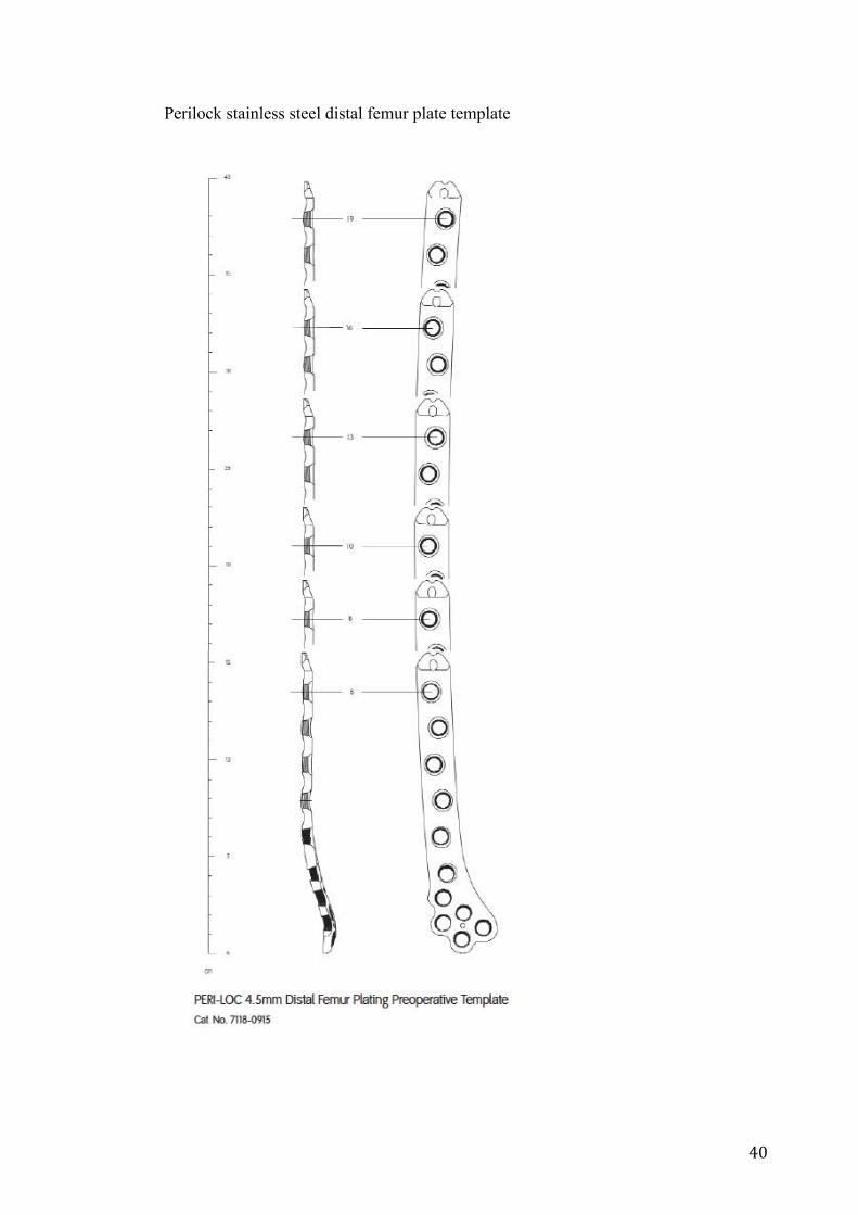
Perilock stainless steel distal femur plate template
" - --I<
"- ,
. --t<
"
•
PERI-LOC 4.5I1m asia! feIlu Il9Ii1g ~ Tempate
Univers
ity of
Cap
e Tow
n
41
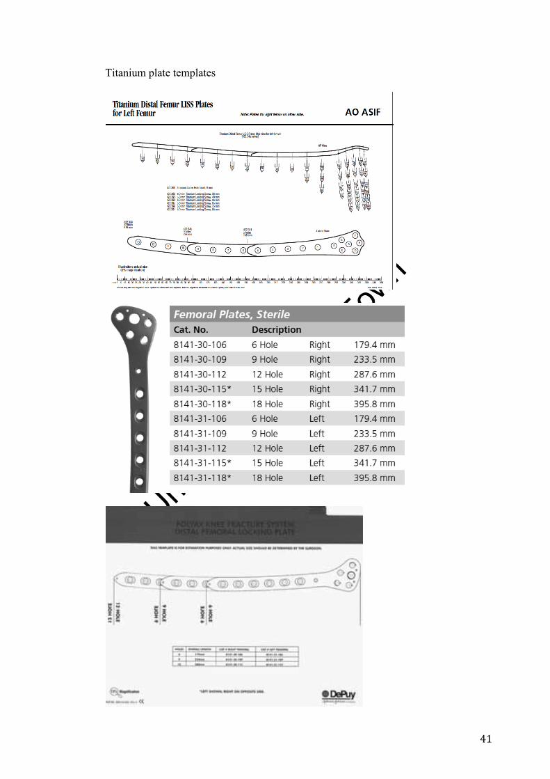
Titanium plate templates
Univers
ity of
Cap
e Tow
n
42
REFERENCES 1 Mooney V, Nickel VL, Harvey JP, Snelson R. Cast brace treatment for fractures of the distal part of the femur. J Bone Joint Surg 1970;52A:1563—78. 2 Neer II CS, Grantham SA, Shelton ML. Supracondylar fracture for the adult femur. J Bone Joint Surg 1967;49A:591—613. 3 Stewart MJ, Sisk TD, Wallace SL. Fractures of the distal third of the femur: a comparison of methods of treatment. J Bone Joint Surg 1966;48A:784—807. 4 Schatzker J, Horne G, Waddell J. The Toronto experience with the supracondylar fracture of the femur, 1966–1972. Injury. 1975;6:113–128 5Schatzker J, Lambert DC. Supracondylar fractures of the femur. Clin Orthop 1979;138:77—83 6 Healy WL, Brooker AF. Distal femoral fractures: comparison of open and closed methods of treatment. Clin Orthop 1983; 174:166—71. 7 Forster MC, Komarsamy B, Davison JN. Distal femoral fractures: a review of fixation methods. Injury 2006; 37:97–108. 8 Davison BL. Varus collapse of comminuted distal femur fractures after open reduction and internal fixation with a lateral condylar buttress plate. Am J Orthop 2003; 32:27–30. 9 Sanders R, Swiontkowski M, Rosen H, Helfet D. Double-‐plating of comminuted, unstable fractures of the distal part of the femur. J Bone Joint Surg Am 1991;73:341—6. 10 Zickel RE, Hobeika P, Robbins DS. Zickel supracondylar nails for fractures of the distal end of the femur. Clin Orthop 1986;212:79. 11 Stromsoe K, Alho A, Ekeland A. The Grosse-‐Kempf nail for distal femoralfractures. 2-‐year follow-‐up of 25 cases. Acta Orthop Scand 1990;61:512—6. 12 Leung, K. S.; Shen, W. Y.; So, W. S.; Mui, L. T.; and |and |Grosse, A.: Interlocking intramedullary nailing for supracondylar and intercondylar fractures of the distal part of the femur. J. Bone and Joint Surg.,73-‐A: 332-‐340, March 1991. 13 Green SA. Distal intramedullary fixation of supracondylar fractures of the femur. Tech Orthop. 1988;3:71–76. 14 Salem KH, Maier D, Keppler P, et al. Limb malalignment and functional outcome after antegrade versus retrograde intramedullary nailing in distal
Univers
ity of
Cap
e Tow
n
43
femoral fractures. J Trauma 2006; 61:375–381 15 Krettek C, Miclau T, Schandelmaier P, Stephan C, Möhlmann U, Tscherne H. The mechanical effect of blocking screws ("Poller screws") in stabilizing tibia fractures with short proximal or distal fragments after insertion of small-diameter intramedullary nails. J Orthop Trauma. 1999 Nov;13(8):550-3. 16 Kumar A, Jasani V, Butt MS. Management of distal femoral fractures in elderly patients using retrograde titanium supracondylar nails. Injury 2000;31:169—73 17 Butt M. S., Krikler S. J. and Ali M. S. Displaced fractures of the distal femur in elderly patients. J. Bone Joint Surg. 1996; 78B: 110±114. 18 Ward PJ, Goodwin MI. The use of the supracondylar nail in the management of femoral fractures in the presence of other femoral implants in the very elderly. Injury 1998;29:671±5. 19 Herrera DA, Kregor PJ, Cole PA, Levy BA, Jonsson A, Zlowodzki M. Treatment of acute distal femur fractures above a total knee arthroplasty: systematic review of 415 cases (1981–2006). Acta Orthop 2008;79:22–7. 20 Martinet O, Cordey J, et al. The epidemiology of fractures of the distal femur Injury. 2000 Sep;31 Suppl 3:C62-3. 21 Seinsheimer F 3rd. Fractures of the distal femur Clin Orthop Relat Res. 1980 Nov-Dec;(153):169-79 22 Müller ME, Allgöwer M, Schneider R, Willenegger H. Manual of internal fixation. 3rd ed. Berlin Heidelberg New York: Springer; 1990. 23 Yamada H. Strength of biological materials. Baltimore: Williams & Wilkins Co, 1970:99-104. 24 Perren SM. Evolution of the internal fixation of long bone fractures. The scientific basis of biological internal fixation: choosing a new balance between stability and biology. J Bone Joint Surg Br. 2002;84: 1093–1110 25 Egol KA, Kubiak EN, Fulkerson E, Kummer FJ, Koval KJ: Biomechanics of locked plates and screws. J Orthop Trauma 2004;18:488-493. 26 Kubiak EN, Fulkerson E, Strauss E, Egol KA: The evolution of locked plates. J Bone Joint Surg Am 2006;88(suppl 4):189-200. 27 Mast J, Jakob R, Ganz R. Planning and Reduction Technique in Fracture Surgery. New York, NY: Springer; 1989. 28 Krettek C, Schandelmaier P, Miclau T, et al. Minimally invasive percutaneous

Univers
ity of
Cap
e Tow
n
44
plate osteosynthesis (MIPPO) using the DCS in proximal and distal femoral fractures. Injury. 1997;28(suppl 1):20–30. 29 Kregor PJ, Stannard J, Zlowodzki M, et al. Distal femora fracture fixation utilizing the Less Invasive Stabilization System (L.I.S.S.): the technique and early results. Injury. 2001;32(suppl 3):32–47. 30 Schutz M, Muller M, Krettek C, et al. Minimally invasive fracture stabilization of distal femoral fractures with the LISS: a prospective multicenter study. Result of a clinical study with special emphasis on difficult cases. Injury. 2001;32(suppl 3):48–54. 31 Weight M, Collinge C. Early results of the less invasive stabilization system for mechanically unstable fractures of the distal femur (AO/OTA types A2, A3, C2, and C3). J Orthop Trauma. 2004;18:503–508. 32 Syed AA, Agarwal M, Giannoudis PV, et al. distal femoral fractures: long–term outcome following stabilization with the LISS. Injury. 2004; 35:599–607. 33 Zlowodzki M, Bhandhari M, Marek DJ, et al. Operative treatment of acute distal femur fractures: systematic review of 2 comparative studies and 45 case series (1989 to 2005). J Orthop Trauma 2006;20:366–71. 34 Herrera DA, Kregor PH, Cole PA, Levy B, Jonsson A, Zlowodzki M. Treatment of acute distal femur fractures above a total knee arthroplasty: Systematic review of 415 cases (1981-2006). Acta Orthopaedics 2008 Feb: 79(1):22-7 35 Marti A, Fankhauser C, Frenk A, et al. Biomechanical evaluation of the less invasive stabilization system for the internal fixation of distal femur fractures. J Orthop Trauma 2001;15:482—7. 36 Zlowodski M, Williamson S, Cole PA, et al. Biomechanical evaluation of the less invasive stabilization system, angled blade plate and retrograde intramedullary nail for the internal fixation of distal femur fractures. J Orthop Trauma 2004;18:494—502. 37 Vallier HA, Hennessey TA, Sontich JK, et al. Failure of LCP condylar plate fixation in the distal part of the femur. A report of 6 cases. J Bone Joint Surg Am. 2006;88:846–853. 38 Sommer C, Babst R, Muller M, Hanson B. Locking compression plate loosening and plate breakage: a report of four cases. J Orthop Trauma. 2004;18:571-7. 39 Bottlang M, Doornink J, Fitzpatrick DC, et al. Effects of construct stiffness on healing of fractures stabilized with locking plates. J Bone Joint Surg Am. 2010;92:12-22. 40 Claes LE, Heigele CA, Neidlinger-Wilke C, Kaspar D, Seidl W, Margevicius KJ, Augat P. Effects of mechanical factors on the fracture healing process. Clin Orthop Relat Res. 1998;355 Suppl:S132-47

Univers
ity of
Cap
e Tow
n
45
41 Uhthoff HK et al The advantages of titanium alloy over stainless steel plates for IF of fractures: an experimental study in dogs. JBJS 1981. 63-B. p 427-434. 42 Gaines R. Titanium versus Stainless Steel Locked Plates for Distal Femur Fractures: Is There Any Difference? Sat., 10/18/08 Femur, Paper #55, 10:28 am OTA-2008
43 Stoffel K, Dieter U, Stachowiak G, Gachter A, Kuster MS. Biomechanical testing of the LCP—How can stability in locked internal fixators be controlled. Injury 2003;34:SB11—9 44 Gautier E, Sommer C. Guidelines for the clinical application of LCP. Injury 2003;34(Suppl. 2):63–76. 45 Cartner J, Hartsell Z, Jones B, Ricci W. Quantifying the Load Sharing Capabilities of Distal Femur Plating through Strain Analyses in Healthy and Osteoporotic Bone in Different Fracture Types. Paper presented at: OTA abstract 2010 Baltimore, Maryland. October 13-16 2010
46 Ricci WM. Risk factors for failure of locked plate fixation of distal femur fractures: an analysis of 305 cases. Paper presented at: OTA abstract 2009, Knee/Tibia. San Diego, CA. October 7–10, 2009 47 Lujan, TJ, Henderson CE, Madey SM, et al. Locked plating of distal femur fractures leads to inconsistent and asymmetric callus formation. J Orthop Trauma. 2010;24:156–162 48 Hammer RRR, Hammerby S, Lindholm B. Accuracy of radiologic assessment of tibial shaft fracture union in humans. Clin Orthop 1985;199:233-8 49 Corrales LA. Variability in the assessment of fracture-healing in orthopaedic trauma studies. J Bone Joint Surg Am 2008 90:1862 50 Button G, Wolinsky P, Hak D. Failure of Less Invasive Stabilization System plates in the distal femur: a report of four cases. J Orthop Trauma. 2004;18:565–570. 51 McClelland D et al. Fracture Healing Assessment Comparing Stiffness Measurements using Radiographs. Clin Orthop 2006 457: 214–219 52Henderson CE, Kuhl LL, Fitzpatrick DC, Marsh JL. Locking plates for distal femur fractures: is there a problem with fracture healing? J Orthop Trauma. 2011 Feb;25 Suppl 1:S8-14. 53 Bhandari M. Variability in the definition and perceived causes of delayed unions and nonunions: a cross- sectional, multinational survey of orthopaedic surgeons. J Bone Joint Surg Am. 2012 Aug 1;94(15):1091-6.

Univers
ity of
Cap
e Tow
n
46
54 Bhandari M, et al. A lack of consensus in the assessment of fracture healing among orthopaedic surgeons. J Orthop Trauma. 2002 Sep;16(8):562-6. 55 Marsh D. Concepts of fracture union, delayed union, and non-union. Clin Orthop 1998;(355 Suppl):S22-S30