Embed Size (px)
Citation preview

2
RAAViewer Manual
Table of Contents
INTRODUCTION – RADIOLOGIC ANATOMY ATLAS ............................................................. 3
HISTORY ................................................................................................................................... 5
SYNOPSIS ................................................................................................................................. 6
INSTALLATION ......................................................................................................................... 7
BUGS ....................................................................................................................................... 10
COMPUTER INTERFACE TERMINOLOGY ............................................................................ 11
PURPOSE ....................................................................................................... 15
TERMINOLOGIA ANATOMICA ...................................................................... 16
ALTERNATE LANGUAGES ........................................................................... 23
INSTRUCTIONS .............................................................................................. 25
DATA SOURCES ............................................................................................ 31
FINAL COMMENTS ................................................................................................................. 33

3
Introduction – Radiologic Anatomy Atlas
RAAViewer is currently under active development. I encourage people to
send their comments and suggestions. If you find features of the program
confusing, drop me a line. I may be able to help you and others by making
the manual clearer.
There are, no doubt, errors in the atlas images themselves with structures
incorrectly labeled or misspelled labels. There is considerable data included
about the anatomic terms that accompanies the program. Much of that
information may be outdated or wrong. Anatomy terminology is confusing
and ambiguous in that many synonyms commonly exits for anatomic
structures and various sources can refer to what are actually different
structures with the same name. The normal variability of human anatomy
from one individual to the next compounds the issue. Many of the sources
for this program, while having the advantage of being in the public domain
are not up to date. Any help with these issues would be appreciated.
The nature of RAAViewer is such that correcting such errors is easy, but I
have to know about them. Please drop me an email when you run across
such things.
ANY such feedback is appreciated and will be acted upon.
Currently, there is a moderate selection of image folders available. I will
be expanding on this and will strive to update the site every few months so
check back later for updates and additional image folders.
I would like even more to have people send me high quality images or
illustrations of some area of anatomic interest that they would like to include
in RAAViewer. These pictures have to be in the public domain or owned by

4
the provider; I cannot use, without permission, material copied from journals
or books that is protected by copyright.
If you send the pictures and indicate how they are labeled then I can
include them in the Atlas. Your contribution will be acknowledged.
In this program, the labels are not embedded in the picture. The labels are
created in the program and applied to the image as a separate “layer”. This
allows modification in the future. If you submit pictures, it is best to send
high quality images in a digital format that are not “pre-labeled”. You can
send a separate set of labeled images, however you wish, to inform me how
the images should be labeled. I would do the final labeling in the program.
Robert R Livingston [email protected] [email protected] 30221 33rd Ave SW Federal Way, WA 98023 (253) 874 6199

5
History
RAAViewer was inspired in part by a web site of J. H. Edmund Lee,
M.D. (<http://homepage.mac.com/frankdcat/mri_atlas/home.html>). What
Dr. Lee did was to make a QuickTime movie of labeled MRI images of
musculoskeletal structures seen with various imaging sequences and in
various planes. The radiologist faced with a piece of anatomy that is
unfamiliar can use the controls embedded in QuickTime to move to the
relevant frame in the movie and thus find the anatomic structure in question
with its label. Standard paper-based anatomy textbooks might have a picture
of the anatomy near where you are looking, but, inevitably, you often find
that the actual cross-section that you are looking at does not correspond to
the handful of images contained in a textbook. Dr. Lee’s approach took
advantage of storage capacity of a computer to overcome this limitation.
Expanding his approach to other areas, I wrote a primitive program that
would look sequentially at all the images in a folder. I then made a number
of folders and filled them with related images that I labeled with anatomic
terms. This works fine, but has some problems. The major problem is that
labeling 30 images in a sequence of MRI images is incredibly tedious and, in
my hands, fraught with error. Writing Rhomboideus Major 30 times on 30
different pictures is painful. I would label all these images and then later I
would find misspellings and incorrect label positionings, and this would be
difficult to correct.
RAAViewer handles this by containing a master database of anatomic
terms and their descriptions. These terms can be applied to any image. In
this sense, the labels and their associated metadata are separate from the
6
image, which has many advantages. The image can be flipped without the
labels being flipped and thus becoming unreadable. The image can be
shrunk down without the labels becoming too small to read. And the labels
can be edited without fussing with the pictures themselves.
Synopsis
The images in RAAViewer have dots on them. The user clicks on a dot
and the anatomic term that corresponds with that dot is displayed. If there is
additional information about the anatomic term, the term is displayed in blue
letters. The term itself can then be clicked on and this additional information
viewed. This is the heart of the program. It is not a complicated program to
use and barely qualifies to have a manual at all.
7
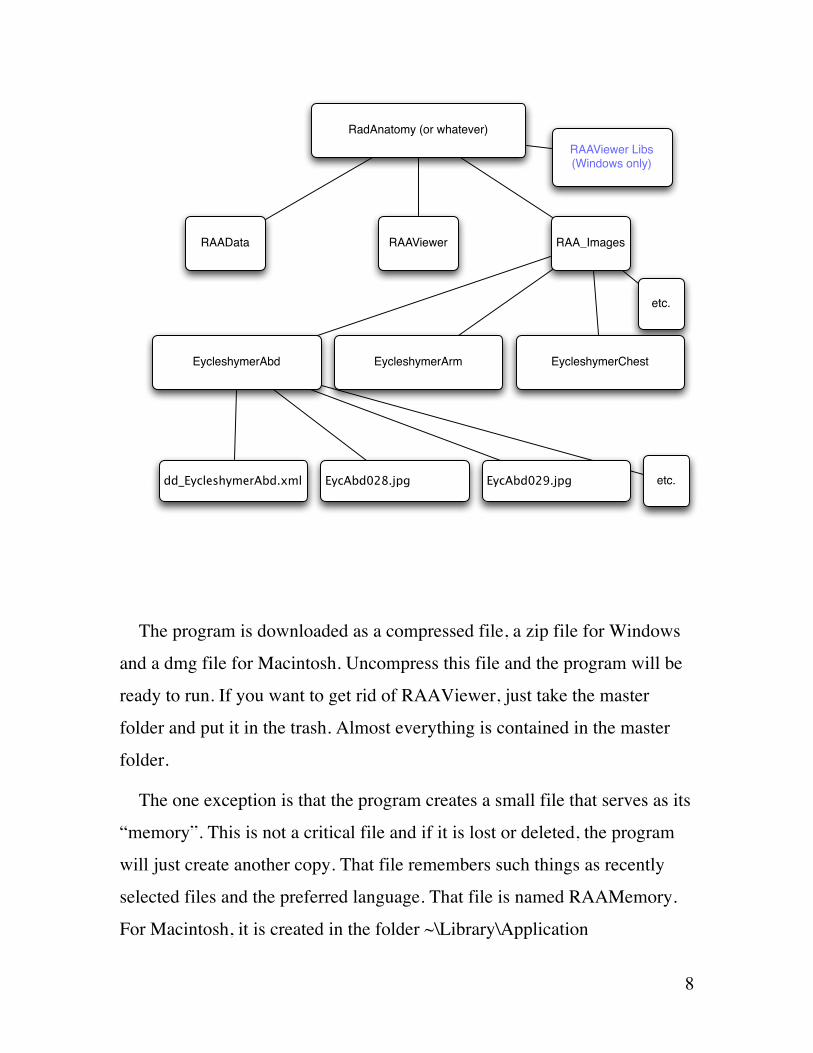
Installation
There are three file/folders that are included with the Macintosh version
of RAAViewer. The Windows version requires an additional folder –
RAAViewer Libs. They should ALL be placed in a single master folder.
That master folder might be named something like RadiologyAnatomyAtlas.
You can name the master folder anything that you want and put it any place
you want on your machine. You should not, however, change the files that
are contained within that master folder.
Inside that master folder/directory go all the files that are needed for this
program.
1. RAAViewer – the program itself
2. RAAData – a file that contains the anatomic terms and data about
them
3. RAA_Images – a folder/directory that contains any number of
subfolders. These subfolders contain the images that are used in the
program. Each subfolder contains a related set of images. When you
use RAAViewer, you select one of these subfolders from within the
program to allow you to see the images in that folder.
4. RAAViewer Libs (Windows version only)
This set-up is simple. But you have to keep these three things (or four
things for the Windows version) all in the same folder for the program to
work. The names of the three (four) file/folders cannot be changed.
Similarly, do not change the names or delete individual images from the
subfolders. Doing so will frustrate the program.
8
The program is downloaded as a compressed file, a zip file for Windows
and a dmg file for Macintosh. Uncompress this file and the program will be
ready to run. If you want to get rid of RAAViewer, just take the master
folder and put it in the trash. Almost everything is contained in the master
folder.
The one exception is that the program creates a small file that serves as its
“memory”. This is not a critical file and if it is lost or deleted, the program
will just create another copy. That file remembers such things as recently
selected files and the preferred language. That file is named RAAMemory.
For Macintosh, it is created in the folder ~\Library\Application
RadAnatomy (or whatever)
RAAData RAAViewer RAA_Images
EycleshymerAbd EycleshymerArm EycleshymerChest
etc.
dd_EycleshymerAbd.xml EycAbd028.jpg etc.EycAbd029.jpg
RAAViewer Libs (Windows only)

9
Support\RAAViewer. For Windows, it is created in the folder ~\Application
Data\RAAViewer
10
Bugs
Non-trivial programs generally have bugs. Therefore I am sure that this
one does. Additionally, I cannot guarantee the accuracy of the output of this
program. Let the user beware. If you miss a question on your final exam in
Anatomy because of RAAViewer, let it be a learning experience and tell me
about it.
Having said this, I am interested in squashing any bugs and content errors.
If you see any anomalous (incorrect) results or strange behaviors when you
are doing things that seem rational, please notify me so I can correct or
clarify the situation.
11
Computer Interface Terminology
This manual strives to be consistent in how it refers to the various widgets
that are used to specify the user’s intents.
DOT
Most of the images in the program have dots on them. You click on the
dot to reveal the anatomic information contained in the dot.
DOT OPACITY SLIDER
The dots can be made more or less conspicuous by use of this dot opacity
slider. There is potential for a dot to obscure the anatomy over which it lies.
This slider addresses that problem.
ORIENTATION BUTTONS
There are four buttons that allow the user to orient the image in various
ways.
1. Flip the image around a horizontal axis (turn a right leg image into a
left leg image)
2. Flip the image around a vertical axis (supine to prone)
3. Rotate the image 90 degree clockwise or counter clockwise.
Clicking on any of these buttons with the Shift key held down will return
the picture to its original orientation.
12
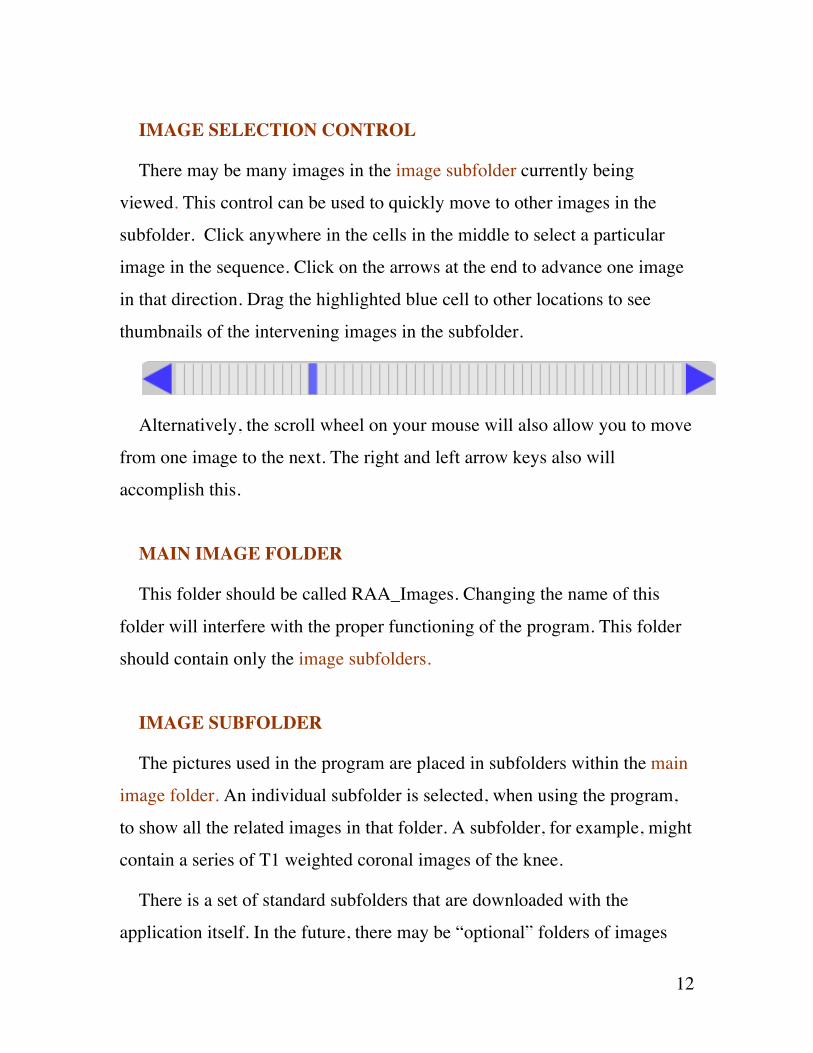
IMAGE SELECTION CONTROL
There may be many images in the image subfolder currently being
viewed. This control can be used to quickly move to other images in the
subfolder. Click anywhere in the cells in the middle to select a particular
image in the sequence. Click on the arrows at the end to advance one image
in that direction. Drag the highlighted blue cell to other locations to see
thumbnails of the intervening images in the subfolder.
Alternatively, the scroll wheel on your mouse will also allow you to move
from one image to the next. The right and left arrow keys also will
accomplish this.
MAIN IMAGE FOLDER
This folder should be called RAA_Images. Changing the name of this
folder will interfere with the proper functioning of the program. This folder
should contain only the image subfolders.
IMAGE SUBFOLDER
The pictures used in the program are placed in subfolders within the main
image folder. An individual subfolder is selected, when using the program,
to show all the related images in that folder. A subfolder, for example, might
contain a series of T1 weighted coronal images of the knee.
There is a set of standard subfolders that are downloaded with the
application itself. In the future, there may be “optional” folders of images

13
that on the website. These optional subfolders, if downloaded, would be put
in the RAA_Images folder so they would be easily accessed.
OPTIONAL SUBFOLDER (NOT IMPLEMENTED)
There is a set of standard subfolders that are downloaded with the
application itself. These are a basic set of axial images of the entire body
from the Eycleshymer atlas A Cross-Section Anatomy and several other
image series. Optional subfolders may become available at some later date
on the web site but do not exist now. These would be folders of images that
could be independently downloaded if the user so chooses. Such folders
would be added to the RAA_Images folder.
I have not actually included optional subfolders to date. For now it is
easier just to download the entire package. This allows me to fix small errors
in any of the images series without having to keep track of everything. Then
the user can just re-download everything and get all the little fixes.
This is an idea for the future when the number of image series gets bigger.
DOT DATA FILE
Within each image subfolder there will probably be a file called dd_[name
of the subfolder]. The file contains the information about the dots that lie on
the images in that folder. Removing or changing this file will interfere with
the proper functioning of the program. This file knows the name of every
image in the subfolder and knows how many of such images are present. If
the user removes some images or adds some images, this file will be aware
that something has changed and will be unable to perform properly.

14
LABEL CHECKBOX
A checkbox allows the user to request that all the dots on the image be
attached to text labels at the side of the picture. If there are many dots, this
can result in a crowded set of labels. If there are lots and lots of dots, some
will not create an attached label. Use at your discretion.
LABEL COLOR SQUARES
If the label checkbox is checked, lines are created connecting the dots to
the text labels at the sides of the image. Near the label checkbox are two
colored squares that allow you to specify line colors. Depending on the
colors contained in the image itself, various line colors can be more or less
conspicuous. Simply clicking on these squares toggles through a default set
of colors. You may move backwards through the defaults by Option-
clicking. You may select your own custom color by Shift-clicking on the
label color square.
There are two squares because colors are used alternately when drawing
the label lines. This makes the lines easier to follow from the label to the dot
when the lines get crowded together. If you want all the lines to be the same
color, Shift-Option click on the color that you want.
IMAGE FOLDER SELECTION BUTTON
A button in the right lower corner of the main window brings up the
image folder selection window, which allows the user to select which image
subfolder to view.

15
MAIN WINDOW
The program starts up with this window displayed. Here will be
displayed the individual anatomic images that the user is reviewing.
IMAGE FOLDER SELECTION WINDOW
Here are listed all the image subfolders. The user highlights an entry on
this list and then clicks on the view button to bring the images in that
subfolder into view. If the Shift key is held down, the thumbnails are not
created and the images become visible more quickly.
ANATOMIC INFORMATION WINDOW
Clicking on the text of the highlighted dot will bring up this window,
which contains information about the anatomic entity.
PURPOSE Paper anatomy textbooks have some advantages. The best of them have
employed skilled medical illustrators to show anatomy in a way that MR or
CT cannot, creating images that show relationships between anatomic
structures in an abstract and, hopefully, informative way. The text further
describes the information.
The problem with these textbooks in the hectic world of the radiologist is
that they never seem to be available at the time that you need them. You
cannot lug a cartload of these things wherever you go. Each textbook has a
different layout that complicates quickly looking up a topic. There are
practical limitations on how many pictures they can show. So often, they do
16
not have a picture that really corresponds to the radiographic image that you
are looking at. On the other hand, the RAAViewer is capable of containing a
huge selection of images and finding a particular image is easy. Essentially
you are scrolling though anatomic images in the same fashion that we scroll
through clinical images all day long. Hopefully, over time, the number of
images in RAAViewer will increase, and it will provide one source to look
at the temporal bone anatomy one day and wrist MR anatomy another.
RAAViewer has become possible because of the speed with which most
people are now able to access the Internet to download pictures. MR and CT
digital images from a radiology practice and public domain images on the
Internet are resources that allow accumulations of useful images and
information that can be bundled into a program of this nature and offered
gratis to the interested.
TERMINOLOGIA ANATOMICA International Anatomical Terminology
Frequently, there are many names for the same anatomy. I have made my
own choices, sometimes arbitrarily. I have also attempted to provide, in
many cases, an “official” name from Terminologia Anatomica.
Ideally, there would be no ambiguity in the naming of anatomic
structures. This is an elusive goal for a number of reasons. A major one is
the Tower of Babel problem: different nationalities develop their own
vocabularies, and there is commonly no simple one-to-one correspondence
between terms used by different languages. Also within a given language,
there may be a plethora of synonyms for the same structure. Sometimes

17
different structures are referred to by identical terms due to the confusions
and ambiguity inherent in a natural language.
Compounding the situation, human anatomy is itself variable from one
individual to the next. A structure may exist in one person that does not exist
in the next. Other structures may be combined in different patterns from
person to person.
The anatomy of the brain in particular is complicated because morphology
and function provide two alternative ways of describing and lumping
structures. On-going brain research makes knowledge of the anatomy here
more fluid than is true for the rest of the body.
To try attack part of this problem, the Federative Committee on
Anatomical Terminology was formed by the International Federation of
Associations of Anatomists and charged with coming up with a list of
anatomic terms that would be generally agreed upon by the member
associations. Their work built upon previous “ official” nomenclatures that
date back to the Basle Nomina Anatomica (BNA) published in 1895. For
historical and political reasons, Latin was chosen as the language in which to
create the “official” terms. The hope was that the individual associations
would be able to create their own language equivalents that would map one-
to-one with the agreed upon Latin terms. The Committee published the
English equivalent terms. Every term in the Terminologia Anatomica has its
own unique code number.
It is an enormous achievement, not the least of which is related to the fact
that it is difficult to coordinate people of different countries and cultures to
complete a cooperative endeavor of this type. Terminologia Anatomica was
published in 1998 as a book with a list of the code numbers, Latin name and

18
English equivalent. An accompanying CD contains a primitive program to
access the various terms.
It is not perfect for the purposes of the RAAViewer, but I have tried to
incorporate it nonetheless. Some of the chosen terms are not the terms
generally used in clinical medicine. The list strictly avoids eponyms so a
term such as “fallopian tube” is rejected despite the depth with which it is
embedded in the medical vocabulary. There are anatomic structures,
important in radiology, which do not rate a separate entry in the book.
One annoyance is that many individual terms do not stand on their own.
They are presented in the book as a hierarchy of terms. The place in the
hierarchy is sometimes required to make sense of the term. An example
given in the preface is
A13.3.05.001 Lymph nodes of lower limb
A13.3.05.011 Popliteal nodes
A13.3.05.013 Deep nodes
A13.3.05.013 refers to Deep popliteal lymph nodes which you can only
know by seeing this particular list entry in context. I would have preferred
the entry itself be Deep popliteal lymph nodes rather than simply Deep
nodes.
Synonyms for the selected Latin and English terms are sometimes
provided, seemingly most often for the purpose of deprecating the
alternative terminology. Sadly, there has not been an attempt to provide any
sort of complete list so it is not really a resource to look up the “correct”
name for an entity that you might know by another name.
19
The Terminologia Anatomica has other weaknesses. There might be two
separate entries for the same entity. This is generally because there are two
places in the hierarchy where it seems appropriate to place this entity. For
example, a vein that drains into a different larger vein in different
individuals due to anatomic variability might end up having two entries. I
doubt that this choice would have been made in a world where computerized
databases have become so ubiquitous and triumphant over text in for this
purpose. Computers provide alternative ways of dealing with this issue. The
most recent version of the Terminologia Anatomica, however, was created at
the time this transition was just starting.
If this work had been done a few years later, the basic Terminologia
Anatomica list might have been made more computer-friendly and available
on the Web. As it is, extracting the terms to use in RAAViewer has been a
tedious and wearying process. The program provided on the CD is of very
limited help.
The following abbreviations are used:
A. = Artery
Aa. = Arteries
Lig. = Ligament
Ligg. = Ligaments
M. = Muscle
Mm. = Muscles
N. = Nerve
Nn. = Nerves

20
R. = Branch (Ramus)
Rr. = Branches
V. = Vein
Vv. = Veins

21
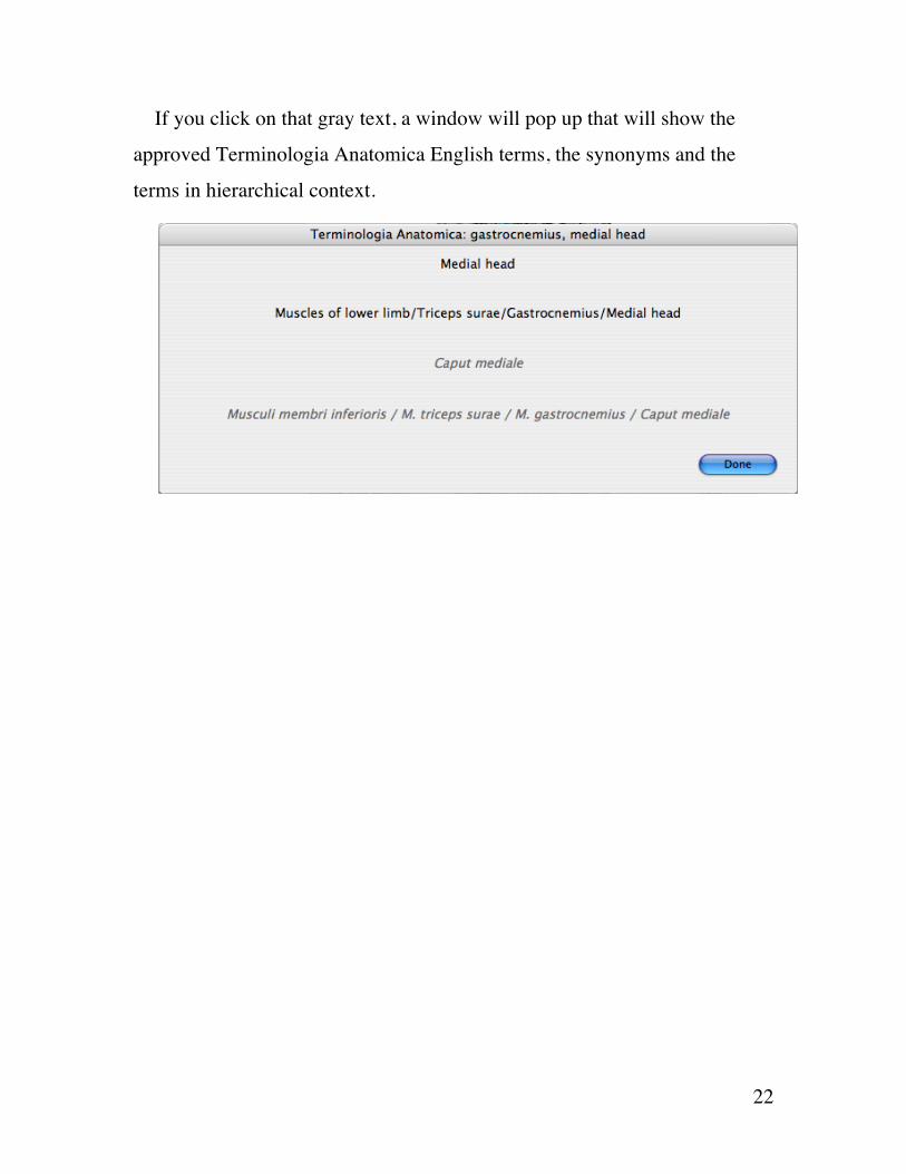
In RAAViewer, when available, the Terminologia Anatomica Latin term
is presented in gray text underneath the English term that I have chosen for
the anatomic entity.
22
If you click on that gray text, a window will pop up that will show the
approved Terminologia Anatomica English terms, the synonyms and the
terms in hierarchical context.

23
ALTERNATE LANGUAGES The program is written in English. I have little facility with any other
languages. In Nov 2009, an option was added to allow the user to specify
Japanese or Spanish as the language for the anatomic terms. I know no
Spanish and less Japanese, but there are sites on the Internet that provide
translations of the Terminologia Anatomica terms into those languages.
Japanese: Kazuya Funato Department of Anatomy, Keio University
School of Medicine, Japan
http://web.sc.itc.keio.ac.jp/anatomy/osteologia/
Spanish: Apuntes de Anatomia
http://www.iqb.es/cbasicas/anatomia/clasificacion/indice.htm
There are a large number of typographical errors in the Spanish listing.
Many of these I was able to find and correct because Spanish is close
enough to Latin that many terms are recognizable to an English speaker. I
have no way of knowing whether there are errors in the Japanese listing.
Using those sources as a starting point, I have incorporated Spanish and
Japanese translations for a large subset of the anatomic terms used in this
program.
In January 2010, French was added to the supported languages. This was
possible due to the assistance of Paul Fabry at the Université de Sherbrooke
who has taken an interest in multilingual versions of Terminologia
Anatomica.
The detailed descriptions and other text remain in English, but if the user
selects one of these alternate languages, many of the actual terms will be

24
displayed in those languages. When the program is launched for the first
time, there is a menu that by default will display English. That menu can be
used to specify one of the other languages.
25
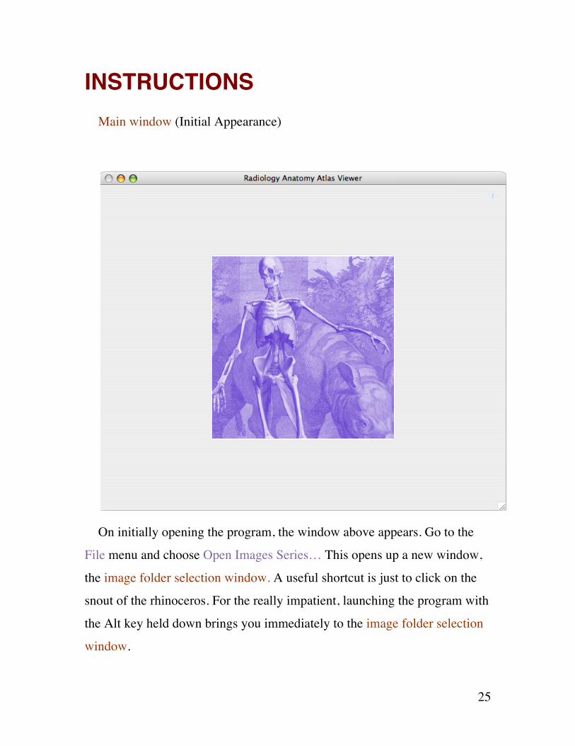
INSTRUCTIONS Main window (Initial Appearance)
On initially opening the program, the window above appears. Go to the
File menu and choose Open Images Series… This opens up a new window,
the image folder selection window. A useful shortcut is just to click on the
snout of the rhinoceros. For the really impatient, launching the program with
the Alt key held down brings you immediately to the image folder selection
window.
26
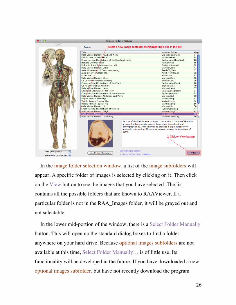
In the image folder selection window, a list of the image subfolders will
appear. A specific folder of images is selected by clicking on it. Then click
on the View button to see the images that you have selected. The list
contains all the possible folders that are known to RAAViewer. If a
particular folder is not in the RAA_Images folder, it will be grayed out and
not selectable.
In the lower mid-portion of the window, there is a Select Folder Manually
button. This will open up the standard dialog boxes to find a folder
anywhere on your hard drive. Because optional images subfolders are not
available at this time, Select Folder Manually… is of little use. Its
functionality will be developed in the future. If you have downloaded a new
optional images subfolder, but have not recently download the program

27
itself, it is possible that this optional folder will not appear in the listing on
the image folder selection window. This button will allow you at access this
new folder of images until such time as you can download the latest version
of the program.
On the left side of the window, there are tools to prioritize the image
subfolders. These do not filter out folders; rather they sort the listing so that
those best meeting the criteria appear toward the top of the list. The picture
of the standing human on the left can serve to sort the folders of interest by
anatomic region. Click on the part of the body that you are interested in (for
example the knee) and the list will re-sort itself such that the image
subfolders that relate to the knee will float to the top. A red line will appear
on the diagram to indicate where it was clicked. It is also possible to click
and drag to indicate a region of interest on the human diagram. In this
situation, two red lines will appear indicating the boundary of the selected
region. There is a Clear button at the top of the picture to clear away the red
lines.
Underneath the picture are two pop-ups that can also be used to prioritize
the list, for example to look for coronal scans. Again, the list will re-sort
itself to place the requested type of images at the top.
The ability to smart sort the list of image folders will become more useful
as the list of such subfolders gets longer.
On the top of the window, there are checkboxes that filter the list. The
Recent checkbox will restrict the list to image folders that have been
examined recently.

28
Main window (Showing Specific Picture)
The various controls for this window are labeled in the above picture. Use
the image selection control to navigate from one picture to the next in the
selected image subfolder. The orientation buttons will flip the image around
which can be useful to match the atlas image with the radiographic image
that you are interested in. For example, an image of the right arm can be
converted into an image of the left.

29
Clicking on a dot will select it. The corresponding anatomic term (in the
example above – subclavius) will appear above the picture. The selected dot
is highlighted as can be seen in the illustration above in the right upper
corner. When the word is written in blue, as it here, then clicking on that
word will bring up the anatomic information window that be seen below.
The anatomic information window contains data about the anatomic entity
that has been selected. In some cases, as seen here, there is additional
information lower down in the window about the etymologic derivation of
the word. When those lines are highlighted in yellow (as the first one can be
30
seen in the example above) then it is possible to click on the line in question
to see yet more information about that line. When applicable, lists of
individuals connected historically with the entity, will appear.
When done with looking at the anatomic information window, click on
the close button at the top of the window to make it disappear.
Click on this button (or go to the File menu and choose Open Images
Series…) to return to image folder selection window.
These instructions should be enough to get you started with using this
program, which is simple in design.

31
DATA SOURCES I am not an anatomist and have done no original research in this field. The
information in the anatomic information window comes from secondary
sources. The most heavily used sources are:
1. Gray’s Anatomy (1918) This anatomy textbook is outdated but has the
advantage of being out of copyright.
http://www.bartleby.com/107/
2. Wikipedia. This web site is an information resource created by the
public. Most of the anatomic information in this encyclopedia is derived
from the Gray’s Anatomy source mentioned above, but it can still be very
useful.
http://en.wikipedia.org/wiki/Main_Page
3. Who named it – is a delightful website that has posted small
biographies of many of the men (and the very rare women) whose names are
remembered in eponyms. The whole topic of eponyms is a contentious one,
but the biographies of those so honored are interesting even if the selection
of such individuals is capricious.
http://www.whonamedit.com/
4. BrainInfo (NeuroNames) Unfortunately, I became aware of this
resource relatively late in the project. The project is an effort to come
up with a rational, consistent and hierarchical lexicon for
neuroanatomic terms. This was developed in the modern computer
era and escapes some of the problems embedded in Terminologia

32
Anatomica. The problems of redundancy and ambiguity and overlap
are severest in the area of brain anatomy, and this resource presents
one approach to the problem.
http://braininfo.rprc.washington.edu/Default.aspx

33
Final Comments
This manual covers the highlights, but to learn the program you have to
use it. It is not very difficult.
For updates of the program, go to
http://www.rrlivingston.com/RAAViewer
Robert Robb Livingston


