Embed Size (px)
Citation preview

8/4/2019 Rabies Flurry Pa Tho Genesis
http://slidepdf.com/reader/full/rabies-flurry-pa-tho-genesis 1/11
JOURNAL OF VIROLOGY, Sept. 2010, p. 8926–8936 Vol. 84, No. 170022-538X/10/$12.00 doi:10.1128/JVI.00787-10Copyright © 2010, American Society for Microbiology. All Rights Reserved.
Molecular Basis of Neurovirulence of Flury Rabies VirusVaccine Strains: Importance of the Polymerase and the
Glycoprotein R333Q Mutation
Lihong Tao,1 Jinying Ge,1 Xijun Wang,1 Hongyue Zhai,1 Tao Hua,1 Bolin Zhao,1 Dongni Kong,1
Chinglai Yang,2 Hualan Chen,1* and Zhigao Bu1*
Veterinary Public Health Laboratory of Ministry of Agriculture and State Key Laboratory of Veterinary Biotechnology, Harbin Veterinary Research Institute of Chinese Academy of Agricultural Sciences, Harbin 150001,
People’s Republic of China,1 and Department of Microbiology and Immunology, Emory University School of Medicine, Rollins Research Center,
Atlanta, Georgia 303222
Received 14 April 2010/Accepted 4 June 2010
The molecular mechanisms associated with rabies virus (RV) virulence are not fully understood. In thisstudy, the RV Flury low-egg-passage (LEP) and high-egg-passage (HEP) strains were used as models to explorethe attenuation mechanism of RV. The results of our studies confirmed that the R333Q mutation in the
glycoprotein (GR333Q) is crucial for the attenuation of Flury RV in mice. The R333Q mutation is stablymaintained in the HEP genome background but not in the LEP genome background during replication inmouse brain tissue or cell culture. Further investigation using chimeric viruses revealed that the polymeraseL gene determines the genetic stability of the GR333Q mutation during replication. Moreover, a recombinant RV containing the LEP G protein with the R333Q mutation and the HEP L gene showed significant attenuation,genetic stability, enhancement of apoptosis, and immunogenicity. These results indicate that attenuation of theRV Flury strain results from the coevolution of G and L elements and provide important information for thegeneration of safer and more effective modified live rabies vaccine.
Rabies virus (RV) belongs to the genus Lyssavirus of thefamily Rhabdoviridae and causes a fatal neurological disease inhumans and animals (6). The RV genome is a nonsegmentednegative-strand (NNS) RNA encoding five structural pro-
teins: nucleoprotein (N), phosphoprotein (P), matrix pro-tein (M), glycoprotein (G), and large polymerase (L). Among these, the G protein is a major contributor to RVpathogenicity (7, 31, 33). The G protein facilitates fast virusentry and transsynaptic spread and regulates the rate of virus replication, together with other viral elements (8, 30,39). The G protein of nonpathogenic RV strains can triggerapoptosis, while the RV G of pathogenic strains induces lessor no apoptosis (35, 59). The amino acid residue at position333 of the G protein (G333) of some fixed strains has beenshown to be an important determinant of virulence in adultmice (5). Strains that have arginine or lysine at positionG333 kill adult mice, whereas mutants with other aminoacids at this site cause a nonlethal infection (1, 5, 25, 36, 49,53). However, the pathogenicity of RV strains is not solelydetermined by substitutions at the G333 position. Othersubstitutions in the G protein, such as N194K, have alsobeen shown to affect viral pathogenicity in mice (10, 21, 50).In addition, other viral elements, such as the N, P, M, and Lgenes, the trailer sequence in the noncoding region, and the
pseudogene, were also reported to modulate RV pathoge-nicity (12, 46, 57, 58). How these viral elements regulate thepathogenicity of RV remains to be fully explored, and fur-ther investigation is needed to understand the molecular
basis of RV pathogenicity. Attenuated Flury RV low-egg-passage (LEP) and high-egg-
passage (HEP) strains were established through serial passagein chicken brain, chicken embryos, and culture cells using aFlury RV isolated from a girl who died of rabies (23, 24). LEPhas Arg at position G333 and kills adult mice after intracere-bral (i.c.) inoculation, while HEP has Gln at G333 and causesonly mild signs in adult mice. It has been demonstrated thatHEP could regain lethality in adult mice by a single amino acidchange at G333 from Gln to Arg (49), which indicated that Argat position G333 is a key determinant of pathogenicity of FluryRV in adult mice. However, whether the Arg at G333 is indis-pensable for the lethal phenotype of LEP has not been dem-
onstrated.In the current study, LEP and HEP Flury RV strains were
used as models to investigate the mechanism of attenuation.We found that both G and L contribute to the attenuationof Flury RV. Substitution of Arg with Gln at G333 (GR333Q)eliminated LEP neuroinvasiveness but not the virus’ lethalphenotype in adult mice after i.c. inoculation. The GR333Q
mutation could be kept stable only in the genome back-ground of HEP but not in that of LEP during replication.The L gene contributes to the attenuation and enhancedimmunogenicity of Flury RV by promoting the stabilizationof the GR333Q mutation during virus replication in braintissues or cells.
* Corresponding author. Mailing address: Harbin Veterinary Re-search Institute of CAAS, 427 Maduan Street, Harbin 150001, Peo-ple’s Republic of China. Phone for Zhigao Bu: 86-451-85935062. Fax:86-451-82733132. E-mail: [email protected]. Phone for Hualan Chen:86-451-85935079. Fax: 86-451-82733132. E-mail: [email protected].
Published ahead of print on 10 June 2010.
8926
8/4/2019 Rabies Flurry Pa Tho Genesis
http://slidepdf.com/reader/full/rabies-flurry-pa-tho-genesis 2/11
MATERIALS AND METHODS
Viruses and cells. Neuroblastoma (NA) cells of A/J mouse origin were grown
in Eagle’s minimum essential medium (MEM) supplemented with 10% fetalbovine serum (FBS). Baby hamster kidney (BHK-21) cells were grown in Dul-becco’s modified Eagle’s MEM (DMEM) supplemented with 10% FBS. The RV
Flury strains LEP (AV2012) and HEP (AV2013) were originally received fromthe China Veterinary Culture Collection Center and propagated in BHK-21cells. The street virus GX/09, isolated from the brain of a dog that died of rabies
in the Guangxi Province of China in 2009, was propagated in the brains of adultmice. All viruses were stored in a 70°C freezer before use for RNA extraction
or challenge studies.Construction of the LEP and HEP full-length cDNA clones. Viral RNA was
extracted with an RNeasy minikit (Qiagen, Valencia, CA). The extracted RNA
was subjected to reverse transcription-PCR (RT-PCR) using high-fidelity Pfx
DNA polymerase (Invitrogen Corp., Carlsbad, CA) to generate three overlap-ping PCR fragments (F1, F2, and F3) of the entire viral genome (Fig. 1). Thefull-length viral genome RNA transcription plasmids were constructed as previ-ously described by Inoue et al. (20). The assembled cDNA containing the se-
quence of the hammerhead ribozyme sequence (HamRz), the full-length(11,925-nucleotide) cDNA of the LEP or HEP strain genome in the antigenomicorientation, and the hepatitis delta virus ribozyme sequence (HdvRz) was in-
serted between the NheI and SmaI sites of the plasmid vector pCI. A PmeIrestriction site was introduced as a genetic marker in the G-L noncoding region
of the LEP and HEP cDNA by changing three nucleotides at position 4907 (T toG), 4910 (G to T), and 4912 (C to A) using a site-directed mutagenesis system(Invitrogen). The resulting full-length plasmids were designated pLEP and
pHEP, respectively.The open reading frames (ORFs) of the N, P, and L genes were PCR amplified
from pLEP to construct helper plasmids. The amplified N, P, and L genes wereeach inserted between EcoRI and KpnI of the plasmid pCAGGS (37), and theresulting helper plasmids were designated pCA-NL, pCA-PL, and pCA-LL,
respectively. The assembled full-length cDNA clones and the helper plasmids were confirmed by sequencing.
Construction of mutants and chimeric viral cDNA clones. The full-length
cDNA clones of parental, mutated, and chimeric RVs are shown in Fig. 2. Themutant virus cDNA clone pLEPG333Q, in which Arg at G333 of the LEP strain
was mutated to Gln, and the pHEPG333R clone, in which Gln at G333 of HEP was mutated to Arg, were constructed by using a site-directed mutagenesissystem (Invitrogen). The chimeric virus cDNA clone pLEP-G(H), in which the G
gene ORF of LEP was replaced by that of HEP, was constructed in the followingmanner. One fragment containing the G gene of HEP was PCR amplified from
pHEP, and another fragment containing the M gene of LEP was PCR amplifiedfrom pLEP. Both fragments were used as templates for an overlapping PCR. ThePCR product was digested with XhoI and PmeI and then inserted into the same
site of plasmid pLEP. The same method was used to construct the chimeric viruscDNA clone pHEP-G(L)333Q, in which the G gene ORF of HEP was replaced bythat of LEP with the GR333Q mutation.
For construction of the chimeric virus cDNA clone pLEPG333Q-L(H), in
which the L gene ORF of pLEPG333Q was replaced by that of HEP, one
fragment containing the L gene and the trailer sequence of HEP was amplifiedfrom pHEP, and another fragment containing part of the G gene, the gene, and
part of the L gene of LEP was amplified from pLEP. Both fragments were thenused as templates for an overlapping PCR. The PCR product was digested with
PmeI and SmaI and then cloned into PmeI- and SmaI- predigested plasmidpLEPG333Q. All of the sequences of the primers used in this study are availablefrom the corresponding author upon request.
Virus rescue. BHK-21 cells were grown overnight to 80% confluence in 6-wellplates in DMEM supplemented with 10% FBS. Cells were transfected with 4.0g of the full-length plasmid, 2 g of pCA-NL, 1 g of pCA-PL, and 1 g of
pCA-LL using Lipofectamine 2000 (Invitrogen) according to the manufacturer’sprotocol. After 4 to 6 h, the transfection medium was replaced with fresh DMEM
supplemented with 10% FBS. After 3 days, supernatants were collected andtransferred into wells containing BHK-21 cells and incubated another 3 days.Rescued viruses were examined by indirect fluorescence assay (IFA) with mouse
serum against RV and fluorescein isothiocyanate (FITC)-conjugated goat-anti-
mouse IgG. Supernatants from virus-positive culture wells were collected topropagate virus stock in BHK-21 cells. The sequences of recovered viruses wereconfirmed by sequencing of the entire viral genome.
Virus titration. Monolayers of NA or BHK-21 cells in 24-well plates were
infected with 10-fold dilutions of virus suspension and incubated at 34°C. At 48 hpostinfection, an IFA was performed. Foci were counted under a fluorescencemicroscope and calculated as focus-forming units/ml (FFU/ml). The in vitro
neurotropism index was expressed as the logarithm of the titer of virus stock inNA cells subtracted by the logarithm of the titer of the same stock virus in
BHK-21 cells.Infection of mice. Virulence of the virus in adult mice was measured in
6-week-old female BALB/c mice (Vitalriver, Beijing). Groups of five mice were
inoculated i.c. or intranasally (i.n.) with 30 l of diluted virus or intramuscularly(i.m.) with 100 l of diluted virus. After infection, mice were observed for clinical
signs of disease, and body weight was recorded daily. The mouse 50% lethaldosage (MLD50) of the viruses was calculated by the method of Reed andMuench (42).
Flow cytometric analysis of FITC annexin V staining. Apoptotic cells of theearly stage were detected using a FITC annexin V apoptosis detection kit (BDPharmingen, San Diego, CA). NA cells were infected with recombinant RVs at
a multiplicity of infection (MOI) of 1 and incubated for 24 h at 34°C. Cells were washed twice with cold PBS and then resuspended in PBS and adjusted to a
concentration of 1 106 cells/ml. Next, 105 cells were incubated with 5 l of FITC annexin V and 5 l propidium iodide (PI) for 15 min at 25°C in the dark. Afterward, 400 l of binding buffer was added to each tube, and flow cytometry
was performed on a BD FACSAria analyzer. Western blotting. NA cells grown in 6-well plates were infected with different
RV strains at an MOI of 1 and incubated for 24 h. G gene expression wasconfirmed by Western blotting. Briefly, cell extracts were analyzed by SDS-PAGE and then blotted to nitrocellulose membrane. The membrane was
incubated with a mixture of mouse anti-G protein polyclonal antiserum,mouse anti-N protein polyclonal antiserum, and anti--actin monoclonalantibody (Santa Cruz Biotechnology). Bindings were visualized with 3,3 -
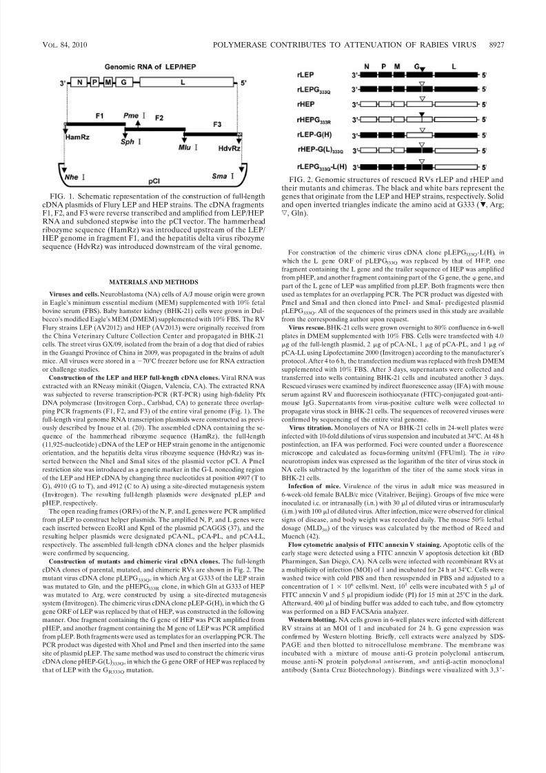
FIG. 1. Schematic representation of the construction of full-lengthcDNA plasmids of Flury LEP and HEP strains. The cDNA fragmentsF1, F2, and F3 were reverse transcribed and amplified from LEP/HEPRNA and subcloned stepwise into the pCI vector. The hammerheadribozyme sequence (HamRz) was introduced upstream of the LEP/HEP genome in fragment F1, and the hepatitis delta virus ribozymesequence (HdvRz) was introduced downstream of the viral genome.
FIG. 2. Genomic structures of rescued RVs rLEP and rHEP andtheir mutants and chimeras. The black and white bars represent thegenes that originate from the LEP and HEP strains, respectively. Solidand open inverted triangles indicate the amino acid at G333 (�, Arg;ƒ, Gln).
VOL. 84, 2010 POLYMERASE CONTRIBUTES TO ATTENUATION OF RABIES VIRUS 8927
8/4/2019 Rabies Flurry Pa Tho Genesis
http://slidepdf.com/reader/full/rabies-flurry-pa-tho-genesis 3/11
diaminobenzidine reagent after incubation with peroxidase-conjugated sec-ondary antibodies.
Immunization and virus challenge. Four-week-old female BALB/c mice(groups of 10) were injected i.m. in the gastrocnemius muscle with 100 l of
serial 10-fold dilutions of rescued live RVs. After 3 weeks, mice were bled andinjected i.m. with 100 l of street virus GX/09 strain containing 50 MLD50. Mice were observed for 4 weeks for clinical signs of rabies. Mice that showed definitive
clinical signs of rabies, such as paralysis, tremors, and spasms, were euthanizedby CO2 intoxication. Survival rates obtained with the different vaccine dilutionsfor the different vaccination groups were compared.
Neutralizing antibodies assay. Mice were bled from the retro-orbital sinusunder isoflurane inhalation anesthesia, and mouse sera were tested for virus-
neutralizing antibodies (VNA) using the rapid fluorescent focus inhibition test
(RFFIT), as described elsewhere (13, 47). Neutralization antibody titers, definedas the highest serum dilution that neutralizes 50% of the challenge virus, were
normalized to international units (IU) using the World Health Organizationanti-RV antibody standard. Geometric mean titers (GMT) were calculated from
the titers of 10 mice of the same vaccination group.Nucleotide sequence accession number. The sequence data for the Flury LEP
and HEP strains in this study have been deposited in GenBank under accession
numbers GU565703 and GU565704, respectively.
RESULTS
Genome comparison of Flury LEP and HEP strains. TheLEP and HEP strains of Flury RV have similar genomic back-grounds but differ in their virulence levels in adult mice. Se-quencing analyses revealed that the genomes of the LEP and
HEP strains share 99.3% nucleotide sequence identity and adeduced amino acid homology of 99.8%, 98.3%, 99.0%, 97.8%,and 99.6% between the individual N, P, M, G, and L proteins,respectively. The two strains have identical intergenic regionsbetween N/P, P/M, and M/G and one substitution in the G-Lintergenic region. Sequences of the 3 and 5 terminal noncod-ing regions, which include the recognition and initiation site of the viral RNA polymerase, were completely conserved in thetwo viruses. The locations of amino acid substitutions in the N,P, M, G, and L proteins are indicated in Table 1. A total of 27amino acid substitutions were found between these two strains.
Biological characterization of the wild type and the rescued
strains. To investigate the molecular mechanism for virulenceand attenuation of Flury RV, two infectious viruses, rLEP andrHEP, were rescued from the genomes of LEP and HEP,respectively. A PmeI restriction endonuclease site in the G-Lnoncoding region was generated as a genetic marker for bothrescued viruses to distinguish them from wild-type LEP andHEP viruses. The growth of the rLEP and rHEP strains incultured cells and their virulence in adult mice were deter-mined and compared with those of their wild-type counter-parts. The growth curves of the rLEP and rHEP strains weresimilar to the wild-type LEP and HEP strains, respectively, inboth neuronal NA and nonneuronal BHK-21 cells (Fig. 3). Thein vitro neurotropism index, defined as the infectivity ratio of agiven virus in NA cells versus in BHK-21 cells, represents a keymeasurement of RV neuroinvasiveness and neurorvirulence(34). The rLEP and wild-type LEP strains appeared to bestrongly neurotropic, having similar index values of 0.84 and
FIG. 3. Multistep growth curves of the rLEP, wtLEP, rHEP, and wtHEP viruses. NA (A) or BHK-21 (B) cells were infected with dif-ferent viruses at an MOI of 0.01. The viral titers in infected cellsupernatants were determined at different time points. Data shown arethe mean titer standard deviations (SD).
TABLE 1. Amino acid substitutions in N, P, M, G, and L proteinsof the HEP strain compared to those
of the LEP strain
Protein PositionSubstitution
LEP strain HEP strain
N 13 Q R
P 59 G D84 D N
160 T I277 I L295 T K
M 22 V A97 I V
G a 19 I L40 E G
194 N H204 G D333 R Q349 G E
425 E K 431 E D436 N K 473 S N476 E G
L 75 V G223 V I431 L R514 K Q
1226 P S1567 R Q1740 H R1795 V I
a The amino acid numbering in the G protein corresponds to the matured formthat does not contain a signal peptide.
8928 TAO ET AL. J. VIROL.

8/4/2019 Rabies Flurry Pa Tho Genesis
http://slidepdf.com/reader/full/rabies-flurry-pa-tho-genesis 4/11
0.78, respectively. As expected, both rHEP and wild-type HEPdid not show any neurotropic characteristics (Table 2).
Virulence of rLEP, rHEP, and wild-type LEP and HEP in
adult mice was determined by different inoculation routes, i.c.,i.n., or i.m. The survival rates and disease progression of miceinfected with different dosages of rLEP or wild-type LEP areshown in Fig. 4A to F. The rLEP strain killed adult mice by anyinoculation route. The MLD50 and death time courses after i.c.or i.n. inoculation of rLEP were comparable to those of wild-type LEP (Fig. 4). Peripheral pathogenicity was assessed byi.m. inoculation. The MLD50 of rLEP by i.m. inoculation was3.4 105 FFU, slightly higher than that of wild-type LEP. For wild-type LEP, only one of the five mice inoculated i.m. with106 FFU wild-type LEP died, and the others exhibited hind legparalysis. In contrast, all mice survived from i.c., i.n., or i.m.inoculation, even with 106 FFU of rHEP or wild-type HEP
(Fig. 4G to L), without exhibiting any neurological signs, ex-cept that an about 3% body weight loss was observed in thefirst 3 days postinoculation with the i.c. group. These resultssuggested that the rescued viruses had biological propertiesand pathogenicities similar to those of their correspondingparental wild-type virus in the adult mouse.
Substitution of Arg with Gln at G333 eliminates LEP neu-
roinvasiveness but not its lethal phenotype in adult mice by i.c.
inoculation. The Arg at position 333 of G is one of the mostimportant determinants for neurotropism of RV. LEP has Argat position 333 of G, while HEP has Gln at this position. Wegenerated a HEP mutant virus, rHEPG333R, in which the Glnat G333 was changed to Arg. As shown in Fig. 6, the multiple-step growth curve of the rHEPG333R strain in NA cells wassimilar to that of the rLEP strain. Meanwhile, the in vitro
neurotropism index of rHEPG333R was significantly increasedfrom 0 to 0.8, indicating that rHEPG333R had acquired theneurotropism property. Infection of adult mice confirmed thatrHEPG333R was lethal (Fig. 4 M to O). The MLD50 by i.c., i.n.,and i.m. inoculation routines were 0.3 FFU, 887 FFU, and1.4 106 FFU, respectively. Theses results were consistent with other reports (49). The mutation at G333 is sufficient tocause HEP to become highly pathogenic in adult mice.
Based on these results, it was hypothesized that the mutationof LEP G333 from Arg to Gln should eliminate the neurotro-pism property and therefore attenuates the LEP virus in adultmice. To investigate this issue, an LEP mutant, rLEPG333Q, in
which the Arg at G333 was changed to Gln, was constructed. As shown in Table 2, rLEPG333Q lost its neurotropism prop-erty and had a neurotropism index value of 0 in cell culture.Infection by the i.m. route with the maximum dosage of 3
106 FFU did not kill adult mice (Fig. 4R), and all survivingmice did not show any signs of neurological disease. However, when inoculated by i.c. and i.n. routes, rLEPG333Q remainedhighly lethal in adult mice (Fig. 4P and Q). The MLD50s by i.c.and i.n. routes were 36 FFU and 2.1 105 FFU, respectively.These results suggested that substitution of Arg with Gln atG333 eliminated only the peripheral neuroinvasiveness of LEPby i.m. inoculation but not its lethal phenotype in adult mice byi.c. or i.n. inoculation.
Gln mutation at G333 of rLEPG333Q reverted to Arg during
replication in neurological tissues or cells. Infection by thei.m. route showed that rLEPG333Q had lost its neurotropism in
vitro and its peripheral pathogenicity in vivo, but it was still ableto kill adult mice by i.c. and i.n. infection. One possible expla-nation for these results is that the Gln mutation at G333 of rLEPG333Q may quickly revert back to Arg during replication
and allow the virus to then spread among neurological cells. Toconfirm this hypothesis, total RNA extracted from the braintissue of each mouse killed after inoculation with rLEPG333Q
was used as a template to amplify the G gene by RT-PCR. Theresults revealed that Gln (CAA) at G333 of rLEPG333Q com-pletely reverted to Arg (CGA) in all mice after one i.c. inoc-ulation (Fig. 5C). Passage of the virus in NA cells showed thatGln (CAA) at G333 in rLEPG333Q was not stably maintainedin NA cells in vitro and partially mutated back to Arg (CGA) within five passages (Fig. 5D). These results indicated thatrLEPG333Q could not stably maintain the GR333Q mutation inmouse brain tissue and NA cells. The virus reverted back toregain its neurotropism and highly pathogenic phenotype. In
contrast, rHEP stably maintained Gln (CAG) at G333, in bothin vivo infection in mice by i.c. inoculation (Fig. 5A) and in vitro
passaging in NA cells for up to five passages (Fig. 5B).The LEP genome backbone is responsible for the instability
of Gln mutation at G333. To investigate if a specific propertyof the G gene itself or if the genome background of LEP isresponsible for the reversion of the Gln mutation at G333 to Arg, we constructed two chimeric viruses. The rHEP-G(L)333Q virus was generated by replacing the ORF of the G gene of HEP with that of LEP in which the amino acid at G333 wasmutated from Arg to Gln. This virus showed a growth patternsimilar to that of rHEP in NA cells (Fig. 6). The neurotropismindex of rHEP-G(L)333Q was 0 and was the same as that of rHEP (Table 2). The pathogenicity of rHEP-G(L)333Q wasevaluated by i.c. inoculation in adult mice. All mice inoculated with 105 FFU of rHEP-G(L)333Q survived the infection and didnot show any signs of neurological disease (Fig. 7). Anotherchimeric virus, rLEP-G(H), was generated by replacing the Ggene ORF of LEP with that of HEP. The titers of rLEP-G(H)in NA cells were lower than that of rLEP and similar to thoseof rHEP (Fig. 6). The in vitro neurotropism index of rLEP-G(H) was 0 (Table 2). All mice inoculated with 105 FFU of rLEP-G(H) died within 12 days postinfection (Fig. 7). The viral genomic RNAs isolated from the brain of infected mice were extracted and sequenced. The results revealed that G333of rLEP-G(H) had changed to Arg (CGG) from Gln (CAG)(Fig. 5E). In comparison, the Gln (CAA) at G333 of rHEP-
TABLE 2. Growth titers of RV strains in NA and BHK cells
StrainTiter (FFU/ml) a Neurotropism
index bNA BHK-21
wtLEP 3.7 108 5.3 107 0.84rLEP 3.0 108 5.0 107 0.78
wtHEP 1.0 107 1.0 107 0
rHEP 2.0 107
2.0 107
0rHEPG333R 1.6 108 2.5 107 0.80rLEPG333Q 3.0 107 3.3 107 0rLEP-G(H) 7.0 106 7.0 106 0rHEP-G(L)333Q 2.0 107 2.0 107 0rLEPG333Q-L(H) 1.0 107 1.0 107 0
a Virus titers were determined by using an IFA as described in Materials andMethods.
b The neurotropism index equals the logarithm of the titer in NA cells sub-tracted by the logarithm of the titer in BHK-21 cells.
VOL. 84, 2010 POLYMERASE CONTRIBUTES TO ATTENUATION OF RABIES VIRUS 8929
8/4/2019 Rabies Flurry Pa Tho Genesis
http://slidepdf.com/reader/full/rabies-flurry-pa-tho-genesis 5/11
G(L)333Q was stably maintained after i.c. inoculation in mice(Fig. 5G).
rHEP-G(L)333Q and rLEP-G(H) was passaged an additionalfive times in NA cells at an MOI of 0.1. The viral genomeRNAs were extracted from the fifth passage and subjected toRT-PCR and sequencing analysis. The results showed that theGln (CAG) at G333 of rLEP-G(H) had partially changed to Arg (CGG) (Fig. 5F), whereas the Gln (CAA) at positionG333 of rHEP-G(L)333Q remained stable after five passages inNA cells (Fig. 5H). These results strongly suggest that muta-tion from Gln to Arg at G333 does not happen randomly.
Certain viral element(s) in the LEP genome backbone otherthan the G gene might be responsible for the instability of theGln mutation at G333.
The L protein is related to the stability of Gln mutation at
G333. The low fidelity of the RNA polymerase of negative-strand RNA viruses is the major reason for virus mutation.For this reason, we constructed another chimeric virus,rLEPG333Q-L(H), in which the ORF of the L gene of rLEPG333Q was replaced with that of rHEP. Multistepgrowth kinetics analysis showed that rLEPG333Q-L(H) rep-licated at a relatively lower rate, about 10-fold lower than
FIG. 4. Pathogenicity of different RVs in adult mice. Death patterns of mice i.c. infected with different dosages (10 0 to 106 FFU) of various viruses are shown. wtLEP (A), rLEP (D), wtHEP (G), rHEP (J), rHEPG333R (M), and rLEPG333Q (P), with different dosages from 100 to 105 FFUare shown. Death patterns of mice i.n. infected with wtLEP (B), rLEP (E), wtHEP (H), rHEP (K), rHEPG 333R (N), and rLEPG333Q (Q), withdifferent dosages of 102 to 106 FFU. Death patterns of mice i.m. infected with wtLEP (C), rLEP (F), wtHEP (I), rHEP (L), rHEPG333R (O), andrLEPG333Q (R), with different dosages from 104 to 106 FFU. The titers of the virus stocks used for inoculation were determined in BHK-21 cells(F, 106 FFU; f, 105 FFU; Œ, 104 FFU; �, 103 FFU; , 102 FFU; E, 101 FFU; ,100 FFU.).
8930 TAO ET AL. J. VIROL.
8/4/2019 Rabies Flurry Pa Tho Genesis
http://slidepdf.com/reader/full/rabies-flurry-pa-tho-genesis 6/11
that of the rLEPG333Q (Fig. 6). All mice infected by the i.c.route with rLEPG333Q-L(H) survived (Fig. 7) and showedno signs of neurological disease, except slight loss of body
weight. This was similar to what was observed for rHEP-G(L)333Q. The viral genome RNA from the brain of infectedmice after inoculation with rLEPG333Q-L(H) was subjected
to RT-PCR and sequencing analysis. The results revealedthat Gln (CAA) at G333 of rLEPG333Q-L(H) remained un-changed (Fig. 5I). Further, rLEPG333Q-L(H) also stablymaintained Gln (CAA) at G333 after five passages in NA
cells (Fig. 5J). These results indicated that the L protein is
responsible for the stability of the Gln mutation at G333 of Flury RV.
The mutation of Arg to Gln at G333 is associated with a
greater induction of early apoptosis by Flury RV. Several stud-ies have shown that the attenuation of RV is associated with itsability to induce apoptosis in neuronal cells and that the G
protein is responsible for triggering the apoptosis cascade (35,39, 51). Annexin V binding, which detects the exposure of phosphatidylserine at the plasma membrane, is an early eventin the apoptotic process (29). Therefore, we employed the
annexin V binding assay to investigate if the attenuation of
FIG. 5. Viral genome sequence analysis. The viral genome RNAs individually isolated from the brain tissues of mice i.c. inoculated with rHEP(A), rLEPG333Q (C), rLEP-G(H) (E), rHEP-G(L)333Q (G), and rLEPG333Q-L(H) (I) were used as templates to perform RT-PCR and sequenceanalyses for the G gene. Viruses were also collected 2 days after infection with rHEP (B), rLEPG 333Q (D), rLEP-G(H) (F), rHEP-G(L)333Q (H),and rLEPG333Q-L(H) (J) viruses that had previously been passaged four times in NA cells. The viral genome RNAs were extracted at this latertime point and used as templates to perform RT-PCR and sequence analyses for the G gene. The three nucleotides encoding the amino acid atG333 are boxed.
VOL. 84, 2010 POLYMERASE CONTRIBUTES TO ATTENUATION OF RABIES VIRUS 8931
8/4/2019 Rabies Flurry Pa Tho Genesis
http://slidepdf.com/reader/full/rabies-flurry-pa-tho-genesis 7/11
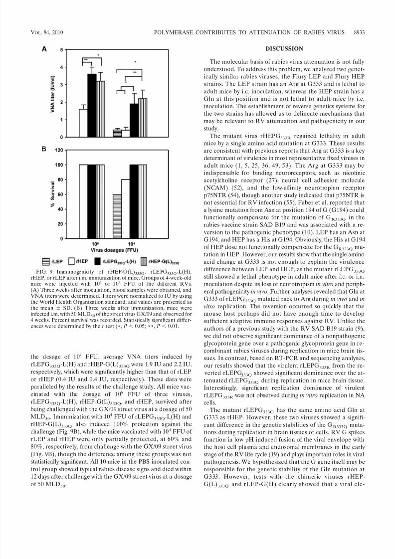
Flury RV changed its ability to induce apoptosis in neuronalcells. The abilities of the different RVs to induce apoptosis ininfected NA cells at 24 h postinfection were compared. Theapoptotic cells were stained by the FITC-labeled annexin V,but were not stained by PI. The apoptotic cells infected byrLEP- or rHEPG333R were about 2.9 0.7% or 2.8 0.6%,respectively. These values were similar to that of uninfectedcells (2.6 0.6%). In contrast, rHEP, rLEPG333Q, rHEP-G(L)333Q, and rLEPG333Q-L(H) induced significantly higherlevels of apoptosis in NA cells, and the percentage of theapoptotic cells were 2.1, 2.4, 2.1, and 2.1 times higher, respec-tively, than that of the uninfected cells (Fig. 8A).
We compared the expression level of viral proteins in in-fected cells. NA cells infected with different recombinant vi-ruses at an MOI of 1 were collected at 24 h postinfection andanalyzed by Western blotting. The G protein and N protein were probed with a polyclonal serum against the G or N pro-tein of LEP. The rLEP virus expressed amounts of G and Nproteins similar to those of rLEPG333Q, rHEP-G(L)333Q, andrLEPG333Q-L(H) in NA cells. Meanwhile, there was no signif-icant difference in the expression level of G or N proteinbetween NA cells infected by rHEP and rHEPG333R (Fig. 8B).
These results indicated that the higher levels of early apoptosisin NA cells induced by the recombinant viruses containingG333Q are not likely associated with an increase in the expres-sion of G protein.
Attenuated recombinant viruses induced enhanced immune
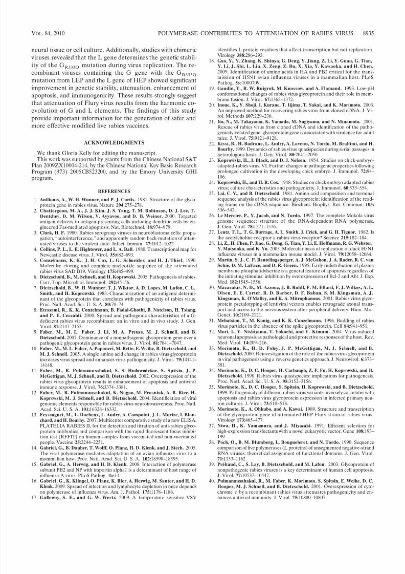
responses and more effective protection. It has been reportedthat attenuated RV is generally more effective at inducingprotective immune responses. rLEPG333Q-L(H) and rHEP-G(L)333Q have shown significant attenuation and genetic sta-bility in vivo and in vitro. To test if these two recombinant viruses could serve as live vaccine candidates with improvedsafety and efficacy, we evaluated their immunogenicity in micein comparison with rLEP and rHEP. Groups of 10 mice werei.m. immunized with 106 or 104 FFU of different RV strainsand then subjected to lethal challenge with the rabies street virus GX/09. Immunization with 106 FFU of rLEPG333Q-L(H)and rHEP-G(L)333Q induced similar levels of VNA, 3.6 IU and3.3 IU, respectively, which were significantly higher than thatinduced by rHEP (1.6 IU [Fig. 9A]). In mice immunized with
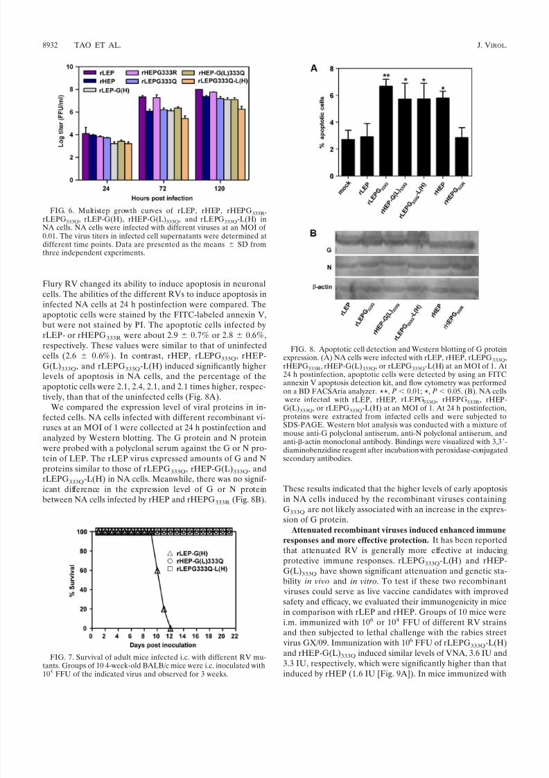
FIG. 6. Multistep growth curves of rLEP, rHEP, rHEPG333R,rLEPG333Q, rLEP-G(H), rHEP-G(L)333Q, and rLEPG333Q-L(H) inNA cells. NA cells were infected with different viruses at an MOI of 0.01. The virus titers in infected cell supernatants were determined atdifferent time points. Data are presented as the means SD fromthree independent experiments.
FIG. 7. Survival of adult mice infected i.c. with different RV mu-tants. Groups of 10 4-week-old BALB/c mice were i.c. inoculated with105 FFU of the indicated virus and observed for 3 weeks.
FIG. 8. Apoptotic cell detection and Western blotting of G proteinexpression. (A) NA cells were infected with rLEP, rHEP, rLEPG333Q,rHEPG333R, rHEP-G(L)333Q, or rLEPG333Q-L(H) at an MOI of 1. At24 h postinfection, apoptotic cells were detected by using an FITCannexin V apoptosis detection kit, and flow cytometry was performedon a BD FACSAria analyzer. **, P 0.01; *, P 0.05. (B). NA cells
were infected with rLEP, rHEP, rLEPG333Q, rHEPG333R, rHEP-
G(L)333Q, or rLEPG333Q-L(H) at an MOI of 1. At 24 h postinfection,proteins were extracted from infected cells and were subjected toSDS-PAGE. Western blot analysis was conducted with a mixture of mouse anti-G polyclonal antiserum, anti-N polyclonal antiserum, andanti--actin monoclonal antibody. Bindings were visualized with 3,3-diaminobenzidine reagent after incubation with peroxidase-conjugatedsecondary antibodies.
8932 TAO ET AL. J. VIROL.
8/4/2019 Rabies Flurry Pa Tho Genesis
http://slidepdf.com/reader/full/rabies-flurry-pa-tho-genesis 8/11
the dosage of 104 FFU, average VNA titers induced byrLEPG333Q-L(H) and rHEP-G(L)333Q were 1.9 IU and 2.2 IU,respectively, which were significantly higher than that of rLEPor rHEP (0.4 IU and 0.4 IU, respectively). These data wereparalleled by the results of the challenge study. All mice vac-
cinated with the dosage of 106 FFU of three viruses,rLEPG333Q-L(H), rHEP-G(L)333Q, and rHEP, survived afterbeing challenged with the GX/09 street virus at a dosage of 50MLD50. Immunization with 104 FFU of rLEPG333Q-L(H) andrHEP-G(L)333Q also induced 100% protection against thechallenge (Fig. 9B), while the mice vaccinated with 104 FFU of rLEP and rHEP were only partially protected, at 60% and80%, respectively, from challenge with the GX/09 street virus(Fig. 9B), though the difference among these groups was notstatistically significant. All 10 mice in the PBS-inoculated con-trol group showed typical rabies disease signs and died within12 days after challenge with the GX/09 street virus at a dosageof 50 MLD50.
DISCUSSION
The molecular basis of rabies virus attenuation is not fullyunderstood. To address this problem, we analyzed two genet-ically similar rabies viruses, the Flury LEP and Flury HEPstrains. The LEP strain has an Arg at G333 and is lethal toadult mice by i.c. inoculation, whereas the HEP strain has a
Gln at this position and is not lethal to adult mice by i.c.inoculation. The establishment of reverse genetics systems forthe two strains has allowed us to delineate mechanisms thatmay be relevant to RV attenuation and pathogenicity in ourstudy.
The mutant virus rHEPG333R regained lethality in adultmice by a single amino acid mutation at G333. These resultsare consistent with previous reports that Arg at G333 is a keydeterminant of virulence in most representative fixed viruses inadult mice (1, 5, 25, 36, 49, 53). The Arg at G333 may beindispensable for binding neuroreceptors, such as nicotinicacetylcholine receptor (27), neural cell adhesion molecule(NCAM) (52), and the low-affinity neurotrophin receptor
p75NTR (54), though another study indicated that p75NTR isnot essential for RV infection (55). Faber et al. reported thata lysine mutation from Asn at position 194 of G (G194) couldfunctionally compensate for the mutation of GR333Q in therabies vaccine strain SAD B19 and was associated with a re- version to the pathogenic phenotype (10). LEP has an Asn atG194, and HEP has a His at G194. Obviously, the His at G194of HEP dose not functionally compensate for the GR333Q mu-tation in HEP. However, our results show that the single aminoacid change at G333 is not enough to explain the virulencedifference between LEP and HEP, as the mutant rLEPG333Q
still showed a lethal phenotype in adult mice after i.c. or i.n.inoculation despite its loss of neurotropism in vitro and periph-
eral pathogenicity in vivo. Further analyses revealed that Gln atG333 of rLEPG333Q mutated back to Arg during in vivo and in
vitro replication. The reversion occurred so quickly that themouse host perhaps did not have enough time to developsufficient adaptive immune responses against RV. Unlike theauthors of a previous study with the RV SAD B19 strain (9), we did not observe significant dominance of a nonpathogenicglycoprotein gene over a pathogenic glycoprotein gene in re-combinant rabies viruses during replication in mice brain tis-sues. In contrast, based on RT-PCR and sequencing analyses,our results showed that the virulent rLEPG333R from the re- verted rLEPG333Q showed significant dominance over the at-tenuated rLEPG333Q during replication in mice brain tissue.Interestingly, significant replication dominance of virulentrLEPG333R was not observed during in vitro replication in NAcells.
The mutant rLEPG333Q has the same amino acid Gln atG333 as rHEP. However, these two viruses showed a signifi-cant difference in the genetic stabilities of the GR333Q muta-tions during replication in brain tissues or cells. RV G spikesfunction in low pH-induced fusion of the viral envelope withthe host cell plasma and endosomal membranes in the earlystage of the RV life cycle (19) and plays important roles in viralpathogenesis. We hypothesized that the G gene itself may beresponsible for the genetic stability of the Gln mutation atG333. However, tests with the chimeric viruses rHEP-G(L)333Q and rLEP-G(H) clearly showed that a viral ele-
FIG. 9. Immunogenicity of rHEP-G(L)333Q, rLEPG333Q-L(H),rHEP, or rLEP after i.m. immunization of mice. Groups of 4-week-oldmice were injected with 106 or 104 FFU of the different RVs.(A) Three weeks after inoculation, blood samples were obtained, andVNA titers were determined. Titers were normalized to IU by usingthe World Health Organization standard, and values are presented asthe mean SD. (B) Three weeks after immunization, mice wereinfected i.m. with 50 MLD50 of the street virus GX/09 and observed for4 weeks. Percent survival was recorded. Statistically significant differ-ences were determined by the t test (*, P 0.05; **, P 0.01.
VOL. 84, 2010 POLYMERASE CONTRIBUTES TO ATTENUATION OF RABIES VIRUS 8933

8/4/2019 Rabies Flurry Pa Tho Genesis
http://slidepdf.com/reader/full/rabies-flurry-pa-tho-genesis 9/11
ment(s) in the genome backbone other than G may beresponsible for the stability of the Gln mutation at G333. Additional tests with rLEPG333Q-L(H) demonstrated thatthe L protein contributed to the stability of the GR333Q
mutation of Flury RV.The most concerning issue regarding the use of live attenu-
ated RV vaccines is their high mutation rate. Several strains of attenuated RV consistently reverted rapidly to regain virulenceafter propagation in NA cells or suckling mouse brain, whileothers acquired increased virulence at a more gradual rate ornot at all (3, 10). Many factors, including duration and route of infection, virus load, host immune response, and virus-hostprotein interaction may be involved (22). Despite these influ-ences, the infidelity of the RNA polymerase of negative-strandRNA viruses could be the major factor responsible for themutations introduced at a relatively high frequency. Recentevidence indicated that the RV polymerase complex also im-pacted virulence (58). The viral replication rate is controlleddirectly by the polymerase complex in tissue culture and hasbeen shown to correlate inversely with the pathogenicity of RV
(9, 35, 41). Analysis of the silver-haired bat-associated RV 18(SHBRV-18) strain showed that the RV polymerase likelycontributed to RV neuroinvasiveness (12). Similarly, specificmutations in influenza virus polymerase have also been shownto considerably increase the virus’ activity in mammalian cellsand to correlate with high virulence in mice (14–16, 18, 28, 48).However, there is still no direct evidence to explain how thepolymerase affects the virulence of RV.
In our study, the L protein appeared to be closely associated with the attenuation of pathogenicity by stabilizing the Gln atG333, suggesting that the attenuation of Flury RV likely re-sulted from the harmonic coevolution of different elements inthe viral genome. The critical importance of the L protein in
transcription and replication is highlighted by its extreme sen-sitivity to mutation (4, 17). The amino acid sequence changesthat we identified in the L protein of the HEP strain thatdiffered from the LEP strain were V75G, V223I, L431R,K514Q, P1226S, R1567Q, H1740R, and I1795V. Phylogeneticanalyses have subdivided the NNS L proteins into six con-served domains linked by variable regions (38). Of all thesesequence differences in the HEP L protein, 1740R seems to beunique for the HEP strain. In contrast, all other RV strains with an Arg at G333, including LEP, SRV9 (AF499686), SAD-B19 (M31046), Ni-CE (AB128149), Nishigahara (AB044824),PV (NC_001542), rabies virus serotype 1 (AY956319), RC-HL(AB009663), SHBRV-18 (AY705373), and Mokola virus(Y09762), had H at position 1740 of the L protein. A statisticalstudy showed that glycine as well as acidic (D and E) and basic(K, R, and H) amino acids are highly conserved and typicallyplay a key role in L protein function and/or structure (26).Therefore, mutation at position 1740 is likely to be related tosome functional change in the L protein that consequentlyinfluences the stability of the Gln mutation at G333. Furtherstudies are needed to elucidate the significance of the aminoacid substitutions.
Apoptosis is used by many neurotropic viruses as a mecha-nism of neuropathogenicity (32). The attenuation of RV isassociated with its apoptotic ability in neuron cells (35). Sev-eral reports have suggested that the level of RV G expressedon the cell surface may be a critical factor in triggering apop-
tosis. Infection with recombinant RV expressing the proapop-totic protein cytochrome c induced a strong increase in apop-tosis, antiviral immune response, and reduced pathogenicity(40). The contribution of apoptosis to immune responses mayinvolve several mechanisms. The apoptotic cells can increaseinnate and adaptive immune responses and trigger the matu-ration and antigen presentation function of dendritic cells (43).Cells undergoing massive apoptosis could release factors toinduce the activation of major histocompatibility complex(MHC) class I- and MHC class II-restricted T cells by maturedendritic cells (2, 44). Moreover, the apoptotic bodies have anexceptional ability to deliver antigens to professional antigenpresenting cells (45). Our in vitro study with NA cells hadshown that the lethal viruses, rLEP and rHEPG333R, rarelyinduced apoptosis, while the highly attenuated viruses, rHEP,rLEPG333Q, rHEP-G(L)333Q, and rLEPG333Q-L(H) inducedsignificant apoptosis in NA cells. Our results are consistent with the observation that the pathogenicity of a particular RVstrain correlates inversely with its ability to trigger apoptosis inneuronal cells (35). A previous study also observed that the
induction of apoptosis is largely dependent on the expressionlevels of G protein (39), and overexpression of this proteinresults in enhanced apoptosis (11). Interestingly, our datashowed that all four RV strains with the GR333Q mutation,rHEP, rLEPG333Q, rHEP-G(L)333Q, and rLEPG333Q-L(H), in-duced significantly higher levels of apoptosis than the viruses with G333R, rLEP and rHEPG333R. However, there was noclear correlation between apoptosis induction and the viralreplication titers or glycoprotein expression level among these viruses. Whether the differences in apoptotic induction ob-served with these viruses was caused by viral replication orchanging the amino acid sequence of their glycoprotein stillremains to be determined. It is possible that the Flury RV
might use an additional direct mechanism to regulate apop-tosis.Due to its excellent immunogenicity, LEP has been widely
used to generate inactivated RV vaccine for human and ani-mals. In several countries, LEP also has been used as a live vaccine to control dog rabies, especially in countryside settings.However, the residual virulence of LEP obviously cannot meetthe current international biosafety standards for a live vaccine.Previous reports demonstrated that enhanced apoptosis andattenuation of RV likely increases the induction of the antiviralimmune response (11, 56). The chimeric virus rHEP-G(L)333Qor rLEPG333Q-L(H) showed significant induction of apoptosisin NA cells and attenuation in mice. We therefore investigated whether these two viruses would be able to serve as modifiedlive vaccine candidates. Immunization and challenge study re-sults showed that rHEP-G(L)333Q or rLEPG333Q-L(H) in-duced significantly higher VNA responses and more effectiveprotection than rLEP and rHEP. Since rHEP-G(L)333Q andrHEP had similar replication titers in NA cells, we can inferthat the G protein of LEP is more immunogenic than thatof HEP.
In summary, we used the RV Flury virus strains LEP andHEP as models to investigate the attenuation mechanism of RV. Our results confirmed that the GR333Q mutation is crucialin the attenuation of Flury RV in mice. The GR333Q mutation was maintained only in the HEP genome background but notin the LEP genome background during replication in mouse
8934 TAO ET AL. J. VIROL.
8/4/2019 Rabies Flurry Pa Tho Genesis
http://slidepdf.com/reader/full/rabies-flurry-pa-tho-genesis 10/11
neural tissue or cell culture. Additionally, studies with chimeric viruses revealed that the L gene determines the genetic stabil-ity of the GR333Q mutation during virus replication. The re-combinant viruses containing the G gene with the GR333Q
mutation from LEP and the L gene of HEP showed significantimprovement in genetic stability, attenuation, enhancement of apoptosis, and immunogenicity. These results strongly suggestthat attenuation of Flury virus results from the harmonic co-evolution of G and L elements. The findings of this studyprovide important information for the generation of safer andmore effective modified live rabies vaccines.
ACKNOWLEDGMENTS
We thank Gloria Kelly for editing the manuscript.This work was supported by grants from the Chinese National S&T
Plan 2009ZX10004-214, by the Chinese National Key Basic ResearchProgram (973) 2005CB523200, and by the Emory University GHIprogram.
REFERENCES
1. Anilionis, A., W. H. Wunner, and P. J. Curtis. 1981. Structure of the glyco-protein gene in rabies virus. Nature 294:275–278.2. Chattergoon, M. A., J. J. Kim, J. S. Yang, T. M. Robinson, D. J. Lee, T.
Dentchev, D. M. Wilson, V. Ayyavoo, and D. B. Weiner. 2000. Targetedantigen delivery to antigen-presenting cells including dendritic cells by en-gineered Fas-mediated apoptosis. Nat. Biotechnol. 18:974–979.
3. Clark, H. F. 1980. Rabies serogroup viruses in neuroblastoma cells: propa-gation, “autointerference,” and apparently random back-mutation of atten-uated viruses to the virulent state. Infect. Immun. 27:1012–1022.
4. Collins, P. L., L. E. Hightower, and L. A. Ball. 1980. Transcriptional map forNewcastle disease virus. J. Virol. 35:682–693.
5. Conzelmann, K. K., J. H. Cox, L. G. Schneider, and H. J. Thiel. 1990.Molecular cloning and complete nucleotide sequence of the attenuatedrabies virus SAD B19. Virology 175:485–499.
6. Dietzschold, B., M. Schnell, and H. Koprowski. 2005. Pathogenesis of rabies.Curr. Top. Microbiol. Immunol. 292:45–56.
7. Dietzschold, B., W. H. Wunner, T. J. Wiktor, A. D. Lopes, M. Lafon, C. L.Smith, and H. Koprowski. 1983. Characterization of an antigenic determi-nant of the glycoprotein that correlates with pathogenicity of rabies virus.Proc. Natl. Acad. Sci. U. S. A. 80:70–74.
8. Etessami, R., K. K. Conzelmann, B. Fadai-Ghotbi, B. Natelson, H. Tsiang,and P. E. Ceccaldi. 2000. Spread and pathogenic characteristics of a G-deficient rabies virus recombinant: an in vitro and in vivo study. J. Gen.Virol. 81:2147–2153.
9. Faber, M., M. L. Faber, J. Li, M. A. Preuss, M. J. Schnell, and B.Dietzschold. 2007. Dominance of a nonpathogenic glycoprotein gene over apathogenic glycoprotein gene in rabies virus. J. Virol. 81:7041–7047.
10. Faber, M., M. L. Faber, A. Papaneri, M. Bette, E. Weihe, B. Dietzschold, andM. J. Schnell. 2005. A single amino acid change in rabies virus glycoproteinincreases virus spread and enhances virus pathogenicity. J. Virol. 79:14141–14148.
11. Faber, M., R. Pulmanausahakul, S. S. Hodawadekar, S. Spitsin, J. P.McGettigan, M. J. Schnell, and B. Dietzschold. 2002. Overexpression of therabies virus glycoprotein results in enhancement of apoptosis and antiviralimmune response. J. Virol. 76:3374–3381.
12. Faber, M., R. Pulmanausahakul, K. Nagao, M. Prosniak, A. B. Rice, H.Koprowski, M. J. Schnell, and B. Dietzschold. 2004. Identification of viral
genomic elements responsible for rabies virus neuroinvasiveness. Proc. Natl. Acad. Sci. U. S. A. 101:16328–16332.
13. Feyssaguet, M., L. Dacheux, L. Audry, A. Compoint, J. L. Morize, I. Blan-chard, and H. Bourhy. 2007. Multicenter comparative study of a new ELISA,PLATELIA RABIES II, for the detection and titration of anti-rabies glyco-protein antibodies and comparison with the rapid fluorescent focus inhibi-tion test (RFFIT) on human samples from vaccinated and non-vaccinatedpeople. Vaccine 25:2244–2251.
14. Gabriel, G., B. Dauber, T. Wolff, O. Planz, H. D. Klenk, and J. Stech. 2005.The viral polymerase mediates adaptation of an avian influenza virus to amammalian host. Proc. Natl. Acad. Sci. U. S. A. 102:18590–18595.
15. Gabriel, G., A. Herwig, and H. D. Klenk. 2008. Interaction of polymerasesubunit PB2 and NP with importin alpha1 is a determinant of host range of influenza A virus. PLoS Pathog. 4:e11.
16. Gabriel, G., K. Klingel, O. Planz, K. Bier, A. Herwig, M. Sauter, and H. D.Klenk. 2009. Spread of infection and lymphocyte depletion in mice dependson polymerase of influenza virus. Am. J. Pathol. 175:1178–1186.
17. Galloway, S. E., and G. W. Wertz. 2009. A temperature sensitive VSV
identifies L protein residues that affect transcription but not replication.Virology 388:286–293.
18. Gao, Y., Y. Zhang, K. Shinya, G. Deng, Y. Jiang, Z. Li, Y. Guan, G. Tian, Y. Li, J. Shi, L. Liu, X. Zeng, Z. Bu, X. Xia, Y. Kawaoka, and H. Chen.2009. Identification of amino acids in HA and PB2 critical for the trans-mission of H5N1 avian influenza viruses in a mammalian host. PLoSPathog. 5:e1000709.
19. Gaudin, Y., R. W. Ruigrok, M. Knossow, and A. Flamand. 1993. Low-pHconformational changes of rabies virus glycoprotein and their role in mem-
brane fusion. J. Virol. 67:1365–1372.20. Inoue, K., Y. Shoji, I. Kurane, T. Iijima, T. Sakai, and K. Morimoto. 2003. An improved method for recovering rabies virus from cloned cDNA. J. Vi-rol. Methods 107:229–236.
21. Ito, N., M. Takayama, K. Yamada, M. Sugiyama, and N. Minamoto. 2001.Rescue of rabies virus from cloned cDNA and identification of the patho-genicity-related gene: glycoprotein gene is associated with virulence for adultmice. J. Virol. 75:9121–9128.
22. Kissi, B., H. Badrane, L. Audry, A. Lavenu, N. Tordo, M. Brahimi, and H.Bourhy. 1999. Dynamics of rabies virus quasispecies during serial passages inheterologous hosts. J. Gen. Virol. 80:2041–2050.
23. Koprowski, H., J. Black, and D. J. Nelsen. 1954. Studies on chick-embryo-adapted-rabies virus. VI. Further changes in pathogenic properties followingprolonged cultivation in the developing chick embryo. J. Immunol. 72:94–106.
24. Koprowski, H., and H. R. Cox. 1948. Studies on chick embryo adapted rabies virus; culture characteristics and pathogenicity. J. Immunol. 60:533–554.
25. Lai, C. Y., and B. Dietzschold. 1981. Amino acid composition and terminal
sequence analysis of the rabies virus glycoprotein: identification of the read-ing frame on the cDNA sequence. Biochem. Biophys. Res. Commun. 103:536–542.
26. Le Mercier, P., Y. Jacob, and N. Tordo. 1997. The complete Mokola virusgenome sequence: structure of the RNA-dependent RNA polymerase.J. Gen. Virol. 78:1571–1576.
27. Lentz, T. L., T. G. Burrage, A. L. Smith, J. Crick, and G. H. Tignor. 1982. Isthe acetylcholine receptor a rabies virus receptor? Science 215:182–184.
28. Li, Z., H. Chen, P. Jiao, G. Deng, G. Tian, Y. Li, E. Hoffmann, R. G. Webster, Y. Matsuoka, and K. Yu. 2005. Molecular basis of replication of duck H5N1influenza viruses in a mammalian mouse model. J. Virol. 79:12058–12064.
29. Martin, S. J., C. P. Reutelingsperger, A. J. McGahon, J. A. Rader, R. C. vanSchie, D. M. LaFace, and D. R. Green. 1995. Early redistribution of plasmamembrane phosphatidylserine is a general feature of apoptosis regardless of the initiating stimulus: inhibition by overexpression of Bcl-2 and Abl. J. Exp.Med. 182:1545–1556.
30. Mazarakis, N. D., M. Azzouz, J. B. Rohll, F. M. Ellard, F. J. Wilkes, A. L.Olsen, E. E. Carter, R. D. Barber, D. F. Baban, S. M. Kingsman, A. J.Kingsman, K. O’Malley, and K. A. Mitrophanous. 2001. Rabies virus glyco-protein pseudotyping of lentiviral vectors enables retrograde axonal trans-port and access to the nervous system after peripheral delivery. Hum. Mol.Genet. 10:2109–2121.
31. Mebatsion, T., M. Konig, and K. K. Conzelmann. 1996. Budding of rabies virus particles in the absence of the spike glycoprotein. Cell 84:941–951.
32. Mori, I., Y. Nishiyama, T. Yokochi, and Y. Kimura. 2004. Virus-inducedneuronal apoptosis as pathological and protective responses of the host. Rev.Med. Virol. 14:209–216.
33. Morimoto, K., H. D. Foley, J. P. McGettigan, M. J. Schnell, and B.Dietzschold. 2000. Reinvestigation of the role of the rabies virus glycoproteinin viral pathogenesis using a reverse genetics approach. J. Neurovirol. 6:373–381.
34. Morimoto, K., D. C. Hooper, H. Carbaugh, Z. F. Fu, H. Koprowski, and B.Dietzschold. 1998. Rabies virus quasispecies: implications for pathogenesis.Proc. Natl. Acad. Sci. U. S. A. 95:3152–3156.
35. Morimoto, K., D. C. Hooper, S. Spitsin, H. Koprowski, and B. Dietzschold.1999. Pathogenicity of different rabies virus variants inversely correlates withapoptosis and rabies virus glycoprotein expression in infected primary neu-
ron cultures. J. Virol. 73:510–518.36. Morimoto, K., A. Ohkubo, and A. Kawai. 1989. Structure and transcription
of the glycoprotein gene of attenuated HEP-Flury strain of rabies virus.Virology 173:465–477.
37. Niwa, H., K. Yamamura, and J. Miyazaki. 1991. Efficient selection forhigh-expression transfectants with a novel eukaryotic vector. Gene 108:193–199.
38. Poch, O., B. M. Blumberg, L. Bougueleret, and N. Tordo. 1990. Sequencecomparison of five polymerases (L proteins) of unsegmented negative-strandRNA viruses: theoretical assignment of functional domains. J. Gen. Virol.71:1153–1162.
39. Prehaud, C., S. Lay, B. Dietzschold, and M. Lafon. 2003. Glycoprotein of nonpathogenic rabies viruses is a key determinant of human cell apoptosis.J. Virol. 77:10537–10547.
40. Pulmanausahakul, R., M. Faber, K. Morimoto, S. Spitsin, E. Weihe, D. C.Hooper, M. J. Schnell, and B. Dietzschold. 2001. Overexpression of cyto-chrome c by a recombinant rabies virus attenuates pathogenicity and en-hances antiviral immunity. J. Virol. 75:10800–10807.
VOL. 84, 2010 POLYMERASE CONTRIBUTES TO ATTENUATION OF RABIES VIRUS 8935
8/4/2019 Rabies Flurry Pa Tho Genesis
http://slidepdf.com/reader/full/rabies-flurry-pa-tho-genesis 11/11
41. Pulmanausahakul, R., J. Li, M. J. Schnell, and B. Dietzschold. 2008. Theglycoprotein and the matrix protein of rabies virus affect pathogenicity byregulating viral replication and facilitating cell-to-cell spread. J. Virol. 82:2330–2338.
42. Reed, L., and Muench, H. 1938. A simple method of estimating fifty percentendpoints. Am. J. Hyg. 27:493–497.
43. Restifo, N. P. 2000. Building better vaccines: how apoptotic cell death caninduce inflammation and activate innate and adaptive immunity. Curr. Opin.Immunol. 12:597–603.
44. Rovere, P., C. Vallinoto, A. Bondanza, M. C. Crosti, M. Rescigno, P. Ric-ciardi-Castagnoli, C. Rugarli, and A. A. Manfredi. 1998. Bystander apoptosistriggers dendritic cell maturation and antigen-presenting function. J. Immu-nol. 161:4467–4471.
45. Sasaki, S., R. R. Amara, A. E. Oran, J. M. Smith, and H. L. Robinson. 2001. Apoptosis-mediated enhancement of DNA-raised immune responses by mu-tant caspases. Nat. Biotechnol. 19:543–547.
46. Shimizu, K., N. Ito, T. Mita, K. Yamada, J. Hosokawa-Muto, M. Sugiyama,and N. Minamoto. 2007. Involvement of nucleoprotein, phosphoprotein, andmatrix protein genes of rabies virus in virulence for adult mice. Virus Res.123:154–160.
47. Smith, J. S., P. A. Yager, and G. M. Baer. 1973. A rapid reproducible test fordetermining rabies neutralizing antibody. Bull. World Health Organ. 48:535–541.
48. Steel, J., A. C. Lowen, S. Mubareka, and P. Palese. 2009. Transmission of influenza virus in a mammalian host is increased by PB2 amino acids 627K or 627E/701N. PLoS Pathog. 5:e1000252.
49. Takayama-Ito, M., K. Inoue, Y. Shoji, S. Inoue, T. Iijima, T. Sakai, I.Kurane, and K. Morimoto.
2006. A highly attenuated rabies virus HEP-Flurystrain reverts to virulent by single amino acid substitution to arginine atposition 333 in glycoprotein. Virus Res. 119:208–215.
50. Takayama-Ito, M., N. Ito, K. Yamada, M. Sugiyama, and N. Minamoto.
2006. Multiple amino acids in the glycoprotein of rabies virus are responsiblefor pathogenicity in adult mice. Virus Res. 115:169–175.
51. Thoulouze, M. I., M. Lafage, J. A. Montano-Hirose, and M. Lafon. 1997.Rabies virus infects mouse and human lymphocytes and induces apoptosis.J. Virol. 71:7372–7380.
52. Thoulouze, M. I., M. Lafage, M. Schachner, U. Hartmann, H. Cremer, andM. Lafon. 1998. The neural cell adhesion molecule is a receptor for rabies virus. J. Virol. 72:7181–7190.
53. Tordo, N., O. Poch, A. Ermine, G. Keith, and F. Rougeon. 1986. Walking
along the rabies genome: is the large G-L intergenic region a remnant gene?Proc. Natl. Acad. Sci. U. S. A. 83:3914–3918.54. Tuffereau, C., J. Benejean, D. Blondel, B. Kieffer, and A. Flamand. 1998.
Low-affinity nerve-growth factor receptor (P75NTR) can serve as a receptorfor rabies virus. EMBO J. 17:7250–7259.
55. Tuffereau, C., K. Schmidt, C. Langevin, F. Lafay, G. Dechant, and M.Koltzenburg. 2007. The rabies virus glycoprotein receptor p75NTR is notessential for rabies virus infection. J. Virol. 81:13622–13630.
56. Wang, Z. W., L. Sarmento, Y. Wang, X. Q. Li, V. Dhingra, T. Tseggai, B. Jiang, and Z. F. Fu. 2005. Attenuated rabies virus activates, while pathogenicrabies virus evades, the host innate immune responses in the central nervoussystem. J. Virol. 79:12554–12565.
57. Wu, X., X. Gong, H. D. Foley, M. J. Schnell, and Z. F. Fu. 2002. Both viraltranscription and replication are reduced when the rabies virus nucleopro-tein is not phosphorylated. J. Virol. 76:4153–4161.
58. Yamada, K., N. Ito, M. Takayama-Ito, M. Sugiyama, and N. Minamoto.2006. Multigenic relation to the attenuation of rabies virus. Microbiol. Im-munol. 50:25–32.
59.Yan, X., M. Prosniak, M. T. Curtis, M. L. Weiss, M. Faber, B. Dietzschold,and Z. F. Fu. 2001. Silver-haired bat rabies virus variant does not induceapoptosis in the brain of experimentally infected mice. J. Neurovirol. 7:518–527.
8936 TAO ET AL. J. VIROL.