Embed Size (px)
Citation preview
8/6/2019 Radiation & Growth of Melanized Fungi
http://slidepdf.com/reader/full/radiation-growth-of-melanized-fungi 1/13
8/6/2019 Radiation & Growth of Melanized Fungi
http://slidepdf.com/reader/full/radiation-growth-of-melanized-fungi 2/13
melanized or not, can withstand doses up to 1.76104 Gy [9], there is
no apparent requirement for melanin as a radiation protector. On
the other hand, biological pigments play a major role in
photosynthesis by converting the energy of light into chemical
energy. Chlorophylls and carotenoids absorb light of certain
wavelengths and help convert photonic energy into chemical energy
during photosynthesis. Given that melanins can absorb visible and
UV light of all wavelengths [16], we hypothesized that exposure to
ionizing radiation would change the electronic properties of melaninand affect the growth of melanized microorganisms. Here we reportthe results of physico-chemical investigations of melanin electronic
properties after radiation exposure and the enhanced growth of
melanized fungi under conditions of radiation flux.
RESULTS
Chemical composition and paramagnetic properties
of melanin influence its interaction with ionizing
radiationOur previous work with the human pathogenic fungus Cryptococco-cus neoformans [24] as well as this study showed that fungal melanin
is concentrated in the cell wall and assembled into multiple
concentric layers of approximately 100 nm in thickness consisting
of closely packed smaller particles [25]. Melanin particles of
hollow spherical shape can be isolated from melanized cells by
digestion in concentrated acid and have been dubbed ‘‘ghosts’’
because they retain the shape and dimensions of the parent cell.
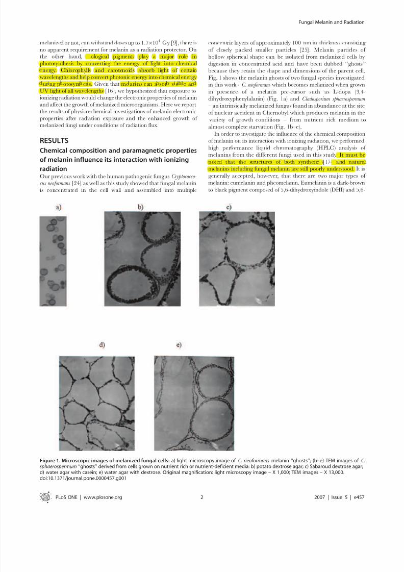
Fig. 1 shows the melanin ghosts of two fungal species investigated
in this work - C. neoformans which becomes melanized when grown
in presence of a melanin pre-cursor such as L-dopa (3,4-
dihydroxyphenylalanin) (Fig. 1a) and Cladosporium sphaerospermum – an intrinsically melanized fungus found in abundance at the site
of nuclear accident in Chernobyl which produces melanin in the
variety of growth conditions – from nutrient rich medium to
almost complete starvation (Fig. 1b–e).
In order to investigate the influence of the chemical composition
of melanin on its interaction with ionizing radiation, we performed
high performance liquid chromatography (HPLC) analysis of
melanins from the different fungi used in this study. It must be
noted that the structures of both synthetic [17] and natural
melanins including fungal melanin are still poorly understood. It is
generally accepted, however, that there are two major types of
melanin: eumelanin and pheomelanin. Eumelanin is a dark-brown
to black pigment composed of 5,6-dihydroxyindole (DHI) and 5,6-
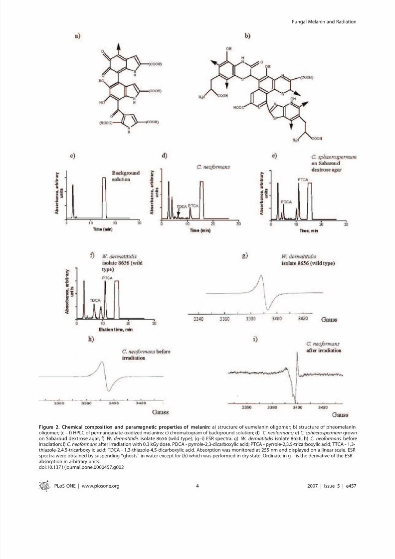
Figure 1. Microscopic images of melanized fungal cells: a) light microscopy image of C. neoformans melanin ‘‘ghosts’’; (b–e) TEM images of C.sphaerospermum ‘‘ghosts’’ derived from cells grown on nutrient rich or nutrient-deficient media: b) potato dextrose agar; c) Sabaroud dextrose agar;d) water agar with casein; e) water agar with dextrose. Original magnification: light microscopy image – X 1,000; TEM images – X 13,000.doi:10.1371/journal.pone.0000457.g001
Fungal Melanin and Radiation
PLoS ONE | www.plosone.org 2 2007 | Issue 5 | e457
biological pigments play a major role in
photosynthesis by converting the energy of light into chemical
nergy. Chlorophylls and carotenoids absorb light of certain
wavelengths and help convert photonic energy into chemical energy
ur ng p otosynt es s.
It must be
noted that the structures of both synthetic and natural
melanins including fungal melanin are still poorly understood.
me an ns can a sor v s e an
UV light of all wavelengths

8/6/2019 Radiation & Growth of Melanized Fungi
http://slidepdf.com/reader/full/radiation-growth-of-melanized-fungi 3/13
dihydroxyindole-2-carboxylic acid (DHICA) monomer units with
6–9% nitrogen and 0–1% sulfur [18,19] (Fig. 2a). In contrast,
pheomelanin is a reddish-brown pigment composed of benzothia-
zine monomer units with 8–11% nitrogen and 9–12% sulfur
[18,19] (Fig. 2b). When subjected to acidic permanganate
oxidation, DHI converts into pyrrole-2,3-dicarboxylic acid
(PDCA); DHICA - into pyrrole-2,3,5-tricarboxylic acid (PTCA);
and pheomelanin oxidation results in 1,3-thiazole-2,4,5-tricarbox-
ylic acid (TTCA) and 1,3-thiazole-4,5-dicarboxylic acid (TDCA)[18,19]. We have previously shown that permanganate-oxidized
melanin from C. neoformans is amenable to HPLC analysis [20,21].
In this study we performed semi-quantitative assessment of the
number of structural subunits of C. neoformans melanins. The HPLC
of oxidized C. neoformans melanin revealed PTCA and TDCA peaks
(Fig. 2d) and the presence of these compounds was confirmed by
matrix assisted laser desorption/ionization time of flight mass
spectrometry (MALDI-TOF). The ratio of PTCA to TDCA was
47.7, which indicates that DHICA subunits predominate in C.
neoformans melanin. Melanins produced by two different intrinsi-
cally melanized fungi Cladosporium sphaerospermum (Fig. 2e and Fig. 3)
and by Wangiella dermatitidis (Fig. 2f) were chemically more diverse
than C. neoformans melanin, revealing also the peaks assigned to
PDCA and peaks at 9–10 min which may be attributed to the small
amounts of oxidized 1,8-dihydroxynaphthalene (DHN) melanin.
Electron spin resonance spectroscopy (ESR) of melanized fungi
showed the presence a stable free radical population (Fig. 2g–i and
Fig. 4) in each of the above fungi, a distinguishing characteristic of
melanin [26]. One important indication of melanin interaction
with ionizing radiation was a large change in ESR signal of C.
neoformans dry melanin ‘‘ghosts’’ after they were subjected to
0.3 kGy irradiation and subsequently suspended in water (Fig. 2h
and i, respectively).
Exposure to ionizing radiation and other forms of
electromagnetic radiation increases electron
transfer properties of melanin
To quantify the effects of ionizing radiation and other forms of electromagnetic radiation on the electron transfer properties of
melanin – we irradiated dry C. neoformans melanin for 20 and 40 min
with 14 Gy/min from a 137Cs source and measured its electron
transfer properties in the coupled oxidation of NADH and reduction
of ferricyanide. In this system, melanin acts as an electron-transfer
agent [27], however, the effects of electromagnetic radiation on
melanin electron-transfer properties are unknown. Irradiation of
melanin for 20 min increased the velocity of the NADH/
ferricyanide coupled reaction 3-fold in comparison to that measured
for non-irradiated melanin, while 40 min irradiation had an even
larger effect, causing a 4-fold increase in velocity (Table 1). When we
investigated the influence of other, non-ionizing forms of radiation
across the electromagnetic spectrum - heat (infrared radiation),
visible light and UV light on the electron-transfer properties of
melanin in NADH/ferricyanide coupled reaction – we found that
each of these types of radiation increased the ability of melanin to
transfer electrons (Table 2). Interestingly, the increase in the
electron-transfer properties of melanin was independent of the
energy of the incident photons (Table 2).
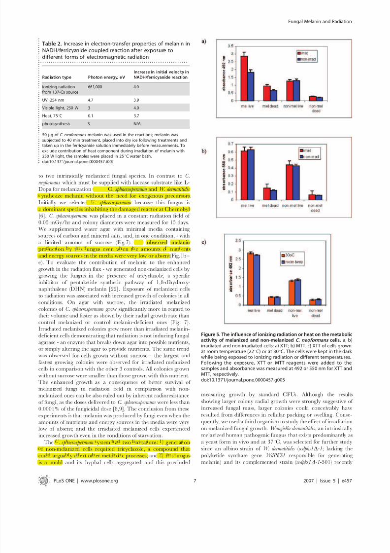
Metabolic activity of melanized and non-melanized
cells in the presence of electromagnetic radiationWe investigated whether the changes in electron transfer
properties of melanin post exposure to ionizing radiation (high-
energy photons, see Table 2) may also be observed in melanized
cells exposed to ionizing radiation. The metabolic activity of C.
neoformans cells was evaluated with 2,3-bis(2-methoxy-4-nitro-5-
sulfophenyl)-5-[(phenylamino) carbonyl]-2H-tetrazolium hydrox-
ide (XTT assay) [28] and 2-(4,5-dimethyl-2-thiazolyl)-3,5-diphe-
nyl-2H-tetrazolium bromide (MTT assay). The use of XTT and
MTT assays in parallel can help to define the location of the
melanin-mediated electron transfer in the cells since positively
charged MTT is taken into the cells via the plasma membrane
potential and is reduced intracellularly; while the negativelycharged XTT is largely cell-impermeable and its reduction occurs
extracellularly, at the cell surface [29]. The melanized and non-
melanized C. neoformans cells were exposed to ionizing radiation in
the dark at 22uC overnight. The irradiation was performed in
a constant field of 0.05 mGy/hr, a non-fungicidal radiation dose
that is comparable to the doses inside the Chernobyl reactor [6].
Following exposure to radiation, the XTT or MTT reagents were
added to the samples and absorbance was measured at 492 or
550 nm, respectively. The XTT assay showed significant increase
in electron-transfer events in the irradiated melanized cells in
comparison with non-irradiated melanized or irradiated non-
melanized cells (Fig. 5a). Increased absorbance at 492 nm was also
observed for dead (heat killed) melanized cells in comparison to
non-melanized ones, showing that melanin can reduce XTT
reagent by itself (Fig 5a). Irradiation of dead cells caused significantincrease in the XTT reduction, thus confirming our hypothesis
that radiation enhances electron-transfer properties of melanin. In
contrast, there was no difference between the irradiated and non-
irradiated melanized and non-melanized cells subjected to MTT
assay (Fig. 5b). The difference between the MTT and XTT assays
may be explained by the occurrence of radiation-related melanin-
mediated electron transfer events near cell wall where melanin is
located that led to higher XTT reduction in irradiated melanized
samples. Interestingly, both irradiated and non-irradiated mela-
nized cells showed higher activity by MTT assay than non-
melanized cells (Fig. 5a, b). Given that melanization is associated
with reduced pore size that could reduce passive nutrient uptake
[25] and that melanin is synthesized from highly reactive cytotoxic
intermediates of the oxidation of L-Dopa - it is possible thatmelanization requires a higher metabolism for cell survival.
In another series of experiments, the melanized and non-
melanized cells were grown overnight in the dark at room
temperature (22uC) or 30uC. Melanized cells demonstrated in-
creased XTT reduction activity at both temperatures in comparison
with non-melanized controls (Fig. 5c), and increasing the temper-
ature to 30uC caused a statistically significant increase in XTT
reduction in melanized cells (P,0.05) while a small decrease was
observed for non-melanized cells. Overall, these experiments showed
the increase in electron transfer properties of melanin in melanized
cells post exposure to ionizing radiation and to less extent - to heat.
Ionizing radiation enhances growth and 14C-acetate
uptake of melanized C. neoformans cellsTo expand the observations of the influence of irradiated melanin
on the growth of melanized cells, we measured the growth of
melanized and non-melanized C. neoformans cells supplied with
limited nutrients and placed into the radiation flux. To maintain
a steady population of melanized cells, we used the same H99
strain of C. neoformans as in XTT/MTT experiments since it is
capable of making melanin when maintained with L-Dopa while
its laccase disrupted [Lac(-)] mutant is incapable of melanization
[30]. The cells were grown into stationary phase up to 30 hr.
There were significantly more (P = 0.006) CFUs for irradiated
melanized wild type H99 samples at 18, 23 and 30 hr than for
Fungal Melanin and Radiation
PLoS ONE | www.plosone.org 3 2007 | Issue 5 | e457
8/6/2019 Radiation & Growth of Melanized Fungi
http://slidepdf.com/reader/full/radiation-growth-of-melanized-fungi 4/13
Figure 2. Chemical composition and paramagnetic properties of melanin: a) structure of eumelanin oligomer; b) structure of pheomelaninoligomer; (c – f) HPLC of permanganate-oxidized melanins: c) chromatogram of background solution; d) C. neoformans; e) C. sphaerospermum grownon Sabaroud dextrose agar; f) W. dermatitidis isolate 8656 (wild type); (g–i) ESR spectra: g) W. dermatitidis isolate 8656; h) C. neoformans beforeirradiation; i) C. neoformans after irradiation with 0.3 kGy dose. PDCA - pyrrole-2,3-dicarboxylic acid; PTCA - pyrrole-2,3,5-tricarboxylic acid; TTCA - 1,3-thiazole-2,4,5-tricarboxylic acid; TDCA - 1,3-thiazole-4,5-dicarboxylic acid. Absorption was monitored at 255 nm and displayed on a linear scale. ESRspectra were obtained by suspending ‘‘ghosts’’ in water except for (h) which was performed in dry state. Ordinate in g–i is the derivative of the ESRabsorption in arbitrary units.doi:10.1371/journal.pone.0000457.g002
Fungal Melanin and Radiation
PLoS ONE | www.plosone.org 4 2007 | Issue 5 | e457

8/6/2019 Radiation & Growth of Melanized Fungi
http://slidepdf.com/reader/full/radiation-growth-of-melanized-fungi 5/13
non-irradiated samples (Fig. 6a), while the difference in CFUs at
18 hr between irradiated and non-irradiated Lac(-) mutant was
not significant (Fig. 6b). Lac(-) without radiation in the presence of
L-dopa grows better than wild type H99 (Fig. 6a and b). There was
also a slight increase in the CFU’s of irradiated Lac(-) cells at 23
and 30 hr. However, the crucial difference between the wild type
H99 and Lac(-) cells is that the exposure to ionizing radiation
produced approximately 2.5 times more CFUs in irradiated
melanized cells than in non-irradiated melanized controls, whileirradiation of Lac(-) cells resulted only in a 1.1-fold increase in CFUs
(Fig. 6e). The dry weight measurements performed at 20 hr showed
a consistent and significant 6.5% increase for irradiated melanized
samples (P = 0.02) while there was no difference in weight for the
mutant strain after irradiation. The relatively small yet significant
increase in dry weight of the melanized cells is a result of the high
percentage of immature cells, with smaller capsules synthesized de novo in the dividing melanized irradiated cell culture. In this regard,
a cell diameter that is one-half to one-third of that for a mature cell
results in a 8- and 26-fold decrease in cell mass, respectively.
Quantification of whole cell sizes using India ink stained cells showed
that proximately 50% of melanized irradiated cells had volumes 2
times smaller than those in the irradiated Lac(-) mutant population
(results not shown), accounting for the relatively small increase in the
dry weight of the melanized H99 samples in comparison to their
larger increase in CFUs.
To obtain additional evidence that exposure to ionizing
radiation enhanced melanized cell growth, we measured the
incorporation of a 14C-labeled carbon source (acetate) into
melanized and non-melanized C. neoformans cells with and withoutradiation flux. In the photosynthesis field the incorporation of 14C-
acetate in bacteria subjected to visible light is considered to be
indicative of their photoheterotrophic capabilities [31]. We
measured a lower absolute uptake of 14C-acetate by wild typeH99 compared to Lac(-) cells (Fig. 6c,d). There was no
incorporation of 14C-acetate into heat killed melanized or non-
melanized cells, which excludes the possibility that radiation
promoted the passive absorption of 14C-acetate on melanin.
Importantly, when melanized and non-melanized Lac(-) H99 cells
were incubated with 14C-acetate with and without radiation –
there was almost 3 times more incorporation of 14C-acetate into
Figure 3. HPLC of melanin derived from C. sphaerospermum grown on different substrates: a) potato dextrose agar; b) Sabaroud dextrose agar;c) water agar with casein; d) water agar with dextrose. PDCA - pyrrole-2,3-dicarboxylic acid; PTCA - pyrrole-2,3,5-tricarboxylic acid; TTCA - 1,3-thiazole-2,4,5-tricarboxylic acid; TDCA - 1,3-thiazole-4,5-dicarboxylic acid. Absorption was monitored at 255 nm and displayed on a linear scale. Cs - C.sphaerospermum.doi:10.1371/journal.pone.0000457.g003
Fungal Melanin and Radiation
PLoS ONE | www.plosone.org 5 2007 | Issue 5 | e457
8/6/2019 Radiation & Growth of Melanized Fungi
http://slidepdf.com/reader/full/radiation-growth-of-melanized-fungi 6/13
irradiated melanized cells than into non-irradiated melanized cells,
while the ratio of 14C-acetate incorporation into irradiated to non-
irradiated Lac(-) cells was only slightly higher than 1 (Fig. 6c,d and
e). Overall, these results demonstrate that the presence of melanin
contributes to the enhancement of cellular growth upon exposure
to ionizing radiation in conditions of limited nutrients.
Intrinsically melanized fungi C. sphaerospermum
and W. dermatitidis manifested enhanced growth in
radiation fluxTo accrue additional data that ionizing radiation can promote
enhanced growth of melanized fungi we extended our observations
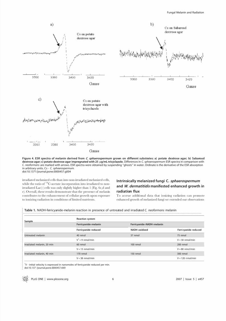
Figure 4. ESR spectra of melanin derived from C. sphaerospermum grown on different substrates: a) potato dextrose agar; b) Sabarouddextrose agar; c) potato dextrose agar impregnated with 25 mg/mL tricyclazole. Differences in C. sphaerospermum ESR spectra in comparison withC. neoformans are marked with arrows. ESR spectra were obtained by suspending ‘‘ghosts’’ in water. Ordinate is the derivative of the ESR absorption
in arbitrary units. Cs - C. sphaerospermum.doi:10.1371/journal.pone.0000457.g004
Table 1. NADH-ferricyanide-melanin reaction in presence of untreated and irradiated C. neoformans melanin. . . . . . . . . . . . . . . . . . . . . . . . . . . . . . . . . . . . . . . . . . . . . . . . . . . . . . . . . . . . . . . . . . . . . . . . . . . . . . . . . . . . . . . . . . . . . . . . . . . . . . . . . . . . . . . . . . . . . . . . . . . . . . . . . . . . . . . . . . . . . . . . . .
SampleReaction system
Ferricyanide+melanin Ferricyanide+NADH+melanin
Ferricyanide reduced NADH oxidized Ferricyanide reduced
Untreated melanin 40 nmol 37 nmol 75 nmol
V1 = 9 nmol/min V = 30 nmol/min
Irradiated melanin, 20 min 60 nmol 100 nmol 200 nmol
V = 13 nmol/min V = 80 nmol/min
Irradiated melanin, 40 min 170 nmol 150 nmol 300 nmol
V = 38 nmol/min V = 120 nmol/min
1V - initial velocity is expressed in nanomoles of ferricyanide reduced per min.doi:10.1371/journal.pone.0000457.t001
.
.
.
.
.
.
.
.
.
.
.
.
.
.
.
.
.
.
.
.
.
.
.
.
.
.
.
.
.
.
.
.
.
. . .
.
.
.
.
.
.
.
.
.
.
.
.
.
.
Fungal Melanin and Radiation
PLoS ONE | www.plosone.org 6 2007 | Issue 5 | e457
8/6/2019 Radiation & Growth of Melanized Fungi
http://slidepdf.com/reader/full/radiation-growth-of-melanized-fungi 7/13
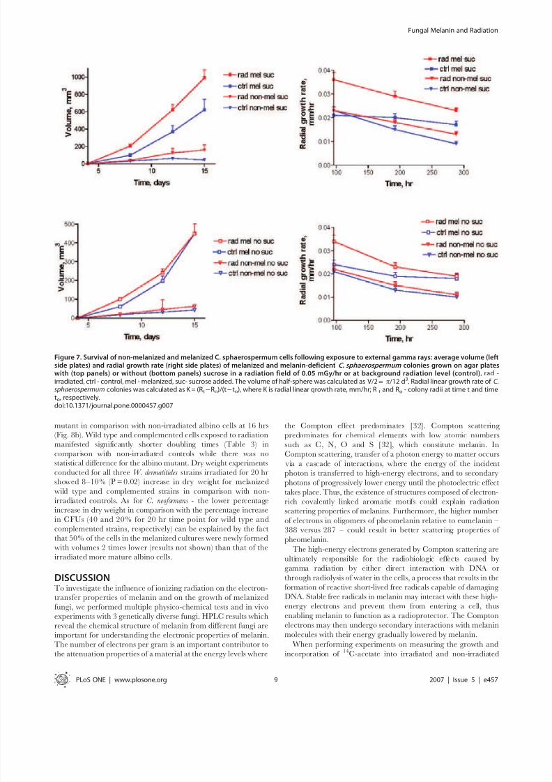
to two intrinsically melanized fungal species. In contrast to C.neoformans which must be supplied with laccase substrate like L-
Dopa for melanization - both C. sphaerospermum and W. dermatitidis
synthesize melanin without the need for exogenous precursors.Initially we selected C. sphaerospermum because this fungus is
a dominant species inhabiting the damaged reactor at Chernobyl
[6]. C. sphaerospermum was placed in a constant radiation field of
0.05 mGy/hr and colony diameters were measured for 15 days.
We supplemented water agar with minimal media containing
sources of carbon and mineral salts, and, in one condition, - with
a limited amount of sucrose (Fig.7). We observed melanin
production by this fungus even when the amounts of nutrients
and energy sources in the media were very low or absent (Fig.1b–
e). To evaluate the contribution of melanin to the enhanced
growth in the radiation flux - we generated non-melanized cells by
growing the fungus in the presence of tricyclazole, a specific
inhibitor of pentaketide synthetic pathway of 1,8-dihydroxy-
naphthalene (DHN) melanin [22]. Exposure of melanized cellsto radiation was associated with increased growth of colonies in all
conditions. On agar with sucrose, the irradiated melanized
colonies of C. sphaerospermum grew significantly more in regard to
their volume and faster as shown by their radial growth rate than
control melanized or control melanin-deficient ones (Fig. 7).
Irradiated melanized colonies grew more than irradiated melanin-
deficient cells demonstrating that radiation is not inducing fungal
agarase - an enzyme that breaks down agar into possible nutrients,
or simply altering the agar to provide nutrients. The same trend
was observed for cells grown without sucrose - the largest and
fastest growing colonies were observed for irradiated melanized
cells in comparison with the other 3 controls. All colonies grown
without sucrose were smaller than those grown with this nutrient.
The enhanced growth as a consequence of better survival of
melanized fungi in radiation field in comparison with non-melanized ones can be also ruled out by inherent radioresistance
of fungi, as the doses delivered to C. sphaerospermum were less than
0.0001% of the fungicidal dose [8,9]. The conclusion from these
experiments is that melanin was produced by fungi even when the
amounts of nutrients and energy sources in the media were very
low of absent; and the irradiated melanized cells experienced
increased growth even in the conditions of starvation.
The C. sphaerospermum system had two limitations: 1) generation
of non-melanized cells required tricyclazole, a compound that
could arguably affect other metabolic processes; and 2) this fungus
is a mold and its hyphal cells aggregated and this precluded
measuring growth by standard CFUs. Although the results
showing larger colony radial growth were strongly suggestive of
increased fungal mass, larger colonies could conceivably have
resulted from differences in cellular packing or swelling. Conse-
quently, we used a third organism to study the effect of irradiation
on melanized fungal growth. Wangiella dermatitidis , an intrinsically
melanized human pathogenic fungus that exists predominantly as
a yeast form in vivo and at 37uC, was selected for further study
since an albino strain of W. dermatitidis ( wdpks1D-1; lacking the
polyketide synthase gene WdPKS1 responsible for generating
melanin) and its complemented strain ( wdpks1D-1-501) recently
Table 2. Increase in electron-transfer properties of melanin inNADH/ferricyanide coupled reaction after exposure todifferent forms of electromagnetic radiation
. . . . . . . . . . . . . . . . . . . . . . . . . . . . . . . . . . . . . . . . . . . . . . . . . . . . . . . . . . . . . . . . . . . . . .
Radiation type Photon energy, eV
Increase in initial velocity in
NADH/ferricyanide reaction
Ionizing radiationfrom 137-Cs source
661,000 4.0
UV, 254 nm 4.7 3.9
Visible light, 250 W 3 4.0
Heat, 75uC 0.1 3.7
photosynthesis 3 N/A
50 mg of C. neoformans melanin was used in the reactions; melanin wassubjected to 40 min treatment, placed into dry ice following treatments andtaken up in the ferricyanide solution immediately before measurements. Toexclude contribution of heat component during irradiation of melanin with250 W light, the samples were placed in 25uC water bath.doi:10.1371/journal.pone.0000457.t002 .
.
.
.
.
.
.
.
.
.
.
.
.
.
.
.
.
.
.
.
.
.
.
.
.
.
.
.
. . . .
.
.
.
.
.
.
.
.
.
.
.
.
.
.
.
.
.
.
.
.
.
.
.
.
.
.
.
Figure 5. The influence of ionizing radiation or heat on the metabolicactivity of melanized and non-melanized C. neoformans cells. a, b)irradiated and non-irradiated cells: a) XTT; b) MTT. c) XTT of cells grownat room temperature (22uC) or at 30uC. The cells were kept in the dark while being exposed to ionizing radiation or different temperatures.Following the exposure, XTT or MTT reagents were added to thesamples and absorbance was measured at 492 or 550 nm for XTT andMTT, respectively.doi:10.1371/journal.pone.0000457.g005
Fungal Melanin and Radiation
PLoS ONE | www.plosone.org 7 2007 | Issue 5 | e457
both C. and W. dermatitidis sphaerospermum
ynthesize melanin without the need for exogenous precursors.. sphaerospermum
dominant species inhabiting the damaged reactor at Chernobyla
We observed melanin
pro uct on y t s ungus even w en t e amounts o nutr ents
and energy sources in the media were very low or absent –
. sp aerospermum ystem a two m tat ons: generat on
f non-melanized cells required tricyclazole, a compound that
cou argua y a e ct ot er meta o c processes; t s ungus
is a mold

8/6/2019 Radiation & Growth of Melanized Fungi
http://slidepdf.com/reader/full/radiation-growth-of-melanized-fungi 8/13
became available [23]. This fungus, which also produces fewer
multicellular forms in vitro in comparison with C. sphaerospermum,
allowed us to quantify the effect of radiation on cell growth by
CFUs instead of measuring colony diameters.
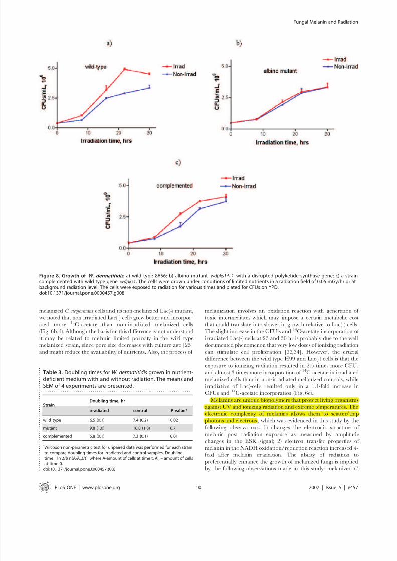
Exposure of W. dermatitidis cells to ionizing radiation resulted in
significantly more cells being produced, as measured by CFUs, for
the melanized strains (P,0.01) than for the non-irradiated
melanized control cells or the irradiated wdpks1D-1 albino mutant
strain (Fig. 8a–c). At 16, 22 and 30 hrs of irradiation the two
melanin-containing strains of W. dermatitides had more colonies
than the amelanotic mutant strain. As in the case of C. neoformans,
some increased growth was observed in the irradiated albino
Figure 6. Growth and incorporation of
14
C-acetate by melanized C. neoformans H99 cells and non-melanized Lac(-) H99 cells lacking the laccaseenzyme under conditions of limited nutrients supply in a radiation field of 0.05 mGy/hr or at background radiation level. a) growth of melanizedH99 cells; b) growth of non-melanized Lac(-) H99 cells; c) incorporation of 14C-acetate into melanized H99 cells; d) incorporation of 14C-acetate intonon-melanized Lac(-) H99 cells; e) ratio of irradiated to non-irradiated cells CFUs and cpms ratios (normalized CFUs and cpms) for melanized H99 andnon-melanized Lac(-) H99 cells.doi:10.1371/journal.pone.0000457.g006
Fungal Melanin and Radiation
PLoS ONE | www.plosone.org 8 2007 | Issue 5 | e457
8/6/2019 Radiation & Growth of Melanized Fungi
http://slidepdf.com/reader/full/radiation-growth-of-melanized-fungi 9/13
mutant in comparison with non-irradiated albino cells at 16 hrs
(Fig. 8b). Wild type and complemented cells exposed to radiation
manifested significantly shorter doubling times (Table 3) in
comparison with non-irradiated controls while there was no
statistical difference for the albino mutant. Dry weight experiments
conducted for all three W. dermatitides strains irradiated for 20 hr
showed 8–10% (P = 0.02) increase in dry weight for melanized
wild type and complemented strains in comparison with non-
irradiated controls. As for C. neoformans - the lower percentage
increase in dry weight in comparison with the percentage increase
in CFUs (40 and 20% for 20 hr time point for wild type and
complemented strains, respectively) can be explained by the fact
that 50% of the cells in the melanized cultures were newly formed
with volumes 2 times lower (results not shown) than that of theirradiated more mature albino cells.
DISCUSSIONTo investigate the influence of ionizing radiation on the electron-transfer properties of melanin and on the growth of melanized
fungi, we performed multiple physico-chemical tests and in vivo
experiments with 3 genetically diverse fungi. HPLC results which
reveal the chemical structure of melanin from different fungi are
important for understanding the electronic properties of melanin.
The number of electrons per gram is an important contributor to
the attenuation properties of a material at the energy levels where
the Compton effect predominates [32]. Compton scattering
predominates for chemical elements with low atomic numbers
such as C, N, O and S [32], which constitute melanin. In
Compton scattering, transfer of a photon energy to matter occurs
via a cascade of interactions, where the energy of the incident
photon is transferred to high-energy electrons, and to secondary
photons of progressively lower energy until the photoelectric effect
takes place. Thus, the existence of structures composed of electron-
rich covalently linked aromatic motifs could explain radiation
scattering properties of melanins. Furthermore, the higher number
of electrons in oligomers of pheomelanin relative to eumelanin –
388 versus 287 – could result in better scattering properties of
pheomelanin.
The high-energy electrons generated by Compton scattering areultimately responsible for the radiobiologic effects caused by
gamma radiation by either direct interaction with DNA or
through radiolysis of water in the cells, a process that results in the
formation of reactive short-lived free radicals capable of damaging
DNA. Stable free radicals in melanin may interact with these high-
energy electrons and prevent them from entering a cell, thus
enabling melanin to function as a radioprotector. The Compton
electrons may then undergo secondary interactions with melanin
molecules with their energy gradually lowered by melanin.
When performing experiments on measuring the growth and
incorporation of 14C-acetate into irradiated and non-irradiated
Figure 7. Survival of non-melanized and melanized C. sphaerospermum cells following exposure to external gamma rays: average volume (leftside plates) and radial growth rate (right side plates) of melanized and melanin-deficient C. sphaerospermum colonies grown on agar plateswith (top panels) or without (bottom panels) sucrose in a radiation field of 0.05 mGy/hr or at background radiation level (control). rad -irradiated, ctrl - control, mel - melanized, suc- sucrose added. The volume of half-sphere was calculated as V/2 = p/12 d3. Radial linear growth rate of C.sphaerospermum colonies was calculated as K = (Rt2Ro)/(t2to), where K is radial linear growth rate, mm/hr; R t and Ro - colony radii at time t and timeto, respectively.doi:10.1371/journal.pone.0000457.g007
Fungal Melanin and Radiation
PLoS ONE | www.plosone.org 9 2007 | Issue 5 | e457
8/6/2019 Radiation & Growth of Melanized Fungi
http://slidepdf.com/reader/full/radiation-growth-of-melanized-fungi 10/13
melanized C. neoformans cells and its non-melanized Lac(-) mutant,
we noted that non-irradiated Lac(-) cells grew better and incorpor-
ated more 14C-acetate than non-irradiated melanized cells
(Fig. 6b,d). Although the basis for this difference is not understood
it may be related to melanin limited porosity in the wild type
melanized strain, since pore size decreases with culture age [25]
and might reduce the availability of nutrients. Also, the process of
melanization involves an oxidation reaction with generation of
toxic intermediates which may impose a certain metabolic cost
that could translate into slower in growth relative to Lac(-) cells.
The slight increase in the CFU’s and 14C-acetate incorporation of
irradiated Lac(-) cells at 23 and 30 hr is probably due to the well
documented phenomenon that very low doses of ionizing radiation
can stimulate cell proliferation [33,34]. However, the crucial
difference between the wild type H99 and Lac(-) cells is that the
exposure to ionizing radiation resulted in 2.5 times more CFUs
and almost 3 times more incorporation of 14C-acetate in irradiated
melanized cells than in non-irradiated melanized controls, while
irradiation of Lac(-cells resulted only in a 1.1-fold increase in
CFUs and14
C-acetate incorporation (Fig. 6e).Melanins are unique biopolymers that protect living organisms
against UV and ionizing radiation and extreme temperatures. The
electronic complexity of melanins allows them to scatter/trap
photons and electrons, which was evidenced in this study by the
following observations: 1) changes the electronic structure of
melanin post radiation exposure as measured by amplitude
changes in the ESR signal; 2) electron transfer properties of
melanin in the NADH oxidation/reduction reaction increased 4-
fold after melanin irradiation. The ability of radiation to
preferentially enhance the growth of melanized fungi is implied
by the following observations made in this study: melanized C.
Figure 8. Growth of W. dermatitidis : a) wild type 8656; b) albino mutant wdpks1D-1 with a disrupted polyketide synthase gene; c) a straincomplemented with wild type gene wdpks1. The cells were grown under conditions of limited nutrients in a radiation field of 0.05 mGy/hr or atbackground radiation level. The cells were exposed to radiation for various times and plated for CFUs on YPD.
doi:10.1371/journal.pone.0000457.g008
Table 3. Doubling times for W. dermatitidis grown in nutrient-deficient medium with and without radiation. The means andSEM of 4 experiments are presented.
. . . . . . . . . . . . . . . . . . . . . . . . . . . . . . . . . . . . . . . . . . . . . . . . . . . . . . . . . . . . . . . . . . . . . .
StrainDoubling time, hr
irradiated control P value*
wild type 6.5 (0.1) 7.4 (0.2) 0.02
mutant 9.8 (1.0) 10.8 (1.8) 0.7
complemented 6.8 (0.1) 7.3 (0.1) 0.01
* Wilcoxon non-parametric test for unpaired data was performed for each strainto compare doubling times for irradiated and control samples. Doublingtime= ln 2/((ln(A/Ao)/t), where A-amount of cells at time t, Ao – amount of cellsat time 0.
doi:10.1371/journal.pone.0000457.t003 .
.
.
.
.
.
.
.
.
.
.
.
.
.
.
.
.
.
.
.
.
.
.
.
.
.
.
.
.
.
.
.
.
. . .
.
.
.
.
.
.
.
.
.
.
.
.
Fungal Melanin and Radiation
PLoS ONE | www.plosone.org 10 2007 | Issue 5 | e457
Melanins are unique biopolymers that protect living organisms
against UV and ionizing radiation and extreme temperatures. The
electronic complexity of melanins allows them to scatter/trap
photons and electrons,
8/6/2019 Radiation & Growth of Melanized Fungi
http://slidepdf.com/reader/full/radiation-growth-of-melanized-fungi 11/13
neoformans and W. dermatitidis cells exposed to levels of radiation
approximately 500 times higher than background grew signifi-
cantly faster as indicated by the presence of more CFUs, greater
biomass as shown by dry weight measurements and/or relative
incorporation of more 14C-acetate than non-irradiated melanized
cells. Furthermore, comparative analysis of MTT/XTT reduction
assays revealed that radiation-induced effects on the electron
transfer properties of melanin were localized to the extracellular
space thus establishing a spatial relationship between the site forelectron-transfer events and the location of the melanin pigment.
In addition, we recorded radiation-induced effects on the growth
of melanized C. sphaerospermum cells under limited nutrients
conditions. Hence, we observed that radiation increased the
growth of melanized cells relative to non-melanized cells using
three fungal species and four measures of cell growth.
The literature already contains some indirect evidence for the
notion that radiation can enhance the growth of melanized
microorganisms. For example, the melanotic fungus C. cladosporioides
manifests radiotropism by growing in the direction of radioactive
particles and this organism has become widely distributed in the
areas surrounding Chernobyl since the nuclear accident in 1986 [7].
Both in the laboratory and in the field several other species of
melanized fungi grew towards soil particles contaminated with
different radionuclides, gradually engulfing and destroying thoseparticles [35,36]. In addition, there are recent reports that certain life
forms can utilize non-conventional forms of energy - microbes in
geothermal vents at the bottom of the ocean can harvest thermal
radiation as an energy source [37] while some microorganisms living
in mines exploit energy from radiolysis of water [38]. On the basis of
these precedents and the results of this study we cautiously suggest
that the ability of melanin to capture electromagnetic radiation
combined with its remarkable oxidation-reduction properties may
confer upon melanotic organisms the ability to harness radiation for
metabolic energy. The enhanced growth of melanotic fungi in
conditions of radiation fluxes suggests the need for additional
investigation to ascertain the mechanism for this effect.
METHODSMicroorganisms American Type Culture Collection (ATCC, Rockville, MD) strain
C. neoformans H99 and its laccase lacking mutant Lac(-) (kind gift
from Dr. A. Idnurm, Duke University, NC) were used in all
experiments. C. neoformans was grown in Sabouraud dextrose broth
(Difco laboratories, Detroit, MI) for 24 hrs at 30uC with constant
shaking at 150 rpm. Melanized C. neoformans cells were generated
by growing the fungus in minimal medium with 1 mM 3,4-
dihydroxyphenylalanine (L-dopa) for 5–10 days. C. sphaerospermum
(ATCC, VA), an intrinsically melanized fungus, was grown on
potato dextrose agar (Becton, Dickinson and Company) for
15 days. Approximately 1,000 C. sphaerospermum cells were plated
on different media: water agar impregnated with minimal media
(no glucose) and casein; agar dissolved in minimal media (noglucose) and 40 g/L dextrose; potato dextrose agar (Becton,
Dickinson and Company); potato dextrose agar impregnated with
25 ug/mL tricyclazole; and Sabaroud dextrose agar. The
laboratory wild-type strain of intrinsically melanized
W. dermatitidis 8656 {ATCC 34100 [ Exophiala dermatitidis CBS
525.76]} a strain with a disrupted polyketide synthase gene
wdpks1D-1, and its complemented isolate ( wdpks1D-1-501) were
a kind gift from Dr. P. Szaniszlo (The University of Texas at
Austin, Austin, TX). Routine propagation of these strains was in
YPD [2% peptone, 1% Bacto Yeast extract, and 2% dextrose] at
37uC with shaking at 150 rpm.
Isolation and purification of fungal melaninsFungal cells were suspended in 1.0 M sorbitol-0.1 M sodium
citrate (pH 5.5). Lysing enzymes from Trichoderma harzarium (Sigma
Chemical Co.) were added to the suspension at 10 mg/mL and
the suspensions were incubated overnight at 30uC. Protoplasts
were collected by centrifugation, and incubated in 4.0 M
guanidine thiocyanate overnight at room temperature. The
resulting particulate material was collected by centrifugation,
and Proteinase K (1.0 mg/mL) in reaction buffer (10.0 mM tris,1.0 mM CaCl2, 0.5% SDS) was added to the particles followed by
overnight incubation at 37uC. The particles were boiled in 6.0 M
HCl for 1 hour. Finally, the resulting material (‘‘ghosts’’) was
washed with PBS, dialyzed against deionized water for 2 days and
dried in air at 65uC overnight.
Transmission electron microscopy (TEM)Samples were processed at the Analytical Imaging Facility,
AECOM. The C. neoformans, C. sphaerospermum and W. dermatitidis ‘‘ghosts’’ or cells were frozen under high pressure using a Leica
EMpact High Pressure Freezer (Leica Microsystems, Austria).
Frozen samples were transferred to a Leica EM AFS Freeze
Substitution Unit and freeze substituted in 1% osmium tetroxide
in acetone. They were brought from 290u
C to room temperatureover 2–3 days, rinsed in acetone and embedded in Spurrs epoxy
resin (Polysciences,Warrington, PA.). Ultrathin sections of 70–
80 nm were cut on a Reichert Ultracut UCT, stained with uranyl
acetate followed by lead citrate and viewed on a JEOL (Tokyo,
Japan) 1200EX transmission electron microscope at 80 kV.
Oxidation of melanins and HPLC of oxidized
melaninsThe melanin ‘‘ghosts’’ were subjected to acidic permanganate
oxidation as described in [18–21]. The pyrrole-2,3,5-tricarboxylic
acid (PTCA), pyrrole-2,3-dicarboxylic acid (PDCA), 1,3-thiazole-
2,4,5-tricarboxylic acid (TTCA) and 1,3-thiazole-4,5-dicarboxylic
acid (TDCA) used as standards were a kind gift from Dr. K.
Wakamatsu of Fujita Health University of the Health Sciences,Toyoake, Japan. The oxidation products were analyzed by HPLC
using a Shimadzu LC-600 liquid chromatograph, Hamilton PRP-
1 C18 column (25064.1 mm dimensions, 7 mm particle size), and
Shimadzu SPD-6AV UV detector. The mobile phase was 0.1%
trifluoroacetic acid in water (solvent A) and 0.1% trifluoroacetic
acid in acetonitrile (solvent B). At 1.0 mL/min, the elution
gradient was (min, %B): 0, 0; 1, 0; 12, 25; 14, 25; 16, 0. The UV
detector was set at a 255 nm absorbance.
MALDI mass spectrometryThe major peaks generated during chromatography of oxidized
melanins were collected and analyzed by MALDI-TOF mass
spectrometry in positive pressure mode on PE-Biosystems Mariner
mass spectrometer. A peptide mixture with molecular weights of 1059.56, 1296.68 and 1672.95 in 2,5-dihydroxybenzoic acid
matrix was used for calibration.
Electron spin resonance spectroscopy (ESR)The ESR of melanin ‘‘ghosts’’ was performed on ER 200D EPR/
ENDOR spectrometer with ESP 300 upgrade (Brucker Instru-
ments, Inc. Billerica, MA). ESR spectra were obtained by
suspending ‘‘ghosts’’ in water. ESR spectra of C. neoformans
‘‘ghosts’’ were also obtained in dry state before irradiation with
0.3 kGy and the ‘‘ghosts’’ were subsequently suspended in water
and ESR was repeated.
Fungal Melanin and Radiation
PLoS ONE | www.plosone.org 11 2007 | Issue 5 | e457
the melanotic fungus . cladosporioides
manifests radiotropism by growing in the direction of radioactive
part c es an t s organ sm as ecome w e y str ute n t e
areas surrounding Chernobyl since the nuclear accident in 1986
ot n t e a oratory an n t e e severa ot er spec es o
melanized fungi grew towards soil particles contaminated with
different radionuclides, gradually engulfing and destroying thoseparticles

8/6/2019 Radiation & Growth of Melanized Fungi
http://slidepdf.com/reader/full/radiation-growth-of-melanized-fungi 12/13
NADH-ferricyanide reaction in the presence of
untreated and irradiated C. neoformans melaninThe ability of melanin to oxidize or reduce NADH and
ferricyanide was determined spectrophotometrically as in [27].
The absorbance of NADH was monitored at 340 nm, of
ferricyanide - at 420 nm. Fifty mg of C. neoformans melanin was
used in the reactions; dry melanin was subjected to 20 and 40 min
irradiation with the 137-Cs source at a dose rate of 14 Gy/min;
put into dry ice following irradiation and taken up in the
ferricyanide solution immediately before measurements.
Determination of metabolic activity of melanized
and non-melanized C. neoformans cells subjected to
ionizing radiation or different temperatures by XTT
and MTT assaysMelanized and non-melanized C. neoformans cells were washed,
suspended in PBS and their concentration was adjusted to 108 per
mL. To account for the possibility of melanin itself changing the
reaction through electron transfer or solubility/retention of
formazan product [28] - the aliquots of both melanized and
non-melanized cells were heat-killed at 65uC (water bath) for one
hour and used as controls. 107 cells were placed into the wells in 96well plates, 5 wells per each condition. The plates were covered
with foil to exclude any light effects and incubated overnight at
room temperature (22uC), at 30uC, or at 22uC in a constant
radiation field of 0.05 mGy/hr. For XTT (2,3-bis(2-methoxy-4-
nitro-5-sulfophenyl)-5-[(phenylamino) carbonyl]-2H-tetrazolium
hydroxide) assay 54 mL (XTT)/menadinone was added to each
well, the plates were covered with foil, shaken for 2 minutes, and
incubated at 37uC for 2 hrs. The absorbance was read at 492 nm
(Labsystem Multiskan, Franklin, MA). For MTT (2-(4,5-dimethyl-
2-thiazolyl)-3,5-diphenyl-2H-tetrazolium bromide) assay, the
MTT solution in PBS was added to the wells with the cells, so
that the final MTT concentration became 0.5 mg/mL. After
incubation at 37uC, the contents of the wells was spun down at
2,000 rpm, supernatant was discarded, followed by addition of 200 ml 0.04 M HCl in absolute isopropanol to each sample. The
samples were transferred into the 96-well plate and the absorbance
was read at 550 nm.
Exposure of C. neoformans to ionizing radiation
under limited nutrient conditions, 14C-acetate
incorporation and dry weight measurementsH99 wild type and Lac(-) mutant cells were grown as above.
Melanization of H99 was achieved by incubation in 1mM L-
Dopa/minimal medium (1/200) in the dark at 30uC, at 150 rpm.
The cells were washed with essential salts solution (3 g/L NaNO3,
1 g/L K2HPO4, 1 g/L MgSO4.7H2O, 0.5 g/L KCl, 0.003 g/L
thiamine, 5.3 g/L NH4Cl), pelleted and taken up in 1 mM Na
acetate solution in essential salts spiked with 0.1 mCi/mL 14C-acetate. The cell concentration was adjusted to 105 cells/ml, 1 mL
samples of each strain were placed in 1.5 mL Eppendorf tubes (4
samples per time point) and subjected either to the background
level of radiation or to a radiation field of 0.05 mGy/hr created by188Re/188W isotope generator for up to 30 hr at 30uC. The cell
uptake of 14C-acetate was quantified by counting the tubes in
a scintillation counter, spinning cells, separating supernatant and
counting the cell pellet again. The cells were also plated for CFUs.
For dry weight experiments 5 mL of cells at 46107 cells/mL cell
density were irradiated for 20 hr at 30uC, filtered through pre-
weighed 0.2 m filters, the filters were dried and weighed again.
Exposure of C. sphaerospermum to ionizing
radiation under limited nutrients conditionsMelanized and non-melanized (tricyclazole-treated) C. sphaerosper-
mum were grown on BBL Sabouraud dextrose agar for 15 days,
harvested and plated on water agar prepared by mixing agar
powder with minimal media lacking glucose. Four-sectional,
100615 mm dishes were used. Identical plates were made for
irradiated and control samples. Five mL of the above agar was
added to each of the left two sections of each plate. These sections
were called ‘‘no sucrose’’ sections. Five mL volumes of sucrose-
containing agar (prepared by mixing agar powder with 100 mg/L
sucrose as described in [6] and with minimal media lacking glucose)
was added to the right two sections of each plate. Non-melanized C.
sphaerospermum was added to the top compartments and melanized C.sphaerospermum was added to the bottom compartments of each plate.
Agar in the top compartments where non-melanized C. sphaeros- permum was plated contained 25 mg/mL tricyclazole to inhibit
melanization. Approximately 20 cells per section were plated. All
plates were prepared in duplicate. The plates were wrapped in foil
and exposed continuously to 0.05 mGy/hr created by 188Re/188W
isotope generator for 15 days while control plates were exposed only
to background radiation (1024 mGy/hr). The colonies were counted
and measured daily.
Exposure of W. dermatitidis to ionizing radiation
under limited nutrients conditions and dry weight
measurementsBefore radiation exposure, wild type, albino mutant and
complemented strains of W. dermatitidis were cultured in the
following manner using the modified procedure from [39]: frozen
cells were diluted and cultured in 20 mL YPD at 37uC with
shaking at 150 rpm for 48 hrs, then diluted in YPD to 10 6 cells/
mL and grown for another 48 hrs. At the end of the 2nd 48 hr
period both wild type and complemented strains developed dark
coloration while albino mutant cells were light yellow. The cells
were again diluted to 106 cells/mL and cultured for 24 hrs, and
this procedure was repeated once. Next, the cells were again
diluted and grown for 24 hours in minimal chemical media (3 g/L
NaNO3, 1 g/L K2HPO4, 1 g/L MgSO4.7H2O, 0.5 g/L KCl,
0.003 g/L thiamine, 5.3 g/L NH4Cl, pH of 6.5) supplemented
with 120 mg/L sucrose as a carbon source. The wild type and
complemented strains maintained their dark color and the albino
mutant remained light yellow. The cells were collected, washed,
and diluted to 56105 cells/mL in the minimal medium. One ml
aliquots in 1.5 ml microfuge tubes were placed at 37uC in the dark
without shaking, either in the cell incubator with the background
level of radiation or in a radiation field of 0.05 mGy/hr created by188Re/188W isotope generator. For each time point, triplicate
samples were used. After 16, 22 and 30 hrs of exposure the cells
were plated on YPD agar for 4 days at room temperature for
determination of CFUs. The cells entered stationary phase around30 hrs. For dry weight determinations, 20 mL of each strain in
essential salts solution supplemented with 120 mg/mL sucrose at
cell density of 107 cell/mL were irradiated for 20 hrs or kept at
background radiation level then filtered through pre-weighed
0.2 m filters that were dried and weighed again.
Statistical analysisWilcoxon non-parametric test for unpaired data was performed
to analyze the differences in CFUs and 14C-acetate uptake.
Differences were considered statistically significant when P values
were,0.05.
Fungal Melanin and Radiation
PLoS ONE | www.plosone.org 12 2007 | Issue 5 | e457

8/6/2019 Radiation & Growth of Melanized Fungi
http://slidepdf.com/reader/full/radiation-growth-of-melanized-fungi 13/13
ACKNOWLEDGMENTSThe authors thank Dr. P.J. Szaniszlo (University of Texas, TX) for the kind
gift of Wangiella dermatitidis and Dr. L. Day (New York University, NY) for
advice on light scattering.
Author Contributions
Conceived and designed the experiments: AC ED RB. Performed theexperiments: RB AS PA JN XH TM. Analyzed the data: AC ED RB XH.Contributed reagents/materials/analysis tools: PA JN. Wrote the paper:
AC ED. Other: Suggested the idea for the study: AC.
REFERENCES
1. Hill HZ (1992) The function of melanin or six blind people examine an elephant.
Bioessays 14: 49–56.
2. Jacobson ES (2000) Pathogenic roles for fungal melanins. Clin. Microbiol. Rev.
13: 708–717.
3. Steenbergen JN, Shuman HA, Casadevall A (2001) Cryptococcus neoformans
interactions with amoebae suggest an explanation for its virulence and
intracellular pathogenic strategy in macrophages. Proc. Natl. Acad. Sci. USA.
98: 15245–15250.
4. Nosanchuk JD, Casadevall A (2003) The contribution of melanin to microbial
pathogenesis. Cell. Microbiol. 5: 203–223.
5. Robinson CH (2001) Cold adaptation in Arctic and Antarctic fungi. New
phytologist 151: 341–353.
6. Wember VV, Zhdanova NN (2001) Peculiarities of linear growth of the melanin-
containing fungi Cladosporium sphaerospermum Penz. and Alternaria alternata
(Fr.) Keissler. Mikrobiol. Z. 63: 3–12.
7. Zhdanova NN, Tugay T, Dighton J, Zheltonozhsky V, McDermott P (2004)
Ionizing radiation attracts soil fungi. Mycol Res. 108: 1089–1096.
8. Mirchink TG, Kashkina GB, Abaturov ID (1972) Resistance of fungi with
different pigments to radiation. Mikrobiologiia 41: 83–86.9. Saleh YG, Mayo MS, Ahearn DG (1988) Resistance of some common fungi to
gamma irradiation. Appl. Environm. Microbiol. 54: 2134–2135.
10. Taylor TN, Hass H, Kerp H, Krings M, Hanlin RT (2005) Perithecial
ascomycetes from the 400 million year old Rhynie chert: an example of ancestral
polymorphism. Mycologia 97: 269–285.
11. Jansonius J, Kalgutkar RM (2000) Redescription of some fossil fungal spores.
Palynology 24: 37–47.
12. Hulot G, Gallet Y (2003) Do superchrons occur without any palaeomagnetic
warning? Earth Planetary Sci. Lett. 210: 191–201.
13. Davis M, Hut P, Muller RA (1985) Terrestrial catastrophism: Nemesis or
Galaxy? Nature 313: 503.
14. Casadevall A (2005) Fungal virulence, vertebrate endothermy, and dinosaur
extinction: is there a connection? Fungal Genet. Biol. 42: 98–106.
15. Redman RS, Sheehan KB, Stout RG, Rodriguez RJ, Henson JM (2002)
Thermotolerance generated by plant/fungal symbiosis. Science 298: 1581–1583.
16. Nicolaus RA (1968) Melanins. Hermann, Paris.
17. Wilczok T, Bilinska B, Buszman E, Kopera M (1984) Spectroscopic studies of
chemically modified synthetic melanins. Arch. Biochem. Biophys. 231: 257–262.
18. Ito S, Fujita K (1985) Microanalysis of eumelanin and pheomelanin in hair and
melanomas by chemical degradation and liquid chromatography. Anal.
Biochem. 144: 527–536.
19. Wakamatsu K, Ito S (2002) Advanced chemical methods in melanin de-
termination. Pigment Cell Res. 15: 174–183.
20. Garcia-Rivera J, Eisenman HC, Nosanchuk JD, Aisen P, Zaragoza O, et al.
(2005) Comparative analysis of Cryptococcus neoformans acid-resistant particles
generated from pigmented cells grown in different laccase substrates. Fungal
Genet Biol. 42: 989–998.
21. Frases S, Chaskes S, Dadachova E, Casadevall A (2006) Induction by Klebsiella
aerogenes of a melanin-like pigment in Cryptococcus neoformans . Appl Environ
Microbiol. 72: 1542–1550.
22. Starratt AN, Ross LM, Lazarovits G (2002) 1,8-Dihydroxynaphthalenemonoglucoside, a new metabolite of Sclerotinia sclerotiorum, and the effect of tricyclazole on its production. Can. J. Microbiol. 48: 320–325.
23. Feng B, Wang X, Hauser M, Kaufmann S, Jentsch S, et al. (2001) Molecularcloning and characterization of WdPKS1, a gene involved in dihydroxy-naphthalene melanin biosynthesis and virulence in Wangiella (Exophiala)dermatitidis . Infect Immun 69: 1781–1794.
24. Wang Y, Aisen P, Casadevall A (1996) Melanin, melanin ‘‘ghosts,’’ and melanincomposition in Cryptococcus neoformans. Infec. Immun. 64: 2420–2424.
25. Eisenman HC, Nosanchuk JD, Webber JB, Emerson RJ, Camesano TA, et al.(2005) Microstructure of cell wall-associated melanin in the human pathogenicfungus Cryptococcus neoformans. Biochemistry 44: 3683–3693.
26. Enochs WS, Nilges MJ, Swartz HM (1993) A standardized test for theidentification and characterization of melanins using electron paramagneticresonance (EPR) spectroscopy. Pigment Cell Res. 6: 91–99.
27. Gan EV, Haberman HF, Menon IA (1976) Electron transfer properties of melanin. Arch. Biochem. Biophys. 173: 666–672.
28. Kuhn DM, Balkis M, Chandra J, Mukherjee PK, Ghannoum MA (2003) Usesand limitations of the XTT assay in studies of Candida growth and metabolism.
J Clin Microbiol. 41: 506–8.29. Berridge MV, Herst PM, Tan AS (2005) Tetrazolium dyes as tools in cell
biology: new insights into their cellular reduction. Biotechnol Annu Rev. 11:127–52.
30. Hicks JK, D’Souza CA, Cox GM, Heitman J (2004) Cyclic AMP-dependentprotein kinase catalytic subunits have divergent roles in virulence factorproduction in two varieties of the fungal pathogen Cryptococcus neoformans.Eukaryot Cell 3: 14–26.
31. Boomer SM, Pierson BK, Austinhirst R, Castenholz RW (2000) Characteriza-tion of novel bacteriochlorophyll-a-containing red filaments from alkaline hotsprings in Yellowstone National Park. Arch Microbiol. 174: 152–161.
32. Sorenson JA, Phelps ME (1987) Physics in Nuclear Medicine. WB SaundersCompany, Philadelphia.
33. Croute F, Soleilhavoup JP, Vidal S, Dupouy D, Planel H (1982) Parameciumtetraurelia growth simulation under low-level chronic irradiation. Investigationsof a possible mechanism. Rad. Res. 92: 560–567.
34. Conter A, Dupouy D, Delteil C, Planel H (1986) Influence of very low doses of ionizing radiation on Synechococcus lividus metabolism during the initialgrowth phase. Arch Microbiol. 144: 286–290.
35. Zhdanova NN, Redchits TI, Lashko TN, Zheltonozhskii VA, Sadovnikov LV(2002) Destruction of radioactive particles by strains of Cladosporiumcladosporoides (FRES.) de Vries. Mikrobiol Z. 64: 47–56.
36. Zhdanova NN, Lashko TN, Redchits TI, Vasilevskaia AI, Borisiuk LG, et al.(1991) The interaction of soil micromycetes with ‘‘hot’’ particles in a modelsystem. Mikrobiol Zh. 53: 9–17.
37. Beatty JT, Overmann J, Lince MT, Manske AK, Lang AS, et al. (2005) Anobligately photosynthetic bacterial anaerobe from a deep-sea hydrothermal vent.Proc. Nat. Acad. Sci. USA 102: 9306–9310.
38. Lin LH, Wang PL, Rumble D, Lippmann-Pipke J, Boice E, et al. (2006) Long-term sustainability of a high-energy, low-diversity crustal biome. Science. 314:479–482.
39. http://www.sbs.utexas.edu/mycology/sza_protocols_media.htm.
Fungal Melanin and Radiation
PLoS ONE | www.plosone.org 13 2007 | Issue 5 | e457
7.
5.
36.
.
1.
.
.
.
.
16.
15.