Embed Size (px)
Citation preview

ORIGINAL RESEARCH PAPER
ROLE OF MRI IN THE EVALUATION OF CEREBELLO-PONTINE ANGLE LESIONS
Dr.Tammineni Asha lata
Final year post graduate, Dept of Radio-Diagnosis, GEMS and hospital, Andhra Pradesh, India
Dr.B.R.Nagaraj*MD(RD), DMRD, FICR,Professor and HOD, Dept of Radio-diagnosis, GEMS and hospital, Andhra Pradesh, India *Corresponding Author
INTRODUCTIONAcoustic schwannomas, were also known as Vestibular schwannomas, and meningiomas were the two most frequent lesions of all CPA tumours. Magnetic resonance imaging (MRI) is the primary modalities for diagnosis of cerebellopontine lesions. MRI is considered superior in differentiating the different types of Cerebellopontine angle (CPA) masses. Knowledge of typical signal characteristics and more specic features such as a hemispheric or ice-cream cone shape, a dural tail, adjacent hyperostosis, extension into one or more skull base foramina, and enlargement of the internal auditory canal (IAC) helps in limiting the differentials considered. Recent advanced MRI techniques that include diffusion-weighted imaging (DWI), MR spectroscopy, and MR perfusion can help provide a more specic diagnosis.
Normal anatomy of Cerebellopontine angle (CPA)The Cerebellopontine Angle (CPA) is the space-bound by the cerebellum, pons, and temporal bone. Space is bound anteriorly by the posterior surface of the temporal bone and posteriorly by the anterior surface of the cerebellum. The cerebellar tonsil forms the inferior border (1). It contains the short intracranial courses of the fth, seventh, and eighth cranial nerves. The seventh and eighth cranial nerves course superiorly and laterally toward the IAC within this space. Superiorly, the fth nerve is visible, with the ninth, tenth, and eleventh nerves located inferiorly. The seventh and eighth nerves are encased in the glial tissue throughout their intracranial course Schwann cells surround these nerves beginning in the IAC, near the porus (Fig 1). The Glial-Schwann junction is also known as Obersteiner- Redlich zone. The vestibular ganglion (Scarpa's ganglion) is located near the mid-portion of the IAC. (1) The division of the eighth nerve into vestibular and cochlear segments occurs in the subarachnoid space or in the medial segment of the IAC. Vestibular segment divides into superior and inferior vestibular nerves, which occupy the posterior half of the IAC.
The tumours can derive from various anatomical structures, including primary origin from internal auditory meatus, pontocerebellar cistern, and the lateral recess of the fourth ventricle, temporal bone, brain stem, or cerebellar tissue. (2-4) Clinical presentation of the CPA tumours is variable, and it depends upon the size and location of the tumour. It can be asymptomatic in early-stage, or it can give vertigo, tinnitus, or hearing loss. Vascular compression of the vestibulocochlear nerve also causes vertigo and tinnitus
MATERIALS AND METHODSThe sample size of this study: 30 patients with clinical suspicion of
cerebellopontine angle lesion
Place of study: Department of Radio-diagnosis Great Eastern Medical College and Hospital, Ragolu, Srikakulam, Andhra Pradesh.
Duration of study: 2019 to 2020
In the present study, 30 patients were analsyed with clinical suspicion of cerebellopontine angle lesions, and MRI was performed based on imaging requirements. T2-W image axial, sagittal and coronal planes, T1W images axial and sagittal plane, and constructive interference in steady-state (CISS) axial images were taken, post gadolinium T1WI in the axial, coronal and sagittal plane were also obtained whenever needed.
RESULTSA total of 30 patients were included in the study. Out of which extra-axial lesions are the most common lesions of the cerebellopontine angle.
In this study, there were 11 cases of Schwannoma, 6 cases of Meningioma, 3 cases of Epidermoid cyst, 2 cases of Arachnoid cyst, 2 cases of Metastasis, 2 cases of medulloblastoma, 1 case of each astrocytoma, glioma, ependymoma, and vertebral artery aneurysm.
Most common extra-axial tumours of CPA were Acoustic schwannomas; second most common tumours were meningiomas. Epidermoid and arachnoid cysts were less common CPA tumours.
Intra-axial tumours at Cerebello-pontine angle (CPA) were astrocytoma, glioma, ependymoma, medulloblastoma.
Non-enhancing lesions were Epidermoid cyst and Arachnoid cyst.
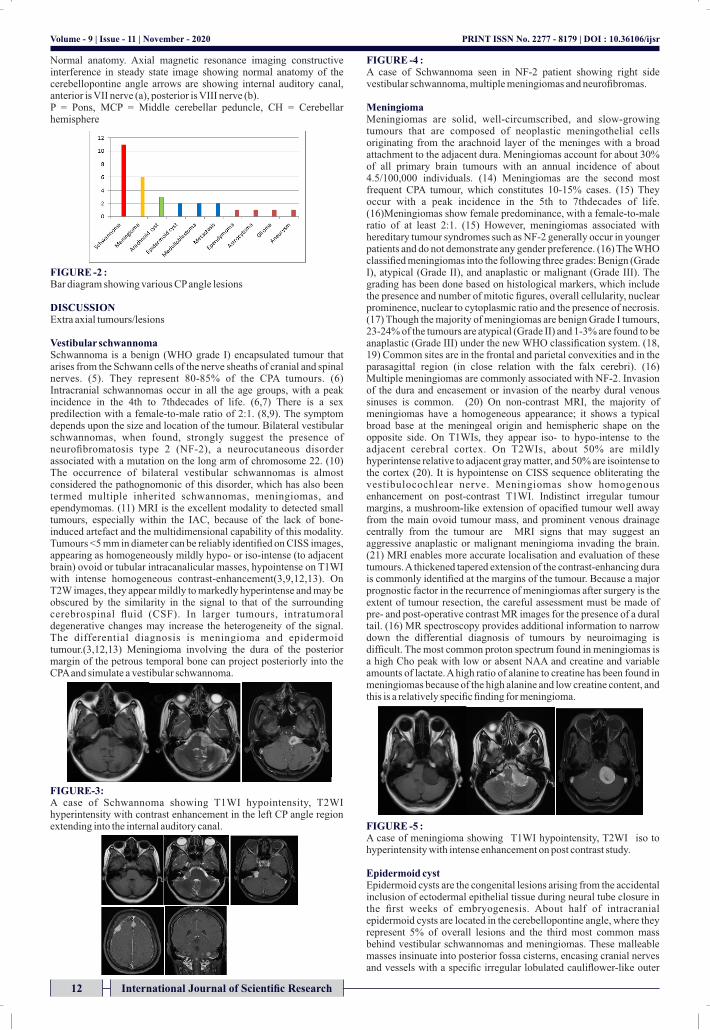
FIGURE -1: Normal Anatomy Of Cerebello Pontine Angle
INTERNATIONAL JOURNAL OF SCIENTIFIC RESEARCH
Radiodiagnosis
International Journal of Scientific Research 11
Volume - 9 | Issue - 11 | November - 2020 | PRINT ISSN No. 2277 - 8179 | DOI : 10.36106/ijsr
ABSTRACTIntroduction: Cerebellopontine angle (CPA) lesions usually were classied into intra-axial and extra-axial tumours. Although vestibular schwannomas and meningiomas constitute the vast majority of the cerebellopontine angle (CPA) lesions, a large variety of unusual lesions can also be encountered in the CPA. To evaluate the role of MRI in various cerebellopontine angle lesions.Aim and objective : Material and Methods: All patients with clinical suspicion of cerebellopontine (CP) angle tumours were subjected to scan on the 1.5T MR imaging system. Special sequences such as DWI, MR spectroscopy were performed. Acoustic schwannomas, which were also known as Vestibular Results: schwannomas, and meningiomas were the two most frequent lesions of all CPA tumours. In this study 30 cases were evaluated, there were 11 cases of Schwannoma, 6 cases of Meningioma, 3 cases of Epidermoid cyst, 2 cases of Arachnoid cyst, 2 cases of Metastasis, 2 cases of medullablastoma, 1 case of each astrocytoma, glioma, ependymoma, and vertebral artery aneurysm. MRI was the most sensitive noninvasive modality Conclusion: to characterise CP angle lesions. MRI identies the exact location and extension of the lesions based on their signal characteristics and contrast enhancement pattern.
KEYWORDSCerebellopontine Angle lesion, Schwannoma, Meningioma
Volume - 9 | Issue - 11 | November - 2020
12 International Journal of Scientific Research
Normal anatomy. Axial magnetic resonance imaging constructive interference in steady state image showing normal anatomy of the cerebellopontine angle arrows are showing internal auditory canal, anterior is VII nerve (a), posterior is VIII nerve (b).P = Pons, MCP = Middle cerebellar peduncle, CH = Cerebellar hemisphere
FIGURE -2 : Bar diagram showing various CP angle lesions
DISCUSSIONExtra axial tumours/lesions
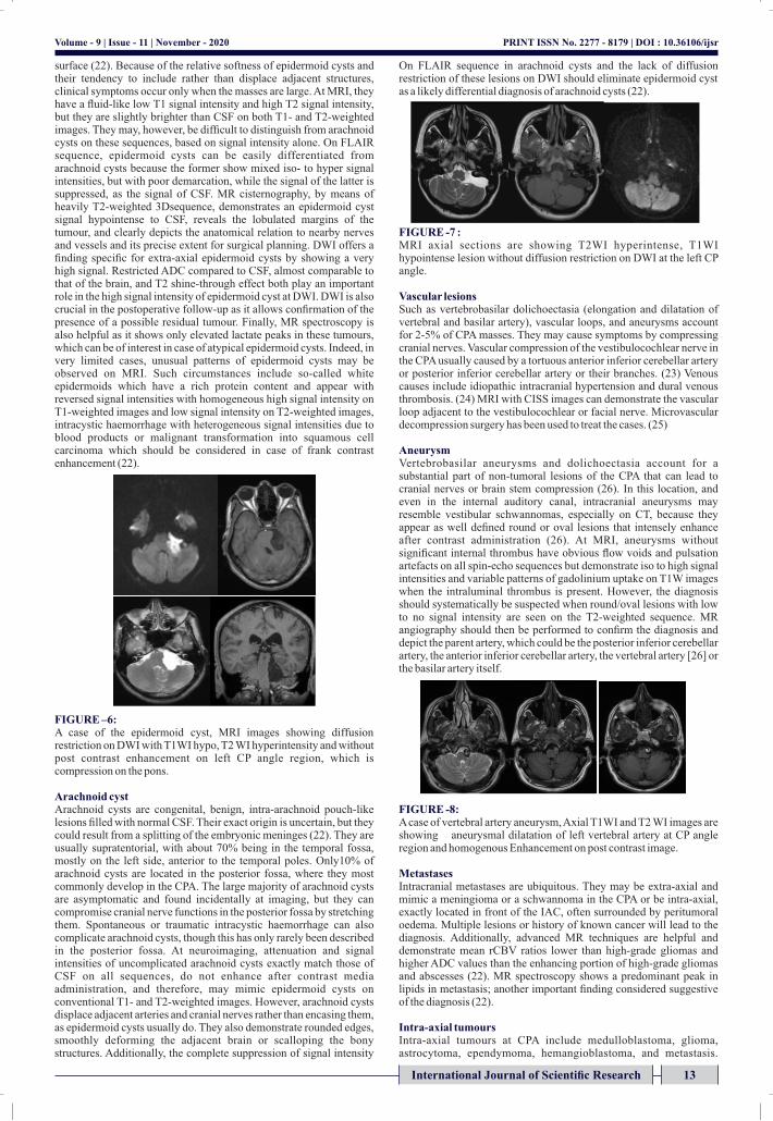
Vestibular schwannomaSchwannoma is a benign (WHO grade I) encapsulated tumour that arises from the Schwann cells of the nerve sheaths of cranial and spinal nerves. (5). They represent 80-85% of the CPA tumours. (6) Intracranial schwannomas occur in all the age groups, with a peak incidence in the 4th to 7thdecades of life. (6,7) There is a sex predilection with a female-to-male ratio of 2:1. (8,9). The symptom depends upon the size and location of the tumour. Bilateral vestibular schwannomas, when found, strongly suggest the presence of neurobromatosis type 2 (NF-2), a neurocutaneous disorder associated with a mutation on the long arm of chromosome 22. (10) The occurrence of bilateral vestibular schwannomas is almost considered the pathognomonic of this disorder, which has also been termed multiple inherited schwannomas, meningiomas, and ependymomas. (11) MRI is the excellent modality to detected small tumours, especially within the IAC, because of the lack of bone-induced artefact and the multidimensional capability of this modality. Tumours <5 mm in diameter can be reliably identied on CISS images, appearing as homogeneously mildly hypo- or iso-intense (to adjacent brain) ovoid or tubular intracanalicular masses, hypointense on T1WI with intense homogeneous contrast-enhancement(3,9,12,13). On T2W images, they appear mildly to markedly hyperintense and may be obscured by the similarity in the signal to that of the surrounding cerebrospinal uid (CSF). In larger tumours, intratumoral degenerative changes may increase the heterogeneity of the signal. The differential diagnosis is meningioma and epidermoid tumour.(3,12,13) Meningioma involving the dura of the posterior margin of the petrous temporal bone can project posteriorly into the CPA and simulate a vestibular schwannoma.
FIGURE-3:A case of Schwannoma showing T1WI hypointensity, T2WI hyperintensity with contrast enhancement in the left CP angle region extending into the internal auditory canal.
FIGURE -4 : A case of Schwannoma seen in NF-2 patient showing right side vestibular schwannoma, multiple meningiomas and neurobromas.
MeningiomaMeningiomas are solid, well-circumscribed, and slow-growing tumours that are composed of neoplastic meningothelial cells originating from the arachnoid layer of the meninges with a broad attachment to the adjacent dura. Meningiomas account for about 30% of all primary brain tumours with an annual incidence of about 4.5/100,000 individuals. (14) Meningiomas are the second most frequent CPA tumour, which constitutes 10-15% cases. (15) They occur with a peak incidence in the 5th to 7thdecades of life. (16)Meningiomas show female predominance, with a female-to-male ratio of at least 2:1. (15) However, meningiomas associated with hereditary tumour syndromes such as NF-2 generally occur in younger patients and do not demonstrate any gender preference. (16) The WHO classied meningiomas into the following three grades: Benign (Grade I), atypical (Grade II), and anaplastic or malignant (Grade III). The grading has been done based on histological markers, which include the presence and number of mitotic gures, overall cellularity, nuclear prominence, nuclear to cytoplasmic ratio and the presence of necrosis. (17) Though the majority of meningiomas are benign Grade I tumours, 23-24% of the tumours are atypical (Grade II) and 1-3% are found to be anaplastic (Grade III) under the new WHO classication system. (18, 19) Common sites are in the frontal and parietal convexities and in the parasagittal region (in close relation with the falx cerebri). (16) Multiple meningiomas are commonly associated with NF-2. Invasion of the dura and encasement or invasion of the nearby dural venous sinuses is common. (20) On non-contrast MRI, the majority of meningiomas have a homogeneous appearance; it shows a typical broad base at the meningeal origin and hemispheric shape on the opposite side. On T1WIs, they appear iso- to hypo-intense to the adjacent cerebral cortex. On T2WIs, about 50% are mildly hyperintense relative to adjacent gray matter, and 50% are isointense to the cortex (20). It is hypointense on CISS sequence obliterating the vestibulocochlear nerve. Meningiomas show homogenous enhancement on post-contrast T1WI. Indistinct irregular tumour margins, a mushroom-like extension of opacied tumour well away from the main ovoid tumour mass, and prominent venous drainage centrally from the tumour are MRI signs that may suggest an aggressive anaplastic or malignant meningioma invading the brain. (21) MRI enables more accurate localisation and evaluation of these tumours. A thickened tapered extension of the contrast-enhancing dura is commonly identied at the margins of the tumour. Because a major prognostic factor in the recurrence of meningiomas after surgery is the extent of tumour resection, the careful assessment must be made of pre- and post-operative contrast MR images for the presence of a dural tail. (16) MR spectroscopy provides additional information to narrow down the differential diagnosis of tumours by neuroimaging is difcult. The most common proton spectrum found in meningiomas is a high Cho peak with low or absent NAA and creatine and variable amounts of lactate. A high ratio of alanine to creatine has been found in meningiomas because of the high alanine and low creatine content, and this is a relatively specic nding for meningioma.
FIGURE -5 :A case of meningioma showing T1WI hypointensity, T2WI iso to hyperintensity with intense enhancement on post contrast study.
Epidermoid cystEpidermoid cysts are the congenital lesions arising from the accidental inclusion of ectodermal epithelial tissue during neural tube closure in the rst weeks of embryogenesis. About half of intracranial epidermoid cysts are located in the cerebellopontine angle, where they represent 5% of overall lesions and the third most common mass behind vestibular schwannomas and meningiomas. These malleable masses insinuate into posterior fossa cisterns, encasing cranial nerves and vessels with a specic irregular lobulated cauliower-like outer
PRINT ISSN No. 2277 - 8179 | DOI : 10.36106/ijsr
surface (22). Because of the relative softness of epidermoid cysts and their tendency to include rather than displace adjacent structures, clinical symptoms occur only when the masses are large. At MRI, they have a uid-like low T1 signal intensity and high T2 signal intensity, but they are slightly brighter than CSF on both T1- and T2-weighted images. They may, however, be difcult to distinguish from arachnoid cysts on these sequences, based on signal intensity alone. On FLAIR sequence, epidermoid cysts can be easily differentiated from arachnoid cysts because the former show mixed iso- to hyper signal intensities, but with poor demarcation, while the signal of the latter is suppressed, as the signal of CSF. MR cisternography, by means of heavily T2-weighted 3Dsequence, demonstrates an epidermoid cyst signal hypointense to CSF, reveals the lobulated margins of the tumour, and clearly depicts the anatomical relation to nearby nerves and vessels and its precise extent for surgical planning. DWI offers a nding specic for extra-axial epidermoid cysts by showing a very high signal. Restricted ADC compared to CSF, almost comparable to that of the brain, and T2 shine-through effect both play an important role in the high signal intensity of epidermoid cyst at DWI. DWI is also crucial in the postoperative follow-up as it allows conrmation of the presence of a possible residual tumour. Finally, MR spectroscopy is also helpful as it shows only elevated lactate peaks in these tumours, which can be of interest in case of atypical epidermoid cysts. Indeed, in very limited cases, unusual patterns of epidermoid cysts may be observed on MRI. Such circumstances include so-called white epidermoids which have a rich protein content and appear with reversed signal intensities with homogeneous high signal intensity on T1-weighted images and low signal intensity on T2-weighted images, intracystic haemorrhage with heterogeneous signal intensities due to blood products or malignant transformation into squamous cell carcinoma which should be considered in case of frank contrast enhancement (22).
FIGURE –6:A case of the epidermoid cyst, MRI images showing diffusion restriction on DWI with T1WI hypo, T2 WI hyperintensity and without post contrast enhancement on left CP angle region, which is compression on the pons.
Arachnoid cystArachnoid cysts are congenital, benign, intra-arachnoid pouch-like lesions lled with normal CSF. Their exact origin is uncertain, but they could result from a splitting of the embryonic meninges (22). They are usually supratentorial, with about 70% being in the temporal fossa, mostly on the left side, anterior to the temporal poles. Only10% of arachnoid cysts are located in the posterior fossa, where they most commonly develop in the CPA. The large majority of arachnoid cysts are asymptomatic and found incidentally at imaging, but they can compromise cranial nerve functions in the posterior fossa by stretching them. Spontaneous or traumatic intracystic haemorrhage can also complicate arachnoid cysts, though this has only rarely been described in the posterior fossa. At neuroimaging, attenuation and signal intensities of uncomplicated arachnoid cysts exactly match those of CSF on all sequences, do not enhance after contrast media administration, and therefore, may mimic epidermoid cysts on conventional T1- and T2-weighted images. However, arachnoid cysts displace adjacent arteries and cranial nerves rather than encasing them, as epidermoid cysts usually do. They also demonstrate rounded edges, smoothly deforming the adjacent brain or scalloping the bony structures. Additionally, the complete suppression of signal intensity
On FLAIR sequence in arachnoid cysts and the lack of diffusion restriction of these lesions on DWI should eliminate epidermoid cyst as a likely differential diagnosis of arachnoid cysts (22).
FIGURE -7 : MRI axial sections are showing T2WI hyperintense, T1WI hypointense lesion without diffusion restriction on DWI at the left CP angle.
Vascular lesionsSuch as vertebrobasilar dolichoectasia (elongation and dilatation of vertebral and basilar artery), vascular loops, and aneurysms account for 2-5% of CPA masses. They may cause symptoms by compressing cranial nerves. Vascular compression of the vestibulocochlear nerve in the CPA usually caused by a tortuous anterior inferior cerebellar artery or posterior inferior cerebellar artery or their branches. (23) Venous causes include idiopathic intracranial hypertension and dural venous thrombosis. (24) MRI with CISS images can demonstrate the vascular loop adjacent to the vestibulocochlear or facial nerve. Microvascular decompression surgery has been used to treat the cases. (25)
AneurysmVertebrobasilar aneurysms and dolichoectasia account for a substantial part of non-tumoral lesions of the CPA that can lead to cranial nerves or brain stem compression (26). In this location, and even in the internal auditory canal, intracranial aneurysms may resemble vestibular schwannomas, especially on CT, because they appear as well dened round or oval lesions that intensely enhance after contrast administration (26). At MRI, aneurysms without signicant internal thrombus have obvious ow voids and pulsation artefacts on all spin-echo sequences but demonstrate iso to high signal intensities and variable patterns of gadolinium uptake on T1W images when the intraluminal thrombus is present. However, the diagnosis should systematically be suspected when round/oval lesions with low to no signal intensity are seen on the T2-weighted sequence. MR angiography should then be performed to conrm the diagnosis and depict the parent artery, which could be the posterior inferior cerebellar artery, the anterior inferior cerebellar artery, the vertebral artery [26] or the basilar artery itself.
FIGURE -8: A case of vertebral artery aneurysm, Axial T1WI and T2 WI images are showing aneurysmal dilatation of left vertebral artery at CP angle region and homogenous Enhancement on post contrast image.
MetastasesIntracranial metastases are ubiquitous. They may be extra-axial and mimic a meningioma or a schwannoma in the CPA or be intra-axial, exactly located in front of the IAC, often surrounded by peritumoral oedema. Multiple lesions or history of known cancer will lead to the diagnosis. Additionally, advanced MR techniques are helpful and demonstrate mean rCBV ratios lower than high-grade gliomas and higher ADC values than the enhancing portion of high-grade gliomas and abscesses (22). MR spectroscopy shows a predominant peak in lipids in metastasis; another important nding considered suggestive of the diagnosis (22).
Intra-axial tumoursIntra-axial tumours at CPA include medulloblastoma, glioma, astrocytoma, ependymoma, hemangioblastoma, and metastasis.
Volume - 9 | Issue - 11 | November - 2020 PRINT ISSN No. 2277 - 8179 | DOI : 10.36106/ijsr
International Journal of Scientific Research 13

Medulloblastoma is a brain tumour of neuroepithelial origin, which represents 15-30% of a pediatric brain tumour and <1% of adult brain tumour. (27) These are hypointense on T1WI and hyperintense on T2WI, it gives intense postcontrast enhancement medulloblastoma can involve CPA in the extreme case of foraminal extension usually in exophytic cerebellar mass. Ependymomas represent 3-9% of neuroepithelial neoplasm and, 6-12% of all pediatric tumours. (28) The majority of intracranial ependymomas located in the posterior fossa, which arise from the oor of the fourth ventricle. Ependymomas are iso- or hypo-intense on T1WI and hyperintense on T2WI with the specks of calcication and haemorrhage are commonly seen. It shows heterogeneous postcontrast enhancement. Ependymomas represent 3-9% of neuroepithelial neoplasm and, 6-12 % of all pediatric tumours. (28) The majority of intracranial ependymomas located in the posterior fossa, which arise from the oor of the fourth ventricle. Ependymomas are iso- or hypo-intense on T1WI and hyperintense on T2WI with the specks of calcication and haemorrhage are commonly seen. It shows heterogeneous postcontrast enhancement.
FIGURE -9: A case of glioma showing T2WI hyperintensity and T1WI hypointensity in right CP angle.
CONCLUSIONA wide variety of lesions can be encountered in the Cerebellopontine angle (CPA). A meticulous analysis of the site of origin, shape, signal intensities and behaviour after contrast media injection allows a systematic approach to the preoperative diagnosis in the majority of cases. Diffusion and perfusion-weighted imaging, as well as MR spectroscopy, will provide crucial information that helps radiologists arrive at the correct diagnosis non-invasively.
REFERENCES1. Bailey BJ, Johnson JT, Shawn D. Head & Neck Surgery: Otolaryngology. Newlands:
Lippincott Williams & Wilkins; 2006.2. Schaller B. Cerebellopontine angle surgery. Part 1: General remarks. HNO
2003;51:284-95.3. Brackmann DE, Kwartler JA. A review of acoustic tumours: 1983-1988. Am J Otol
1990;11:216-32.4. Smirniotopoulos JG, Yue NC, Rushing EJ. Cerebellopontine angle masses: Radiologic-
pathologic correlation. Radiographics 1993;13:1131-47.5. Woodruff JM, Kourea HP, Louis DN. Tumors of cranial and peripheral nerves. In:
Kleihues P, Cavenee WK, editors. Pathology and Genetics of Tumors of the Nervous System. Lyon: International Agency for Research on Cancer; 2000. p. 163-74.
6. Bonneville F, Sarrazin JL, Dupuch KM, IffeneckerC, Cordoliani YS, Doyon D, Bonneville JF. Unusual Lesions of the Cerebellopontine Angle: A Segmental Approach. Radiographics 2001;21:419-38.
7. Brunori A, Scarano P, Chiappetta F. Non-acoustic neuroma tumour (NANT) of the cerebello-pontine angle: A 15-year experience.JNeurosurgSci 1997;41:159-68.
8. Casadei GP, Komori T, Scheithauer BW, Miller GM, Parisi JE, Kelly PJ. Intracranial parenchymal Schwannoma. A clinicopathological and neuroimaging study of nine cases. J Neurosurg 1993;79:217-22.
9. Smirniotopoulos JG, editor. Extra axial masses of the central nervous system. In: Categorical Course in Diagnostic Radiology: Neuroradiology. Oak Brook, Ill: Radiological Society of North America; 2000. p. 123-32.
10. Mulvihill JJ, Parry DM, Sherman JL, Pikus A, Kaiser-Kupfer MI, Eldridge R. NIH conference. Neurobromatosis 1 (Recklinghausen disease) and neurobromatosis 2 (bilateral acoustic neurobromatosis). An update. Ann Intern Med1990;113:39-52.
11. Smirniotopoulos JG, Murphy FM. The phakomatoses. AJNR Am J Neuroradiol 1992;13:725-46.
12. Ricci PE. Imaging of adult brain tumours. Neuroimaging ClinNAm 1999;9:651-69.13. Osborn AG, editor. Meningiomas and other nonglial neoplasms.In: Diagnostic
Neuroradiology. St. Louis: Mosby; 1994.p. 579-625.14. CBTRUS. Statistical Report: Primary Brain Tumors in the United States, 1998-2002.
Central Brain Tumor Registry of the United States; 2005.15. Sarrazin JL. Infra tentorial tumors. J Radiol 2006;87(6 Pt 2):748-63.16. Woodruff JM, Kourea HP, Louis DN, Scheitauer BW. Tumours of cranial and peripheral
nerves. In: Kleihues P, CaveneeWK, editors. Pathology and Genetics of Tumours of the Nervous System. Lyon: International Agency for Research on Cancer;2000. p. 163-74.
17. Commins DL, Atkinson RD, Burnett ME. Review of meningioma histopathology. Neurosurg Focus 2007;23:E3.
18. Smith SJ, Boddu S, Macarthur DC. Atypical meningiomas: WHO moved the goalposts? Br J Neurosurg 2007;21:588-92.
19. Willis J, Smith C, Ironside JW, Erridge S, Whittle IR, Everington D. The accuracy of meningioma grading: A 10-year retrospective audit. Neuropathol Appl Neurobiol 2005;31:141-9.
20. Atlas SW, Lavi E, Goldberg HI. Extra axial brain tumours. In: Atlas SW, editor. Magnetic Resonance Imaging of the Brain and Spine. 3rd ed. Philadelphia: Lippincott Williams & Wilkins;2002. p. 695-772.
21. New PF, Hesselink JR, O'Carroll CP, Kleinman GM. Malignant meningiomas: CT and histologic criteria, including a new CT sign. AJNR Am J Neuroradiol 1982;3:267-76.
22. Fabrice Bonneville Julien Savatovsky Jacques ChirasEurRadiol (2007) 17: 2908–2920 DOI 10.1007/s00330-007-0680-4
23. Bertrand RA, Molina P, Hardy J. Vestibular syndrome and vascular anomaly in the
cerebellopontine angle. Acta Otolaryngol 1977;83:187-94.24. Hofmann E, Behr R, Neumann-Haefelin T, Schwager K. Pulsatile tinnitus: Imaging and
differential diagnosis. DtschArzteblInt 2013;110:451-8.25. Wuertenberger CJ, Rosahl SK. Vertigo and tinnitus caused by vascular compression of
the vestibulocochlear nerve, not intracanalicular vestibular Schwannoma: Review and case presentation. Skull Base 2009;19:417-24.
26. Fabrice Bonneville Julien Savatovsky Jacques ChirasEurRadiol (2007) 17: 2472–2482 DOI 10.1007/s00330-007-0679-x
27. Bourgouin PM, Tampieri D, Grahovac SZ, Léger C, Del Carpio R, Melançon D. CT and MR imaging ndings in adults with cerebellar medulloblastoma: Comparison with ndings in children. AJR Am J Roentgenol 1992;159:609-12.
28. Koeller KK, Sandberg GD; Armed Forces Institute of Pathology. From the archives of the AFIP. Cerebral intraventricular neoplasms: Radiologic-pathologic correlation. Radiographics 2002;22:1473-505.
Volume - 9 | Issue - 11 | November - 2020 PRINT ISSN No. 2277 - 8179 | DOI : 10.36106/ijsr
14 International Journal of Scientific Research