Embed Size (px)
Citation preview

Core Curriculum V5
Radiographic evaluation of the SpineAlex M. Buteera, MD
Associate Professor and Chief Consultant Orthopedic SurgeonRwanda Military and King Faisal Hospital, Kigali

Core Curriculum V5
Objectives of Radiographic Examinations
• Adjunct to history and physical examination in process of establishing diagnosis of spine injury.
• Ascertain as definitively as possible whether there is a Spine injury
• Define fully the nature of the Spine injury

Core Curriculum V5
Radiographic Examination

Core Curriculum V5
Radiographic Exam•Systematic Approach- Steps

Core Curriculum V5
Radiographic ExamSystematic Approach• If a Step is missed
Core Curriculum V5
Challenges of Radiographic Examination
• Extremely sensitive but relatively non specific
• Reveal abnormalities in 1/3 of asymptomatic patients
• Differentiating between abnormalities with clinical implications and effects of ageing or healing

Core Curriculum V5
Radiographic Examination
• Studies that are routine- Lateral C-Spine
(Part of Trauma Series)
• Or Equivalent ( CT Scan with Sagittal recon)
Cervical spine imaging in patients with blunt traumaBlackmore CC, Emerson SS et al, 1999

Core Curriculum V5
Radiographic Examination
•If 1 Spine injury is detected
•Do complete C, T, L and S of the Spine10-20% non contiguous injury
Evaluation of risk of noncontiguous fractures of the spine in blunt trauma. Daniel William Nelson et al. J Trauma Acute Care Surg.2013 Jul.

Core Curriculum V5
Radiographic examination
•Presence of facial trauma- C-Spine radiographs•Presence of face or neck abrasions from sit belts-C-Spine radiographs
•Presence of lap belt contusion- T-L Spine radiographs
•Presence of calcaneal fractures- T-L/ L-Spine radiographs

Core Curriculum V5
Cervical Spine Spine Examination

Core Curriculum V5
• Systematic
• Upper Cervical
• Lower Cervical
• Start with PLAIN LATERAL FILM85% of injuries
Initial radiographic evaluation of the spine after trauma , France John CM, Bono Christopher et al, 2005
Core Curriculum V5
Occipital Cervical junction injuries
• Dislocations and Dissociation
• Associated major trauma
• Injury Detection is a challenge leading to missed diagnosis
• CT scan is best option for these injuries.

Core Curriculum V5
Detecting O-C Junction injuries
• Harris Lines• Basiondental Interval (BDI)
• Distance from basion to the tip of the dens
• Basionposterior Axial Line Interval (BAI)
• Distance from the basion to a line drawn on the posterior aspect of C2
• Harris Rule of 12• Both of these lines should be less
than 12 mm
BDI
BAI

Core Curriculum V5
Detecting O-C Junction injuries
• Power’s Ratio• Describes relationship
between occiput and C1• Line drawn from
• Basion to Posterior Aspects of the C1 Arch (BC)
• Opisthion to Anterior Arch of C1 (OA)
• Ratio of these lines should be less than 1 in normal patients
• BC/OA < 1
B
C
OA

Core Curriculum V5
Upper Cervical Instability
• Widened ADI• Atlanto-dens Interval (ADI)
• Horizontal distance between posterior border of anterior arch of C1 and the anterior border of the Dens
• > 3.5 mm indicative of instability• Posterior atlanto-dens interval
(PADI)• Horizontal distance between posterior
border of dens and the anterior border of the posterior arch of C1
• Commonly evaluated as Space Available for the Cord
• The AP diameter of the canal at this level
PADIADI

Core Curriculum V5
Upper Cervical: Open Mouth View: C1-C2
Normal C1-C2

Core Curriculum V5
Measuring Lateral Mass Overhang

Core Curriculum V5
CT scan- C- spine C1-C2 (Odontoid fracture)
Defines the nature of spine injury better
Aids decision on management
C-arm Image post fixation
Core Curriculum V5
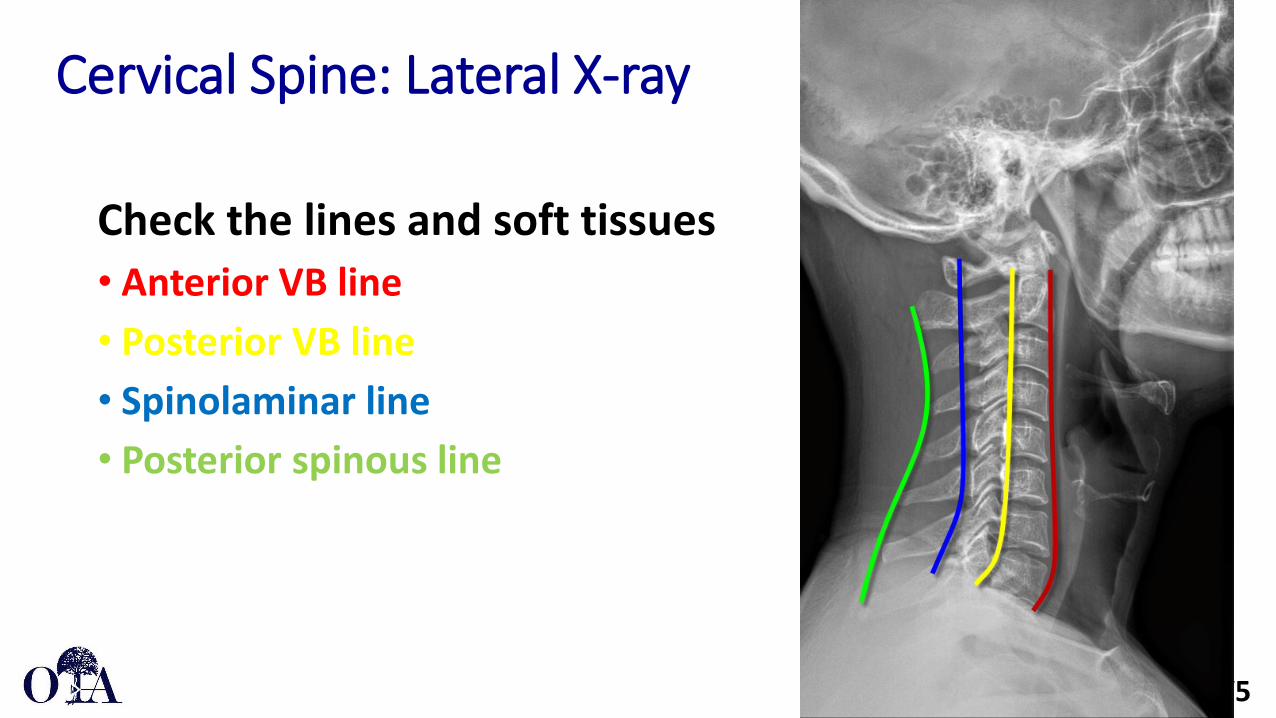
Cervical Spine: Lateral X-ray
Check the lines and soft tissues• Anterior VB line• Posterior VB line• Spinolaminar line• Posterior spinous line

Core Curriculum V5
Lower C-Spine detection
• Spinous process gapping• Facet joint apposition• Intervertebral gaping• Angulation• Translation
Core Curriculum V5
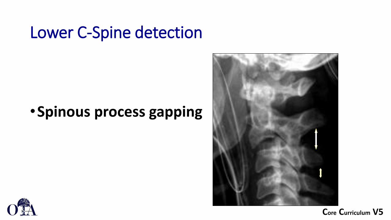
Lower C-Spine detection
•Spinous process gapping

Core Curriculum V5
Lower C-Spine detection
• Facet Joint Apposition• Normal facets should have overlap
(green)• Subluxed or Dislocated facets no
longer show this overlap (red)
Core Curriculum V5
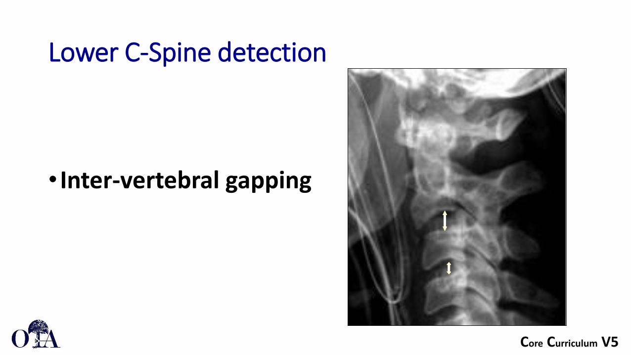
Lower C-Spine detection
• Inter-vertebral gapping

Core Curriculum V5
Lower C-spine detection
•Vertebral Angulation

Core Curriculum V5
Lower C-Spine detection
•Vertebral translation

Core Curriculum V5
Subtle signs of injury
•No obvious fracture/dislocation•Check retrophangeal orPrevertebral soft tissue swelling
Presence:--> + injuryAbsence: may not rule out injury

Core Curriculum V5
Soft tissue swellingUsing: • 6mm at C3 ---> 59% Sensitivity
• 22mm at C6 ---> 5% Sensitivity
Doesn’t mean much if not there
DeBehn and Havel, 1994

Core Curriculum V5
C-Spine: Anteroposterior view
•Spinous process deviation
•Lateral translation
•Coronal deformity

Core Curriculum V5
Cervicothoracic junction
•Complete lateral (Upper part of T1)•Swimmers view•CT Scan is better for transition zones

Core Curriculum V5
CT Scan as- Screening Modality
• CT with sagittal recon l• Most sensitive for fracturedetection• Especially transition zones( C0-C1 and C7-T1)• Difficult with X-rays• Vascular injury
Michael Utz, Shadab Khan et al, Insights Imaging, 2014

Core Curriculum V5
MRI- best soft tissue definition
• Negative plain Films• Negative CT Scan• But Clinically Suspicious• Check for: Continuity of ligaments Edema in soft tissues Cord injury?

Core Curriculum V5
Safety: Contra-indications for MRI
Implanted devices that:
•Subject to magnetically induced malfunction
•Potentially harmful movement

Core Curriculum V5
MRI- best soft tissues definition
• Clinical suspicion• Has neural deficitHerniated discCord injury

Core Curriculum V5
MRI- soft tissue definition
T1 sequences:• Excellent for surveying anatomy and caliber of spinal
cord
T2 images with or without fat saturation:• epidural fluid collection, ligamentous disruption,
edema and herniated discs

Core Curriculum V5
‘Clearing’ the C-Spine
•Standardized Protocol•No consensus

Core Curriculum V5
Clearing C-Spine
•Avoid missed injuries•Identify patients without significant injuries
•Delay in diagnosis associated with worse outcome
Levi AD, Hubert RJ et al, Spine 2006

Core Curriculum V5
Injury detection- Thoracic and Lumbar Spine
• Same principles• Landmarks and lines: Lateral ViewPosterior vertebral body lineAnterior vertebral body line Inter-spinous DistanceTranslation

Core Curriculum V5
Injury detection- T and L Spine
AP View:•Spinous process to pedicles
• Should be symmetric• Interpedicular distance
• May be widened in burst fractures
•Translation

Core Curriculum V5
CT Scan: T-L Spine
• More Common as initial study• Indicated if plain x-ray is suspicious• Best bony detail• Request multiple planes and recon• Axial alone can miss translation

Core Curriculum V5
Thoracic and lumbar injuries
• What is normal angulation?

Core Curriculum V5
T-L Spine injuries
•Height loss
Adjacent fracture

Core Curriculum V5
MRI- Best at soft tissues
• MRI Can be useful to detect injuries to soft tissues, such as the posterior ligamentous complex (PLC)
• Consists of • Supraspinous Ligament• Interspinous Ligament• Ligamentum Flavum• Facet Capsule

Core Curriculum V5
MRI- best for soft tissues
Assessing PLC using MRIContinuity of the ligamentumflavum

Core Curriculum V5
Summary
• Radiographic imaging serves as an adjunct to history and physical examination in process of diagnosing traumatic spinal injuries
• Radiographic evaluation should be approached in a systematic manner
• The advent of advanced imaging systems has led to improved detection, understanding, and diagnosis of spine trauma …
• But understanding the principles of these injuries on plain films remains critically important