Embed Size (px)
Citation preview

1
Mina Le, HMS IIIGillian Lieberman, MD March 2006
Radiologic Evaluation ofCarotid Body Tumor
Mina Le, Harvard Medical School Year IIIGillian Lieberman, MD

2
Mina Le, HMS IIIGillian Lieberman, MD
A.G.’s Story
A.G. was a 71-year-old Ethiopian woman who periodically traveled to Boston to visit her daughter.
She would frequent the BIDMC for diverse GI issues.
One day, a mass was felt on the left side of her neck.

3
Mina Le, HMS IIIGillian Lieberman, MD
By radiology and biopsy, the mass in A.G.’s neck wasfound to be a carotid body tumor (paraganglioma).
She was advised to have it removed, but opted not to.
Over the years, on subsequent visits from Ethiopia toBoston, A.G.’s tumor was followed as it slowly grew.
We will discuss its appearance on MRI, MRA, CT, CTA.
Ever Larger With Time

4
Mina Le, HMS IIIGillian Lieberman, MD
Four Ways of Looking at a Tumor
(1) MRI, August 2000. (2) MRA, August 2000.Images courtesy of Dr. Lai

5
Mina Le, HMS IIIGillian Lieberman, MD
(3) CT, August 2004. (4) CTA, February 2006.Images courtesy of Dr. Lai
Four Ways of Looking at a Tumor

6
Mina Le, HMS IIIGillian Lieberman, MD
DDx for a Solid Mass in the Carotid Sheath
1. Lymphadenopathy – inflammatory, infectious2. Traumatic neuroma3. Benign tumor
a. Granular cell tumor (rhabdomyoblastoma)b. Hemangiolymphangiomac. Lipomad. Nerve sheath tumor (schwannoma, neurofibroma, ganglioneuroma, ganglioblastoma, ganglioneuroblastoma)e. Paraganglioma (glomus jugulare tumor, glomus vagale tumor, carotid body tumor)f. Thyroid and parathyroid tumors (e.g. adenoma)
4. Malignant tumor – lymphoma, metastatic lymphadenopathy
Reeder MM. Gamuts in Radiology, p. 169.

7
Mina Le, HMS IIIGillian Lieberman, MD
DDx for a Solid Mass in the Carotid Sheath
1. Lymphadenopathy – inflammatory, infectious2. Traumatic neuroma3. Benign tumor
a. Granular cell tumor (rhabdomyoblastoma)b. Hemangiolymphangiomac. Lipomad. Nerve sheath tumor (schwannoma, neurofibroma, ganglioneuroma, ganglioblastoma, ganglioneuroblastoma)e. Paraganglioma (glomus jugulare tumor, glomus vagale tumor, carotid body tumor)f. Thyroid and parathyroid tumors (e.g. adenoma)
4. Malignant tumor – lymphoma, metastatic lymphadenopathy
Reeder MM. Gamuts in Radiology, p. 169.
http://webpath.dmsf.edu.ph/ENDOHTML/ENDO084.HTM
8
Mina Le, HMS IIIGillian Lieberman, MD
Biopsy Not Recommended
The diagnosis of paraganglioma was made in A.G.’scase through fine-needle aspiration and cytology.
However, when paraganglioma is being considered,biopsy is generally not advised because of thehypervascular nature of these tumors.
Imaging is the preferred way to make the diagnosis.
Boedeker CC et al. Fam Cancer. 2005;4(1):55-59.

9
Mina Le, HMS IIIGillian Lieberman, MD
Boedeker CC et al. Fam Cancer. 2005;4(1):55-59.
B-mode sonography+ color-coded
Doppler sonography
Magneticresonance
imaging
Computedtomography
Digital subtractionangiography
Menu of Imaging Choices for Diagnosis and Evaluation of a Lateral Neck Mass

10
Mina Le, HMS IIIGillian Lieberman, MD
Merits of Ultrasound
Useful as the first step in assessment but not the last.
Inexpensive.
Non-invasive.
Readily available.
Boedeker CC et al. Fam Cancer. 2005;4(1):55-59.

11
Mina Le, HMS IIIGillian Lieberman, MD
Carotid Body Tumor on Ultrasound
Stoeckli SJ et al. Laryngoscope. 2002 Jan;112(1):143-6.
12
Mina Le, HMS IIIGillian Lieberman, MD
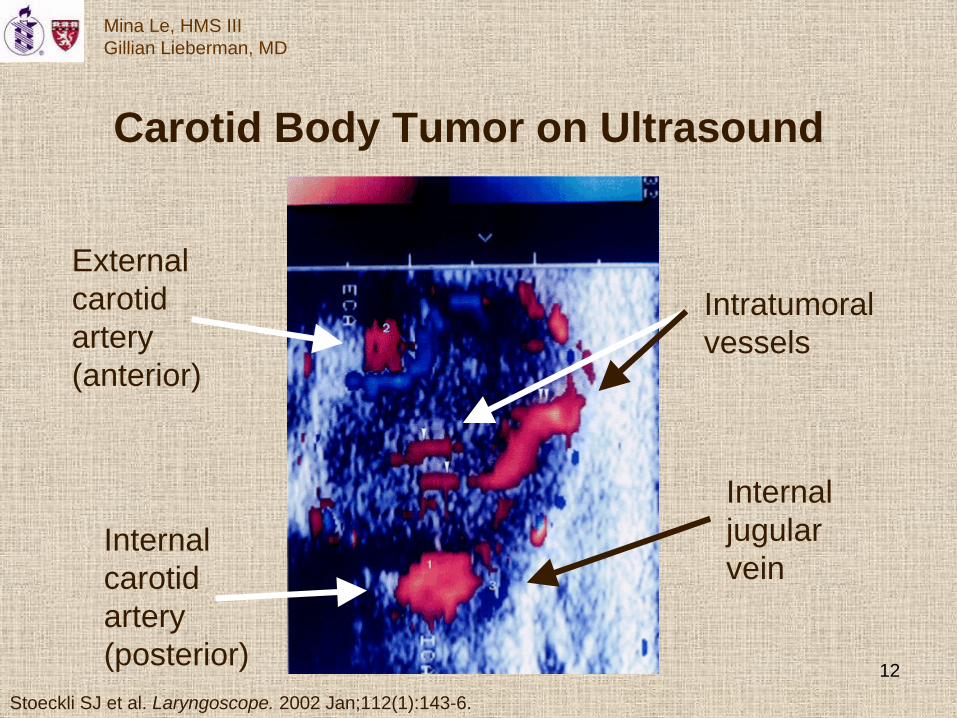
Carotid Body Tumor on Ultrasound
Stoeckli SJ et al. Laryngoscope. 2002 Jan;112(1):143-6.
Internalcarotidartery(posterior)
Externalcarotidartery(anterior)
Intratumoralvessels
Internaljugularvein

13
Mina Le, HMS IIIGillian Lieberman, MD
Abnormalities on Ultrasound
Stoeckli SJ et al. Laryngoscope. 2002 Jan;112(1):143-6.
Solid,well-defined,hypoechoic,
hypervascularmass
Splaying atthe bifurcation:external carotid
displaced anteriorly,
…internal carotidand internal jugular
displaced posteriorly
Tumor-vesselflow directionpredominantly
upward(red)
Boedeker CC et al. Fam Cancer. 2005;4(1):55-59.

14
Mina Le, HMS IIIGillian Lieberman, MD
Differential Diagnosis on Ultrasound
Stoeckli SJ et al. Laryngoscope. 2002 Jan;112(1):143-6. Boedeker CC et al. Fam Cancer. 2005;4(1):55-59.
Non-paraganglioma masses,e.g. enlarged lymph node:would look similar on B-modesonography, would not behypervascular on Doppler.
Vagal paraganglioma: canalso splay the bifurcationand look hypervascular, butthe intratumoral flow signalis directed downward (blue).

15
Mina Le, HMS IIIGillian Lieberman, MD
Contribution of MRI
MRI helps diagnose paragangliomas by representing theirhypervascularity as multiple low-signal areas due to flow void.
It is superior to CT scanning in delineating these tumorsand in distinguishing them from inflammation and hemorrhage.
MRI is also better able to demonstrate the relationship of carotid body tumors to adjacent vascular structures.
Mafee MF et al. Radiol Clin North Am. 2000 Sep;38(5):1059-76.

16
Mina Le, HMS IIIGillian Lieberman, MD
Carotid Body Tumor on MRI
Courtesy of Dr. Lai

17
Mina Le, HMS IIIGillian Lieberman, MD
Carotid Body Tumor on MRI
Courtesy of Dr. Lai
Externalcarotid
Internalcarotid
Carotid body

18
Mina Le, HMS IIIGillian Lieberman, MD
Carotid Body Tumor on MRA
Courtesy of Dr. Lai

19
Mina Le, HMS IIIGillian Lieberman, MD
Carotid Body Tumor on MRA
Courtesy of Dr. Lai
R externalcarotid
R internalcarotid
R vertebralartery
L externalcarotid
L internalcarotid
L vertebralartery

20
Mina Le, HMS IIIGillian Lieberman, MD
Abnormalities on MRI
Courtesy of Dr. Lai
Mass splaying the internal andexternal carotid arteries
No definite flow voidsHigh signal on STIREnhances with gadolinium
Courtesy of Dr. Lai

21
Mina Le, HMS IIIGillian Lieberman, MD
Most Likely DDx on MRI
Courtesy of Dr. Lai
Schwannomavs.
paraganglioma
Courtesy of Dr. Lai

22
Mina Le, HMS IIIGillian Lieberman, MD
Role of CT
CT is excellent for defining the exact location of a massand visualizing its effect on adjacent structures.
Combined with a knowledge of anatomy and epidemiology, it can help distinguish among congenital, inflammatory, and neoplastic processes that all result in neck masses.
It is quicker and more available than MRI.
Reede DL et al. Radiol Clin North Am. 1984 Mar;22(1):239-50.

23
Mina Le, HMS IIIGillian Lieberman, MD
Courtesy of Dr. Lai
Carotid Body Tumor on CT

24
Mina Le, HMS IIIGillian Lieberman, MD
Courtesy of Dr. Lai
Carotid Body Tumor on CT
ECA
ICA
IJV

25
Mina Le, HMS IIIGillian Lieberman, MD
Courtesy of Dr. Lai
Soft-tissue relations
Anteriorscalenemuscles
Sternocleido-mastoid m.
Middlescalene m.
Silver AJ et al. Radiol Clin North Am. 1984 Mar;22(1):219-238.

26
Mina Le, HMS IIIGillian Lieberman, MD
AbnormalitiesOn CTA
Coarse hypervascularity
Incorporation offeeding vessels
Silver AJ et al. Radiol Clin North Am. 1984 Mar;22(1):219-238.
Courtesy of Dr. Lai

27
Mina Le, HMS IIIGillian Lieberman, MD
Courtesy of Dr. Lai
DifferentialDiagnosis
on CT
Schwannoma Lucent Displaces adjacent vesselsParaganglioma Isodense Incorporates adjacent vessels
Silver AJ et al. Radiol Clin North Am. 1984 Mar;22(1):219-238.
Courtesy of Dr. Lai

28
Mina Le, HMS IIIGillian Lieberman, MD
Definitive Confirmation by DSA
Digital subtraction angiography provides definitive preoperative characterization.
It provides endovascular access for possible embolization;best maps the arterial supply;and, in later phases, visualizes the venous drainage,
which allows the surgeon to avoid cutting it until the end, decreasing intraoperative blood loss.
Boedeker CC et al. Fam Cancer. 2005;4(1):55-59.

29
Mina Le, HMS IIIGillian Lieberman, MD
Angiograms ofparagangliomas
Carotid body tumor,regular angiogram.
Vagalpara-
ganglioma,digital
subtractionangiogram.
van den Berg R et al. Am J Neuroradiol. 2000 Jan;21(1):162-70.

30
Mina Le, HMS IIIGillian Lieberman, MD
ReferencesBoedeker CC, Ridder GJ, Schipper J. Paragangliomas of the head and neck: diagnosis and treatment. Fam Cancer. 2005;4(1):55-59.
Klatt EC. “Carotid body, paraganglioma, low power microscopic.” WebPath: The Internet Pathology Laboratory. 1998. http://webpath.dmsf.edu.ph/ENDOHTML/ENDO084.HTM.
Mafee MF, Raofi B, Kumar A, Muscato C. Glomus faciale, glomus jugulare, glomus tympanicum, glomus vagale, carotid body tumors, and simulating lesions. Radiol Clin North Am. 2000 Sep;38(5):1059-76.
Reede DL, Whelan MA, Bergeron RT. CT of the soft tissue structures of the neck. Radiol Clin North Am. 1984 Mar;22(1):239-50.
Reeder MM. Reeder and Felson’s Gamuts in Radiology: Comprehensive Lists of Roentgen Differential Diagnosis, 4th ed. New York: Springer-Verlag New York, Inc., 2003.
Silver AJ, Ganti SR, Hilal SK. The carotid region: normal and pathologic anatomy on CT. Radiol Clin North Am. 1984 Mar;22(1):219-238.
Stoeckli SJ, Schuknecht B, Alkadhi H, Fisch U. Evaluation of paragangliomas presenting as a cervical mass on color-coded Doppler sonography. Laryngoscope. 2002 Jan;112(1):143-6.
Van den Berg R et al. Vascularization of head and neck paragangliomas: comparison of three MR angiographic techniques with digital subtraction angiography. Am J Neuroradiol. 2000 Jan;21(1):162-70.

31
Mina Le, HMS IIIGillian Lieberman, MD
Acknowledgments
Dr. Ken Lai
Dr. Katherine Krajewski
Pamela Lepkowski
Dr. Gillian Lieberman
Larry Barbaras, webmaster