Embed Size (px)
Citation preview

lable at ScienceDirect
Food Control 65 (2016) 14e20
Contents lists avai
Food Control
journal homepage: www.elsevier .com/locate/ foodcont
Ralstonia insidiosa serves as bridges in biofilm formation by foodbornepathogens Listeria monocytogenes, Salmonella enterica, andEnterohemorrhagic Escherichia coli
Nancy T. Liu a, c, Gary R. Bauchan b, Charlotte B. Francoeur a, 1, Daniel R. Shelton a,Y. Martin Lo c, Xiangwu Nou a, *
a Environmental Microbial and Food Safety Laboratory, USDA Agricultural Research Service Beltsville Agricultural Research Center, Beltsville, MD 20705,USAb Electron and Confocal Microscopy Research Unit, USDA Agricultural Research Service Beltsville Agricultural Research Center, Beltsville, MD 20705, USAc Department of Nutrition and Food Science, University of Maryland, College Park, MD 20740, USA
a r t i c l e i n f o
Article history:Received 1 October 2015Received in revised form26 December 2015Accepted 4 January 2016Available online 6 January 2016
Keywords:Ralstonia insidiosaBridging bacteriaDual-species biofilmsFoodborne pathogens
* Corresponding author. Environmental MicrobialUSDA Agricultural Research Service, Beltsville, MD 20
E-mail address: [email protected] (X. No1 Current address: Department of Cell Biology and M
of Maryland, College Park, MD 20740.
http://dx.doi.org/10.1016/j.foodcont.2016.01.0040956-7135/Published by Elsevier Ltd.
a b s t r a c t
Biofilm formation on abiotic surfaces in fresh produce processing facilities may play a role in foodborneoutbreaks by providing protective microniches for pathogenic bacteria. Our previous study showed that astrain of Ralstonia insidiosa isolated from a fresh produce processing plant could enhance the incorpo-ration of Escherichia coli O15:H7 in biofilms under various environmental conditions. These results raisedthe concern that R. insidiosa might have the ability to incorporate other foodborne pathogens andpromote their survival and growth in biofilms. To test this hypothesis, 6 strains of Shiga toxin producingE. coli, 2 strains of Salmonella, and 6 strains of Listeria monocytogenes were examined for dual-speciesbiofilm formation with R. insidiosa. A significant increase in biomass formation was observed in 7 ofthe 14 R. insidiosa-pathogen combinations, while significantly enhanced incorporation of pathogeniccells into biofilms was seen in 12 of the 14 R. insidiosa-pathogen combinations. The synergistic in-teractions between R. insidiosa and the tested foodborne pathogens seemed dependent on intimatecellular contact between the two strains. Overall, this study showed that R. insidiosa could enhance theincorporation of biofilms of different types of foodborne pathogenic bacteria and should be considered abridging bacterium for biofilm formation in various food processing environments.
Published by Elsevier Ltd.
1. Introduction
Fresh-cut produce is widely recognized as a potential vehicle forfoodborne outbreaks of bacterial infections (Harris et al., 2003;Lynch, Tauxe, & Hedberg, 2009). Various serotypes of Listeriamonocytogenes, Salmonella enterica, and Shiga toxin producingEscherichia coli have been implicated as causal agents for freshproduce associated outbreaks (Bowen, Fry, Richards, & Beuchat,2006; Warriner & Namvar, 2010). One particular concern relatedto these outbreaks is the formation of multispecies biofilms inproduce production and processing environments (Carpentier &
and Food Safety Laboratory,705, USA.u).olecular Genetics, University
Chassaing, 2004; Silagyi, Kim, Lo, & Wei, 2009).It has been previously shown that native microflora in dairy and
meat processing facilities provided potential transference routesfor foodborne pathogens (Carpentier & Chassaing, 2004; Lynchet al., 2009; Penteado, Eblen, & Miller, 2004). Particularly, certainenvironmental bacteria could play a key role in multispecies bio-films formation, thereby providing protective niches to planktonicbacteria for gaining enhanced resistance to daily cleaning anddisinfections (Jeong & Frank, 1994; Rickard, Gilbert, High,Kolenbrander, & Handley, 2003; Van der Veen & Abee, 2011). Aci-netobacter calcoaceticus, a strong biofilm former frequently isolatedfrom meat processing facilities, enhanced the survival of E. coliO157:H7 in a dynamic culture system (Habimana, Heir, Langsrud,Asli, & Moretro, 2010). A. calcoaceticus was also found to serve asa “bridge” for other bacteria isolated from drinking water informing multispecies biofilms. The absence of A. calcoaceticusresulted in a 75% reduction in biomass of multispecies biofilms
N.T. Liu et al. / Food Control 65 (2016) 14e20 15
(Simoes, Simoes, & Vieira, 2008). Therefore, these type of residentbacteria, known as bridging bacteria, may not only facilitateincorporation of pathogenic bacteria species into biofilms but alsoenhance the biomass production of biofilms, increasing the likeli-hood of pathogen survival (Uhlich, Rogers, &Mosier, 2010; Van derVeen & Abee, 2011).
Our previous study (Liu, Nou, Lefcourt, Shelton, & Lo, 2014)demonstrated that Ralstonia insidiosa isolated from a fresh-cutprocessing environment could enhance the incorporation ofE. coli O157:H7 into dual-species biofilms. Other strong biofilmproducers isolated from fresh-cut processing plants, includingKlebsiella pneumoniae, Stenotrophomonas rhizophila, were shown tobe ineffective in increasing the incorporation of E. coli O157:H7 inbiofilms, suggesting that the enhanced incorporation of E. coliO157:H7 in biofilms was species or strain dependent. In anotherstudy, we observed that R. insidiosa increased the incorporation ofmultiple E. coli O157:H7 strains into dual-species biofilms undervarious temperature and nutrient availability conditions (Liu et al.,2015), indicating that R. insidiosa was able to adapt to diverse mi-croenvironments in fresh produce processing plants. Based onthese observations, we hypothesized that R. insidiosamay serve as auniversal bridging species for multispecies biofilm formationallowing for enhanced incorporation of other types of pathogenicmicroorganisms. Thus, the goal of this research was to evaluate theeffects of R. insidiosa on biofilm formation by foodborne pathogenicbacteria most commonly associated with outbreaks involving freshproduce, including L. monocytogenes, S. enterica, and Shiga toxinproducing E. coli (STEC). In addition, selected key factors that mightaffect incorporation of pathogenic bacteria in biofilms, such asbiofilm-cell attraction, cell-cell adherence and metabolite produc-tion, were tested using E. coli O157:H7-R. insidiosa combination as amodel.
2. Materials and methods
2.1. Bacterial strains and media
The bacterial strains used in this study are listed in Table 1.R. insidosa strain FC1138 was isolated from food contact surfaces ina fresh-cut processing plant (Liu et al., 2013). E. coli O157:H7 strainFS4052 is a derivative from the non-virulent strain CDC B6-914 thatcarries a stable plasmid expressing green fluorescent protein(pGFP) (Fratamico, Deng, Strobaugh, & Palumbo, 1997). E. coli
Table 1List of environmental isolate and foodborne human pathogens.
Strain Serotype Source of isolation Source
Ralstonia insidiosaFC1138 NA Produce processing plant EMFSL
Listeria monocytogenesFS2005 1/2a Milk EMFSLFS2008 4b Milk EMFSLB-57616 1/2b Clinical isolate NRRLB-57617 1/2a Clinical isolate NRRLB-57618 1/2a Clinical isolate NRRLB-57622 1/2a Clinical isolate NRRL
Salmonella entericaFS3022 Newport Mango EMFSLFS3060 Poona Cantaloupe EMFSL
Escherichia coliFS4052 O157:H7 Human feces EMFSLFS4137 O111:NM Clinical isolate EMFSLFS4140 O45:H2 Clinical isolate EMFSLFS4143 O26:H11 Clinical isolate EMFSLFS4146 O103:H2 Clinical isolate EMFSLTW16133 O104:H4 Clinical isolate MSU
O104:H4 strain TW16133, which was associated with the 2011German sprout outbreak (CDC, 2013), was obtained from MichiganState University EHEC Stock Center (East Lansing, MI.).L. monocytogenes strains NRRL B-57616, 57617, 57618 and 57622,associated with 2011 Colorado cantaloupe outbreaks, were ob-tained from ARS Culture Collections (Peoria, Il.). All other strainswere obtained from the Environmental Microbial and Food SafetyLaboratory (EMFSL) (USDA-ARS, Beltsville, MD) laboratory stockculture collections. Tryptic soy broth (TSB) and tryptic soy agar(TSA) (BD Biosciences, San Jose, CA.) were used for routine bacterialgrowth. Diluted (10%) TSB was used for culturing bacteria to formbiofilms in static culture, while 1% TSB was used for forming bio-films in the drip flow biofilm reactor. TSAwas used for enumerationof total aerobic bacteria. All strains used in this study grew well onTSA, with R. insidiosa forming characteristic small round coloniesafter 24 h incubation, allowing easy distinction of R. insidiosa fromother strains inmixed cultures. In addition, modified Oxford Listeriaagar (MOC, BD Biosciences), Xylose lysine tergitol 4 agar (XLT-4,Neogen, Lansing, MI), and sorbitol MacConkey agar (sMAC, BDBiosciences) were used for enumeration of L. monocytogenes,S. enterica, and Shiga toxin-producing E. coli, respectively.
2.2. Biofilm formation by pathogenic bacteria and R. insidiosa
Monoculture or dual-species biofilms were produced in poly-styrene 12-well tissue culture plates (BD Biosciences) following theprocedure described previously (Liu et al., 2014), with slightmodification. Briefly, overnight cultures of individual strains werewashed in phosphate buffered saline (PBS) and cell numbersadjusted to approximately 109 CFU/ml. Three ml of 10% TSB waspipette into 12-well tissue culture plates along with 30 ml aliquotsof bacteria suspensions, resulting in approximately 107 CFU/ml. Fordual-species biofilms formation, equal amounts of each bacterialstrain (30 ml) were co-inoculated into the same well. Inoculatedculture plates were then incubated at 30 �C for 24 h with moderateorbital shaking (80 RPM).
2.3. Biofilm formation with physical separation of tested strains
E. coli O157:H7 strain FS4052 and R. insidiosa were used to testthe key factors that might affect incorporation into dual-speciesbiofilms. Special cell culture plates (12-well) with base andmatching insert compartments with 0.4 mm polyethylene tere-phthalate membranes (Corning, Tewksbury, MA) were used tophysically separate R. insidiosa and E. coli O157:H7 strains duringbiofilm formation. E. coli O157:H7 was inoculated into the basecompartments of the tissue culture plates, while an equal amountof R. insidiosawas added into the insert compartments on top of thewells, thereby allowing for free exchange of growth medium andmetabolites between the well and the insert compartments. Afterincubation at 30 �C for 24 h, the inserts were removed; biofilms inthe base compartments were washed, and cell counts of E. coliO157:H7 in the biofilm were determined.
2.4. E. coli biofilm formation on primed surface with R. insidiosabiofilm
The ability of E. coli O157:H7 to become incorporated intoexisting viable R. insidiosa biofilms was determined by inoculatingR. insidiosa into 12 well tissue culture plates and allowing for bio-film formation for 24 h at 30 �C. After removing the overnightgrowth culture, the resultant monoculture biofilms were rinsedwith PBS 3 times followed by inoculation with E. coli O157:H7 ontothe culture plates with pre-formed R. insidiosa biofilms in freshmedium. The plates were incubated at 30 �C for another 24 h.
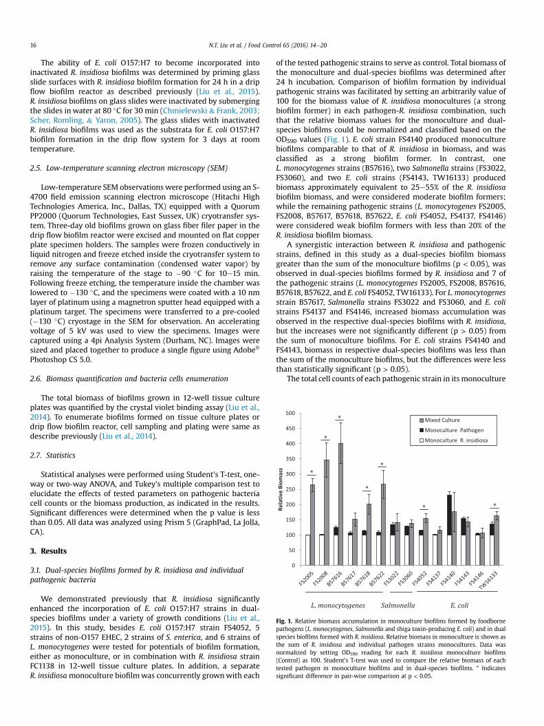
Fig. 1. Relative biomass accumulation in monoculture biofilms formed by foodbornepathogens (L. monocytogenes, Salmonella and shiga toxin-producing E. coli) and in dualspecies biofilms formed with R. insidiosa. Relative biomass in monoculture is shown asthe sum of R. insidiosa and individual pathogen strains monocultures. Data wasnormalized by setting OD590 reading for each R. insidiosa monoculture biofilms(Control) as 100. Student's T-test was used to compare the relative biomass of eachtested pathogen in monoculture biofilms and in dual-species biofilms. * Indicatessignificant difference in pair-wise comparison at p < 0.05.
N.T. Liu et al. / Food Control 65 (2016) 14e2016
The ability of E. coli O157:H7 to become incorporated intoinactivated R. insidiosa biofilms was determined by priming glassslide surfaces with R. insidiosa biofilm formation for 24 h in a dripflow biofilm reactor as described previously (Liu et al., 2015).R. insidiosa biofilms on glass slides were inactivated by submergingthe slides inwater at 80 �C for 30 min (Chmielewski& Frank, 2003;Scher, Romling, & Yaron, 2005). The glass slides with inactivatedR. insidiosa biofilms was used as the substrata for E. coli O157:H7biofilm formation in the drip flow system for 3 days at roomtemperature.
2.5. Low-temperature scanning electron microscopy (SEM)
Low-temperature SEM observations were performed using an S-4700 field emission scanning electron microscope (Hitachi HighTechnologies America, Inc., Dallas, TX) equipped with a QuorumPP2000 (Quorum Technologies, East Sussex, UK) cryotransfer sys-tem. Three-day old biofilms grown on glass fiber filer paper in thedrip flow biofilm reactor were excised and mounted on flat copperplate specimen holders. The samples were frozen conductively inliquid nitrogen and freeze etched inside the cryotransfer system toremove any surface contamination (condensed water vapor) byraising the temperature of the stage to �90 �C for 10e15 min.Following freeze etching, the temperature inside the chamber waslowered to �130 �C, and the specimens were coated with a 10 nmlayer of platinum using a magnetron sputter head equipped with aplatinum target. The specimens were transferred to a pre-cooled(�130 �C) cryostage in the SEM for observation. An acceleratingvoltage of 5 kV was used to view the specimens. Images werecaptured using a 4pi Analysis System (Durham, NC). Images weresized and placed together to produce a single figure using Adobe®
Photoshop CS 5.0.
2.6. Biomass quantification and bacteria cells enumeration
The total biomass of biofilms grown in 12-well tissue cultureplates was quantified by the crystal violet binding assay (Liu et al.,2014). To enumerate biofilms formed on tissue culture plates ordrip flow biofilm reactor, cell sampling and plating were same asdescribe previously (Liu et al., 2014).
2.7. Statistics
Statistical analyses were performed using Student's T-test, one-way or two-way ANOVA, and Tukey's multiple comparison test toelucidate the effects of tested parameters on pathogenic bacteriacell counts or the biomass production, as indicated in the results.Significant differences were determined when the p value is lessthan 0.05. All data was analyzed using Prism 5 (GraphPad, La Jolla,CA).
3. Results
3.1. Dual-species biofilms formed by R. insidiosa and individualpathogenic bacteria
We demonstrated previously that R. insidiosa significantlyenhanced the incorporation of E. coli O157:H7 strains in dual-species biofilms under a variety of growth conditions (Liu et al.,2015). In this study, besides E. coli O157:H7 strain FS4052, 5strains of non-O157 EHEC, 2 strains of S. enterica, and 6 strains ofL. monocytogenes were tested for potentials of biofilm formation,either as monoculture, or in combination with R. insidiosa strainFC1138 in 12-well tissue culture plates. In addition, a separateR. insidiosamonoculture biofilmwas concurrently grownwith each
of the tested pathogenic strains to serve as control. Total biomass ofthe monoculture and dual-species biofilms was determined after24 h incubation. Comparison of biofilm formation by individualpathogenic strains was facilitated by setting an arbitrarily value of100 for the biomass value of R. insidiosa monocultures (a strongbiofilm former) in each pathogen-R. insidiosa combination, suchthat the relative biomass values for the monoculture and dual-species biofilms could be normalized and classified based on theOD590 values (Fig. 1). E. coli strain FS4140 produced monoculturebiofilms comparable to that of R. insidiosa in biomass, and wasclassified as a strong biofilm former. In contrast, oneL. monocytogenes strains (B57616), two Salmonella strains (FS3022,FS3060), and two E. coli strains (FS4143, TW16133) producedbiomass approximately equivalent to 25e55% of the R. insidiosabiofilm biomass, and were considered moderate biofilm formers;while the remaining pathogenic strains (L. monocytogenes FS2005,FS2008, B57617, B57618, B57622, E. coli FS4052, FS4137, FS4146)were considered weak biofilm formers with less than 20% of theR. insidiosa biofilm biomass.
A synergistic interaction between R. insidiosa and pathogenicstrains, defined in this study as a dual-species biofilm biomassgreater than the sum of the monoculture biofilms (p < 0.05), wasobserved in dual-species biofilms formed by R. insidiosa and 7 ofthe pathogenic strains (L. monocytogenes FS2005, FS2008, B57616,B57618, B57622, and E. coli FS4052, TW16133). For L. monocytogenesstrain B57617, Salmonella strains FS3022 and FS3060, and E. colistrains FS4137 and FS4146, increased biomass accumulation wasobserved in the respective dual-species biofilms with R. insidiosa,but the increases were not significantly different (p > 0.05) fromthe sum of monoculture biofilms. For E. coli strains FS4140 andFS4143, biomass in respective dual-species biofilms was less thanthe sum of the monoculture biofilms, but the differences were lessthan statistically significant (p > 0.05).
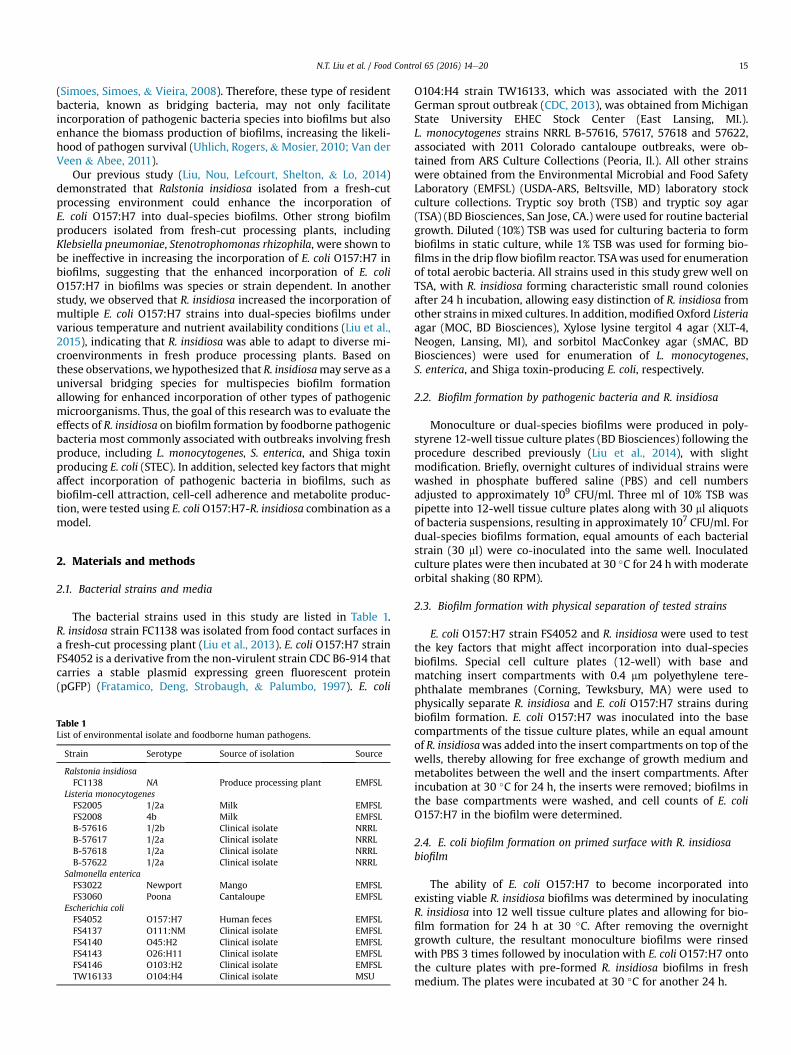
The total cell counts of each pathogenic strain in its monoculture
N.T. Liu et al. / Food Control 65 (2016) 14e20 17
and dual-species biofilms are shown in Fig. 2. In monoculturebiofilms, the cell counts for pathogenic stains ranged from 4.54 to7.67 log CFU/cm2. Most (12 out of 14) of the pathogenic strainsshowed significant increases (p < 0.05) in cell counts in dual-species biofilms as compared to their respective monoculture bio-films. The increases ranged from 0.36 log (E. coli strain TW16133) to1.84 logs (Salmonella strain FS3022). On the other hand, cell countsof E. coli FS4143 (p > 0.05) and FS4140 (p < 0.05) decreased in dual-species biofilms as compared to the monoculture biofilms.
Fig. 3. E. coli O157:H7 cell counts in biofilms formed in absence and presence ofR. insidiosa. Capital letters under the horizontal axis represent E. coli (E), R. insidiosa (R),or no bacterium (0) in the culture, letters outside the parentheses indicate bacteriagrown in the base compartments, and that inside the parentheses bacterium grown inthe insert. Tukey's multiple-comparison test was used to compare each set of data.Different lower case letters above the bars indicate significant difference at p < 0.05.
3.2. Biofilm formation by E. coli O157:H7 and R. insidiosa inmonocultures separated by permeable membrane
Since secreted signal molecules are a primary means of inter-species communications during biofilm formation (Karatan &Watnick, 2009), we examined the possibility that the incorpora-tion of pathogenic bacteria cells in the dual-species biofilms wasaffected by a mechanism akin to quorum sensing (Simoes, Simoes,& Vieira, 2007). R. insidiosa and E. coli O157:H7 cells were inocu-lated in two separate compartments separated by 0.4 mm poly-ethylene terephthalate filter membrane that supported freeexchange of culture medium and metabolites. Then E. coli O157:H7cell counts in the monoculture biofilms formed on the basecompartment surfacewere determined after incubation at 30 �C for24 h. No significant difference (p> 0.05) was observed in cell countsof E. coli O157:H7 in the monoculture biofilms formed with orwithout the presence of R. insidiosa in the permeable membrane-lined insert compartment (Fig. 3). This observation does not sup-port the hypothsis that R. insidiosa metabolites or secreted signalmolecules play a significant role in promoting the incorporation ofother pathogenic bacteria cells into dual-species biofilms.
3.3. Incorporation of E. coli O157:H7 in existing R. insidiosa biofilm
Since bacterial interspecies interactions can also occur by directcell-cell contact, we examined the incorporation of E. coli O157:H7cells into established R. insidiosa biofilms. Compared to E. coliO157:H7monoculture biofilms, a significant increase (p < 0.05) was
Fig. 2. Cell counts of tested foodborne pathogens (L. monocytogenes, Salmonella andshiga toxin-producing E. coli) in monoculture biofilms and in dual species biofilmsformed with R. insidiosa. Student's T-test was used to compare cell counts of eachtested pathogen in monoculture biofilms and in dual-species biofilms. *Indicates sig-nificant difference in pair-wise comparison at p < 0.05.
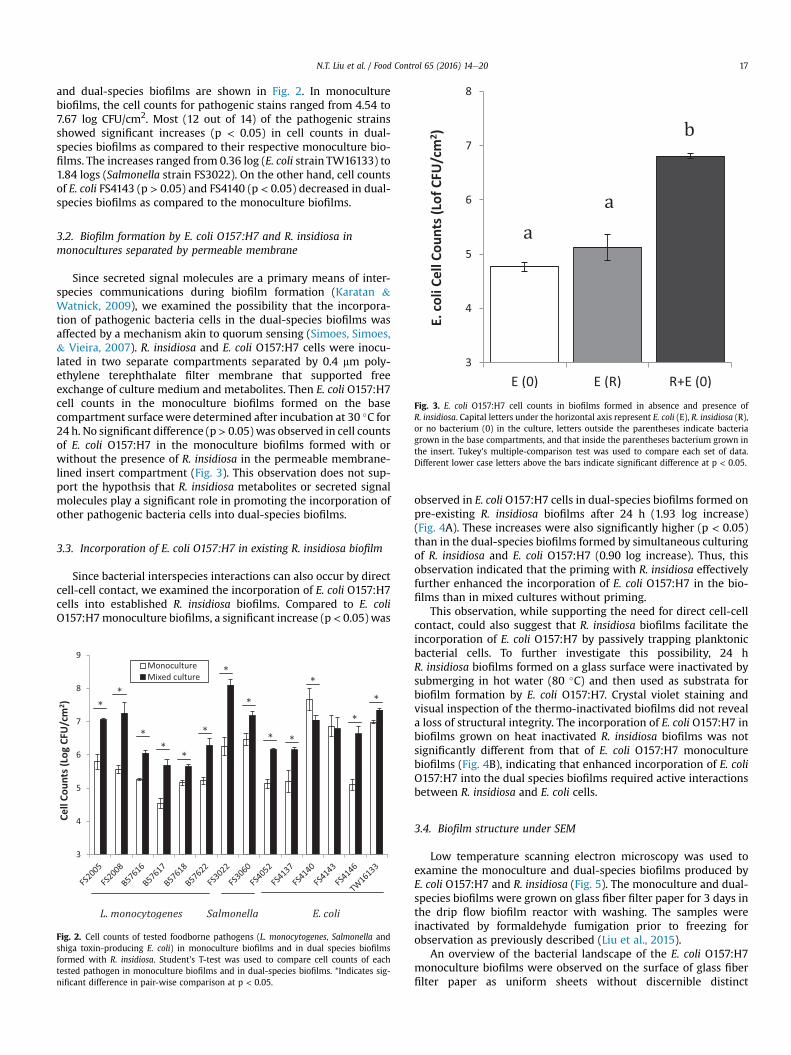
observed in E. coli O157:H7 cells in dual-species biofilms formed onpre-existing R. insidiosa biofilms after 24 h (1.93 log increase)(Fig. 4A). These increases were also significantly higher (p < 0.05)than in the dual-species biofilms formed by simultaneous culturingof R. insidiosa and E. coli O157:H7 (0.90 log increase). Thus, thisobservation indicated that the priming with R. insidiosa effectivelyfurther enhanced the incorporation of E. coli O157:H7 in the bio-films than in mixed cultures without priming.
This observation, while supporting the need for direct cell-cellcontact, could also suggest that R. insidiosa biofilms facilitate theincorporation of E. coli O157:H7 by passively trapping planktonicbacterial cells. To further investigate this possibility, 24 hR. insidiosa biofilms formed on a glass surface were inactivated bysubmerging in hot water (80 �C) and then used as substrata forbiofilm formation by E. coli O157:H7. Crystal violet staining andvisual inspection of the thermo-inactivated biofilms did not reveala loss of structural integrity. The incorporation of E. coli O157:H7 inbiofilms grown on heat inactivated R. insidiosa biofilms was notsignificantly different from that of E. coli O157:H7 monoculturebiofilms (Fig. 4B), indicating that enhanced incorporation of E. coliO157:H7 into the dual species biofilms required active interactionsbetween R. insidiosa and E. coli cells.
3.4. Biofilm structure under SEM
Low temperature scanning electron microscopy was used toexamine the monoculture and dual-species biofilms produced byE. coli O157:H7 and R. insidiosa (Fig. 5). The monoculture and dual-species biofilms were grown on glass fiber filter paper for 3 days inthe drip flow biofilm reactor with washing. The samples wereinactivated by formaldehyde fumigation prior to freezing forobservation as previously described (Liu et al., 2015).
An overview of the bacterial landscape of the E. coli O157:H7monoculture biofilms were observed on the surface of glass fiberfilter paper as uniform sheets without discernible distinct

Fig. 4. Effect of R. insidiosa priming on cell counts of E. coli O157:H7 in biofilms. (A)E. coli cell counts in monoculture (MN), mixed culture (MX), and R. insidiosa primed(PR) biofilms grow for 24 h in microplate. (B) E. coli cell counts in monoculture (MN),untreated R. insidiosa primed (PR), and heat inactivated R. insidiosa primed biofilmsgrown on glass slides in a drip flow system for 72 h. Tukey's multiple-comparison testwas used to compare each set of data. Different lower case letters above the barsindicate significant difference at p < 0.05.
Fig. 5. Scanning Electron Microscopy (SEM) of Biofilms. (A). Landscape view ofR. insidiosa monoculture biofilm showing the microcolonies and valleys. Box highlightsthe valleys; (B). Valley area of R. insidiosa biofilm. Lower-right corner is the edge of amicrocolony. Arrows point to the joints of web-like structures and filamentous pro-jections; and (C) Valley area of E. coli O157:H7 - R. insidiosa dual-species biofilm. Left isa partial microcolony.
N.T. Liu et al. / Food Control 65 (2016) 14e2018
structures. The porous property of the glass fiber paper surface wassupportive of E. coli O157:H7 cellular proliferation, however, theobserved uniform layer of cells did not show 3D biofilm structures(Image not shown). In contrast, R. insidiosa growth was observed asmicrocolonies with distinct domes and valleys, and the domedareas were seen connected through multiple conduit-like struc-tures (Fig. 5A). Under higher magnification, R. insidiosa cells wereseen as closely packed and possibly multilayered masses in thedomed areas (Not shown). The domed areas were connected byextensive web-like extracellular structures, which were mostevident in the valleys with low cell density (Fig. 5B). In addition,filamentous projections were seen extruding from the surface ofmicrocolonies to the surrounding valley areas, with the web-likestructures attached to these filamentous cells. There seemed tobe numerous sub-cellular sized particles distributed throughoutthe web-like structures and especially on the surfaces of the fila-mentous projections. The morphology of dual-species biofilms was
very similar to that of the R. insidiosa monoculture biofilms(Fig. 5C). Currently we are unable to confidently distinguish E. colicells from R. insidiosa using SEM. However, when the dual species-biofilms were examined using transmission electron microscope(TEM), R. insidiosa and E. coli O157:H7 cells exhibited segregatedspatial distribution, with E. coli O157:H7 microcolonies primarilyfound proximal to the matrix surface and R. insidiosa cells
N.T. Liu et al. / Food Control 65 (2016) 14e20 19
overlaying on top and in the domed areas (Liu et al., 2014).Therefore, it is likely that the observation of the dual-species bio-films using SEM is indeed simply a reflection of the characteristicsof R. insidiosa biofilms.
4. Discussion
4.1. R. insidiosa behaves like a bridging bacterium
In a polymicrobial community, certain species or strains interactsynergistically with amultitude of other species in forming biofilmsand are referred to as a bridge bacterium (Rickard et al., 2003). Forexample, Fusobacterium nucleatum, a prevalent bacterium in hu-man plaque, positively interacts with bacteria from 7 differentgenera, thereby connecting the planktonic bacteria to the biofilmsand enhancing biofilm production by mediating their adhesions tosurfaces (Shaniztki, Ganeshkumar, & Weiss, 1998). R. insidiosa iswidely present in diverse environmental niches, including watertreatment and supply systems (Coenye, Goris, De Vos, Vandamme,& LiPuma, 2003; Ryan, Pembroke, & Adley, 2011), making it one ofthe most common contaminants of food and food processing en-vironments. Our preliminary data (unpublished) suggests thatR. insidiosa is highly efficient in nutrient utilization and proliferateswell in oligonutrient environments, which could be advantageousfor bacteria serving as a bridging or pioneering species.
We have previously demonstrated that the species-specificinteraction between R. insidiosa and E. coli O157:H7 results inincreased populations of E. coli O157:H7 in dual-species biofilms(Liu et al., 2014). In this study, we show that this enhanced incor-poration can occur with other foodborne pathogenic bacteria,including 6 L. monocytogenes strains (representing 3 serotypes), 2serovars of S. enterica, and 4 of 6 serotypes of Shiga toxin-producingE. coli; although the increased cell counts of the foodborne patho-gens were not always accompanied by equally increased biomassproduction in dual-species biofilms. Especially notable is thesignificantly elevated biomass levels in dual-species biofilms formost (5 of 6) R. insidiosa-L. monocytogenes combinations. Thissuggests that large amounts of additional extracellular matriceswere produced in the dual-species biofilms, which could render thebiofilms more robust and more protective than either of themonoculture biofilms. Zammer and colleagues also observedincreased biomass production when co-culturing L. monocytogeneswith Staphylococcus epidermidis (Zameer, Kreft, & Gopal, 2010).Increased biomass production was also observed in the dual-species biofilms formed by R. insidiosa with environmental strainsisolated from fresh-cut processing facilities (unpublished data),including Pantoea agglomerans and Rahnella aquatilis. Takentogether, these observations suggest that R. insidiosa plays animportant role as a bridging bacterium (Rickard et al., 2003) that iscritical for multispecies biofilm formation, and provides a micro-environment for pathogenic bacterial accumulation and survival.
4.2. Role of cellular contacts in the interactions between R. insidiosaand the pathogenic strains
Interspecies communication and interactions can occur throughdiffusible signaling molecules or by intimate cellular contact(Flemming & Wingender, 2010; Karatan & Watnick, 2009). Dataacquired in the present study suggested that cellular contact withR. insidiosa cells was required for increased incorporation of E. coliO157:H7 into dual-species biofilms, as opposed to a diffusible signalThese observations are consistent with previous findings by Uhlichand colleagues (Uhlich et al., 2010), who also observed that directcell-cell contact with companion strains was required for E. coliO157:H7 biofilm formation.
There could be at least two mechanisms for R. insidiosaenhancing the incorporation of other species (potentially patho-gens) in heterogeneous biofilms. Bridging bacterium could serve asa primary colonizer by attaching to solid substrates and excretingextracellular polymeric substances that provides microniches forthe secondary colonizers (Rickard et al., 2003). Alternatively, itcould co-aggregate with other bacteria to form clusters that candeposit on surfaces to initiate colonization (Bos, van der Mei,Meinders, & Busscher, 1994; Rickard et al., 2003). ExistingR. insidiosa biofilms strongly promoted the incorporation of E. coliO157:H7 cells into biofilms, suggesting that co-aggregationwas notobligatory. However, heat inactivated R. insidiosa biofilms failed topromote the incorporation of E. coli O157:H7 into biofilms, indi-cating the necessity of active cell-cell interactions. Scanning elec-tron microscopy showed extensive web-like structures andfilamentous projections connecting R. insidiosa biofilm micro-colonies. The role of those structures in attracting other bacterialspecies should be further investigated.
Although we were unable to demonstrate that the compoundsor metabolites secreted by R. insidiosa could promote E. coliO157:H7 incorporation into biofilms, it cannot be ruled out thatsignal molecules play a role in R. insidiosa communicating withother bacteria species and in promoting multispecies biofilm for-mation. Ralstonia solanacearum, a closely related phytopathogen, iswell known for production of acyl-homoserine lactones (AHL), theputative cell-cell signaling molecules in biofilm development(Flavier, Ganova-Raeva, Schell, & Denny, 1997). It has been docu-mented that both E. coli and Salmonella produced receptors for AHLmolecules in order to detect surrounding microbial community(Dyszel et al., 2010; Michael, Smith, Swift, Heffron,& Ahmer, 2001).Further investigation of the potential roles of signal molecules inR. insidiosa interacting with foodborne pathogens may shed morelight on the understanding of polymicrobial biofilm formation infood processing environments.
Acknowledgments
The author thanks Dr. Todd Ward (ARS National Center forAgricultural Utilization Research, Peoria, Il.) and Michigan StateUniversity EHEC Stock Center (East Lansing, MI) for providing someof the strains used in this study. Mention of trade names or com-mercial products in this publication is solely for the purpose ofproviding specific information and does not imply recommenda-tion or endorsement by the USDA; USDA is an equal opportunityprovider and employer.
References
Bos, R., van der Mei, H. C., Meinders, J. M., & Busscher, H. J. (1994). A quantitativemethod to study co-adhesion of microorganisms in a parallel plate flowchamber: basic principles of the analysis. Journal of Microbiological Methods,20(4), 289e305.
Bowen, A., Fry, A., Richards, G., & Beuchat, L. (2006). Infections associated withcantaloupe consumption: a public health concern. Epidemiology and Infection,134(4), 675e685.
Carpentier, B., & Chassaing, D. (2004). Interactions in biofilms between Listeriamonocytogenes and resident microorganisms from food industry premises. In-ternational Journal of Food Microbiology, 97(2), 111e122.
CDC. (2013). Outbreak of Escherichia coli O104:H4 infections associated with sproutconsumption - Europe and North America, May-July 2011. MMWR Morbility andMortality Weekly Reports, 62(50), 1029e1031.
Chmielewski, R. A. N., & Frank, J. F. (2003). Biofilm formation and control in foodprocessing facilities. Comprehensive Reviews in Food Science and Food Safety,2(1), 22e32.
Coenye, T., Goris, J., De Vos, P., Vandamme, P., & LiPuma, J. J. (2003). Classification ofRalstonia pickettii-like isolates from the environment and clinical samples asRalstonia insidiosa sp. nov. International Journal of Systematic EvolutionaryMicrobiology, 53(Pt 4), 1075e1080.
Dyszel, J. L., Soares, J. A., Swearingen, M. C., Lindsay, A., Smith, J. N., & Ahmer, B. M.(2010). E. coli K-12 and EHEC genes regulated by SdiA. PLoS One, 5(1), e8946.
N.T. Liu et al. / Food Control 65 (2016) 14e2020
Flavier, A. B., Ganova-Raeva, L. M., Schell, M. A., & Denny, T. P. (1997). Hierarchicalautoinduction in Ralstonia solanacearum: control of acyl-homoserine lactoneproduction by a novel autoregulatory system responsive to 3-hydroxypalmiticacid methyl ester. Journal of Bacteriology, 179(22), 7089e7097.
Flemming, H. C., & Wingender, J. (2010). The biofilm matrix. Nature ReviewsMicrobiology, 8(9), 623e633.
Fratamico, P. M., Deng, Y. M., Strobaugh, T. P., & Palumbo, S. A. (1997). Constructionand characterization of Escherichia coli O157:H7 strains expressing fireflyluciferase and green fluorescent protein and their use in survival studies.Journal of Food Protection, 60(10), 1167e1173.
Habimana, O., Heir, E., Langsrud, S., Asli, A. W., & Moretro, T. (2010). Enhancedsurface colonization by Escherichia coli O157:H7 in biofilms formed by an Aci-netobacter calcoaceticus isolate from meat-processing environments. Appliedand Environmental Microbiology, 76(13), 4557e4559.
Harris, L. J., Farber, J. N., Beuchat, L. R., Suslow, T. V., Garrett, E. H., & Busta, F. F.(2003). Outbreaks associated with fresh produce: incidence, growth, and sur-vival of pathogens in fresh and fresh- cut produce. Comprehensive Reviews inFood Science and Food Safety, 2(suppliment), 78e141.
Jeong, D. K., & Frank, J. F. (1994). Growth of Listeria monocytogenes at 10 oC inbiofilms with microorganisms isolated from meat and dairy processing envi-ronments. Journal of Food Protection, 57(7), 576e586.
Karatan, E., & Watnick, P. (2009). Signals, regulatory networks, and materials thatbuild and break bacterial biofilms. Microbiology and Molecular Biology Reviews,73(2), 310e347.
Liu, N. T., Lefcourt, A. M., Nou, X., Shelton, D. R., Zhang, G., & Lo, Y. M. (2013). Nativemicroflora in fresh-cut produce processing plants and their potentials for bio-film formation. Journal of Food Protection, 5, 827e832.
Liu, N. T., Nou, X., Bauchan, G. R., Murphy, C., Lefcourt, A. M., Shelton, D. R., et al.(2015). Effects of environmental parameters on the dual-species biofilmsformed by Escherichia coli O157:H7 and Ralstonia insidiosa, a strong biofilmproducer isolated from a fresh-cut produce processing plant. Journal of FoodProtection, 78(1), 121e127.
Liu, N. T., Nou, X., Lefcourt, A. M., Shelton, D. R., & Lo, Y. M. (2014). Dual-speciesbiofilm formation by Escherichia coli O157: H7 and environmental bacteriaisolated from fresh-cut processing facilities. International Journal of FoodMicrobiology, 171, 15e20.
Lynch, M. F., Tauxe, R. V., & Hedberg, C. W. (2009). The growing burden of foodborneoutbreaks due to contaminated fresh produce: risks and opportunities. Epide-miology and Infection, 137(3), 307e315.
Michael, B., Smith, J. N., Swift, S., Heffron, F., & Ahmer, B. M. (2001). SdiA of Sal-monella enterica is a LuxR homolog that detects mixed microbial communities.
Journal of Bacteriology, 183(19), 5733e5742.Penteado, A. L., Eblen, B. S., & Miller, A. J. (2004). Evidence of Salmonella internal-
ization into fresh mangos during simulated postharvest insect disinfestationprocedures. Journal of Food Protection, 67(1), 181e184.
Rickard, A. H., Gilbert, P., High, N. J., Kolenbrander, P. E., & Handley, P. S. (2003).Bacterial coaggregation: an integral process in the development of multi-species biofilms. Trends in Microbiology, 11(2), 94e100.
Ryan, M. P., Pembroke, J. T., & Adley, C. C. (2011). Genotypic and phenotypic diversityof Ralstonia pickettii and Ralstonia insidiosa isolates from clinical and environ-mental sources including high-purity water. Diversity in Ralstonia pickettii. BMCMicrobiology, 11, 194.
Scher, K., Romling, U., & Yaron, S. (2005). Effect of heat, acidification, and chlori-nation on Salmonella enterica serovar Typhimurium cells in a biofilm formed atthe air-liquid interface. Applied and Environmental Microbiology, 71(3),1163e1168.
Shaniztki, B., Ganeshkumar, N., & Weiss, E. (1998). Characterization of a novel N-acetylneuraminic acid-specific Fusobacterium nucleatum PK1594 adhesin. Oralmicrobiology and immunology, 13(1), 47e50.
Silagyi, K., Kim, S. H., Lo, Y. M., & Wei, C. I. (2009). Production of biofilm and quorumsensing by Escherichia coli O157:H7 and its transfer from contact surfaces tomeat, poultry, ready-to-eat deli, and produce products. Food Microbiology, 26(5),514e519.
Simoes, L. C., Simoes, M., & Vieira, M. J. (2007). Biofilm interactions between distinctbacterial genera isolated from drinking water. Applied and EnvironmentalMicrobiology, 73(19), 6192e6200.
Simoes, L. C., Simoes, M., & Vieira, M. J. (2008). Intergeneric coaggregation amongdrinking water bacteria: evidence of a role for Acinetobacter calcoaceticus as abridging bacterium. Applied and Environmental Microbiology, 74(4), 1259e1263.
Uhlich, G. A., Rogers, D. P., & Mosier, D. A. (2010). Escherichia coli serotype O157:H7retention on solid surfaces and peroxide resistance is enhanced by dual-strainbiofilm formation. Foodborne Pathogens and Disease, 7(8), 935e943.
Van der Veen, S., & Abee, T. (2011). Mixed species biofilms of Listeria monocytogenesand Lactobacillus plantarum show enhanced resistance to benzalkonium chlo-ride and peracetic acid. International Journal of Food Microbiology, 144(3),421e431.
Warriner, K., & Namvar, A. (2010). The tricks learnt by human enteric pathogensfrom phytopathogens to persist within the plant environment. Current Opinionin Biotechnology, 21(2), 131e136.
Zameer, F., Kreft, J., & Gopal, S. (2010). Interaction of Listeria monocytogenes andStaphylococcus epidermidis in dual species biofilms. Journal of Food Safety, 30(4),954e968.