Embed Size (px)
Citation preview
Rapid and Sensitive Detection of an IntracellularPathogen in Human Peripheral Leukocytes withHybridizing Magnetic Relaxation NanosensorsCharalambos Kaittanis1, Hamza Boukhriss1, Santimukul Santra1, Saleh A. Naser2, J. Manuel Perez1,2,3*
1NanoScience Technology Center, University of Central Florida, Orlando, Florida, United States of America, 2 Burnett School of Biomedical Sciences, College of Medicine,
University of Central Florida, Orlando, Florida, United States of America, 3Department of Chemistry, University of Central Florida, Orlando, Florida, United States of
America
Abstract
Bacterial infections are still a major global healthcare problem. The quick and sensitive detection of pathogens responsiblefor these infections would facilitate correct diagnosis of the disease and expedite treatment. Of major importance areintracellular slow-growing pathogens that reside within peripheral leukocytes, evading recognition by the immune systemand detection by traditional culture methods. Herein, we report the use of hybridizing magnetic nanosensors (hMRS) for thedetection of an intracellular pathogen, Mycobacterium avium spp. paratuberculosis (MAP). The hMRS are designed to bind toa unique genomic sequence found in the MAP genome, causing significant changes in the sample’s magnetic resonancesignal. Clinically relevant samples, including tissue and blood, were screened with hMRS and results were compared withtraditional PCR analysis. Within less than an hour, the hMRS identified MAP-positive samples in a library of laboratorycultures, clinical isolates, blood and homogenized tissues. Comparison of the hMRS with culture methods in terms ofprediction of disease state revealed that the hMRS outperformed established culture methods, while being significantlyfaster (1 hour vs 12 weeks). Additionally, using a single instrument and one nanoparticle preparation we were able to detectthe intracellular bacterial target in clinical samples at the genomic and epitope levels. Overall, since the nanoparticles arerobust in diverse environmental settings and substantially more affordable than PCR enzymes, the potential clinical andfield-based use of hMRS in the multiplexed identification of microbial pathogens and other disease-related biomarkers viaa single, deployable instrument in clinical and complex environmental samples is foreseen.
Citation: Kaittanis C, Boukhriss H, Santra S, Naser SA, Perez JM (2012) Rapid and Sensitive Detection of an Intracellular Pathogen in Human Peripheral Leukocyteswith Hybridizing Magnetic Relaxation Nanosensors. PLoS ONE 7(4): e35326. doi:10.1371/journal.pone.0035326
Editor: Baochuan Lin, Naval Research Laboratory, United States of America
Received December 20, 2011; Accepted March 12, 2012; Published April 9, 2012
Copyright: � 2012 Kaittanis et al. This is an open-access article distributed under the terms of the Creative Commons Attribution License, which permitsunrestricted use, distribution, and reproduction in any medium, provided the original author and source are credited.
Funding: This work was supported by the National Institute of General Medical Sciences GM084331 grant awarded to JMP. The funders had no role in studydesign, data collection and analysis, decision to publish, or preparation of the manuscript.
Competing Interests: The authors have declared that no competing interests exist.
* E-mail: [email protected]
Introduction
Pathogenesis caused by intracellular pathogens, such as
Mycobacterium tuberculosis among others, relies on the survival of
the microorganism within host cells, such as macrophages and
dendritic cells. This mode of infection with or without clinical
symptomology hampers bacterial detection, preventing correct
diagnosis. Consequently, the physician cannot proceed to the
assignment of an appropriate treatment course, delaying the
clearance of the pathogen from the body. Serological immunoas-
says cannot effectively detect intracellular pathogens in biological
fluids (e.g. blood, lymphatic fluid) as the microorganism is hidden
away within the immune cells. For this reason, isolation of the
infected cells and extraction of their DNA, followed by detection of
specific bacterial genomic markers via Polymerase Chain Reaction
(PCR), is needed to facilitate identification of the pathogen.
Although highly specific and sensitive, PCR methods are generally
laborious, time-consuming and typically require homogeneous and
pure DNA samples [1]. Evidently, complex biological samples,
such as biopsies and blood, have to be thoroughly processed in
multistep elaborate protocols to obtain pure DNA samples,
significantly increasing the final readout time, and affecting the
overall DNA yield. An alternative way to detect intracellular
pathogens, particularly when they are in low numbers, is by
isolating the infected leukocytes from the blood and expanding the
number of viable bacteria via culturing methods before PCR
analysis. Although very effective, bacterial culturing methods
require a significant amount of time, such as in the case of slow-
growing intracellular pathogens that have to be cultivated for even
weeks, assuming that there is an adequate amount of viable
pathogens in the clinical sample. Another drawback of culturing
methods is that some pathogens do not grow effectively in culture,
either because of the nature of the pathogen itself or the presence
of biological interferences present in the clinical sample that
prevent growth in culture. Because of these hurdles, new
technologies that can detect the presence of a pathogen in human
clinical samples are urgently needed.
It has been predicted that nanotechnology will have a major
impact in medicine, agriculture and biotechnology among other
fields [2,3,4,5,6,7,8,9,10]. Innovative technologies that take
advantage of the unique size-dependent electronic, magnetic and
luminescence properties that some nanomaterials exhibit when
they specifically interact with a biological marker have been
PLoS ONE | www.plosone.org 1 April 2012 | Volume 7 | Issue 4 | e35326
developed for faster and more accurate sensing of various
pathogens [2,3,4,5]. However, the translation of these technologies
to the clinical diagnosis of an infectious disease and how it relates
to the presence of the pathogen in clinical samples has been
limited. The slow clinical translation of some of these nano-
technologies has been partly due to their poor performance in
crude or minimally processed clinical samples. Among the most
promising nanomaterials are magnetic relaxation nanosensors
(MRS). These magnetic nanosensors are composed of a polymer-
coated iron oxide nanoparticle onto which affinity ligands are
conjugated to facilitate binding and magnetic detection of
a particular target [11,12]. Upon specific binding of a target to
ligands on the magnetic nanoparticle, changes in the sample’s
magnetic resonance signal (specifically the water proton relaxation
time; T2) occur that correlate with the target concentration in
solution. By measuring the changes in T2 relaxation times upon
target interaction and correlating the intensity of the change with
the target concentration, one can develop a sensitive detection
method, therefore the acronym of magnetic relaxation nanosen-
sors (MRS).
In contrast to other methods that utilize magnetic nanopar-
ticles as labels and directly measure an intrinsic physical
property, such as a magnetic field [13,14], or a chemical
property [6], the MRS method relies on the effect that the
nanoparticle’s induced magnetic field exerts on the hundreds of
thousands of water molecules surrounding the nanoparticle.
This effect results in an amplification of the signal, allowing for
sensitive detection even in turbid and minimally processed
samples, without the need of further amplification, which is
critical for assays such as PCR and ELISA. Hence, MRS-based
assays can be more affordable than traditional assays, since they
use a single reagent that can be easily produced in large
quantities, and a single-step that avoids time- and labor-
consuming procedures. Additionally, MRS can detect their
targets at various point-of-care settings, since the nanosensors
are stable at ambient conditions and utilize deployable
instrumentation. We recently reported a unique MRS nano-
particle-target interaction that resulted in rapid T2 increases
upon target binding [12]. This unique nanoparticle-target
interaction is different from other MRS-based methods, because
clustering of the nanoparticles is not required to achieve
detection. In contrast, binding of MRS to the target caused
fast, target-concentration-dependent increases in T2. Kinetic
studies also revealed that the binding of MRS was faster and
more sensitive than the MRS clustering, while achieving lower
detection thresholds. In addition, nanoparticle valency – the
amount of a targeting ligand on the surface of the nanoparticle
– plays a role in the magnetic relaxation response. Nanoparticle
valency controls the trend of the change in the spin-spin
relaxation time (DT2). Specifically, it has been found that low
valency results in decreases in the DT2 as the target
concentration increases, while at high valency conditions the
DT2 increases as the target concentration increases.
Hence considering the faster detection kinetics of MRS binding-
based detection assays and the higher sensitivity of the MRS low-
valency nanoparticle system, we developed hybridizing MRS
(hMRS) that switched (increased) the sample’s water proton
relaxation times (T2) upon binding to a unique bacterial genomic
marker and tested their performance in clinical samples. The
hMRS were designed to bind to a specific genomic marker in
Mycobacterium avium spp. paratuberculosis (MAP); an intracellular
pathogen known to cause Johne’s disease in cattle [15] and has
been also implicated in the cause of Crohn’s disease in humans
[16]. Furthermore, acknowledging the limitations of PCR in
detecting nucleic acid biomarkers of intracellular pathogens in
clinical samples, we investigated the hMRS performance in
detecting MAP in human peripheral blood samples of Crohn’s
disease patients as well as animal tissues with Johne’s disease.
MAP is found within the white blood cells of infected animals
with Johne’s disease, a form of animal paratuberculosis, which is
associated with chronic enteritis, reminiscent of Crohn’s disease
in humans [16]. As early as 1913, Dalziel noted the clinical
similarities of animal paratuberculosis, intestinal tuberculosis and
human chronic granulomatous enteritis (Crohn’s disease) [17].
In humans, Crohn’s disease is a debilitating chronic inflamma-
tory syndrome of the gastrointestinal track and adjacent lymph
nodes [17,18,19]. The detection of MAP in tissues from patients
with Crohn’s disease has been extensively reported. Of
particular importance to our study is the report of the presence
of MAP in human peripheral blood [20,21,22]. In these studies,
MAP was identified by a culture method followed by PCR
identification of a MAP genomic marker. The whole process
took several months to complete, due to the slow growing
nature of this pathogen. Such a slow detection method not only
delays the diagnosis, but also slows any potential therapeutic
intervention [20,21]. Likewise, difficulties in detecting an
intracellular pathogen, such as MAP, hamper studies aimed at
the investigation of the potential role of MAP in Crohn’s disease
pathology, as well as the pathogen’s impact on the dairy and
beef industries.
Attracted by MAP’s slow growing characteristics, intracellular
residence and clinical and agricultural relevance, we used hMRS
for the identification of a highly conserved MAP genomic element
(IS900). This unique DNA insertion sequence has been previously
used to identify MAP in clinical samples and cultures by PCR, as it
has not been found in other microorganisms or mycobacteria
[23,24,25]. Detection is primarily achieved through either direct
or culture-based nested PCR (nPCR), which use pure DNA
extracts from infected leukocytes. Both PCR methods sequentially
amplify a 398 bp fragment (amplification round 1) and a 298 bp
internal sequence (amplification round 2). We reasoned that the
binding of hMRS to IS900 would achieve faster and more
sensitive detection of MAP DNA via magnetic relaxation in
minimally processed clinical samples. Initial studies revealed that
hMRS can specifically detect MAP’s genomic marker without
showing cross-reactivity with other mycobacteria. Furthermore,
the presence of MAP in homogenized tissues from Johne’s disease
cattle was achieved at the genomic and epitope levels using hMRS
and anti-MAP antibody-carrying MRS, demonstrating that the
MRS technology can achieve detection at the genome and
organism levels using diverse molecular probes. Finally, a library
of 60 blood samples from Crohn’s disease patients and healthy
individuals was screened with hMRS and nPCR assays. Using
minimally processed blood samples, within 60 minutes the hMRS
were able to detect the intracellular pathogen and better predict
the clinical condition of an individual (healthy vs Crohn’s disease)
than the nPCR methodologies. Specifically, hMRS outperformed
the 12-week-long culture-based nPCR in clinical prognosis, as well
as the least reliable direct nPCR, which are currently used in
clinical and research studies. Overall, we show for the first time the
use and validation of a novel translational nanotechnology, where
the unique water relaxation properties of iron oxide nanoparticles
and the dynamics of the nanoparticle/target interaction were
harnessed to achieve a sensitive genomic biomarker detection
system for an intracellular pathogen in samples directly obtained
from human patients.
Intracellular Pathogen Detection Using Nanosensors
PLoS ONE | www.plosone.org 2 April 2012 | Volume 7 | Issue 4 | e35326
Results
Design and Synthesis of hMRSThe detection of MAP’s IS900 genomic marker relies on
DNA isolation and subsequent amplification and detection via
PCR. Typically, DNA from peripheral leukocytes found in the
buffy coat (Direct nPCR) or leukocyte-derived cultured bacteria
(Culture-based nPCR) is first isolated in a multistep protocol
[21], yielding bacterial DNA of high quality (Figure 1a).However, this approach often times compromises DNA yield,
which may impede MAP identification. In subsequent steps, the
bacterial DNA is subjected to two 3-hour-long polymerase-
mediated amplification rounds of the IS900 locus, followed by
gel electrophoresis. To facilitate faster detection via magnetic
relaxation, we designed a single hMRS probe that hybridizes
with the unique IS900 sequence within the MAP plasmid.
These hMRS are composed of a polyacrylic acid-coated iron
oxide nanoparticle onto which an oligonucleotide sequence
(ATGTGGTTGCTGTGT) complementary to the IS900 se-
quence in MAP is conjugated to facilitate binding and
detection. The resulting MAP specific hMRS had a diameter
of 78 6 3 nm, zeta potential value of –35 mV, an average of
55 oligonucleotides per nanoparticle, with R1 and R2
relaxivities (at 0.47T) of 45 mM-1s-1 and 60 mM-1s-1 respective-
ly. We anticipated that hMRS could utilize crude DNA,
obtained from isolated buffy coats that have been subjected to
heating (Figure 1a). It is well reported that heating of
peripheral leukocytes accomplishes first the release of a pathogen
from intracellular compartments [21], such as phagosomes, due
to extensive pore formation on the host cell’s plasma membrane
and organelles’ phospholipid-containing membrane. Once ex-
posed to high temperatures, the pathogen’s structural integrity is
affected, leading to release of its genomic DNA to the aqueous
milieu. A series of heating and cooling procedures on the
isolated MAP DNA plasmid in the presence of hMRS reduces
the DNA torsional strain, facilitating annealing and hydridiza-
tion of the hMRS onto the IS900 region of the MAP genome
(Figure 1b). Decreasing the sample’s temperature can return
the DNA to a higher structural order, with the hMRS still
binding to the MAP genome and inducing increases in the
sample’s magnetic resonance signal. Specifically, the hMRS
were able to bind to MAP DNA after two short heating rounds
(3 min each at 95uC), followed by unassisted cooling to room
temperature. The fast changes in the magnetic resonance signal
were monitored with a compact nuclear magnetic resonance
instrument (0.47T, Bruker, Minispec), recording increases in the
sample’s transverse relaxation times (DT2+, where T2(sample) .
T2(control)). Within 30 minutes after removing the samples from
heat, significant and reproducible changes were observed
prompting us to set this time-point as the hMRS readout point
(Figure S1). Based on the observation that the sample’s T2
times were higher than those of the sterile control throughout
the experimental time course, we deduced that the hMRS first
bound to the relaxed circular MAP DNA that was heated, while
the hMRS still remained bound to the cooled DNA as it was
returning to its supercoil form. These results were corroborated
by dynamic light scattering studies that showed no clustering of
the nanoparticle suspension upon addition of the isolated MAP
genome, since the nanoparticle diameter was found to be 81 6
5 nm. Likely, this may be attributed to the fact that during
sample cooling the DNA returned to its supercoiled conforma-
tion, which can be in the order of a few nanometers, and as
a result nominally altering the overall size of the hybridizing
nanoparticle – DNA complex.
hMRS are Specific and Sensitive ProbesTo determine the specificity of the hMRS, we incubated the
hMRS (6 mg Fe/mL) with pure DNA extracts from various
mycobacteria and monitored changes in T2 signal.
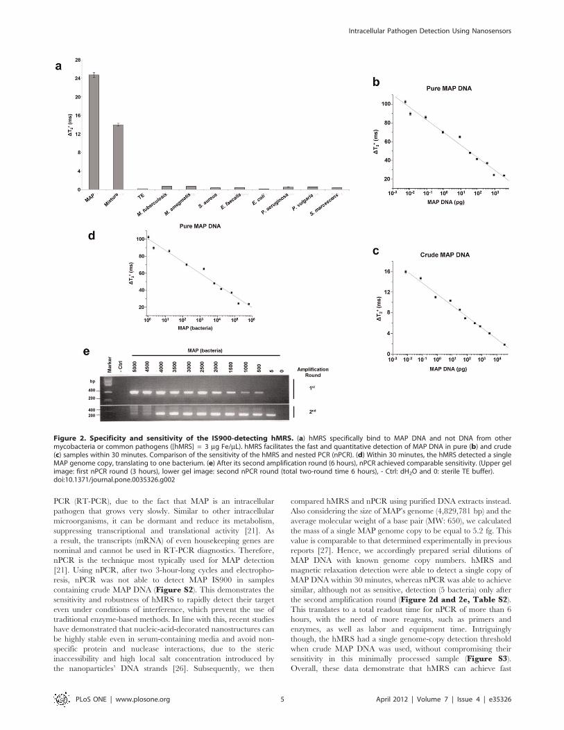
Within 30 minutes, results indicated that only samples contain-
ing MAP DNA exhibited a high DT2+ signal, being attributed to
the hMRS – MAP DNA binding (Figure 2a). Additionally, thehMRS were still able to identify their target in a solution with
a mixture of MAP and other mycobacterial DNA (Figure 2a).This indicates that the hMRS are sensitive and specific, without
being affected by the presence of other mycobacterial DNA.
Furthermore, addition of a synthetic oligonucleotide complemen-
tary to the IS900 sequence abrogated the signal of the hMRS upon
MAP DNA addition, confirming the specificity of the hMRS
probe (data not shown). Since other microorganisms’ DNA might
be found in a clinical sample, we examined if the hMRS could
differentiate between MAP and common Gram positive and Gram
negative bacteria, as well as different types of mycobacteria.
Results showed that only the MAP sample yielded a high DT2+,
whereas the other bacterial DNA samples exhibited signal
proximal to that of the sterile control (Figure 2a). These data
hint the potential use of the hMRS and magnetic relaxation
detection for the fast and specific detection of MAP DNA.
In subsequent studies, we determined if the hMRS could
quantify MAP DNA. MAP DNA was isolated using either an
elaborate multistep extraction protocol to obtain pure DNA, or
a fast 30-minute boiling-based methodology to obtain crude DNA.
After a 30-minute incubation period, the hMRS were able to
detect MAP DNA in both pure and crude DNA preparations,
exhibiting concentration-dependent DT2+ changes (Figures 2b
and 2c, Table S1). The observed MAP concentration response
pattern exhibited a high magnetic resonance signal (DT2+) at low
target concentration levels and low signals at higher DNA
concentrations. This suggests that the binding between the hMRS
and MAP DNA was facilitated via low valency interaction, as
previously described in other models [11]. This unique interaction
mechanism provides enhanced sensitivity during the screening of
scarce biomarkers, since high signal is desired at low target
concentrations. Quantification of the number of oligonucleotides
on the hMRS probe revealed that on average there were 55
oligonucleotides (oligos) per hMRS. This level of oligos per
nanoparticle is in line with previous reports, where valency
grafting of 46 oligos per nanoparticle exhibited a low valency
binding behavior, whereas a nanoparticle with 368 oligos obeyed
a high valency mechanism [12]. A low-valency-based detection
approach is ideal for the identification of MAP in clinical samples,
since it will induce a prominent DT2+ signal at low numbers of
MAP. Based on the findings that both pure and crude MAP DNA
exhibited similar hMRS quantification patterns, it was deduced
that the hMRS assay can detect IS900 independent of the sample’s
DNA extraction methodology. Although higher changes were
recorded in pure MAP DNA samples, both extraction method-
ologies had equivalent detection thresholds, reaching low femto-
gram (10–15) levels. Potentially the differences in the magnitude of
the DT2+ from pure and crude MAP DNA might be attributed to
the sample’s unique characteristics. It is plausible that the high
purity of the pure DNA extracts is associated with marginal
protein and lipid levels, resulting in lower viscosity. Alternatively,
a crude MAP DNA sample may have substantially higher content
of proteins and lipids, thus higher viscosity, which may be reflected
in lower DT2+ values upon IS9000 – hMRS hybridization We then
examined whether nested PCR (nPCR) can achieve comparable
sensitivity using crude MAP DNA. We decided to compare our
hMRS method with nPCR as opposed to reverse transcriptase
Intracellular Pathogen Detection Using Nanosensors
PLoS ONE | www.plosone.org 3 April 2012 | Volume 7 | Issue 4 | e35326
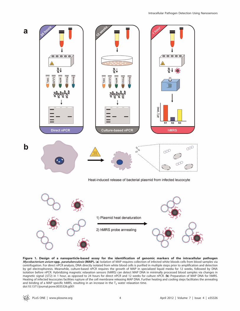
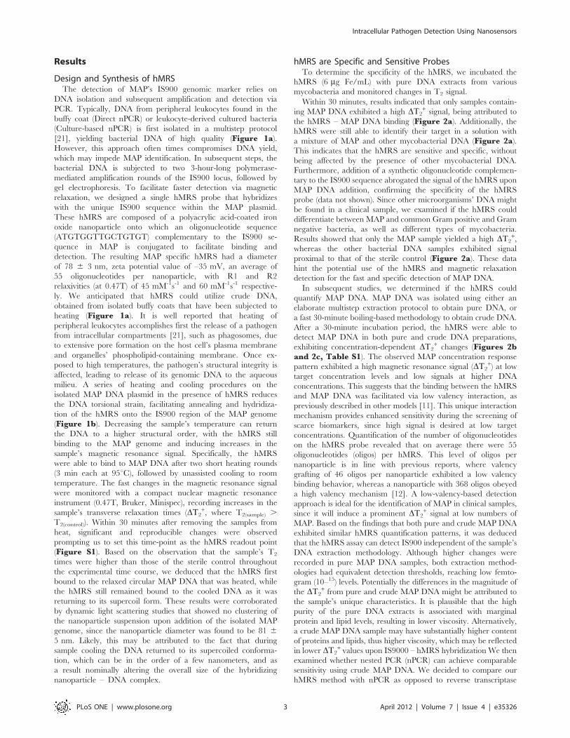
Figure 1. Design of a nanoparticle-based assay for the identification of genomic markers of the intracellular pathogenMycobacterium avium spp. paratuberculosis (MAP). (a) Isolation of MAP requires collection of infected white bloods cells from blood samples viacentrifugation. For direct nPCR analysis, DNA directly isolated from white blood cells is purified in multiple steps prior to amplification and detectionby gel electrophoresis. Meanwhile, culture-based nPCR requires the growth of MAP in specialized liquid media for 12 weeks, followed by DNAisolation before nPCR. Hybridizing magnetic relaxation sensors (hMRS) can detect MAP DNA in minimally processed blood samples via changes inmagnetic signal (DT2) in 1 hour, as opposed to 24 hours for direct nPCR and 12 weeks for culture nPCR. (b) Preparation of MAP DNA for hMRS.Heating of infected leucocytes facilities rupture of the cell membrane releasing MAP DNA. Further heating and cooling steps facilitates the annealingand binding of a MAP specific hMRS, resulting in an increase in the T2 water relaxation time.doi:10.1371/journal.pone.0035326.g001
Intracellular Pathogen Detection Using Nanosensors
PLoS ONE | www.plosone.org 4 April 2012 | Volume 7 | Issue 4 | e35326
PCR (RT-PCR), due to the fact that MAP is an intracellular
pathogen that grows very slowly. Similar to other intracellular
microorganisms, it can be dormant and reduce its metabolism,
suppressing transcriptional and translational activity [21]. As
a result, the transcripts (mRNA) of even housekeeping genes are
nominal and cannot be used in RT-PCR diagnostics. Therefore,
nPCR is the technique most typically used for MAP detection
[21]. Using nPCR, after two 3-hour-long cycles and electropho-
resis, nPCR was not able to detect MAP IS900 in samples
containing crude MAP DNA (Figure S2). This demonstrates the
sensitivity and robustness of hMRS to rapidly detect their target
even under conditions of interference, which prevent the use of
traditional enzyme-based methods. In line with this, recent studies
have demonstrated that nucleic-acid-decorated nanostructures can
be highly stable even in serum-containing media and avoid non-
specific protein and nuclease interactions, due to the steric
inaccessibility and high local salt concentration introduced by
the nanoparticles’ DNA strands [26]. Subsequently, we then
compared hMRS and nPCR using purified DNA extracts instead.
Also considering the size of MAP’s genome (4,829,781 bp) and the
average molecular weight of a base pair (MW: 650), we calculated
the mass of a single MAP genome copy to be equal to 5.2 fg. This
value is comparable to that determined experimentally in previous
reports [27]. Hence, we accordingly prepared serial dilutions of
MAP DNA with known genome copy numbers. hMRS and
magnetic relaxation detection were able to detect a single copy of
MAP DNA within 30 minutes, whereas nPCR was able to achieve
similar, although not as sensitive, detection (5 bacteria) only after
the second amplification round (Figure 2d and 2e, Table S2).This translates to a total readout time for nPCR of more than 6
hours, with the need of more reagents, such as primers and
enzymes, as well as labor and equipment time. Intriguingly
though, the hMRS had a single genome-copy detection threshold
when crude MAP DNA was used, without compromising their
sensitivity in this minimally processed sample (Figure S3).Overall, these data demonstrate that hMRS can achieve fast
Figure 2. Specificity and sensitivity of the IS900-detecting hMRS. (a) hMRS specifically bind to MAP DNA and not DNA from othermycobacteria or common pathogens ([hMRS] = 3 mg Fe/mL). hMRS facilitates the fast and quantitative detection of MAP DNA in pure (b) and crude(c) samples within 30 minutes. Comparison of the sensitivity of the hMRS and nested PCR (nPCR). (d) Within 30 minutes, the hMRS detected a singleMAP genome copy, translating to one bacterium. (e) After its second amplification round (6 hours), nPCR achieved comparable sensitivity. (Upper gelimage: first nPCR round (3 hours), lower gel image: second nPCR round (total two-round time 6 hours), - Ctrl: dH2O and 0: sterile TE buffer).doi:10.1371/journal.pone.0035326.g002
Intracellular Pathogen Detection Using Nanosensors
PLoS ONE | www.plosone.org 5 April 2012 | Volume 7 | Issue 4 | e35326
single MAP genome detection even in crude DNA samples,
outperforming nPCR in readout time, sensitivity and robustness.
hMRS Achieve Genome-based Detection of theIntracellular Pathogen Mycobacterium avium spp.Paratuberculosis in Cultured Clinical Isolates and TissueSamplesSince the hMRS detected MAP DNA in samples from pure
bacterial cultures, we investigated if the nanosensors could identify
their targets in more complex samples, such as cultured clinical
isolates and homogenized tissue preparations. We first screened
cultures of clinical isolates from Crohn’s disease patients with the
hMRS and compared these results with nPCR. Specifically, we
used crude DNA for the hMRS and pure DNA for the nPCR,
since nPCR cannot utilize crude DNA. Following a single-step
procedure, crude DNA obtained from cultured isolates of healthy
individuals yielded a magnetic signal change that was comparable
to that of the sterile culture medium (DT2+ = 1.3 6 0.2 ms),
establishing this magnetic signal change as the hMRS negative
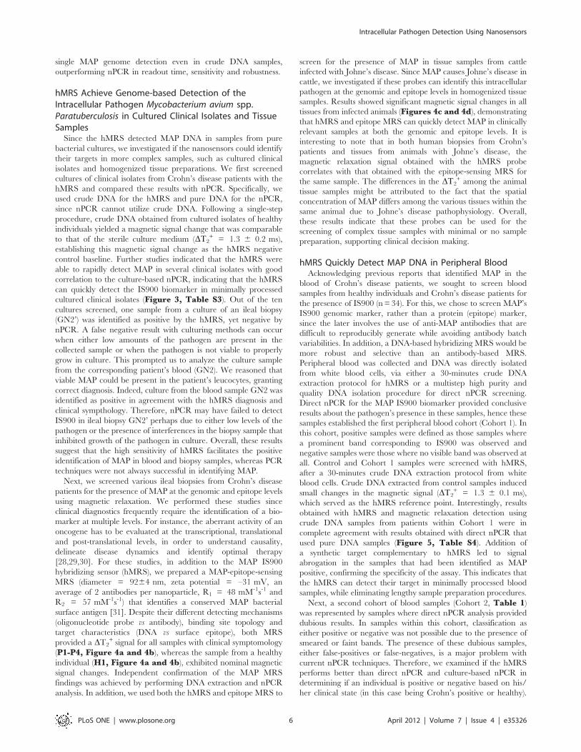
control baseline. Further studies indicated that the hMRS were
able to rapidly detect MAP in several clinical isolates with good
correlation to the culture-based nPCR, indicating that the hMRS
can quickly detect the IS900 biomarker in minimally processed
cultured clinical isolates (Figure 3, Table S3). Out of the ten
cultures screened, one sample from a culture of an ileal biopsy
(GN2’) was identified as positive by the hMRS, yet negative by
nPCR. A false negative result with culturing methods can occur
when either low amounts of the pathogen are present in the
collected sample or when the pathogen is not viable to properly
grow in culture. This prompted us to analyze the culture sample
from the corresponding patient’s blood (GN2). We reasoned that
viable MAP could be present in the patient’s leucocytes, granting
correct diagnosis. Indeed, culture from the blood sample GN2 was
identified as positive in agreement with the hMRS diagnosis and
clinical sympthology. Therefore, nPCR may have failed to detect
IS900 in ileal biopsy GN2’ perhaps due to either low levels of the
pathogen or the presence of interferences in the biopsy sample that
inhibited growth of the pathogen in culture. Overall, these results
suggest that the high sensitivity of hMRS facilitates the positive
identification of MAP in blood and biopsy samples, whereas PCR
techniques were not always successful in identifying MAP.
Next, we screened various ileal biopsies from Crohn’s disease
patients for the presence of MAP at the genomic and epitope levels
using magnetic relaxation. We performed these studies since
clinical diagnostics frequently require the identification of a bio-
marker at multiple levels. For instance, the aberrant activity of an
oncogene has to be evaluated at the transcriptional, translational
and post-translational levels, in order to understand causality,
delineate disease dynamics and identify optimal therapy
[28,29,30]. For these studies, in addition to the MAP IS900
hybridizing sensor (hMRS), we prepared a MAP-epitope-sensing
MRS (diameter = 9264 nm, zeta potential = –31 mV, an
average of 2 antibodies per nanoparticle, R1 = 48 mM-1s-1 and
R2 = 57 mM-1s-1) that identifies a conserved MAP bacterial
surface antigen [31]. Despite their different detecting mechanisms
(oligonucleotide probe vs antibody), binding site topology and
target characteristics (DNA vs surface epitope), both MRS
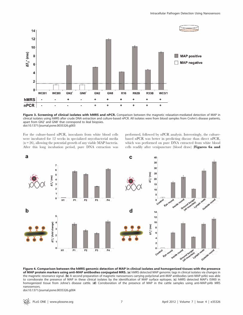
provided a DT2+ signal for all samples with clinical symptomology
(P1-P4, Figure 4a and 4b), whereas the sample from a healthy
individual (H1, Figure 4a and 4b), exhibited nominal magnetic
signal changes. Independent confirmation of the MAP MRS
findings was achieved by performing DNA extraction and nPCR
analysis. In addition, we used both the hMRS and epitope MRS to
screen for the presence of MAP in tissue samples from cattle
infected with Johne’s disease. Since MAP causes Johne’s disease in
cattle, we investigated if these probes can identify this intracellular
pathogen at the genomic and epitope levels in homogenized tissue
samples. Results showed significant magnetic signal changes in all
tissues from infected animals (Figures 4c and 4d), demonstrating
that hMRS and epitope MRS can quickly detect MAP in clinically
relevant samples at both the genomic and epitope levels. It is
interesting to note that in both human biopsies from Crohn’s
patients and tissues from animals with Johne’s disease, the
magnetic relaxation signal obtained with the hMRS probe
correlates with that obtained with the epitope-sensing MRS for
the same sample. The differences in the DT2+ among the animal
tissue samples might be attributed to the fact that the spatial
concentration of MAP differs among the various tissues within the
same animal due to Johne’s disease pathophysiology. Overall,
these results indicate that these probes can be used for the
screening of complex tissue samples with minimal or no sample
preparation, supporting clinical decision making.
hMRS Quickly Detect MAP DNA in Peripheral BloodAcknowledging previous reports that identified MAP in the
blood of Crohn’s disease patients, we sought to screen blood
samples from healthy individuals and Crohn’s disease patients for
the presence of IS900 (n= 34). For this, we chose to screen MAP’s
IS900 genomic marker, rather than a protein (epitope) marker,
since the later involves the use of anti-MAP antibodies that are
difficult to reproducibly generate while avoiding antibody batch
variabilities. In addition, a DNA-based hybridizing MRS would be
more robust and selective than an antibody-based MRS.
Peripheral blood was collected and DNA was directly isolated
from white blood cells, via either a 30-minutes crude DNA
extraction protocol for hMRS or a multistep high purity and
quality DNA isolation procedure for direct nPCR screening.
Direct nPCR for the MAP IS900 biomarker provided conclusive
results about the pathogen’s presence in these samples, hence these
samples established the first peripheral blood cohort (Cohort 1). In
this cohort, positive samples were defined as those samples where
a prominent band corresponding to IS900 was observed and
negative samples were those where no visible band was observed at
all. Control and Cohort 1 samples were screened with hMRS,
after a 30-minutes crude DNA extraction protocol from white
blood cells. Crude DNA extracted from control samples induced
small changes in the magnetic signal (DT2+ = 1.3 6 0.1 ms),
which served as the hMRS reference point. Interestingly, results
obtained with hMRS and magnetic relaxation detection using
crude DNA samples from patients within Cohort 1 were in
complete agreement with results obtained with direct nPCR that
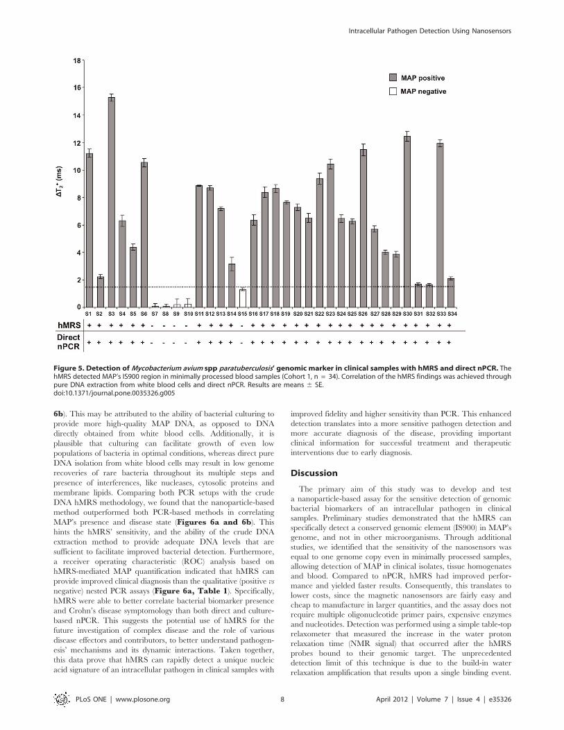
used pure DNA samples (Figure 5, Table S4). Addition of
a synthetic target complementary to hMRS led to signal
abrogation in the samples that had been identified as MAP
positive, confirming the specificity of the assay. This indicates that
the hMRS can detect their target in minimally processed blood
samples, while eliminating lengthy sample preparation procedures.
Next, a second cohort of blood samples (Cohort 2, Table 1)was represented by samples where direct nPCR analysis provided
dubious results. In samples within this cohort, classification as
either positive or negative was not possible due to the presence of
smeared or faint bands. The presence of these dubious samples,
either false-positives or false-negatives, is a major problem with
current nPCR techniques. Therefore, we examined if the hMRS
performs better than direct nPCR and culture-based nPCR in
determining if an individual is positive or negative based on his/
her clinical state (in this case being Crohn’s positive or healthy).
Intracellular Pathogen Detection Using Nanosensors
PLoS ONE | www.plosone.org 6 April 2012 | Volume 7 | Issue 4 | e35326
For the culture-based nPCR, inoculants from white blood cells
were incubated for 12 weeks in specialized mycobacterial media
(n = 26), allowing the potential growth of any viable MAP bacteria.
After this long incubation period, pure DNA extraction was
performed, followed by nPCR analysis. Interestingly, the culture-
based nPCR was better in predicting disease than direct nPCR,
which was performed on pure DNA extracted from white blood
cells readily after venipuncture (blood draw) (Figures 6a and
Figure 3. Screening of clinical isolates with hMRS and nPCR. Comparison between the magnetic relaxation-mediated detection of MAP inclinical isolates using hMRS after crude DNA extraction and culture-based nPCR. All isolates were from blood samples from Crohn’s disease patients,apart from GN2’ and GN8’ that correspond to ileal biopsies.doi:10.1371/journal.pone.0035326.g003
Figure 4. Comparison between the hMRS genomic detection of MAP in clinical isolates and homogenized tissues with the presenceof MAP protein markers using anti-MAP antibodies conjugated MRS. (a) hMRS detected MAP genomic tags in clinical isolates via changes inthe magnetic resonance signal. (b) A second preparation of magnetic nanosensors carrying polyclonal anti-MAP antibodies (anti-MAP-pAb) was ableto corroborate the presence of MAP in these clinical isolates by the identification of MAP surface epitopes. (c) hMRS detected MAP’s IS900 inhomogenized tissue from Johne’s disease cattle. (d) Corroboration of the presence of MAP in the cattle samples using anti-MAP-pAb MRSnanosensors.doi:10.1371/journal.pone.0035326.g004
Intracellular Pathogen Detection Using Nanosensors
PLoS ONE | www.plosone.org 7 April 2012 | Volume 7 | Issue 4 | e35326
6b). This may be attributed to the ability of bacterial culturing to
provide more high-quality MAP DNA, as opposed to DNA
directly obtained from white blood cells. Additionally, it is
plausible that culturing can facilitate growth of even low
populations of bacteria in optimal conditions, whereas direct pure
DNA isolation from white blood cells may result in low genome
recoveries of rare bacteria throughout its multiple steps and
presence of interferences, like nucleases, cytosolic proteins and
membrane lipids. Comparing both PCR setups with the crude
DNA hMRS methodology, we found that the nanoparticle-based
method outperformed both PCR-based methods in correlating
MAP’s presence and disease state (Figures 6a and 6b). Thishints the hMRS’ sensitivity, and the ability of the crude DNA
extraction method to provide adequate DNA levels that are
sufficient to facilitate improved bacterial detection. Furthermore,
a receiver operating characteristic (ROC) analysis based on
hMRS-mediated MAP quantification indicated that hMRS can
provide improved clinical diagnosis than the qualitative (positive vs
negative) nested PCR assays (Figure 6a, Table 1). Specifically,hMRS were able to better correlate bacterial biomarker presence
and Crohn’s disease symptomology than both direct and culture-
based nPCR. This suggests the potential use of hMRS for the
future investigation of complex disease and the role of various
disease effectors and contributors, to better understand pathogen-
esis’ mechanisms and its dynamic interactions. Taken together,
this data prove that hMRS can rapidly detect a unique nucleic
acid signature of an intracellular pathogen in clinical samples with
improved fidelity and higher sensitivity than PCR. This enhanced
detection translates into a more sensitive pathogen detection and
more accurate diagnosis of the disease, providing important
clinical information for successful treatment and therapeutic
interventions due to early diagnosis.
Discussion
The primary aim of this study was to develop and test
a nanoparticle-based assay for the sensitive detection of genomic
bacterial biomarkers of an intracellular pathogen in clinical
samples. Preliminary studies demonstrated that the hMRS can
specifically detect a conserved genomic element (IS900) in MAP’s
genome, and not in other microorganisms. Through additional
studies, we identified that the sensitivity of the nanosensors was
equal to one genome copy even in minimally processed samples,
allowing detection of MAP in clinical isolates, tissue homogenates
and blood. Compared to nPCR, hMRS had improved perfor-
mance and yielded faster results. Consequently, this translates to
lower costs, since the magnetic nanosensors are fairly easy and
cheap to manufacture in larger quantities, and the assay does not
require multiple oligonucleotide primer pairs, expensive enzymes
and nucleotides. Detection was performed using a simple table-top
relaxometer that measured the increase in the water proton
relaxation time (NMR signal) that occurred after the hMRS
probes bound to their genomic target. The unprecedented
detection limit of this technique is due to the build-in water
relaxation amplification that results upon a single binding event.
Figure 5. Detection ofMycobacterium avium spp paratuberculosis’ genomic marker in clinical samples with hMRS and direct nPCR. ThehMRS detected MAP’s IS900 region in minimally processed blood samples (Cohort 1, n = 34). Correlation of the hMRS findings was achieved throughpure DNA extraction from white blood cells and direct nPCR. Results are means 6 SE.doi:10.1371/journal.pone.0035326.g005
Intracellular Pathogen Detection Using Nanosensors
PLoS ONE | www.plosone.org 8 April 2012 | Volume 7 | Issue 4 | e35326
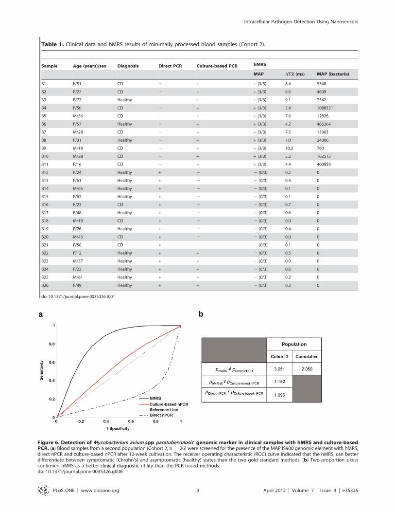
Table 1. Clinical data and hMRS results of minimally processed blood samples (Cohort 2).
Sample Age (years)/sex Diagnosis Direct PCR Culture-based PCR hMRS
MAP DT2 (ms) MAP (bacteria)
B1 F/51 CD 2 + + (3/3) 8.4 5348
B2 F/27 CD 2 + + (3/3) 8.6 4609
B3 F/73 Healthy 2 + + (3/3) 9.1 2542
B4 F/50 CD 2 + + (3/3) 3.4 1088531
B5 M/56 CD 2 + + (3/3) 7.6 12826
B6 F/57 Healthy 2 + + (3/3) 4.2 465264
B7 M/28 CD 2 + + (3/3) 7.5 13963
B8 F/31 Healthy 2 + + (3/3) 7.0 24006
B9 M/18 CD 2 + + (3/3) 10.3 760
B10 M/28 CD 2 + + (3/3) 5.2 162513
B11 F/16 CD 2 + + (3/3) 4.4 400959
B12 F/24 Healthy + 2 2 (0/3) 0.2 0
B13 F/61 Healthy + 2 2 (0/3) 0.4 0
B14 M/65 Healthy + 2 2 (0/3) 0.1 0
B15 F/62 Healthy + 2 2 (0/3) 0.1 0
B16 F/23 CD + 2 2 (0/3) 0.7 0
B17 F/48 Healthy + 2 2 (0/3) 0.6 0
B18 M/19 CD + 2 2 (0/3) 0.0 0
B19 F/26 Healthy + 2 2 (0/3) 0.4 0
B20 M/43 CD + 2 2 (0/3) 0.0 0
B21 F/50 CD + 2 2 (0/3) 0.1 0
B22 F/12 Healthy + + 2 (0/3) 0.5 0
B23 M/57 Healthy + + 2 (0/3) 0.0 0
B24 F/23 Healthy + + 2 (0/3) 0.6 0
B25 M/61 Healthy + + 2 (0/3) 0.2 0
B26 F/49 Healthy + + 2 (0/3) 0.3 0
doi:10.1371/journal.pone.0035326.t001
Figure 6. Detection of Mycobacterium avium spp paratuberculosis’ genomic marker in clinical samples with hMRS and culture-basedPCR. (a) Blood samples from a second population (Cohort 2, n = 26) were screened for the presence of the MAP IS900 genomic element with hMRS,direct nPCR and culture-based nPCR after 12-week cultivation. The receiver operating characteristic (ROC) curve indicated that the hMRS, can betterdifferentiate between symptomatic (Chrohn’s) and asymptomatic (healthy) states than the two gold standard methods. (b) Two-proportion z-testconfirmed hMRS as a better clinical diagnostic utility than the PCR-based methods.doi:10.1371/journal.pone.0035326.g006
Intracellular Pathogen Detection Using Nanosensors
PLoS ONE | www.plosone.org 9 April 2012 | Volume 7 | Issue 4 | e35326
The T2 relaxation times of hundreds of thousands of water
molecules surrounding the hMRS probes increase upon binding of
a single probe to the target. This is also facilitated by the low
valency of the hMRS probe that induces drastic changes at low
concentration of the target, or in this case low copy numbers of the
genomic target. This approach contrasts with previous magnetic
relaxation approaches that rely on the use of two magnetic
nanoparticle probes that hybridize to adjacent sequences on
a DNA target, facilitating clustering of the nanoparticles with
a corresponding decrease in the T2 relaxation times [32,33]. Since
our approach does not rely on nanoparticle clustering and only the
binding of the nanoparticle is sufficient for detection, a faster,
more sensitive magnetic relaxation assay that rivals PCR has been
developed for the screening of an intracellular pathogen genomic
marker. Apart from a table-top relaxometer, portable magnetic
relaxometers have been developed, suitable for point-of-care
screening with hMRS [34].
hMRS can be used for the detection of diverse targets in
complex media, including turbid and opaque clinical samples
[11,12]. Herein, our results demonstrate that the hMRS binding
can achieve the unprecedented and fast detection of a single
genome copy of an intracellular pathogen. This is critical for the
identification of extremely virulent pathogens. As MAP is an
intracellular pathogen similar to M. tuberculosis, its detection is
difficult, due to the pathogen’s residence within an infected
leukocyte’s organelles. Thus contemporary diagnostic methodol-
ogies rely on cell isolation, cell lysis and nucleic acid isolation, in
order to yield high purity DNA. This renders MAP’s detection
problematic since the microorganism has to be isolated from
highly complex samples, such as blood or severely inflamed tissue
biopsies, before amplification and detection via PCR analysis.
Apart from DNA quality, the quantity of MAP DNA can affect the
outcome of PCR. For instance, low MAP DNA levels from
a clinical sample may not be successfully amplified through direct
nPCR or biological matrix components can affect the polymerase
activity, yielding false-negative or dubious results. In our studies,
a significant number of samples that were identified as MAP
dubious by direct nPCR were found to be positive by culture-
based nPCR and hMRS (Cohort 2), indicating that the pathogen
was originally present in the patient’s blood (albeit in low amounts)
and successfully grew in culture at adequate levels.
The culture-based nPCR, relies on the availability of viable
bacteria in the clinical sample that can be grown in culture in
sufficient amounts before nPCR. Most clinical microbial diag-
nostics rely on the growth of the pathogen in culture, before
performing the appropriate immunological or PCR tests [35,36].
Growth can be observed within 12 to 24 hours for fast-growing
microorganisms, but for slow-growing intracellular microorgan-
isms like MAP it can take from a week up to several months.
Another problem with this technique is that some microorganisms
are difficult to culture in the laboratory, which limits their
detection. Although highly reliable, culture-based PCR can result
in the identification of samples as false-negative, due to the
absence of a viable pathogen or the inability of the pathogen to
grow in culture. Therefore, it is plausible that samples that might
be found to be positive by direct nPCR and hMRS to be identified
as negative 12 weeks after by culture-based nPCR. This is
common for low bacterial loads of slow-growing microorganisms
that require highly specialized media and cultural conditions,
whose inadequacy might drastically affect bacterial viability
leading to cell death and DNA degradation [37]. These problems
in detection with PCR methods are encountered in the detection
of any intracellular pathogen, as the pathogen has optimized its
adaptation and growth within a host cell. Due to their sensitivity
and minimal sample processing, hMRS are capable of circum-
venting PCR’s obstacles and achieving improved clinical di-
agnosis. Our findings indicate that hMRS can detect an
intracellular pathogen’s genomic marker in biological fluids (i.e.
blood) and tissue homogenates within one hour, where the first 30
minutes are spent on crude DNA extraction and the remaining
time on readout.
Herein, we have also demonstrated that MAP DNA can be
detected in various tissues from Johne’s disease animals, as well as
in biopsies and blood from Crohn’s disease patients, at the epitope
and genomic biomarker levels. Furthermore, our results indicate
that hMRS can detect the presence of MAP in Crohn’s disease
patients more reliably than direct or cultured-based nPCR. As
a matter of fact, hMRS is a better predictor of the state of the
disease, correlating the clinical state of a patient with the presence
of MAP in blood samples (Figure 6). The use of hMRS could be
a powerful new tool in the study of the mechanism of infection of
intracellular pathogens and its relation to disease state, as well as in
elucidating intracellular pathogens’ adaptation strategies [38].
Difficulties in detecting MAP hamper studies aimed at the
investigation of the potential role of MAP in Crohn’s disease
pathology, as well as the pathogen’s impact on the dairy and beef
industries [39]. Overall, these studies provide evidence for the use
of hybridizing magnetic nanosensors and magnetic relaxation
detection to reliably identify the presence of an intracellular
pathogen in clinical samples in a fast, cost-effective and highly
sensitive way.
Materials and Methods
Preparation of IS900-specific hMRSThe IS900-specific hMRS were prepared from propargylated
polyacrylic-acid-coated iron oxide nanoparticles, utilizing litera-
ture available protocols [11]. Specifically, incorporation of
propargyl groups on polyacrylic-acid-coated iron oxide nanopar-
ticles was achieved via the carbodiimide chemistry, followed by
magnetic separation of the nanoparticles, using an LS25 MACS
column (Miltenyi) [11]. Subsequently, the propargyl-functiona-
lized iron oxide nanoparticles were reacted with an azide-modified
oligonucleotide, which is complementary to a segment of MAP’s
IS900 genomic element. Conjugation of the azide-terminated 15-
bp oligonucleotide 59-ATGTGGTTGCTGTGT-39 was achieved
via ‘‘click’’ chemistry, as previously described [40]. Briefly, 400 mLpropargylated iron oxide nanoparticles were resuspended in
1,100 mL NaHCO3 buffer (0.1 M, pH 8.4). To this, 200 mL of
10 mM TCEP (tris(2-carboxyethyl)phosphane hydrochloride,
Sigma) were added as a reducing agent. Twenty mL of 6.3 M
oligo were diluted in 80 mL NaHCO3 buffer (0.1 M, pH 8.4) and
added to the nanoparticle solution. The reaction was initiated with
the dropwise addition of 150 mL Cu(I)-TBTA complex (tris(ben-
zyltriazolylmethyl) amine, 10 mM, Sigma), which was previously
prepared in DI water and tbutanol (9:1). The reaction was
incubated at room temperature under continuous mixing for 3
hours, followed by overnight incubation at 4uC under constant
mixing. The resulting IS900 hMRS were dialyzed against DI
water using a 6,000 – 8,000 MWCO membrane (Spectrum),
followed by magnetic separation with an LS25 MACS column
(Miltenyi). The epitope-sensing MRS were formulated as pre-
viously reported, and the MAP antibody was obtained from Dr.
Naser’s lab [12]. Briefly, polyacrylic-acid-coated iron oxide
nanoparticles were conjugated to Protein G via the EDC/NHS
chemistry, in order to have Protein G as a high-affinity
immunoglobulin linker that provides optimal antibody orientation
[12]. The reaction was performed according to the literature,
Intracellular Pathogen Detection Using Nanosensors
PLoS ONE | www.plosone.org 10 April 2012 | Volume 7 | Issue 4 | e35326
followed by magnetic separation through a 1X-PBS-equilibrated
LS25 column and antibody conjugation [12]. The nanoparticle
valency of the antibody-carrying nanoparticles was assessed
through quantification of the nanoparticles’ protein content, using
published methodologies [11]. Size determination of all hMRS
was achieved through dynamic light scattering, using the PDDLS
CoolBatch 40T instrument and Precision Deconvolve 32 software.
Zeta potential measurements were performed on a Malvern
zetasizer, while the R1 and R2 relaxivities were determined after
determination of the nanoparticle’s iron content and relaxation
studies on a compact relaxometer (Minispec, Bruker). Determina-
tion of the hMRS oligonucleotide concentration was achieved by
monitoring the absorbance at 260 and 305 nm (background),
using a Nanodrop 1000 spectrophotometer (Thermo Scientific), as
previously described [12]. The hMRS were stored at 4uC until
further use.
Bacterial Cultures, Isolates and Homogenized TissueLab strains of MAP and clinical isolates were grown in 12B*
BACTEC bottles (Becton Dickinson) as previously described [21].
Quantification of MAP grown in the BACTEC bottles was
assessed with the BACTEC 460 TB Analyzer (Becton Dickinson).
Heat-inactivation of MAP was performed by autoclaving the
BACTEC bottle for 10 mins. Proteus vulgaris # 8427, Staphylococcus
aureus #33862, Pseudomonas aeruginosa # 27853, Enterococcus faecalis,
Escherichia coli # 8739, and Serratia marcensis were grown in culture
tubes with nutrient broth, and bacterial growth was monitored
spectrophotometrically. Heat inactivation of these bacteria was
performed by autoclaving the culture tubes for 10 minutes. Upon
inactivation, all bacterial stocks were placed in a Fisher Isotemp
freezer (Fisher Scientific), until further use. Clinical isolates and
tissue specimens from animals with Johne’s disease were obtained
from tissue collections stored in Dr. Naser’s laboratory.
Extraction of Pure and Crude Bacterial DNA fromBacterial CulturesDNA was extracted from cultured bacteria in a class II biosafety
cabinet, according to literature-available procedures [21]. One mL
of bacterial cultures was aseptically transferred to microcentrifuge
tubes, followed by centrifugation at 13,200 rpm. The resulting
pellets were resuspended in 120 mL sterile TE buffer (10 mM Tris,
1 mM EDTA, pH 8.0) and incubated in a dry heat bath at 100uC.Samples without further processing were used as crude DNA
extracts in MRnS and nPCR studies. In order to obtain pure
bacterial DNA, after heating the samples, we placed them on ice
for 15 minutes and then centrifuged them at 12,000 rpm for 10
minutes at 4uC. The supernatants were transferred to Phase-lock
gel tubes (Eppendorf), which were supplemented with 200 mL of
phenol/chloroform/isoamyl alcohol (1:1:24 v:v, Acros). The tubes
were centrifuged for 5 minutes (12,000 rpm, 4uC), and the
supernatants were precipitated using 400 mL 100% ethanol cooled
to 220uC. The precipitated DNA was washed, dried, and finally
reconstituted in 50 mL TE buffer. Pure and crude bacterial DNA
was stored at 4uC until further analysis, whereas spectrophoto-
metric quantification of DNA content was achieved using
a compact spectrophotometer (Nanodrop 1000, Thermo Scientif-
ic). Crude and pure DNA extractions from clinical isolates were
similarly performed.
hMRS and Nested PCR ExperimentsSerial dilutions of bacterial DNA were prepared in TE buffer.
For the relaxation-mediated experiments, 200 mL of the nano-
particle suspension (6 mg Fe/mL in 0.1 M phosphate buffer,
0.1 M NaCl, pH 7.4) were incubated with 1 mL of bacterial DNA
or control (TE buffer). The samples were heated twice for 3
minutes at 95uC. After this, the samples were transferred to
relaxometer tubes and cooled down at room temperature, while
taking relaxation measurements on a magnetic relaxometer
(Minispec, Bruker). For the specificity assays using a complemen-
tary synthetic target (59-TCCTTCGGCCATCCAACACAG-
CAACCACAT-39), 1 mL of the target (1.1 M) was added to the
hMRS mycobacterial samples containing the same DNA concen-
tration (3.5 ng/mL). Similar DNA concentration was used for the
specificity experiment utilizing other Gram-positive and Gram-
negative bacteria and hMRS (3 mg Fe/mL). For the sensitivity
experiments, serial dilutions of pure and crude MAP DNA were
prepared from pure cultures of MAP grown in 12B* BACTEC
bottles, which had been processed and quantified as described in
the preceding section (DNA extraction methodology). Nested PCR
(nPCR) was performed as previously described [21].
Blood Samples, Extraction of Bacterial DNA from WhiteBlood Cells and MAP CulturingBlood samples were collected from healthy individuals and
patients with Crohn’s disease at the University of Florida, College
of Medicine, in accordance to Institutional Review Board
guidelines. The samples were coded and sent to the University
of Central Florida for blind screening via PCR and hMRS
analyses, without knowing in advance the individual’s condition,
demographics and MAP’s presence (Cohorts 1 and 2). Isolation of
white blood cells was achieved via centrifugation at 3,000 rpm for
10 minutes at room temperature, as previously described [21].
Pure DNA was extracted via phenol/chloroform/isoamyl alcohol
precipitation, similar to previously published methodologies. The
pure DNA was subjected to direct nested PCR, as described
above. Crude DNA extraction from the isolated white blood cells
was achieved via centrifugation at 13,200 rpm for 2 minutes. The
resulting pellets were resuspended in 120 mL sterile TE buffer
(10 mM Tris, 1 mM EDTA, pH 8.0) and incubated in a dry heat
bath at 100uC for 30 minutes, followed by cooling and
centrifugation at 12,000 rpm that provided us with supernatants
rich in crude DNA. For bacterial culturing, buffy coat samples
were prepared in sterile phosphate buffered saline as described
before and inoculated on MGIT bottles. The inoculants were first
incubated at 37uC in a 5% CO2 atmosphere for 12 weeks, and
then pure DNA extraction was performed as stated above.
Measurement of Proton Relaxation TimesSpin-spin relaxation times (T2) were measured using
a 0.47 T mq20 NMR analyzer (Minispec, Bruker). T2 values
were obtained before and after addition of the sample, and
through the time course of the study. All T2 measurements were
performed using a CPMG pulse-echo train with a 1.5 ms
interpulse spacing (Bruker Corp., Billerica, MA). The change in
magnetic resonance signal (DT2+) was defined as the sample’s T2
minus the magnetic signal of the corresponding negative control.
All experiments and measurements were carried out in triplicate
and data were expressed as mean 6 standard error, unless
otherwise denoted.
Statistical AnalysisClinical diagnosis and sample collection was performed at the
College of Medicine, University of Florida, based on clinical and
endoscopic criteria [21]. Samples identified as IS900-positive by
the hMRS method had an average DT2+ higher than 1.5 ms, while
positive samples by the nested PCR method had a unique 298-bp
Intracellular Pathogen Detection Using Nanosensors
PLoS ONE | www.plosone.org 11 April 2012 | Volume 7 | Issue 4 | e35326
nucleotide sequence. The averages of three independent experi-
ments are reported, unless otherwise stated. Two-proportion z-test
statistics were determined through the SPSS package, with the
confidence level set at 95% (IBM Co.). ROC analysis was
performed based on hMRS determination of the sample’s MAP
load. This was achieved through the correlation of the DT2+ signal
to bacterial genome copies, using a crude extracted DNA standard
curve (Figure S3). For nPCR, the presence of a band
corresponding to IS900 was marked as positive (value = 1),
whereas its absence was assigned as negative (value = 0). The
overall ROC methodology was performed as previously reported
in literature [34]. Specifically, the classification variable was the
individual’s clinical condition (Crohn’s disease vs healthy), whereas
the output curve was fitted using logistic regression.
Ethics StatementThe use of human subjects was approved by the University of
Florida’s Institutional Review Board (protocol number 354–2007).
A signed individual written informed consent agreement was
obtained from each subject or in the case of children from their
parent or legally authorized representative before enrolment in the
study.
Supporting Information
Figure S1 Kinetics for MAP’s IS900 genomic markerdetection with hMRS. After heating the samples to facilitate
DNA stand separation and hMRS hybridization, the changes in
the T2 magnetic resonance signal were recorded over time, with
marked changes occurring within less than an hour (Means6SE).
(TIF)
Figure S2 Nested PCR (nPCR) cannot quantify crudeMAP DNA. – Ctrl: negative control (dH2O) of the first nPCR
round, – Ctrl: negative control (dH2O) of the second nPCR
round, TE: TE buffer, + Ctrl: two controls of pure extracted MAP
DNA.
(TIF)
Figure S3 Genome-copy-based quantification of MAPwith hMRS and crude extracted DNA. Samples with known
amounts of crude DNA from cultured MAP were utilized to
correlate the changes in the T2 signal (DT2+) and the number of
bacteria originally present in the sample, using the MAP’s genome
size as a reference. (Means6SE. SE too small to depict in high
bacterial levels.)
(TIF)
Table S1 Spin-spin relaxation times (T2) of crude MAPDNA samples. Serial dilutions of crude MAP DNA samples
have been utilized to assess the sensitivity of the hMRS method in
minimally processed bacterial cultures. The averages of each
independent experiment are listed and the studies’ mean.
(PDF)
Table S2 Spin-spin relaxation times (T2) of pure MAPDNA samples with known genome copies. Three in-
dependent experiments were performed on pure DNA samples
obtained from cultured MAP. Correlation between DNA levels
and bacterial populations was achieved by quantifying DNA
spectrophotometrically and using the MAP genome size as
a reference.
(PDF)
Table S3 Demographics of cultured clinical isolatesthat were screened with hMRS and nPCR. (CD: Crohn’s
disease, IBD: inflammatory bowel disease)
(PDF)
Table S4 Clinical data and hMRS results of Cohort 1blood samples. Samples from Crohn’s disease (CD) patients,
healthy individuals or asymptomatic carriers were analyzed by
direct nPCR and hMRS. Quantification of the MAP genome
copies was achieved in positive (+) samples using a training
standard curve for crude extracted MAP DNA.
(PDF)
Acknowledgments
The authors thank Dr. John F. Valentine for providing samples and
performing clinical diagnoses.
Author Contributions
Conceived and designed the experiments: CK JMP. Performed the
experiments: CK HB SS. Analyzed the data: JMP CK SAN. Contributed
reagents/materials/analysis tools: SAN. Wrote the paper: CK JMP.
References
1. Batt CA (2007) Materials science. Food pathogen detection. Science 316:
1579–1580.
2. Ferrari M (2005) Cancer nanotechnology: opportunities and challenges. Nat
Rev Cancer 5: 161–171.
3. Jain KK (2005) Nanotechnology in clinical laboratory diagnostics. Clin Chim
Acta 358: 37–54.
4. Rosi NL, Mirkin CA (2005) Nanostructures in biodiagnostics. Chem Rev 105:
1547–1562.
5. Kaittanis C, Santra S, Perez JM (2010) Emerging nanotechnology-based
strategies for the identification of microbial pathogenesis. Adv Drug Deliv Rev
62: 408–423.
6. Gao L, Zhuang J, Nie L, Zhang J, Zhang Y, et al. (2007) Intrinsic peroxidase-like
activity of ferromagnetic nanoparticles. Nat Nanotechnol 2: 577–583.
7. Gao J, Gu H, Xu B (2009) Multifunctional magnetic nanoparticles: design,
synthesis, and biomedical applications. Acc Chem Res 42: 1097–1107.
8. Gaster RS, Xu L, Han SJ, Wilson RJ, Hall DA, et al. (2011) Quantification of
protein interactions and solution transport using high-density GMR sensor
arrays. Nat Nanotechnol 6: 314–320.
9. Kaittanis C, Naser SA, Perez JM (2007) One-step, nanoparticle-mediated
bacterial detection with magnetic relaxation. Nano Lett 7: 380–383.
10. Laurent S, Bridot JL, Elst LV, Muller RN (2010) Magnetic iron oxide
nanoparticles for biomedical applications. Future Med Chem 2: 427–449.
11. Kaittanis C, Santra S, Perez JM (2009) Role of nanoparticle valency in the
nondestructive magnetic-relaxation-mediated detection and magnetic isolation
of cells in complex media. J Am Chem Soc 131: 12780–12791.
12. Kaittanis C, Santra S, Santiesteban OJ, Henderson TJ, Perez JM (2011) The
Assembly State between Magnetic Nanosensors and Their Targets OrchestratesTheir Magnetic Relaxation Response. J Am Chem Soc 133: 3668–3676.
13. Grossman HL, Myers WR, Vreeland VJ, Bruehl R, Alper MD, et al. (2004)
Detection of bacteria in suspension by using a superconducting quantuminterference device. Proc Natl Acad Sci U S A 101: 129–134.
14. Wang SX, Li G (2008) Advances in Giant Magnetoresistance Biosensors With
Magnetic Nanoparticle Tags: Review and Outlook. IEEE TRANSACTIONS
ON MAGNETICS 44: 1687–1702.
15. Cocito C, Gilot P, Coene M, de Kesel M, Poupart P, et al. (1994)Paratuberculosis. Clin Microbiol Rev 7: 328–345.
16. Chiodini RJ (1989) Crohn’s disease and the mycobacterioses: a review and
comparison of two disease entities. Clin Microbiol Rev 2: 90–117.
17. Dalziel TK (1913) Chronic Interstitial Enteritis. The British Medical Journal 2:1068–1070.
18. Crohn BB, Ginzburg L, Oppenheimer GD (1932) REGIONAL ILEITIS.
Journal of the American Medical Association 99: 1323–1329.
19. Greenstein RJ, Greenstein AJ (1995) Is there clinical, epidemiological and
molecular evidence for two forms of Crohn’s disease? Mol Med Today 1:343–348.
20. Chiodini RJ, Van Kruiningen HJ, Thayer WR, Coutu JA (1986) Spheroplastic
phase of mycobacteria isolated from patients with Crohn’s disease. J ClinMicrobiol 24: 357–363.
21. Naser SA, Ghobrial G, Romero C, Valentine JF (2004) Culture of
Mycobacterium avium subspecies paratuberculosis from the blood of patients
with Crohn’s disease. Lancet 364: 1039–1044.
Intracellular Pathogen Detection Using Nanosensors
PLoS ONE | www.plosone.org 12 April 2012 | Volume 7 | Issue 4 | e35326
22. Romero C, Hamdi A, Valentine JF, Naser SA (2005) Evaluation of surgical
tissue from patients with Crohn’s disease for the presence of Mycobacteriumavium subspecies paratuberculosis DNA by in situ hybridization and nested
polymerase chain reaction. Inflamm Bowel Dis 11: 116–125.
23. Green EP, Tizard ML, Moss MT, Thompson J, Winterbourne DJ, et al. (1989)Sequence and characteristics of IS900, an insertion element identified in
a human Crohn’s disease isolate of Mycobacterium paratuberculosis. NucleicAcids Res 17: 9063–9073.
24. Lisby G, Andersen J, Engbaek K, Binder V (1994) Mycobacterium
paratuberculosis in intestinal tissue from patients with Crohn’s diseasedemonstrated by a nested primer polymerase chain reaction. Scand J Gastroen-
terol 29: 923–929.25. Sanderson JD, Moss MT, Tizard ML, Hermon-Taylor J (1992) Mycobacterium
paratuberculosis DNA in Crohn’s disease tissue. Gut 33: 890–896.26. Cutler JI, Zhang K, Zheng D, Auyeung E, Prigodich AE, et al. (2011) Polyvalent
nucleic acid nanostructures. J Am Chem Soc 133: 9254–9257.
27. Fang Y, Wu WH, Pepper JL, Larsen JL, Marras SA, et al. (2002) Comparison ofreal-time, quantitative PCR with molecular beacons to nested PCR and culture
methods for detection of Mycobacterium avium subsp. paratuberculosis inbovine fecal samples. J Clin Microbiol 40: 287–291.
28. Hiratsuka S, Watanabe A, Aburatani H, Maru Y (2006) Tumour-mediated
upregulation of chemoattractants and recruitment of myeloid cells predeter-mines lung metastasis. Nat Cell Biol 8: 1369–1375.
29. Morgan RA, Dudley ME, Wunderlich JR, Hughes MS, Yang JC, et al. (2006)Cancer regression in patients after transfer of genetically engineered lympho-
cytes. Science 314: 126–129.
30. Kruse JP, Gu W (2009) Modes of p53 regulation. Cell 137: 609–622.
31. Naser SA, Shafran I, Schwartz D, El-Zaatari F, Biggerstaff J (2002) In situidentification of mycobacteria in Crohn’s disease patient tissue using confocal
scanning laser microscopy. Mol Cell Probes 16: 41–48.
32. Grimm J, Perez JM, Josephson L, Weissleder R (2004) Novel nanosensors forrapid analysis of telomerase activity. Cancer Res 64: 639–643.
33. Perez JM, Grimm J, Josephson L, Weissleder R (2008) Integrated nanosensors todetermine levels and functional activity of human telomerase. Neoplasia 10:
1066–1072.
34. Haun JB, Castro CM, Wang R, Peterson VM, Marinelli BS, et al. Micro-NMRfor rapid molecular analysis of human tumor samples. Sci Transl Med 3: 71ra16.
35. Bancroft EA (2007) Antimicrobial resistance: it’s not just for hospitals. Jama 298:1803–1804.
36. Taubes G (2008) The bacteria fight back. Science 321: 356–361.37. Engelberg-Kulka H, Amitai S, Kolodkin-Gal I, Hazan R (2006) Bacterial
programmed cell death and multicellular behavior in bacteria. PLoS Genet 2:
e135.38. Homolka S, Niemann S, Russell DG, Rohde KH (2010) Functional genetic
diversity among Mycobacterium tuberculosis complex clinical isolates: de-lineation of conserved core and lineage-specific transcriptomes during in-
tracellular survival. PLoS Pathog 6: e1000988.
39. Pierce ES (2009) Where are all the Mycobacterium avium subspeciesparatuberculosis in patients with Crohn’s disease? PLoS Pathog 5: e1000234.
40. Seela F, Sirivolu VR (2007) Nucleosides and oligonucleotides with diynyl sidechains: the huisgen-sharpless cycloaddition ‘‘click reaction’’ performed on DNA
and their constituents. Nucleosides Nucleotides Nucleic Acids 26: 597–601.
Intracellular Pathogen Detection Using Nanosensors
PLoS ONE | www.plosone.org 13 April 2012 | Volume 7 | Issue 4 | e35326