Embed Size (px)
Citation preview

Rapid Mass Movement of Chloroplasts during SegmentFormation of the Calcifying Siphonalean Green Alga,Halimeda macrolobaAnthony W. D. Larkum1,4*, Anya Salih2, Michael Kuhl3,4
1 School of Biological Sciences, University of Sydney, Camperdown, New South Wales, Australia, 2 Confocal Bio-Imaging Facility, School of Natural Sciences, University of
Western Sydney, Richmond, New South Wales, Australia, 3 Marine Biological Section, Department of Biology, University of Copenhagen, Helsingør, Denmark, 4 Plant
Functional Biology and Climate Change Cluster, University of Technology, Sydney, Broadway, New South Wales, Australia
Abstract
Background: The calcifying siphonalean green alga, Halimeda macroloba is abundant on coral reefs and is important in theproduction of calcium carbonate sediments. The process by which new green segments are formed over-night is revealedhere for the first time.
Methodology/Principal Findings: Growth of new segments was visualised by epifluorescence and confocal microscopy andby pulse amplitude modulation (PAM) fluorimetry. Apical colourless proto-segments were initiated on day 1, and formed aloose network of non-calcified, non-septate filaments, containing no chloroplasts. Rapid greening was initiated at dusk by i)the mass movement of chloroplasts into these filaments from the parent segment and ii) the growth of new filamentscontaining chloroplasts. Greening was usually complete in 3–5 h and certainly before dawn on day 2 when the first signs ofcalcification were apparent. Mass chloroplast movement took place at a rate of ,0.65 mm/s. Photosynthetic yield and rateremained low for a period of 1 to several hours, indicating that the chloroplasts were made de novo. Use of the inhibitorscolchicine and cytochalasin d indicated that the movement process is dependent on both microtubules and microfilaments.
Significance: This unusual process involves the mass movement of chloroplasts at a high rate into new segments during thenight and rapid calcification on the following day and may be an adaptation to minimise the impact of herbivorous activity.
Citation: Larkum AWD, Salih A, Kuhl M (2011) Rapid Mass Movement of Chloroplasts during Segment Formation of the Calcifying Siphonalean Green Alga,Halimeda macroloba. PLoS ONE 6(7): e20841. doi:10.1371/journal.pone.0020841
Editor: Ivan Baxter, United States Department of Agriculture, Agricultural Research Service, United States of America
Received November 9, 2010; Accepted May 13, 2011; Published July 5, 2011
Copyright: � 2011 Larkum et al. This is an open-access article distributed under the terms of the Creative Commons Attribution License, which permitsunrestricted use, distribution, and reproduction in any medium, provided the original author and source are credited.
Funding: The study was financed by the Australian Research Council (AWDL, AS), the Danish Natural Science Research Council (MK) and the CarlsbergFoundation (MK). The funders had no role in study design, data collection and analysis, decision to publish, or preparation of the manuscript.
Competing Interests: The authors have declared that no competing interests exist.
* E-mail: [email protected]
Introduction
Calcifying siphonalean green algae in the genus Halimeda are
abundant on coral reefs around the world [1] and are important in
the sequestration of carbon from the atmosphere [1,2]. Halimeda
species typically produces segmented plants that are attached by a
holdfast, which can be attached to rock or coral or buried in
sediment. Bundles of the aragonite crystals from dead Halimeda
plants are an abundant source for the fine-grained sediments on
many coral reefs [1]. As in all members of the Order Siphonales,
the thallus is formed from branching filaments, which have few
septa and thus the plant is coenocytic. In Halimeda, the thallus has
central filaments arranged in an axial orientation, giving rise to
radial filaments in the cortex. The cortical radial filaments branch
extensively and fuse at the apices of branches to form a closed
surface [3]; beneath this closed surface there is free space between
the filaments known as the inter-utricular space. Large numbers of
aragonite crystals are found in the inter-utricular space. Chloro-
plasts occur mainly in the outer cortical filaments adjacent to the
inter-utricular spaces. The coenocytic morphology renders the
plant potentially vulnerable to damage and grazing, and plants in
this Order possess very rapid wound responses and produce
feeding deterrent chemicals [4,5].
Halimeda-dominated communities have been found to be impor-
tant primary producers in many regions of the world [2,6,7,8
,9,10]. On shallow reefs, they are significant contributors to soft
sediments [1], and Halimeda species have been shown to produce
massive carbonate deposits in deeper-water sites, both in the
modern world [9,10,11] and in the geological past [12]. Halimeda is
thus important both in geological sediment production [2,11,12]
and in making a significant contribution to carbon sequestration [2].
A number of studies have been carried out on Halimeda spp. with
a view to clarifying processes of photosynthesis, growth, calcifica-
tion and chloroplast movement. Borowitztka and Larkum studied
calcification and chloroplast formation [3,13,14,15,16,17,18].
They concluded that photosynthesis during the day causes
alkalinisation of the inter-utricular space and that this is the
trigger for calcification [3]. Subsequent microelectrode studies
[19] supported this hypothesis, although it has not yet been
possible to obtain in situ results for the inter-utricular space with
pH microelectrodes because wound reactions lead to the release of
acids around the microelectrode. While photosynthesis undoubt-
PLoS ONE | www.plosone.org 1 July 2011 | Volume 6 | Issue 7 | e20841

edly plays a key role, ion transport processes across the utricular
filament membranes [3,15] and nucleation sites in the inter-
utricular space [20] are also important for the calcification process.
Borowitzka & Larkum [13,14] also studied chloroplast formation:
they showed that new plastids formed from proplastids and that a
‘thylakoid organising body’ was an intermediate step in the
maturation process [14].
Plastid movement in Halimeda species has been studied by Drew
and coworkers. In the dark, chloroplasts are withdrawn inwards in
the radial outer filaments [21], returning to the perimeter the
following day just before dawn. The segments thus pale visibly in
the dark, turning from a dark green colour to almost white. This is
largely a response to light, since segments will also pale during the
day if shaded, but there is also a regulatory component with an
endogenous rhythm [22].
New segments of Halimeda plants are formed monthly [23]. In
this process, a new colourless proto-segment is initiated during the
course of the first day. This flaccid young segment is formed of a
loose network of non-calcified, non-septate filaments, bathed in
seawater and initially containing no chloroplasts. During the
following night, chloroplasts move from the mature parent
segment below into the developing segment. Apart from
qualitative descriptions of this process, no detailed reports of the
sequence of events or the possible mechanisms are available. Here
we have used confocal microscopy and variable chlorophyll
fluorescence imaging, with inhibitors and light manipulations, to
study the process of the formation and greening of new segments
in Halimeda macroloba.
Materials and Methods
SamplingPlants of Halimeda macroloba Decaisne were collected on the reef
flat adjacent to the Heron Island Research Station (152u069E,
20u299S) on the Great Barrier Reef, Australia. Plants were kept in
running seawater tanks under shaded conditions at 23–25uC. For
the confocal work, material was kept in a seawater aquarium at the
University of Sydney over a period of 3 months at 25uC under
white growth-light fluorescence tubes at an irradiance of
,250 mmol photons m22 s21 during a 12 h day.
MicroscopyInitial work was carried out using a compound microscope
(BH2-RFL, Olympus, Japan) with a reflected light fluorescence
attachment at 10X (DPLAPO10XUV, Olympus, Japan) and 40X
(DAPO40UV/RiO, Olympus, Japan) magnification. The excita-
tion light source was a 100W high-pressure mercury lamp (HBO
100W/2, Olympus, Japan). An excitation filter (UG-1, Olympus,
Japan) was used in combination with a dichroic filter U-V (400–
455 nm) for chlorophyll fluorescence (,680 nm), and an excita-
tion filter BP-545 in combination with a dichroic filter B–G for
green (cell wall) fluorescence (500–580 nm).
Confocal MicroscopyH. macroloba thallus segments were imaged in seawater mounted
on a glass cover slip, which was fitted over a drilled glass slide.
Typically, three regions of interest were imaged at different time
intervals – the bottom, the middle and the growing edge of each
sampled new segment. Confocal imaging was done on a Leica
DMIRBE inverted microscope fitted with a TCS SPII confocal
head (Leica Microsystems, Heidelberg, Germany) using 10X NA
0.35 dry, 40X PLAPO CS 0.75 oil or 63X HCX PLAPO CS
water immersion objectives (Leica Microsystems, Heidelberg,
Germany). We used the 488 nm excitation line of an Argon
multi-line laser and the triple dichroic TD 488/543/633 nm beam
splitter. Fluorescence emissions of cell walls and the meshwork of
growing filaments were detected in photomultiplier (PMT) 1 at
500–530 nm and PMT 2 at 570–620 nm; chlorophyll in chloro-
plasts was imaged by fluorescence at 670–700 nm. Structural
analysis was performed by 3-dimensional (3D) reconstruction of
z-stacks containing serial sections (‘‘xyz’’ scan mode) taken from
each sample surface at 5 mm steps and to a depth of 40–120 mm.
Complete 3D models of the specimen were rendered and
examined at different orientations using Leica Microsystems
software. Chloroplast streaming was imaged using the ‘‘xyt’’
time-scan mode. Spectral changes during segment growth and
maturation were monitored at 488 nm or 514 nm laser line
excitation and imaging was done by microspectral detection in
‘‘xyl’’ mode at 500–700 nm or 520–700 nm, respectively, using a
500 RSP dichroic filter to block excitation light.
Cellular aragonite (calcite) deposition was imaged using the
reflection mode of the confocal microscope with a violet 458 nm
laser line (high light scattering) positioned on top of PMT 1 at
450–470 nm. Fluorescence emissions in green-yellow wavebands
were imaged in PMT 2 and chlorophyll fluorescence emissions at
680–700 nm were detected in PMT3. 3D sections were collected
at 2.28 mm increments to a depth of 200 mm. All imaging was
done at a scan speed of 400 Hz and image size of 5126512 pixels.
Variable chlorophyll fluorescence imagingThe distribution and photosynthetic activity of chloroplasts in
Halimeda plants was mapped simultaneously with an imaging
pulse-amplitude-modulated chlorophyll fluorimeter (Imaging-
PAM, Walz GmbH, Germany)[24]. The system uses weak modu-
lated blue light to probe the status of PSII by measuring the
chlorophyll fluorescence yield in the dark-adapted state (F0) and
during a strong saturation pulse (Fm), which drives photosystem II
into a closed state. By this saturation pulse method [24,25] it is
possible to determine the maximum quantum yield as:
Wmax~Fm{F0
Fm
~DF
Fm
Similarly, the effective quantum yield of PSII (WPSII ) can be
determined under a known actinic irradiance, using the fluores-
cence yield under ambient irradiance (F) and the fluorescence
yield from a saturating pulse (F0m), as follows:
WPSII~F0m{F
F0m
~DF
F0m
These quantum yields can be transformed to a relative measure of
photosynthetic electron transport rate (ETR) as follows:
rETR~A:PAR:WPSII
where the absorptivity, A, is either set to a constant or estimated
from reflectance measurements. The imaging-PAM (i-PAM)
system estimates A from imaging the reflectance of red
(photosynthetically active) light (R) and near-infrared (photosyn-
thetically inactive) light (RNIR) from a sample:
A~1{R
RNIR
Many other parameters of photochemical and non-photochemical
Mass Chloroplast Movement in Halimeda
PLoS ONE | www.plosone.org 2 July 2011 | Volume 6 | Issue 7 | e20841

quenching can be determined by the saturation pulse method
[24,25]. In this study we took advantage of the ability of the i-PAM
system to map i) the distribution of chlorophyll/chloroplasts via
fluorescence yield measurements, ii) the absorption of red light,
and iii) chloroplast activity via the quantum yield of PSII over
several time points at intervals of 5–15 minutes.
Oxygen microelectrode measurementsWe recorded the O2 conditions in the proto-segment during its
maturation with an O2 microelectrode with a tip diameter of
10 mm [26]. The microelectrode was connected to a pA-meter
(PA2000, Unisense AS, Denmark) and measuring signals were
acquired on a strip-chart recorder (SE110, ABB Goertz, Austria).
The microelectrode was linearly calibrated from readings in
aerated seawater and water deoxygenated with gaseous nitrogen.
The microsensor was mounted in a manually operated microma-
nipulator (MM33, Martzhauser, Germany) and the measuring tip
was carefully positioned into the center of the Halimeda proto-
segment, which was kept in a small chamber with aerated seawater
at a temperature of 25uC. We used a fiber-optic tungsten-halogen
lamp with a collimating lens (KL2500, Schott GmbH, Germany)
with the output fiber as a light source. The irradiance used in the
morning after measuring the O2 depletion during darkness was
,365 mmol photon m22 s21.
Inhibitors and statistical analysisInhibitors were obtained from Sigma Chemical Co. A standard
‘‘R’’ statistics package was use for the chi-square analysis.
Results
General Observations and MicroscopyObservations in the field of .200 plants and over two summers
indicated that white to yellowish proto-segments were formed
during daylight hours (day 1) (Fig. 1A–B). Ten plants with proto-
segments were collected in the late afternoon and were kept under
ambient light in flowing seawater at 25uC. The proto-segment is
formed of large-diameter (40–80 mm) poorly branched ramuli,
called scaffold filaments in the following. There appears to be a
general synchronicity in the process of proto-segment formation
with most plants showing new growth at the same time, i.e. the
same day of the month, once a month. However, on any given
day, apart from the major event, about 10% of plants showed one
or more new segments on one or more branches of the thallus.
Epifluorescence microscopy showed strong green fluorescence
of the cell walls of filaments in newly formed proto-segments
(Fig. 1C; 2A); after 1–2 hours, this fluorescence gradually
increased and changed from green to bright yellow-orange
emission (compare Fig 2A–D vs E–H). Confocal microspectro-
scopic analysis of filament tips showed peak emissions at 540 and
583 nm, with 488 and 514 nm excitation, respectively. The
yellow-orange fluorescence was especially prominent and was
present in cell walls and the cytoplasm (Fig. 2J).
After dusk on the first day (ca 18.30 h in summer), greening of
proto-segments occurred as a result of two processes: i) phase 1,
movement of plastids in the cytoplasm inside the preformed
scaffold filaments, originating from the parent segment, and then
into smaller branches (Fig. 2A–H), and ii) phase 2, production of
new green filaments by the parent segment, which grew into the
proto-segments, intertwining the scaffold filaments (Fig. 2I, K).
Phase 1. In the first process, the rate of movement of
chloroplasts was observed by confocal ‘‘xyt’’ imaging. Plastids
moved by cytoplasmic streaming, with plastids being carried
randomly; some individual chloroplasts moved in the reverse
direction, but there was a net movement of chloroplasts in the
distal direction (see Fig. 3). In 56 randomly selected plastids, in 14
separate filaments, we obtained a rate of 0.48 (60.24) mm s21
(mean 6 SD; n = 56). The greening process was observed visually
many times on Heron Island with freshly collected material as well
as in the laboratory with confocal microscopy, over 6 non-
consecutive nights using 5 plants kept in the aquarium under
normal (dark conditions) and 5 plants kept illuminated over night.
Much binary fission of chloroplasts was observed under high
power microscopy (x1200).Phase 2. The initial rapid movement of chloroplasts into the
scaffold filaments was followed by ramification of the initial
ramuli, which formed many sub-branches, and by the growth of
new ramuli from the parent segment. By dawn on day 2 (ca
05.30 h in summer), the secondary filaments had penetrated
throughout the proto-segment and projected further in all
directions, but particularly perpendicular to the surface, where
many small branchlets were formed that became young utricles.
These young utricles could be seen packed closely together from
Figure 1. Morphology of proto-segments and greened youngsegments of Halimeda macroloba. Photographs of A) a white proto-segment of H. macroloba, B) the same segment greened 14 h later, andC) epifluorescence microscopy image of a young, non-green proto-segment as in (A), showing surface scaffold filaments.doi:10.1371/journal.pone.0020841.g001
Mass Chloroplast Movement in Halimeda
PLoS ONE | www.plosone.org 3 July 2011 | Volume 6 | Issue 7 | e20841
dawn on day 2 (Fig. 2I) and thereafter began to fuse forming a
closed, hexagonal surface identical to the surface of mature
segments (Fig. 2L). During the morning of day 2, calcium
carbonate (aragonite) crystals [3] were observed in the inter-
utricular spaces, close to the cell walls of the young utricles (Fig. 4);
aragonite crystals are highly light-scattering and their appearance
within cells was visualized by using confocal microscopy in reflec-
tion mode (see Materials and Methods).
This developmental pattern was typical for most plants;
however, in a few plants (,10%) it took most of the night to
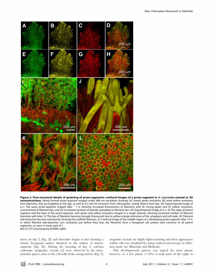
Figure 2. Fine-structural details of greening of proto-segments: confocal images of a proto-segment in H. macroloba viewed as 3Dreconstructions. Newly formed proto-segment imaged under 488 nm excitation showing (A) mainly green emissions, (B) weak yellow emissionsfrom filaments, that are brightest at the tips, as well as (C) red Chl emissions from chloroplasts, mainly absent from tips. (D) Superimposed image ofa–c. The same proto-segment imaged after ,1 h, showing increased fluorescence of filaments with (E) strong green and (F) yellow emissions,concentrated at filament tips, and (G) increased numbers of plastids spreading to filament tips. (H) Superimposed image of a–c. (I) The edge of parentsegment and the base of the proto-segment, with green and yellow emissions imaged in a single channel, showing increased number of filamentbranches with time. (J) The tips of filaments become strongly fluorescent due to yellow-orange emissions of the cytoplasm and cell walls. (K) Filamentside-branches become intertwined, forming the scaffold filaments. (L) Confocal image of the middle region of a developing proto-segment after 14 h,in which filament side-branches turn outwards; just before they fuse, the filaments form a hexagonal cell surface view common to all parentsegments, as seen in lower part of I.doi:10.1371/journal.pone.0020841.g002
Mass Chloroplast Movement in Halimeda
PLoS ONE | www.plosone.org 4 July 2011 | Volume 6 | Issue 7 | e20841
complete the growth of green filaments. Such slow growth was
particularly noted in young plants with thick mature segments and
few branches; in one such case the new segments (8 in all) were still
white the following morning and greening occurred only over the
course of 5 h in daylight on day 2.
In all cases, growth continued on day 3 and by day 4 the new
segments approached the size of mature segments. In the
aquarium-grown material, segment formation did not appear to
follow any periodicity and occurred randomly in plants from day
to day, possibly as a result of disruption of their natural diurnal
light cycles. Nevertheless, it was still possible to identify proto-
segments on the afternoon of day 1 and to then observe greening
during the night and calcification on the following day.
In an experiment with 10 plants under illumination (,200 mmol
photon m22 s21, white light) from 16.00 h on day 1 until 07.00 h
on day 2 no green filaments were produced during the night. After
transfer to natural light, green filaments began to grow in the early
morning (ca 8.00 h, 2.5 h after dawn) of day 2, and by the
afternoon of day 2 they were similar to filaments on plants
darkened overnight. Plants (n = 5) collected after 17.00 h on day 1,
but illuminated overnight, as before, tended to have new segments
that greened overnight, although the timing was delayed. Plants
(n = 5) irradiated with red light (,100 mmol photon m22 s21, light
.600 nm) overnight all had white proto-segments on day 2 and
greened up (in daylight) on day 2.
Imaging-PAM fluorometryOn 4 nights using field material, a sub-sample of the normal
(dark) treatment was taken for imaging PAM fluorimetric analysis.
This necessitated subjecting the plants to low modulated blue light
pulses and short (,1 s) saturating blue light pulses every 5–15 min.
A typical dataset is shown in Fig. 5 for one such treatment (and an
animation is shown in Supporting Information: Movie S1). At time
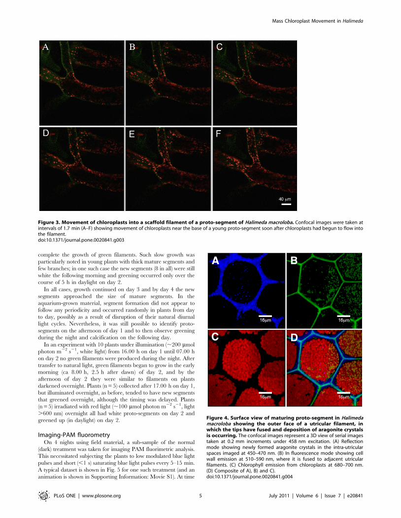
Figure 3. Movement of chloroplasts into a scaffold filament of a proto-segment of Halimeda macroloba. Confocal images were taken atintervals of 1.7 min (A–F) showing movement of chloroplasts near the base of a young proto-segment soon after chloroplasts had begun to flow intothe filament.doi:10.1371/journal.pone.0020841.g003
Figure 4. Surface view of maturing proto-segment in Halimedamacroloba showing the outer face of a utricular filament, inwhich the tips have fused and deposition of aragonite crystalsis occurring. The confocal images represent a 3D view of serial imagestaken at 0.2 mm increments under 458 nm excitation. (A) Reflectionmode showing newly formed aragonite crystals in the intra-utricularspaces imaged at 450–470 nm. (B) In fluorescence mode showing cellwall emission at 510–590 nm, where it is fused to adjacent utricularfilaments. (C) Chlorophyll emission from chloroplasts at 680–700 nm.(D) Composite of A), B) and C).doi:10.1371/journal.pone.0020841.g004
Mass Chloroplast Movement in Halimeda
PLoS ONE | www.plosone.org 5 July 2011 | Volume 6 | Issue 7 | e20841

zero, the white proto-segment exhibited weak chlorophyll fluores-
cence and absorptivity and low quantum yield confined to the area
closest to the old segment. Over the course of 5–6 hours, the new
green segment was formed and chloroplasts migrated into its
filaments. The greening of the segment was largely completed after
,3–5 h, while active photosynthesis was first detected after 5 hours.
In the following 12 hours, the segment calcified and gained volume,
while the maximum quantum yield was largely unchanged.
Usually, chloroplasts had reached the tips of the scaffold
filaments after 180 min and the process of chloroplast movement
was complete in ,210 min (Fig. 5). The chloroplasts moved at a
fairly constant rate, as evidenced by the rate of increase and spread
of Ft throughout the new segment. Based on a mean base-to-tip
length for the proto-segment of 7.060.1 mm (mean 6 SD; n = 10)
and a greening rate of 3 h, this would represent a chloroplast
movement rate of 0.65 mm s21, which is similar to rates observed
Figure 5. Sequence of new segment development and the onset of photosynthetic activity in Halimeda macroloba. The imageds wereobtained by digital photography, absorptivity (Abs), fluorescence yield (F) and quantum yield of PSII (WPSII) over a 17 h period using microscopy andimaging-PAM fluorimetry. An animation of the full dataset, i.e., images taken every 5–10 minutes is available in Supporting Information: Movie S1. Allcolour images were normalised to the same relative colour code shown in the lower part of the figure.doi:10.1371/journal.pone.0020841.g005
Mass Chloroplast Movement in Halimeda
PLoS ONE | www.plosone.org 6 July 2011 | Volume 6 | Issue 7 | e20841

directly under confocal imaging (unpublished data), but
somewhat slower than the more random movent observed above
for individual chloroplasts. Initially the areas with chlorophyll
fluorescence showed little or no quantum yield, i.e., they were
unable to carry out photosynthesis. The capacity for photosyn-
thesis lagged ,1 h behind completion of the chloroplast migration
(see Fig. 5), as did the ability to carry out non-photochemical
quenching (NPQ)(not shown).
Oxygen concentration in the proto-segmentMicroelectrode measurements showed O2 depletion (0–5% air
saturation) in the center of the proto-segment throughout the dark
incubation period where the maturation of the new segment
occurred (data not shown). When actinic light was provided in the
morning after the greening process had completed, the O2 quickly
rose to levels exceeding those in the surrounding seawater kept at
100% atmospheric saturation. These measurements thus showed a
pronounced O2 respiration in the proto-segment during matura-
tion and fully competent photosynthesis only upon completion of
the maturation process.
Inhibitor studiesTable 1 shows the results of the experiment with colchicine
(0.5 mM) and cytochalasin d (2 mM), with 20 replicates. Alone,
each inhibitor partially inhibited greening. However, with both
inhibitors present together there was a total inhibition of new
filament growth, where the proto-segments (n = 20) were approx-
imately the same size on the morning of the second day, as at
dusk the previous day, and without formation of utricle initials.
Cytoplasmic streaming and filament growth were only fully
inhibited in the presence of both inhibitors, while partial inhibition
was induced when only one of the inhibitors was applied.
Although the numbers are small, results are clearly significant
and this was confirmed by using Pearson’s chi-square test on the
complete set of data (p,2.2e216).
Discussion
The siphonalean green alga Halimeda has been studied for many
years because of its special features in terms of calcification
[3,15,16,17,18] and circadian chloroplast movement [21,22]. The
mechanisms involved in new segment formation have hitherto
been unknown and our study shows for the first time that segment
formation involves a complex series of events, whose further study
could be a very valuable tool in plant cell research.
Once the proto-segment is formed, greening begins at dusk.
Maturation is a three-part process, depending firstly on the import
of green chloroplasts and proplastids into the scaffold filaments
from the parent segment, together with replication of those plastids
and secondly on the formation of new green filaments, which grow
a) by ramification of the existing ramuli, and movement of
chloroplasts in the large ramuli (Fig. 3) and b) by new filaments
growing from the parent segment that interpenetrate the scaffold
filaments of the proto-segment (Fig. 2), taking chloroplasts
generated in the parent segment and proplastids. The incapacity
of freshly imported plastids to perform full photosynthesis initially
(Fig. 5) is an interesting observation, suggesting that these are
newly formed organelles, whose maturation occurs during the
night. As the new segment greens overnight maturation affects
several photosynthetic fluorescence parameters (Fig. 5), most
notably Fm, Fm’ and NPQ, suggesting that proplastids mature
into fully functional plastids during this process. The third stage of
the maturation process is sealing off the inter-utricular space from
the outside, the formation of aragonite crystals (Fig 4) and the
evolution of photosynthetic oxygen on the morning of the second
day. Following this, new segments continued to grow over the
third and fourth day.
The rapid production of new plastids, over a typical time of 2–
3 h, represents one of the fastest recorded maturations of photo-
synthetic tissue and therefore deserves closer scrutiny in future
studies. The chloroplast movement into the initially colourless
filaments via the scaffold filaments (Figs. 2, 3 & 5) occurred at a
fast rate of ,0.65 mm s21. Ten fold higher intracellular transport
rates have been observed for cytoplasmic streaming in Caulerpa
prolifera, a member of the Siphonales, while in Nitella, a strepto-
phyte alga, an hundred fold higher rate was recorded [27].
However, these higher rates were not for plastid movement, nor
were they a mass unidirectional movement.
The chloroplast movement in H. macroloba could only be
completely inhibited by the simultaneous presence of the inhibitors
colchine and cytochalasin c (Table 1), which inhibit microtubule-
and microfilament-dependent cell organelle movement, respec-
tively [28]. Thus both types of cellular transport mechanisms are
involved in chloroplast movement of H. macroloba. The formation
and transport of chloroplasts is, however, only one aspect of the
remarkable growth spurt in H. macroloba and, for example, the rate
of protein synthesis must also be very high. The proto-segment
interior became anoxic during night-time maturation indicating a
high respiratory load during this period, presumably as a result of
the high demand for ATP. Thus this system should provide a
useful tool for future studies of protein synthesis and growth in
algae.
The temporal triggering of new segment formation in H.
macroloba is intriguing as it apparently involves a periodicity in the
majority of plants, which may, for example, be entrained to a
lunar cycle, as has been found for other green macroalgae [29].
However, about 10% of plants do not follow this cycle. Once the
proto-segment is initiated, the timing of the greening process seems
dependent on a light/dark sensor system, whereby growth is
initiated at dusk on day 1. Continuous light overnight could delay
Table 1. The effect of the inhibitors colchicine (0.5 mM in seawater) and cytochalasin c (2 mM in seawater), on the greening ofproto-segments in Halimeda macroloba (see text for further description).
Treatment Replicates Colour after 12 h (in darkness)
White Partially green Fully green
Control 20 0 0 20
Colchicine (0.5 mM) 20 2 16 2
Cytochalasin d (2 mM) 20 4 16 0
Colchicine + Cytochalasin d 20 20 0 0
doi:10.1371/journal.pone.0020841.t001
Mass Chloroplast Movement in Halimeda
PLoS ONE | www.plosone.org 7 July 2011 | Volume 6 | Issue 7 | e20841

development until the next morning when inhibition was
alleviated and growth proceeded. Our finding that red light had
a similar effect to white light in causing such a delay could indicate
that a phytochrome system is involved.
The proto-segment is formed of scaffold filaments recruiting
chloroplasts and becoming intertwined by secondary green
filaments from the parent segment at night-time. We hypothesize
that grazers attempting to feed on the newly arising structure
during daylight of day 1 would be deterred by the tough nature of
the proto-segment filaments, lack of nutrition and the possible
presence of secondary metabolites. The walls of the proto-segment
filaments are thickened and the fluorescent nature of the walls
suggests that tannins or other phenolics may be present as a
feeding deterrent. It is known that Halimeda spp. produce
diterpenoid feeding deterrents [4,5,30] and such compounds
may also be present in the proto-segment.
The end result of the greening process is a mature segment with
protection from grazing by the presence of secondary metabolites
and the production of unpalatable aragonite crystals (Fig 4).
Borowitzka & Larkum [3] suggested that a major factor in the
calcification process was the alkalinisation of the inter-utricular
compartment, as a result of photosynthesis in adjacent chloroplast-
packed utricular filaments in the light. This hypothesis was
supported by the later work of Borowitzka [18] and De Beer &
Larkum [19]. Unfortunately, it was not possible in the latter study
to place a microsensor in the inter-utricular space owing to wound
reactions. In mature segments, at night, chloroplasts are
withdrawn deeper into the thallus and out of the inter-utricular
filaments [21,22]; thus illumination at night should not result in
alkalinisation and calcification.
The reason(s) for the complex set of events leading to the
generation of a new green segment in H. macroloba is(are) still a
matter of conjecture. The phenomenon and its timing may be
explained as a process minimizing herbivory. Herbivorous fish and
molluscs would potentially be attractive to young, green segments.
However mature segments of Halimeda species, even at the ends of
branches, are tough, packed with aragonite crystals and filled with
diterpenoid feeding deterrents [4,5,30], which are avoided by
many reef herbivores. Although white proto-segments would be
easy to ingest, they would be relatively unattractive, low in food
value and, furthermore, may be replete with feeding deterrents.
Our observations suggest that proto-segments are not actively
grazed at night-time when there are few active herbivores, and this
is when the chloroplasts are imported into the proto-segment, and
maturation occurs. By first light on the following day, the utricles
generally have been fused, whereafter calcification quickly fills the
new green segment with aragonite crystals, increasing its
indigestibility.
The hypothesis of herbivory avoidance could be tested by
identifying, for example, when secondary metabolites are imported
into the proto-segments and by assessing the palatability of proto-
segments and new green segments to herbivorous fish and other
grazers. It should be noted, however, that Mantyka & Bellwood
[31] have shown that many macroalgae on coral reefs, including
several species of Halimeda, are susceptible to heavy grazing by reef
fish, when more attractive algal species are not available.
In summary, the white proto-segment of Halimeda is a novel
structure that has received little attention in the past. We present
the first detailed study of the rapid structural changes and cellular
transport processes involved in segment formation and matura-
tion. The fast movement of H. macroloba chloroplasts, inactive in
photosynthesis, into scaffold filaments seems to represent a new
phenomenon of mass migration of plastids not previously reported.
The whole system provides a fascinating field of research for future
studies in plant cell biology
Supporting Information
Movie S1 An animation showing the greening of segments of
Halimeda macroloba over 6 hours as imaged by an imaging-PAM
(conditions were the same as for Fig. 5).
(WMV)
Acknowledgments
We wish to thank the staff of the Heron Island Research Station and the
Australian Centre for Microscopy and Microanalysis, University of Sydney
for support. We are grateful to Dr EA Drew and Professor R Overall,
University of Sydney, who gave helpful general advice, and to Professor
John Robinson, University of Sydney, who gave statistical advice. The field
part of this study was carried out under Research Permit GB1000659,
kindly supplied by the Great Barrier Reef Marine Park Authority,
Townsville.
Author Contributions
Conceived and designed the experiments: AWDL AS MK. Performed the
experiments: AWDL AS MK. Analyzed the data: AWDL AS MK.
Contributed reagents/materials/analysis tools: AWDL AS MK. Wrote the
paper: AWDL AS MK.
References
1. Hillis-Colinvaux L (1980) Ecology and taxonomy of Halimeda: primary producerof coral reefs. Advances in Marine Biology 17: 1–327.
2. Rees SA (2007) Significance of Halimeda bioherms to the global carbonate budgetbased on a geological sediment budget for the Northern Great Barrier Reef,
Australia. Coral Reefs 26: 177–188.
3. Borowitzka MA, Larkum AWD (1976b) Calcification in the green alga Halimeda.
3. The sources of inorganic carbon for photosynthesis and calcification and amodel of the mechanism of calcification. Journal of Experimental Botany 27:
879–893.
4. Paul VJ, van Alstyne KL (1992) Activation of chemical defenses in the tropicalgreen algae Halimeda spp. Journal of Experimental Marine Biology and Ecology
160: 191–203.
5. Hay ME (1996) Marine chemical ecology: what’s known and what’s next.
(Antifeedant chemicals in Halimeda). Journal of Experimental Marine Biologyand Ecology 200: 103–134.
6. Drew EA (1983) Halimeda biomass, growth rates and sediment generation in thecentral Great Barrier Reef Province. Coral Reefs 2: 101–110.
7. Drew EA, Abel K (1988) Studies on Halimeda I. The distribution and speciescomposition of Halimeda meadows throughout the Great Barrier Reef Province,
Australia. Coral Reefs 6: 195–206.
8. Wolanski E, Drew EA, Abel KM, O’Brien J (1988) Tidal jets nutrient upwelling
and their influence on the productivity of the alga Halimeda in the ribbon reefs,
Great Barrier Reef, Australia. Estuarine and Coastal Shelf Science 26: 169–202.
9. Marshall JF, Davies PJ (1988) Halimeda bioherms of the northern Great Barrier
Reef. Coral Reefs 5: 139–148.
10. Orme GR, Salama MS (1988) Form and seismic stratigraphy of Halimeda banksin part of the northern Great Barrier Reef Province. Coral Reefs 6: 131–137.
11. Fornos JJ, Forteza V, Jaume C, Martinez-Taberner A (1992) Present-day
Halimeda carbonate sediments in temperate Mediterranean embayments:Fornells, Balearic Islands. Sedimentary Geology 75: 283–293.
12. Braga JC, Martin JM, Riding R (1996) Internal structure of segment reefs:
Halimeda algal mounds in the Mediterranean Miocene. Geology 24: 35–38.
13. Borowitzka MA, Larkum AWD (1974a) Chloroplast development in the
caulerpalean alga Halimeda. Protoplasma 81: 131–144.
14. Borowitzka MA, Larkum AWD (1974b) The caulerpalean thylakoid organizingbody. Eighth International Congress on Electron Microscopy, Canberra 2:
588–589.
15. Borowitzka MA, Larkum AWD (1976a) Calcification in the green alga Halimeda.2. The exchange of Ca2+ and the occurrence of age gradients in calcification and
photosynthesis. Journal of Experimental Botany 27: 846–878.
16. Borowitzka MA, Larkum AWD (1976c) Calcification in the green alga Halimeda.
4. The action of metabolic inhibitors on photosynthesis and calcification. Journal
of Experimental Botany 27: 894–907.
17. Borowitzka MA, Larkum AWD (1977) Calcification in the green alga Halimeda.
1. An ultrastructure study of thallus development. Journal of Phycology 13:
6–16.
Mass Chloroplast Movement in Halimeda
PLoS ONE | www.plosone.org 8 July 2011 | Volume 6 | Issue 7 | e20841

18. Borowitzka MA (1986) Physiology and biochemistry of calcification in the
Chlorophyceae. In: Leadbeater B, Riding H, eds. Biomineralization in the lowerplants and animals. Oxford: Oxford University Press. pp 107–124.
19. De Beer D, Larkum AWD (2001) Photosynthesis and calcification in the
calcifying algae Halimeda discoidea studied with microsensors. Plant Cell andEnvironment 24: 1209–1217.
20. Borowitzka MA (1982) Carbonate calcification in algae – initiation and control.In: Mann S, Webb J, Williams RJP, eds. Biomineralization: Chemical and
biochemical perspectives. Weinheim: VCH Verlag. pp 63–94.
21. Drew EA, Abel K (1990) Studies on Halimeda III. A daily cycle of chloroplastmigration within segments. Botanica Marina 33: 31–46.
22. Drew EA, Abel K (1992) Studies on Halimeda IV. An endogenous rhythm ofchloroplast migration in the siphonous green alga Halimeda distorta. Journal of
Interdisciplinary Cycle Research 22: 128–13.23. Wilbur KM, Collinvaux LM, Watabe N (1969) Electron microscope study of
calcification in Halimeda (Order Siphonales). Phycologia 8: 27–35.
24. Ralph PJ, Schreiber U, Gademann R, Kuhl M, Larkum AWD (2005) Coralphotobiology studied with a new imaging pulse amplitude modulated
fluorometer. Journal of Phycology 41: 335–342.
25. Schreiber U (2004) Pulse-amplitude-modulation (PAM) fluorometry and
saturation pulse method: an overview. In: Papageorgiou GC, Govindjee, eds.Chlorophyll a fluorescence: a signature of photosynthesis, (Advances in
Photosynthesis and respiration, Vol 11). Berlin: Springer Verlag. pp 279–319.
26. Larkum AWD, Koch E, Kuhl M (2003) Diffusive boundary layers andphotosynthesis of the epilithic algal community of coral reefs. Marine Biology
142: 1073–1082.27. Sabnis DD, Jacobs WP (1967) Cytoplasmic streaming and microtubules in the
coenocytic alga, Caulerpa prolifera. Journal of Cell Science 2: 465–472.
28. Williamson RE (1993) Organelle movements. Annual Review of PlantPhysiology and Plant Molecular Biology 44: 181–202.
29. Luning K, Kadel P, Pang S (2008) Control of reproduction rhythmicity byenvironmental and endogenous signals in Ulva pseudocurvata (Chlorophyta).
J Phycol 44: 866–873.30. Paul VJ, van Alstyne KL (1988) Chemical defense and chemical variation in
some tropical Pacific species of Halimeda (Halimedaceae; Chlorophyta). Coral
Reefs 6: 263–269.31. Mantyka CS, Bellwood DR (2007) Direct evaluation of macroalgal removal by
herbivorous coral reef fishes. Coral Reefs 26: 435–442.
Mass Chloroplast Movement in Halimeda
PLoS ONE | www.plosone.org 9 July 2011 | Volume 6 | Issue 7 | e20841