Embed Size (px)
Citation preview
725
doi: 10.2169/internalmedicine.5733-20
Intern Med 60: 725-730, 2021
http://internmed.jp
【 CASE REPORT 】
Rapidly Progressing Aseptic Abscesses in a Patientwith Ulcerative Colitis
Yoshiharu Yamaguchi, Marie Nakagawa, Shoko Nakagawa, Kazuhiro Nagao, Satoshi Inoue,
Tomoya Sugiyama, Shinya Izawa, Yasutaka Hijikata, Masahide Ebi, Yasushi Funaki,
Naotaka Ogasawara, Makoto Sasaki and Kunio Kasugai
Abstract:Aseptic abscesses (AAs) are extraintestinal manifestations of inflammatory bowel disease (IBD). IBD-
associated AAs are rare in Japan. We treated a 45-year-old man with ulcerative colitis (UC)-associated AAs.
During remission, multiple progressive abscesses were detected in the spleen; he underwent splenectomy be-
cause an infectious disease was suspected. Although his condition improved temporarily after splenectomy, a
large liver abscess was noted, and a diagnosis of UC-associated AAs was made. Granulocytapheresis (GCAP)
and infliximab (IFX) administration resolved the abscess. This is the first reported case of UC-associated
AAs in a Japanese patient treated by splenectomy, GCAP, and IFX.
Key words: aseptic abscesses, ulcerative colitis, splenectomy, granulocytapheresis, infliximab
(Intern Med 60: 725-730, 2021)(DOI: 10.2169/internalmedicine.5733-20)
Introduction
Aseptic abscesses (AAs) are focal enclosed lesions char-
acterized by the presence of abscesses, failure of antibiotic
therapy, and negative blood and aspirate cultures; however,
improvement is seen after corticosteroid therapy with or
without adjunctive immunosuppressant therapy. AAs are
closely related to neutrophilic dermatoses and inflammatory
bowel disease (IBD) (1). Although the prevalence of IBD in
Japan has increased substantially (2), few cases of IBD-
associated AAs have been reported to date (3-5).
We herein report a rare case of IBD-associated AAs in a
Japanese patient.
Case Report
A 45-year-old man was referred to our hospital from an
outpatient clinic with a 30-day history of a fever and epigas-
tric pain, weight loss of 8 kg, and skin lesions on both legs.
His family history was unremarkable. His bowel habits were
unchanged, with 2-3 non-bloody stools per day. He had a
30-year history of pancolonic ulcerative colitis (UC) and
ankylosing spondylitis (AS) and was undergoing treatment
with mesalazine (3.6 g/day) and adalimumab (ADA) (40 mg
every 2 weeks). The patient’s condition was stable with the
occasional abdominal pain or slightly bloody diarrhea (par-
tial Mayo score 3).
A physical examination conducted upon his admission
was unremarkable, except for hyperthermia (38.8℃). Labo-
ratory findings (Table 1) showed an erythrocyte sedimenta-
tion rate of 57 mm in the first hour (normal value, 1-7 mm);
hemoglobin, 8.9 g/dL (normal value, 13.9-16 g/dL); white
blood cell (WBC) count, 15.3×10³/μL (normal value, 5-8×
103/μL), with neutrophils at 13,479/μL, lymphocytes at
1,056/μL, and monocytes at 734/μL; platelet count, 448×103/
μL (normal value, 138-309×103/μL); C-reactive protein
(CRP), 11.79 mg/dL (normal value, <0.3 mg/dL); aspartate
aminotransferase, 7 U/L (normal value, 13±33 U/L); alanine
aminotransferase, 6 U/L (normal 6-30 U/L); total bilirubin,
0.29 mg/dL (normal value, 0.3-1.2 mg/dL); alkaline phos-
phatase, 205 U/L (normal value, 115±359 U/L); and gam-
maglutamyl transferase, 19 U/L (normal value, 10-47 U/L).
Colonoscopy at the time of admission demonstrated a
Department of Gastroenterology, Aichi Medical University School of Medicine, Japan
Received: June 29, 2020; Accepted: August 9, 2020; Advance Publication by J-STAGE: September 30, 2020
Correspondence to Dr. Yoshiharu Yamaguchi, [email protected]
Intern Med 60: 725-730, 2021 DOI: 10.2169/internalmedicine.5733-20
726
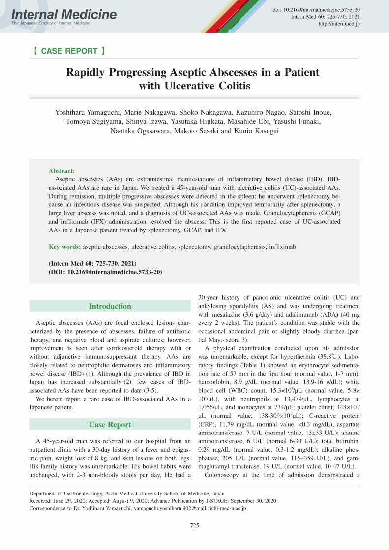
Figure 1. Colonoscopy showed a lack of vascular patterning and friable mucosa (Mayo endoscopic score 1) in the ascending (a) and sigmoid (b) colon.
Table 1. Results of Blood Examination of the Patient.
Parameter Value Parameter Value Parameter Value
ALB 2.9 g/dL T-Bil 0.29 mg/dL WBC 15,300 /μL
UN 4.5 mg/dL Glu 102 mg/dL Neutrophils 13,479 /μL
CRE 0.61 mg/dL Na 135 mEq/L Lymphocytes 1,056 /μL
AST 7 U/L K 3.9 mEq/L Monocytes 734 /μL
ALT 6 U/L Cl 100 mEq/L Eosinocytes 0.1 %
ALP 205 U/L Ca 8.3 mg/dL Hb 8.9 g/dL
LDH 105 U/L CRP 11.79 mg/L Ht 27.5 %
γ-GTP 19 U/L ESR 57 mm Plt 448×103 /μL
Alb: albumin, UN: urea nitrogen, CRE: creatinine, AST: aspartate aminotransferase, ALT: alanine amino-
transferase, ALP: alkaline phosphatase, LDH: lactate dehydrogenase, γ -GTP: γ -glutamyl transpeptidase,
T-Bil: total bilirubin, Glu: glucose, Na: sodium, K:potassium, Cl: chloride, Ca: calcium, CRP: C-reactive
protein, ESR: erythrocyte sedimentation rate, WBC: white blood cell, Hb: hemoglobin, Ht: hematocrit,
Plt: platelet
lack of vascular patterning and friable mucosa in the entire
colon (Mayo endoscopic score 1) (Fig. 1a, b). In addition,
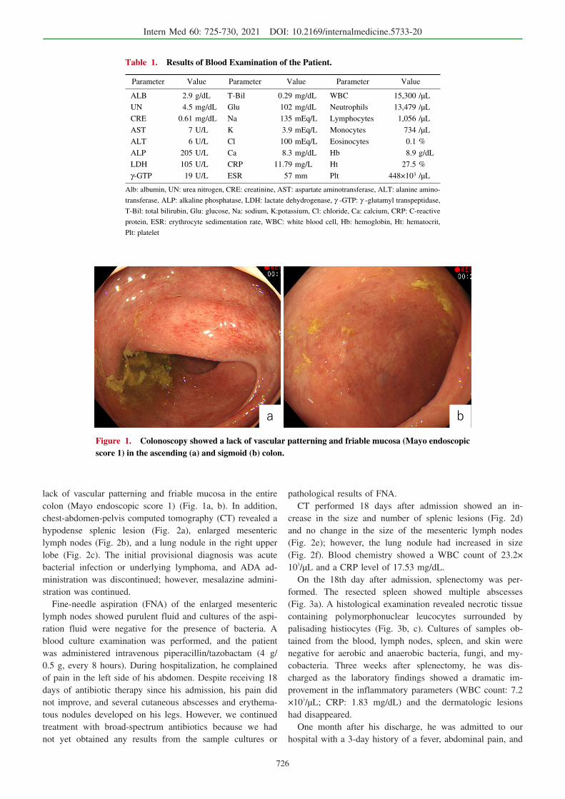
chest-abdomen-pelvis computed tomography (CT) revealed a
hypodense splenic lesion (Fig. 2a), enlarged mesenteric
lymph nodes (Fig. 2b), and a lung nodule in the right upper
lobe (Fig. 2c). The initial provisional diagnosis was acute
bacterial infection or underlying lymphoma, and ADA ad-
ministration was discontinued; however, mesalazine admini-
stration was continued.
Fine-needle aspiration (FNA) of the enlarged mesenteric
lymph nodes showed purulent fluid and cultures of the aspi-
ration fluid were negative for the presence of bacteria. A
blood culture examination was performed, and the patient
was administered intravenous piperacillin/tazobactam (4 g/
0.5 g, every 8 hours). During hospitalization, he complained
of pain in the left side of his abdomen. Despite receiving 18
days of antibiotic therapy since his admission, his pain did
not improve, and several cutaneous abscesses and erythema-
tous nodules developed on his legs. However, we continued
treatment with broad-spectrum antibiotics because we had
not yet obtained any results from the sample cultures or
pathological results of FNA.
CT performed 18 days after admission showed an in-
crease in the size and number of splenic lesions (Fig. 2d)
and no change in the size of the mesenteric lymph nodes
(Fig. 2e); however, the lung nodule had increased in size
(Fig. 2f). Blood chemistry showed a WBC count of 23.2×
103/μL and a CRP level of 17.53 mg/dL.
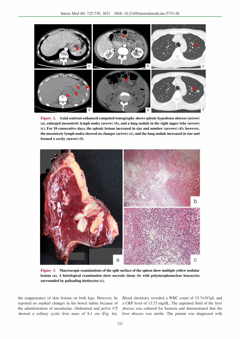
On the 18th day after admission, splenectomy was per-
formed. The resected spleen showed multiple abscesses
(Fig. 3a). A histological examination revealed necrotic tissue
containing polymorphonuclear leucocytes surrounded by
palisading histiocytes (Fig. 3b, c). Cultures of samples ob-
tained from the blood, lymph nodes, spleen, and skin were
negative for aerobic and anaerobic bacteria, fungi, and my-
cobacteria. Three weeks after splenectomy, he was dis-
charged as the laboratory findings showed a dramatic im-
provement in the inflammatory parameters (WBC count: 7.2
×103/μL; CRP: 1.83 mg/dL) and the dermatologic lesions
had disappeared.
One month after his discharge, he was admitted to our
hospital with a 3-day history of a fever, abdominal pain, and
Intern Med 60: 725-730, 2021 DOI: 10.2169/internalmedicine.5733-20
727
Figure 2. Axial contrast-enhanced computed tomography shows splenic hypodense abscess (arrow) (a), enlarged mesenteric lymph nodes (arrow) (b), and a lung nodule in the right upper lobe (arrow) (c). For 10 consecutive days, the splenic lesions increased in size and number (arrows) (d); however, the mesenteric lymph nodes showed no changes (arrow) (e), and the lung nodule increased in size and formed a cavity (arrow) (f).
Figure 3. Macroscopic examinations of the split surface of the spleen show multiple yellow nodular lesions (a). A histological examination show necrotic tissue (b) with polymorphonuclear leucocytes surrounded by palisading histiocytes (c).
the reappearance of skin lesions on both legs. However, he
reported no marked changes in his bowel habits because of
the administration of mesalazine. Abdominal and pelvic CT
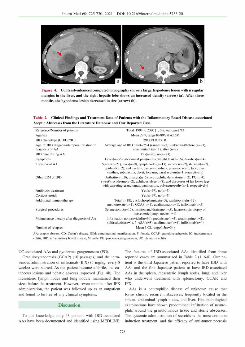
showed a solitary cystic liver mass of 8.1 cm (Fig. 4a).
Blood chemistry revealed a WBC count of 15.7×10³/μL and
a CRP level of 13.73 mg/dL. The aspirated fluid of the liver
abscess was cultured for bacteria and demonstrated that the
liver abscess was sterile. The patient was diagnosed with
Intern Med 60: 725-730, 2021 DOI: 10.2169/internalmedicine.5733-20
728
Figure 4. Contrast-enhanced computed tomography shows a large, hypodense lesion with irregular margins in the liver, and the right hepatic lobe shows an increased density (arrow) (a). After three months, the hypodense lesion decreased in size (arrow) (b).
Table 2. Clinical Findings and Treatment Data of Patients with the Inflammatory Bowel Disease-associated Aseptic Abscesses from the Literature Database and Our Reported Case.
Reference/Number of patients Total. 1994 to 2020 [1, 6-8, our case] /43
Age/sex Mean 29.7, range10-80/27F&16M
IBD phenotype (CD/UC/IC) 29CD/13UC/1IC
Age of IBD diagnosis/temporal relation to
diagnosis of AA
Average age of IBD onset=25.4 (range10-72, 3unknown)/before (n=23),
concomitant (n=11), after (n=9)
IBD flare during AA Yes(n=20), no(n=23)
Symptoms Fever(n=36), abdominal pain(n=30), weight loss(n=16), diarrhea(n=14)
Location of AA Spleen(n=21), liver(n=9), lymph nodes(n=13), muscles(n=2), sternum(n=2),
epidural(n=2), and eyelids, pancreas, kidney, pharynx, scalp, face, inner
canthus, submaxilla, chest, forearm, nasal septum(n=1, respectively)
Other EIM of IBD Arthritis(n=10), myalgia(n=5), neutrophilic dermatosis(n=2), PG(n=4),
sweet’s syndrome(n=2), aphthous ulcer(n=8), and abscesses of his lower legs
with caseating granulomas, panniculitis, polyneuropathy(n=1, respectively)
Antibiotic treatment Yes(n=39), no(n=4)
Corticosteroids Yes(n=39), no(n=4)
Additional immunotherapy Total(n=18), cyclophosphamide(n=3), azathioprine(n=12),
methotrexate(n=1), GCAP(n=1), adalimumab(n=1), infliximab(n=5)
Surgical procedures Splenectomy(n=17), incision and drainage(n=5), laparoscopic biopsy of
mesenteric lymph nodes(n=1)
Maintenance therapy after diagnosis of AA Information not provided(n=30), prednisone(n=4), azathioprine(n=2),
sulfasalazine(n=1), 5-ASA(n=3), adalimumab(n=1), infliximab(n=4)
Number of relapses Mean 1.02, range0-5(n=34)
AA: aseptic abscess, CD: Crohn’s disease, EIM: extraintestinal manifestation, F: female, GCAP: granulocytapheresis, IC: indeterminate
colitis, IBD: inflammatory bowel disease, M: male, PG: pyoderma gangrenosum, UC: ulcerative colitis
UC-associated AAs and pyoderma gangrenosum (PG).
Granulocytapheresis (GCAP) (10 passages) and the intra-
venous administration of infliximab (IFX) (5 mg/kg, every 8
weeks) were started. As the patient became afebrile, the cu-
taneous lesions and hepatic abscess improved (Fig. 4b). The
mesenteric lymph nodes and lung nodule maintained their
sizes before the treatment. However, seven months after IFX
administration, the patient was followed up as an outpatient
and found to be free of any clinical symptoms.
Discussion
To our knowledge, only 43 patients with IBD-associated
AAs have been documented and identified using MEDLINE.
The features of IBD-associated AAs identified from these
reported cases are summarized in Table 2 (1, 6-8). Our pa-
tient is the third Japanese patient reported to have IBD with
AAs and the first Japanese patient to have IBD-associated
AAs in the spleen, mesenteric lymph nodes, lung, and liver
who underwent treatment with splenectomy, GCAP, and
IFX.
AAs is a neutrophilic disease of unknown cause that
forms chronic recurrent abscesses, frequently located in the
spleen, abdominal lymph nodes, and liver. Histopathological
examinations have shown predominant infiltration of neutro-
phils around the granulomatous tissue and sterile abscesses.
The systemic administration of steroids is the most common
induction treatment, and the efficacy of anti-tumor necrosis
Intern Med 60: 725-730, 2021 DOI: 10.2169/internalmedicine.5733-20
729
factor-α (TNF-α) antibody in addition to steroid pulse ther-
apy and immunosuppressants has been recently reported in
some refractory cases (Table 2) (1, 6-8). An AA diagnosis is
a diagnosis of exclusion, and there is currently no estab-
lished diagnostic method. However, Andre et al. reported the
following diagnostic clues for AAs: (a) deep abscesses with
neutrophilic features; (b) negative serologic tests for bacteria
and fungi and cultures of blood and aspirate; (c) when ad-
ministered, failure of broad-spectrum antibiotic therapy, in-
cluding antituberculosis therapy; and (d) rapid clinical im-
provement on corticosteroid therapy with or without addi-
tional immunosuppressant therapy and subsequent radiologic
evidence of abscess resolution (9). Our case presented with
these features of AAs. If a patient has a background of dis-
eases with immune abnormalities, the diagnosis should be
reached as soon as possible using evidence to reject the pos-
sibility of infection and prove the pathology.
In our case, multiple splenic abscesses were initially sus-
pected to have been caused by bacterial infection. For this
reason, the patient underwent splenectomy, and the ab-
scesses were found to be sterile. Although there were many
possibilities of infections causing similar life-threating ab-
scesses (10), we could not initially exclude all infections in
this case. Therefore, we chose to perform splenectomy to ar-
rive at an exact diagnosis.
Because of the considerable overlap with other infections
based on the appearance on imaging modalities, such as ul-
trasound, CT, and MRI, and the small number of AAs, AAs
are extremely difficult to distinguish. Abscess formation is
essentially a persistent and intense inflammatory irritation,
so it is unlikely that differences in abscess formation found
on imaging studies due to variable causes of irritation can
be noted.
To prove that the abscesses are sterile, an invasive exami-
nation, such as laparotomy, is required. Furthermore, a tem-
porary improvement in systemic symptoms after splenec-
tomy can be attributed to a decrease in the systemic immune
response, owing to the removal of the largest accumulation
of lymphoid tissue, which controls immunity, in the
body (11).
The present patient refused the administration of steroids,
and eventually, the symptoms improved after splenectomy
with GCAP and IFX.
The event in the present patient occurred during the use
of ADA; however, we did not use ADA again because
ADA’s secondary loss of response could not be ruled out.
Furthermore, we did not use steroids due to the lack of clar-
ity regarding the accumulation of steroids during long-term
UC treatment and the patient’s strong refusal of steroids,
which required long-term administration after hospital ad-
mission. Therefore, IFX treatment was chosen in this case.
AA remission can be induced without using steroids and
sustained with GCAP and IFX. GCAP is reported to be use-
ful for the treatment of IBD and PG (12, 13). Indeed, Kato
et al. described a Japanese patient with AAs and Crohn’s
disease (CD) who showed improvement following treatment
with GCAP (5). The advantages of GCAP are that it rarely
causes adverse events and is considered to be as useful as
corticosteroids for induction therapy of AAs.
The precise mechanism underlying the action of GCAP
has not been clearly described. However, GCAP selectively
adsorbs and removes activated monocytes and MAC-1-
expressing neutrophils that infiltrate the inflamed regions. In
addition, GCAP reduces the circulating levels of inflamma-
tory cytokines, such as TNF-α, interleukin (IL)-1β, IL-6,
and IL-8 (14, 15). Therefore, GCAP is effective for treating
AAs as a systemic disease.
Andre et al. reported that 60% of AAs relapsed at least
once in a 7-year follow-up period (9). Our patient required
careful follow-up when he was on remission maintenance
therapy. When a patient experiences a relapse, novel anti-
body formulations, such as vedolizumab, which is effective
for PG associated with CD, and ustekinumab, which has
been reported to improve inflammatory cutaneous lesions,
are expected to be effective (16, 17).
Possible mechanisms underlying the pathogenesis of AAs
associated with UC have been discussed. The pathology of
UC, AAs, PG, and spondylitis is considered to be related to
aseptic neutrophil infiltration. Pathological conditions based
on neutrophil dysfunction were considered to have resulted
in the condition of our patient. The patient had no family
history of a similar disease; however, he did not wish to un-
dergo genetic testing, such as DNA typing, to determine if
he had any genetic mutations. Therefore, these tests were
not performed. However, human leukocyte antigen (HLA)-
B27 is positive in 25-78% of patients with concomitant IBD
and AS. In addition, other mutations, such as those in the
PSTPIP1 gene, which cause pyogenic arthritis, pyoderma
gangrenosum, and acne syndrome, may also be pre-
sent (18, 19).
Extraintestinal manifestations of IBD correlate with the
IBD activity. In contrast, AS and PG associated with IBD
have no correlation with the IBD activity (18, 20). In our
case, AS and PG did not correlate with the IBD activity, and
AA did not correlate with the UC activity. However, the AA
and PG activity were correlated. We must be careful when
managing cases with AAs without IBD relapse, as AAs may
not be related to the intestinal activity. Further research will
be needed to determine the relationship between AAs and
IBD.
Risk factors for AAs may include a high likelihood of
skin conditions. However, the number of cases has been
small (43 cases, including our case), so the risk factors are
not clear. Therefore, it will be necessary to clarify these risk
factors by accumulating more cases in the future.
Given the present findings, even when patients are in re-
mission in chronic IBD, gastroenterologists must pay close
attention to AAs and provide prompt treatment.
The authors state that they have no Conflict of Interest (COI).
Intern Med 60: 725-730, 2021 DOI: 10.2169/internalmedicine.5733-20
730
References
1. Bollegala N, Khan R, Scaffidi MA, et al. Aseptic abscesses and
inflammatory bowel disease: two cases and review of literature.
Can J Gastroenterol Hepatol 2017: 5124354, 2017.
2. Murakami Y, Nishiwaki Y, Oba MS, et al. Estimated prevalence of
ulcerative colitis and Crohn’s disease in Japan in 2014: an analysis
of a nationwide survey. J Gastroenterol 54: 1070-1077, 2019.
3. Maeshima K, Ishii K, Inoue M, Himeno K, Seike M. Behçet’s dis-
ease complicated by multiple aseptic abscesses of the liver and
spleen. World J Gastroenterol 19: 3165-3168, 2013.
4. Ito T, Sato N, Yamazaki H, Koike T, Emura I, Saeki T. A case of
aseptic abscesses syndrome treated with corticosteroids and TNF-
alpha blockade. Mod Rheumatol 23: 195-199, 2013.
5. Kato S, Hosomi E, Amano F, et al. The efficacy of intensive
granulocyte and monocyte adsorption apheresis in a patient with
Crohn’s disease complicated by extensive subcutaneous aseptic
neutrophilic abscesses. J Crohns Colitis 6: 787-791, 2012.
6. Yang Y, Chen D. Treatment of aseptic liver abscess due to Crohn’s
disease using infliximab. Clin Gastroenterol Hepatol 15: A27-A28,
2017.
7. Herskovitz I, Maderal AD, Alonso-Llamazares J. Caseating granu-
lomas manifesting as aseptic abscesses in the setting of ulcerative
colitis. Int J Dermatol 57: 475-476, 2018.
8. Bavaro DF, Ingravallo G, Signorile F, et al. Splenic abscesses as a
first manifestation of Crohn’s disease: a case report. BMC Gastro-
enterol 19: 144, 2019.
9. Andre MF, Piette JC, Kemeny JL, et al. Aseptic abscesses: a study
of 30 patients with or without inflammatory bowel disease and re-
view of the literature. Medicine (Baltimore) 86: 145-161, 2007.
10. Kamaya A, Weinstein S, Desser TS. Multiple lesions of the
spleen: differential diagnosis of cystic and solid lesions. Semin Ul-
trasound CT MR 27: 389-403, 2006.
11. Hansen K, Singer DB. Asplenic-hyposplenic overwhelming sepsis:
postsplenectomy sepsis revisited. Pediatr Dev Pathol 4: 105-121,
2001.
12. Matsuda K, Ohno K, Okada Y, et al. Adsorptive granulocyte and
monocyte apheresis is effective in ulcerative colitis patients both
with and without concomitant prednisolone. Inflamm Intest Dis 5:
36-41, 2020.
13. Russo I, Miotto S, Colpo A, et al. Successful treatment of pyo-
derma gangrenosum with granulocyte and monocyte adsorption
apheresis. Int Wound J 14: 282-284, 2017.
14. Hanai H, Takeda Y, Eberhardson M, et al. The mode of actions of
the Adacolumn therapeutic leucocytapheresis in patients with in-
flammatory bowel disease: a concise review. Clin Exp Immunol
163: 50-58, 2011.
15. Cuadrado E. Granulocyte/monocyte apheresis as immunotherapic
tool: cellular adsorption and immune modulation. Autoimmun Rev
8: 292-296, 2009.
16. Vernero M, Ribaldone DG, Cariti C, et al. Dual-targeted therapy
with apremilast and vedolizumab in pyoderma gangrenosum asso-
ciated with Crohn’s disease. J Dermatol 47: e216-e217, 2020.
17. Phillips FM, Verstockt B, Sebastian S, et al. Inflammatory cutane-
ous lesions in inflammatory bowel disease treated with Vedolizu-
mab or Ustekinumab: an ECCO CONFER multicentre case series.
J Crohns Colitis 14: 1488-1493, 2020.
18. Ott C, Scholmerich J. Extraintestinal manifestations and complica-
tions in IBD. Nat Rev Gastroenterol Hepatol 10: 585-595, 2013.
19. Martinez-Rios C, Jariwala MP, Highmore K, et al. Imaging find-
ings of sterile pyogenic arthritis, pyoderma gangrenosum and acne
(PAPA) syndrome: differential diagnosis and review of the litera-
ture. Pediatr Radiol 49: 23-36, 2019.
20. Marzano AV, Borghi A, Stadnicki A, Crosti C, Cugno M. Cutane-
ous manifestations in patients with inflammatory bowel diseases:
pathophysiology, clinical features, and therapy. Inflamm Bowel
Dis 20: 213-227, 2014.
The Internal Medicine is an Open Access journal distributed under the Creative
Commons Attribution-NonCommercial-NoDerivatives 4.0 International License. To
view the details of this license, please visit (https://creativecommons.org/licenses/
by-nc-nd/4.0/).
Ⓒ 2021 The Japanese Society of Internal Medicine
Intern Med 60: 725-730, 2021