Embed Size (px)
Citation preview
case RePORT OPeN access
www.edoriumjournals.com
International Journal of Case Reports and Images (IJCRI)International Journal of Case Reports and Images (IJCRI) is an international, peer reviewed, monthly, open access, online journal, publishing high-quality, articles in all areas of basic medical sciences and clinical specialties.
Aim of IJCRI is to encourage the publication of new information by providing a platform for reporting of unique, unusual and rare cases which enhance understanding of disease process, its diagnosis, management and clinico-pathologic correlations.
IJCRI publishes Review Articles, Case Series, Case Reports, Case in Images, Clinical Images and Letters to Editor.
Website: www.ijcasereportsandimages.com
Rare sequelae of a testicular tumor: A ‘burnt-out’ tumor
Francesca Kum, Faisal Ghumman, Matin Sheriff
ABSTRACT
Introduction: Testicular tumors typically present as a palpable lump. Some tumors have been known to spontaneously regress, thus have been referred to as ‘burnt-out’ tumors or vanishing tumors. Case Report: A 28-year-old male was seen in urology clinic with clinical findings of a small atrophic left testis, with a prior history of a painful left testicular lump not responsive to antibiotics, which had spontaneously resolved. Ultrasound scan revealed an initial solid testicular lesion of 19x12x10 mm with internal vascularity, which upon repeat ultrasound scan two months later found a 12-mm coarse calcification and a hypoechoic region, but regression of the initial solid lesion. The diagnosis of a burnt-out tumor was made and he underwent urgent radical orchidectomy and prosthesis insertion. Computed tomoraphy (CT) staging scan showed significant paraortic lymphadenopathy, therefore the patient further underwent chemotherapy. Treatment has been successful and the patient is progressing well with no complications. Conclusion: The diagnosis of a burnt-out testicular tumor is an important differential to consider in patients with spontaneously regressing testicular lumps, abnormal findings on ultrasound scan and subsequent presentation of testicular atrophy of unknown cause. This diagnosis should also be considered in patients with secondary metastatic tumors of unknown primary
(This page in not part of the published article.)
International Journal of Case Reports and Images, Vol. 6 No. 1, January 2015. ISSN – [0976-3198]
Int J Case Rep Images 2015;6(1):16–20. www.ijcasereportsandimages.com
Kum et al. 16
CASE REPORT OPEN ACCESS
Rare sequelae of a testicular tumor: A ‘burnt-out’ tumor
Francesca Kum, Faisal Ghumman, Matin Sheriff
AbstrAct
Introduction: testicular tumors typically present as a palpable lump. some tumors have been known to spontaneously regress, thus have been referred to as ‘burnt-out’ tumors or vanishing tumors. case report: A 28-year-old male was seen in urology clinic with clinical findings of a small atrophic left testis, with a prior history of a painful left testicular lump not responsive to antibiotics, which had spontaneously resolved. Ultrasound scan revealed an initial solid testicular lesion of 19x12x10 mm with internal vascularity, which upon repeat ultrasound scan two months later found a 12-mm coarse calcification and a hypoechoic region, but regression of the initial solid lesion. the diagnosis of a burnt-out tumor was made and he underwent urgent radical orchidectomy and prosthesis insertion. computed tomoraphy (ct) staging scan showed significant paraortic lymphadenopathy, therefore the patient further underwent chemotherapy. treatment has been successful and the patient is progressing well with no complications. conclusion: the diagnosis of a burnt-out testicular tumor is an important differential to consider in patients
Francesca Kum1, Faisal Ghumman2, Matin Sheriff3
Affiliations: 1MBBS, BSc, Foundation Year 2 Doctor, Department of Urology, Medway Maritime Hospital, UK; 2FRCS(Urol), FCPS(Pak), Urology Specialist Registrar, Department of Urology, Medway Maritime Hospital, UK; 3PhD, FRCS, FRCS(Urol), FEBU, Consultant Urological Surgeon, Department of Urology, Medway Maritime Hospital, Canterbury and Christ Church University, UK.Corresponding Author: Dr. Francesca E. C. Kum, E-mail: [email protected]
Received: 07 October 2014Accepted: 18 October 2014Published: 01 January 2015
with spontaneously regressing testicular lumps, abnormal findings on ultrasound scan and subsequent presentation of testicular atrophy of unknown cause. this diagnosis should also be considered in patients with secondary metastatic tumors of unknown primary.
Keywords: burnt-out tumor, cancer, Fast-track (2-week-wait) referral, Orchidectomy, self-re-solving tumor, testicular cancer, testicular tu-mor, Vanishing tumor
How to cite this article
Kum F, Ghumman F, Sheriff M. Rare sequelae of a testicular tumor: A ‘burnt-out’ tumor. Int J Case Rep Images 2015;6(1):16–20.
doi:10.5348/ijcri-201503-CR-10464
INtrODUctION
Testicular cancer primarily affects younger men typically aged 20–55 years. Presence of a testicular lump is an indication for referral to urology under the fast-track cancer pathway as an estimated 97% of testicular cancers present with a palpable mass [1]. Some tumors have exhibited the ability to spontaneously regress in size, thus presenting as an occult primary tumor. The diagnostic workup and management of these ‘burnt-out’ or vanishing tumors is identical to that of a typical testicular lump. This article presents a case of a burnt-out tumor, the important characteristic radiological findings and subsequent management.
cAsE rEPOrt
A 28-year-old male presented via the fast-track cancer pathway to urology clinic with a preceding history of a
International Journal of Case Reports and Images, Vol. 6 No. 1, January 2015. ISSN – [0976-3198]
Int J Case Rep Images 2015;6(1):16–20. www.ijcasereportsandimages.com
Kum et al. 17
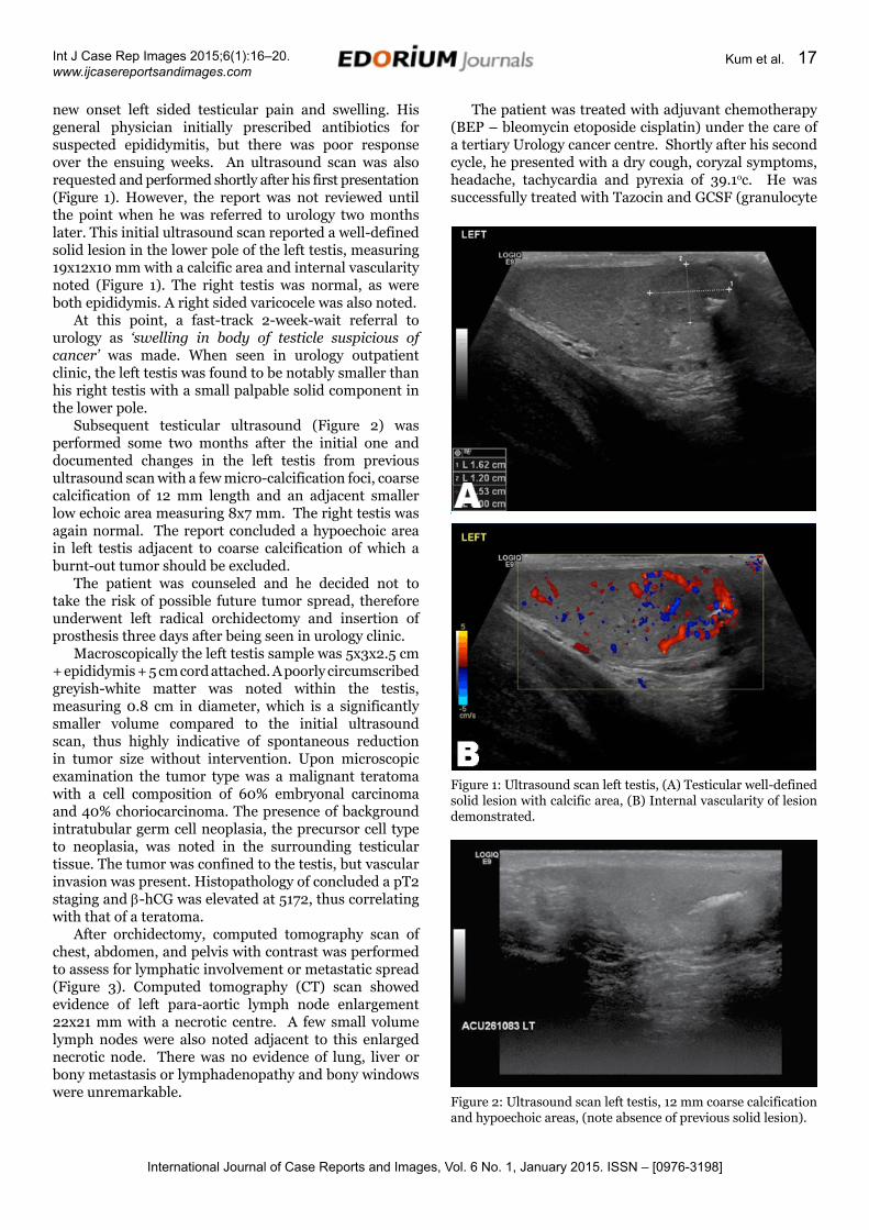
new onset left sided testicular pain and swelling. His general physician initially prescribed antibiotics for suspected epididymitis, but there was poor response over the ensuing weeks. An ultrasound scan was also requested and performed shortly after his first presentation (Figure 1). However, the report was not reviewed until the point when he was referred to urology two months later. This initial ultrasound scan reported a well-defined solid lesion in the lower pole of the left testis, measuring 19x12x10 mm with a calcific area and internal vascularity noted (Figure 1). The right testis was normal, as were both epididymis. A right sided varicocele was also noted.
At this point, a fast-track 2-week-wait referral to urology as ‘swelling in body of testicle suspicious of cancer’ was made. When seen in urology outpatient clinic, the left testis was found to be notably smaller than his right testis with a small palpable solid component in the lower pole.
Subsequent testicular ultrasound (Figure 2) was performed some two months after the initial one and documented changes in the left testis from previous ultrasound scan with a few micro-calcification foci, coarse calcification of 12 mm length and an adjacent smaller low echoic area measuring 8x7 mm. The right testis was again normal. The report concluded a hypoechoic area in left testis adjacent to coarse calcification of which a burnt-out tumor should be excluded.
The patient was counseled and he decided not to take the risk of possible future tumor spread, therefore underwent left radical orchidectomy and insertion of prosthesis three days after being seen in urology clinic.
Macroscopically the left testis sample was 5x3x2.5 cm + epididymis + 5 cm cord attached. A poorly circumscribed greyish-white matter was noted within the testis, measuring 0.8 cm in diameter, which is a significantly smaller volume compared to the initial ultrasound scan, thus highly indicative of spontaneous reduction in tumor size without intervention. Upon microscopic examination the tumor type was a malignant teratoma with a cell composition of 60% embryonal carcinoma and 40% choriocarcinoma. The presence of background intratubular germ cell neoplasia, the precursor cell type to neoplasia, was noted in the surrounding testicular tissue. The tumor was confined to the testis, but vascular invasion was present. Histopathology of concluded a pT2 staging and β-hCG was elevated at 5172, thus correlating with that of a teratoma.
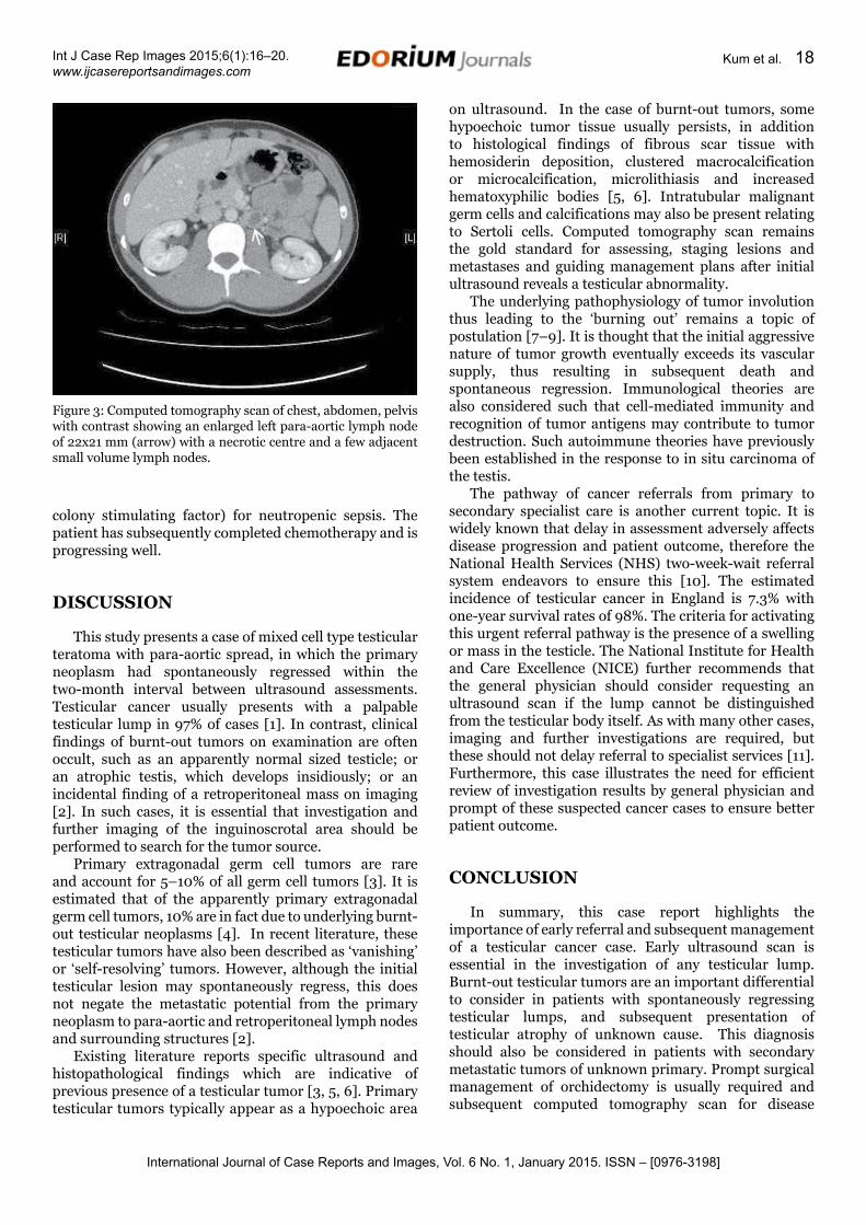
After orchidectomy, computed tomography scan of chest, abdomen, and pelvis with contrast was performed to assess for lymphatic involvement or metastatic spread (Figure 3). Computed tomography (CT) scan showed evidence of left para-aortic lymph node enlargement 22x21 mm with a necrotic centre. A few small volume lymph nodes were also noted adjacent to this enlarged necrotic node. There was no evidence of lung, liver or bony metastasis or lymphadenopathy and bony windows were unremarkable.
The patient was treated with adjuvant chemotherapy (BEP – bleomycin etoposide cisplatin) under the care of a tertiary Urology cancer centre. Shortly after his second cycle, he presented with a dry cough, coryzal symptoms, headache, tachycardia and pyrexia of 39.1oc. He was successfully treated with Tazocin and GCSF (granulocyte
Figure 1: Ultrasound scan left testis, (A) Testicular well-defined solid lesion with calcific area, (B) Internal vascularity of lesion demonstrated.
Figure 2: Ultrasound scan left testis, 12 mm coarse calcification and hypoechoic areas, (note absence of previous solid lesion).
International Journal of Case Reports and Images, Vol. 6 No. 1, January 2015. ISSN – [0976-3198]
Int J Case Rep Images 2015;6(1):16–20. www.ijcasereportsandimages.com
Kum et al. 18
on ultrasound. In the case of burnt-out tumors, some hypoechoic tumor tissue usually persists, in addition to histological findings of fibrous scar tissue with hemosiderin deposition, clustered macrocalcification or microcalcification, microlithiasis and increased hematoxyphilic bodies [5, 6]. Intratubular malignant germ cells and calcifications may also be present relating to Sertoli cells. Computed tomography scan remains the gold standard for assessing, staging lesions and metastases and guiding management plans after initial ultrasound reveals a testicular abnormality.
The underlying pathophysiology of tumor involution thus leading to the ‘burning out’ remains a topic of postulation [7–9]. It is thought that the initial aggressive nature of tumor growth eventually exceeds its vascular supply, thus resulting in subsequent death and spontaneous regression. Immunological theories are also considered such that cell-mediated immunity and recognition of tumor antigens may contribute to tumor destruction. Such autoimmune theories have previously been established in the response to in situ carcinoma of the testis.
The pathway of cancer referrals from primary to secondary specialist care is another current topic. It is widely known that delay in assessment adversely affects disease progression and patient outcome, therefore the National Health Services (NHS) two-week-wait referral system endeavors to ensure this [10]. The estimated incidence of testicular cancer in England is 7.3% with one-year survival rates of 98%. The criteria for activating this urgent referral pathway is the presence of a swelling or mass in the testicle. The National Institute for Health and Care Excellence (NICE) further recommends that the general physician should consider requesting an ultrasound scan if the lump cannot be distinguished from the testicular body itself. As with many other cases, imaging and further investigations are required, but these should not delay referral to specialist services [11]. Furthermore, this case illustrates the need for efficient review of investigation results by general physician and prompt of these suspected cancer cases to ensure better patient outcome.
cONcLUsION
In summary, this case report highlights the importance of early referral and subsequent management of a testicular cancer case. Early ultrasound scan is essential in the investigation of any testicular lump. Burnt-out testicular tumors are an important differential to consider in patients with spontaneously regressing testicular lumps, and subsequent presentation of testicular atrophy of unknown cause. This diagnosis should also be considered in patients with secondary metastatic tumors of unknown primary. Prompt surgical management of orchidectomy is usually required and subsequent computed tomography scan for disease
Figure 3: Computed tomography scan of chest, abdomen, pelvis with contrast showing an enlarged left para-aortic lymph node of 22x21 mm (arrow) with a necrotic centre and a few adjacent small volume lymph nodes.
colony stimulating factor) for neutropenic sepsis. The patient has subsequently completed chemotherapy and is progressing well.
DIscUssION
This study presents a case of mixed cell type testicular teratoma with para-aortic spread, in which the primary neoplasm had spontaneously regressed within the two-month interval between ultrasound assessments. Testicular cancer usually presents with a palpable testicular lump in 97% of cases [1]. In contrast, clinical findings of burnt-out tumors on examination are often occult, such as an apparently normal sized testicle; or an atrophic testis, which develops insidiously; or an incidental finding of a retroperitoneal mass on imaging [2]. In such cases, it is essential that investigation and further imaging of the inguinoscrotal area should be performed to search for the tumor source.
Primary extragonadal germ cell tumors are rare and account for 5–10% of all germ cell tumors [3]. It is estimated that of the apparently primary extragonadal germ cell tumors, 10% are in fact due to underlying burnt-out testicular neoplasms [4]. In recent literature, these testicular tumors have also been described as ‘vanishing’ or ‘self-resolving’ tumors. However, although the initial testicular lesion may spontaneously regress, this does not negate the metastatic potential from the primary neoplasm to para-aortic and retroperitoneal lymph nodes and surrounding structures [2].
Existing literature reports specific ultrasound and histopathological findings which are indicative of previous presence of a testicular tumor [3, 5, 6]. Primary testicular tumors typically appear as a hypoechoic area
International Journal of Case Reports and Images, Vol. 6 No. 1, January 2015. ISSN – [0976-3198]
Int J Case Rep Images 2015;6(1):16–20. www.ijcasereportsandimages.com
Kum et al. 19
staging and grading.
*********
Author contributionsFrancesca Kum – Substantial contributions to conception and design, Acquisition of data, Analysis and interpretation of data, Drafting the article, Revising it critically for important intellectual content, Final approval of the version to be publishedFaisal Ghumman – Substantial contributions to conception and design, Acquisition of data, Analysis and interpretation of data, Revising it critically for important intellectual content, Final approval of the version to be publishedMatin Sheriff – Substantial contributions to conception and design, Revising it critically for important intellectual content, Final approval of the version to be published
GuarantorThe corresponding author is the guarantor of submission.
conflict of InterestAuthors declare no conflict of interest.
copyright© 2015 Francesca Kum et al. This article is distributed under the terms of Creative Commons Attribution License which permits unrestricted use, distribution and reproduction in any medium provided the original author(s) and original publisher are properly credited. Please see the copyright policy on the journal website for more information.
rEFErENcEs
1. East Midlands Cancer Network, Operational Guidelines for the Management of Testicular Tumours, online, August 2011, available:http://www.eastmidlandscancernetwork.nhs.uk/library/urologynssgclinicalguidelines.pdf
2. Coulier B, Lefebvre Y, de Visscher L, et al. Metastases of clinically occult testicular seminoma mimicking primary extragonadal retroperitoneal germ cell tumors. JBR-BTR 2008 Jul-Aug;91(4):139–44.
3. Choyke PL, Hayes WS, Sesterhenn IA. Primary extragonadal germ cell tumors of the retroperitoneum: Differentiation of primary and secondary tumors. Radiographics 1993 Nov;13(6):1365–75.
4. Kühn MW, Weissbach L. Localization, incidence, diagnosis and treatment of extratesticular germ cell tumors. Urol Int 1985;40(3):166–72.
5. Tasu JP, Faye N, Eschwege P, Rocher L, Bléry M. Imaging of burned-out testis tumor: Five new cases and review of the literature. J Ultrasound Med 2003 May;22(5):515–21.
6. Comiter CV, Renshaw AA, Benson CB, Loughlin KR. Burned-out primary testicular cancer: Sonographic and pathological characteristics. J Urol 1996 Jul;156(1):85–8.
7. Lehmann D, Müller H. Analysis of the autoimmune response in an ‘in situ’ carcinoma of the testis. Int J Androl 1987 Feb;10(1):163–8.
8. Fabre E, Jira H, Izard V, et al. ‘Burned-out’ primary testicular cancer. BJU Int 2004 Jul;94(1):74–8.
9. Tefany FJ, Barnetson RS, Halliday GM, McCarthy SW, McCarthy WH. Immunocytochemical analysis of the cellular infiltrate in primary regressing and non-regressing malignant melanoma. J Invest Dermatol 1991 Aug;97(2):197–202.
10. NHS England Service Specifications, Cancer: Testicular (Adult), online, 2013, available: http://www.england.nhs.uk/wp-content/uploads/2013/06/b14-cancr-testic.pdf
11. NICE Clinical Guidelines, Referral guidelines for suspected cancer, online, June 2005, available: https://www.nice.org.uk/guidance/CG27
ABOUT THE AUTHORS
Article citation: Kum F, Ghumman F, Sheriff M. Rare sequelae of a testicular tumor: A ‘burnt-out’ tumor. Int J Case Rep Images 2015;6(1):16–20.
Francesca E. c. Kum is graduated with an MBBS degree from King’s College London School of Medicine (formerly Guy’s, King’s and St. Thomas’ Medical School) with Distinctions. She has recently completed her Foundation Training Programme and is now studying for a Masters in Surgical Education at Imperial College London. Her future interests are a career in surgery in either Urology or ENT Surgery. Email: [email protected]
Faisal Iftikhar A. Ghumman is a Specialty Registrar in Urology at Medway NHS Trust, Gillingham, Kent, UK. He has achieved the following qualifications: MBBS (Army Medical College at Quaid-i-Azam University, Islamabad, Pakistan). FCPS General Surgery (College of Physicians and Surgeons, Karachi, Pakistan). FRCS (Urology), Glasgow, UK. Mr Ghumman is pursuing a career with a specialist interest in laparoscopic urology.
International Journal of Case Reports and Images, Vol. 6 No. 1, January 2015. ISSN – [0976-3198]
Int J Case Rep Images 2015;6(1):16–20. www.ijcasereportsandimages.com
Kum et al. 20
Matin K. M. sheriff (PhD, FRCS, FRCS(Urol), FEBU) is a Consultant Urological Surgeon and Urology Specialty Cancer Lead for the West Kent Urology Cancer Centre at Medway Maritime Hospital, Gillingham, Kent, UK. He is also Professor of Minimally Invasive Surgery (MIS) and Programme Director for MSc MIS at Canterbury and Christ Church University, UK. Matin Sheriff graduated from University College London and completed postgraduate training at the Institute of Urology, London. His specialist interests are laparoscopic surgery, cases include laparoscopic radical prostatectomy, cystectomy and nephrectomy.
Access full text article onother devices
Access PDF of article onother devices
EDORIUM JOURNALS AN INTRODUCTION
Edorium Journals: On Web
About Edorium JournalsEdorium Journals is a publisher of high-quality, open ac-cess, international scholarly journals covering subjects in basic sciences and clinical specialties and subspecialties.
Edorium Journals www.edoriumjournals.com
Edorium Journals et al.
Edorium Journals: An introduction
Edorium Journals Team
But why should you publish with Edorium Journals?In less than 10 words - we give you what no one does.
Vision of being the bestWe have the vision of making our journals the best and the most authoritative journals in their respective special-ties. We are working towards this goal every day of every week of every month of every year.
Exceptional servicesWe care for you, your work and your time. Our efficient, personalized and courteous services are a testimony to this.
Editorial ReviewAll manuscripts submitted to Edorium Journals undergo pre-processing review, first editorial review, peer review, second editorial review and finally third editorial review.
Peer ReviewAll manuscripts submitted to Edorium Journals undergo anonymous, double-blind, external peer review.
Early View versionEarly View version of your manuscript will be published in the journal within 72 hours of final acceptance.
Manuscript statusFrom submission to publication of your article you will get regular updates (minimum six times) about status of your manuscripts directly in your email.
Our Commitment
Mentored Review Articles (MRA)Our academic program “Mentored Review Article” (MRA) gives you a unique opportunity to publish papers under mentorship of international faculty. These articles are published free of charges.
Favored Author programOne email is all it takes to become our favored author. You will not only get fee waivers but also get information and insights about scholarly publishing.
Institutional Membership programJoin our Institutional Memberships program and help scholars from your institute make their research accessi-ble to all and save thousands of dollars in fees make their research accessible to all.
Our presenceWe have some of the best designed publication formats. Our websites are very user friendly and enable you to do your work very easily with no hassle.
Something more...We request you to have a look at our website to know more about us and our services.
We welcome you to interact with us, share with us, join us and of course publish with us.
Browse Journals
CONNECT WITH US
Invitation for article submissionWe sincerely invite you to submit your valuable research for publication to Edorium Journals.
Six weeksYou will get first decision on your manuscript within six weeks (42 days) of submission. If we fail to honor this by even one day, we will publish your manuscript free of charge.
Four weeksAfter we receive page proofs, your manuscript will be published in the journal within four weeks (31 days). If we fail to honor this by even one day, we will pub-lish your manuscript free of charge and refund you the full article publication charges you paid for your manuscript.
This page is not a part of the published article. This page is an introduction to Edorium Journals and the publication services.