Embed Size (px)
Citation preview
1
Paul D. Johnson, MD www.RavenNeurologyReview.com
Raven Neurology Review Stroke Free e-book
2
Acute stroke is a medical emergency and requires rapid assessment, diagnosis and treatment. Evaluating for stroke is a significant component of the workload in inpatient neurology. Stroke is a broad term, referring to several types of cerebrovascular disease (i.e. dealing with the blood vessels of the brain). Typically, disease in specific blood vessels causes injury to the portion of the brain they supply – therefore disease in particular vessels typically causing focal brain injury and focal neurologic symptoms. The management of stroke can vary greatly depending on the type of stroke, stroke etiology and the elapsed time from stroke onset. We will cover the basics for the most common stroke types here. Ischemic stroke is the most frequent stroke type, accounting for about 85% of all strokes. Almost 800,000 Americans have strokes each year, making this a very common neurologic emergency. Ischemia occurs when blood flow is decreased or stopped altogether, resulting in the death of brain tissue. Ischemia typically occurs because of chronic atherosclerosis causing stenosis and thrombosis within a blood vessel, or from blood clots that form elsewhere in the body and travel (embolize) to the brain vasculature and block blood flow in the vessel they occlude. A major cause of embolic stroke is cardiac embolism from atrial fibrillation. Hemorrhagic strokes are less common, but account for a large number of stroke related deaths. These are typically due to the rupture of a small blood vessel, made fragile over time due to long standing high blood pressure. The result is a hematoma within the brain tissue, known as an intraparenchymal hemorrhage (IPH). Another type of hemorrhagic stroke is subarachnoid hemorrhage, in which blood fills the spaces around the brain. These usually occur from ruptured aneurysms, fragile outpouchings which grow from a weak spot in a blood vessel. Aneurysms usually develop at the junction of two blood vessels. These are typically managed by neurosurgeons, and will be discussed in more detail in the neurosurgical section of the book.
3
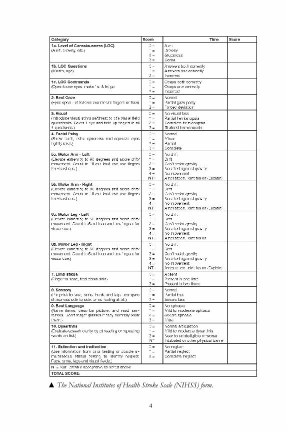
Transient Ischemic Attack (TIA) is a stroke like episode with spontaneous resolution. In the past a TIA was defined as a stroke like episode that resolved completely within 24 hours. However, now that MRI is easily accessible, a TIA is defined as a stroke like event that leaves no evidence of stroke on MRI. Typically these will last 5 – 15 minutes, but can be shorter or longer. A TIA is a warning sign for impending stroke, and should be taken very seriously. A full stroke evaluation is indicated, including evaluation for starting medication for primary stroke prevention, such as aspirin or statins. The most commonly used exam for the evaluation of a patient with a known or suspected stroke is the National Institutes of Health Stroke Scale, or NIHSS. The NIHSS is a standardized, widely used exam whose purpose is to help determine the severity of stroke symptoms. It was not designed to make the diagnosis of stroke, and a high stroke scale does not mean stroke is more likely – rather, the more severe the stroke, the higher the patient scores on the NIHSS. The American Heart Association offers an online NIHSS certification: https://learn.heart.org/Activity/2695217/Detail.aspx
Statins and Ischemic Stroke How are statins used in stroke? First, they are used in ischemic strokes, not hemorrhagic stroke – as very low lipids are thought to increase the risk of bleeding. Secondly, there is evidence that high dose statins – usually atorvastatin 40 mg – 80 mg, reduces the risk of recurrent stroke and other vascular disease. It is standard practice to start a high dose statin, like atorvastatin or rosuvastatin after ischemic stroke or TIA, unless a contraindication exists.
4
▲ The National Institutes of Health Stroke Scale (NIHSS) form.
5
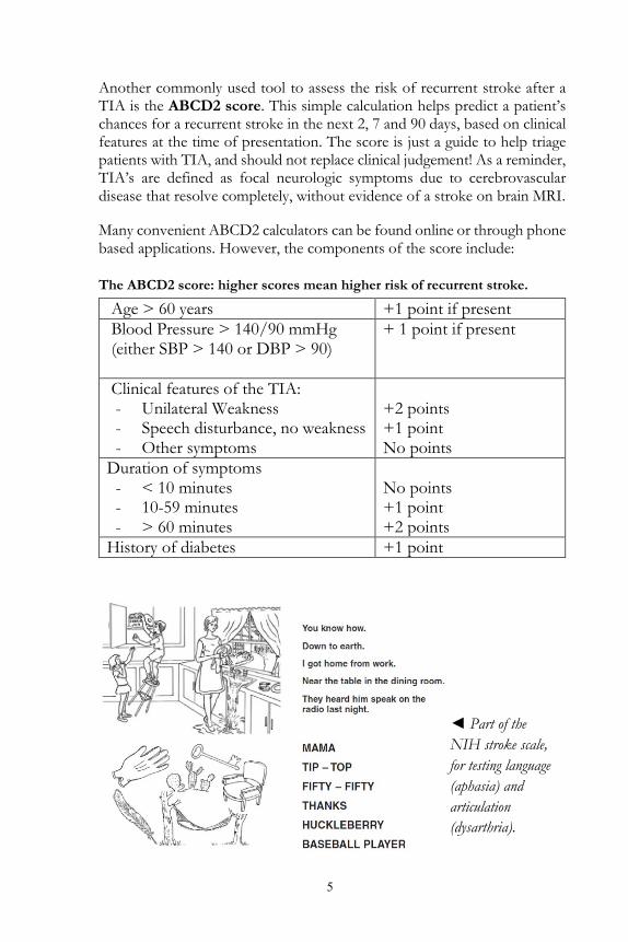
Another commonly used tool to assess the risk of recurrent stroke after a TIA is the ABCD2 score. This simple calculation helps predict a patient’s chances for a recurrent stroke in the next 2, 7 and 90 days, based on clinical features at the time of presentation. The score is just a guide to help triage patients with TIA, and should not replace clinical judgement! As a reminder, TIA’s are defined as focal neurologic symptoms due to cerebrovascular disease that resolve completely, without evidence of a stroke on brain MRI. Many convenient ABCD2 calculators can be found online or through phone based applications. However, the components of the score include: The ABCD2 score: higher scores mean higher risk of recurrent stroke.
Age > 60 years +1 point if present Blood Pressure > 140/90 mmHg (either SBP > 140 or DBP > 90)
+ 1 point if present
Clinical features of the TIA: - Unilateral Weakness - Speech disturbance, no weakness - Other symptoms
+2 points +1 point No points
Duration of symptoms - < 10 minutes - 10-59 minutes - > 60 minutes
No points +1 point +2 points
History of diabetes +1 point
◄ Part of the NIH stroke scale, for testing language (aphasia) and articulation (dysarthria).
6
Diagnostic Principles for Ischemic Stroke and TIA The following are general rules of thumb and should be tailored to the individual patient. Brain Imaging – patients usually undergo both non-contrast CT and MRI. Head CT – The initial study for acute stroke because it is fast and
readily available. Its main uses are identifying hemorrhagic strokes, as there is no clinical way to differentiate between hemorrhagic and ischemic strokes. Large strokes may be visible, but small or acute strokes can be missed.
Brain MRI – Typically done within 24 hours of presentation for acute stroke. MRI is best for identifying small strokes, and helps differentiate between TIA’s and strokes whose symptoms resolve quickly.
Vessel Imaging – It is generally not necessary to obtain both a CTA and MRA. Head and neck imaging is needed for all ischemic stroke patients, whereas head imaging only is needed for hemorrhagic strokes. CT angiogram – Often performed at initial presentation (i.e. in the
ER) to evaluate for acute large vessel occlusion, symptomatic carotid stenosis, or large artery atherosclerosis.
MR angiogram – A non-contrast MRA of the head, known as a “time of flight” study, is available for those who need to avoid iodinated contrast.
Carotid duplex – Largely replaced by CTA or MRA, unless explicitly needed to evaluate high grade carotid stenosis prior to surgery.
Cardiac Monitoring Telemetry – Guidelines stipulate that patients should undergo at
least 48 hours of cardiac telemetry to evaluate for occult atrial fibrillation as a cause of stroke. Most undergo much longer monitoring, especially if embolism is suspected.
Outpatient cardiac telemetry – If occult atrial fibrillation is a possible stroke mechanism and isn’t detected as an inpatient, 14-30 day outpatient cardiac monitors are frequently used. Implantable devices for even longer monitoring are now available.
Echocardiogram – A transthoracic echocardiogram is a standard part of the stroke/TIA evaluation, usually performed with agitated saline (called a “bubble study”) to evaluate for PFO.
7
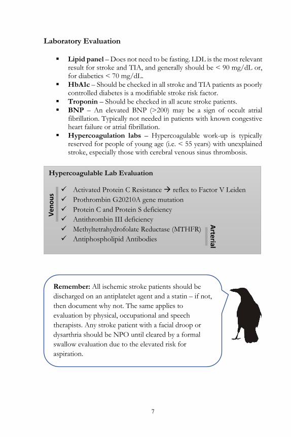
Laboratory Evaluation Lipid panel – Does not need to be fasting. LDL is the most relevant
result for stroke and TIA, and generally should be < 90 mg/dL or, for diabetics < 70 mg/dL.
HbA1c – Should be checked in all stroke and TIA patients as poorly controlled diabetes is a modifiable stroke risk factor.
Troponin – Should be checked in all acute stroke patients. BNP – An elevated BNP (>200) may be a sign of occult atrial
fibrillation. Typically not needed in patients with known congestive heart failure or atrial fibrillation.
Hypercoagulation labs – Hypercoagulable work-up is typically reserved for people of young age (i.e. < 55 years) with unexplained stroke, especially those with cerebral venous sinus thrombosis.
Hypercoagulable Lab Evaluation
Activated Protein C Resistance reflex to Factor V Leiden Prothrombin G20210A gene mutation Protein C and Protein S deficiency Antithrombin III deficiency Methyltetrahydrofolate Reductase (MTHFR) Antiphospholipid Antibodies
Remember: All ischemic stroke patients should be discharged on an antiplatelet agent and a statin – if not, then document why not. The same applies to evaluation by physical, occupational and speech therapists. Any stroke patient with a facial droop or dysarthria should be NPO until cleared by a formal swallow evaluation due to the elevated risk for aspiration.
Veno
us
Arterial
8
Case 1 – Stroke A 64 year old, right handed woman is brought to the emergency room by her daughter. The patient was in the kitchen when she became weak in the left hand, dropping a plate. She wasn’t able to move the left side of her mouth, her left foot dragged and her voice sounded slurred. The symptoms had resolved completely by the time medics arrived 5 minutes later, although her blood pressure was 172/104 mmHg. The patient came to the ER where her blood pressure had returned to normal, and her NIHSS showed no deficits, including normal left sided strength. The patient’s history was notable only for high blood pressure, for which she took Lisinopril, and type 2 diabetes.
1. Is the normal head CT enough to rule out an acute ischemic stroke? A. Yes C. Only when done with IV contrast B. No 2. What is this patient’s ABCD2 score? A. 1 D. 4 B. 2 E. 5 C. 3 F. 6 3. The patient only takes Lisinopril. Which of the following medications should be started to reduce the risk of recurrent stroke? A. Metoprolol 20mg BID B. Warfarin 5mg C. Aspirin 81mg D. Apixaban 5mg BID
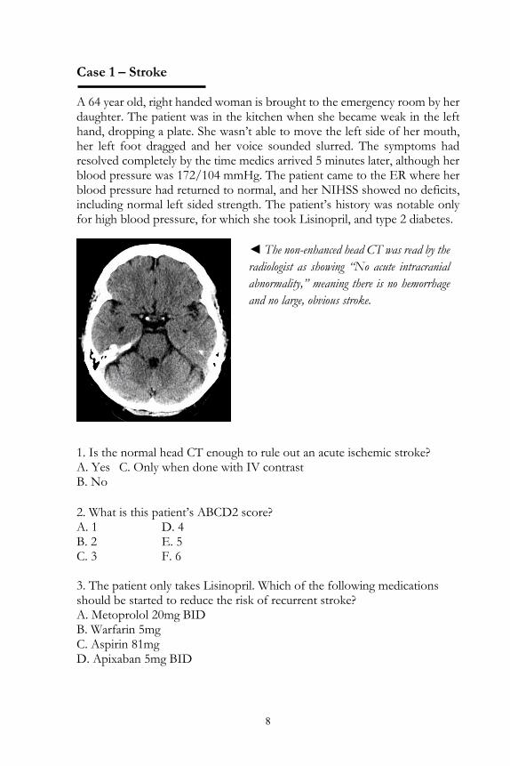
◄ The non-enhanced head CT was read by the radiologist as showing “No acute intracranial abnormality,” meaning there is no hemorrhage and no large, obvious stroke.
9
Case 1 – Diagnosis: TIA This patient most likely had a transient ischemic attack, as the symptoms resolved within 5 minutes. However, a brain MRI would be necessary to fully exclude a small stroke – the head CT rules out hemorrhage, but does a poor job of evaluating acute ischemic stroke. A transient ischemic attack should be taken as a high risk sign for impending stroke – the ABCD2 score of 5 suggests a 4% risk of stroke within 48 hours and almost 10% risk of stroke within three months. Clearly, preventative medications such as aspirin and statins are needed. All ischemic stroke and TIA patients should be on an antiplatlet agent – either aspirin, clopidogrel or combination aspirin with extended-release dipyridamole (aggrenox) – unless contraindications exist. Antiplatelets reduce recurrent stroke risk by about 25%. Anticoagulants are used to prevent strokes in patients with atrial fibrillation. If someone has a TIA while already on an antiplatelet agent, the evidence is less clear about how best to treat them. Many providers will change from one antiplatelet agent to another. Research is ongoing about the benefit of short term use of two antiplatelet agents. Terminology Hemiparesis – Mild to moderate weakness of one half of the body Hemiplegia – Complete paralysis of one half of the body
1. Is the normal head CT enough to rule out an acute ischemic stroke? B. No – acute stroke is usually not seen on head CT, you need MRI 2. What is the patient’s ABCD2 score? E. 5 (age, BP > 140/90, unilateral weakness, and history of diabetes) 3. The patient only takes Lisinopril. Which other medication should be started to reduce the risk of recurrent stroke? C. Aspirin 81 mg - alternatively clopidogrel 75 mg could be used
Teaching Point: Pure motor strokes causing isolated hemiparesis are frequently caused by small vessel disease involving the basal ganglia or thalamus, less frequently in the descending motor fibers in the pons.
10
Case 2 – Stroke A 67 year old right handed man has a history of hypertension and hyperlipidemia. He has been on aspirin 81mg, rosuvastatin 10mg and hydrochlorothiazide 25mg only. Early in the morning he developed sudden onset of isolated right arm weakness. He arrives in the ER two hours after symptom onset, where his NIHSS was 3, for isolated right arm weakness. His blood pressure was 212/120 mmHg. He had no prior history of stroke, TIA or heart disease.
1. What is the next best step in this patient’s treatment? A. Administer IV tPA B. Administer aspirin 325mg C. Administer IV labetalol D. Emergency carotid endarterectomy 2. Which of the following is a likely location for this stroke? A. Left occipital lobe B. Right cortical motor strip C. Right parietal lobe D. Left cortical motor strip
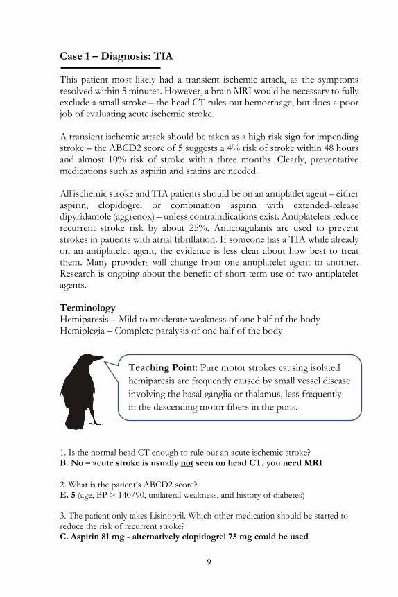
▲ The non-enhanced head CT showed no hemorrhage, no prior strokes and no overt abnormalities. The cortical motor strip, and specifically the so called ‘hand knob’ (arrowhead) where hand motor function is located, is well visualized.
▲ The CT angiogram shows a large filling defect (arrow) in the proximal internal carotid artery, with just over 70% narrowing of the lumen of the artery.
11
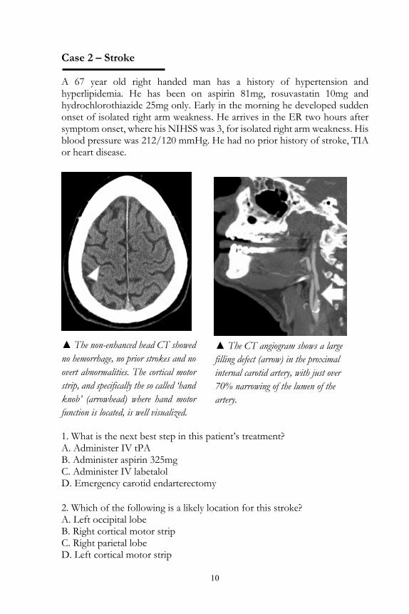
Case 2 – Stroke This patient received IV tPA after his blood pressure was controlled with IV labetalol. Uncontrolled hypertension is a strict contraindication to giving IV tPA because of the risk of brain hemorrhage. Blood pressure must be lowered to < 185/110 mmHg prior to giving tPA and kept below that level for the next 24 hours. Given the high grade (>70%) carotid artery stenosis on the symptomatic side, the stroke mechanism is presumed thromboembolism from ruptured carotid plaque. 1. What is the next best step in this patient’s treatment? C. Administer IV labetalol – blood pressure must be brought down to 185/110 mmHg or less before giving IV tPA 2. Which of the following is a likely location for this stroke? D. Left cortical motor strip – carotid stenosis leads to embolism which most often affects the cortex
3. This patient had a left MCA stroke with high grade (>70%) left internal carotid artery stenosis, and was scheduled for left carotid endarterectomy. Which of the following patients would be least likely to benefit from carotid endarterectomy? A. The patient from this case, undergoing surgery 1 week after stroke B. The patient from this case, undergoing surgery 3 months after stroke C. An asymptomatic patient with 75% carotid artery stenosis D. A symptomatic patient with 30% carotid artery stenosis
▲ Brain MRI showed diffusion restriction (arrow) in the left motor strip, in the hand area, which was confirmed on the ADC (arrowhead). Note that cortical motor strokes can cause pure motor weakness!
12
Case 2 – Diagnosis: Symptomatic Carotid Artery Stenosis 3. This patient had a left MCA stroke with high grade (>70%) left internal carotid artery stenosis, and was scheduled for left carotid endarterectomy. Which of the following patients would be least likely to benefit from endarterectomy? D. A symptomatic patient with 30% carotid artery stenosis Carotid endarterectomy (CEA) and carotid stenting are frequently performed for patients with symptomatic internal carotid artery stenosis, causing TIA or minor stroke. Asymptomatic patients with high grade stenosis may be considered for CEA if their life expectancy is long and the surgeon performing the procedure has a record of very low mortality rates. For more information, the AAN has published guidelines for the use of CEA in symptomatic and asymptomatic patients: http://tools.aan.com/professionals/practice/guideline/pdf/Clinician_guideline.pdf Complications of Carotid Endarterectomy • Hyperperfusion: after surgically opening the artery and restoring
blood flow there is a risk of the brain receiving more blood than it can handle – this can cause ipsilateral headache, seizure and hemorrhagic stroke. Patients must be watched closely after CEA for hypertension and development of these symptoms.
• Acute stroke: there is a small chance of blood clots forming in the carotid artery after the surgery and causing new stroke.
• Nerve injury: rarely the vagus, facial or sympathetic nerves which run within the carotid sheath are injured.
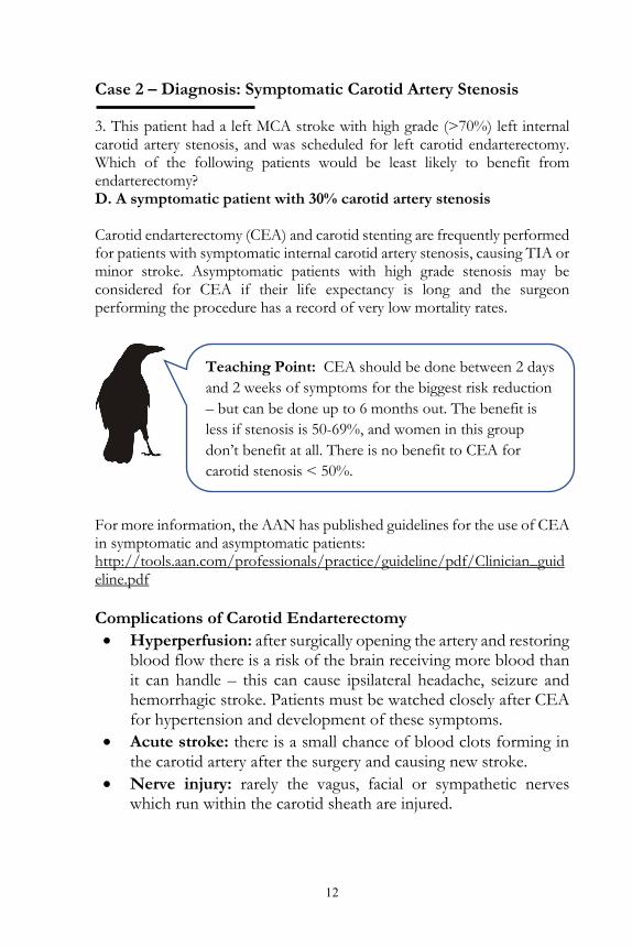
Teaching Point: CEA should be done between 2 days and 2 weeks of symptoms for the biggest risk reduction – but can be done up to 6 months out. The benefit is less if stenosis is 50-69%, and women in this group don’t benefit at all. There is no benefit to CEA for carotid stenosis < 50%.
13
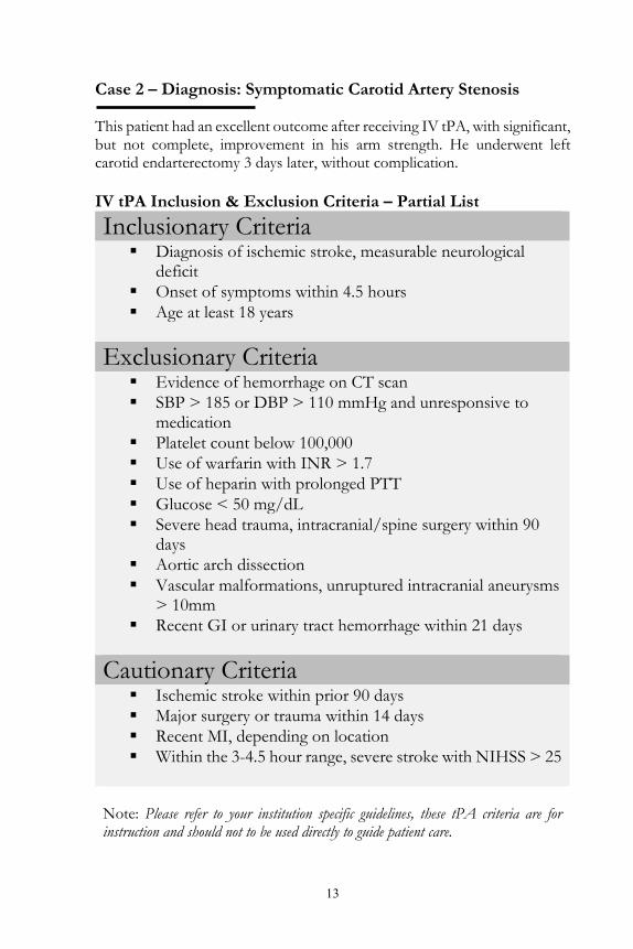
Case 2 – Diagnosis: Symptomatic Carotid Artery Stenosis This patient had an excellent outcome after receiving IV tPA, with significant, but not complete, improvement in his arm strength. He underwent left carotid endarterectomy 3 days later, without complication. IV tPA Inclusion & Exclusion Criteria – Partial List Inclusionary Criteria Diagnosis of ischemic stroke, measurable neurological
deficit Onset of symptoms within 4.5 hours Age at least 18 years
Exclusionary Criteria Evidence of hemorrhage on CT scan SBP > 185 or DBP > 110 mmHg and unresponsive to
medication Platelet count below 100,000 Use of warfarin with INR > 1.7 Use of heparin with prolonged PTT Glucose < 50 mg/dL Severe head trauma, intracranial/spine surgery within 90
days Aortic arch dissection Vascular malformations, unruptured intracranial aneurysms
> 10mm Recent GI or urinary tract hemorrhage within 21 days
Cautionary Criteria Ischemic stroke within prior 90 days Major surgery or trauma within 14 days Recent MI, depending on location Within the 3-4.5 hour range, severe stroke with NIHSS > 25
Note: Please refer to your institution specific guidelines, these tPA criteria are for instruction and should not to be used directly to guide patient care.
14
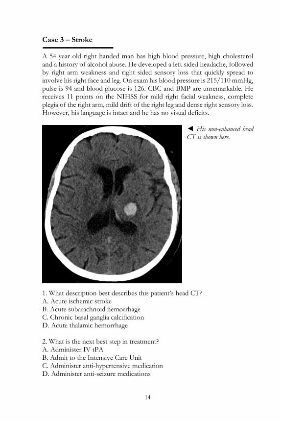
Case 3 – Stroke A 54 year old right handed man has high blood pressure, high cholesterol and a history of alcohol abuse. He developed a left sided headache, followed by right arm weakness and right sided sensory loss that quickly spread to involve his right face and leg. On exam his blood pressure is 215/110 mmHg, pulse is 94 and blood glucose is 126. CBC and BMP are unremarkable. He receives 11 points on the NIHSS for mild right facial weakness, complete plegia of the right arm, mild drift of the right leg and dense right sensory loss. However, his language is intact and he has no visual deficits.
◄ His non-enhanced head CT is shown here.
1. What description best describes this patient’s head CT? A. Acute ischemic stroke B. Acute subarachnoid hemorrhage C. Chronic basal ganglia calcification D. Acute thalamic hemorrhage 2. What is the next best step in treatment? A. Administer IV tPA B. Admit to the Intensive Care Unit C. Administer anti-hypertensive medication D. Administer anti-seizure medications
15
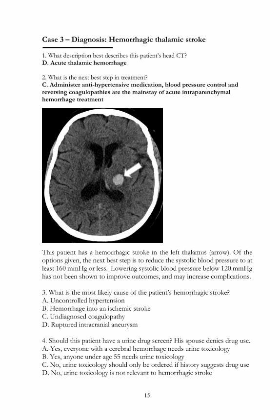
Case 3 – Diagnosis: Hemorrhagic thalamic stroke 1. What description best describes this patient’s head CT? D. Acute thalamic hemorrhage 2. What is the next best step in treatment? C. Administer anti-hypertensive medication, blood pressure control and reversing coagulopathies are the mainstay of acute intraparenchymal hemorrhage treatment
This patient has a hemorrhagic stroke in the left thalamus (arrow). Of the options given, the next best step is to reduce the systolic blood pressure to at least 160 mmHg or less. Lowering systolic blood pressure below 120 mmHg has not been shown to improve outcomes, and may increase complications. 3. What is the most likely cause of the patient’s hemorrhagic stroke? A. Uncontrolled hypertension B. Hemorrhage into an ischemic stroke C. Undiagnosed coagulopathy D. Ruptured intracranial aneurysm 4. Should this patient have a urine drug screen? His spouse denies drug use. A. Yes, everyone with a cerebral hemorrhage needs urine toxicology B. Yes, anyone under age 55 needs urine toxicology C. No, urine toxicology should only be ordered if history suggests drug use D. No, urine toxicology is not relevant to hemorrhagic stroke
16
Case 3 – Diagnosis: Hemorrhagic thalamic stroke 3. What is the most likely cause of the patient’s hemorrhagic stroke? A. Uncontrolled hypertension – the thalamus is a typical location 4. Should this patient have a urine drug screen? His spouse denies drug use. B. Yes, anyone under age 55 needs urine toxicology
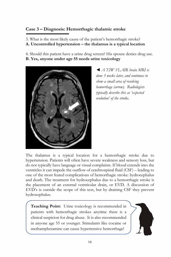
The thalamus is a typical location for a hemorrhagic stroke due to hypertension. Patients will often have severe weakness and sensory loss, but do not typically have language or visual complaints. If blood extends into the ventricles it can impede the outflow of cerebrospinal fluid (CSF) – leading to one of the most feared complications of hemorrhagic stroke: hydrocephalus and death. The treatment for hydrocephalus due to a hemorrhagic stroke is the placement of an external ventricular drain, or EVD. A discussion of EVD’s is outside the scope of this text, but by draining CSF they prevent hydrocephalus.
◄ A T2W FLAIR brain MRI is done 3 weeks later, and continues to show a small area of resolving hemorrhage (arrow). Radiologists typically describe this as ‘expected evolution’ of the stroke.
Teaching Point: Urine toxicology is recommended in patients with hemorrhagic strokes anytime there is a clinical suspicion for drug abuse. It is also recommended in anyone age 55 or younger. Stimulants like cocaine or methamphetamine can cause hypertensive hemorrhage!
17
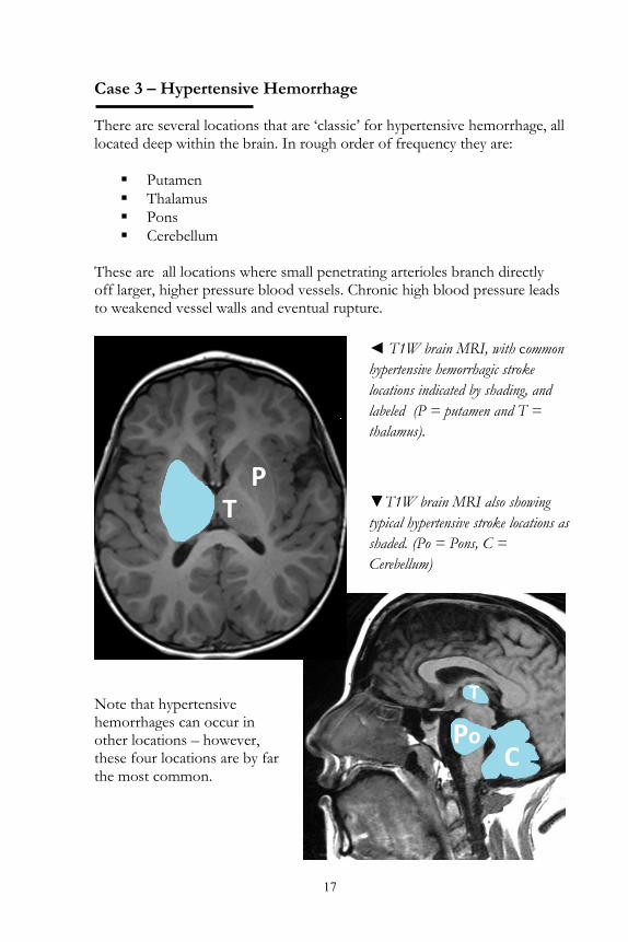
Case 3 – Hypertensive Hemorrhage There are several locations that are ‘classic’ for hypertensive hemorrhage, all located deep within the brain. In rough order of frequency they are: Putamen Thalamus Pons Cerebellum
These are all locations where small penetrating arterioles branch directly off larger, higher pressure blood vessels. Chronic high blood pressure leads to weakened vessel walls and eventual rupture.
Note that hypertensive hemorrhages can occur in other locations – however, these four locations are by far the most common.
◄ T1W brain MRI, with common hypertensive hemorrhagic stroke locations indicated by shading, and labeled (P = putamen and T = thalamus).
▼T1W brain MRI also showing typical hypertensive stroke locations as shaded. (Po = Pons, C = Cerebellum)
P T
Po C
T
18
Case 4 – Stroke While shopping, a 52 year old right handed woman suddenly develops left sided weakness, right gaze deviation and confusion. She is rushed to the ER within an hour of symptom onset, where her BP is 180/100 mmHg, pulse is 110 and irregularly irregular, and glucose is 96. She has hypertension and known atrial fibrillation, for which she takes warfarin. On exam she is alert and follows commands, but has a dense left hemiplegia and right gaze deviation. Although sensation on the left is diminished, she can feel the left arm when touched, but has neglect when both arms or legs are touched simultaneously. Her NIHSS is 13. Her labs show an INR of 2.7 and platelet count of 230,000.
1. Which vascular territory would you localize the patient’s symptoms to? A. Right middle cerebral artery C. Left anterior cerebral artery B. Left posterior cerebral artery D. Basilar artery 2. Given the patient’s symptoms, CT and exam (NIHSS 13), which of the following options is the most likely cause of the stroke? A. Small vessel disease (lacunar stroke) C. Atrial Fibrillation B. Stroke mimic D. TIA 3. What is the next best therapeutic step in management for this patient? A. Administer IV tPA B. Administer labetalol C. Evaluate for mechanical thrombectomy D. Admit to the ICU
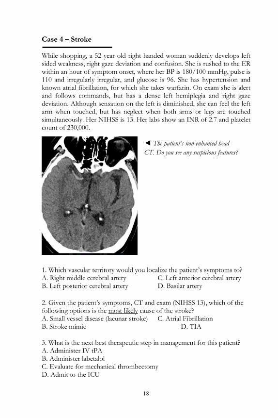
◄ The patient’s non-enhanced head CT. Do you see any suspicious features?
19
Case 4 – Stroke 1. To which vascular territory would you localize the patient’s symptoms? A. Right middle cerebral artery – note signs such as gaze deviation and neglect indicate that the cortex is involved, not only deep structures 2. Given the patient’s symptoms, CT and exam (NIHSS 13), which of the following options is the most likely cause of the stroke? C. Atrial Fibrillation – of the options, it is most likely to cause a large vessel occlusion 3. What is the next best therapeutic step in this patient’s management? C. Evaluate for mechanical thrombectomy
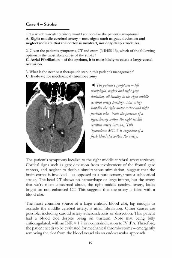
The patient’s symptoms localize to the right middle cerebral artery territory. Cortical signs such as gaze deviation from involvement of the frontal gaze centers, and neglect to double simultaneous stimulation, suggest that the brain cortex is involved – as opposed to a pure sensory/motor subcortical stroke. The head CT shows no hemorrhage or large infarct, but the artery that we’re most concerned about, the right middle cerebral artery, looks bright on non-enhanced CT. This suggests that the artery is filled with a blood clot. The most common source of a large embolic blood clot, big enough to occlude the middle cerebral artery, is atrial fibrillation. Other causes are possible, including carotid artery atherosclerosis or dissection. This patient had a blood clot despite being on warfarin. Note that being fully anticoagulated, with an INR > 1.7, is a contraindication to IV tPA. Therefore, the patient needs to be evaluated for mechanical thrombectomy – emergently removing the clot from the blood vessel via an endovascular approach.
◄ The patient’s symptoms – left hemiplegia, neglect and right gaze deviation, all localize to the right middle cerebral artery territory. This artery supplies the right motor cortex and right parietal lobe. Note the presence of a hyperdensity within the right middle cerebral artery (arrows). This ‘hyperdense MCA’ is suggestive of a fresh blood clot within the artery.
20
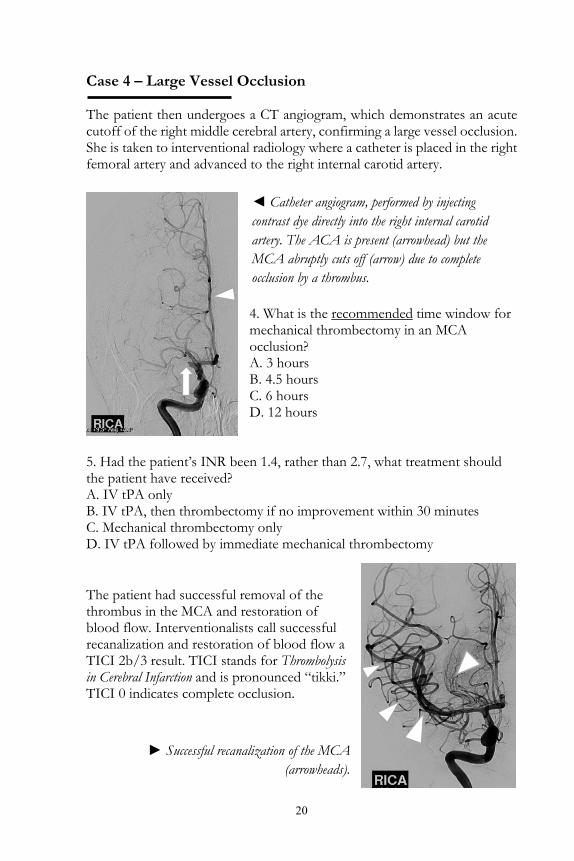
Case 4 – Large Vessel Occlusion The patient then undergoes a CT angiogram, which demonstrates an acute cutoff of the right middle cerebral artery, confirming a large vessel occlusion. She is taken to interventional radiology where a catheter is placed in the right femoral artery and advanced to the right internal carotid artery.
4. What is the recommended time window for mechanical thrombectomy in an MCA occlusion? A. 3 hours B. 4.5 hours C. 6 hours D. 12 hours
5. Had the patient’s INR been 1.4, rather than 2.7, what treatment should the patient have received? A. IV tPA only B. IV tPA, then thrombectomy if no improvement within 30 minutes C. Mechanical thrombectomy only D. IV tPA followed by immediate mechanical thrombectomy The patient had successful removal of the thrombus in the MCA and restoration of blood flow. Interventionalists call successful recanalization and restoration of blood flow a TICI 2b/3 result. TICI stands for Thrombolysis in Cerebral Infarction and is pronounced “tikki.” TICI 0 indicates complete occlusion.
◄ Catheter angiogram, performed by injecting contrast dye directly into the right internal carotid artery. The ACA is present (arrowhead) but the MCA abruptly cuts off (arrow) due to complete occlusion by a thrombus.
► Successful recanalization of the MCA (arrowheads).
21
Case 4 – Large Vessel Occlusion 4. What is the recommended time window for mechanical thrombectomy in an MCA occlusion? C. 6 hours from last known well – although note that recent evidence has shown that in carefully selected patients, using perfusion imaging to identify patients with small core infarcts but large areas of brain tissue potentially ‘at risk’, the time window can be extended up to 24 hours from last known well. 5. Had the patient’s INR been 1.4, rather than 2.7, what treatment should the patient have received? D. IV tPA followed by immediate mechanical thrombectomy – combined therapy, when possible, is the standard of care for large vessel occlusion.
National guidelines recommend mechanical thrombectomy in the anterior circulation (i.e. the carotid or proximal middle cerebral arteries) within 6 hours of symptom onset. However, patient treatment is often individualized, and advanced imaging techniques such as CT perfusion are often used to select patients for thrombectomy outside the traditional time window. In the posterior circulation (i.e. the basilar artery), the time window is often extended due to the very poor prognosis associated with most brainstem strokes. Thrombectomy confers enormous benefit in reduced mortality and disability, with a number needed to treat to prevent disability of 3.
Patients with an acute stroke due to a large vessel occlusion who are eligible for IV tPA should receive it. They should also be evaluated for thrombectomy – these two treatments are often performed together.
As of the publication of this book, rapid advances are being made in the selection of patients for thrombectomy. Both the DAWN and DEFUSE 3 trials have identified benefit in using perfusion imaging to select patients who may benefit from thrombectomy in an extended the time window from 6 – 24 hours from last known well. The traditional criteria still apply within the 6 hour window. Keep your eyes open for more updates in this rapidly evolving field!
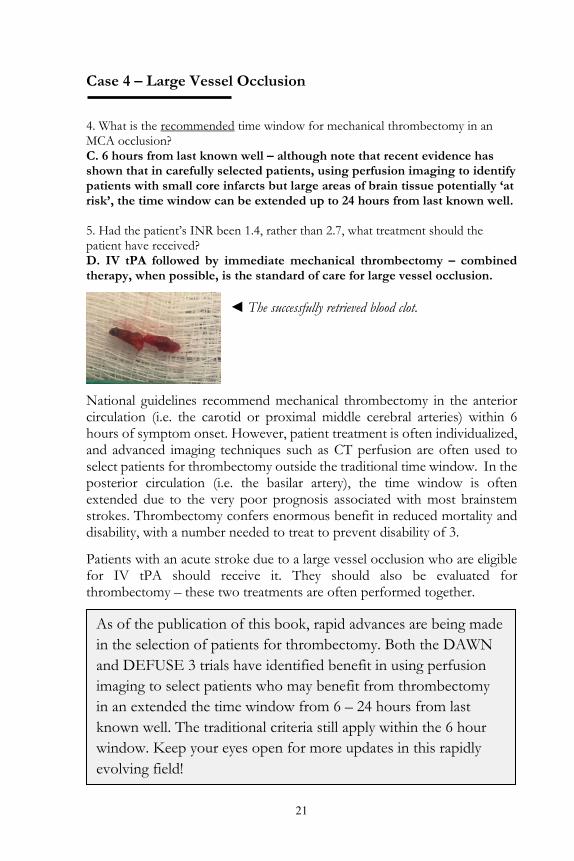
◄ The successfully retrieved blood clot.
22
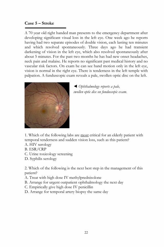
Case 5 – Stroke A 70 year old right handed man presents to the emergency department after developing significant visual loss in the left eye. One week ago he reports having had two separate episodes of double vision, each lasting ten minutes and which resolved spontaneously. Three days ago he had transient darkening of vision in the left eye, which also resolved spontaneously after about 5 minutes. For the past two months he has had new onset headaches, neck pain and malaise. He reports no significant past medical history and no vascular risk factors. On exam he can see hand motion only in the left eye, vision is normal in the right eye. There is tenderness in the left temple with palpation. A funduscopic exam reveals a pale, swollen optic disc on the left.
1. Which of the following labs are most critical for an elderly patient with temporal tenderness and sudden vision loss, such as this patient? A. HIV serology B. ESR/CRP C. Urine toxicology screening D. Syphilis serology 2. Which of the following is the next best step in the management of this patient? A. Treat with high dose IV methylprednisolone B. Arrange for urgent outpatient ophthalmology the next day C. Empirically give high dose IV penicillin D. Arrange for temporal artery biopsy the same day
◄ Ophthalmology reports a pale, swollen optic disc on funduscopic exam.
23
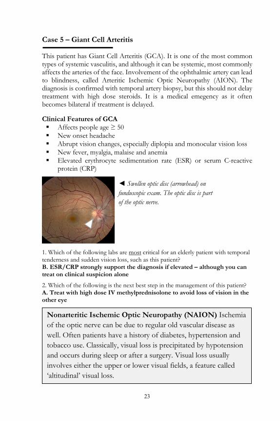
Case 5 – Giant Cell Arteritis This patient has Giant Cell Arteritis (GCA). It is one of the most common types of systemic vasculitis, and although it can be systemic, most commonly affects the arteries of the face. Involvement of the ophthalmic artery can lead to blindness, called Arteritic Ischemic Optic Neuropathy (AION). The diagnosis is confirmed with temporal artery biopsy, but this should not delay treatment with high dose steroids. It is a medical emegency as it often becomes bilateral if treatment is delayed. Clinical Features of GCA Affects people age ≥ 50 New onset headache Abrupt vision changes, especially diplopia and monocular vision loss New fever, myalgia, malaise and anemia Elevated erythrocyte sedimentation rate (ESR) or serum C-reactive
protein (CRP)
1. Which of the following labs are most critical for an elderly patient with temporal tenderness and sudden vision loss, such as this patient? B. ESR/CRP strongly support the diagnosis if elevated – although you can treat on clinical suspicion alone
2. Which of the following is the next best step in the management of this patient? A. Treat with high dose IV methylprednisolone to avoid loss of vision in the other eye
◄ Swollen optic disc (arrowhead) on funduscopic exam. The optic disc is part of the optic nerve.
Nonarteritic Ischemic Optic Neuropathy (NAION) Ischemia of the optic nerve can be due to regular old vascular disease as well. Often patients have a history of diabetes, hypertension and tobacco use. Classically, visual loss is precipitated by hypotension and occurs during sleep or after a surgery. Visual loss usually involves either the upper or lower visual fields, a feature called ‘altitudinal’ visual loss.
24
Case 5 – Stroke A 34 year old left handed man awakens with persistent vertigo and nausea, as well as left face pain. He presents to the emergency department where he is found to have loss of pain and temperature sensation on the left face, as well as in the right arm and leg. On finger to nose exam, however, he seems ataxic only on the right side. He seems to have difficulty swallowing, needing to frequently cough to clear his secretions, and he speaks with a slightly hoarse voice. Further history reveals that he had struck the back of his head two days ago while at his job as a construction worker. For the past two days he has had moderate left neck pain, which improved with over the counter naproxen. 1. Given this constellation of symptoms, which other exam finding would you expect to see? A. Right sided Horner syndrome B. Left sided Horner syndrome C. Right sided third nerve palsy D. Right sided facial nerve palsy 2. What is the most common cause of this syndrome? A. Basilar artery occlusion B. Vertebral artery dissection C. Superior cerebellar artery thrombosis D. Anterior inferior cerebellar artery dissection 3. Which stroke syndrome does this patient have? A. Lateral medullary syndrome (Wallenberg syndrome) B. Superior alternating hemiplegia (Weber syndrome) C. Paramedian midbrain syndrome (Benedikt syndrome) D. Medial medullary syndrome (Dejerine syndrome)
25
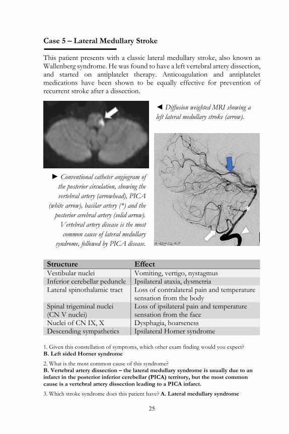
Case 5 – Lateral Medullary Stroke This patient presents with a classic lateral medullary stroke, also known as Wallenberg syndrome. He was found to have a left vertebral artery dissection, and started on antiplatelet therapy. Anticoagulation and antiplatelet medications have been shown to be equally effective for prevention of recurrent stroke after a dissection.
1. Given this constellation of symptoms, which other exam finding would you expect? B. Left sided Horner syndrome
2. What is the most common cause of this syndrome? B. Vertebral artery dissection – the lateral medullary syndrome is usually due to an infarct in the posterior inferior cerebellar (PICA) territory, but the most common cause is a vertebral artery dissection leading to a PICA infarct.
3. Which stroke syndrome does this patient have? A. Lateral medullary syndrome
Structure Effect Vestibular nuclei Vomiting, vertigo, nystagmus Inferior cerebellar peduncle Ipsilateral ataxia, dysmetria Lateral spinothalamic tract Loss of contralateral pain and temperature
sensation from the body Spinal trigeminal nuclei (CN V nuclei)
Loss of ipsilateral pain and temperature sensation from the face
Nuclei of CN IX, X Dysphagia, hoarseness Descending sympathetics Ipsilateral Horner syndrome
◄ Diffusion weighted MRI showing a left lateral medullary stroke (arrow).
► Conventional catheter angiogram of the posterior circulation, showing the vertebral artery (arrowhead), PICA
(white arrow), basilar artery (*) and the posterior cerebral artery (solid arrow).
Vertebral artery disease is the most common cause of lateral medullary
syndrome, followed by PICA disease.
*
26
Raven Neurology Review
Free Stroke e-book
We hope you enjoyed this free e-book. The book is
a representative sample of the full Raven Neurology Review: Clinical Neurology for the Medical Student Clerkship textbook, which is
available on Amazon.com.
Visit us at www.RavenNeurologyReview.com to
subscribe to Neurons, a newsletter with easily digestible tidbits of neurology and neuroscience review, perfect to help you stay current in your
neurology clerkship or class.