Embed Size (px)
Citation preview
Official reprint from UpToDate®
www.uptodate.com©2011 UpToDate®
AuthorS Vincent Rajkumar, MD
Section EditorRobert A Kyle, MD
Deputy EditorRebecca F Connor, MD
Recognition of monoclonal proteins
Last literature review version 19.1: January 2011 | This topic lastupdated: October 4, 2010
INTRODUCTION — The monoclonal gammopathies (paraproteinemias ordysproteinemias) are a group of disorders characterized by the proliferation of asingle clone of plasma cells, which produces an immunologically homogeneousprotein commonly referred to as a paraprotein or monoclonal protein (M-protein,where the "M" stands for monoclonal). (See 'Definition of an M-protein' below.)
A complete immunoglobulin consists of two heavy polypeptide chains of the sameclass designated by a capital letter and a corresponding Greek letter:
Gamma in IgGAlpha in IgAMu in IgMDelta in IgDEpsilon in IgE
The heavy polypeptide chains are further subdivided: IgG has four subclasses(IgG1, IgG2, IgG3, and IgG4) and IgA has two (IgA1 and IgA2). To form the intactimmunoglobulin, the paired heavy chains are associated with two light chains of thesame type, either kappa or lambda, but not both. (See "Structure ofimmunoglobulins".)
Analysis of serum or urine to detect the presence of an M-protein and identify itaccording to its heavy chain class and light chain type requires a sensitive, rapid,and dependable screening method [1]. This review will present an overview of themethodologies currently available for these purposes. Discussions of monoclonalgammopathies and the diagnosis of multiple myeloma are presented separately.(See "Diagnosis of monoclonal gammopathy of undetermined significance" and"Clinical features, laboratory manifestations, and diagnosis of multiple myeloma".)
DEFINITION OF AN M-PROTEIN — An M-protein (paraprotein, monoclonalprotein, M-component) is a monoclonal immunoglobulin secreted by an abnormally
expanded clone of plasma cells in an amount that can be detected byimmunofixation of serum and/or urine, or rarely in other body fluids (eg, jejunalfluid in alpha heavy chain disease) [2].
The M-protein can be an intact immunoglobulin (ie, containing both heavy and lightchains), can be composed of only light chains (ie, light chain myeloma, light chaindeposition disease, AL (light chain) amyloidosis), or rarely can consist of heavychains only (ie, heavy chain disease, heavy chain deposition disease). (See"Pathogenesis and clinical features of AL (primary) amyloidosis and light and heavychain deposition diseases" and "The heavy chain diseases".)
CLINICAL RELEVANCE
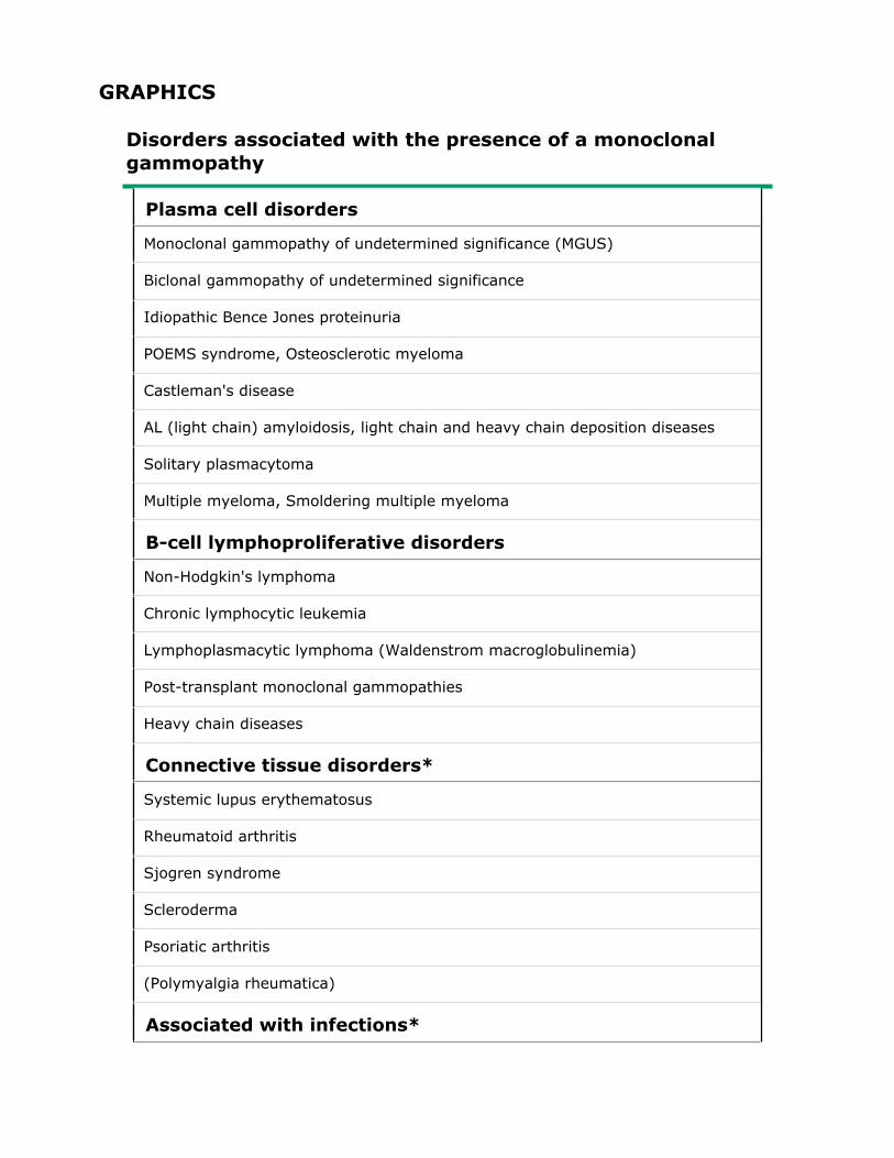
Clonal size — The presence of a monoclonal protein in the serum or urineindicates an underlying clonal plasma cell or lymphoproliferative disorder (table 1).In some cases, the monoclonal protein is the result of an underlying malignancyand is associated with evidence of disease infiltrating lymph nodes, liver, spleen,bone, or other organs (eg, multiple myeloma, solitary plasmacytoma,Waldenstrom's macroglobulinemia). In other cases, the monoclonal protein is theresult of a small limited clonal expansion, and causes no symptoms (eg,monoclonal gammopathy of undetermined significance, MGUS). (See "Diagnosis ofmonoclonal gammopathy of undetermined significance".)
However, even when the clonal expansion is limited, the monoclonal protein maylead to disabling or fatal disease through one or more of its adverse properties,such as [3]:
Ability to agglutinate red cells (eg, cold agglutinin disease)Insolubility at low temperatures (eg, cryoglobulinemia)Increased viscosity (eg, Waldenstrom macroglobulinemia)Deposition in tissues with resulting organ dysfunction (eg, AL (light chain)amyloidosis or the immunoglobulin deposition diseases)Neuropathy (paraneoplastic neuropathy; eg, MGUS, Waldenstrommacroglobulinemia, primary amyloid, POEMS syndrome).
Accordingly, monoclonal proteins may be part of an asymptomatic limited clonalexpansion of plasma cells (eg, MGUS), be a manifestation of a malignancy (eg,myeloma, macroglobulinemia), or may lead to life-threatening complications even ifthe clonal expansion that produced the M protein is limited (eg, primaryamyloidosis). The clinician seeing a patient with a monoclonal protein musttherefore make the appropriate diagnosis (table 1) and initiate effective therapy ina timely manner, in order to prevent irreversible organ damage and/or shorteningof life [3].
Interference with laboratory tests — The presence of a circulating monoclonal

protein may interfere with one of more laboratory tests performed on liquid-basedautomated analyzers, either by precipitating during the analysis [4], or by virtue ofits specific binding properties [5].
The most common artifacts are a low value for HDL cholesterol, a high value forbilirubin, as well as altered measurement of inorganic phosphate [4,6-9]. Otherexamples include interference with measurement of LDL cholesterol, C-reactiveprotein, antistreptolysin-O, creatinine, glucose, urea nitrogen, iron [10], andinorganic calcium.
Re-analysis using a different method or sample dilution can be employed forobtaining accurate measurements [4]. These events may occur in patients whoseclinicians are unaware of the presence of an underlying monoclonal protein andmight result in the mismanagement of patients with monoclonal gammopathy,especially as regards measurement of HDL and LDL cholesterol and estimation ofcardiovascular risk [7,9].
Sia water test — The phenomenon of protein precipitation when plasma orserum is diluted in low ionic strength solutions or water (ie, positive Sia water test)is neither specific nor sensitive for the presence of monoclonal proteins, and hasbeen described in patients with IgG myeloma, Waldenstrom's macroglobulinemia,in sera with a positive rheumatoid factor and IgG/IgM complexes, as well as inpatients with diffuse hypergammaglobulinemia (eg, leishmaniasis) [11,12].
ANALYSIS OF SERUM — Analysis of serum for the presence of M-proteins, or forthe evaluation of a patient with increased total serum proteins, is classicallyperformed using electrophoretic techniques, supplemented with additional tests forprotein quantification and methodologies to determine whether the protein arisesfrom a single clone (ie, monoclonal).
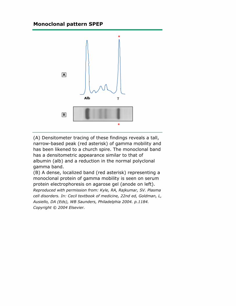
SERUM PROTEIN ELECTROPHORESIS (SPEP) — Serum protein electrophoresis(SPE or SPEP) is an inexpensive and easy to perform screening procedure. Serumprotein electrophoresis is usually done by the agarose gel method (agarose gelelectrophoresis), or less commonly by the capillary zone electrophoretic method. Itis the recommended method for the detection of an M-protein. The resultingM-protein, if found (figure 1), can then be quantitated by means of a densitometertracing of the gel (figure 1). (See 'Capillary zone electrophoresis' below.)
In the electrophoretic methodologies (agarose or capillary zone), proteins areclassified by their final position after electrophoresis is complete into five generalregions: albumin, alpha-1, alpha-2, beta, and gamma (figure 1). These regions,which also use a Greek lettering system, do not refer to the immunoglobulin classto which an M-protein may belong, and refer only to mobility through the supportmedium. The various immunoglobulin classes (IgG, IgA, IgM, IgD, and IgE) are
usually of gamma mobility and make up most of the gamma region, but they mayalso be found in the beta-gamma and beta regions, and may occasionally extendinto the alpha-2 globulin area.
Indications — Serum protein electrophoresis is indicated in all patients in whommultiple myeloma, Waldenström's macroglobulinemia, primary amyloidosis, or arelated disorder is suspected. The SPEP should always be performed in combinationwith serum immunofixation in order to determine clonality (see 'Serumimmunofixation' below). (See "Clinical features, laboratory manifestations, anddiagnosis of multiple myeloma" and "Epidemiology, pathogenesis, clinicalmanifestations and diagnosis of Waldenstrom macroglobulinemia".)
Serum protein electrophoresis should also be considered in any patient with anelevated total serum protein or otherwise unexplained signs and symptomssuggestive of the presence of a plasma cell disorder. These include any one ormore of the following [13]:
Elevated erythrocyte sedimentation rate or serum viscosityUnexplained anemia, back pain, weakness, or fatigueOsteopenia, osteolytic lesions, or spontaneous fracturesRenal insufficiency with a bland urine sedimentHeavy proteinuria in a patient over age 40HypercalcemiaHypergammaglobulinemiaImmunoglobulin deficiencyBence Jones proteinuriaUnexplained peripheral neuropathyRecurrent infections
As an example, the presence of a localized band or spike on SPEP in adults withnephrotic syndrome, refractory heart failure, orthostatic hypotension, peripheralneuropathy, carpal tunnel syndrome, or malabsorption strongly suggests thepossibility of primary amyloidosis (AL), and requires confirmation withimmunofixation studies. (See "Diagnosis of primary (AL) amyloidosis".)
Monoclonal gammopathy — A monoclonal protein (M-protein) usually presentsas a single narrow peak, like a church spire, in the gamma, beta, or alpha-2 regionof the densitometer tracing or as a dense, discrete band on the agarose gel (figure1). In approximately 5 percent of sera with an M-protein, two M proteins arepresent (biclonal gammopathy) (figure 2) [14].
A monoclonal protein can be present in a number of different disorders, includingB-cell and plasma cell proliferations. The most common of these are listed in thetable (table 1).
Polyclonal gammopathy — The presence of a broad-based peak or band, usuallyof gamma mobility, suggests a polyclonal increase in immunoglobulins, most oftendue to an infectious, inflammatory, or reactive process (figure 3). In chronichepatitis, for example, the gamma component may reach 6 or 7 g/dL. Polyclonalgammopathy may on occasion be present without evidence of an underlyingprocess.
In a retrospective cohort study of 148 patients seen at the Mayo Clinic, in whom apolyclonal gamma globulin level ≥3.0 g/dL was found, a single associated medicaldisorder was present in 130. No patient developed myeloma or a clonal plasma cellproliferative disorder. The most common disorders were [15]:
Liver disease — 61 percentConnective tissue disease — 22 percentChronic infection — 6 percentHematologic disorders — 5 percentNon-hematologic malignancy — 3 percentOther — 3 percent
Polyclonal gammopathy, along with bone marrow plasmacytosis, is a commonfinding in HIV-infected patients [16]. However, these patients also have anincreased incidence of clonal plasma cell disorders (eg, MGUS, multiple myeloma,plasmablastic lymphoma) [17-19]. (See "Overview of non-AIDS-definingmalignancies in HIV infection", section on 'Plasma cell disorders' and "AIDS-relatedlymphomas: Epidemiology, risk factors, and pathobiology".)
Hypogammaglobulinemia — Hypogammaglobulinemia (<0.7 g/dL) ischaracterized by a decrease in size of the gamma mobility component on SPEP, andshould be documented by quantitation of serum IgG, IgA, and IgM levels. Thiscondition may be congenital, sex-linked, and/or part of a combinedimmunodeficiency state [20]. (See "Primary humoral immune deficiencies: Anoverview" and "Severe combined immunodeficiency (SCID): An overview".) It mayalso be acquired, as in multiple myeloma, primary amyloidosis, chronic lymphocyticleukemia, lymphoma, or the nephrotic syndrome:
Panhypogammaglobulinemia occurs in about 10 percent of patients withmultiple myeloma. Most of these patients have a Bence Jones protein(monoclonal free kappa or lambda light chains) in the urine, but lack intact
immunoglobulins in the serum [21,22].
Panhypogammaglobulinemia is seen in approximately 20 percent of patientswith primary amyloidosis, often associated with a nephrotic pattern (mainlyalbumin with nonselective globulin loss) in the urine.
False negative results — A small M-protein may be present even whenquantitative immunoglobulin values, beta and gamma mobility components onSPEP, and total serum protein concentrations are all within normal limits. If a clonalplasma cell disorder is suspected, the most sensitive tests to screen for presence ofa monoclonal protein are serum and urine immunofixation, and the serum free lightchain assay. In addition, the following caveats need to be kept in mind:
In alpha heavy chain disease (HCD), which occurs in patients with a form ofsmall intestinal lymphoma called immunoproliferative small intestinal disease(IPSID), one never sees a localized band or sharp peak, presumably becauseof the tendency of these chains to polymerize, or due to their highcarbohydrate content [23-26]. In some patients these proteins can be foundin jejunal fluid but not in the serum. (See "Clinical presentation and diagnosisof primary gastrointestinal lymphomas", section on 'Lymphoma of the smallintestine' and "The heavy chain diseases", section on 'Alpha HCD'.)
In mu HCD, panhypogammaglobulinemia is a prominent feature and alocalized band is found in only 40 percent [27]. (See "The heavy chaindiseases", section on 'Mu HCD'.)
In an occasional patient with gamma HCD, the electrophoretic tracing mayappear broad and heterogeneous rather than showing a localized band[25,28,29]. (See "The heavy chain diseases", section on 'Gamma HCD'.)
An M-protein may produce a broad band in the agarose gel, suggesting apolyclonal pattern. This can occur when an M-protein complexes with otherplasma components, or when there are IgM dimers and pentamers, IgApolymers, or IgG aggregates.
Some patients make only monoclonal light chains (Bence Jones proteinemia),which are usually present in concentrations too low to be visible as a spike inthe agarose gel, because of rapid excretion in the urine [30]. The serumconcentrations will rise if such patients develop renal failure.
In some patients with IgD myeloma, the M-protein spike may be small andeasily overlooked.
False positive results — Serum protein elements other than immunoglobulinsmay falsely suggest the presence of an M-protein. As examples:

Fibrinogen (in plasma) is seen as a discrete band between the beta andgamma mobility regions. This is indistinguishable from an M-protein; additionof thrombin to the specimen will produce a clot if fibrinogen is present. Thepresence of fibrinogen is established if the discrete band is no longer detectedwhen electrophoresis is repeated after the addition of thrombin.
Hemoglobin-haptoglobin complexes secondary to hemolysis may appear as alarge band in the alpha-2-globulin region.
High concentrations of transferrin in patients with iron-deficiency anemia mayproduce a localized band in the beta region.
Nephrotic syndrome is often associated with increased alpha-2 and beta bandswhich can be mistaken for an M-protein. Serum albumin and gamma globulinconcentrations are usually reduced in this setting.
Nonspecific increases in acute phase reactants or certainhyperlipoproteinemias may result in increases in alpha-1 bands.
A common artifact is the presence of a protein band at the point of applicationof the sample. A clue to the presence of this artifact is the presence of thisband on simultaneously performed samples from multiple patients.
Other SPEP patterns — A number of other patterns may be observed on theSPEP:
A decrease in serum albumin and an increase in alpha-1 and alpha-2 globulinsmay be present in patients with infection or metastatic malignancy.
Marked reduction of the alpha-1 globulin component is usually due to adeficiency of alpha-1 antitrypsin.
Two albumin bands (bisalbuminemia) may be found. This familial abnormalityproduces no symptoms [31].
SERUM IMMUNOFIXATION — Serum protein electrophoresis is a useful screeningprocedure, although, as noted above, an M-protein may be easily overlooked or anapparent M-protein may actually represent a polyclonal increase inimmunoglobulins or a nonimmunoglobulin. Consequently, the laboratory mustperform additional studies, usually serum immunofixation, in order to ascertain thepresence of an M-protein and to determine its type. (See "Function and clinicalapplications of immunoglobulins".)
Serum immunofixation is critical for the differentiation of a monoclonal from apolyclonal increase in immunoglobulins. This technique may be performed using

commercial kits or laboratory personnel can pour their own plates and cut theappropriate troughs.
In immunofixation, the patient's serum is electrophoresed into at least fiveseparate lanes. Following electrophoretic separation of the serum proteins, eachsample is overlaid with a different monospecific antibody, usually three for theheavy chain component and two for the light chain component (eg, anti-gamma,anti-mu, anti-alpha, anti-kappa, and anti-lambda, respectively). Precipitation ofproteins (ie, the antigen-antibody complex) is allowed to occur, followed bywashing (nonprecipitated proteins wash out) and staining of the remainingimmunoprecipitates.
An M-protein is characterized on immunofixation by the combined presence of asharp, well-defined band associated with a single heavy-chain class and a sharpand well-defined band with similar mobility characteristics which reacts with eitherkappa or lambda light chain antisera, but not both (figure 4).
Indications — Serum immunofixation is more sensitive than serum proteinelectrophoresis, and also determines the heavy and light chain type of themonoclonal protein. However, unlike serum protein electrophoresis, immunofixationdoes not give an estimate of the size of the M protein (ie, its serum concentration),and thus should be done in conjunction with electrophoresis. Serumimmunofixation should be performed when a sharp band or peak is found in theagarose gel (ie, a monoclonal protein on SPEP) or when multiple myeloma,macroglobulinemia, primary amyloidosis, solitary or extramedullary plasmacytoma,or a related disorder is suspected, despite a normal serum protein electrophoresispattern [3].
Immunofixation will detect a serum M-protein at a concentration of at least 0.02g/dL and a urine M-protein at a concentration of ≥0.004 g/dL [13]. It shouldalways be performed in the presence of otherwise unexplained sensory motorperipheral neuropathy, nephrotic syndrome, refractory heart failure, orthostatichypotension, carpal tunnel syndrome, malabsorption, or whenever the clinicalsituation suggests the possibility of primary amyloidosis (AL). (See "Diagnosis ofprimary (AL) amyloidosis".)
There are also a variety of other indications for immunofixation:
Detection of a small M-protein in the presence of normal or increasedbackground immunoglobulins.
In patients with multiple myeloma or macroglobulinemia in whom treatmenthas resulted in disappearance of the band on routine electrophoresis.
Recognition and distinction of biclonal (two M-proteins) (figure 5) or triclonal
(three M-proteins) (figure 6) gammopathies [32]. Such multiple M-proteinsmay have only a single band or spike on SPEP, and may otherwise be missed.
In addition, the possibility of IgD and IgE monoclonal proteins must be excluded byimmunofixation using IgD and IgE antisera in all patients with a monoclonal lightchain in the serum but no reactivity to anti-G, anti-M, or anti-A.
When following patients with multiple myeloma, MGUS, or a related disorder, oncethe presence of a monoclonal protein and its type are initially confirmed byimmunofixation, it is not necessary to repeat immunofixation unless needed todocument complete response to therapy. Patients can usually be followed withelectrophoresis of serum (SPEP) or urine (UPEP) proteins.
Biclonal gammopathy — A biclonal gammopathy is suspected when there are twoproteins with different mobilities comprising two different monoclonal heavy chainswith their respective monoclonal light chains (figure 5) [33]. A biclonalgammopathy may also consist of two heavy chains of the same class andmonoclonal light chains of the same type. In this setting, one must be careful toexclude monomers and aggregates as well as monomers and polymers of anM-protein. As an example, if two IgA spikes having the same class light chain arepresent, the two components should be considered parts of the same abnormalclone, since they most likely represent a monomer and polymer, respectively, of asingle protein. Similarly, IgM pentamers (19S IgM) and monomers (7S IgM) mayappear as two distinct bands on immunofixation.
Two bands may also be seen in the presence of monomers and aggregates of IgG.As a general rule, if both bands migrate toward the cathode or anode, it is likelythat a monoclonal protein (with monomers, polymers, or aggregates) is present;biclonal gammopathy is almost certain when one of the bands migrates toward theanode and the other band migrates toward the cathode.
Patients with biclonal gammopathy of undetermined significance have the sameclinical spectrum as those with monoclonal gammopathy of undeterminedsignificance (MGUS), and should be followed in the same manner.
QUANTITATION OF IMMUNOGLOBULINS — Quantitation of immunoglobulins isthe most useful technique for the detection of hypogammaglobulinemia. The use ofa rate nephelometer is a good method for this purpose. The degree of turbidityproduced by antigen-antibody interaction is measured by nephelometry in the nearultraviolet regions. Because the method is not affected by the molecular size of theantigen, the nephelometric technique accurately measures 7S IgM, polymers ofIgA, or aggregates of IgG.
Estimation of quantitative immunoglobulins by nephelometry does not allow anassessment of monoclonality. Increased levels can be due to polyclonal or
monoclonal elevations; clonality needs to be established using SPEP andimmunofixation, as discussed above.
Although the reason is not well understood, nephelometric levels of IgM are often1000 to 2000 mg/dL greater than expected on the basis of the SPEP densitometrytracing [34]. Because of the overestimation of IgM by nephelometry, it has beenrecommended that serum protein electrophoresis followed by densitometry ispreferable, the results of which usually more closely agree with the clinicalpresentation [35]. IgG and IgA may also be spuriously increased usingnephelometry.
Quantitation of immunoglobulins using nephelometry is a useful adjunct to theSPEP and UPEP in following patients with multiple myeloma specifically to assessresponse to therapy. However, when assessing response, SPEP values should onlybe compared to SPEP values, and quantitative immunoglobulin values only toquantitative immunoglobulin values.
In certain situations quantitative immunoglobulin values may be more reliable thanthe SPEP:
Small beta-migrating M-proteins (usually IgA M-proteins) are contaminated bynormal immunoglobulins that are often greater in quantity than the M spikeitself.
When the M-spike is so large (>4 g/dL) and narrow on agarose that the SPEPunderestimates the actual immunoglobulin level (by more than 1.5 g/dL), dueto technical staining properties of the agarose gel.
Quantitation of immunoglobulins can also be performed with radialimmunodiffusion. However, this method, which is standardized with 19S IgM and7S IgA, often gives a spuriously high IgM value because of the presence of 7S IgMand a spuriously low IgA level because of polymers of the IgA monoclonal proteins.Thus, radial immunodiffusion is not recommended for immunoglobulin quantitation.
CAPILLARY ZONE ELECTROPHORESIS — Capillary zone electrophoresis refers toan alternative method of performing serum protein electrophoresis compared to theagarose gel technique. It measures protein on-line via light absorbance techniques;protein stains are not necessary and no point of application is seen [36,37]. Theelectrophoretograms are similar to those seen with high resolution agarose gelserum protein electrophoresis.
Capillary electrophoresis appears to be slightly more sensitive than agarose gelelectrophoresis.
IMMUNOSUBTRACTION — Immunosubtraction is an alternative procedure to
serum immunofixation. In this procedure the serum sample is incubated withSepharose beads coupled with anti-gamma, -alpha, -mu, -kappa, and -lambdaantisera. After incubation with each of the heavy and light chain antisera, thesupernatants are reanalyzed to determine which reagent(s) removed theelectrophoretic abnormality. The immunosubtraction procedure is technically lessdemanding, is automated, and is therefore a useful alternative to serumimmunofixation [36,37]. It has the same indications, and accomplishes the samegoals, as serum immunofixation.
IMMUNOELECTROPHORESIS — Immunoelectrophoresis may also be used for thedetection of an M-protein. This technique differs from immunofixation in that theend-point is a precipitin arc rather than a distinct band. Most laboratories no longerperform immunoelectrophoresis and rely instead on immunofixation techniques.
SERUM VISCOSITY — Serum viscosity should be performed in any patient with amonoclonal gammopathy and signs and symptoms suggesting the hyperviscositysyndrome. These symptoms include: oronasal bleeding, blurred vision, dilatation ofretinal veins, flame-shaped retinal hemorrhages, unexplained heart failure, orneurologic symptoms such as headaches, vertigo, nystagmus, deafness, ataxia,diplopia, paresthesias, stupor, or somnolence [38].
Waldenström macroglobulinemia, with increased concentrations of IgM, is the mostcommon cause of hyperviscosity, but hyperviscosity can also occur in patients withhigh concentrations of monoclonal IgA or IgG. (See "Epidemiology, pathogenesis,clinical manifestations and diagnosis of Waldenstrom macroglobulinemia", sectionon 'Hyperviscosity syndrome'.) Serum viscosity should be determined whenever themonoclonal IgM protein spike is >4 g/dL or the IgA or IgG protein spike is >6 g/dL.
The Ostwald-100 viscometer is an inexpensive and satisfactory instrument formeasurement of viscosity, but a Wells-Brookfield (Brookfield EngineeringLaboratory, Stoughton, MA) is preferred because it is more accurate, requires lessserum (1.0 mL), and can be performed at different shear rates and at varioustemperatures. In addition, determinations can be made much more rapidly thanwith the Ostwald device, especially if the viscosity of the serum is high. Anotheroption is the Sonoclot Coagulation and Platelet Function Analyzer (Sienco, Inc.Wheat Ridge, CO).
The normal value for serum viscosity is 1.5 centipoise (CP), but hyperviscositysymptoms are rarely present unless the viscosity is >4 CP. However, manylaboratories report viscosity in relative terms (eg, relative to distilled water orsaline). The normal relative viscosity in our laboratory is 1.8. Because of the oftenpoor correlation between viscosity and symptoms, and the fact that relative andabsolute (in centipoise, see below) viscosities of plasma are similar, these two unitscan be used interchangeably.
The relationship between serum viscosity and IgM protein concentration isnonlinear. Thus, with low serum IgM concentrations, an increase of 1 to 2 g/dLproduces only a small increase in serum viscosity, but with IgM levels of 4 to 5g/dL, an increment of 1 to 2 g/dL greatly increases viscosity. In addition, therelationship between serum viscosity and symptoms of hyperviscosity is notprecise. Although many patients have symptoms when the viscosity is >4centipoise (CP), most have symptoms when the viscosity reaches 6 to 7 CP.However, we have also seen higher viscosities in the absence of symptoms orphysical findings of the hyperviscosity syndrome.
The specific viscosity level at which clinical symptoms occur is affected not only bythe serum protein concentration but also by molecular characteristics of theprotein, aggregation of protein molecules, simultaneous presence of diseasesinvolving the microvasculature, hematocrit, and cardiac status. Consequently,clinical evaluation of the patient is important. The decision to performplasmapheresis should be made on the basis of signs and symptoms ofhyperviscosity rather than the viscosity value per se. (See "Epidemiology,pathogenesis, clinical manifestations and diagnosis of Waldenstrommacroglobulinemia", section on 'Hyperviscosity syndrome' and "Treatment andprognosis of Waldenstrom macroglobulinemia".)
It is useful to measure the serum viscosity and to perform SPEP before and aftereach plasmapheresis to determine effectiveness. Such patients should also bemonitored by periodic SPEP. If the serum M-protein values increase or if symptomsor signs of hyperviscosity recur, the serum viscosity should be repeated.
SERUM FREE LIGHT CHAINS — In 16 percent of our patients with multiplemyeloma, only a Bence Jones protein (light chain) was produced [22], often inconcentrations too low to be detected by routine immunofixation techniques appliedto serum samples. Under such circumstances, or when primary systemicamyloidosis or light-chain-deposition disease are suspected, it is routine to analyzea 24-hour urine collection for the presence of free light chains (see 'Analysis ofurine' below and "Clinical features, laboratory manifestations, and diagnosis ofmultiple myeloma", section on 'Monoclonal proteins').
Immunoassays are now available for detection of low concentrations of monoclonalfree light chains in serum. In this assay, normal levels were as follows (95 percentconfidence intervals) [39]:
Free serum kappa light chains — 3.3 to 19.4 mg/LFree serum lambda light chains — 5.7 to 26.3 mg/LRatio of kappa to lambda free light chains — 0.26 to 1.65
This assay has been proven to have value for the diagnosis, prognosis, and

response to treatment in a number of monoclonal plasma cell disorders, includingMGUS, AL amyloid, and multiple myeloma [40].
This free light chain assay is more sensitive for the detection of monoclonal freelight chains than urine immunofixation. This was illustrated in a study of pairedserum and urine samples from patients with multiple myeloma who tested positivefor either kappa or lambda monoclonal free light chains in their serum; only 51 and35 percent, respectively, of their paired urine samples were positive for BenceJones protein on urine immunofixation [41].
When this assay was applied to sera from patients previously diagnosed as havingnonsecretory myeloma, the improved sensitivity allowed 19 of 28 to be reclassifiedas secretory light chain myeloma (figure 7) [42,43]. (See "Clinical features,laboratory manifestations, and diagnosis of multiple myeloma", section on'Non-secretory myeloma'.)
Indications and uses — Measurement of free light chains is useful in a number ofsettings, such as:
Diagnosis and monitoring progress of patients with non-secretory myelomaand oligosecretory (<1g/dL monoclonal protein in the serum and <200mg/daymonoclonal protein in the urine) myeloma.
Diagnosis and monitoring progress of patients with light chain myeloma aswell as primary systemic amyloidosis, in whom the underlying clonal plasmacell disorder may otherwise be difficult to detect and monitor [44,45].
Predicting risk of progression of MGUS. (See "Clinical course and managementof monoclonal gammopathy of undetermined significance", section on 'Riskstratification'.)
Predicting risk of progression of smoldering multiple myeloma. (See "Clinicalfeatures, laboratory manifestations, and diagnosis of multiple myeloma",section on 'Smoldering multiple myeloma'.)
Predicting risk of progression of solitary plasmacytoma of bone. (See"Diagnosis and management of solitary plasmacytoma of bone", section on'Prognosis'.)
Diagnosis, monitoring during and after treatment, and perhaps prognosis ofpatients with multiple myeloma and an intact immunoglobulin [46,47].
Potentially obviating the initial need for urine protein studies in the screeningalgorithm for monoclonal gammopathies when serum free light chain analysisis performed along with serum protein electrophoresis and serum
immunoelectrophoresis [48]. If a monoclonal protein is found, electrophoresisand immunofixation of an aliquot from a 24-hour urine collection should thenbe performed.
Use in patients with renal failure — The normally rapid renal clearance of serumfree light chains is reduced in the presence of renal failure. As a result, serum freelight chain concentrations rise as the glomerular filtration rate falls, and may be 20to 30 times normal in end-stage renal failure [49]. In addition, the kappa/lambdaratio, which is normally in the range of 0.26 to 1.65, may rise to as high as 3.1 inthe presence of renal failure. The change in kappa/lambda ratios with increasingrenal impairment is of potential clinical relevance in the following settings:
Patients might be misclassified as having a kappa monoclonal gammopathybecause of an increased kappa/lambda ratio due solely to renal insufficiency.
Patients with a lambda monoclonal gammopathy along with renal insufficiencymight have a relatively normal kappa/lambda ratio and be missed because ofthe relative increase in kappa chains due to the renal insufficiency.
ANALYSIS OF URINE — A number of methods are available for determination ofthe amount of protein excreted into the urine and for determining the nature andclonality of these proteins. Although many methods are listed below, the main testsrequired for the evaluation of monoclonal plasma cell disorders, including multiplemyeloma and primary amyloidosis, are urine protein electrophoresis and urineimmunofixation.
Dipstick testing — Dipsticks are used in many laboratories to screen for thepresence of protein in the urine. The dipstick is impregnated with a bufferedindicator dye that binds to protein and produces a color change proportional to theamount of protein bound to it. However, dipsticks are insensitive to the presence ofBence Jones protein (free kappa or lambda light chains) and should not be used forthis purpose [50,51]. To the contrary, it is the presence of a negative or tracedipstick for protein in a patient over the age of 50 who presents with otherwiseunexplained renal failure with a bland urine sediment that should raise suspicion ofmyeloma kidney. Urinary protein should be measured by other methods in all ofthese patients. (See "Types of renal disease in multiple myeloma".)
Other detection methods — Detection of protein in the urine of patients withmonoclonal gammopathy can be accomplished with sulfosalicylic acid or Exton'sreagent. These agents detect albumin, globulin, Bence Jones protein, polypeptidesand proteases. False-positive reactions may be induced by penicillin or itsderivatives, tolbutamide metabolites, sulfisoxazole metabolites, and certain organicroentgenographic contrast media [52]. The biuret method is satisfactory for the
quantitation of total protein.
Sulfosalicylic acid test — The SSA test is performed by mixing one part urinesupernatant (eg, 2.5 mL) with three parts 3 percent sulfosalicylic acid, and gradingthe resultant turbidity according to the following schema (the numbers inparentheses represent the approximate protein concentration):
0 = no turbidity (0 mg/dL)
trace = slight turbidity (1 to 10 mg/dL)
1+ = turbidity through which print can be read (15 to 30 mg/dL)
2+ = white cloud without precipitate through which heavy black lines on a whitebackground can be seen (40 to 100 mg/dL)
3+ = white cloud with fine precipitate through which heavy black lines cannot beseen (150 to 350 mg/dL)
4+ = flocculent precipitate (>500 mg/dL).
The combination of a 3+ or 4+ SSA test and a negative or trace dipstick is usuallyindicative of a nonalbumin protein in the urine. In the adult over age 50, this ismost often a monoclonal light chain. Further testing is required for confirmation.
Heat test for light chains — Monoclonal light chains in the urinecharacteristically precipitate at 40 to 60ºC, dissolve at 100ºC, and reprecipitate oncooling to between 40 and 60ºC [53]. However, the heat test may give a falsepositive result. In this setting, a broad-based gamma-mobility band and normalappearing kappa and lambda bands are seen on immunofixation. Presumably, thefalse positive result is due to an excess of polyclonal light chains, which occursmost often in patients with renal insufficiency [54]. False negative tests also maybe seen.
For these reasons, the heat test is not recommended for the detection of BenceJones proteins. Immunofixation of an adequately concentrated urine specimen isthe recommended test.
24-HOUR URINE PROTEIN ELECTROPHORESIS (UPEP) — The urine proteinelectrophoresis is analogous to the serum protein electrophoresis (SPEP), and isused to detect monoclonal (M) proteins in the urine by an electrophoretic method.
A 24-hour urine collection is necessary for determination of the total amount ofprotein excreted in the urine per day. The quantity of M-protein excreted isdetermined by measuring the size (percent) of the M-spike in the densitometertracing and multiplying it by the total 24-hour urinary protein excretion. Theamount of protein can be expressed as mg/dL or mg/L but it is much more usefulto report the M-protein in g/24 hours because of wide variability in the daily urinary
volume. The 24-hour urine specimen requires no preservative and may be kept atroom temperature during collection.
On UPEP, a urinary M-protein is seen as a dense localized band on agarose or a tallnarrow peak on the densitometer tracing (figure 8). Generally, the amount ofurinary monoclonal protein correlates directly with the size of the plasma cellburden, as long as renal function is relatively normal. Consequently, urinaryM-protein excretion is useful in determining the response to chemotherapy orprogression of disease.
Indications — All patients with a diagnosis of a plasma cell dyscrasia should havea baseline urine protein electrophoresis (and immunofixation) of an aliquot from a24-hour urine collection. This test is essential to detect the presence of potentiallynephrotoxic concentrations of urinary light chains. (See "Pathogenesis anddiagnosis of myeloma cast nephropathy (myeloma kidney)".)
Urine electrophoresis testing is subsequently required to detect progression and tomonitor response to therapy in patients who have urinary monoclonal proteins atbaseline.
Urine protein electrophoresis (and immunofixation) has been used also as astandard screening test for patients in whom there is clinical suspicion for amonoclonal plasma cell proliferative disorder such as myeloma or primaryamyloidosis. The serum free light chain assay can be used as an alternativemethod.
URINE IMMUNOFIXATION — Urine immunofixation is the preferred method foridentification of a monoclonal protein in the urine. As in the case of serumimmunofixation it is more sensitive than urine electrophoresis, and allowsdetermination of the heavy and light chain type of the urinary monoclonal protein.It does not estimate the size of the monoclonal protein and hence is done inconjunction with urine protein electrophoresis.
The presence of a urinary monoclonal light chain on immunofixation ischaracterized by a discrete band with reactivity to either kappa or lambda antisera,but not both (figure 9). Two discrete bands with either kappa or lambda antisera(but not both) may be found; this is usually due to the presence of monomers anddimers of the monoclonal light chain protein. However, a discrete band with heavychain antisera reactivity corresponding to the type of heavy chain present in thepatient's serum and coinciding with one of the two light chain bands indicates thepresence of a fragment of the intact immunoglobulin (figure 10). This is of noparticular clinical relevance.
A biclonal gammopathy with one monoclonal protein having a kappa chain and theother having a lambda chain rarely occurs. Occasionally, regularly spaced faint but
discrete multiple bands are seen on immunofixation. These restricted bandsrepresent related polyclonal free light chains and are not to be confused withmonoclonal light chains. This phenomenon has been described as "ladder lightchain" or a "pseudo-oligoclonal pattern" [55].
If the patient has nephrotic syndrome, the presence of a monoclonal light chainstrongly suggests either primary amyloidosis (AL) or light chain deposition diseasein almost all instances. If electrophoresis of the urine reveals a localized globulinband and immunofixation does not demonstrate a monoclonal light chain, oneshould suspect the possibility of heavy chain deposition disease. Immunofixationshould then be performed with antisera to IgG (gamma heavy chains). (See"Diagnosis of primary (AL) amyloidosis" and "Pathogenesis and clinical features ofAL (primary) amyloidosis and light and heavy chain deposition diseases".)
Theoretically, antisera that recognize only free kappa or free lambda light chainsshould be used rather than antisera that recognize both free light chains as well aslight chains which are part of an intact immunoglobulin. However, such antisera areoften either nonspecific or insufficiently potent. In addition, a patient may have animmunoglobulin fragment that free kappa or free lambda antisera do not recognize.Thus, it is advisable to use kappa and lambda antisera that are monospecific andpotent and able to recognize both free and combined light chains when performingimmunofixation. This is especially important, as some patients have isolated BenceJones proteinuria with no M-spike in the serum [30,42].
Indications — Urine immunofixation should be performed at baseline in allpatients with a diagnosis of a plasma cell dyscrasia. Immunofixation should beperformed in these patients even if the routine urine analysis is negative forprotein, 24-hour urine protein concentration is within normal limits, andelectrophoresis of a concentrated urine specimen shows no globulin peak.Immunofixation is sufficiently sensitive to detect a urine M-protein of ≥0.004 g/dL[13]. Urine immunofixation is subsequently required to assess and documentcomplete response in patients receiving therapy.
Urine immunofixation has also been used as a standard screening test for patientsin whom there is clinical suspicion for a monoclonal plasma cell proliferativedisorder such as myeloma or primary amyloidosis. The serum free light chain assaycan be used as an alternative method.
EVALUATION OF MONOCLONAL GAMMOPATHIES — This issue is discussedseparately. (See "Diagnosis of monoclonal gammopathy of undeterminedsignificance", section on 'Summary and recommendations'.)
It is of great importance for the physician to be in contact with the laboratoryconcerning interpretation of these laboratory tests. It is also essential that the
laboratory know when the physician suspects primary amyloidosis or a plasmacell/lymphocytic disorder so that a greater effort can be made for the detection ofan M-protein, which may be present in such a low concentration that it is not bedetected by routine serum or urine protein electrophoresis, but may be detected bymore sensitive techniques (eg, immunofixation or free light chain assay) [56,57].
Use of UpToDate is subject to the Subscription and License Agreement.
REFERENCES
Katzmann, JA, Kyle, RA. Immunochemical characterization of immunoglobulinsin serum, urine, and cerebrospinal fluid. In: Manual of Molecular and ClinicalLaboratory Immunology, 7th ed, Detrick, B (Ed), American Society forMicrobiology Press, Washington, DC 2006. p.88.
1.
Bird J, Behrens J, Westin J, et al. UK Myeloma Forum (UKMF) and NordicMyeloma Study Group (NMSG): guidelines for the investigation of newlydetected M-proteins and the management of monoclonal gammopathy ofundetermined significance (MGUS). Br J Haematol 2009; 147:22.
2.
Merlini G, Stone MJ. Dangerous small B-cell clones. Blood 2006; 108:2520.3.
Berth M, Delanghe J. Protein precipitation as a possible important pitfall in theclinical chemistry analysis of blood samples containing monoclonalimmunoglobulins: 2 case reports and a review of the literature. Acta Clin Belg2004; 59:263.
4.
Merlini G, Farhangi M, Osserman EF. Monoclonal immunoglobulins withantibody activity in myeloma, macroglobulinemia and related plasma celldyscrasias. Semin Oncol 1986; 13:350.
5.
Smogorzewska A, Flood JG, Long WH, Dighe AS. Paraprotein interference inautomated chemistry analyzers. Clin Chem 2004; 50:1691.
6.
Tsai LY, Tsai SM, Lee SC, Liu SF. Falsely low LDL-cholesterol concentrationsand artifactual undetectable HDL-cholesterol measured by direct methods in apatient with monoclonal paraprotein. Clin Chim Acta 2005; 358:192.
7.
Pantanowitz L, Horowitz GL, Upalakalin JN, Beckwith BA. Artifactualhyperbilirubinemia due to paraprotein interference. Arch Pathol Lab Med 2003;127:55.
8.
Murali MR, Kratz A, Finberg KE. Case records of the Massachusetts GeneralHospital. Case 40-2006. A 64-year-old man with anemia and a low level ofHDL cholesterol. N Engl J Med 2006; 355:2772.
9.
Alyanakian MA, Taes Y, Bensaïd M, et al. Monoclonal immunoglobulin withantitransferrin activity: A rare cause of hypersideremia with increasedtransferrin saturation. Blood 2007; 109:359.
10.

Keystone E, Pruzanski W. Immunochemical and physical studies of the Siatest. Am J Med Sci 1976; 271:151.
11.
Kumar R, Pai K, Kumar P, et al. Sero-epidemiological study of kala-azar in avillage of Varanasi district, India. Trop Med Int Health 2006; 11:41.
12.
International Myeloma Working Group. Criteria for the classification ofmonoclonal gammopathies, multiple myeloma and related disorders: a reportof the International Myeloma Working Group. Br J Haematol 2003; 121:749.
13.
Kyle RA, Robinson RA, Katzmann JA. The clinical aspects of biclonalgammopathies. Review of 57 cases. Am J Med 1981; 71:999.
14.
Dispenzieri A, Gertz MA, Therneau TM, Kyle RA. Retrospective cohort study of148 patients with polyclonal gammopathy. Mayo Clin Proc 2001; 76:476.
15.
De Milito A, Nilsson A, Titanji K, et al. Mechanisms of hypergammaglobulinemiaand impaired antigen-specific humoral immunity in HIV-1 infection. Blood2004; 103:2180.
16.
Amara S, Dezube BJ, Cooley TP, et al. HIV-associated monoclonalgammopathy: a retrospective analysis of 25 patients. Clin Infect Dis 2006;43:1198.
17.
Yee TT, Murphy K, Johnson M, et al. Multiple myeloma and humanimmunodeficiency virus-1 (HIV-1) infection. Am J Hematol 2001; 66:123.
18.
Dezube BJ, Aboulafia DM, Pantanowitz L. Plasma cell disorders in HIV-infectedpatients: from benign gammopathy to multiple myeloma. AIDS Read 2004;14:372.
19.
Rosen FS, Cooper MD, Wedgwood RJ. The primary immunodeficiencies. N EnglJ Med 1995; 333:431.
20.
Kyle RA. Multiple myeloma: review of 869 cases. Mayo Clin Proc 1975; 50:29.21.
Kyle RA, Gertz MA, Witzig TE, et al. Review of 1027 patients with newlydiagnosed multiple myeloma. Mayo Clin Proc 2003; 78:21.
22.
Rambaud JC, Halphen M, Galian A, Tsapis A. Immunoproliferative smallintestinal disease (IPSID): relationships with alpha-chain disease and"Mediterranean" lymphomas. Springer Semin Immunopathol 1990; 12:239.
23.
Seligmann M, Mihaesco E, Preud'homme JL, et al. Heavy chain diseases:current findings and concepts. Immunol Rev 1979; 48:145.
24.
Fermand JP, Brouet JC. Heavy-chain diseases. Hematol Oncol Clin North Am1999; 13:1281.
25.
Al-Saleem T, Al-Mondhiry H. Immunoproliferative small intestinal disease(IPSID): a model for mature B-cell neoplasms. Blood 2005; 105:2274.
26.
Wahner-Roedler DL, Kyle RA. Mu-heavy chain disease: presentation as abenign monoclonal gammopathy. Am J Hematol 1992; 40:56.
27.
Fermand JP, Brouet JC, Danon F, Seligmann M. Gamma heavy chain "disease":heterogeneity of the clinicopathologic features. Report of 16 cases and reviewof the literature. Medicine (Baltimore) 1989; 68:321.
28.
Wahner-Roedler DL, Witzig TE, Loehrer LL, Kyle RA. Gamma-heavy chaindisease: review of 23 cases. Medicine (Baltimore) 2003; 82:236.
29.
Kyle RA, Greipp PR. "Idiopathic" Bence Jones proteinuria: long-term follow-upin seven patients. N Engl J Med 1982; 306:564.
30.
Pola V, Tichý M. [Bisalbuminemia. Critical review and report of a case of anacquired form in a myeloma patient]. Folia Haematol Int Mag Klin MorpholBlutforsch 1985; 112:208.
31.
Grosbois B, Jégo P, de Rosa H, et al. [Triclonal gammopathy and malignantimmunoproliferative syndrome]. Rev Med Interne 1997; 18:470.
32.
Bakkus MH, Schots R, Gomez La Fuente PB, et al. Clonally related IgA- andIgE-secreting plasma cells in a myeloma patient. Eur J Haematol 2000;65:348.
33.
Riches PG, Sheldon J, Smith AM, Hobbs JR. Overestimation of monoclonalimmunoglobulin by immunochemical methods. Ann Clin Biochem 1991; 28 ( Pt3):253.
34.
Sinclair D, Ballantyne F, Shanley S, et al. Estimation of paraproteins byimmunoturbidimetry and electrophoresis followed by scanning densitometry.Ann Clin Biochem 1990; 27 ( Pt 4):335.
35.
Katzmann JA, Clark R, Wiegert E, et al. Identification of monoclonal proteins inserum: a quantitative comparison of acetate, agarose gel, and capillaryelectrophoresis. Electrophoresis 1997; 18:1775.
36.
Katzmann JA, Clark R, Sanders E, et al. Prospective study of serum proteincapillary zone electrophoresis and immunotyping of monoclonal proteins byimmunosubtraction. Am J Clin Pathol 1998; 110:503.
37.
Gertz MA, Kyle RA. Hyperviscosity syndrome. J Intensive Care Med 1995;10:128.
38.
Katzmann JA, Clark RJ, Abraham RS, et al. Serum reference intervals anddiagnostic ranges for free kappa and free lambda immunoglobulin light chains:relative sensitivity for detection of monoclonal light chains. Clin Chem 2002;48:1437.
39.
Pratt G. The evolving use of serum free light chain assays in haematology. Br JHaematol 2008; 141:413.
40.
Nowrousian MR, Brandhorst D, Sammet C, et al. Serum free light chainanalysis and urine immunofixation electrophoresis in patients with multiplemyeloma. Clin Cancer Res 2005; 11:8706.
41.
Drayson M, Tang LX, Drew R, et al. Serum free light-chain measurements for42.

identifying and monitoring patients with nonsecretory multiple myeloma. Blood2001; 97:2900.
Bradwell AR, Carr-Smith HD, Mead GP, et al. Serum test for assessment ofpatients with Bence Jones myeloma. Lancet 2003; 361:489.
43.
Abraham RS, Katzmann JA, Clark RJ, et al. Quantitative analysis of serum freelight chains. A new marker for the diagnostic evaluation of primary systemicamyloidosis. Am J Clin Pathol 2003; 119:274.
44.
Lachmann HJ, Gallimore R, Gillmore JD, et al. Outcome in systemic ALamyloidosis in relation to changes in concentration of circulating freeimmunoglobulin light chains following chemotherapy. Br J Haematol 2003;122:78.
45.
Mead GP, Carr-Smith HD, Drayson MT, et al. Serum free light chains formonitoring multiple myeloma. Br J Haematol 2004; 126:348.
46.
van Rhee F, Bolejack V, Hollmig K, et al. High serum-free light chain levels andtheir rapid reduction in response to therapy define an aggressive multiplemyeloma subtype with poor prognosis. Blood 2007; 110:827.
47.
Katzmann JA, Dispenzieri A, Kyle RA, et al. Elimination of the need for urinestudies in the screening algorithm for monoclonal gammopathies by usingserum immunofixation and free light chain assays. Mayo Clin Proc 2006;81:1575.
48.
Abadie JM, van Hoeven KH, Wells JM. Are renal reference intervals requiredwhen screening for plasma cell disorders with serum free light chains andserum protein electrophoresis? Am J Clin Pathol 2009; 131:166.
49.
Hinberg IH, Katz L, Waddell L. Sensitivity of in vitro diagnostic dipstick tests tourinary protein. Clin Biochem 1978; 11:62.
50.
Scarpioni L, Ballocchi S, Bergonzi G, et al. Glomerular and tubular proteinuriain myeloma. Relationship with Bence Jones proteinuria. Contrib Nephrol 1981;26:89.
51.
Morcos SK, el-Nahas AM, Brown P, Haylor J. Effect of iodinated water solublecontrast media on urinary protein assays. BMJ 1992; 305:29.
52.
PUTNAM FW, EASLEY CW, LYNN LT, et al. The heat precipitation ofBence-Jones proteins. I. Optimum conditions. Arch Biochem Biophys 1959;83:115.
53.
Perry MC, Kyle RA. The clinical significance of Bence Jones proteinuria. MayoClin Proc 1975; 50:234.
54.
Harrison HH. The "ladder light chain" or "pseudo-oligoclonal" pattern in urinaryimmunofixation electrophoresis (IFE) studies: a distinctive IFE pattern and anexplanatory hypothesis relating it to free polyclonal light chains. Clin Chem1991; 37:1559.
55.

Kyle RA. Sequence of testing for monoclonal gammopathies. Arch Pathol LabMed 1999; 123:114.
56.
Vermeersch P, Van Hoovels L, Delforge M, et al. Diagnostic performance ofserum free light chain measurement in patients suspected of a monoclonalB-cell disorder. Br J Haematol 2008; 143:496.
57.
GRAPHICS
Disorders associated with the presence of a monoclonalgammopathy
Plasma cell disorders
Monoclonal gammopathy of undetermined significance (MGUS)
Biclonal gammopathy of undetermined significance
Idiopathic Bence Jones proteinuria
POEMS syndrome, Osteosclerotic myeloma
Castleman's disease
AL (light chain) amyloidosis, light chain and heavy chain deposition diseases
Solitary plasmacytoma
Multiple myeloma, Smoldering multiple myeloma
B-cell lymphoproliferative disorders
Non-Hodgkin's lymphoma
Chronic lymphocytic leukemia
Lymphoplasmacytic lymphoma (Waldenstrom macroglobulinemia)
Post-transplant monoclonal gammopathies
Heavy chain diseases
Connective tissue disorders*
Systemic lupus erythematosus
Rheumatoid arthritis
Sjogren syndrome
Scleroderma
Psoriatic arthritis
(Polymyalgia rheumatica)
Associated with infections*

Hepatitis C virus infection
HIV/AIDS
Dermatologic disorders
Scleredema (scleromyxedema), Lichen myxoedematosus
Diffuse plane xanthomatosis
Urticaria and IgM (Schnitzler's syndrome)
Subcorneal pustular dermatosis
Necrobiotic xanthogranuloma
Pyoderma gangrenosum
Miscellaneous disorders*
Acquired von Willebrand disease
Acquired C1 esterase inhibitor deficiency (angioedema)
Eosinophilic fasciitis
Cryoglobulinemia, cryofibrinogenemia
Myelodysplastic syndrome
Chronic neutrophilic leukemia
Sensorimotor neuropathy with MGUS
Capillary leak syndrome
T-cell large granular lymphocyte leukemia
Cold agglutinin disease
* Some of the disorders listed here may be associated with the presence of anunderlying lymphoproliferative disorder.
Monoclonal pattern SPEP
(A) Densitometer tracing of these findings reveals a tall,narrow-based peak (red asterisk) of gamma mobility andhas been likened to a church spire. The monoclonal bandhas a densitometric appearance similar to that ofalbumin (alb) and a reduction in the normal polyclonalgamma band.(B) A dense, localized band (red asterisk) representing amonoclonal protein of gamma mobility is seen on serumprotein electrophoresis on agarose gel (anode on left).Reproduced with permission from: Kyle, RA, Rajkumar, SV. Plasmacell disorders. In: Cecil textbook of medicine, 22nd ed, Goldman, L,Ausiello, DA (Eds), WB Saunders, Philadelphia 2004. p.1184.Copyright © 2004 Elsevier.
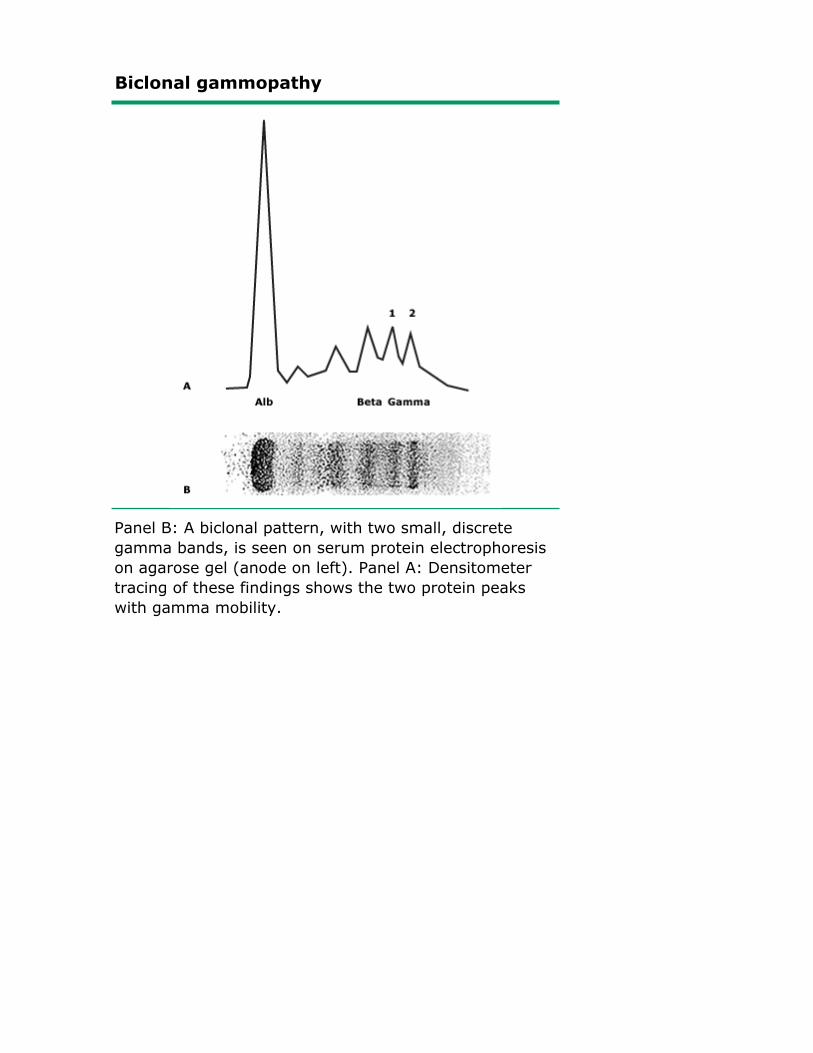
Biclonal gammopathy
Panel B: A biclonal pattern, with two small, discretegamma bands, is seen on serum protein electrophoresison agarose gel (anode on left). Panel A: Densitometertracing of these findings shows the two protein peakswith gamma mobility.
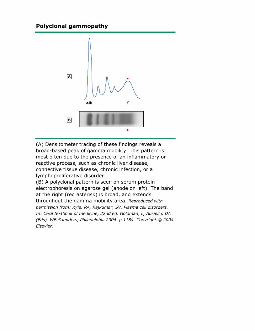
Polyclonal gammopathy
(A) Densitometer tracing of these findings reveals abroad-based peak of gamma mobility. This pattern ismost often due to the presence of an inflammatory orreactive process, such as chronic liver disease,connective tissue disease, chronic infection, or alymphoproliferative disorder.(B) A polyclonal pattern is seen on serum proteinelectrophoresis on agarose gel (anode on left). The bandat the right (red asterisk) is broad, and extendsthroughout the gamma mobility area. Reproduced withpermission from: Kyle, RA, Rajkumar, SV. Plasma cell disorders.In: Cecil textbook of medicine, 22nd ed, Goldman, L, Ausiello, DA(Eds), WB Saunders, Philadelphia 2004. p.1184. Copyright © 2004Elsevier.
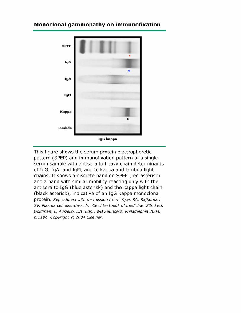
Monoclonal gammopathy on immunofixation
This figure shows the serum protein electrophoreticpattern (SPEP) and immunofixation pattern of a singleserum sample with antisera to heavy chain determinantsof IgG, IgA, and IgM, and to kappa and lambda lightchains. It shows a discrete band on SPEP (red asterisk)and a band with similar mobility reacting only with theantisera to IgG (blue asterisk) and the kappa light chain(black asterisk), indicative of an IgG kappa monoclonalprotein. Reproduced with permission from: Kyle, RA, Rajkumar,SV. Plasma cell disorders. In: Cecil textbook of medicine, 22nd ed,Goldman, L, Ausiello, DA (Eds), WB Saunders, Philadelphia 2004.p.1184. Copyright © 2004 Elsevier.

Biclonal gammopathy
This figure illustrates the immunofixation pattern of asingle serum specimen with antisera to heavy chaindeterminants of IgA, IgG, and IgM, and to kappa andlambda light chain determinants. It shows a discrete IgGband (seen as a dark column) and a discrete lambdaband with similar mobility, indicative of the presence ofan IgG lambda monoclonal protein. There is also an IgMkappa monoclonal protein.
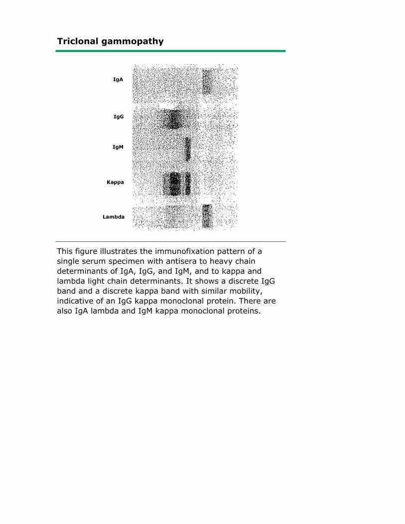
Triclonal gammopathy
This figure illustrates the immunofixation pattern of asingle serum specimen with antisera to heavy chaindeterminants of IgA, IgG, and IgM, and to kappa andlambda light chain determinants. It shows a discrete IgGband and a discrete kappa band with similar mobility,indicative of an IgG kappa monoclonal protein. There arealso IgA lambda and IgM kappa monoclonal proteins.

Serum free light chains
This figure shows simultaneous measurements of free serumkappa (horizontal axis) and lambda (vertical axis) light chains. Thedashed line indicates a 1:1 kappa/lambda ratio (weight basis).Results are shown for 282 normal subjects (+), 120 patients withkappa light chain myeloma (closed circles), 140 patients withlambda light chain myeloma (closed triangles), 31 patients withrenal impairment from non light chain disorders (open squares)and 28 subjects with non-secretory myeloma (open circles). Notethat monoclonality could be detected in many of the latter groupusing this technique. Data from Bradwell, AR, et al. Lancet 2003; 361:489.
Urinary monoclonal protein
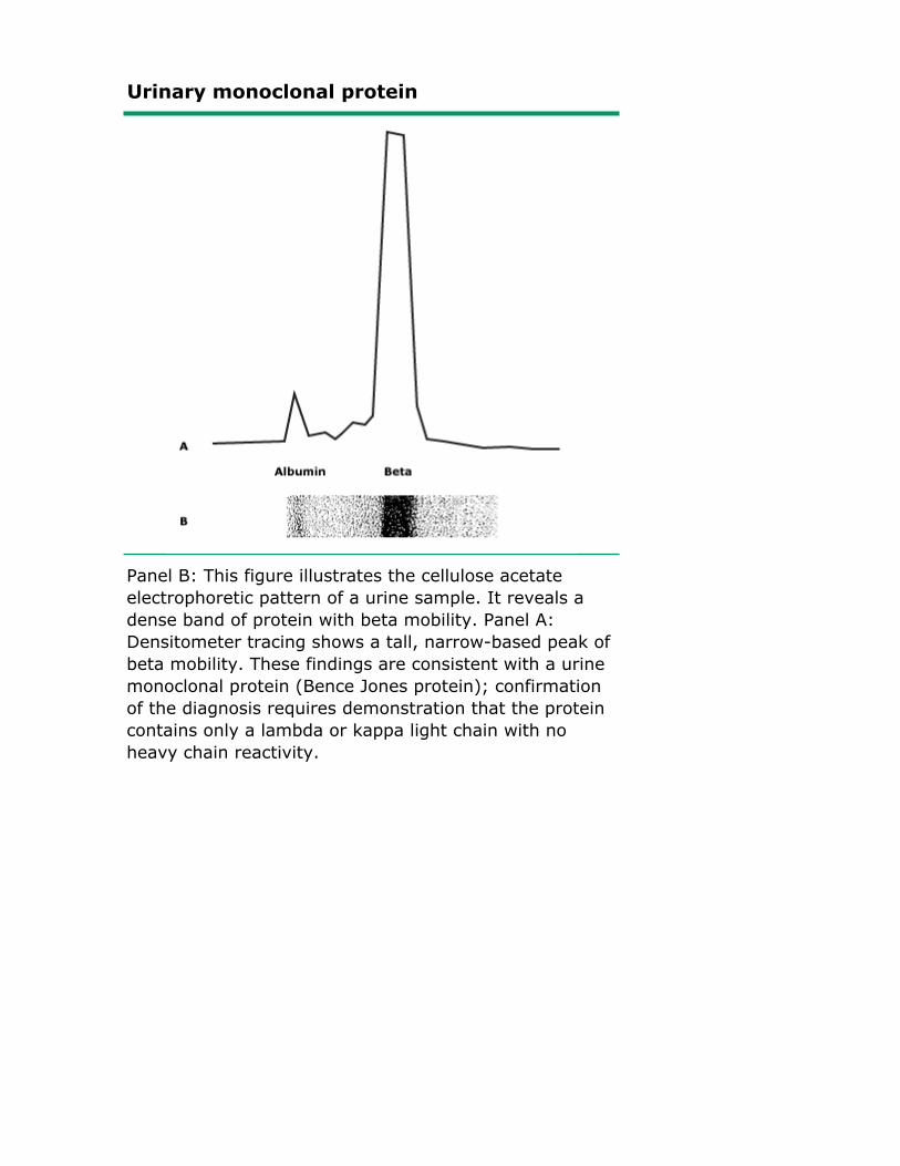
Panel B: This figure illustrates the cellulose acetateelectrophoretic pattern of a urine sample. It reveals adense band of protein with beta mobility. Panel A:Densitometer tracing shows a tall, narrow-based peak ofbeta mobility. These findings are consistent with a urinemonoclonal protein (Bence Jones protein); confirmationof the diagnosis requires demonstration that the proteincontains only a lambda or kappa light chain with noheavy chain reactivity.
Urinary monoclonal protein
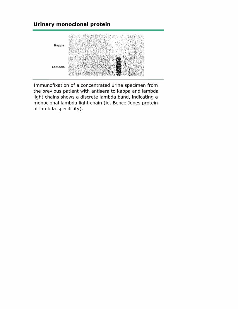
Immunofixation of a concentrated urine specimen fromthe previous patient with antisera to kappa and lambdalight chains shows a discrete lambda band, indicating amonoclonal lambda light chain (ie, Bence Jones proteinof lambda specificity).
Urinary immunoglobulin fragment
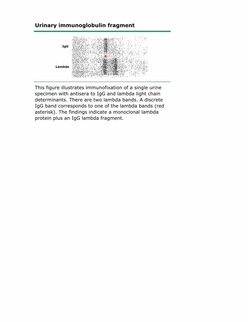
This figure illustrates immunofixation of a single urinespecimen with antisera to IgG and lambda light chaindeterminants. There are two lambda bands. A discreteIgG band corresponds to one of the lambda bands (redasterisk). The findings indicate a monoclonal lambdaprotein plus an IgG lambda fragment.

© 2011 UpToDate, Inc. All rights reserved. | Subscription and License Agreement | Support Tag:[ecapp1004p.utd.com-206.176.168.52-8F13E9C827-1273.1354.14]Licensed to: Lsu Hlth Sciences Ctr In