Embed Size (px)
Citation preview
Short Communications Pakistan J. Zool., vol. 42(6), pp. 825-827, 2010. Recovery of Snow Leopard Uncia uncia in Tomur National Nature Reserve of Xinjiang, Northwestern China Feng Xu, Ming Ma*, and Yiqun Wu Xinjiang Institute of Ecology and Geography, Chinese Academy of Sciences, Urumqi 830011, China (FX, MM), and Department of Environment and Life Sciences, Weinan Teachers University, Weinan 714000, China
Abstract.- Tomur National Nature Reserve (TNNR) is one of the biggest reserves in northwestern China containing the endangered snow leopards. But only a few snow leopard signs were found here by Schaller in 1988. And it was reported that more than 10 snow leopards were illegally killed by local herders the next year after Schaller’s survey, so the snow leopard was assumed to have nearly disappeared in this nature reserve. From October to November 2004 and October to December 2005, we conducted snow leopard sign surveys in four major valleys in TNNR using the Snow Leopard Information Management System (SLIMS). We ran 69 transects covering c. 39.7 km and located 56 sites with 131 snow leopard signs. Combined with the results of previous snow leopard sign surveys and camera trapping studies in Muzart valley of TNNR, we conclude that snow leopards appear to have re-inhabited the nature reserve. But conflict between snow leopard and herders still exist in this area and we are far from resolving it. Keywords: snow leopard, Uncia uncia, sign surveys, Tomur National Nature Reserve, Xinjiang
The Tomur National Nature Reserve (TNNR) is on the southern flank of the Tianshan mountains, and encompasses c. 3000 km2 of rugged ridges and _________________________________ *Corresponding author: [email protected]/2010/0006-0825 $ 8.00/0 Copyright 2010 Zoological Society of Pakistan.
narrow valleys with the elevation ranging from 2000 m to 7000 m. The reserve support varieties of mammals (e.g., snow leopard, wolf Canis lupus, red fox Vulpes vulpes, ibex Capra sibirica, and argali sheep Ovis ammon), birds (e.g., snowcock Tetraogallus altaicus, chukar partridge Alectoris chukar), and rodents (Mountain Investigation Team of Chinese Academy Sciences, 1985). The wildlife resources survey team, in the 1960s, recorded snow leopard in TNNR (Qian and Zhang, 1965). In the 1980s, studies also revealed the presence of snow leopard in the area (Mountain Investigation Team of Chinese Academy Sciences, 1985), but the following years snow leopards were widely prosecuted by local shepherds in the TNNR (Schaller et al., 1988). With the establishment of the nature reserve in Tomur area in 1989 (Yuan, 1998), which was upgraded to a national nature reserve in 2003, no-hunting rule was imposed since then. Populations of ibex, snow leopard’s major prey, burgeoned in the TNNR (Xu et al., 2007). So in this paper, we try to detect if the snow leopard population had recovered in TNNR.
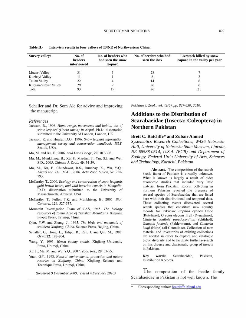
Materials and methods From October to November 2004 and October to December 2005, we conducted sign surveys in four major valleys of the TNNR - Muzart, Kuzbayi, Tailan, Kurgan-Yinyer - to ascertain the status and distribution of snow leopards (Fig. 1). We interviewed 93 herders to collect information on whether they had lost any livestock to snow leopards or if they had seen or heard about snow leopards and ibex. We followed the survey protocol of the Snow Leopard Information Management System (Jackson and Hunter, 1996; Jackson, 1996; McCarthy, 2000; McCarthy et al., 2005). Transect routes comprised landforms such as ridgelines, valley-bottom, and bases of cliffs where snow leopard signs were likely to leave signs.
Results and discussion Along 69 transects (c. 39.7 km), we located 56 sites with 131 signs (Table I). We found no sign in Kuzbayi valley. Muzart valley had highest mean density of the sign sites per km of transect among the three valleys surveyed (Kruskal-Wallis H=7.857,
SHORT COMMUNICATIONS 826
df=2, p=0.020). Pugmarks (48%) were the most common sign, followed by scrapes (30%), claw-rakes (11%), feces (8%), and scent-spray (3%). In the winter, we discovered several sets of pugmarks on snow, indicating their confirmed presence in the area.
Fig. 1. Distribution of the snow leopards in Tomur National Nature Reserves of northwestern China
Table I.- Number of sign sites, and signs (with mean
densities per km of transect in parentheses) in four valleys of TNNR of Northwestern China.
Location Total
transect length (km)
No. sign sites (mean km-1)
No. signs (mean km-1)
Muzart 16.4 35 (2.13) 74 (4.51) Kuzbayi 4.3 0 (0) 0 (0) Tailan 7.6 4 (0.53) 16 (3.72) Kurgan-Yinyer 11.4 17 (1.49) 41 (3.60)
Total 39.7 56 (1.41) 131 (3.30)
Snow leopard signs confirmed the presence of snow leopards in three of the four valleys (Fig. 1). Compared to Schaller et al. (1988) findings, snow leopards are now more abundant and covered much larger area. Schaller et al. (1988) reported - based on their findings from the upper part of the Muzart valley – snow leopards survived only in small, remote parts of the reserve, and cautioned that unless received prompt and complete protection,
snow leopards would disappear from the reserve. Our sign surveys indicated snow leopard-presence in three of the four major valleys. These results are consistent with pilot snow leopard signs survey in TNNR (Ma et al., 2005). During that survey only limited snow leopard signs were found in Tailan and Kurgan-Yinyer valley. After more detailed surveys in this study, we found more snow leopard signs and ascertained the presence of the snow leopard in more sites among three of four valleys in TNNR. Besides that in the winter of 2005, Ma et al conducted a camera trapping study on snow leopards in Muzart valley of TNNR (Ma and Xu, 2006; Ma et al., 2006), their photo also confirmed the presence of snow leopard in this valley. And the interviews confirmed our results. The number of snow leopard encountered by the local shepherds was consistent with our sign surveys results (Table II). With above results, we suggest the survival of the snow leopard in TNNR has improved since the establishment of the reserve in the 1980s. Snow leopard-herder conflict still exists in the reserve as reported by Schaller et al. (1988) as does the heavy-grazing problem during the summer months. The snow leopard attacks happened in all four valleys (Table II). Livestock increased from 400,000 in 1976 to c. 600,000 in 1993 (Wang, 1993). Except the core and remote areas of the reserve, livestock have access to all areas (Xu et al., 2007). This can be threatening to the other wildlife species sharing the habitat. Promotion of tourism and mining in and around the reserve may impact snow leopard’s survival. In spite of these threats, snow leopards have increased in distribution and abundance since 1988 and this should be attributed to the effective control of illegal hunting.
Acknowledgements We thank the International Snow Leopard Trust (ISLT) and Xinjiang Institute of Ecology and Geography, Chinese Academy of Sciences for financial support. We thank Xinjiang Forestry Bureau and Tomur National Nature Reserve Management Bureau for supporting our research. We thank R.S. Chundawat and Thomas McCarthy for help planning the research, and Kuban, Mulamin for help with the field work. We thank Dr. George
SHORT COMMUNICATIONS 827
Table II.- Interview results in four valleys of TNNR of Northwestern China. Survey valleys No. of
herders interviewed
No. of herders who had seen the snow
leopard
No. of herders who had seen the ibex
Livestock killed by snow leopard in the valley per year
Muzart Valley 31 5 28 7 Kuzbayi Valley 11 1 8 2 Tailan Valley 22 4 14 6 Kurgan-Yinyer Valley 29 9 26 6 Total 93 19 76 21
Schaller and Dr. Som Ale for advice and improving the manuscript.
References Jackson, R., 1996. Home range, movements and habitat use of
snow leopard (Uncia uncia) in Nepal. Ph.D. dissertation submitted to the University of London, London, UK.
Jackson, R. and Hunter, D.O., 1996. Snow leopard information management survey and conservation handbook. ISLT, Seattle, USA.
Ma, M. and Xu, F., 2006. Arid Land Geogr., 29: 307-308. Ma, M., Munkhtsog, B., Xu, F., Mardan, T., Yin, S.J. and Wei,
S.D., 2005. Chinese J. Zool., 40: 34-39. Ma, M., Xu, F., Chundawat, R.S., Jumabay, K., Wu, Y-Q.,
Aizezi and Zhu, M-H., 2006. Acta Zool. Sinica, 52: 788-793.
McCarthy, T., 2000. Ecology and conservation of snow leopards, gobi brown bears, and wild bactrian camels in Mongolia. Ph.D. dissertation submitted to the University of Massachusetts, Amherst, USA.
McCarthy, T., Fuller, T.K. and Munkhtsog, B., 2005. Biol. Conserv., 124, 527-537.
Mountain Investigation Team of CAS, 1965. The biology resources of Tomur Area of Tianshan Mountains. Xinjiang People Press, Urumqi, China.
Qian, Y.W. and Zhang. J., 1965. The birds and mammals of southern Xinjiang, China. Science Press, Beijing, China.
Schaller, G., Hong, L., Talipu, R., Ren, J. and Qin, M., 1988. Oryx, 22: 197-204.
Wang, Y., 1993. Wensu county annals. Xinjiang University Press, Urumqi, China
Xu, F., Ma, M. and Wu, Y.Q., 2007. Zool. Res., 28: 53-55. Yuan, G.Y., 1998. Natural environmental protection and nature
reserves in Xinjiang, China. Xinjiang Science and Technique Press, Urumqi, China.
(Received 9 December 2009, revised 4 February 2010)
Pakistan J. Zool., vol. 42(6), pp. 827-830, 2010.
Additions to the Distribution of Scarabaeidae (Insecta: Coleoptera) in Northern Pakistan Brett C. Ratcliffe* and Zubair Ahmed Systematics Research Collections, W436 Nebraska Hall, University of Nebraska State Museum, Lincoln, NE 68588-0514, U.S.A. (BCR) and Department of Zoology, Federal Urdu University of Arts, Sciences and Technology, Karachi, Pakistan
Abstract.- The composition of the scarab beetle fauna of Pakistan is virtually unknown. What is known is largely a result of older taxonomic studies that included very little material from Pakistan. Recent collecting in northern Pakistan revealed the presence of several species of Scarabaeidae that are listed here with their distributional and temporal data. These collecting events discovered several scarab species that constitute new country records for Pakistan: Popillia cyanea Hope (Rutelinae), Oryctes elegans Prell (Dynastinae), Clinteria confinis pseudoconfinis Schürhoff, Gametis jucunda (Faldermann), and Clinteria klugi (Hope) (all Cetoniinae). Collection of new material and inventories of existing collections are needed in order to explore and catalogue biotic diversity and to facilitate further research on this diverse and charismatic group of insects in Pakistan. Key words: Scarabaeidae, Pakistan, Distribution Records.
The composition of the beetle family Scarabaeidae in Pakistan is not well known. The ________________________ * Corresponding author: [email protected]
SHORT COMMUNICATIONS 828
first and perhaps only serious attempt at cataloguing and describing the scarabaeoid beetles for this part of the world was Gilbert Arrow’s three volumes on Lamellicornia in the series The Fauna of British India (Arrow, 1917, 1925, 1931), but this work was extremely preliminary and is now very outdated. Since that time, there have been several revisional works of various taxa that incidentally included Pakistan, e.g., Balthasar (1963a-b, 1964) for Scarabaeinae (dung beetles), Keith (2001) for some Melolonthinae (chafers), Endrödi (1985) for Dynastinae (rhinoceros beetles), and Miksic (1976, 1977, 1982, 1987) for Cetoniinae (flower chafers). Abdullah and Roohi (1968a-c, 1969) published a series of papers on Rutelinae (metallic leaf chafers) of Pakistan, wherein they described a few new species and constructed keys (by modifying those in Arrow, 1917) to species of Anomala and Mimela occurring or potentially occurring in the country. However, there has never been a contribution dedicated exclusively to elucidating the biodiversity of the Scarabaeidae of Pakistan. Accordingly, our knowledge of what scarabs occur in Pakistan is fragmentary at best. To our knowledge, there is no species list of Scarabaeidae for the country. The catalogue of Palearctic Coleoptera (Löbl and Smetana, 2006) lists Pakistan for some of the species, but it is, again, not complete for listing the taxa occurring in Pakistan. We provide here an annotated list of scarabs recently collected in northern Pakistan with the hopes of adding to the faunal composition of scarabs occurring in Pakistan. Some of these species are recorded from the country for the first time.
Materials and methods One of us (AZ) was able to collect in Kashmir (Rawalakot, Pallandri, and Bagh) and the Northwest Frontier Province (Dera Ismail Khan, Wari (Upper Dir), and Paras) from April to September 2009. Collecting was conducted at lights at night for Dynastinae and some Rutelinae and by foliage and flower gleaning by hand during the day for Cetoniinae and some Rutelinae. Dera Ismail Khan and associated areas are currently relatively unsafe for collecting insects, either during the day or night, and there are also restrictions on travel and
curfews that further limit collecting activities. Accordingly, we believe these new collecting records are particularly valuable and informative. The specimens were sent to the first author for identification, and the vouchers for those specimens have been deposited in the systematics research collections of the University of Nebraska State Museum. Even though the collecting effort was relatively short in duration and the number of each species small, the results have provided us with a better idea of the faunal composition of certain scarabs in northern Pakistan, thus adding incrementally to a list of scarabs that occur in the entire country. Based upon our scan of the literature, which is based upon specimens in collections, some of these species are reported from this area for the first time. Results and discussion The following is a list of localities and the scarabs collected there. Each species is annotated with its known distributional data.
NW Frontier Province: Dera Ismail Khan, VI-19-2009
Family: Dynastinae Tribe: Pentodontini
Alissonotum crassum Arrow. Broadly distributed in
the Oriental Region, including Pakistan (Endrödi, 1985; Löbl and Smetana, 2006).
Pentodon algernum indicum Endrödi. Occurs in the Oriental Region, including Pakistan (Endrödi, 1985; Löb l and Smetana, 2006).
Phyllognathus dionysius (Fabr.). Widely distributed on the Indian subcontinent. Siddiqui et al. (2005) were the first to record this species from Pakistan, and its presence in the northern half of the country was confirmed by Drumont et al. (2009), who noted that this species prefers habitats below 2000 meters in elevation.
Tribe: Oryctini
Oryctes elegans Prell. Known from the Middle East and Asia (Endrödi, 1985; Löbl and Smetana, 2006) but not previously recorded from Pakistan. New country record.

SHORT COMMUNICATIONS 829
NW Frontier Province: Paras, VIII-20-2009 Family: Rutelinae Tribe: Anomalini
Mimela passerini passerini Hope. Occurs in
southwestern China, Bhutan, Nepal, northern India, and Pakistan (Löbl and Smetana, 2006).
Mimela pectoralis Blanchard. Known from Nepal, northern India, southwestern China, and Pakistan (Löbl and Smetana, 2006)..
Popillia cyanea Hope. Recorded from northern India (including Kashmir), Tibet, and southwestern China (Arrow, 1917; Löbl and Smetana, 2006). New country record for Pakistan.
NW Frontier Province: Wari, Upper Dir, IX-22-
2009 Family: Rutelinae Tribe: Anomalini.
Mimela passerini passerini Hope. See preceding
information.
Family: Cetoniinae Tribe: Cetoniini
Protaetia indica Miksic. This species is known from
Pakistan, Afghanistan, and parts of India (Löbl and Smetana, 2006). When Miksic (1987) described it, he considered it a subspecies of the very broadly distributed (Europe to Asia) P. cuprea (Fabr.).
Kashmir: Bagh, IV-13-2009
Family: Cetoniinae Tribe: Gymnetini
Clinteria confinis pseudoconfinis Schürhoff. Occurs in Nepal, Bhutan, and the Indian states of Himachal Pradesh, Kashmir, Sikkim, and Uttar Pradesh (Miksic, 1977). New country record for Pakistan.
Tribe: Cetoniini
Gametis jucunda (Faldermann). Widely distributed in Asia (Japan, China, Laos, Korea, Tibet, Nepal and northern India (Miksic, 1982). Previously unknown in Pakistan. New country record.
Kashmir: Pallandri, VI-22-2009 Family: Cetoniinae Tribe: Gymnetini
Clinteria confinis pseudoconfinis Schürhoff. See
preceding information.. Clinteria klugi (Hope). Known from Nepal and
northern India (Miksic, 1977). New country record for Pakistan.
Kashmir: Rawalakot, VII-15-2009
Family: Dynastinae Tribe: Oryctini
Oryctes nasicornis illigeri Minck. Broadly
distributed from western Europe to Asia with many subspecies (Löbl and Smetana, 2006). This subspecies is known from Kashmir.
Tribe: Dynastini Xylotrupes mniszechii mniszechii Thomson. Occurs
in northern India, and Pakistan (Endrödi, 1985; Löbl and Smetana, 2006).
Discoveries like these, combined with the well-documented threats to many natural habitats globally, add importance to conducting extensive field studies and faunistic surveys in Pakistan. Collection of new material and inventories of existing collections are needed in order to explore and catalogue biotic diversity and to facilitate further research. Results of faunistic research will be available to researchers, Pakistani students at all levels, natural resource managers, government entities, and amateur naturalists. Research infrastructure in Pakistan will be enhanced by collaborative activities such as this one, as well as establishment of authoritatively identified collections, creation/augmentation of databases, and future partnerships to explore biodiversity. Benefits to society include a better understanding of the importance of and threats to biodiversity, enhanced ability to monitor habitats (conservation, invasive species, pest species) using taxonomic knowledge, educating students and the public about science, training future scientists or technicians, and instilling in the people of Pakistan a sense of responsibility and pride in their own rich biota so they may better care for it.
SHORT COMMUNICATIONS 830
Acknowledgements This report was supported, in part, by a Biotic Survey and Inventory grant from the U. S. National Science Foundation (DEB0716899) to B.C. Ratcliffe and R. D. Cave. References Abdullah, M. and Roohi, R.A., 1968a. Pak. J. Sci. Ind. Res., 11:
415-422. Abdullah, M. and Roohi, R.A., 1968b. Pak. J. Sci. Ind. Res., 11:
427-440. Abdullah, M. and Roohi, R.A., 1968c. Pak. J. Sci. Ind. Res., 11:
441-445. Abdullah, M. and Roohi, R.A., 1969. Pak. J. Sci. Ind. Res., 12:
121-126. Arrow, G.J., 1917. The Fauna of British India, Including Ceylon
and Burma. Coleoptera Lamellicornia, Part II (Rutelinae, Desmonycinae, and Euchirinae). Taylor and Francis, London, United Kingdom. 387 pp., 5 plates.
Arrow, G.J., 1925. The Fauna of British India, Including Ceylon and Burma. Coleoptera Lamellicornia (Cetoniinae and Dynastinae). Taylor and Francis, London, United Kingdom. 322 pp., 2 plates.
Arrow, G.J., 1931. The Fauna of British India, Including Ceylon and Burma. Coleoptera Lamellicornia, Part III (Coprinae). Taylor and Francis, London, United Kingdom. 428 pp., 13 plates, 1 map.
Balthasar, V. 1963a. Monographie der Scarabaeidae und Aphodiidae der Palaearktischen und Orientalischen Region, Vol. 1. Tschechoslowakische Academie der Wissenschaften, Prague, Czechoslovakia. 391 pp., 24 plates.
Balthasar, V., 1963b. Monographie der Scarabaeidae und Aphodiidae der Palaearktischen und Orientalischen Region, Vol. 2. Tschechoslowakische Academie der Wissenschaften, Prague, Czechoslovakia. 627 pp., 16 plates.
Balthasar, V., 1964. Monographie der Scarabaeidae und Aphodiidae der Palaearktischen und Orientalischen Region, Vol. 3. Tschechoslowakische Academie der Wissenschaften, Prague, Czechoslovakia. 652 pp., 2 plates.
Drumont, A., Saltin, J.-P. and Akhter, M.A., 2009. Lambillonea, 109: 398-402.
Endrödi, S., 1985. The Dynastinae of the World. Dr. W. Junk Publisher, Dordrecht, Netherlands. 800 p.
Keith, D., 2001. Bull. Mens. Soc. Linn. Lyon, 70: 6-14. Löbl, I. and Smetana, A. (editors). 2006. Catalogue of Palearctic
Coleoptera. Vol. 3. Scarabaeoidea – Scirtoidea – Dascilloidea – Buprestoidea – Byrrhoidea. Apollo Books, Stenstrup, Denmark. 690 pp.
Miksic, R., 1976. Monographie der Cetoniinae der Paläarktischen und Orientalischen Region. Coleoptera: Lamellicornia. Band 1. Gymnetini (Taenioderina, Chalcotheina). Forstinstitute in Sarajevo, Yugoslavia. 444 pp., 10 plates.
Miksic, R., 1977. Monographie der Cetoniinae der Paläarktischen und Orientalischen Region. Coleoptera: Lamellicornia. Band 2. Gymnetini (Lomapterina, Clinteriina), Phaedimini, Gnathocerini, Heterorrhinini. Forstinstitute in Sarajevo, Yugoslavia. 400 pp., 15 plates.
Miksic, R., 1982. Monographie der Cetoniinae der Paläarktischen und Orientalischen Region. Coleoptera: Lamellicornia. Band 3. Cetoniini I. Forstinstitute in Sarajevo, Yugoslavia. 530 pp., 14 plates.
Miksic, R., 1987. Monographie der Cetoniinae der Paläarktischen und Orientalischen Region. Coleoptera: Lamellicornia. Band 4. Cetoniini II. Forstinstitute in Sarajevo, Yugoslavia. 608 pp., 12 plates.
Siddiqui, M. J. I., Rana, S. A. Rana, N. and Sohail, A. 2005. Pak. Entomol., 27: 25-28.
(Received 13 October 2009, revised 29 April 2010)
Pakistan J. Zool., vol. 42 (6), pp. 830-833, 2010.
A Fin Anomaly in Thinlip Mullet Liza ramada (Risso, 1810) Caught From Homa Lagoon (Izmir Bay/Aegean Sea) Bahar Bayhan, Murat Kaya and Deniz Acarlı Departments of Hydrobiology (BB, MK) and Fisheries (DA), Faculty of Fisheries, Ege University, 35100 Bornova, Izmir-Turkey
Abstract.- A report on fin anomaly in thinlip mullet fish, Liza ramada was firstly described in one specimen caught from Homa Lagoon, Aegean Sea. The anomalous specimen showed the initial six rays of upper and lower lobes in the caudal fin and all the pelvic fin rays to have lengthened. Fin rays did not significantly vary in number, while considerable variations were established among some morphometric values comparing the meristic aspects of normal with those of abnormal specimens. Finally, we conclude that the anomaly may have resulted from domestic and industrial chemicals discharged into the area. Key words: Fin anomaly, Liza ramada, Environmental pollution, Homa Lagoon, Izmir Bay, Aegean Sea.
A total of 8915 tonnes of mullet was caught in Turkish seas, with 5538 tonnes and 1764 tonnes being harvested from the Black Sea and Aegean Sea, ____________________________ * Corresponding author: [email protected]

SHORT COMMUNICATIONS 831
respectively (Anonymous, 2006). In one of the most significant fisheries areas in the Aegean Sea, Izmir Bay is composed of three sections by their physical features, namely the outer, the middle and the inner bays; and mullet harvest is the leading process in commercial value and annual catching performance. All fisheries activities are prohibited in the inner bay which has been severely exposed to the pollution caused by domestic and industrial wastes dumped into it. Thicklip grey mullet Chelon labrosus (Risso, 1827), Golden grey mullet Liza (Risso, 1810), Leaping mullet Liza saliens (Risso, aurata (Risso, 1810), Thinlip mullet Liza ramada 1810), Flathead mullet Mugil cephalus Linnaeus, 1758 and So-iuy mullet Mugil soiuy Basilewsky, 1855 (Bilecenoglu et al., 2002). The study is of great interest because it has established that such a morphological modification in an individual of the species consumed in and around Izmir accounted for by domestic and/or industrial pollution as well as an increase of similar anomalies could constitute a long term risk factor for human health. Accordingly, we compared meristic and morphometric characteristics of an individual having fin differentiation with those of the normal specimen of the same species and examine the factors likely to account for the concerned anomaly in the L. ramada specimen. Materials and methods Located on 38º33'28'' north latitude and on the 26º50'42'' east longitude, the Homa Lagoon is currently the largest and only active area to harvest fish around Izmir Bay. The lagoon is 7.4 km long and 3 km wide with a total area of 1800 hectares and a depth of not over 1 m. The only fish specimen with fin differentiation was caught in the study field on 17th of October, 2007 with a multifilament netting gear of 32 mm mesh size and sent to the laboratory where it was weighed by a 0.01 g sensitive scale and morphometric measurements were made by a 0.01 mm sensitive digital compass. Such physicochemical parameters as temperature, salinity, dissolved oxygen and pH were measured. Water temperature plus pH, dissolved oxygen (DO) and salinity (‰S) were measured by Hanna Instruments HI 8314 with Winkler and Mohr-Knudsen methods respectively.
Morphometric and meristic characteristics of the fin-anomalous fish were compared with those of the L. ramada individual of the same size without fin anomalies caught in the same area and at the same date in order to find out whether there could be any variation between them. The table illustrates morphometric and meristic variables of both individuals. Additionaly, the anomalous fish was examined radiographically. Results The only fish specimen having fin differentiation was caught in the study field on 17th of October, 2007. The specimen found to be abnormal showed that the initial six rays of the upper and lower lobes in the caudal fin in particular and all Table I.- Morphometric (mm) and meristic
characteristics of the normal and abnormal L.ramada specimen from the Homa Lagoon (Aegean Sea).
Characteristics Normal Abnormal Morphometric Total length (LT) 390 405 Fork length (LF) 359 328 Standard length (LS) 330 305 Head length 78.75 71.33 First predorsal length 152.23 149.83 Second predorsal length 227 226 Preanal length 213 213 Prepectoral length 79.43 78.06 Preventral length 110.56 105.85 First dorsal fin length 20.85 23.86 Maximal ray lenght of first dorsal fin 35.56 36.99 Second dorsal fin length 27.50 27.36 Maximal ray lenght of second dorsal fin 39.09 45.23 Anal fin length 35.18 37.11 Maximal ray lenght of anal fin 35.20 38.96 Pectoral fin length 46.59 47.99 Pelvic fin length 24.79 33.44 Maximal ray lenght of pelvic fin 38.47 62.81 Caudal fin length 68.29 112.12 Maximal body depth 63.13 69.22 Minimal body depth 33.67 30.76 Eye diameter 12.13 10.77 Interorbital length 34.57 32.97 Preorbital length 16.72 17.03 Postorbital length 47.99 44.19 Meristic First dorsal fin IV IV Second dorsal fin 8 8 Anal fin II+9 II+9 Pectoral fins 15 14 Pelvic fins I+5 I+5 Caudal fin 16 (8/8) 16 (8/8)

SHORT COMMUNICATIONS 832
pelvic fin rays were lengthened in such a way to become filaments. In addition, initial rays of the second dorsal and anals fins were somewhat lengthened. Some of the physicochemical parameters measured in the Homa Lagoon during the sampling period are as follows: dissolved oxygen 8.8 mg/l, salinity 33.45 %o, pH 8.45, temperature 24°C. This study measured both anomalous (Fig. 1A) and normal individuals (Fig. 1B) morpholometrically and meristically and presented their related results in Table I in detail. No neither specimen showed any variations in meristic characteristics, especially in number of fin rays (Table I). Radiographs showed that fin deformaties can not have originated from skeleton or vertebrae (Fig. 1C).
A
B
C
Fig. 1. Liza ramada; A, abnormal (LT 405 mm, W 452.86 g); B, normal (LT 390 mm, W 435.07 g); C, X-ray photograph of abnormal fish.
Discussion Homa Lagoon is located both near the most important industrialization center of the Aegean region and the area where artificial composts and
pesticides are widely used to increase the agricultural yielding. Consequences a research carried out in the field found that Gediz River carries a certain amount of organic material and microbiological pollution into Izmir Bay, thus the water quality of the area exposed to a threat of dense pollution should be monitored sistematically (Anonymous, 1999). Genotypes of fish and environmental conditions play an important role in development of fish (Blaxter, 1988). Embryos and larvaes are less tolerant to environmental pollutants than those adults of the same species (Connor, 1972). Anomalies in fish can be accounted for by various etiologies, they are usually considered to have resulted from mutations and teratogenic effects of adverse environmental factors such as mutagenic chemicals in water (Longwell et al., 1992; Lien, 1997; Brown and Nuñez, 1998). For example, heavy pollution caused by slicks of oil can result in skeletal anomalies in fish (Al-Hassan, 1982) and adverse environmental factors such as chemical pollution in the habitat lead to skeletal deformities in Mugil cephalus (Jawad, 2004). In addition, some chemical pollutants in natural environment such xeno-hormones as pesticides, industrial chemicals, plastics, or endocrine-distrupter chemicals etc. can be responsible for hermaphroditism in Liza ramada (Bayhan and Acarlı, 2006). Nevertheless, the biological studies carried out across Turkish seas on mullets have thus far reported no fin anomalous individuals. Caudal fin deformities in particular affect swimming and feeding processes of fish. Formation of fins in fish appears following egg fertilisation. Initial fins during embriyonic phase are pectoral, tail and primordial ones chronologically, which is followed by the pelvic fin at early larval phase (Russell, 1976; Lagler et al., 1977). For this reason, the deformities associated with caudal and pelvic fin anomalies in our specimen must have certainly emerged during the egg fertilisation and early larval stages. Because Izmir Bay covering Homa Lagoon is an area exposed to pollution caused by domestic and chemical discharges and to eutrification, highest concentrations of heavy metals (Hg, Cd, Pb and Cr) were found in the inner part of the bay which is intensely industrialized (mainly iron, paper and pulp factories, antifouling paints, chlorine-alkali plants, chemical industries, textile industries, metal
SHORT COMMUNICATIONS 833
processing etc.) compared to the middle and outer parts of the bay (Küçüksezgin et al., 2006). Therefore it is greatly likely that eggs and larvae of L. ramada spawning in the outer and middle bays drift away into the inner part and undergo contamination to create such a fin anomaly. References Al-Hassan, L.A.J., 1982. Iraqi J. mar. Sci., 1: 13-23. Anonymous, 1999. Project of wetlands management of Gediz
estuary. No. 97K100020, Final Report I: 248 pp. Anonymous, 2006. Fisheries statistics. State Institute of
Statistics Prime Ministry, Republic of Turkey, ISSN: 1013-6177.
Bayhan, B. and Acarlı, D., 2006. Aquac. Res., 37: 1050-1052. Bilecenoğlu, M., Taşkavak, E., Mater, S. and Kaya, M., 2002.
Zootaxa, 113: 194 pp. Blaxter, J.H.S., 1988. In: Fish physiology (eds. W.S. Hoar and
D.J. Randall), XIA. Academic Press, London. pp. 1-58. Brown, C.L. and Nuñez, J.M., 1998. In: Fish diseases and
disorders, CABI Publishing, Vol. 2: 340 pp. Connor, P.M., 1972. Mar. Pollut. Bull., 3: 190-192. Jawad, L.A., 2004. Tuhinga, 15: 121-124. Küçüksezgin, F., Kontas, A., Altay, O., Uluturhan, E. and
Darılmaz, E., 2006. Environ. Int., 32: 41–51. Lagler, K.F., Bardach, J.E., Mıller, R.R. and Passıno, D.R.M.,
1977. Ichthyology. Second edition, by John Wiley and Sons, Canada, pp. 506.
Lien, N.T.H., 1997. Chemosphere, 35: 1475-1486. Longwell, A.C., Chang, S., Hebert, A., Hughes, J.B. and Perry,
D., 1992. Environ. Biol. Fish. 35: 1-21. Russell, F.S., 1976. The eggs and planktonic stages of British
marine fishes. Academic Press, London, pp. 524.
(Received 18 February 2010, revised 5 May 2010) Pakistan J. Zool., vol. 42(6), pp. 833-836, 2010. Major Strains of Canine Parvovirus Present in Dog Population of Pakistan Farhan Towakal, Masood Rabbani*, Khushi Muhammad, Muhammad Sarwar Khan, and Muhammad Zubair Shabbir University Diagnostic Lab, University of Veterinary and Animal Sciences, Lahore. (FT, MR and MZS), Department of Microbiology, University of Veterinary and Animal Sciences, Lahore (KM), and
Department of Clinical Medicine and Surgery, University of Veterinary and Animal Sciences, Lahore (MSK)
Abstract.- Active prevalence of canine parvovirus (CPV) was studied in the dog population of Pakistan. A total of 40 fecal samples were taken aseptically from dogs clinically suspected for parvovirus infection. The dogs were showing enteritis, bloody and smelly diarrhea. Each of the samples was processed for hemagglutinating (HA) activity and polymerase chain reaction (PCR) based detection of capsid protein using specific primers against CPV2 and CPV2b. Only 30 samples showed HA titer ranging from 1:16 to 1:512. Added to this, only 29 and 27 HA positive samples affirmed CPV2 and CPV 2b amplification, respectively whereas one HA positive sample (1:32) was found negative through PCR. The results suggest that HA is a rapid screening test for diagnosis of CPV infection. The PCR technique is more reliable and rules out the possibility of other hemagglutinating pathogens causing hemorrhagic enteritis in dogs. Further, it confirms that both CPV2 and CPV2b are pathogenic strains prevailing in Pakistan. Key words: Parvovirosis, hemagglutinating activity, hemorrhagic enteritis, PCR.
Canine parvovirus was first recognized during 1978 (Appel et al., 1979). The virus belongs to family parvoviridae and its type 2 (CPV-2), differs antigenically from type 1 (CPV-1). The CPV-2 is associated with enteric and respiratory illness in newborn pups (Binn et al., 1970). Analysis of isolates collected since its emergence has revealed antigenic differences from earlier isolates (Parrish et al., 1988). Further, monoclonal antibodies based strain differentiation and restriction enzyme analysis of viral DNA have shown post year1980 viruses similar to earlier isolates, but some restriction site differences have been observed in the new strains CPV2a and CPV 2b (Parrish et al., 1985). Since the emergence of CPV-2, two new antigenic types of CPV, designated CPV-2a and CPV-2b, have arisen consecutively. These new virus types have now _________________________ * Corresponding Author: [email protected]
SHORT COMMUNICATIONS 834
almost completely replaced CPV-2 viruses as the dominant infectious agents. In another study Parrish et al. (1991) have identified another antigenic variant CPV 2b, that emerged around 1984 and replaced CPV-2a after 1986. Similar kind of pattern of spread, emergence and subsequent replacement by the new antigenic strains with different proportions have been shown through phylogenetic analysis of isolates from Denmark, Germany, France, Spain, Japan, Australia and Africa (Parrish et al., 1988; Mochizuki et al., 1993; Yabanez et al., 1995; Truyen et al., 1996; Steinel et al., 2000). Although the exact mechanisms of these evolutions are not clear, the emergence of these new antigenic types of CPV can likely be ascribed to the adaptation of CPV-2 type viruses in dogs. Clinically, the canine parvovirus disease is prevalent at rate of over 21 percent (Jafri and Rabbani, 1999). In another study made by Khan et al. (2006) at Lahore, the disease occurrence was found more in German shephered (84%) followed by Rottweiller (9%), Doberman (5%) and Bull terriers (2%). They further described its prevalence more in male (85%) during hot humid environment (61%). Subsequently, widespread outbreaks of canine hemorrhagic enteritis with high morbidity and mortality occurred over the whole country. There is scanty information concerning the antigenic types of CPV prevailing in Pakistan. This study, therefore, has been conducted to set apart canine parvovirus CPV2 and CPV2b using haemagglutination (HA) test and PCR from fecal samples of the infected dogs.
Materials and methods A total of 40 fecal samples were collected from clinically suspected dogs showing vomiting, enteritis bloody and smelly diarrhea. These samples were diluted as 1:5 using 0.85 per cent aqueous solution of sodium chloride and centrifuged at 1000x g for 10 minutes. The supernatant of each sample was passed through syringe filter of 0.2 micron porosity and processed for HA and PCR based detection of viral DNA. Feces from normal dogs were taken as negative control whereas commercially available canine parvo vaccine (Primodog Merial, France) was used as positive
control for PCR reaction. The supernatant of all the samples was processed for hemagglutination (HA) test according to method described by Carmichael et al. (1980) with some modifications done by Umer (2003). For polymerase chain reaction CPV DNA was extracted using DNA Purification Kit (Fermentas Life Sciences) according to manufacturer’s instruction (Pereria et al., 2000, Sagazio et al., 1998; Ozkul et al., 2001). The amplification of extracted DNA was performed as described by Pereria et. al. (2000) using specific primers. For CPV2, sense (5´GAAGAGTGGTTGTAAATAATA3´) and anti-sense (5´CCTATATCACCAAAGTTAGTAG3´) primers were at 3025-3045 and 3685-3706 of the CPV2 genome, respectively that yields a 681 bp product, while sense (5´CTTTAACCTTCCTGTAACAG3´) and antisense (5´CATAGTTAAATTGGTTATCTAC3´) primers for CPV2b were located at 4043-4062 and 4449-4470, respectively that yields a 427 bp product. The PCR amplification was performed in a total of 25 µl volume by adding extracted DNA to the PCR master mix containing Tris-HCl, Ammonium sulphate (NH4(SO4)2), Magnesium chloride (MgCl2), primer pair, dNTPs, and Taq DNA polymerase. The thermal cycler was set up as follows: an initial denaturation step at 94°C for 5 min was followed by a cycle of 30 sec. at 55°C, 2 min. at 72°C and 2min. at 94°C, repeated 30 times. Amplification was terminated by a final extension at 70°C for 10 min followed by electrophoresis on 1% agarose gel containing 0.5 µg per milliliter of ethidium bromide. The bands were compared for their particular size against the ladder under UV light and photographic records were made.
Results and discussion In order to isolate infected dogs and prevent secondary infections of susceptible animals, rapid diagnosis of CPV-2 infection is important especially in kennels and shelters (Desario et al., 2005). Since a clinical diagnosis is not definitive, several laboratory methods have been developed to detect CPV-2 in the faeces of infected dogs. These include HA, PCR or direct electron microscopy. Despite the advancement in diagnostic techniques such as DNA
SHORT COMMUNICATIONS 835
hybridization, electron microscopy and ELISA, HA still has been used as initial screening test for parvovirus identification (Teramoto et al., 1984; Cavalli et al., 2001) and is routinely used for diagnosis of canine parvovirus (Senda et. al., 1988). Of the 40 fecal samples examined, only 30 showed agglutination to one percent washed chicken erythrocytes. HA titer of these sample ranged from 1:16 to 1:512, whereas, sample taken from healthy dog showed no HA activity. To confirm parvovirus induced HA, all the samples were processed for PCR based amplification resulting in capsid gene amplification from 29 and 27 samples for CPV2 and CPV2b, respectively. Two samples that showed HA titer 1:256 were found negative to CPV2b but positive to CPV2 gene amplification. Though CPV2a and CPV2b have emerged consecutively with similar frequencies of disease occurrence in UK, Germany and Spain (Ybanfez et al., 1995; Greenwood et al., 1996), CPV2 is still the major cause of acute infectious diarrhea in dogs (Hirasawa et al., 1996). Added to this, Parrish et al. (1991) and Steinel et al. (1998) have described CPV2b as a predominant virus type responsible for canine hemorrhagic enteritis. This is, further, in agreement with our finding as PCR amplified product were obtained only from HA positive fecal samples. Although, Parrish et al. (1988) and Cavalli et al. (2001) have reported lack of HA activity among CPV-2 strains, nevertheless, in this study all HA negative samples were found negative through PCR for either CPV2 or CPV2b gene amplification. This deviates from the previous observation demonstrated in natural and experimental infection that CPV2 is detectable for few days post infection and can give false-negative results despite high amounts of viral DNA detected by real-time PCR analysis (Desario et al., 2005). Decaro et al. (2004) have described poor correlation between HA and real-time PCR titres since samples with low HA titres were found to contain high viral DNA titres. This may be attributed to the presence of high levels of CPV-2 antibodies in the feces of the infected dogs. Only one HA positive sample with titer 1:32 was found negative by PCR that may be due to other hemagglutinating agents in the feces such as orthomyxovruses (Marulappa and Kepil, 2009) and
E. coli (Hogan et al., 1990. This shows the specificity of the PCR that can detect only genome of the CPV using specific primers (Sakulwira et al., 2001). Moreover, the primer pair used for identification of both the CPV2 and CPV2b was highly sensitive and specific (Parrish et al., 1988; Buonavoglia et al., 2001). The sequences of the PCR primers were selected from variable regions in the VP1/VP2 capsid genes sequences of CPV-2 and 2b Reed el al. (1988). The CPV2 genome was amplified from vaccinal strains and 29 HA positive samples, whereas, two samples were found negative with CPV2b primers that may be due to the reason that this strain is specific for wild strains (Parrish et al., 1991; Steinel et al., 2000). This is first study showing prevailing strains of canine parvovirus in dog population of Pakistan. Though we are not able to describe the evolutionary process of CPV2 in Pakistan into new strains like CPV2a and CPV2b but these findings suggest that there exist a considerable number of parvo affected dogs with both the CPV2 and CPV2b. This virus represents a threat to dogs, especially young pups in animal shelters and pups from unvaccinated dams. Accurate standardisation of laboratory tests for the detection of CPV-2 is required to provide veterinary scientists with an effective tool for a precise etiological diagnosis of CPV-2 infection. The study concludes that HA test is a rapid screening test for diagnosis of CPV infection. Nonetheless, PCR is more reliable and indicates that both the CPV2 and CPV2b strains of the virus are prevailing in Pakistan.
References Appel, M.J., Scott, F.W. and Carmichael, L.E., 1979. Vet Rec.,
105: 156–159.Binn, L.N., Lazar, E.C., Eddy, G.A. and Kajima, A., 1970.
Infect. Immun., 1: 503-508. Buonavoglia, C., Martella, V., Pratelli, A., Tempesta, M.,
Cavalli, A. and Buonavoglia, D., 2001. J. Gen. Virol., 82: 3021-3025.
Carmichael, L.E., Joubert, J.C. and Pollock, R.V., 1980. Am. J. Vet. Res., 41: 784-791.
Cavalli, A., Bozzo, G., Decaro, N., Tinelli, A., Aliberti, A. and Buonavoglia, D., 2001. Microbiologica, 23: 23-96.
Decaro, N., Desario, C., Campolo, C., Cavalli, A., Ricci, D., Martella, V., Tempesta, M. and Buonavoglia, C., 2004. New Microbiol., 27: 375–379
Desario, C., Decaro, N., Campolo, M., Cavalli, A., Cirone, F.,
SHORT COMMUNICATIONS 836
Elia, G., Martella, V., Lorusso, E., Camero, M. and Buonavoglia, C., 2005. J. Virol. Meth., 126: 179-185
Greenwood, N.M., Chalmers, W.S.K., Baxendale, W. and Thompson, H., 1996. Vet. Rec., 138: 495-449.
Hirasawa, T., Kaneshige, T. and Mikazuki, K., 1996. Vet. Microbiol., 41: 135-145.
Hogan, J. S., Todhunter, D.A., Smith, L. and Choenbergerh, P.S., 1990. J. Dairy Sci., 73: 3126-3131.
Jafri, S.A. and Rabbani, M., 1999. Pak. Vet. J., 19: 40-42. Khan, M.A., Rabbani, M., Muhammad, K., Murtaza, N. and
Nazir. J., 2006. Int. J. Agric. Biol., 8: 898-900. Marulappa, S.Y. and Kapil, S., 2009. Clin. vacc. Immunol., 16:
127–131. Mochizuki, M., Gabriel, S., Nakatani, M. and Harasawa, R.,
1993. Res. Vet. Sci., 55: 60-63. Özkul, A., Kelefi, H. and Karaolu, T., 2001. Turk. J. Vet. Anim.
Sci., 26: 1201-1203. Parrish, C.R., Aquadro, C.F. and Carmichael, L. E., 1988.
Virology, 166: 293–307. Parrish, C.R., Aquadro, C.F., Strassheim, M.L., Evermann, J. F.,
Sgro, J. Y. and Mohammed, H.O., 1991. J. Virol., 65: 6544–6552.
Parrish, C.R., O'Connell, P.H., Evermann, J.F. and Carmichael, L.E., 1985. Science, 230: 4729, 1046-1048.
Pereira, C.A., Monezi, T.A., Mehnert, D.U., Angelo, M.D. and Durigon, E.L., 2000. Vet. Microbiol., 75: 127-33.
Reed, A.P., Jones, E.V. and Miller, T.J., 1988. J. Virol., 62: 266-276.
Sagazio, P., Tempesta, M., Bunavoglia, D. and Bunavoglia, C., 1998. J. Virol. Meth., 73: 197-200.
Sakulwira, K., Oraveerakul, K. and Poovorawan, Y., 2001. Sci. Asia, 27: 143-147.
Senda, M., Hirayama, N., Itoh, O. and Yamamoto, H., 1988. J. Gen. Virol., 69: 349–354.
Steinel, A., Munson, L. and Truyen, U., 2000. J. Gen. Virol., 81: 345-350.
Teramoto, Y.A., Mildbrand, M.M., Carlson, J., Collins, J. K. and Winston, S., 1984. J. Clin. Microbiol., 20: 373-378.
Truyen, U., Platzer, G. and Parrish, C.R., 1996. Vet. Rec., 138: 365-366.
Umar, A., 2003. Passive immunization against canine parvovirus in dogs. M.Sc (Hons) thesis, Department of Microbiology, University of Veterinary and animal Sciences Lahore.
Yabanez, R.R., Vela, C., Cortes, E., Simarro, I. and Casal, J.I., 1995. Vet. Rec., 136: 174-175.
(Received 10 December 2009, revised 3 February 2010)
Pakistan J. Zool., vol. 42(6), pp. 836-839, 2010.
Obestatin Induces Testosterone But Not Prolactin Secretion in Male Sprague Dawley Rats Sarwat Jahan*, Shakeel Ahmed, Nazifa Taqvim and Hizbullah Reproductive Physiology Laboratory, Department of Animal Sciences, Quaid-i-Azam University, Islamabad.
Abstract.- The present study was designed to investigate the in vivo effect of an anorexigenic gut peptide obestatin on plasma testosterone and prolactin secretion, in adult male Sprauge Dawley rats. One group of animals served as control (n=5), while the treatment group (n=5) received a single i.v. dose of 8 nmol/Kg of obestatin. Blood samples (200µl-250 µl) were collected at -10, 0, 10, 20, 30 and 40 min of treatment. In treated group obestatin caused a significant (p<0.05) increase in plasma testosterone concentrations at 20 minutes. However, at 30 and 40 min post treatment, the plasma testosterone concentrations declined to non-significantly (P>0.05) higher values. A significant (P<0.05) increase in plasma testosterone concentrations was observed after treatment, compared to that before treatment. Obestatin showed no profound effect on plasma prolactin concentration in all the treated groups. It was concluded that obestatin i.v infusion leads to increase in plasma testosterone in adult male rats and may thus be involved in testosterone secretion, however, it has no effect on the prolactin secretion. Key words: Obestatin, anorexigenic, reproduction, testosterone, prolactin.
The gastrointestinal tract functions as an endocrine organ and also acts as an important source of regulatory peptide hormones. There are about 20 different gut peptides which are sensitive to the change in nutrient content within the gut (Kevin et al., 2006). These gut peptides also regulate body functioning by affecting various parts of the body like brain, endocrine glands and reproductive tissues (Peeters, 2003). One of the most important gut ___________________________ * Corresponding author: [email protected]
SHORT COMMUNICATIONS 837
peptide is ghrelin which was discovered in 1999 (Kojima et al., 1999). Ghrelin is anorexigenic gut peptide, produced from preproghrelin and secreted from oxyntic gland in fundus of the stomach. Obestatin is another gut peptide (Ariyasu et al., 2001). The expression of obestatin has been found in various species, however rats and mice are the only species in which its physiological function has been tested (Nogueiras et al., 2006). It was reported that obestatin peptide was present in the large and small intestines, stomach, spleen and cerebral cortex of rats (Zhang et al.,2005). Obestatin immunoreactivity was detected in cells of the gastric mucosa, myenteric plexus, and in the Leydig cells of the testis in mice (Dun et al., 2006). Experiments using labeled obestatin revealed that obestatin binds jejunum, ileum, stomach, pituitary and hypothalamus with high affinity. Later on, it was found that obestatin binds to the G-protein coupled receptor 39 (GPR39) in these areas of the gut. Expression of GPR39 has been found in a variety of rat tissues including hypothalamus, amygdala, cerebrum, cerebellum, testis and ovary (Zhang et al., 2005: Dong et al., 2009). Obestatin is considered as functional antagonist of ghrelin (Zizzari et al., 2007), as it was observed that obestatin is encoded by the same gene and has opposing effect to ghrelin. It was expected that obestatin may be involved in modulation of reproductive functions. Obestatin was found to increase ovarian cell functions. It was also found that obestatin was able to increase progesterone secretion in cultured porcine ovarian granulosa cells (Meszarosova et al., 2008). A previous study has observed that obestatin gene is expressed in Leydig cells of testis (Dun et al., 2006). Similarly, GPR39 mRNA the receptor of obestatin was also reported to be expressed in rat testis, pituitary and hypothalamus (Zhang et al., 2005; Moechars et al., 2006; Dong et al., 2009). The present study was designed to observe the effect of iv administration of obestatin on testosterone and prolactin secretion in intact male Sprauge Dawley rats. Materials and methods Animals Adult 10-12 weeks old male Sprauge Dawley
rats weighing 200-220g were used in this experiment. All animals were maintained in groups of five animals/cage at 22°C in 12 hour light/12 hour dark cycle with free access to pelleted food and water in the animal house of Quaid-i-Azam university Islamabad, Pakistan. Experimental protocols for animals use and care were approved by Animal Sciences Department, Quaid-i-Azam University Islamabad, Pakistan. Experimental design Animals were divided into two groups. Control rats (n=5) received saline, while treated rats (n=5) were administered with 8 nmol/Kg obestatin (Ana-Spec, USA) intravenously. Before the initiation of treatment all rats were anaesthetized with diethyl ether (Sigma-Aldrich, USA) and a Teflon cannula was inserted into the lateral tail vein, as per a previously described procedure (Omaye et al., 1987). Blood (200-250 µL) was collected in heparin (Sino-Chem, China) containing syringes at 10 min intervals starting from 10 min before obestatin treatment till 40 min after obestatin treatment (-10, 0, 10, 20, 30 and 40). The blood samples were centrifuged at 4000 rpm at 4°C to separate the plasma that was stored at -20°C until assay. Plasma testosterone and prolactin were quantitatively determined by using commercial enzyme immuno-assay kits (Amgenix,USA) according to the manufacturers instructions. Statistical analysis Student “t” test. was applied to analyze the data and P<0.05 was considered as significant. Results and discussion In adult male rats, following the obestatin (8nmol/kg) administration the plasma testosterone concentrations increased significantly at 20 min post administration (Table I) and then decreased at 30 and 40 min. This finding shows that in the adult male rats the 8nmol/kg dose of obestatin remains effective for at least 20 min duration. It has been previously described, that plasma degradation time of obestatin in the blood is 22 min, supporting the view that obestatin degradation resulted in the loss of its impact on testosterone production (Zizzari et al., 2007). This increase in testosterone
SHORT COMMUNICATIONS 838
concentration might be through hypothalamus pituitary gonadal axis (HPG) or hypothalamus pituitary adrenal axis (HPA). The exact mechanism of this increase is yet not known. Table I.- Mean±SEM plasma testosterone and prolactin
concentrations before and after Obestatin/ Saline administration in adult male rats.
Plasma testosterone conc.
(ng/ml) Plasma prolactin conc.
(ng/ml) Time
Saline Obestatin (8 nmol/kg)
Saline Obestatin (8 nmol/kg)
-10 1.24±0.25 1.34±0.49 0.91±0.30 0.74±0.37
0 1.15±0.24 1.85±0.42 0.80±0.09 0.69±0.19
10 1.27±0.23 2.59±0.84 0.52±0.07 0.35±0.12
20 1.15±0.25 3.37±0.77* 0.57±0.14 0.89±0.20
30 1.18±0.32 2.54±0.74 0.82±0.21 0.98±0.13
40 1.22±0.17 2.92±0.68 0.86±0.13 1.31±0.24
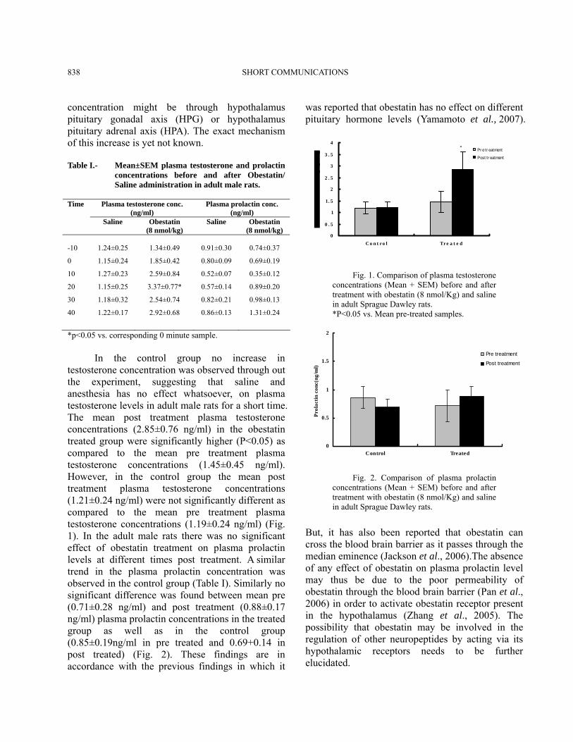
*p<0.05 vs. corresponding 0 minute sample. In the control group no increase in testosterone concentration was observed through out the experiment, suggesting that saline and anesthesia has no effect whatsoever, on plasma testosterone levels in adult male rats for a short time. The mean post treatment plasma testosterone concentrations (2.85±0.76 ng/ml) in the obestatin treated group were significantly higher (P<0.05) as compared to the mean pre treatment plasma testosterone concentrations (1.45±0.45 ng/ml). However, in the control group the mean post treatment plasma testosterone concentrations (1.21±0.24 ng/ml) were not significantly different as compared to the mean pre treatment plasma testosterone concentrations (1.19±0.24 ng/ml) (Fig. 1). In the adult male rats there was no significant effect of obestatin treatment on plasma prolactin levels at different times post treatment. A similar trend in the plasma prolactin concentration was observed in the control group (Table I). Similarly no significant difference was found between mean pre (0.71±0.28 ng/ml) and post treatment (0.88±0.17 ng/ml) plasma prolactin concentrations in the treated group as well as in the control group (0.85±0.19ng/ml in pre treated and 0.69+0.14 in post treated) (Fig. 2). These findings are in accordance with the previous findings in which it
was reported that obestatin has no effect on different pituitary hormone levels (Yamamoto et al., 2007).
0
0 . 5
1
1. 5
2
2 . 5
3
3 . 5
4
C o n t r o l Tr e a t e d
Pr e tr eatment
Post tr eatment
*
Fig. 1. Comparison of plasma testosterone concentrations (Mean + SEM) before and after treatment with obestatin (8 nmol/Kg) and saline in adult Sprague Dawley rats. *P<0.05 vs. Mean pre-treated samples.
0
0.5
1
1.5
2
Control Treated
Prol
actin
con
c(ng
/ml)
Pre treatment
Post treatment
Fig. 2. Comparison of plasma prolactin concentrations (Mean + SEM) before and after treatment with obestatin (8 nmol/Kg) and saline in adult Sprague Dawley rats.
But, it has also been reported that obestatin can cross the blood brain barrier as it passes through the median eminence (Jackson et al., 2006).The absence of any effect of obestatin on plasma prolactin level may thus be due to the poor permeability of obestatin through the blood brain barrier (Pan et al., 2006) in order to activate obestatin receptor present in the hypothalamus (Zhang et al., 2005). The possibility that obestatin may be involved in the regulation of other neuropeptides by acting via its hypothalamic receptors needs to be further elucidated.
SHORT COMMUNICATIONS 839
Acknowledgement This piece of work was fully funded and supported by Department of Animal Sciences, Quaid-i-Azam University, Islamabad. Pakistan. References Ariyasu, H., Takaya, K., Tagami, T., Ogawa, Y., Hosoda, K.,
Akamizu, T., Suda, M., Koh, T., Natsui, K., Toyooka, S., Shirakami, G., Usui, T., Shimatsu, A., Doi, K., Hosoda, H., Kojima, M., Kangawa, K. and Nakao, K., 2001. J. clin. Endocrinol. Meth., 86: 4753–4758.
Dong, X.Y., He, J.M., Tang, S.Q., Li, H.Y., Jiang, Q.Y. and Zou, X.T., 2009. Peptides. 30: 431-438.
Dun, S.L., Brailoiu, G.C.B.E., Yang, J., Chang, J.K. and Dun, N.J., 2006. J. Endocrinol., 191: 481–489.
Jackson, V.R., Nothacker, H.P. and Civelli, O., 2006. Neurorep., 17: 813–816.
Kevin, G.M., Dhillo, W.S. and Stephen, R.B., 2006. Endocr. Rev., 27: 719-727.
Kojima, M., Hosoda, H., Date, Y., Nakazato, M., Matsuo, H. and Kangawa, K., 1999. Nature, 402: 656–660.
Meszarosova, M., Sirotkin, A.V., Grossmann, R., Darlak, K.N. and Valenzuela, F., 2008. Anim. Reprod. Sci., 108: 196-207.
Moechars, D., Depoortere, I., Moreaux, B., Desmet, B., Goris, I., Hoskens, L., Daneels, G., Kass, S., Ver, D.L., Peeters, T.L. and Coulie, B., 2006. Gastroenterology, 131: 1131–1144.
Nogueiras, R., Pfluger, P., Tovar, S., Myrtha, A., Mitchell, S., Morris, A., Perez-Tilve, D., Vazquez, M.J., Wiedmer, P., Castanedaz, T.R., Marchi, D.R., Tschop, M., Schurmann, A., Joost, H.G., Williams, L.M., Langhans, W. and Dieguez, C., 2006. J. Endocrinol., 148: 21–26.
Omaye, S.T., Skala, J.H., Gretz, M.D., Schaus, E.E. and Wade, C.E., 1987. Lab. Anima., 21: 261-264.
Pan, W., Tu, H. and Kastin, A.J., 2006. Peptide., 27: 911-916. Peeters, T.L., 2003. J. Physiol. Pharmacol., 54: 95-103. Yamamoto, D., Ikeshita, N., Daito, R., Herningtyas, E.H., Toda,
K., Takahashi, K., Iida, K., Takahashi, Y., Kaji, H., Chihara, K. and Okimura, Y., 2007. Reg. Pep., 138: 141–144.
Zhang, J.V., Ren, P.G., Kretchmer, O.A., Luo, C.W., Rauch, R., Klein, C. and Hsueh, A.J., 2005. Science, 310: 996–999.
Zizzari, P., Longchamps, R., Epelbaum, J. and Bluet-Pajot, M.T., 2007. J. Endocrinol., 148: 1648–1653.
(Received 5 April 2009, revised 10 May 2010)
Pakistan J. Zool., vol. 42(6), pp. 839-841, 2010. Gross Anatomical Studies of Digestive System of Japanese Quails (Coturnix japonica) of Different Age Groups Razia Kausar*, Anas Sarwar Qureshi and Ayesha Masood Department of Anatomy, Faculty of Veterinary Sciences, University of Agriculture, Faisalabad, Pakistan
Abstract.- Gross anatomy of digestive system of Japanese quails (Coturnix japonica) of different age groups viz., 4, 8, >12 weeks were investigated. The esophagus length was 55.14±3.76, 59.86±1.21, 129.71±5.35mm in the three groups, respectively and was significantly higher in group C as compare to other age groups. The crop width and diameter, the length and greater width of proventriculus, the weight, breath circumference of gizzard the length of ilium and length of right and left ceaca followed the same pattern. Key words: Crop, ceaca, gizzard, proventriculus, liver, macroscopy.
Poultry industry today is an important industry in our country. Growth is a priority trait in the poultry industry (Balcıoglu et al., 2005). The Japanese quails (Coturnix japonica) have been used widely as a model species in research on poultry breeding and the genetics of growth traits, because they are small, get early maturity (Qureshi, 1996), less expensive than chickens and turkeys, have a short generation interval and show genetic variation for growth traits in most populations (Wilson et al., 1961; Balcıoglu et al., 2005). The present study determines the age related changes in digestive system of Japanese quails to consider it as a criteria for developing strategy for more production quail farming. Materials and methods A total of 24 clinically healthy Japanese quails (Coturnix japonica) of both sexes, and of 3 _______________________________ * Corresponding author: [email protected]
SHORT COMMUNICATIONS 840
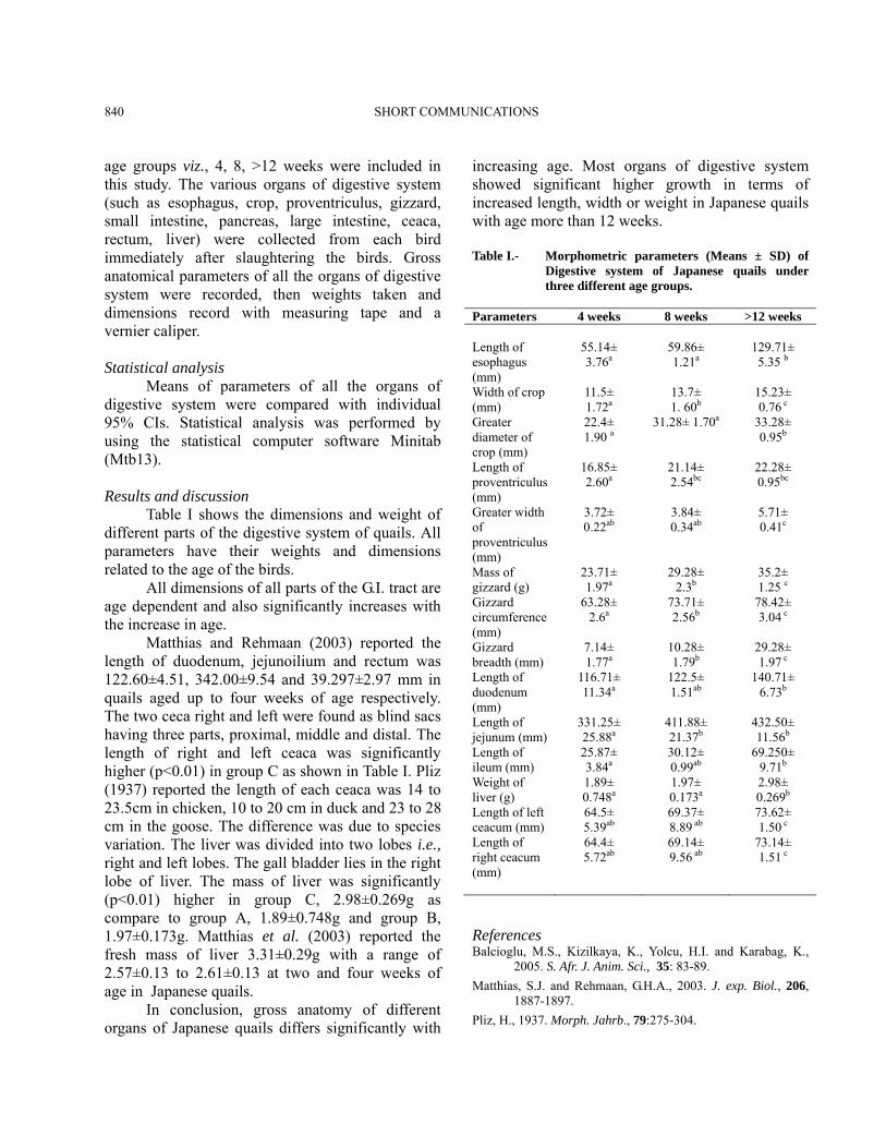
age groups viz., 4, 8, >12 weeks were included in this study. The various organs of digestive system (such as esophagus, crop, proventriculus, gizzard, small intestine, pancreas, large intestine, ceaca, rectum, liver) were collected from each bird immediately after slaughtering the birds. Gross anatomical parameters of all the organs of digestive system were recorded, then weights taken and dimensions record with measuring tape and a vernier caliper. Statistical analysis Means of parameters of all the organs of digestive system were compared with individual 95% CIs. Statistical analysis was performed by using the statistical computer software Minitab (Mtb13). Results and discussion Table I shows the dimensions and weight of different parts of the digestive system of quails. All parameters have their weights and dimensions related to the age of the birds. All dimensions of all parts of the G.I. tract are age dependent and also significantly increases with the increase in age. Matthias and Rehmaan (2003) reported the length of duodenum, jejunoilium and rectum was 122.60±4.51, 342.00±9.54 and 39.297±2.97 mm in quails aged up to four weeks of age respectively. The two ceca right and left were found as blind sacs having three parts, proximal, middle and distal. The length of right and left ceaca was significantly higher (p<0.01) in group C as shown in Table I. Pliz (1937) reported the length of each ceaca was 14 to 23.5cm in chicken, 10 to 20 cm in duck and 23 to 28 cm in the goose. The difference was due to species variation. The liver was divided into two lobes i.e., right and left lobes. The gall bladder lies in the right lobe of liver. The mass of liver was significantly (p<0.01) higher in group C, 2.98±0.269g as compare to group A, 1.89±0.748g and group B, 1.97±0.173g. Matthias et al. (2003) reported the fresh mass of liver 3.31±0.29g with a range of 2.57±0.13 to 2.61±0.13 at two and four weeks of age in Japanese quails. In conclusion, gross anatomy of different organs of Japanese quails differs significantly with
increasing age. Most organs of digestive system showed significant higher growth in terms of increased length, width or weight in Japanese quails with age more than 12 weeks. Table I.- Morphometric parameters (Means ± SD) of
Digestive system of Japanese quails under three different age groups.
Parameters 4 weeks 8 weeks >12 weeks Length of esophagus (mm)
55.14± 3.76a
59.86± 1.21a
129.71± 5.35 b
Width of crop (mm)
11.5± 1.72a
13.7± 1. 60b
15.23± 0.76 c
Greater diameter of crop (mm)
22.4± 1.90 a
31.28± 1.70a 33.28± 0.95b
Length of proventriculus (mm)
16.85± 2.60a
21.14± 2.54bc
22.28± 0.95bc
Greater width of proventriculus (mm)
3.72± 0.22ab
3.84± 0.34ab
5.71± 0.41c
Mass of gizzard (g)
23.71± 1.97a
29.28± 2.3b
35.2± 1.25 c
Gizzard circumference (mm)
63.28± 2.6a
73.71± 2.56b
78.42± 3.04 c
Gizzard breadth (mm)
7.14± 1.77a
10.28± 1.79b
29.28± 1.97 c
Length of duodenum (mm)
116.71± 11.34a
122.5± 1.51ab
140.71± 6.73b
Length of jejunum (mm)
331.25± 25.88a
411.88± 21.37b
432.50± 11.56b
Length of ileum (mm)
25.87± 3.84a
30.12± 0.99ab
69.250± 9.71b
Weight of liver (g)
1.89± 0.748a
1.97± 0.173a
2.98± 0.269b
Length of left ceacum (mm)
64.5± 5.39ab
69.37± 8.89 ab
73.62± 1.50 c
Length of right ceacum (mm)
64.4± 5.72ab
69.14± 9.56 ab
73.14± 1.51 c
References Balcioglu, M.S., Kizilkaya, K., Yolcu, H.I. and Karabag, K.,
2005. S. Afr. J. Anim. Sci., 35: 83-89. Matthias, S.J. and Rehmaan, G.H.A., 2003. J. exp. Biol., 206,
1887-1897. Pliz, H., 1937. Morph. Jahrb., 79:275-304.

SHORT COMMUNICATIONS 841
Qureshi, M.S., 1996. Batair farming (Urdu). Department of Livestock and Dairy Development, Lahore, pp. 8-9.
Wilson, W.O., Abbott, U.K. and Abplanalp, H., 1961. Poult. Sci., 40: 651-657.
(Received 14 December 2009, revised 25 January 2010)
Pakistan J. Zool., vol. 42(6), pp. 841-843, 2010. Epidemic of Dengue Hemorrhagic Fever During 2008 in Lahore, Pakistan Muhammad Khurram Shahzad*, Tayyaba Ijaz, Muhammad Younus, Muhammad Athar Khan, Nasir Mahmood, Zahida Fatima and Sibtain Ahmad Department of Pathology (MKS, MY), Department of Epidemiology and Public Health (MAK, ZF) and Department of Physiology and Biochemistry (SA), University of Veterinary and Animal Sciences, Lahore, Research Cell, Mayo Hospital, Lahore (TI), and School of Biological Sciences, University of the Punjab, Lahore (NM)
Abstract.- Dengue haemorrhagic fever (DHF), an acute arthropod-borne viral disease, is worldwide in distribution and caused by four antigenically distinct serotypes of Dengue virus. The disease appeared in Karachi, Pakistan in 1994 and with uninterrupted epidemics in Lahore during 2006, 2007 and 2008. In the fulminating and massive epidemic of 2008 more than 1200 suspected patients were admitted in a referral public hospital along with more than 300 patients admitted in other hospitals of Lahore. This epidemic investigation comprises 903 confirmed cases of DHFwith IgG and IgM capture ELISA by adopting the standard protocols. Among the confirmed patients 60% were males and 40% were females. Key words: Cross protection, Dengue haemorrhage fever, Aedes aegypti.
Dengue haemorrhagic fever (DHF) is an acute febrile disease caused by any of the four serotypes of dengue virus (DENV 1, DENV 2, DENV 3 and DENV 4) belonging to flaviviruses. It is a public health ____________________________ * Corresponding author: [email protected]
problem of growing importance in areas where the insect vector, Aedes aegypti mosquitoes are present (Rigau-Perez et al., 1998). Although most DENV infections are characterized as mild illnesses with low mortality, many countries report increasing incidence with high mortality (Gubler and Clark, 1995). Immunity against any of the four serotypes does not provide cross protection against each other. In the absence of an effective vaccine, the primary method of prevention is only possible through vector surveillance and its control. All ages and both sexes are susceptible to the DENV infection. Several DENV epidemics or pandemics have been reported world wide. In Pakistan 4 of 30 sera randomly collected in the late sixties were found positive to neutralization antibodies against DENV 1 from Rawalpindi and Peshawar cities (Burney, 1966). Studies to test for antibodies to flaviviruses were done among residents of Chiniot and Changa Manga National Forest areas of Punjab Province between 1968 and 1978 in which 124 had hemagglutination inhibition (HI) antibodies against DENV 3. Between 1983 and 1985, HI antibodies against dengue virus were reported from the sera of outpatients and healthy volunteer controls in a study at a government hospital in Karachi. In 1994, there were confirmed reports of DHF attributed to unusually heavy rainfall (Igarashi et al., 1994). During the same epidemic, acute phase sera from 16 patients in one hospital were tested by IgM capture ELISA. The results showed that 15 patients had IgM DENV 2 (Chan et al., 1995). Laboratory based surveillance was initiated to investigate the epidemic to asses the impact on population and to determine the secular trends of the dengue fever epidemic in Lahore, Pakistan. Materials and methods The study was conducted in the Microbiology Diagnostic and Research Lab for DHF diagnosis Mayo hospital, Lahore. Blood samples of clinically suspected and admitted patients and/or referred from private practitioners and family physicians were tested by using IgM and IgG capture ELISA from Nova Tec Immunodiagnostica GmbH, Dietzenbach, Germany. The epidemiological parameters like incidence rate, bar graphs and time-series graphs were made by using SPSS software (Version 16). Results The blood samples from patients suspected to
SHORT COMMUNICATIONS 842
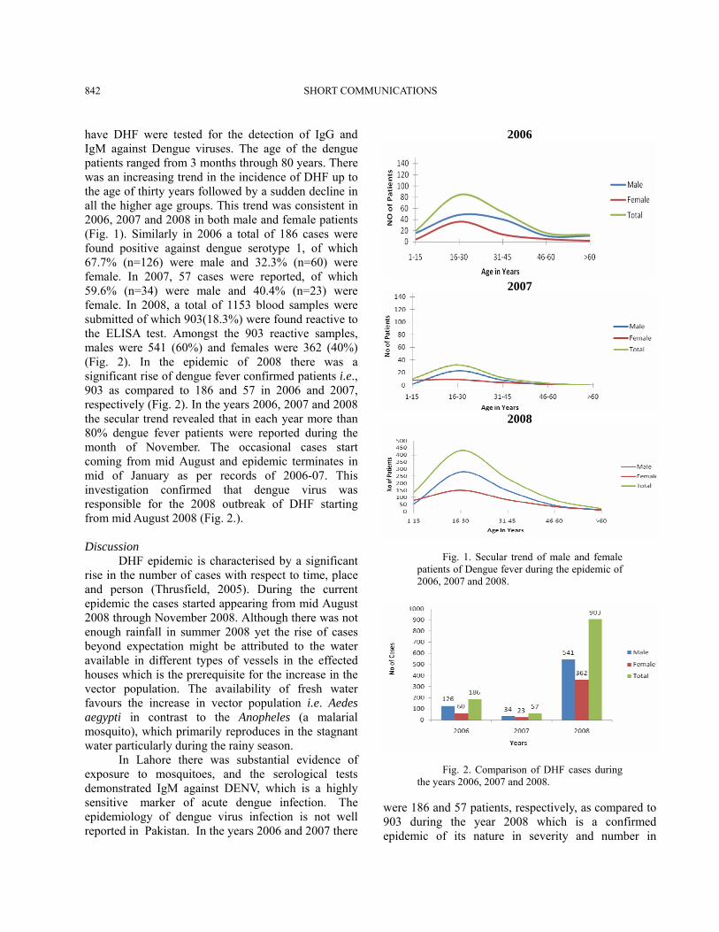
have DHF were tested for the detection of IgG and IgM against Dengue viruses. The age of the dengue patients ranged from 3 months through 80 years. There was an increasing trend in the incidence of DHF up to the age of thirty years followed by a sudden decline in all the higher age groups. This trend was consistent in 2006, 2007 and 2008 in both male and female patients (Fig. 1). Similarly in 2006 a total of 186 cases were found positive against dengue serotype 1, of which 67.7% (n=126) were male and 32.3% (n=60) were female. In 2007, 57 cases were reported, of which 59.6% (n=34) were male and 40.4% (n=23) were female. In 2008, a total of 1153 blood samples were submitted of which 903(18.3%) were found reactive to the ELISA test. Amongst the 903 reactive samples, males were 541 (60%) and females were 362 (40%) (Fig. 2). In the epidemic of 2008 there was a significant rise of dengue fever confirmed patients i.e., 903 as compared to 186 and 57 in 2006 and 2007, respectively (Fig. 2). In the years 2006, 2007 and 2008 the secular trend revealed that in each year more than 80% dengue fever patients were reported during the month of November. The occasional cases start coming from mid August and epidemic terminates in mid of January as per records of 2006-07. This investigation confirmed that dengue virus was responsible for the 2008 outbreak of DHF starting from mid August 2008 (Fig. 2.). Discussion DHF epidemic is characterised by a significant rise in the number of cases with respect to time, place and person (Thrusfield, 2005). During the current epidemic the cases started appearing from mid August 2008 through November 2008. Although there was not enough rainfall in summer 2008 yet the rise of cases beyond expectation might be attributed to the water available in different types of vessels in the effected houses which is the prerequisite for the increase in the vector population. The availability of fresh water favours the increase in vector population i.e. Aedes aegypti in contrast to the Anopheles (a malarial mosquito), which primarily reproduces in the stagnant water particularly during the rainy season. In Lahore there was substantial evidence of exposure to mosquitoes, and the serological tests demonstrated IgM against DENV, which is a highly sensitive marker of acute dengue infection. The epidemiology of dengue virus infection is not well reported in Pakistan. In the years 2006 and 2007 there
2006
2007
2008
Fig. 1. Secular trend of male and female patients of Dengue fever during the epidemic of 2006, 2007 and 2008.
Fig. 2. Comparison of DHF cases during the years 2006, 2007 and 2008.
were 186 and 57 patients, respectively, as compared to 903 during the year 2008 which is a confirmed epidemic of its nature in severity and number in

SHORT COMMUNICATIONS 843
Lahore. As this data is derived from the referral laboratory of a public hospital the total cases in all the other Hospitals (public and private) of Lahore city were reported to be more than 3000 (Shahzad et. al., 2007, 2008). The epidemic since 1994 and 1995 clearly document the presence of dengue viral infections in Pakistan, most probably due to increased rainfall in Karachi that resulted in an increase in mosquito population. No information on the mosquito species and/or other vector/ reservoir involved at that time is available. Later on the most common vector of DHF was found to be Aedes aegypti. The elevation of IgM ELISA titers to DENV 1 and DENV 2 serotypes suggests that viruses in this outbreak are of two different types. Dengue is endemic in the neighbouring countries like India and Sri Lanka, and the presence of more than one dengue serotype of DHF is migrating outside Pakistan epidemic DHF is also increasingly reported (Anonymous, 1985; Kabra et al., 1992; Vitarana and Jayasekera, 1990; Halstead, 1990). In 1996, an epidemic of DHF in India resulted in at least 227 deaths and more than 4700 persons being admitted in Delhi government hospitals. Most previous episodes of DHF in India have also been reported (Anonymous, 1996; Halstead, 1992; Mudur, 1996). In tropical and subtropical Asia where dengue is reported, changing lifestyles, urbanization, explosive population growth, destruction of city water supplies, migration, and increased air travel are some of the reasons cited for increase in the prevalence of dengue infections. As there is no vaccine available against this disease and in the absence of sentinel surveillance efforts to decrease breeding places of Aedes aegypti, community participation in capping, cleaning, or emptying water containers and eliminating hidden fresh water in the storage tanks and addition of larvicidal agents to stored water in the containers at house may control the dengue fever right in the
beginning of an outbreak. References Anonymous, 1985. Arthropod-borne and rodent-borne viral
diseases. WHO report, Geneva: Anonymous, 1996. Nature, 383: 654. Burney, M.I., 1966. Pakistan J. med. Res., 5:215–225. Chan, Y.C., Salahuddin, N.I. and Khan, J., 1995. Trans. R. Soc.
trop. Med. Hyg., 89: 619-620. Gubler, D.J., 1993. Dengue and dengue hemorrhagic fever in
the Americas. Monograph on dengue/dengue hemorrhagic fever. WHO: Regional Office for South East Asia, New Delhi. Regional Publication, SEAR 22: 9-22.
Gubler D. J. and Clark, G.G., 1995. Emerg. Infect. Dis., 1: 55-57 Halstead, S.B., 1990. Southeast Asian J. trop. Med. Publ. Hlth.,
21: 636-641. Halstead, S.B., 1992. Wld. Hlth. Stat. Q., 45: 292-298. Igarashi, A., Tanaka, M. and Morita, K., 1994. Microbial
Immunol., 38: 827-830. Kabra, S.K., Verma, I.C., Arora, N.K., Jain, Y. and Kalra, V.,
1992. Wld. Hlth. Org. Bull., 70: 105-108. Mudur, G., 1996. Br. med. J., 313: 1034. Rigau-Perez, J.G., Clark, G.G., Gubler, D.J., Reiter, P., Sanders,
E.J. and Vorndam, A.V., 1998. Lancet, 352(9132): 971-977.
Shahzad, M.K., Ijaz, T., Ijaz, S. and Younus, M., 2007. Int. J. Agro Vet. Med. Sci., 1: 13-16
Shahzad, M. K., Ijaz, T. and Ijaz, S., 2008. Int. J. Agro Vet. Med. Sci., 2: 36
Thrusfield, M., 2005. Veterinary epidemiology; 3rd ed. Blackwell Science Ltd, UK.
Vitarana, T. and Jayasekera, N., 1990. Southeast Asian J. trop. Med. Publ. Hlth., 21: 682
(Received 19 June 2009, revised 23 July 2009)