Embed Size (px)
Citation preview
NEURO-ONCOLOGY
College voor Oncologie Nationale Richtlijnen
V1.2008 © 2008 College of Oncology
College voor Oncologie Nationale Richtlijnen
College voor Oncologie Nationale Richtlijnen
College voor Oncologie Nationale Richtlijnen
RReeccttuumm CCaanncceerr
VVeerrssiioonn 11..22000044
NNeeuurroo--OOnnccoollooggyy
VVeerrssiioonn 11..22000088
Continue
CCOOLLLLEEGGEE OOFF OONNCCOOLLOOGGYY
NNaattiioonnaall CClliinniiccaall PPrraaccttiiccee GGuuiiddeelliinneess
NEURO-ONCOLOGY
College voor Oncologie Nationale Richtlijnen
V1.2008 © 2008 College of Oncology
College voor Oncologie Nationale Richtlijnen
College voor Oncologie Nationale Richtlijnen
College voor Oncologie Nationale Richtlijnen
College of Oncology National Guidelines
Expert panel
Neuro-Oncology Guidelines Expert Panel Prof. dr. Jacques De Grève Coordinator National Neuro-Oncology Guidelines Chairman Working Party Manuals, College of Oncology Universitair Ziekenhuis Brussel
Prof. dr. Bart Neyns Co-Coordinator National Neuro-Oncology Guidelines Universitair Ziekenhuis Brussel
Prof. dr.Tom Boterberg Ghent University Hospital
Dr. Daniël Devriendt Jules Bordet Institute Brussels
Dr. Lionel D'Hondt Grand Hôpital de Charleroi
Prof. dr. Serge Goldman Cliniques Universitaires de Bruxelles -
Hôpital Erasme Dr. Thierry Gustin Cliniques Universitaires UCL de Mont Godinne
Prof. dr. Alex Michotte Universitair Ziekenhuis Brussel
Prof. dr. Paul Parizel Universitair Ziekenhuis Antwerpen
Prof. dr. Frank Van Calenbergh Universitaire Ziekenhuizen Leuven
Dr. Margareta Haelterman Federal Public Service Health, Food Chain Safety and Environment
Prof. dr. Simon Van Belle Chairman College of Oncology University Hospital Ghent
Continue
1
NEURO-ONCOLOGY College voor Oncologie Nationale Richtlijnen
College voor Oncologie Nationale Richtlijnen
College voor Oncologie Nationale Richtlijnen
College voor Oncologie Nationale Richtlijnen
College of Oncology National Guidelines
External reviewers
External reviewers Prof. dr. Paul Clement
Belgian Society of Medical Oncology
Dr. Dirk Van den Berge
Belgische Vereniging voor Radiotherapie-Oncologie - Association Belge de Radiothérapie-Oncologie
Prof. dr. Jean-François Baurain Dr. Micheline Mouchamps
Belgian Association for Neuro-Oncology
Conflict of interest: All authors and external reviewers are employed in centres specialized in the treatment of brain tumors. One member of the expert panel received fees or compensation for writing a publication or participate in its development, received a scholarship, fees or other funds or compensation to perform research, received payments to speak or participate in a conference, training fees or travel support and did consultancy for an organization that can win or lose financially by the results of this report. Another member of the expert panel received a scholarship, fees or other funds or compensation to perform research and received payments to speak or participate in a conference, training fees or travel support. Two external reviewers received payments to speak or participate in a conference, training fees or travel support. Another external reviwer did consultancy for an organization that can win or lose financially by the results of this report. Other conflicts of interest were not communicated.
Continue
2 V1.2008 © 2008 College of Oncology
NEURO-ONCOLOGY
College voor Oncologie Nationale Richtlijnen
V1.2008 © 2008 College of Oncology
College voor Oncologie Nationale Richtlijnen
College voor Oncologie Nationale Richtlijnen
College voor Oncologie Nationale Richtlijnen
Table 2: WHO-classification
College of Oncology National Guidelines
Table of contents
• Neuro-oncology guidelines expert panel
• External reviewers
• National guidelines neuro-oncology (Full text) • Introduction • Search for evidence • General treatment option overview • Brain stem gliomas • Pilocytic astrocytomas • ioma, WHO grade II low-grade oligodendrogl
mixed glioma and diffuse astrocytoma • endroglioma, WHO grade III anaplastic oligod
mixed glioma and astrocytoma • WHO grade IV glioma (glioblastoma) • Ependymal Tumours • Gliomatosis cerebri
• Pineal parenchymal tumours • Primitive neuroectodermal tumour • Pituitary adenomas • Craniopharyngiomas • Germ cell tumours • Meningeal tumours • Hemangioblastomas • Malignant Peripheral Nerve Sheath Tumours • Chordomas and chondrosarcomas • Leptomeningeal carcinomatosis • Brain metastases • Supportive care
• Glossary
• idence Table 1: NCI-PDQ Levels of Ev
•
3
NEURO-ONCOLOGY College voor Oncologie
Nationale Richtlijnen College voor Oncologie
Nationale Richtlijnen College voor Oncologie
Nationale Richtlijnen College voor Oncologie
Nationale Richtlijnen
College of Oncology National Guidelines
Full Text
Table of contents
National Guidelines Neuro-Oncology
INTRODUCTION This document provides an overview of the clinical practice guidelines for neuro-oncology. The guidelines are developed by a panel of experts (see 'expert panel') comprising clinicians of different specialties and were reviewed by relevant professional associations (see 'external reviewers'). The guidelines are based on the best evidence available at the time they are derived. The aim of these guidelines is to assist all care providers involved in the care of people with a brain tumour. To be completed: statement that these guidelines comprise only adults
SEARCH FOR EVIDENCE Clinical practice guidelines
Sources A broad search of electronic databases (Medline), specific guideline websites (NCI-PDQ, Cochrane) and websites of oncology organisations (NCCN) was conducted.
In- and exclusion criteria Both national and international clinical practice guidelines (CPGs) on brain tumours were searched. A language (English, Dutch, French) restriction was used. Relevant literature until april 2008 was included. CPGs without references were excluded, as were CPGs without clear recommendations.
Additional evidence For each clinical question, the evidence – identified through the included CPGs – was updated by searching Medline and the Cochrane Database of Systematic Reviews from the search date of the CPG on.
Level of evidence A level of evidence was assigned to each recommendation using the NCI-PDQ levels of evidence (Table 1). All statements regarding treatment recommendations are level 3 unless otherwise specified.
4 V1.2008 © 2008 College of Oncology
NEURO-ONCOLOGY
College of Oncology National Guidelines
Full Text
Table of contents
GENERAL CONSIDERATIONS FOR DIAGNOSIS AND TREATMENT OF PRIMARY BRAIN TUMOURS The treatment of brain tumours should be carried out in dedicated centres with experienced and specialized multidisciplinary teams.
Standard diagnostic imaging consists of magnetic resonance (MR)-imaging (T1 +/- gadolinium and T2 sequences with axial, coronal & sagittal slices) except for patients with implanted stimulating devices in which contrast enhanced CT should be used. Additional information obtained by perfusion/diffusion weighted MR-imaging, MR-Spectroscopy and PET-imaging (e.g. C11-methionine or FDG-PET) can be helpful in the differential diagnosis with non-malignant lesions, the evaluation of the metabolic activity or better delineation of anatomical extent in selected cases. Functional MR imaging can be useful preoperatively for lesions near functional areas. Surgical removal is recommended for most types of brain tumours and in most locations. The removal should be as complete as possible within the constraints of preservation of neurological function (= maximal safe resection). For patients with primary brain tumours, the primary goals of surgery include (1) establishing a histological diagnosis, (2) reducing intracranial pressure, (3) cytoreduction. Exceptions to this rule are brainstem tumours, which are diagnosed on imaging and clinical evidence and treated without initial surgery in approximately half of the cases (because of the risks that are associated with the surgical intervention in this anatomical region). In most deep-seated lesions as in tumours located in functional areas, a diagnostic biopsy is indicated, using a stereotactic frame or by neuronavigation-guided craniotomy.
A conclusive pathological diagnosis should be obtained in most cases of primary brain tumours before primary treatment is initiated. All tissue biopsies (gross removal and stereotactic samples) should be considered for complete diagnostic work-up (neuropathological, immunohistochemical and molecular-genetic characterization). Cryopreservation of fresh tumour tissue should be strongly recommended to allow molecular-genetic characterisation that might gain value in the near future. All primary brain tumours should be classified according to the WHO classification (Table 2). The most important prognostic factors at diagnosis for patients with primary brain tumours are (1) the histology and differentiation grade, (2) the performance status following recovery from surgery and (3) age at diagnosis. Patients with a poor performance status (WHO-PS>2, KPS <60) following maximal safe resection and optimal medical treatment (corticosteroids, anti-convulsants, …) have a poor prognosis and are in general ineligible for prospective study protocols. No treatment has demonstrated to improve the survival for such patients as opposed to best supportive care. Treatment decisions in such patients need to be made on an individual basis (MOC and discussion with the patient and his/her relatives). Radiation therapy and chemotherapy are the two most often used treatment modalities for brain tumour patients who cannot be controlled with surgery alone. Their activity varies according to the anatomopathological and molecular-genetic characteristics of the brain tumour. In the absence of a curative treatment option many patients who are diagnosed with a primary brain tumour should in the first place be considered as candidates for participation in a clinical trial. Novel biologic therapies under clinical evaluation for patients with brain tumours include inhibitors of growth factor receptors, agents with anti-angiogenesis activity, viral-based gene therapy, oncolytic viruses, and dendritic cell vaccination.
V1.2008 © 2008 College of Oncology
5
NEURO-ONCOLOGY
College of Oncology National Guidelines
Full Text
Table of contents
Supportive treatment. Corticosteroids (methylprednisolone, dexamethasone), are used to treat the peritumoural brain edema. Mannitol and diuretics are useful to control intracranial hypertension intraoperatively. The use of anticonvulsants is mandatory for patients undergoing surgery for supratentorial gliomas and presenting with seizures. In case of preventive use, anticonvulsivants should be discontinued one week after surgery, except in cases demonstrated at risk for postoperative seizures. Many patients with primary brain tumours are dependent on substantial psycho-social support and should be directed towards existing psychosocial services and supportive/palliative teams (including a nutritionist and psychologist).
V1.2008 © 2008 College of Oncology
6
NEURO-ONCOLOGY
College of Oncology National Guidelines
Full Text
Table of contents
BRAIN STEM GLIOMAS Intrinsic brain stem gliomas can be divided into three groups: (1) diffuse pontine gliomas that have a poor prognosis correlated with histology (if biopsies are performed), location, and extent of tumour, (2) exophytic, well delineated gliomas of the cervicomedullary region and (3) tectal plate gliomas, esp. in children, that have a very indolent course.
These patients are appropriate candidates for clinical trials. Information about clinical trials open for inclusion of patients can be found at the website of the U.S. National Cancer Institute (http://www.nci.nih.gov/clinicaltrials ).
Treatment options • Radiotherapy is the standard treatment for diffuse pontine gliomas
(Level of evidence: 2) [1-9]. There is no role for hyperfractionated radiotherapy (Level of evidence: 1) [1-3].
• Surgery followed by radiotherapy is the standard treatment for exophytic brain stem gliomas (Level of evidence: 2) [1-3].
• Treatment of hydrocephalus and follow-up is the standard treatment for tectal plate gliomas [10].
• No standard of care has been established at recurrence following initial radiotherapy. Chemotherapy can be considered at recurrence following radiotherapy [11-12].
References 1 Freeman CR, Farmer JP. Pediatric brain stem gliomas: A review. Int J
Radiat Oncol Biol Phys 40: 265-71, 1998.
2 Mandell LR, Kadota R, Freeman C, et al. There is no role for hyperfractionated radiotherapy in the management of children with newly diagnosed diffuse intrinsic brainstem tumors: results of a pediatric oncology group phase III trial comparing conventional vs hyperfractionated radiotherapy. Int J Radiat Oncol Biol Phys 43(5): 959-64, 1999.
3 Freeman CR, Suissa S. Brain stem tumors in children: Results of a survey of 62 patients treated with radiotherapy. Int J Radiat Oncol Biol Phys 12: 1823-28, 1986.
4 Greenberger JS, Cassady JR, Levene MB. Radiation therapy of thalamic, midbrain and brain stem gliomas. Radiology 122(2): 463-8, 1977.
5 Levin VA, Edwards MS, Wara WM, et al. 5-Fluorouracil and 1-(2-chloroethyl)-3-cyclohexyl-1-nitrosourea (CCNU) followed by hydroxyurea, misonidazole, and irradiation for brain stem gliomas: a pilot study of the Brain Tumor Research Center and the Childrens Cancer Group. Neurosurgery 14 (6): 679-81, 1984.
6 Allen JC, Bloom J, Ertel I, et al. Brain tumors in children: current cooperative and institutional chemotherapy trials in newly diagnosed and recurrent disease. Semin Oncol 13 (1): 110-22, 1986.
7 Eifel PJ, Cassady JR, Belli JA. Radiation therapy of tumors of the brainstem and midbrain in children: experience of the Joint Center for Radiation Therapy and Children's Hospital Medical Center (1971-1981). Int J Radiat Oncol Biol Phys 13(6): 847-52, 1987.
8 Shrieve DC, Wara WM, Edwards MS, et al. Hyperfractionated radiation therapy for gliomas of the brainstem in children and in adults. Int J Radiat Oncol Biol Phys 24(4): 599-610, 1992.
9 Surma-aho O, Niemelä M, Vilkki J, et al. Adverse long-term effects of brain radiotherapy in adult low-grade glioma patients. Neurology 56(10): 1285-90, 2001.
V1.2008 © 2008 College of Oncology
7
NEURO-ONCOLOGY
College of Oncology National Guidelines
Full Text
Table of contents
10 Javadpour M, Malluci C. The role of neuroendoscopy in the management of tectal gliomas. Childs Nerv Syst 20: 852-7, 2004.
11 Fulton DS, Levin VA, Wara WM, et al. Chemotherapy of pediatric brain-stem tumors. J Neurosurg 54(6): 721-5, 1981.
12 Rodriguez LA, Prados M, Fulton D, et al. Treatment of recurrent brain stem gliomas and other central nervous system tumors with 5-fluorouracil, CCNU, hydroxyurea, and 6-mercaptopurine. Neurosurgery 22(4): 691-3, 1988.
V1.2008 © 2008 College of Oncology
8
NEURO-ONCOLOGY
College of Oncology National Guidelines
Full Text
Table of contents
PILOCYTIC ASTROCYTOMAS Most cases of pilocytic astrocytomas occur in paediatric patients which is not the scope of these guidelines. Neurofibromatosis 1 predisposes to the development of central nervous system tumours. Histopathologically, most of these tumours are pilocytic astrocytomas [1,2]. They occur in children (and young adults) and are predominantly located in the optic pathways or in the brainstem.
Treatment options • If the tumour is totally resectable, surgery alone is the standard
treatment (Level of evidence: 2) [3]. • If the tumour is not resectable, adjuvant radiotherapy should be
discussed in a multidisciplinary meeting (Level of evidence: 2) [4]. • Primary stereotactic radiotherapy or radiosurgery in inoperable patients
is an alternative option [5]. • At recurrence, patients should be considered for reoperation and
radiation therapy if not previously given. Patients who have already received radiation therapy can be considered candidates for chemotherapy [6]. Treatment options at recurrence should be discussed in a multidisciplinary meeting. These patients are appropriate candidates for clinical trials. Information about clinical trials open for inclusion of patients can be found at the website of the U.S. National Cancer Institute (http://www.nci.nih.gov/clinicaltrials ).
References 1 Guillamo JS, Créange A, Kalifa C, Grill J, Rodriguez D, Doz F,
Barbarot S, Zerah M, Sanson M, Bastuji-Garin S, Wolkenstein P; Réseau NF France. Prognostic factors of CNS tumours in Neurofibromatosis 1 (NF1): a retrospective study of 104 patients. Brain 126(Pt 1):152-60, 2003.
2 Rodriguez FJ, Perry A, Gutmann DH, et al. Gliomas in neurofibromatosis type 1: a clinicopathologic study of 100 patients. J Neuropathol Exp Neurol. 67(3):240-9, 2008.
3 Wallner KE, Gonzales MF, Edwards MS, et al. Treatment results of juvenile pilocytic astrocytoma. J Neurosurg 69(2): 171-6, 1988.
4 Shaw EG, Daumas-Duport C, Scheithauer BW, et al. Radiation therapy in the management of low-grade supratentorial astrocytomas. J Neurosurg 70(6): 853-61, 1989.
5 Hadjipanayis CG, Kondziolka D, Gardner P, et al. Stereotactic radiosurgery for pilocytic astrocytomas when multimodal therapy is necessary. J Neurosurg 97(1): 56-64, 2002.
6 Quinn Ja, Reardon Da, Friedman Ah, et al. Phase II trial of temozolomide in patients with progressive low-grade glioma. J Clin Oncol 21: 646-51, 2003.
V1.2008 © 2008 College of Oncology
9
NEURO-ONCOLOGY
College of Oncology National Guidelines
Full Text
Table of contents
WHO GRADE II LOW GRADE OLIGODENDROGLIOMA, MIXED GLIOMA AND DIFFUSE ASTROCYTOMA The WHO grade II gliomas or low-grade gliomas have a worse prognosis than WHO grade I glioma. Overall survival is extremely variable and ranges between <1y to >15y. No randomized clinical trial has demonstrated to improve overall survival of these patients. These patients are appropriate candidates for clinical trials. Information about clinical trials open for inclusion of patients can be found at the website of the U.S. National Cancer Institute (http://www.nci.nih.gov/clinicaltrials ).Clinical, histopathological and molecular-genetical features determine the prognosis of patients with WHO grade II gliomas. An age of over 40 years at diagnosis, an astrocytoma histology, a largest diameter of the tumour of more than 6 cm, a tumour crossing the midline and the presence of a neurological deficit before surgery have been identified as negative prognostic clinical factors for survival in adult patients with WHO grade II gliomas. According to these clinical factors, patients can be classified within a high or low risk group with a significantly different prognosis [1]. According to the histopathological features WHO grade II gliomas are classified as oligodendroglioma, mixed glioma and astrocytoma. In addition WHO grade II low-grade gliomas can also be characterized by their molecular-genetic features. Gliomas with a chromosomal loss of 1p/19q (unbalanced translocation, [2]) have the best prognosis following treatment with radiation and are highly chemosensitive (although no randomized clinical trial today has proven chemotherapy to influence survival). The prognosis following surgery-only might not be different according to the 1p and 19q status [3]. Loss of 1p/19q is associated with
oligodendroglial histology and mixed glioma. Taken into account the high inter-observer differences in histopathological classification of WHO grade II gliomas, it is recommended to characterize the 1p/19q status of all WHO grade II gliomas. Metabolic activity of WHO grade II low-grade gliomas, evaluated by PET imaging, might have additional prognostic value and might help in the early detection of anaplastic transformation [4-7].
Treatment options • Surgery at diagnosis
• Patients who are suspected to have a WHO grade II glioma on MRI should be considered for surgery in order to establish the anatomopathological diagnosis and determine the molecular-genetic characteristics of the glioma.
• A maximal safe resection should be aimed for if possible [8]. • If the tumour is not completely resectable, a stereotactic biopsy is
preferred: preferably with metabolic or functional guidance to ensure proper tumour sampling [6,7].
• Post-surgical treatment options • At present considerable controversy exists with regards to treatment
of patients with WHO grade II gliomas following surgery. No randomized clinical trial has demonstrated to improve overall survival of these patients. As such these patients should be considered candidates for treatment within the context of a clinical trial. Post-operative treatment should be discussed at a multidisciplinary meeting.
• In general, the following guidelines can be followed:
V1.2008 © 2008 College of Oncology
10
NEURO-ONCOLOGY
College of Oncology National Guidelines
Full Text
Table of contents
o Low risk, asymptomatic patients (= with the best clinical and molecular genetic prognostic factors) or patients who have undergone a complete resection can be followed-up by MRI every 3-6 months. Tumour growth should be evaluated on sequential scans and against the baseline measurements and by functional /molecular imaging [expert opinion].
o For high risk patients and for low-risk patients without complete resection and persistent glioma related symptoms: surgery should be followed by radiotherapy. In a study of 300 patients who had surgery were randomized to either radiation therapy or watch and wait. There was no difference in overall survival (OS) in the two groups (Level of evidence: 1ii). Median progression-free survival was 5.3 years in the radiation therapy group and 3.4 years in the control group (Level of evidence 1ii). The recommended radiotherapy dose is 54 Gy [9-11].
o Two phase III trials investigating respectively 45 Gy in 5 weeks or 59.4 Gy in 6.6 weeks could not demonstrate a benefit for the higher doses of RT (Level of evidence: 1ii) [10,12].
o Symptomatic patients with chemosensitive gliomas (=1p loss) and a large inoperable tumour can be considered candidates for 6 to 12 months of upfront chemotherapy. Chemotherapy with temozolomide or PCV has activity in patients with low-grade oligodendrogliomas [13,14].
• Treatment options at recurrence will depend on the previously administered treatment. Patients should be considered for surgical resection, chemotherapy or radiotherapy and for participation in clinical trials [13,15].
References 1 Pignatti F, van den Bent M, Curran D, et al. Prognostic factors for
survival in adult patients with cerebral low-grade glioma. J Clin Oncol. 20(8): 2076-84, 2002.
2 Jenkins RB, Blair H, Ballman KV, et al. A t(1;19)(q10;p10) mediates the combined deletions of 1p and 19q and predicts a better prognosis of patients with oligodendroglioma. Cancer Res 66(20): 9852-61, 2006.
3 Weller M, Berger H, Hartmann C, et al. Combined 1p/19q loss in oligodendroglial tumours: predictive or prognostic biomarker? Clin Cancer Res 13: 6933-7, 2007.
4 Floeth FW, Pauleit D, Sabel M, et al. Prognostic value of O-(2-18F-fluoroethyl)-L-tyrosine PET and MRI in low-grade glioma. J Nucl Med 48: 519-27. 2007.
5 De Witte O, Goldberg I, Wikler D, et al. Positron emission tomography with injection of methionine as a prognostic factor in glioma. J Neurosurg 95: 746-50. 2001.
6 Levivier M, Wikler D, Jr., Massager N, et al. The integration of metabolic imaging in stereotactic procedures including radiosurgery: a review. J Neurosurg 97: 542-50. 2002.
7 Pirotte B, Goldman S, Massager N, et al. Comparison of 18F-FDG and 11C-methionine for PET-guided stereotactic brain biopsy of gliomas. J Nucl Med 45: 1293-8. 2004.
8 Smith Js, Chang Ef, Lamborn Kr, et al. Role of extent of resection in the long-term outcome of low-grade hemispheric gliomas. J Clin Oncol 26: 1338-45, 2008.
9 Karim Ab, Afra D, Cornu P, et al. Randomized trial on the efficacy of radiotherapy for cerebral low-grade glioma in the adult: European Organization for Research and Treatment of Cancer Study 22845 with
V1.2008 © 2008 College of Oncology
11
NEURO-ONCOLOGY
College of Oncology National Guidelines
Full Text
Table of contents
the Medical Research Council study BRO4: an interim analysis. Int J Radiat Oncol Biol Phys 52: 316-324, 2002.
10 Karim Ab, Maat B, Hatlevoll R, et al. A randomized trial on dose-response in radiation therapy of low-grade cerebral glioma: European Organization for Research and Treatment of Cancer (EORTC) Study 22844. Int J Radiat Oncol Biol Phys 36: 549-556, 1996.
11 van den Bent MJ, Afra D, de Witte O, et al. Long-term efficacy of early versus delayed radiotherapy for low-grade astrocytoma and oligodendroglioma in adults: the EORTC 22845 randomised trial. Lancet 366 (9490): 985-90, 2005.
12 Shaw EG, Daumas-Duport C, Scheithauer BW, et al. Radiation therapy in the management of low-grade supratentorial astrocytomas. J Neurosurg 70 (6): 853-61, 1989.
13 Hoang-Xuan K, Capelle L, Kujas M, et al. Temozolomide as initial treatment for adults with low-grade oligodendrogliomas or oligoastrocytomas and correlation with chromosome 1p deletions. J Clin Oncol 22 (15): 3133-8, 2004.
14 Stege EM, Kros JM, de Bruin HG, et al. Successful treatment of low-grade oligodendroglial tumors with a chemotherapy regimen of procarbazine, lomustine, and vincristine. Cancer 103 (4): 802-9, 2005.
15 Quinn Ja, Reardon Da, Friedman Ah, et al. Phase II trial of temozolomide in patients with progressive low-grade glioma. J Clin Oncol 21: 646-51, 2003.
V1.2008 © 2008 College of Oncology
12
NEURO-ONCOLOGY
College of Oncology National Guidelines
Full Text
Table of contents
WHO GRADE III OLIGODENDROGLIOMA, MIXED GLIOMA AND ASTROCYTOMA WHO grade III gliomas (also referred to as anaplastic gliomas) have a very low cure rate with available treatment options. Median overal survival of patients with WHO grade III glioma has been 3-4 years with available treatment options. Such patients are appropriate candidates for clinical trials. Information about clinical trials open for inclusion of patients can be found at the website of the U.S. National Cancer Institute (http://www.nci.nih.gov/clinicaltrials ).Clinical, histopathological and molecular-genetical features determine the prognosis of patients with WHO grade III gliomas. Age, astrocytoma histopathology, a larger diameter of the tumour, crossing of the midline and a poor performance status at diagnosis are negative prognostic clinical factors for survival in adult patients with WHO grade III gliomas. WHO grade III anaplastic gliomas can be classified according to their histopathological and molecular-genetic features. Gliomas with a chromosomal loss of chromosal arms 1p/19q (most often found in pure oligodendrogliomas and to a lesser extent mixed gliomas) have a better natural prognosis and are more chemosensitive (although no randomized clinical trial today has proven chemotherapy to influence survival). Loss of 1p/19 is associated with oligodendroglial histology but mixed WHO grade III glioma may also carry this deletion. Taking into account the high inter-observer differences in pathological classification of anaplastic gliomas, it is recommended that all WHO grade III oligodendroglioma and mixed gliomas be characterized for 1p/19q deletions [1,2]. Anaplastic astrocytomas with a mutated EGFR-gene (most often the EGFRvIII mutant) or amplification of the EGFR gene have a worse natural
prognosis that is comparable to the prognosis of patients with glioblastoma (WHO grade IV glioma) [3,4]. Taking into account the high inter-observer differences in pathological classification of anaplastic gliomas, it is recommended that all WHO grade III mixed gliomas and astrocytoma be characterized for EGFR mutation or amplification. Patients with WHO grade III glioma with an EGFR mutation or amplification can be considered for treatment according to the guidelines for glioblastoma (expert opinion). Treatment options • Surgery at diagnosis
• Patients who are suspect to have WHO grade III glioma on MRI should be considered for surgery in order to establish the anatomopathological diagnosis and determine the molecular-genetic characteristics of the glioma.
• A maximal safe resection should be aimed for if possible. • If the tumour is not resectable, a stereotactic biopsy is preferred.
• Post-surgical treatment options • Radiation therapy (60 Gy fractionated radiotherapy, 30 x 2Gy)
(Level of evidence: 2) [5]. • Two phase III trials demonstrated that patients with WHO grade III
oligodendroglioma or mixed glioma gain only limited benefit in time to progression and no significant benefit in OS from (neo)adjuvant PCV-based chemotherapy. Lack of benefit was observed irrespective of the 1p/19q status (Level of evidence: 2) [6-8].
• Treatment options at recurrence • Chemotherapy. Both temozolomide and nitrosurea based regimens
have activity. Nitrosurea based regimens are more toxic as compared to temozolomide (Level of evidence: 2) [9-12].
V1.2008 © 2008 College of Oncology
13
NEURO-ONCOLOGY
College of Oncology National Guidelines
Full Text
Table of contents
• Selected patients with a focal recurrence might benefit from a second resection and/or course of radiation therapy. The therapeutic plan in such patients should be discussed at a MOC.
Remarks • An EORTC phase III study (CATNON trial) that investigates the
combined radiotherapy/temozolomide and or adjuvant temozolomide in patients with anaplastic glioma without 1p/19q deletion is ongoing.
References 1 Van Den Bent Mj. Anaplastic oligodendroglioma and
oligoastrocytoma. Neurol Clin 25: 1089-109, ix-x, 2007. 2 Van Den Bent Mj and Kros Jm: Predictive and prognostic markers in
neuro-oncology. J Neuropathol Exp Neurol 66: 1074-81, 2007. 3 Buckner Jc, Aldape Kd, Ballman K, et al. Immunohistochemical
detection of EGFRvIII and prognostic significance in patients with malignant glioma enrolled in NCCTG clinical trials. Journal of Clinical Oncology, 2004 ASCO Annual Meeting Proceedings (Post-Meeting Edition) 22: 1508, 2004.
4 Pelloski Ce, Ballman Kv, Furth Af, et al. Epidermal growth factor receptor variant III status defines clinically distinct subtypes of glioblastoma. J Clin Oncol 25: 2288-94, 2007.
5 Walker Md, Green Sb, Byar Dp, et al. Randomized comparisons of radiotherapy and nitrosoureas for the treatment of malignant glioma after surgery. N Engl J Med 303: 1323-9, 1980.
6 Cairncross G, Berkey B, Shaw E, et al. Phase III trial of chemotherapy plus radiotherapy compared with radiotherapy alone for pure and mixed anaplastic oligodendroglioma: Intergroup Radiation Therapy Oncology Group Trial 9402. J Clin Oncol 24: 2707-14, 2006.
7 Van Den Bent Mj, Carpentier Af, Brandes Aa, et al. Adjuvant procarbazine, lomustine, and vincristine improves progression-free survival but not overall survival in newly diagnosed anaplastic oligodendrogliomas and oligoastrocytomas: a randomized European Organisation for Research and Treatment of Cancer phase III trial. J Clin Oncol 24: 2715-22, 2006.
8 Quon H, Abdulkarim B. Adjuvant treatment of anaplastic oligodendrogliomas and oligoastrocytomas. Cochrane Database Syst Rev. Apr 16;(2):CD007104.
9 Van Den Bent Mj, Taphoorn Mj, Brandes Aa, et al. Phase II study of first-line chemotherapy with temozolomide in recurrent oligodendroglial tumors: the European Organization for Research and Treatment of Cancer Brain Tumor Group Study 26971. J Clin Oncol 21: 2525-8, 2003.
10 Van Den Bent Mj, Chinot O, Boogerd W, et al. Second-line chemotherapy with temozolomide in recurrent oligodendroglioma after PCV (procarbazine, lomustine and vincristine) chemotherapy: EORTC Brain Tumor Group phase II study 26972. Ann Oncol 14: 599-602, 2003.
11 Brandes Aa, Ermani M, Basso U, et al. Temozolomide as a second-line systemic regimen in recurrent high-grade glioma: a phase II study. Ann Oncol 12: 255-7, 2001.
12 Chinot Ol, Honore S, Dufour H, et al. Safety and efficacy of temozolomide in patients with recurrent anaplastic oligodendrogliomas after standard radiotherapy and chemotherapy. J Clin Oncol 19: 2449-55, 2001.
V1.2008 © 2008 College of Oncology
14
NEURO-ONCOLOGY
College of Oncology National Guidelines
Full Text
Table of contents
WHO GRADE IV GLIOMA (GLIOBLASTOMA) WHO GRADE IV GLIOMA (GLIOBLASTOMA) WHO grade IV gliomas (glioblastoma) are the most aggressive subtype of glioma. Glioblastoma patients have a very low cure rate with the available treatment options. Median overal survival of glioblastoma patients has been 8-14 months with available treatment options. Glioblastoma patients are appropriate candidates for clinical trials.
WHO grade IV gliomas (glioblastoma) are the most aggressive subtype of glioma. Glioblastoma patients have a very low cure rate with the available treatment options. Median overal survival of glioblastoma patients has been 8-14 months with available treatment options. Glioblastoma patients are appropriate candidates for clinical trials. In patients diagnosed with glioblastoma more extensive tumour resection, younger age, Mini-Mental State Examination (MMSE) score of 27 or higher, and no corticosteroid treatment following surgery are associated with better survival [1,2].
In patients diagnosed with glioblastoma more extensive tumour resection, younger age, Mini-Mental State Examination (MMSE) score of 27 or higher, and no corticosteroid treatment following surgery are associated with better survival [1,2]. Loss of 1p/19q is not associated with glioblastoma and testing is not recommended. Mutation/amplification of the EGFR gene is found in 20-40% of glioblastoma and has been correlated with prognosis [3]. At present EGFR status has no implementation in the clinical decision making outside a clinical trial.
Loss of 1p/19q is not associated with glioblastoma and testing is not recommended. Mutation/amplification of the EGFR gene is found in 20-40% of glioblastoma and has been correlated with prognosis [3]. At present EGFR status has no implementation in the clinical decision making outside a clinical trial.
Treatment options Treatment options • Surgery at diagnosis • Surgery at diagnosis
• Patients who are suspect to have gliobblastoma on MRI should be considered for surgery in order to establish the anatomopathological diagnosis.
• Patients who are suspect to have gliobblastoma on MRI should be considered for surgery in order to establish the anatomopathological diagnosis.
• A maximal safe resection should be aimed for if possible. • A maximal safe resection should be aimed for if possible. • If the tumour is not resectable, a stereotactic biopsy is preferred. • If the tumour is not resectable, a stereotactic biopsy is preferred.
• Post-surgical treatment options • Post-surgical treatment options • Post-surgical treatment recommendations are based on the age and
performance status following surgery. • Post-surgical treatment recommendations are based on the age and
performance status following surgery.
• Poor performance status (WHO PS of 4 or KPS <• Poor performance status (WHO PS of 4 or KPS < 60): no postoperative treatment has demonstrated to improve the outcome. Outside a clinical trial these patients should be offered best supportive care only (expert opinion).
• Good performance status (WHO-PS<2 or KPS > 60) and age <70 years: postoperative radiation therapy (60 Gy fractionated radiotherapy, 30 x 2 Gy) with concomitant temozolomide (75 mg/m²/day) and pneumocystis pneumonia prophylaxis followed by 6 cycles of adjuvant temozolomide (first cycle 150 mg, following cycles at 200 mg/m²/d x5 q28d) (Level of evidence: 1) [4,5]. Combination treatment was demonstrated to improve survival as compared to radiation therpay alone. Improved survival was observed without a negative effect on health related quality of life (HRQOL) [6].
• Good performance status (WHO-PS<2 or KPS > 60) and age >70 years: o Postoperative radiotherapy (50 Gy fractionated radiotherapy, 1.8
Gy) (Level of evidence: 1) [7]. o Postoperative radiotherapy (40 Gy, 15 fractions over 3 weeks) [8].
• Elderly patients might benefit from the combination of temozolomide and radiotherapy but there are no data available on the activity and toxicity of this regimen in patients above 70 years. In subgroup analysis patients with an older age, a PS of 2 and a stereotactic biopsy deribed a lesser benefit from the combination of temozolomide and radiotherapy [4].
• Treatment at recurrence • No randomized trial has demonstrated to improve the outcome of
glioblastoma patients who have a recurrence following radiation therapy with concomitant temozolomide followed by adjuvant
V1.2008 © 2008 College of Oncology
15

NEURO-ONCOLOGY
College of Oncology National Guidelines
Full Text
Table of contents
temozolomide. Such patients should be considered as candidates for participation in ongoing clinical trials.
• Patients who were treated with radiation therapy only at the time of diagnosis can benefit from temozolomide (200 mg/m²/d x5 q28d). at the time of recurrence. Improvement of HRQOL has been associated with progression-free survival in this setting (Level of evidence: 1ii) [8].
• Selected patients with a focal recurrence might benefit from a second resection and/or course of re-irradiation therapy [9].
• In selected cases of recurrence, second surgery followed by dendritic cell based immunotherapy may be considered [10].
• The treatment options at recurrence should be discussed in a multidisciplinary meeting.
Remarks • Evidence from retrospective studies strongly suggests that response to
and survival benefit from temozolomide and nitrosurea based chemotherapy in glioblastoma is correlated with a methylation of the MGMT-gene promoter [11,12]. Genetic testing for MGMT-gene promoter methylation is currently under evaluation in prospective studies. In the absence of prospective validation of this predictive marker it is not recommended to use MGMT methylation status for treatment decision making in glioblastoma patients.
• A randomized trial in 240 patients with high-grade glioma showed a survival advantage for patients who had BCNU-impregnated polymer (Gliadel wafer) placed intraoperatively at the time of initial surgery [13]. There was no statistical survival advantage for the subgroup of patients with glioblastoma. Neither has the Gliadel wafer been combined with standard postoperative temozolomide treatment. The use of the gliadel wafer is therefore not recommended at present.
References 1 Smith JS, Chang EF, Lamborn KR, et al. Role of extent of resection in
the long-term outcome of low-grade hemispheric gliomas. J Clin Oncol 26(8): 1338-45, 2008.
2 Gorlia T, van den Bent MJ, Hegi ME, et al. Nomograms for predicting survival of patients with newly diagnosed glioblastoma: prognostic factor analysis of EORTC and NCIC trial 26981-22981/CE.3. Lancet Oncol 9(1): 29-38, 2008.
3 Pelloski CE, Ballman KV, Furth AF, et al. Epidermal growth factor receptor variant III status defines clinically distinct subtypes of glioblastoma. J Clin Oncol 25(16): 2288-94, 2007.
4 Stupp R, Mason WP, van den Bent MJ, et al. Radiotherapy plus concomitant and adjuvant temozolomide for glioblastoma. N Engl J Med 352(10): 987-96, 2005.
5 Mirimanoff RO, Gorlia T, Mason W, et al. Radiotherapy and temozolomide for newly diagnosed glioblastoma: recursive partitioning analysis of the EORTC 26981/22981-NCIC CE3 phase III randomized trial. J Clin Oncol 24(16): 2563-9, 2006.
6 Taphoorn MJ, Stupp R, Coens C, et al. Health-related quality of life in patients with glioblastoma: a randomised controlled trial. Lancet Oncol 6(12): 937-44, 2005.
7 Keime-Guibert F, Chinot O, Taillandier L, et al. Association of French-Speaking Neuro-Oncologists. Radiotherapy for glioblastoma in the elderly. N Engl J Med 356(15): 1527-35, 2007.
8 Osoba D, M. BradaM, Yung WK, et al. Health-related quality of life in patients treated with temozolomide versus procarbazine for recurrent glioblastoma multiforme.J Clin Oncol 18(7): 1481-1491, 2000.
9 Nieder C, Astner ST, Mehta MP, et a. Improvement, clinical course, and quality of life after palliative radiotherapy for recurrent glioblastoma. Am J Clin Oncol 31(3):300-5, 2008.
V1.2008 © 2008 College of Oncology
16
NEURO-ONCOLOGY
College of Oncology National Guidelines
Full Text
Table of contents
10 De Vleeschouwer S, Fieuws S, Rutkowski S, et al. Postoperative adjuvant dendritic cell-based immunotherapy in patients with relapsed glioblastoma multiforme. Clin Cancer Res 14(10) :3098-104, 2008.
11 Esteller M, Garcia-Foncillas J, Andion E, et al. Inactivation of the DNA-repair gene MGMT and the clinical response of gliomas to alkylating agents. N Engl J Med 343(19): 1350-4, 2000.
12 Hegi ME, Diserens AC, Godard S, et al. Clinical trial substantiates the predictive value of O-6-methylguanine-DNA methyltransferase promoter methylation in glioblastoma patients treated with temozolomide. Clin Cancer Res 10(6): 1871-4, 2004.
13 Westphal M, Hilt DC, Bortey E, et al. A phase 3 trial of local chemotherapy with biodegradable carmustine (BCNU) wafers (Gliadel wafers) in patients with primary malignant glioma. Neuro Oncol 5(2): 79-88, 2003.
V1.2008 © 2008 College of Oncology
17
NEURO-ONCOLOGY
College of Oncology National Guidelines
Full Text
Table of contents
EPENDYMAL TUMOURS Ependymomas are rare intracranial tumours in adults. Evidence for prognostic factors and treatment guidelines is lacking, and most treatment options are based on studies in children. Diagnostic spinal axis imaging by Gd-MRI should be done, and in selected cases, esp. in children, lumbar puncture with cytological examination of CSF may be helpful to determine adjuvant treatment. Spinal ependymomas, both intramedullary and in the conus-cauda region, are more often seen in adults.
Grade I ependymal tumours: myxopapillary and subependymal types After complete resection of myxopapillary ependymoma or subependymoma, high cure rates have been reported, although local recurrence remains possible and long term surveillance with MRI seems appropriate. The role for adjuvant radiotherapy after complete resection is controversial. In incompletely resected myxopapillary ependymoma or subependymoma, adjuvant local radiotherapy is the treatment of choice. Not previously irradiated recurrences will benefit from second surgery and radiotherapy.
Grade II ependymoma and anaplastic (grade III) ependymoma The prognosis of WHO grade II ependymomas and anaplastic (WHO grade III) ependymomas in adults is variable. Predictive factors include localization (supratentorial tumours associated with better survival than
infratentorial tumours), extent of disease (presence of metastasis and completeness of surgical resection. Tumour grade seems to have a modest impact on prognosis in adults. Recently several attempts have been made to improve histological grading systems. Treatment options • Complete surgical resection can be curative in low-grade
ependymoma. • The role of postoperative local field radiotherapy in completely resected
low-grade ependymoma is controversial. The role of postoperative local field radiotherapy in completely resected high-grade is more widely accepted but definitive proof is lacking.
• Second look surgery has recently been advocated when an operable part of the tumour has inadvertently been left.
• In cases where the neurological risk makes complete resection impossible, subtotal resection followed by radiotherapy is the treatment of choice.
• Only in cases with spinal axis metastasis, craniospinal radiotherapy is given.
• At recurrence following surgery, patients should be considered for reoperation and radiation therapy if not previously given. In patients who have received radiation therapy, chemotherapy can be an option.
References 1 Chamberlain MC. Ependymomas. Curr Neurol Neurosci Rep 3: 193-9,
2003. 2 Metellus P, Barrie M, Figarella-Branger D, et al. Multicentric French
study on adult intracranial ependymomas: prognostic factors analysis
V1.2008 © 2008 College of Oncology
18
NEURO-ONCOLOGY
College of Oncology National Guidelines
Full Text
Table of contents
and therapeutic considerations from a cohort of 152 patients. Brain 130: 1338-49, 2007.
3 Wolfsberger S, Fischer I, Höftberger R, et al. Ki-67 immunolabeling index is an accurate predictor of outcome in patients with intracranial ependymoma. Am J Surg Pathol 28: 914-20, 2004.
V1.2008 © 2008 College of Oncology
19
NEURO-ONCOLOGY
College of Oncology National Guidelines
Full Text
Table of contents
GLIOMATOSIS CEREBRI Gliomatosis cerebri is a rare and usually fatal diffusely infiltrating glial neoplasm of the central nervous system. At least two cortical lobes have to be involved. There often is extension to the infratentorial structures though with preservation of the anatomic architecture and sparing of the neurons. The diagnosis is based on radiological imaging and histological examination. MRI typically shows widespread tumour invasion involving both cerebral hemispheres with isointensity or hypointensity on T1-weighted MR images and diffuse hyperintensity on T2-weighted or FLAIR images [1]. Contrast enhancement is present in only 10% of cases, and is associated with very poor survival, as are genetic aberrations like loss of 13q and 10q and gain of 7q. Magnetic resonance spectroscopy (MRS) shows elevated Cho/Cr and Cho/NAA levels as well as decreased NAA/Cr ratios in the abnormal areas on T2-weighted images; sometimes a lactate doublet is present. There is a statistically significant (p = 0.05) inverse relation between Cho/Cr ratio and survival time [2]. Histologically, an astrocytic, oligodendroglial or mixed phenotype can be seen [3]. The overall median survival is highest for oligodendroglial tumours (36 m) and lowest for astrocytic tumours (11 m). Treatment options • Partial resection may be an option [4]. • Radiotherapy and chemotherapy (PCV or temozolomide) have
demonstrated activity in this disease. • The treatment plan should be based on expected toxicity [4-10] and be
discussed in a multidisciplinary meeting.
References 1 Yu A, Li K, Li H. Value of diagnosis and differential diagnosis of MRI
and MR spectroscopy in gliomatosis cerebri. Eur J Radiol. 59(2): 216-221, 2006.
2 Guzmán-de-Villoria JA, Sánchez-González J, Muñoz L, et al. 1H MR spectroscopy in the assessment of gliomatosis cerebri. AJR Am J Roentgenol. 188(3): 710-714, 2007.
3 Ware ML, Hirose Y, Scheithauer BW, et al. Genetic aberrations in gliomatosis cerebri. Neurosurgery 60(1): 150-8, 2007.
4 Taillibert S, Chodkiewicz C, Laigle-Donadey F, et al. Gliomatosis cerebri: a review of 296 cases from the ANOCEF database and the literature. J Neurooncol 76(2): 201-5, 2006.
5 Franceschi E, Cavallo G, Scopece L, et al. Temozolomide-induced partial response in a patient with primary diffuse leptomeningeal gliomatosis. J Neurooncol 73(3): 261-4, 2005.
6 Levin N, Gomori JM, Siegal T. Chemotherapy as initial treatment in gliomatosis cerebri: results with temozolomide. Neurology 63(2): 354-6, 2004.
7 Sanson M, Cartalat-Carel S, Taillibert S, et al. Initial chemotherapy in gliomatosis cerebri. Neurology 63(2):270-5, 2004.
8 Elshaikh MA, Stevens GHJ, Peereboom DM, et al. Gliomatosis cerebri: treatment results with radiotherapy alone. Cancer 95(9): 2027-31, 2002.
9 Vates GE, Chang S, Lamborn KR, et al. Gliomatosis cerebri: a review of 22 cases. Neurosurgery 53(2): 261-271, 2003.
V1.2008 © 2008 College of Oncology
20
NEURO-ONCOLOGY
College of Oncology National Guidelines
Full Text
Table of contents
PINEAL PARENCHYMAL TUMOURS Pineocytoma (WHO II) and pineoblastoma (WHO IV) are the most common tumours originating from the pineal parenchymal cells. They often occur in childhood and will usually be treated together with a paediatric oncologist. Germ cell tumours and gliomas are the most common tumours of the pineal region, but have a different origin and are discussed elsewhere in these guidelines. Pineocytomas are slowly growing tumours with usually a rather good prognosis. Their treatment is similar to that of low-grade gliomas. Pineoblastomas are aggressive tumours that have a tendency to give drop metastases. Their prognosis is clearly worse. They are usually treated in a manner similar to medulloblastomas. There are also pineal tumours with an intermediate differentiation that have an unpredictable growth and clinical behaviour. The diagnosis of pineal parenchymal tumours is based on histology, if available. In some cases, surgery, and even biopsy, is omitted because it is associated with a high risk of neurologic sequelae. However, the availability of a histological diagnosis favours the treatment choice.
Treatment options • Surgery (biopsy or resection if possible) followed by local radiotherapy
is recommended for pineocytoma. Patients with complete surgical resection may be observed [1].
• Pineoblastomas are treated with maximally safe resection followed by radiotherapy (craniospinal irradiation with a local boost) and usually chemotherapy [2-4].
• There are even less data on the treatment of pineal tumours with an intermediate differentiation. Surgery followed by radiotherapy and chemotherapy seems recommended [3]. Chemotherapy may preceed radiation therapy; cisplatin containing therapy is preferred over nitrosurea [4].
References 1 Deshmukh VR, Smith KA, Rekate HL, et al. Diagnosis and
management of pineocytomas. Neurosurgery 55(2):349-55, 2004. 2 Hinkes B, von Hoff K, Deinlein F, et al. Childhood pineoblastoma:
experiences from the prospective multicenter trials HIT-SKK87, HIT-SKK92 and HIT91. J Neurooncol 81(2):217-23, 2007.
3 Anan M, Ishii K, Nakamura T, et al. Postoperative adjuvant treatment for pineal parenchymal tumour of intermediate differentiation. J Clin Neurosci 13(9):965-8, 2006.
4 Galanis E, Buckner JC, Schomberg PJ, et al. Effective chemotherapy for advanced CNS embryonal tumors in adults. J Clin Oncol. 15(8):2939-44, 1997.
V1.2008 © 2008 College of Oncology
21
NEURO-ONCOLOGY
College of Oncology National Guidelines
Full Text
Table of contents
PRIMITIVE NEUROECTODERMAL TUMOUR (PNET) The most common type of PNET is medulloblastoma (WHO grade 4). These tumours are malignant invasive embryonal tumours of the cerebellum occurring preferentially in children. However 30% of these arise in adulthood mostly between 21 and 40 years of age. One of the main characteristics is early CSF dissemination. Unlike their pediatric counterpart localized predominantly in the vermis, about 50% of the medulloblastomas in adults are located within the cerebellar hemispheres. The WHO defines different subtypes with prognostic significance, among them the classic, the desmoplastic, the extensive nodularity, the anaplastic and the large-cell variants. The desmoplastic variant has a more favorable outcome than anaplastic and large-cell medulloblastomas. Other types of primitive neuroectodermal tumours found in the central nervous system, also known as PNET’s, include neuroblastomas, ganglioneuroblastomas, medulloepitheliomas and ependymoblastomas. They occur predominantly in children and adolescents and will not be discussed. Treatment options • Surgical resection is essential for diagnostic purposes and has been
shown to improve survival in randomised trials in children. The risk of postoperative morbidity is lower for tumours located in cerebellar hemispheres.
• For staging purposes a CSF sample should be obtained by lumbar puncture 2 weeks after surgery to avoid false-positive cytology from the initial resection. Assessment of CSF dissemination is crucial because up to 10% of adults and 30% of children have evidence of disseminated
disease at presentation [1]. High-risk patients include those with metastatic disease at presentation and a significant residual postoperative tumour volume [1,2].
• Radiation therapy is a standard part of the treatment for children > 3 years and adults. It improves survival compared to surgery alone. Usually a local dose of 55,5 Gy is given with 23,4 Gy on the craniospinal axis (36,0-39,6 Gy in case of CSF dissemination) [1,2].
• Chemotherapy given during and after radiotherapy is now the standard of care for children in all risk groups. Post craniospinal radiotherapy, high-dose chemotherapy and stem-cell rescue has shown improved survival rates with acceptable toxicity [1]. There is less evidence for chemotherapy in the adult setting but activity has been demonstrated [4].
• At relapse patients should be re-considered for chemotherapy (expert opinion).
References 1 Crawford JR, McDonald TJ, Packer RJ. Medulloblastoma in childhood:
new biological advances. Lancet Neurology 6: 1073-85, 2007. 2 Gajjar A, Chintagumpala M, Ashley D et al. Risk-adapted craniospinal
radiotherapy followed by high-dose chemotherapy and stem-cell rescue in children with newly diagnosed medulloblastoma (St Jude Medulloblastoma-96): long-term results from a prospective, multicentre trial. Lancet Oncology 7 : 813-20, 2006.
3 Giangaspero F, Eberhart CG, Haapasalo H, et al. Medulloblastoma. In WHO classification of CNS tumors. IARC Press, 132-40, 2007.
4 Brandes AA, Franceschi E, Tosoni A, et al. Long-term results of a prospective study on the treatment of medulloblastoma in adults. Cancer 110(9):2035-41, 2007.
V1.2008 © 2008 College of Oncology
22
NEURO-ONCOLOGY
College of Oncology National Guidelines
Full Text
Table of contents
PITUITARY ADENOMAS Pituitary adenomas are benign tumours arising from the anterior lobe of the pituitary gland. Some of them are producing clinical symptoms related to hormonal hypersecretion (Cushing's disease, acromegaly, amenorrhea-galactorrhea and hyperthyroidism). In case of non-functional adenomas the symptoms are due to mass effect (hypopituitarism, visual defect or other neurological defects). Radiologic diagnosis should be made by MRI. Treatment options • Prolactin-secreting adenomas and rarely growth-hormone producing
adenomas can be treated by medication only. The indication for surgery in prolactinomas is based on the lack of efficacy of dopaminergic drugs, or on patient’s choice.
• The first-line treatment for the other tumours is surgery, most of the time by transsphenoidal route.
• Radiosurgery may be indicated in residual tumours after surgery, especially if cavernous sinus is invaded. Minimum doses to the margin of the non-functional pituitary adenomas typically range from 15 to 20 Gy in a single fraction. For secreting adenomas, minimal margin doses as high as 20 to 30 Gy are recommended.
• Fractionated stereotactic radiotherapy is recommended for residual tumour in proximity to the visual pathways. Doses between 45 and 54 Gy are recommended. The higher doses are used for functioning or large (>2 cm) adenomas.
• In case of pituitary apoplexy, the indication for surgical decompression is based on clinical symptoms of mass effect upon the visual apparatus
or the cavernous sinus. Emergency hormone replacement therapy must be instituted.
References 1 Biermasz NR, Romijn JA, Pereira AM, Roelfsema F. Current
pharmacotherapy for acromegaly: a review. Expert Opin Pharmacother. 6(14):2393-405, 2005.
2 Casanueva FF, Molitch ME, Schlechte JA, et al. Guidelines of the Pituitary Society for the diagnosis and management of prolactinomas. Clin Endocrinol (Oxf). 65(2):265-73, 2006.
3 Chanson P, Brochier S. Non-functioning pituitary adenomas. J Endocrinol Invest. 28(11 Suppl International):93-9, 2005.
4 Chanson P, Salenave S. Diagnosis and treatment of pituitary adenomas. Minerva Endocrinol. 29(4):241-75, 2004.
5 Kong DS, Lee JI, Lim do H, Kim KW, Shin HJ, Nam DH, Park K, Kim JH. The efficacy of fractionated radiotherapy and stereotactic radiosurgery for pituitary adenomas: long-term results of 125 consecutive patients treated in a single institution. Cancer. 110(4):854-60, 2007.
6 Laws ER, Sheehan JP, Sheehan JM, Jagnathan J, Jane JA Jr, Oskouian R. Stereotactic radiosurgery for pituitary adenomas: a review of the literature. J Neurooncol. 69(1-3):257-72, 2004.
V1.2008 © 2008 College of Oncology
23
NEURO-ONCOLOGY
College of Oncology National Guidelines
Full Text
Table of contents
CRANIOPHARYNGIOMAS Craniopharyngioma (WHO grade I) has two subtypes: the adamantinomatous type occurs mostly in children, the papillary type in adults. Although histologically benign, craniopharyngiomas have a high tendency to recur. Treatment is associated with important morbidity (visual, hypothalamic and pituitary deficits). There are no high-level evidence-based guidelines. A delicate balance has to be sought between an attempt at complete cure and quality of life, esp. hypothalamic damage. Treatment options • Complete surgical resection can be curative but is in most series
associated with recurrence rates of more than 15%. Some authors advocate postoperative radiotherapy even after apparent complete resection.
• Subtotal resection followed by radiotherapy is the treatment of choice when complete resection appears to carry an important risk.
• Fractionated radiotherapy has been the modality most often used. A possible role for stereotactic radiotherapy and radiosurgery has been suggested. Timing of radiotherapy is still unclear: immediate adjuvant vs delayed until recurrence.
• In large cystic recurrences, bleomycine or radioisotope injection using an Ommaya-Rickham device after the absence of leakage through the cyst has been reported with conflicting results. Recently the intratumoural injection of interferon alpha has also been reported.
References 1 Garrè ML, Cama A. Craniopharyngioma: modern concepts in
pathogenesis and treatment. Curr Opin Pediatr. 2007;19:471-9. 2 Hukin J, Steinbok P, Lafay-Cousin L, et al. Intracystic bleomycin
therapy for craniopharyngioma in children: the Canadian experience. Cancer 109:2124-31, 2007.
3 Ierardi DF, Fernandes MJ, Silva IR, et al. Apoptosis in alpha interferon (IFN-alpha) intratumoral chemotherapy for cystic craniopharyngiomas. Childs Nerv Syst 23:1041-6, 2007.
4 Kalapurakal JA, Goldman S, Hsieh YC, et al. Clinical outcome in children with craniopharyngioma treated with primary surgery and radiotherapy deferred until relapse. Med Pediatr Oncol. 40(4):214-8, 2003.
5 Spoudeas HA, Saran F, Pizer B. A multimodality approach to the treatment of craniopharyngiomas avoiding hypothalamic morbidity: a UK perspective. J Pediatr Endocrinol Metab. 19 Suppl 1:447-51, 2006.
6 Suh JH, Gupta N. Role of radiation therapy and radiosurgery in the management of craniopharyngiomas. Neurosurg Clin N Am 17:143-8, 2006.
7 Thompson D, Phipps K, Hayward R. Craniopharyngioma in childhood: our evidence-based approach to management. Childs Nerv Syst. 21(8-9):660-8, 2005.
V1.2008 © 2008 College of Oncology
24
NEURO-ONCOLOGY
College of Oncology National Guidelines
Full Text
Table of contents
GERM CELL TUMOURS Central nervous germ cell tumours most frequently occur in the pineal region. The peak incidence is in the second decade, although some can present after the third decade. Since they often occur in childhood, they will usually be treated together with a paediatric oncologist. Some tumours may have a bifocal presentation, with a second localisation at the pituitary region. Histological subtypes are germinoma, embryonal carcinoma, choriocarcinoma, yolk sac tumour. Because of its rarety in the adult patient, treatment of teratoma is not discussed in these guidelines. Diagnosis • Biopsy is recommended, since the optimal treatment depends upon the
histologic subtype. However, empiric treatment with early radiological reassessment may be considered if a biopsy is considered to be too riskful [1].
• In addition to MRI of the whole brain and neuraxis, determination of markers and cytological examination of CSF usually contributes to the final diagnosis and treatment, especially if histology was not obtained. HCG (human chorionic gonadotrophin) production is typical for choriocarcinoma and embryonal carcinoma. It may be slightly elevated in germinoma (as are LDH (lactic dehydrogenase) and PLAP (placental alkaline phosphatase. AFP (alpha-fetoprotein) is found in yolk sac tumours and sometimes in embryonal carcinoma, but AFP is always negative in germinoma. Teratomas may produce some AFP. Mixed tumours may occur and are often missed with stereotactic biopsy due to sample error. If resection is not attempted, treatment of hydrocephalus may be necessary to relieve symptoms [2].
Treatment options • Surgical resection is not indicated. Germinoma can usually cured by
radiotherapy alone. Routine prophylactic craniospinal irradiation is not recommended unless there is spill at surgery, positive CSF cytology or leptomeningeal metastases on MRI. In recent studies, whole brain irradiation followed by boost has been successfully replaced by whole ventricular irradiation followed by boost. The addition of chemotherapy may allow reduction of the radiotherapy dose and volume. Chemotherapy can also be used in disseminated disease and at relapse [3-6].
• Non-germinoma malignant germ cell tumours are less sensitive to radiotherapy and maximally safe resection is recommended for most patients. Neoadjuvant or adjuvant chemotherapy is used, followed by local radiotherapy or craniospinal irradiation in case of positive neuraxis [6].
• In analogy with extracranial germ cell tumours, two cycles of a BEP like regimen should be administered beyond normalisation of markers (expert opinion).
References 1 Regis J, Bouillot P, Rouby-Volot F, et al. Pineal region tumors and the
role of stereotactic biopsy: review of the mortality, morbidity, and diagnostic rates in 370 cases. Neurosurgery. 39(5):907-12, 1996.
2 Sawamura Y, de Tribolet N, Ishii N, et al. Management of primary intracranial germinomas: diagnostic surgery or radical resection? J Neurosurg. 87(2):262-6, 1997.
3 Nguyen QN, Chang EL, Allen PK, et al. Focal and craniospinal irradiation for patients with intracranial germinoma and patterns of failure. Cancer 107(9): 2228-36, 2006.
4 Schoenfeld GO, Amdur RJ, Schmalfuss IM, et al. Low-dose
V1.2008 © 2008 College of Oncology
25
NEURO-ONCOLOGY
College of Oncology National Guidelines
Full Text
Table of contents
prophylactic craniospinal radiotherapy for intracranial germinoma. Int J Radiat Oncol Biol Phys 65(2): 481-5, 2006.
5 Rogers SJ, Mosleh-Shirazi MA, Saran FH. Radiotherapy of localised intracranial germinoma: time to sever historical ties? Lancet Oncol 6(7): 509-19, 2005.
6 Haas-Kogan DA, Missett BT, Wara WM, et al. Radiation therapy for intracranial germ cell tumors. Int J Radiat Oncol Biol Phys 56(2): 511-8, 2003.
7 Balmaceda C, Modak S, Finlay J. Central nervous system germ cell tumors. Semin Oncol. 25(2):243-50, 1998.
V1.2008 © 2008 College of Oncology
26
NEURO-ONCOLOGY
College of Oncology National Guidelines
Full Text
Table of contents
MENINGEAL TUMOURS Meningiomas are the most common extra-axial intracranial tumours in adults and account for approximately 25% to 30% of all intracranial tumours. The most common locations of meningiomas, in descending order of frequency, are convexity (19%–34%), parasagittal (18%–25%), sphenoid and middle cranial fossa (17%–25%), frontal base (10%), posterior fossa (9%–15%), cerebellar convexity (5%), cerebellopontine angle (2%–4%), intraventricular (1%–5%), and clivus (<1%). According to the WHO classification, meningiomas are divided in WHO grade I tumours, WHO grade II (atypical, chordoid and clear cell meningiomas) and WHO grade III tumours (anaplastic or malignant, rhabdoid and papillary meningiomas). WHO grade II and WHO grade III meningiomas account respectively for 4.7% to 7.2% and 1.0% to 2.8% of meningiomas. Treatment options • Gross total surgical resection of benign meningiomas is the mainstay of
treatment and is curative in most cases. Complete resection is however often not possible in skull base meningiomas including optic nerve, cavernous sinus, clival and foramen magnum tumours because of the significant risk of associated morbidity [1].
• Incomplete resections and histopathological findings (higher tumour grade) are the main predictors of recurrence [1].
• Preoperative endovascular embolization (devascularization) of meningiomas is beneficial for large meningiomas because it diminishes the necessity of intraoperative transfusions and significantly decreased blood loss [2]. However, particle embolization of menigiomas is associated with a substantial risk of ischemic and hemorrhagic events
[3]. Therefore, the individual risk-to-benefit ratio of embolization should be considered on a case per case basis and depends on the size, vascular supply and location of the tumour.
• Radiotherapy improves the long-term (10-20 year) control rate of subtotally resected meningiomas in the order of 70 to > 90% for benign meningiomas and 50-70% for atypical meningiomas [4].
• Radiation treatment options include fractionated radiation (50-54 Gy in 1,8-2 Gy fractions), or single dose stereotactic radiosurgery (SRS) (10-20 Gy at the periphery of the tumour) [4]. Patients with recurrent high-grade meningiomas may be treated by SRS with a marginal dose exceeding 20 Gy or fractionated radiotherapy with doses of 54-66 Gy [5,6].
• Radiosurgery, either gamma-knife or LINAC based, is generally offered for lesions < 3 cm and located at the distance of > 1-2 mm from optic chiasm and nerves. Optic nerves and chiasm can tolerate a radiosurgical dose of 8 Gy, the brainstem can tolerate 10 Gy. Thus fractionated radiation is generally preferred for lesions abutting these critical structures. Nevertheless SRS can be used in these situations with planning systems that allow dose painting [4].
• Chemotherapy has no established role in the treatment of meningiomas. Results of uncontrolled clinical studies have been contradictory [7].
Hemangiopericytoma Meningeal hemangiopericytomas are tumours derived from the pericytes of capillaries and venules. They constitute less than 1% of all intracranial tumours. These tumours differ from meningiomas because of their more aggressive behaviour, their tendency for early recurrence and extracranial metastases and a worse prognosis. According to the WHO they are graded as grade 2 and grade 3 (anaplastic) tumours.
V1.2008 © 2008 College of Oncology
27
NEURO-ONCOLOGY
College of Oncology National Guidelines
Full Text
Table of contents
• Management requires a gross total resection followed by postoperative radiotherapy [8,9].
• Preoperative embolization may be performed in selected cases to reduce the preoperative blood loss [9].
• Extensive follow-up is mandatory to rule out local recurrences and delayed extracranial metastases [9].
• Radiosurgery may be an effective treatment for recurrent hemangiopericytomas [10,11].
References 1 Asthagiri AR, Helm GA, Sheehan JP. Current concepts in
management of meningiomas and schwannomas. Neurol Clin 25: 1209-30, 2007.
2 Dean BL, Flom RA, Wallace RC, et al. Efficacy of endovascular treatment of meningiomas: evaluation with matched samples. AJNR Am J Neuroradiol 15(9): 1675-80, 1994.
3 Bendszus M, Monoranu CM, Schütz A, et al. Neurologic complications after particle embolization of intracranial meningiomas. AJNR Am J Neuroradiol 26(6): 1413-19, 2005.
4 Johnson MD, Sade B, Milano MT, et al. New prospects for management and treatment of inoperable and recurrent base meningiomas. J Neurooncol 86: 109-22 , 2008.
5 Kano H, Takahashi JA, Katsuki T, et al. Stereotactic radiosurgery for atypical and anaplastic meningiomas. J Neurooncol 84: 41-7, 2007.
6 Pasquier D, Bijmolt S, Veninga T, et al. Atypical and Malignant Meningioma: Outcome and Prognostic Factors in 119 Irradiated Patients. A Multicenter, Retrospective Study of the Rare Cancer Network. Int J Radiat Oncol Biol Phys. In press 2008.
7 Newton HB. Hydroxyurea chemotherapy in the treatment of meningiomas. Neurosurg Focus 23, 2007.
8 Soyuer S, Chang EL, Selek U, et al. Intracranial meningeal hemangiopericytoma: the role of radiotherapy. Report of 29 cases and review of the literature. Cancer 100: 1491-97, 2004.
9 Fountas KN, Kapsalaki E, Kassam M, et al. Management of intracranial meningeal hemangiopericytomas: outcome and experience. Neurosurg Rev 29: 145-53, 2006.
10 Ecker RD, Marsh WR, Pollock BE, et al. Hemangiopericytoma in the central nervous system: treatment, pathological features, and long-term follow up in 38 patients. J Neurosurg 98: 1182-87, 2003.
11 Combs SE, Thilmann C, Debus J, et al. Precision radiotherapy for hemangiopericytomas of the CNS. Cancer 104: 2457-65, 2005.
V1.2008 © 2008 College of Oncology
28
NEURO-ONCOLOGY
College of Oncology National Guidelines
Table of contents
Full Text
HEMANGIOBLASTOMAS Hemangioblastomas are rare tumours, occurring predominantly in the posterior fossa and spinal cord. They require special consideration from an oncological point of view because approx. 50 % are found in patients with von Hippel Lindau (VHL) disease, an autosomal dominant disorder, caused by inactivation of the VHL tumour suppressor gene on chromosome 3 p. Patients with VHL may develop CNS hemangioblastomas, retinal hemangioblastomas, renal and pancreatic cysts, pheochromocytoma and renal cell carcinoma (RCC). Regular surveillance is mandatory, esp. for the early stage development of RCC.
Diagnosis • In most cases, the neuroradiological diagnosis of a posterior fossa or
spinal hemangioblastoma is established by MRI. Hemangioblastoma typically presents on MRI as an intensely enhancing, well-circumscribed mass, often associated with a cyst.
• Since the introduction of high quality MRI, the indication for angiography in hemangioblastomas of the posterior fossa or spinal cord has become increasingly debatable. The benefits (showing the arterial supply and venous drainage, as well as the angio-archtecture of the tumor) are outweighed by the risk of complications. One should keep in mind that neuro-angiography is an invasive investigation, and may result in severe complications.
• The clinical utility of pre-operative embolization of hemangioblastomas is ambiguous, and may result in some cases, in posterior fossa swelling requiring emergency craniotomy.
Treatment options in solitary hemangioblastoma • Surgery for accessible lesions. • Stereotactic radiosurgery for selected inoperable lesions.
Treatment options in hemangioblastoma in VHL • Surgery for symptomatic accessible lesions. • Stereotactic radiosurgery for selected inoperable lesions. • Fractionated radiotherapy may have a role in extensive disease. • Anti-angiogenic therapy is under evaluation in clinical trials.. • Patients should be offered genetic counseling.
References 1 Gläsker S, Van Velthoven V. Risk of hemorrhage in
hemangioblastomas of the central nervous system. Neurosurgery 57: 71-6, 2005.
2 Jagannathan J, Lonser RR, Smith R, et al. Surgical management of cerebellar hemangioblastomas in patients with von Hippel-Lindau disease. J Neurosurg 108(2):210-22, 2008.
3 Koh ES, Nichol A, Millar BA, et al. Role of fractionated external beam radiotherapy in hemangioblastoma of the central nervous system. Int J Radiat Oncol Biol Phys 69(5):1521-6, 2007.
4 Madhusudan S, Deplanque G, Braybrooke JP, et al. Antiangiogenic therapy for von Hippel-Lindau disease. JAMA 291(8):943-4, 2004.
5 Tago M, Terahara A, Shin M, et al. Gamma knife surgery for hemangioblastomas. J Neurosurg 102 Suppl:171-4, 2005.
6 Tampieri D, Leblanc R, ter Brugge K: Preoperative embolization of brain and spinal hemangioblastomas. Neurosurgery 33: 502–5, 1993.
7 Vazquez-Anon V, Botella C, Beltran A, et al. Preoperative embolization of solid cervicomedullary junction hemangioblastomas: Report of two cases. Neuroradiology 39:86–9, 1997.
8 Willinsky RA, Taylor SM, ter Brugge K, et al. Neurologic complications of cerebral angiography: Prospective analysis of 2,899 procedures and review of the literature. Radiology 227:522–28, 2003.
V1.2008 © 2008 College of Oncology
29
NEURO-ONCOLOGY
College of Oncology National Guidelines
Full Text
Table of contents
MALIGNANT PERIPHERAL NERVE SHEATH TUMOURS Malignant peripheral nerve sheath tumours (MPNST) are essentially found in the lower extremity (sciatic nerve is the most common site), neck, retroperitoneum and paraspinal region. Except for localized, small and completely resectable lesions, prognosis is dismal with frequent local and distant relapses. Patients with genetic diseases like neurofibromatosis type 1, neurofibromatosis type 2 and schwannomatosis are prone to develop nerve sheath tumours. NF-1 patients have a 10% lifetime risk of malignant transformation. Diagnosis • MRI is the imaging procedure of choice; however it cannot provide
histological diagnosis, because of significant overlap between the imaging characteristics of MPNST and their benign counterparts. Signs of malignant transformation include tumour heterogeneity, areas of necrosis, size and irregular margins. FDG-PET can be used to distinguish MPNST from benign nerve sheath tumour in selected cases.
Treatment options • Surgery (total resection with negative margins), if possible, is the
treatment of choice in MPNST since it can be curative, relieves symptoms and provides definitive histological diagnosis. Resection of distant metastases can sometimes be proposed.
• Radiotherapy is an adjuvant treatment in all cases of MPNST.
• The role of chemotherapy remains uncertain although some cases of complete responses have been reported. Many drugs are used: cisplatin, carboplatin, VP16, adriamycine, ifosfamide, cyclophosphamide and vincristine. Successful neoadjuvant strategies have been described.
• There is an established role for chemotherapy in the palliative setting. Adriamycine based chemotherapy has the highest activity.
References 1 Bredella MA, Torriani M, Hornicek F, et al. Value of PET in the
assessment of patients with neurofibromatosis type 1. AJR Am J Roentgenol. 189(4):928-35, 2007.
2 Evans DG, Baser ME, McGaughran J, et al. Malignant peripheral nerve sheath tumours in neurofibromatosis 1. J Med Genet 39: 311–14, 2002.
3 Kim DH, Murovic JA, Tiel RL, et al. Operative outcomes of 546 Louisiana State University Health Sciences Center peripheral nerve tumors. Neurosurg Clin N Am 15(2): 177-92, 2004.
4 Landy H, Feun L, Markoe A, et al. Extended remission of a recurrent median nerve malignant peripheral nerve sheath tumor after multimodal treatment. Case report. J Neurosurg 103(4): 760-3, 2005.
5 Minovi A, Basten O, HunterB, et al. Malignant peripheral nerve sheath tumors of the head and neck: management of 10 cases and literature review. Head Neck 29(5): 439-45, 2007.
6 Perrin RG, Guha A. Malignant peripheral nerve sheath tumors. Neurosurg Clin N Am 15(2): 203-16, 2004.
7 Wong WW, Hirose T, Scheithauer BW, et al. Malignant peripheral nerve sheath tumor: analysis of treatment outcome. Int J Radiat Oncol Biol Phys 42(2):351-60, 1998.
V1.2008 © 2008 College of Oncology
30
NEURO-ONCOLOGY
College of Oncology National Guidelines
Full Text
Table of contents
CHORDOMAS AND CHONDROSARCOMAS Chordomas are rare, slowly growing tumours that have a locally aggressive behavior and frequently recur. They are found mainly in the sacrococcygeal region and in the skull base. About 40% of chordomas are intracranial and 60% develop in the spine. Metastases have been reported with an average rate of 17%. Chondrosarcomas are generally more indolent tumours but are radiologically not reliably distinguishable from chordomas. Optimal treatment of chordomas and chondrosarcomas remains controversial. Treatment options • Maximal surgical resection (multiple procedures if necessary) is the
most effective first-line treatment. Only en-bloc radical surgery with adequate margins is curative, but is rarely feasible and is associated with a significant morbidity.
• After subtotal surgical resection, adjuvant radiation therapy at high doses is recommended and provides a significant increase in both progression-free and overall survival. Proton beam therapy seems to achieve a better local tumour control than conventional photon therapy, since higher doses can be administrated (70-80 Gy). After piecemeal complete surgical resection, timing of radiation therapy remains controversial. Multimodal therapy is accompanied by 5-year survival rates > 70%.
• Experience with radiosurgery and carbon ion radiotherapy is limited. • In inoperable patients, radiation therapy after diagnostic biopsy is a
reasonable option. • There is no established role for chemotherapy.
References 1 Casali PG, Stacchiotti S, Sangalli C, et al. Chordoma. Curr Opin Oncol
19(4): 367-70, 2007. 2 Lanzino G, Dumont AS, Lopes MB, et al. Skull base chordomas:
overview of disease, management options and outcome. Neurosurg Focus 10(3): E12, 2001.
3 Muro K, Das S, Raizer JJ. Chordomas of the craniospinal axis: multimodality surgical, radiation and medical management strategies. Expert Rev Neurother 7(10): 1295-312, 2007.
V1.2008 © 2008 College of Oncology
31
NEURO-ONCOLOGY
College of Oncology National Guidelines
Full Text
Table of contents
LEPTOMENINGEAL CARCINOMATOSIS Leptomeningeal metastasis is a rare but devastating complication of solid tumours. The prognosis is very grim: without treatment, patients die within 4 to 6 weeks due to progressive neurological deficit. In about half of the cases, breast cancer is the primary tumour (in particular lobular carcinoma). Other common primaries are lung cancers (especially small cell lung cancer) and melanoma but leptomeningeal metastasis may develop in any cancer. Diagnosis • Demonstration of tumour cells in the CSF is diagnostic. Cytology is
positive in about 50 to 80% at first lumbar puncture and 90% at the second. False positive cytology is not found in solid tumours so CSF should be performed if clinically possible when diagnostic imaging is inconclusive.
• Diagnostic imaging should include gadolinium enhanced MRI scanning (sensitivity and specificity of about 75%). MRI scanning should be done before lumbar puncture.
Treatment options • The treatment plan should be discussed in a multidisciplinary meeting. • Local radiotherapy on symptomatic masses can be given in order to
palliate symptoms like pain (favourable clinical effect reported in 40 to 70% of patients) or relieve CSF flow blocks. Radiotherapy on the entire neuraxis is associated with significant toxicity and used very rarely in selected cases.
• Intrathecal chemotherapy has a disputable efficacy and clear toxicity. Methotrexate is the most commonly used drug. It can be proposed outside clinical trials if systemic treatment is not feasible, if
leptomeningeal carcinomatosis is the major site of the disease, and this treatment should not be prolonged for over 6 weeks.
• Effective systemic anticancer therapy (incl. new molecular-targeted agents with activity against extracranial disease) should be proposed.
• Symptomatic therapy including corticosteroids is of major importance. References 1 Boogerd W, van den Bent MJ, Koehler PJ, et al. The relevance of
intraventricular chemotherapy for leptomeningeal metastasis in breast cancer: a randomised study. Eur J Cancer 40: 2726-33, 2004.
2 Boogerd W; Leptomeningeal metastasis in solid tumors: is there a role for intrathecal therapy? Eur J Cancer (suppl 5) : 41-51, 2007.
3 Glantz M, Jaeckle K, Chamberlain M, et al. A randomized controlled trial comparing intrathecal sustained-release cytarabine (DepoCyt) to intrathecal methotrexate in patients with neoplastic meningitis from solid tumors. Clin Cancer Res 5: 3394-402, 1999.
4 Gomori JM, Heching N, Siegal T; Leptomeningeal metastases. Evaluation by gadolinium enhanced spinal magnetic resonance imaging. J Neuro Oncol 36: 55-60, 1998.
5 Hermann B, Hultenschmidt B, Sautter-BihlML. Radiotherapy of the neuroaxis for palliative treatment of leptomeningeal carcinomatosis. Strahlenther Onkol 177: 195-9, 2001.
6 Lashford LS, Davies AG, Richardson RB, et al. A pilot study of I131 monoclonal antibodies in the therapy of leptomeningeal tumors. Cancer 61: 857-68, 1988.
7 Oschmann P, Kaps M, Volker J, et al. Meningeal carcinomatosis CSF cytology, immunocytochemistery and biochemical tumor markers. Acta Neurol Scand 89: 395-9, 1994.
8 Rogers LR, Remer SE, Tejwani S. Durable response of breast cancer leptomeningeal metastasis to capectabine monotherapy. Neuro-oncol 6: 63-4, 2004.
V1.2008 © 2008 College of Oncology
32
NEURO-ONCOLOGY
College of Oncology National Guidelines
Full Text
Table of contents
BRAIN METASTASES Brain metastases are the most common intracranial tumours in adults. They occur in 10-30 % of adult cancer patients. The incidence of brain metastases seems to increase, due to both improved detection of small metastases by contrast enhanced MRI and better control of extracerebral disease resulting from improved systemic therapy. Diagnosis Brain metastases must be distinguished from primary brain tumours and other non-tumour processes like abscesses, cerebral infarction or bleeding. • Imaging is usually sufficient, but histology is necessary in some cases
for a definitive diagnosis. • Contrast enhanced MRI is recommended in all cases, especially to
confirm or rule out single metastases. • Diffusion weighted MRI is useful to differentiate necrotic tumours from
pyogenic abscesses. Treatment options • Corticosteroids, anticonvulsants, whole brain radiotherapy (WBRT),
surgery, and stereotactic radiosurgery (SRS) have an established place in the management of brain metastases. Systemic therapy is limited to patients with brain metastases from sensitive tumours (Level of evidence 2).
• In patients with multiple brain metastases, WBRT is recommended. • In patients with a single brain metastasis, surgery is the treatment of
choice, if feasible and especially for lesions larger than 3,5 cm or with a large cystic component, where immediate decompression can be
obtained. Additional WBRT improves survival. SRS on the surgical bed is currently studied. Surgery is not recommended in case of extensive or uncontrolled systemic disease.
• SRS is an alternative to surgery, especially for lesions smaller than 3 cm, depending on the symptoms and status of the patient and in carefully selected oligometastatic patients. No randomized trials compared surgery and SRS. The addition of WBRT to SRS improves local tumour control but does not increase survival. WBRT might be withheld in selected patients with a single brain metastasis, if the development of additional brain lesions is less likely. The patient can be retreated if there is a new metastasis or a recurrence. This approach may avoid the long-term toxicity of WBRT in patients with a prolonged survival and may also be an option in oligometastatic patients.
• In case of recurrent brain metastases after WBRT, SRS or repeated WBRT can be considered, depending on the number of metastases. For repeated WBRT, a minimum interval of 6-12 months is recommended. In case of recurrence after SRS in patients in good general condition (RTOG class I), surgery can be considered.
References 1 Andrews DW, Scott CB, Sperduto PW, et al. Whole brain radiation
therapy with or without stereotactic radiosurgery boost for patients with one to three brain metastases: phase III results of the RTOG 9508 randomised trial. Lancet 363: 1665-72, 2004.
2 Aoyama H, Shirato H, Tago M, et al. Stereotactic radiosurgery plus whole-brain radiation therapy vs stereotactic radiosurgery alone for treatment of brain metastases: a randomized controlled trial. JAMA 295: 2483-91, 2006.
3 Arbit E, Wronski M, Burt M, et al. The treatment of patients with recurrent brain metastases. Cancer 76:765-73, 1995.
V1.2008 © 2008 College of Oncology
33
NEURO-ONCOLOGY
College of Oncology National Guidelines
Full Text
Table of contents
4 Bindal RK, Sawaya R, Leavens ME, et al. Reoperation for recurrent metastatic brain tumors. J Neurosurg. 83(4):600-4, 1995.
5 Kondziolka D, Patel A, Lunsford LD, et al. Stereotactic radiosurgery plus whole brain radiotherapy versus radiotherapy alone for patients with multiple brain metastases. Int J Radiat Oncol Biol Phys 45(2): 427-34, 1999.
6 Mehta MP, Tsao MN, Whelan TJ, et al. The American Society for Therapeutic Radiology and Oncology (ASTRO) evidence-based review of the role of radiosurgery for brain metastases. Int J Radiat Oncol Biol Phys 63(1): 37-46, 2005.
7 Mintz AH, Kestle J, Rathbone MP, et al. A randomized trial to assess the efficacy of surgery in addition to radiotherapy in patients with a single cerebral metastasis. Cancer 78(7):1470-6, 1996.
8 Noordijk EM, Vecht CJ, Haaxma-Reiche H, et al. The choice of treatment of single brain metastasis should be based on extracranial tumor activity and age. Int J Radiat Oncol Biol Phys 29: 711-7, 1994.
9 Patchell RA, Tibbs PA, Walsh JW, et al. A randomized trial of surgery in the treatment of single metastases to the brain. N Engl J Med 322: 494-500, 1990.
10 Patchell RA, Tibbs PA, Regine WF, et al. Postoperative radiotherapy in the treatment of single brain metastases to the brain. JAMA 280: 1485-9, 1998.
11 Sawaya R, Hammoud M, Schoppa D, et al. Neurosurgical outcomes in a modern series of 400 craniotomies for treatment of parenchymal tumors. Neurosurgery 42: 1044-55, 1998.
12 Schellinger PD, Meinck HM, Thron A. Diagnostic accuracy of MRI compared to CCT in patients with brain metastases. J Neurooncol. 44(3):275-81, 1999.
13 Smalley SR, Schray MF, Laws ER Jr, et al. Adjuvant radiation therapy after surgical resection of solitary brain metastasis: association with pattern of failure and survival. Int J Radiat Oncol Biol Phys 13: 1611-6,
1987. 14 Soltys SG, Adler JR, Lipani JD, et al. Stereotactic radiosurgery of the
postoperative resection cavity for brain metastases. Int J Radiation Oncology Biol Phys 70(1):187-93, 2008.
15 Wong WW, Schild SE, Sawyer TE, et al. Analysis of outcome in patients reirradiated for brain metastases. Int J Radiat Oncol Biol Phys. 34(3):585-90, 1996.
V1.2008 © 2008 College of Oncology
34
36
NEURO-ONCOLOGY
College of Oncology National Guidelines
Full Text
Table of contents
SUPPORTIVE CARE Brain tumour patients frequently experience multifocal neurological deficits. These symptoms may be present early in the disease history but ultimately the neurological status deteriorates during the end-stage process. Palliative supports must be started early in the disease history in order to relieve rapidly patient and familial symptoms or distress. The neurological deterioration is frequently associated with major anxiety. Due to speech impairment, the patient may not always be able to verbalize it. Active listening is important trying to understand patient’s thoughts and feelings. In order to respect patient’s dignity in advanced state, their wishes should be determined during early stages when communication is still clear. Brain tumour patients present more severe and more specific symptoms compared with other cancer patients. Epilepsy is frequent and often resistant to treatment. Drug interactions with systemic treatments have to be considered. Focal deficits may reduce autonomy and impair quality of life. In addition thromboembolic complications may be more frequent. Low-dose molecular weight heparine should be considered in this case. Cognitive disorders can be related to disease location or to treatment (chemotherapy, radiotherapy, steroids, antiepileptic drugs, …). Pain is present in half of brain tumour patients. The origin can be multifactorial but is frequently due to an increased intracranial pressure. Steroids are effective in this setting. Steroids may induce severe side effects such as myopathy, osteoporosis, etc. They should be tapered rapidly and adequate nutritional support
associated with physiotherapy and daily physical activity should be provided. Fatigue is frequently encountered and is usually severe. It is not only a physical but also a mental and emotional fatigue. Daily physical exercises, healthy diet and regular sleeping habits should be promoted. If anaemia is present erythropoietin or darbepoietin could be administered. Appetite is frequently disabled in brain tumour patients. Megestrol acetate is frequently prescribed in this setting although its impact on quality of life is still debated. Other substances are under investigation like anabolic androgens. Psychological disturbances are frequently related to symptoms previously described but may also be linked to sexual dysfunction, speech impairment, sphincter dysfunction, etc. Brain tumour patients are usually very concerned about their dependency, their appearance and their future. Finally, management by multidisciplinary care teams is mandatory for these brain tumour patients. References 1 Taillibert S, Laigle-Donadey F, Sanson M. The palliative care in
patients with primary brain tumors. Curr Opin Oncol, 16(6): 587-592, 2004.
2 Watanabe K, MacLeod R. Care for dying patients with primary malignant brain tumor. Neurol Med Chir, 45: 657-659, 2005.
V1.2008 © 2008 College of Oncology
35
NEURO-ONCOLOGY
College of Oncology National Guidelines
Glossary
Table of contents
NCI-PDQ: Natinonal Cancer Institute - Physician Data Query NCCN: National Comprehensive Cancer Network CPG: Clinical Practice Guideline MRI: Magnetic Resonance Imaging CT: Computed tomography PET: Positron Emission Tomography FDG-PET: Fluorodeoxyglucose Positron Emission Tomography WHO: World Health OrganiZation PS: Performance Status MOC: Multidisciplinary Oncologic Consult Gy: Gray OS: Overall Survival EGFR: Epidermal Growth Factor Receptor PCV: Procarbazine, Lomustine, Vincristine EORTC: European Organization for Research and Treatment of Cancer MMSE: Mini-Mental State Examination HRQOL: Health Related Quality Of Life MGMT: methylguanine-DNA methyltransferase BCNU: 1,3-bis (2-chloroethyl)-1-nitrosourea Gd-MRI: Gadalinium Magnetic Resonance Imaging CSF: Cerebrospinal Fluid FLAIR images: Fluid Attenuated Inversion Recovery Images MRS: Magnetic Resonance Spectroscopy Cho/CR: Choline / Creatinine Cho/NAA: Choline / N-acetyl aspartate PNET: Primitive Neuroectodermal Tumour HCG: Human Chorionic Gonadotrophin LDH: Lactic Dehydrogenase PLAP: Placental Alkaline Phosphatase
AFP: Alfa-fetoprotein BEP: Bleomycine, Etoposide, cis-Platinum SRS: Stereotactic Radiosurgery LINAC: Linear Accelerator VHL: von Hippel Lindau Disease CNS: Central Nervous System MPNST: Malignant Peripheral Nerve Sheath Tumour VP16: Etoposide WBRT: Whole Brain Radiation Therapy
36 V1.2008 © 2008 College of Oncology
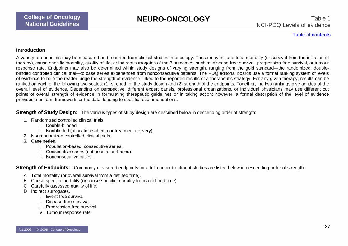
NEURO-ONCOLOGY
College of Oncology National Guidelines
Table 1 NCI-PDQ Levels of evidence
Table of contents
Introduction A variety of endpoints may be measured and reported from clinical studies in oncology. These may include total mortality (or survival from the initiation of therapy), cause-specific mortality, quality of life, or indirect surrogates of the 3 outcomes, such as disease-free survival, progression-free survival, or tumour response rate. Endpoints may also be determined within study designs of varying strength, ranging from the gold standard—the randomized, double-blinded controlled clinical trial—to case series experiences from nonconsecutive patients. The PDQ editorial boards use a formal ranking system of levels of evidence to help the reader judge the strength of evidence linked to the reported results of a therapeutic strategy. For any given therapy, results can be ranked on each of the following two scales: (1) strength of the study design and (2) strength of the endpoints. Together, the two rankings give an idea of the overall level of evidence. Depending on perspective, different expert panels, professional organizations, or individual physicians may use different cut points of overall strength of evidence in formulating therapeutic guidelines or in taking action; however, a formal description of the level of evidence provides a uniform framework for the data, leading to specific recommendations. Strength of Study Design: The various types of study design are described below in descending order of strength:
1. Randomized controlled clinical trials. i. Double-blinded. ii. Nonblinded (allocation schema or treatment delivery).
2. Nonrandomized controlled clinical trials. 3. Case series.
i. Population-based, consecutive series. ii. Consecutive cases (not population-based). iii. Nonconsecutive cases.
Strength of Endpoints: Commonly measured endpoints for adult cancer treatment studies are listed below in descending order of strength:
A Total mortality (or overall survival from a defined time). B Cause-specific mortality (or cause-specific mortality from a defined time). C Carefully assessed quality of life. D Indirect surrogates.
i. Event-free survival ii. Disease-free survival iii. Progression-free survival iv. Tumour response rate
37 V1.2008 © 2008 College of Oncology
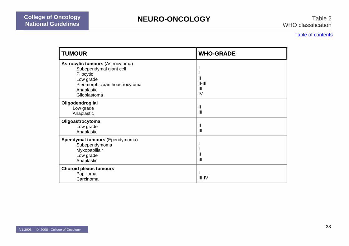
NEURO-ONCOLOGY
College of Oncology National Guidelines
Table 2 WHO classification
Table of contents
TUMOURTUMOUR WHO-GRADEWHO-GRADEAstrocytic tumours (Astrocytoma)
Subependymal giant cell Pilocytic Low grade Pleomorphic xanthoastrocytoma Anaplastic Glioblastoma
I I II II-III III IV
Oligodendroglial Low grade Anaplastic
II III
Oligoastrocytoma Low grade Anaplastic
II III
Ependymal tumours (Ependymoma) Subependymoma Myxopapillair Low grade Anaplastic
I I II III
Choroïd plexus tumours Papilloma Carcinoma
I III-IV
38 V1.2008 © 2008 College of Oncology
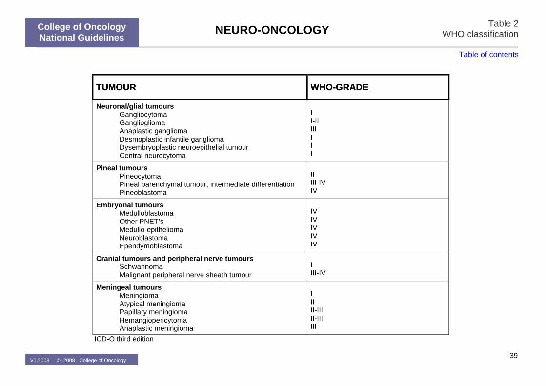
NEURO-ONCOLOGY
College of Oncology National Guidelines
Table 2 WHO classification
Table of contents
TUMOURTUMOUR WHO-GRADEWHO-GRADE
Neuronal/glial tumours Gangliocytoma Ganglioglioma Anaplastic ganglioma Desmoplastic infantile ganglioma Dysembryoplastic neuroepithelial tumour Central neurocytoma
I I-II III I I I
Pineal tumours Pineocytoma Pineal parenchymal tumour, intermediate differentiation Pineoblastoma
II III-IV IV
Embryonal tumours Medulloblastoma Other PNET’s Medullo-epithelioma Neuroblastoma Ependymoblastoma
IV IV IV IV IV
Cranial tumours and peripheral nerve tumours Schwannoma Malignant peripheral nerve sheath tumour
I III-IV
Meningeal tumours Meningioma Atypical meningioma Papillary meningioma Hemangiopericytoma Anaplastic meningioma
I II II-III II-III III
ICD-O third edition
39 V1.2008 © 2008 College of Oncology