Embed Size (px)
Citation preview

Redirecting Specificity of T-Cell Populations For CD19 Using the
Sleeping Beauty System
Harjeet Singh,1Pallavi R. Manuri,
1Simon Olivares,
1Navid Dara,
1Margaret J. Dawson,
1
Helen Huls,1Perry B. Hackett,
3Donald B. Kohn,
4Elizabeth J. Shpall,
2
Richard E. Champlin,2and Laurence J.N. Cooper
1
Divisions of 1Pediatrics and 2Cancer Medicine, University of Texas M. D. Anderson Cancer Center, Houston, Texas; 3Department ofGenetics, Cell Biology and Development, University of Minnesota, St. Paul, Minnesota; and 4Division of Research Immunology/BoneMarrow Transplantation, Children’s Hospital Los Angeles, Los Angeles, California
Abstract
Genetic modification of clinical-grade T cells is undertaken toaugment function, including redirecting specificity for desiredantigen. We and others have introduced a chimeric antigenreceptor (CAR) to enable T cells to recognize lineage-specifictumor antigen, such as CD19, and early-phase human trialsare currently assessing safety and feasibility. However, a sig-nificant barrier to next-generation clinical studies is devel-oping a suitable CAR expression vector capable of geneticallymodifying a broad population of T cells. Transduction ofT cells is relatively efficient but it requires specializedmanufacture of expensive clinical grade recombinant virus.Electrotransfer of naked DNA plasmid offers a cost-effectivealternative approach, but the inefficiency of transgeneintegration mandates ex vivo selection under cytocidalconcentrations of drug to enforce expression of selectiongenes to achieve clinically meaningful numbers of CAR+
T cells. We report a new approach to efficiently generatingT cells with redirected specificity, introducing DNA plasmidsfrom the Sleeping Beauty transposon/transposase systemto directly express a CD19-specific CAR in memory andeffector T cells without drug selection. When coupled withnumerical expansion on CD19+ artificial antigen-presentingcells, this gene transfer method results in rapid outgrowthof CD4+ and CD8+ T cells expressing CAR to redirect specificityfor CD19+ tumor cells. [Cancer Res 2008;68(8):2961–71]
Introduction
The most robust example of successful T-cell therapy occursfollowing allogeneic hematopoietic stem-cell transplantation wherethe engrafted donor-derived T cells recognize recipient tumor-associated antigens in the context of MHC. However, the graft-versus-tumor effect after allogeneic-hematopoietic stem celltransplantation is incomplete, resulting in relapse as the majorcause of mortality. To augment the graft-versus-tumor effect forB-lineage neoplasms, we have previously shown that geneticallymodified peripheral blood– and umbilical cord blood–derivedT cells can be rendered specific for CD19, a molecule constitutivelyexpressed on B-cell malignancies (1, 2). The redirected specificitywas achieved by electrotransfer of a linearized DNA plasmidcoding for a first-generation chimeric antigen receptor (CAR),
designated CD19R, which recognizes CD19 via the scFv of a murineCD19-specific monoclonal antibody (mAb) fused to a chimericCD3-~–derived activation endodomain. A phase I trial (BB-IND1141,clinicalTrials.gov identifier: NCT00182650; ref. 3) is currentlyevaluating the safety and feasibility of infusing autologous T cellselectroporated to coexpress CD19R CAR and the hygromycinphosphotransferase (Hy) and herpes simplex virus-1 thymidinekinase selection/suicide fusion transgene (4).We anticipated that the therapeutic efficacy of adoptive transfer of
CD19-specific Tcells would be improved by developing a CARwith afully competent activation signal and introducing the CAR intocentral memory (CM) T cells. As a result, a second-generation CAR,designated CD19RCD28, has been developed that provides CD19-dependent signaling through chimeric CD3-~ and CD28, resulting inimproved in vivo persistence and antitumor effect, compared withCD19R+ Tcells (5). To further optimize the clinical potential of CAR+
Tcells, while taking advantage of the cost-efficiency of nonviral genetransfer, we desired a clinically feasible approach to the efficientpropagation of CAR+ T-cell populations, including TCM, in theabsence of expression immunogenic drug selection genes, such asHy . We reasoned that genetically modified Tcells could be selectivelypropagated, upon activating T cells for sustained proliferation,through the introduced second-generation CAR. To maximizetransgene expression, we codon-optimized (CoOp) the CAR asreports have shown that codon optimization of genes toward humanconsensus codon usage increases protein expression (6, 7).The focus on developing nonviral gene transfer technologies is
justified based on the cost and time savings compared withdeveloping recombinant clinical-grade viral supernatant, which aresubject to rigorous regulatory oversight and rely on specializedmanufacturing experience of a limited number of productionfacilities. Although the transfection efficiency of nonviral genetransfer is inferior to viral-mediated transduction, naked DNAplasmids expressing desired transgenes such as CAR can be rapidlyproduced at a fraction of the cost compared with clinical gradeg-retrovirus and lentivirus. A potential drawback to nonviralgene transfer, compared with viral gene transfer, is the lengthyex vivo manufacturing time to selectively propagate electroporatedT cells with stable expression of transgene, during which time thecells may become susceptible to replicative senescence, loseexpression of desired homing receptors, and furthermore becleared in vivo due to recognition of immunogenic drug selectiontransgene (8, 9). What is needed is an approach that when coupledwith nonviral gene transfer shortens the culture time to generateT cells with durably expressed transgene and maintains a desiredT-cell immunophenotype.To introduce the CAR, we evaluated whether the efficient
transposition and long-lasting transgene expression of the
Requests for reprints: Laurence J.N. Cooper, University of Texas M. D. AndersonCancer Center, Pediatrics-Research, Unit 907, 1515 Holcombe Boulevard, Houston, TX77030. Phone: 713-563-3208; Fax: 713-792-9832; E-mail: [email protected].
I2008 American Association for Cancer Research.doi:10.1158/0008-5472.CAN-07-5600
www.aacrjournals.org 2961 Cancer Res 2008; 68: (8). April 15, 2008
Research Article
Research. on May 20, 2020. © 2008 American Association for Cancercancerres.aacrjournals.org Downloaded from
Sleeping Beauty (SB) DNA transposon derived from Tc1/marinersuperfamily of transposons (10, 11) can improve transgene transferefficiency. The SB transposable element from a DNA donor plasmidcan be adapted for nonviral gene transfer in T cells, using a SBtransposase supplied in trans to mediate integration of atransposon CAR expression cassette flanked by terminal invertedrepeats (IR), which each contain two copies of a short direct repeat(DR) that have binding sites for the transposase enzyme (Fig. 1D).The SB transposase mediates transposition by binding to IRs,excising a precise DNA sequence flanked by the IRs, and insertingthe transposon into any of f200 million TA sites in a mammaliangenome (12). Previously, the SB system has been used as a nonviralgene delivery into multiple murine and human cell lines, includingliver, keratinocytes, endothelial cells, lung, hematopoietic pro-genitor cells, embryonic stem cells, and tumor cells (11). Ofparticular relevance is that SB-mediated integration has beenshown in human T cells (13), signifying the potential application ofthis technology.We report that electrotransfer of a two-component DNA SB
system into primary human T cells from umbilical cord blood andperipheral blood results in efficient and stable CAR gene transfer,which can be numerically expanded to clinically meaningfulnumbers within 4 weeks on CD19+ artificial antigen-presentingcell (aAPC), without the need for addition of cytocidal concen-trations of drug for selection, and with the outgrowth of CD8+ andCD4+ CM and effector CAR+ T-cell subpopulations. This wasachieved through the rationale design of (a) a next-generation
codon-optimized CD19-specific CAR, (b) CD19+ aAPC expressingdesired costimulatory and cytokine molecules, and (c) SB DNAplasmids expressing CAR transposon and an improved transposase.The relative ease of DNA plasmid production, electroporation, andoutgrowth of stable integrants on a thawed g-irradiated bank ofaAPC can be readily transferred to the facilities operating incompliance with current good manufacturing practice (cGMP) forphase I/II trials. This is predicted to greatly facilitate trial designinfusing CD4+ and CD8+ CAR+ T cells that have desiredimmunophenotype, including TCM.
Materials and Methods
Plasmids. The plasmid pT-MNDU3-eGFP containing salmonid fish–
derived SB IR flanking the constitutive promoter, derived from the U3
region of the MND retrovirus (14), to drive an eGFP reporter gene (15), wasderived from the plasmid pT-MCS (16) that was derived from pT/neo (10).
The second-generation CD19RCD28 CAR (5) was human codon optimized
(CoOp), substituting codons with those optimally used in mammals
(GENEART) without altering anticipated amino acid sequence. Thecodon-optimized CD19RCD28 (CoOpCD19RCD28) CAR was subcloned
into pT-MNDU3 DNA plasmid by replacing the eGFP sequence with
the CAR to create CoOpCD19RCD28/pT-MNDU3 (Fig. 1A). The DNAplasmid pCMV-SB11 (Fig. 1B ) expresses the SB11 transposase (17).
Plasmid CoOpCD19RCD28/Hy-pVitro4 (Fig. 1C ) was generated from
pVitro4-mcs DNA vector (InvivoGen) by subcloning CoOpCD19RCD28 at
NheI in multiple cloning site two and replacing the internal ribosome entrysite (IRES) for the foot and mouth disease virus with that of the
encephalomyocarditis virus ( from pMG vector described; ref. 6). To
Figure 1. Schematic of the expression plasmids and experimental design. A, CoOpCD19RCD28/pT-MNDU3 (Transposon ).MNDU3 promoter, the constitutive promoterfrom the U3 region of the MND retrovirus; CoOpCD19RCD28, codon-optimized CD19RCD28 CAR; IR, SB -inverted/direct repeats; bGh pAn, polyadenylation signalfrom bovine growth hormone; AmpR, ampicillin resistance gene. B, pCMV-SB11 (Transposase). SB11, SB-transposase SB11; CMV promoter, CMV enhancer/promoter; SV40pAN, polyadenylation signals from SV40. C, CoOpCD19CD28/Hy-pVitro4. EF1a promoter, composite promoter comprising the elongation factor-1a(EF1a ) core promoter and the R segment and part of the U5 sequence (R-U5¶) of the human T-cell leukemia virus type 1 LTR; pMB1 ori, a minimal E. coliorigin of replication; SpAn, synthetic pause; CAGp, a composite promoter that combines the human CMV immediate-early enhancer and a modified chicken h-actinpromoter and first intron; Hy, hygromycin B resistance gene (hygromycin phosphotransferase); bGh pAn, polyadenylation signal from bovine growth hormone;EM7 , synthetic prokaryotic promoter. D, an expression cassette in a plasmid (blue ) provides only transient expression unless incorporated into an integratingtransposon vector that can be cleaved from the plasmid and integrated into a host genome by a source of transposase (red ).
Cancer Research
Cancer Res 2008; 68: (8). April 15, 2008 2962 www.aacrjournals.org
Research. on May 20, 2020. © 2008 American Association for Cancercancerres.aacrjournals.org Downloaded from
generate cell surface–bound human interleukin 15 (IL-15), the granulocytemacrophage colony-stimulating factor signal peptide sequence was fused to
the coding sequence of mature human IL-15 at the 5¶ end of a modified
human IgG4 Fc region (5) fused in frame to human CD4 transmembrane
domain and correct assembly was verified by DNA sequence analysis. Themembrane-bound IL-15-Fc cytokine fusion gene was subcloned into
pIRESpuro3 (Clontech) to obtain IL-15-Fc/pIRESpuro3.
Cell lines and primary human T cells. Daudi (Burkitt lymphoma) and
HLAnull K562 (erythroleukemia) cells were obtained from American TypeCulture Collection. Lymphoblastoid cells (LCL) were a kind gift of Dr. Helen
Heslop (Cell and Gene Therapy, Baylor College of Medicine, Houston TX).
These cell lines were cultured in HyQ RPMI 1640 (Hyclone) supplemen-
ted with 2 mmol/L Glutamax-1 (Life Technologies-Invitrogen) and 10%heat-inactivated defined FCS (Hyclone), referred to as culture medium (1).
Human T cells were isolated by density gradient centrifugation over Ficoll-
Paque-Plus (GE Healthcare Bio-Sciences AB), from umbilical cord blood or
peripheral blood mononuclear cells (PBMC) after consent, and were
cultured in culture medium.Generation of aAPC. As previously reported, K562 cells were electro-
porated with DNA plasmids to enforce expression of all of the following:
truncated CD19, 4-1BBL, and MICA fused to GFP (18). These aAPCs were
further modified to express membrane-bound IL-15 to provide a cytokinestimulus at the site of CAR-binding and T-cell costimulation (19).
Electroporation and T-cell coculture with aAPC. On day 0, PBMCs
and umbilical cord blood mononuclear cells (107) were suspended in 100 ALof Amaxa Nucleofector solution (CD34 kit) and mixed with 5 Ag of
supercoiled CoOpCD19RCD28/pT-MNDU3 and 5 Ag pCMV-SB11 DNA
plasmids, transferred to a cuvette, and electroporated (Program U-14).
After a 10-min room temperature incubation, the cells were transferred to a
six-well plate containing 3 to 4 mL incomplete phenol-free RPMI and rested
for 2 to 3 h. The cells were cultured overnight in 6 to 7 mL 10% phenol-free
RPMI and stimulated the next day (day 1) with g-irradiated (100 Gy) aAPC
at a 1:10 T cell/aAPC ratio. The g-irradiated aAPC were re-added every 7 d.
Recombinant human interleukin 2 (rhIL-2; Chiron) was added to the
cultures at 50 units/mL on a Monday-Wednesday-Friday schedule,
beginning day 1 of each 7-d expansion cycle. The supercoiled plasmid
CoOpCD19RCD28/Hy-pVitro4 (expressing CAR under control of EF1apromoter and Hy under control of CAG promoter) was electroporated
(10 Ag) into PBMCs (107) using Nucleofector technology and T cells were
propagated by cross-linking CD3 using an OKT3-mediated 14-d rapid
expansion protocol (REP) as described previously using allogeneic
g-irradiated PBC and LCL feeder cells in the presence of exogenous
(soluble) rhIL-2 (20). T cells were enumerated every 7 d, and viable cells
were counted based on trypan blue exclusion.Western blot. Expression of the chimeric 66-kD (CD19R) and 79-kD
(CD19RCD28) CD3-~ was accomplished using a primary mouse anti-human
CD3-~ mAb (1 Ag/mL; BD Biosciences) and secondary horseradish
peroxidase (HRP)–conjugated goat anti-mouse IgG (1:75,000; Pierce) underreducing conditions, based on methods previously described (20). Protein
lysates were transferred onto nitrocellulose membrane using iBlot Dry
Blotting System (Invitrogen) and developed with SuperSignal West Femto
Maximum Sensitivity substrate (Pierce) per the manufacturer’s instructionsand chemiluminescence was captured after 1-min exposure using VersaDoc
MP 4000 Imaging System (Bio-Rad).
Generation of monoclonal antibody recognizing a CD19-specificCAR. Female BALB/c mice were injected six times in the foot at 3-d
intervals with syngeneic NS0 cells expressing CD19R CAR. Three days
after the last immunization, mice were sacrificed, popliteal lymph nodes
were removed, and cells were fused with P3-8AG-X653 myeloma cells ata ratio of 3:5, using 30% polyethylene glycol 1450 (in serum free RPMI
containing 5% DMSO). After 10 d, hybridoma colonies were picked,
cloned by limiting dilution in 96-well plates, and 100 AL of supernatants
were screened by ELISA for differential binding to round-bottomed96-plates containing adsorbed (105/well) CD19R+ and CD19Rneg Jurkat
cells as detected by 1:500 dilution of HRP-conjugated goat anti-mouse
IgG (Santa Cruz Biotechnology). Detection was achieved by TMBMicrowell peroxidase substrate system (KPL). Protein G column (Roche)
purified mAb was conjugated to Alexa Fluor 488 (Invitrogen-MolecularProbes) per manufacturer’s instructions.
Flow cytometry. Fluorochrome-conjugated reagents were obtained
from BD Biosciences: anti-CD4, anti-CD8, anti-CD25, anti-CD27, anti-
CD28, anti-CD62L, anti-CD45RA, anti-CD45RO, and anti-CD95. Affinity-purified F(ab¶)2 fragment of FITC-conjugated goat anti-human Fcg(Jackson Immunoresearch) was used at 1/20 dilution to detect cell
surface expression of CD19-specific CAR. Purified CAR-specific mAb clone
2D3, conjugated to Alexa Fluor 488, was used at a dilution of 1/30, givinga concentration of f30 Ag/mL. In some experiments, binding of this mAb
to the Fc region of CAR was blocked (30 min at 4jC) using goat human
Fc-specific antiserum (Sigma). Blocking of nonspecific antibody binding
was achieved using FACS wash buffer (2% FCS in PBS). T-cell receptor(TCR)-Vh expression was determined with a panel of 24 TCR-Vh–specificmAbs (IO TEST Beta Mark TCR-Vh repertoire kit, Beckman Coulter) used
in association with anti-CD3 and appropriate isotype-matched controlmAbs. Data acquisition was on a FACSCalibur (BD Biosciences) using
CellQuest version 3.3 (BD Biosciences). Analyses and calculation of mean
fluorescence intensity (MFI) was undertaken using FCS Express version
3.00.007 (Thornhill).Intracellular IL-2 cytokine staining. Intracellular IL-2 was assayed
using the Intracellular Cytokine Staining Starter Kit (BD PharMingen) per
the manufacturer’s instructions. Briefly, 105 T cells were incubated with
0.5 � 106 stimulator cells in 200 AL culture medium along with protein
transport inhibitor (BD Golgi Plug containing Brefeldin A) in a 96-well plate.
Following a 4- to 6-h incubation at 37jC, the cells were stained for CAR
expression using hybridoma mAb clone 2D3 at 4jC for 30 min. After
washing, the cells were fixed and permeabilized (100 AL, Cytofix/Cytopermbuffer) and phycoerythrin-conjugated mAb specific for IL-2 was added.
Cells were further washed and analyzed by FACSCalibur. T cells
were treated with a leukocyte activation cocktail (phorbol 12-myristate
13-acetate and ionomycin) as a positive control.Confocal microscopy. Jurkat parental and CD19R+ Jurkat cells were
stained with the hybridoma clone mAb 2D3, at a 1:50 dilution for 15 min at
4jC, washed in FACS wash buffer, and fixed with 0.1% paraformaldehyde.
After fixing, the cells were washed twice with FACS wash buffer andtransferred onto slides, and coverslips were mounted with Prolong Gold
anti-fade agent (Invitrogen). Cells were examined under a confocal
microscope (Leica TCS SP2-SE) using oil immersion lens (�63 objective).
Single-scan images were obtained with a 4.76� zoom in a 1,024 � 1,024format with a line averaging of 8.
Chromium release assay. The cytolytic activity of T cells was
determined by 4-h chromium release assay (1). CD19-specific T cells
were incubated with 5 � 103 51Cr-labeled target cells in a V-bottomed96-well plate (Costar). The percentage of specific cytolysis was calculated
from the release of 51Cr, as described earlier, using a TopCount NXT
(Perkin-Elmer Life and Analytical Sciences, Inc.). Data are reported asmean F SD.
DNA PCR for SB transposon and transposase. DNA was isolated from
PBMC using the QIAmp DNA mini kit (Qiagen). PCR was carried out using
CD19RCD28-specific forward primer 5¶-AGATGACCCAGACCACCTCCAGC-3¶ and reverse primer 5¶-GGTATCCTTGGTGGCGGTGCT-3¶ for the transpo-son. The PCR reaction used 1 Ag of DNA/sample in a mix containing
10� PCR buffer, 2.5 mmol/L deoxynucleotide triphosphates, 3 Amol/L
MgCl2, and 0.5 units of DNA polymerase (AmpliTaq Gold, AppliedBiosystems) in a final volume of 50 AL amplified in a thermal Cycler
(PTC-200 DNA Engine Cycler, Bio-Rad). After an initial denaturation at 95jCfor 5 min, the samples underwent 34 cycles of 95jC for 30 s, 65jC for 30 s,72jC for 1 min 15 s, followed by a prolonged extension step at 72jC for
7 min. For the transposase gene, PCR was carried out using SB11-specific
forward primer 5¶-ATGGGACCACGCAGCCG-3¶ and reverse primer 5¶-CGTTTCGGGTAGCCTTCCACA-3¶. After an initial denaturation at 95jCfor 5 min, the samples underwent 34 cycles of 95jC for 15 s, 58jC for 30 s,
74jC for 2 min followed by a prolonged extension step at 74jC for 7 min.
The housekeeping gene GAPDH was also amplified in the same samples
using the forward primer 5¶-TCTCCAGAACATCATCCCTGCCAC-3¶ andreverse primer 5¶-TGGGCCATGAGGTCCACCACCCTG-3¶.
Sleeping Beauty to Generate CD19-Specific T Cells
www.aacrjournals.org 2963 Cancer Res 2008; 68: (8). April 15, 2008
Research. on May 20, 2020. © 2008 American Association for Cancercancerres.aacrjournals.org Downloaded from
Chromosome banding analysis. Exponentially growing SB-transfected
T-cell cultures ( freshly fed 24 h earlier) were incubated for 2 h at
37jC with colcemid (20 AL of 0.04 Ag/mL) per 10 mL medium followed by0.075 mol/L KCl at room temperature for 15 min, fixed with acetic acid/
methanol (1:3), and washed thrice on a glass slide. For Giemsa (G)
banding, 5- to 6-d-old slides treated with trypsin were stained with
Giemsa stain following standard techniques described previously (21).A total of 15 G-banded metaphases were photographed and 5 complete
karyotypes were prepared using a karyotyping system from Applied
Imaging Corporation.
Results
We describe a new approach to using nonviral gene transfer ofDNA plasmids to efficiently obtain populations of memory andeffector T cells with desired specificity (Fig. 1D). The system wehave devised provides for robust antigen-driven expansion of CD4+
and CD8+ CAR+ T cells to clinically meaningful numbers.Monoclonal antibody with specificity for CD19-specific CAR.
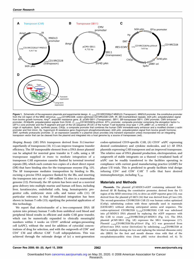
The cell surface expression of the introduced CAR was predictedto increase with outgrowth of T-cell populations that haveundergone CAR-mediated numerical expansion on CD19+ aAPC.Currently, the only-commercially available flow cytometryreagents that recognize our CARs are polyclonal anti-Fc anti-bodies raised in goat, but we desired a homogeneous monoclonalproduct for use in the release of CAR+ T cells for clinical trials. Tolongitudinally follow the transgene expression, we developed aCAR-specific mAb by immunizing mice with syngeneic NS0 cellsexpressing CD19R. A hybridoma mAb clone 2D3 (IgG1) wasselected by flow cytometry that selectively bound to CD19R+
Jurkat cells, but not parental Jurkat cells. The binding of 2D3 can
be blocked using a Fc-specific antibody (Fig. 2A). The 2D3 clonebound a CD20-specific CAR that shares the IgG4 Fc region withCD19R and CD19RCD28 (data not shown). The pattern of stainingby confocal microscopy showed 2D3 binding to CAR on the cellsurface (Fig. 2B). These data are consistent with a mAb bindingspecifically to the CD19-specific CAR and recognizing themodified human IgG Fc region. Of note, the production of thismAb avoided the need to purity recombinant CAR protein as theimmunogen was genetically modified NS0 cells and the ELISAscreening used genetically modified Jurkat cells.Electrotransfer of SB two-plasmid DNA system.We have used
a nonviral gene transfer approach to introduce codon optimizedDNA expression plasmids because these expression vectors can bereadily and cheaply manufactured to clinical grade. Althoughcodon modification of TCR genes has been shown to enhanceexpression of transgenic TCR in primary human T cells (22), wenow show the usage of a codon optimized second-generation CAR.Previously, our electroporation approach based on the Multi-porator (Eppendorf; refs. 23, 24) used T cells that had beenstimulated to proliferate by cross-linking CD3 with OKT3 to allowaccess of the introduced naked DNA to the nucleus afterdissolution of the nuclear envelope during prometaphase. However,T cells nonspecially activated to proliferate, such as by cross-linkingCD3 as occurs in the REP (25), would preclude subsequentimmediate antigen-mediated propagation and thus directedoutgrowth of CAR+ T cells. Nucleofector technology has been usedto electroporate nonreplicating cells by direct transfer of DNA tothe nucleus (26). Thus, we investigated whether this electrotransfersystem could be used to genetically modify circulating T cells fromperipheral blood and umbilical cord blood, which are in a
Figure 2. Specificity of mouse-derivedCAR-specific mAb (clone 2D3). A, Jurkat cellswere genetically modified and sorted toexpress CD19R. Jurkat parental (gray line )and CD19R+ (black line ) cells were stainedwith (i) Alexa 488–conjugated clone 2D3and (ii ) F(ab¶)2-fragment of goat-derivedpolyclonal antibody specific for human Fc;iii, binding of 2D3 (solid line ) was blockedby polyclonal Fc-specific antisera (dashedline ). B, cell surface staining of Alexa Fluor488–conjugated clone 2D3 by confocalmicroscopy on (i) CD19R+ Jurkat cells and (ii )Jurkat parental cells. Cells were stained, fixed,and mounted as described in Materials andMethods.
Cancer Research
Cancer Res 2008; 68: (8). April 15, 2008 2964 www.aacrjournals.org
Research. on May 20, 2020. © 2008 American Association for Cancercancerres.aacrjournals.org Downloaded from
quiescent state. To assist with subsequent translation to clinicalpractice, the Nucleofector solution is available for use in cGMP.Both the SB transposase and IR have been independently
manipulated to improve efficiency of transposition, but changes toboth do not generally seem to be additive. In preliminaryexperiments, we too compared the relative transposition efficiencyof the SB10 (10, 16) and SB11 transposases (the latter exhibitingimproved enzymatic activity; ref. 17) in a two-by-two matrix usingthe Amaxa 96-well Shuttle system to introduce these transposasesand pT (15, 16) and pT2 transposons (the latter exhibitingimproved transposition; ref. 27) into Jurkat T cells. As observedwith other cell lines, we found a similar increase in transpositionusing SB11 with pT and using SB10 with pT2 (27) although, asreported, overproduction of transposase inhibited transposition(13, 17). When the pT2-improved transposon was combined withSB11, no further increase in transgene expression was observedover that achieved when these components were used with SB10 orpT, respectively. In the present study, a combination of pTtransposon ( for integration) and SB11 transposase ( for transientexpression) was used for experiments with primary T cells.Generation of CD19+ aAPC. We determined whether peripheral
blood and umbilical cord blood–derived T cells could be selectivelypropagated by stimulating through an introduced immunorecep-tor. This experiment would evaluate our underlying hypothesis ofwhether the presence of the SB transposase would improveefficiency of CAR transposon integration in T cells. Our initialattempts at CAR-dependent T-cell propagation after electrotransferof the SB system used allogeneic LCL because these are widelyavailable as master cell banks (including at M. D. Anderson CancerCenter) manufactured in compliance with cGMP for phase I/IItrials. However, these LCL resulted in nonspecific outgrowth ofCARnull T cells that had undergone electrotransfer of SB plasmids,independent of CAR expression (data not shown), presumably dueto outgrowth of alloreactive T cells. Because our SB transposon bydesign does not include a drug resistance gene, we avoidednonspecific propagation of T cells using K562 as aAPC becausethese do not express classical HLA molecules. K562 cells are widelyrecognized as a platform suitable for the numerical expansion oflymphocytes because they (a) can be cultured in compliance withcGMP, (b) express desired endogenous T-cell costimulatorymolecules, (c) secrete pro-inflammatory cytokines, and (d) can be
readily modified to enforce the expression of antigen and desiredendogenous T-cell costimulatory molecules (28, 29). To provide anIL-15–mediated growth stimulus coordinated with recognition ofCD19 antigen, the aAPC expressing tCD19, 4-1BBL, and MICA werefurther modified to express the IL-15 cytokine on the cell surface(IL-15-Fc; Fig. 2A). Membrane-bound IL-15 has been used before topropagate natural killer (NK) cells on K562 (19). The ability of theseK562 aAPCs to propagate CAR+ T cells after electrotransfer of SBtransposon and transposase plasmids is described in the nextsection.SB-mediated gene transfer of CAR transposon in primary
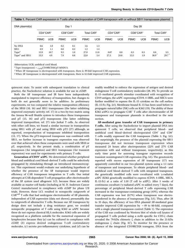
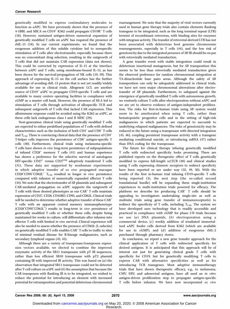
T cells. After using the Nucleofector to import plasmid DNA intoquiescent T cells, we observed that peripheral blood– andumbilical cord blood–derived electroporated CD4+ and CD8+
T cells readily expressed the CAR transposon (Table 1; Fig. 3A).Not surprisingly, the presence of the plasmid expressing the SB11transposase did not increase transposon expression whenmeasured 24 hours after electroporation (22% and 27% CARexpression with and without transposase, respectively), as thisearly time point for assessing transgene expression recordstransient nonintegrated CAR expression (Fig. 3A). The genotoxicityreported with excess expression of SB transposase (17) wasapparently controlled in our two-plasmid system using a 1:1 ratioof transposon and transposase. To obtain peripheral blood– andumbilical cord blood–derived T cells with integrated transposon,the genetically modified cells were cocultured with g-radiatedaAPC (K562 genetically modified to express tCD19, 4-1BBL, MICA,IL-15-Fc) at a ratio of 1:10 (T cell to aAPC). After 5 weeks ofcontinuous coculture (g-radiated aAPC re-added every 7 days), thepercentage of peripheral blood–derived T cells expressing CARincreased in the transposase-containing group (43%), whereas theCAR expression was lost (0.7%) when transposon was electro-transferred in the absence of transposase (Fig. 3A). Thus, after 28to 35 days, the efficiency of two DNA plasmid SB-mediated genetransfer improved CAR expression by f49 to 60-fold, comparedwith a single plasmid transposon control (Table 1). The expressionof the CAR was confirmed by Western blot of whole-cell lysates ofpropagated T cells probed using a mAb specific for CD3-~ chainrevealed the 79-kDa chimeric ~ chain in addition to the 21-kDaendogenous ~ chain (Fig. 3C). To monitor for the presence orabsence of the integrated CD19RCD28 transgene, DNA from the
Table 1. Percent CAR expression in Tcells after electroporation of CAR transposon with or without SB11 transposase plasmid
DNA plasmid(s) Day 1 Day 28
CD4+CAR+ CD8+CAR+ Total CAR+ CD4+CAR+ CD8+CAR+ Total CAR+
PBMC UCB PBMC UCB PBMC UCB PBMC UCB PBMC UCB PBMC UCB
No DNA 0.6 1.0 0.2 0.1 1.6 1.3SB11 0.9 1.1 0.8 0.2 1.2 1.4
Txpn* 15.9 10.7 9.5 1.0 27.0 11.8 0.07 0.8 0.5 0.3 0.8 3.5
Txpn* and SB11 13.3 4.9 7.9 0.8 22.0 5.4 27.5 24.8 13.5 1.9 38.9c
29.7b
Abbreviation: UCB, umbilical cord blood.
*Txpn (transposon) = CoOpCD19RCD28/pT-MNDU3.cWhen SB transposase is electroporated with transposon, there is 49-fold improved CAR expression.bWhen SB transposase is electroporated with transposon, there is 8.4-fold improved CAR expression.
Sleeping Beauty to Generate CD19-Specific T Cells
www.aacrjournals.org 2965 Cancer Res 2008; 68: (8). April 15, 2008
Research. on May 20, 2020. © 2008 American Association for Cancercancerres.aacrjournals.org Downloaded from

numerically expanded T cells, electroporated with and withoutSB11, were PCR amplified using CAR-specific primers. A 1,900-bpband corresponding to the CD19RCD28 transgene was observed inT cells electroporated using the SB two-plasmid system, whereas
no similar band was observed in cells electroporated with SBtransposon in the absence of transposase, which is consistent withimproved SB11-mediated transposition in T cells expressing CARprotein (Fig. 3D).
Figure 3. Characterization of CAR expression on peripheral blood–derived T cells after electrotransfer of SB plasmid system. A, expression of CAR on CD8+ andCD4+ T cells after electrotransfer of SB transposon with or without SB11 transposase at 24 h, and 4 and 5 wk of coculture on g-irradiated K562-derived aAPCexpressing tCD19, IL-15-Fc, MICA, and 4-1BBL. B, i, immunophenotype of memory cell markers (CD27, CD28, CD62L) on genetically modified T cells generated after5 wk of coculture on aAPC. The gray-filled histograms reveal the percentage of T cells expressing CD27, CD28, and CD62L in the lymphocyte-gated population. Thoseexpressing the memory cell markers were analyzed for coexpression of CAR (detected by mAb clone 2D3) and CD8 or CD4. ii, expression of CD45RO, CD45RA, andCD62L on T cells generated after coculture. CAR+ CD4 or CD8 cells were analyzed for the expression of CD45RA and CD45RO. The MFI of the unmanipulatedT cells was 867/50 (CD45RA/CD45RO) compared with 28/38 for the SB-transfected T cells. CD45RO and CD62L double-positive cells were also analyzed forcoexpression of CAR. iii, TCM, defined as double-positive for CD28 and CD95 (TEM, CD28
negCD95pos), were analyzed for coexpression of CD62L and CAR. C,Western blot analysis of CAR expression detected by mAb specific for CD3-~ . Whole-cell protein (20 Ag) lysates from primary T cells genetically modified with
CoOpCD19RCD28 (lane 1, f79 kDa chimeric protein) or no plasmid control (lane 2 ); CD19R+ Jurkat cells (lane 3, f66 kDa chimeric protein) or parental Jurkat (lane 4 )were resolved by SDS-PAGE under reducing conditions. D, integration of CoOpCD19RCD28 by PCR. DNA was isolated from T cells after mock electroporation(no DNA, lanes 1 and 4 ), from T cells 28 d after electroporation with SB transposon in the absence of transposase (lanes 2 and 5 ), and from T cells 28 d afterelectroporation with transposon in the presence of SB11 transposase (lanes 3 and 6 ). PCR was accomplished using transposon-specific primers (lanes 1–3 ) orGAPDH-specific primers (lanes 4–6 ). The data showing SB system in peripheral blood/cord blood are from a representative experiment.
Cancer Research
Cancer Res 2008; 68: (8). April 15, 2008 2966 www.aacrjournals.org
Research. on May 20, 2020. © 2008 American Association for Cancercancerres.aacrjournals.org Downloaded from

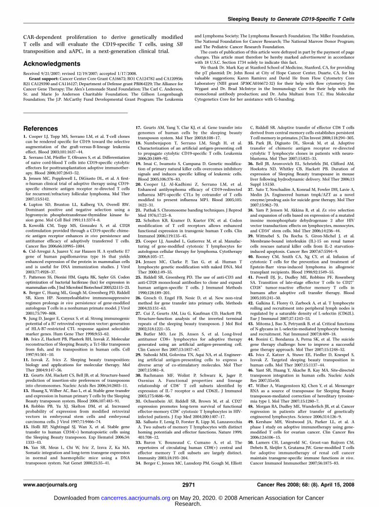
Propagation of CAR+ T cells. The K562-derived aAPC wascalculated to give a 20-fold growth of genetically modified T cellsat the end of 4 weeks with continued and accelerated expansionthereafter (Fig. 4A). A subset analysis revealed that populationsof both CD4+CAR+ and CD8+CAR+ T cells could be propagated(Table 1). Initially, the rates of CD4+ and CD8+ T-cell growth onaAPC were similar, but after f8 weeks there was an outgrowth ofCD4+CAR+ T cells (Fig. 4B). Thus, continued time in tissue culturecould be used to derive CAR+ T cells with an increased CD4 to CD8ratio. We also followed the percentage expression and density ofthe CAR on the T-cell surface by flow cytometry. With coculture,there was outgrowth of percentage of T cells expressing the CD19-specific CAR (22% on day 1 and peaking at 99% on day 70).However, as the percentage of CAR+ T cells increased, there was adecrease in the density of CAR expression, as the MFI droppedfrom a peak of 109 arbitrary units at 21 days, early in thecoculturing process, and then declined over culture time. Theamount of CAR for the population peaked at f70 days after
electroporation (percentage expression multiplied by MFI). Thus,adding a fixed ratio of aAPC (with a fixed density of CD19 antigen)to T cells seems to have supported the growth for populations ofT cells that either expressed high density of CAR or high percentageof CAR.Immunophenotype of CAR+ memory T cells. Previously, T cells
from healthy donors electroporated to express a CD19-specific CARand nonspecifically activated for proliferation by cross-linking CD3with OKT3 using REP have shown a predominant phenotypeconsistent with differentiated effector CD8+ T cells (20). In contrast,after electrotransfer of SB plasmids and numerical expansion onaAPC, T cells exhibited a heterogeneous immunophenotype andapparently included populations of CAR+ TCM. We showed that theCAR+ T cells expressed memory cell markers (CD27, CD28, CD62L;refs. 30–32) as well as determinants of an effector-cell phenotype(Fig. 3Bi). For example, over half of CD27+, CD28+, and CD62L+
T cells expressed CAR. Indeed, as a marker for TCM, 88% of theCD62L+CD45RO+ cells expressed the CAR (Fig. 3Bii ; ref. 33). Upon
Figure 4. Sustained proliferation of genetically modifiedprimary peripheral blood–derived T cells with analysis ofCAR expression and TCR repertoire. A, kinetics of propa-gation of T cells in culture with aAPC. The average T-cellnumerical expansion was 22-fold (range 20–31) every 7 d forup to 10 wk of continuous coculture with aAPC. B, percentexpression (unbroken lines, left axis ) and density asmeasured by MFI (dotted line, right axis ) of CAR on total,CD4+, CD8+ T cells as measured by flow cytometry overculture time on aAPC. C, TCR Vh analysis by flow cytometry4 wk after electrotransfer of SB plasmids (filled columns )or CoOpCD19CD28/Hy-pVitro4 plasmid (open columns ).Data are representative of two different experiments.
Sleeping Beauty to Generate CD19-Specific T Cells
www.aacrjournals.org 2967 Cancer Res 2008; 68: (8). April 15, 2008
Research. on May 20, 2020. © 2008 American Association for Cancercancerres.aacrjournals.org Downloaded from

gating CD45RO+ cells, we observed a preferential expansion ofT cells with this memory-cell marker in the cultured SB-transfectedT cells (90%; MFI, 38) compared with unmanipulated T cellsobtained directly from PBMC (36%; MFI, 50). TEM and TCM havealso been distinguished based on relative expression of CD28 andFas (34). Using these markers, we were able to identify thatgenetically modified and propagated TCM constituted f40% of thetotal cell population and CD28negCD95+ TEM represented theremainder of the propagated Tcells. Multiparameter flow cytometryfurther revealed that 39% of CAR+CD28+CD95+ TCM expressedCD62L. In comparison, only 7% of the CAR+ TEM expressed CD62L(Fig. 3Biii). These data reveal that CAR+ T cells are present in T cellsthat express markers consistent with TCM. Preferential expansion ofT cells in tissue culture with an apparent memory phenotype canalso be inferred by from the ratio of CD45RA/CD45RO, whichdecreased from 2.75 in unmanipulated freshly derived PBMC to0.9 for SB-transfected and ex vivo propagated T cells (Fig. 3Bii).The relative percentage increase of observed CD45RO+ cells, orthe decrease in CD45RA/CD45RO ratio, is presumably due to therepetitive antigenic stimulation of cultured T cells resulting indown-regulation of the high molecular weight CD45RA isoform andreciprocal up-regulation of the low molecular weight isoformCD45RO during time in culture. Coexpression of both CD45RA andCD45RO has been associated with the phenotype of effector T cells(35) but as in circulating peripheral blood–derived T cells expressboth CD45RA and CD45RO, the markers are presumably alsopresent on memory cells. These data have implications forimproved in vivo efficacy as TCM are associated with long-termpersistence after adoptive transfer.TCR VB repertoire. We tracked the expression of TCR Vh usage
by flow cytometry over time with the hypothesis that animprovement in DNA-plasmid integration would be reflected by
maintenance of a broad pre-electroporation TCR Vh repertoire. Thepattern of TCR Vh usage observed after electrotransfer of the twoDNA SB plasmids and propagation on aAPC wasmuch broader thanwhen T cells were electroporated using the single CoOpCD19RCD28/Hy-pVitro4 plasmid and expanded by REP by cross-linking CD3 withOKT3 in cytocidal concentrations of hygromycin B. We observedthat f80% of the T cells electroporated with CoOpCD19RCD28/Hy-pVitro4 plasmid expressed a single TCR Vh family (Vh5.3). Incontrast, f80% of the T cells electroporated with the complete SBsystem expressed 30% of the TCR Vh families (Fig. 4C). This isconsistent with less efficient integration of the CoOpCD19RCD28/Hy-pVitro4 plasmid compared with the SB system. These data haveimplications for design of adoptive immunotherapy trials asmaintaining a broad TCR diversity is desired to restore immunereconstitution after myeloablative preparative regimens.Redirected function of CAR+ T cells after electrotransfer of
SB plasmids. The numerically expanded T cells were evaluated forredirected killing. The genetically modified T cells were able to lyseCD19+ targets, and specificity of killing was shown by thebackground lysis of CD19neg K562 cells (Fig. 5A). We showed a25-fold increase in specific lysis of CD19+ K562 at effector-to-targetration of 50:1. The lack of killing of CD19neg K562 is consistent withabsence of resident NK cell function in the culture, as these targetcells are sensitive to NK cell–mediated lysis. Because the CARcontains a CD28 endodomain, we investigated whether T cell–derived IL-2 could be produced when CAR contacted CD19 antigenin the absence of binding CD80 or CD86. An intracellular cytokineassay showed that IL-2 could be detected in the CAR+ T cells onlywhen cultured with CD19+ stimulator cells and not with CD19neg
cells (Fig. 5B). There was an f4-fold increase in IL-2 expressionwhen CAR+ T cells were stimulated by CD19+CD80negCD86neg K562cells compared with CD19neg K562 parental controls. No significant
Figure 5. Redirected specificity of peripheral blood–derived T cells geneticallymodified with SB system. A, killing of CD19+ target cells (HLA Ineg Daudi; HLA classI/IIneg K562 cells transfected to express truncated CD19) in a standard 4-h chromiumrelease assay. Background lysis of CD19neg (parental K562) cells is shown. Points,mean specific lysis of triplicate wells at effector to target (E:T ) cell ratios; bars, SD.B, CAR and intracellular IL-2 expression after incubating with panel of stimulatorcells by multiparameter flow cytometry gating on CD3+ lymphocytes. Phorbol12-myristate 13-acetate (PMA ) and ionomycin were added as a positive control.
Cancer Research
Cancer Res 2008; 68: (8). April 15, 2008 2968 www.aacrjournals.org
Research. on May 20, 2020. © 2008 American Association for Cancercancerres.aacrjournals.org Downloaded from

IL-2 production was observed when T cells were cultured in absenceof stimulator cells. These data are consistent with activation ofT cells for killing and IL-2 cytokine production through the CAR.Lack of integration of SB11 transposase in propagated
T cells. Continued presence of the SB11 transposase in geneticallymodified T cells may cause genotoxicity. We evaluated for thepresence of integrated transposase plasmid by genomic PCR. Noband corresponding to the SB11 transposase gene (size f830 bp)was detected in T cells that were electroporated with the SBtransposon and transposase and had undergone 4 weeks ofcoculture with aAPC (Fig. 6A), which is consistent with the rapidloss of transposase expression activity over the first few dayspostdelivery in mice (36). These results indicate that the SB11transposase was not integrated into the genome of cells stablyexpressing the CD19RCD28 CAR.Karyotype of genetically modified T cells. As a measure of
global genotoxicity associated with undesired and continuedtransposition, we evaluated the integrity of the chromosomestructure. G-banding analysis of the SB-transfected T cells showeda normal female karyotype, 46, XX with no apparent numerical orstructural chromosome alterations (Fig. 6B). Although this does notexclude chromosomal damage below the limit of detection of thistechnique, it supports the premise that SB transposition in T cells isnot associated with translocations and chromosomal aberrations.
Discussion
We have previously showed that peripheral blood– and umbilicalcord blood–derived T cells can be rendered specific for CD19, basedon using a CAR capable of providing a fully competent activationsignal, development of aAPC-expressing antigen, and desiredcostimulatory signals. In this report, we describe the use of SBtransposon/transposase plasmids to introduce CD19-specific CARleading to efficient outgrowth of CAR+ T cells on aAPC withpreservation of CD4+, CD8+, central memory, and effector-cellimmunophenotypes. This is expected to be of widespread interestas many institutions are evaluating the clinical potential ofgenetically modified T cells with redirected specificity. The majorityof these programs use recombinant viral vectors, which, althoughefficient at gene transfer, are generally cost-prohibitive tomanufacture to clinical grade and still permit incremental changesto clinical trial design. Yet, at this early stage of gene therapyplanning with clinical grade T cells, what is needed, and is providedhere, is a cost-effective gene transfer system that encouragesreiterative changes to expression vector and/or CAR design to beused in proof-of-concept clinical trials that support hypothesistesting from the bench to the bedside and back again. Theapproximate cost for manufacture and release of a clinical gradeplasmid DNA is between $20,000 and $40,000 depending onsupplier and degree of release testing needed. This release testingtypically requires restriction enzyme analyses, sequencing, andmeasures of (a) homogeneity/purity/contamination (protein, RNA,and other DNA) and (b) sterility including endotoxin. For early-phase proof-of-concept trials, this pricing compares favorably withthe relatively high cost of recombinant retrovirus, includinglentivirus as manufacture and release of clinical grade virusesmay exceed 10 times the cost of DNA-plasmid production.Furthermore, there is downward pressure on the unit cost forDNA because there are many vendors worldwide with thecapability to produce clinical grade plasmids. The manufacture/release of recombinant retrovirus is highly specialized, requiring
the expertise of a small number of GMP facilities that contributesto high cost and can introduce delays to production and thusavailability for clinical use.Previously, the relatively low levels of nonviral gene transfer
efficiency to introduce naked DNA plasmid coding CAR transgene,compared with viral-mediated transduction, has been compensat-ed by lengthy periods of ex vivo tissue culture to select-out T cellsexpressing drug-metabolizing enzymes. Thus, an attractive featureof the SB gene transfer system to introduce CAR into T cells isavoidance of the need to express immunogenic section genes, suchas bacteria-derived Hy transgene. Some human-derived drug-resistant transgenes are available for use in hematopoietic cells(37, 38), but they typically incorporate amino acid changes from thenative protein sequence that may compromise their inability toremain nonimmunogenic and the continued presence of chemo-selective drugs may slow kinetics of ex vivo numerical expansionand alter T-cell function.Coupling electrotransfer of SB system with selective propagation
of CAR+ T cells was made possible using K562 cells that had been
Figure 6. Safety issues regarding SB transposase and chromosomalaberrations. A, lack of integration of SB11 transposase by genomic PCR fromgenetically modified and propagated peripheral blood–derived T cells. DNA wasisolated from T cells after mock electroporation (no DNA, lanes 1 and 4),from T cells 28 d after electroporation with the two-plasmid SB system(lanes 2 and 5), or from Tcells 1 d after electroporation with the two-plasmid SBsystem (lanes 3 and 6 ). PCR was accomplished using transposase-specificprimers (lanes 1–3 ) or GAPDH-specific primers (lanes 4–6 ). B, idiogram of aG-banded karyotype of the SB-transfected peripheral blood–derived T cellsshowing no apparent numerical or structural chromosome alterations.
Sleeping Beauty to Generate CD19-Specific T Cells
www.aacrjournals.org 2969 Cancer Res 2008; 68: (8). April 15, 2008
Research. on May 20, 2020. © 2008 American Association for Cancercancerres.aacrjournals.org Downloaded from
genetically modified to express costimulatory molecules tofunction as aAPC. We have previously shown that the presence of4-1BBL and MICA on CD19+ K562 could propagate CD19R+ T cells(18). However, sustained antigen-driven numerical expansion ofgenetically modified T cells on aAPC has required the presence ofrhIL-15 (18). In our current experiments, we found that theexogenous addition of this soluble cytokine led to nonspecificstimulation of T cells after electrotransfer, especially because therewas no concomitant drug selection, resulting in the outgrowth ofT cells that did not maintain CAR expression (data not shown).This could be corrected by expression of IL-15 at the interfacebetween aAPC and T cells, using membrane-bound IL-15, as hasbeen shown for the survival/propagation of NK cells (19, 39). Thisapproach of expressing IL-15 on the cell surface has the furtheradvantage of avoiding rhIL-15 protein that is not yet readily/widelyavailable for use in clinical trials. Allogeneic LCL are anothersource of CD19+ aAPC to propagate CD19-specific T cells and areavailable to many centers operating facilities in compliance withcGMP as a master cell bank. However, the presence of HLA led tostimulation of T cells through activation of allospecific TCR andsubsequent outgrowth of T cells that lacked CAR expression. Thisalloimmune response could be avoided using the K562 as aAPC, asthese cells lack endogenous class I and II MHC (29).Next-generation clinical trials using genetically modified T cells
are expected to infuse predefined populations of T cells with definedcharacteristics such as the inclusion of both CD4+ and CD8+ T cellsand TCM. There is convincing clinical data that the presence of CD4
+
T-helper cells improves the persistence of CD8+ antigen-specific Tcells (40). Furthermore, clinical trials using melanoma-specificT cells have shown in vivo long-term persistence of subpopulationsof infused CD28+ memory T cells (41) and human experiencehas shown a preference for the selective survival of autologousHIV-specific CD27+ versus CD27neg adoptively transferred T cells(31). These data are supported by nonhuman experimentsin which adoptive transfer of ex vivo propagated macaqueCD28+CD95+CD62L+ TCM resulted in longer in vivo persistencecompared with infusion of numerically expanded effector T cells(34). We note that the electrotransfer of SB plasmids and subsequentCAR-mediated propagation on aAPC supports the outgrowth ofT cells with these desired phenotypes as our CAR+ T cells maintainexpression of CD27, CD28, CD45RO, CD95, and CD62L. Clinical trialswill be needed to determine whether adoptive transfer of these CAR+
T cells with an apparent central memory immunophenotype(CD28+CD95+CD62L+) results in long-term in vivo persistence ofgenetically modified T cells or whether these cells, despite beingmaintained for weeks in culture, will differentiate after infusion intoeffector T cells with limited in vivo survival. Clinical experience willalso be needed to assess whether the presence of CD62L (L-selectin)on genetically modified T cells enables CAR+ T cells to traffic to sitesof minimal residual disease for B-lineage malignancies, such assecondary lymphoid organs (42, 43).Although there are a variety of transposase/transposon expres-
sion vectors available, we elected to combine the improvedenzymatic activity of the SB11 transposase with pT IR sequences,rather than less efficient SB10 transposase with pT2 plasmidcontaining IR with improved IR activity. This was based on (a) theobservation that integrated SB11 transposase could not be detectedafter T-cell culture on aAPC and (b) the assumption that because theCAR transposon with flanking IR is to be integrated, we wished toreduce the potential for introducing an element with increasedpotential for retransposition and potential deleterious chromosomal
rearrangement. We note that the majority of viral vectors currentlyused in human gene therapy trials also contain elements flankingtransgene to be integrated, such as the long terminal repeat (LTR)termini of recombinant retrovirus, with binding sites for enzymeswith integrase activity. The transfer of retroviral-derived LTR has notbeen associated with deleterious host genome chromosomerearrangements, especially in T cells (44), and the low risk ofgenotoxicity due to the integrated presence of SB IR should be on parwith retrovirally mediated transduction.A gene transfer event with stable integration could result in
deleterious insertional mutagenesis, but for SB transposition thisseems to be less than retrovirally mediated transduction giventhe observed preference for random chromosomal integration atTA-dinucleotide base pairs areas. Although the safety of SBtransposition can only be adequately addressed in clinical trials,we have not seen major chromosomal aberrations after electro-transfer of SB plasmids. Furthermore, to safeguard against theemergence of genetically modified T cells with autonomous growth,we routinely culture T cells after electroporation without aAPC andwe are yet to observe evidence of antigen-independent prolifera-tion. The risks for first-in-human trials using SB system wouldseem to be ameliorated when using T cells, rather thanhematopoietic progenitor cells and in the setting of high-riskmalignancies in which patients are expected to succumb tounderlying relapsed malignancies. The risks of genotoxicity may bereduced in the future using a transposase with directed integration(45, 46), coupling persistent transposase activity with a transgenemediating conditional suicide, or introducing mRNA (47) ratherthan DNA coding for the transposase.The future for clinical therapy infusing genetically modified
T cells with redirected specificity looks promising. There arepublished reports on the therapeutic effect of T cells geneticallymodified to express full-length ahTCR (48) and clinical studiesusing T cells expressing chimeric receptors to redirect specificityhave been reported or are under way (35, 49, 50). With theresults of the first in-human trial infusing CD19-specific T cellsbeing reported (3), the next step (the so-called secondtranslational hurdle) will be expanding these single-institutionexperiences to multi-institution trials powered for efficacy. Theplatform we describe for producing CAR+ T cells should beappealing to investigators undertaking single-site as well asmultisite trials using gene transfer of immunoreceptor(s) toredirect the specificity of T cells, including TCM. The system wehave developed uses technology that is readily accessible andpracticed in compliance with cGMP for phase I/II trials becausewe use (a) DNA plasmids, (b) electroporation using acommercial device, (c) weekly addition of irradiated immortal-ized aAPC feeder cells derived from K562 (which are availablefor use in cGMP), and (d) addition of exogenous rhIL-2purchased through pharmacy stores.In conclusion, we report a new gene transfer approach for the
clinical application of T cells with redirected specificity fordesired antigens. It is anticipated that this approach will be ofinterest not just for generating clinical grade T cells withspecificity for CD19, but for genetically modifying T cells toexpress CAR with alternative specificities as well as forintroducing TCR transgenes. Most adoptive immunotherapytrials that have shown therapeutic efficacy, e.g., to melanoma,CMV, EBV, and adenoviral antigens, have all used an in vitroantigen-driven proliferation step to propagate antigen-specificT cells before infusion. We have now incorporated ex vivo
Cancer Research
Cancer Res 2008; 68: (8). April 15, 2008 2970 www.aacrjournals.org
Research. on May 20, 2020. © 2008 American Association for Cancercancerres.aacrjournals.org Downloaded from
CAR-dependent proliferation to derive genetically modifiedT cells and will evaluate the CD19-specific T cells, using SBtransposition and aAPC, in a next-generation clinical trial.
Acknowledgments
Received 9/21/2007; revised 12/19/2007; accepted 1/17/2008.Grant support: Cancer Center Core Grant CA16672; RO1 CA124782 and CA120956;
R21 CA129390 and CA116127; Department of Defense grant PR064229; The Alliance forCancer Gene Therapy; The Alex’s Lemonade Stand Foundation; The Carl C. Anderson,Sr. and Marie Jo Anderson Charitable Foundation; The Gillson LongenbaughFoundation; The J.P. McCarthy Fund Developmental Grant Program; The Leukemia
and Lymphoma Society; The Lymphoma Research Foundation; The Miller Foundation,The National Foundation for Cancer Research; The National Marrow Donor Program;and The Pediatric Cancer Research Foundation.
The costs of publication of this article were defrayed in part by the payment of pagecharges. This article must therefore be hereby marked advertisement in accordancewith 18 U.S.C. Section 1734 solely to indicate this fact.
We thank Dr. Mark Kay at Stanford School of Medicine, Stanford, CA, for providingthe pT plasmid; Dr. John Rossi at City of Hope Cancer Center, Duarte, CA, for hisvaluable suggestions; Karen Ramirez and David He from Flow Cytometry CoreLaboratory (NIH grant 5P30CA016672-32) for their help with flow cytometry; JimWygant and Dr. Brad McIntrye in the Immunology Core for their help with themonoclonal antibody production; and Dr. Asha Multani from T.C. Hsu MolecularCytogenetics Core for her assistance with G-banding.
References1. Cooper LJ, Topp MS, Serrano LM, et al. T-cell clonescan be rendered specific for CD19: toward the selectiveaugmentation of the graft-versus-B-lineage leukemiaeffect. Blood 2003;101:1637–44.
2. Serrano LM, Pfeiffer T, Olivares S, et al. Differentiationof naive cord-blood T cells into CD19-specific cytolyticeffectors for posttransplantation adoptive immunother-apy. Blood 2006;107:2643–52.
3. Jensen MC, Popplewell L, DiGiusto DL, et al. A first-n-human clinical trial of adoptive therapy using CD19-specific chimeric antigen receptor re-directed T cellsfor recurrent/refractory follicular lymphoma. Mol Ther2007;15:S142.
4. Lupton SD, Brunton LL, Kalberg VA, Overell RW.Dominant positive and negative selection using ahygromycin phosphotransferase-thymidine kinase fu-sion gene. Mol Cell Biol 1991;11:3374–8.
5. Kowolik CM, Topp MS, Gonzalez S, et al. CD28costimulation provided through a CD19-specific chime-ric antigen receptor enhances in vivo persistence andantitumor efficacy of adoptively transferred T cells.Cancer Res 2006;66:10995–1004.
6. Cid-Arregui A, Juarez V, zur Hausen H. A synthetic E7gene of human papillomavirus type 16 that yieldsenhanced expression of the protein in mammalian cellsand is useful for DNA immunization studies. J Virol2003;77:4928–37.
7. Patterson SS, Dionisi HM, Gupta RK, Sayler GS. Codonoptimization of bacterial luciferase (lux) for expression inmammalian cells. J IndMicrobiol Biotechnol 2005;32:115–23.
8. Berger C, Huang ML, Gough M, Greenberg PD, RiddellSR, Kiem HP. Nonmyeloablative immunosuppressiveregimen prolongs in vivo persistence of gene-modifiedautologous T cells in a nonhuman primate model. J Virol2001;75:799–808.
9. Jung D, Jaeger E, Cayeux S, et al. Strong immunogenicpotential of a B7 retroviral expression vector: generationof HLA-B7-restricted CTL response against selectablemarker genes. Hum Gene Ther 1998;9:53–62.
10. Ivics Z, Hackett PB, Plasterk RH, Izsvak Z. Molecularreconstruction of Sleeping Beauty, a Tc1-like transposonfrom fish, and its transposition in human cells. Cell1997;91:501–10.
11. Izsvak Z, Ivics Z. Sleeping beauty transposition:biology and applications for molecular therapy. MolTher 2004;9:147–56.
12. Geurts AM, Hackett CS, Bell JB, et al. Structure-basedprediction of insertion-site preferences of transposonsinto chromosomes. Nucleic Acids Res 2006;34:2803–11.
13. Huang X, Wilber AC, Bao L, et al. Stable gene transferand expression in human primary T cells by the SleepingBeauty transposon system. Blood 2006;107:483–91.
14. Robbin PB, Yu XJ, Skelton DM, et al. Increasedprobability of expression from modified retroviralvectors in embryonal stem cells and embryonalcarcinoma cells. J Virol 1997;71:9466–74.
15. Holli RP, Nightingal SJ, Wan X, et al. Stable genetransfer to human CD34(+) hematopoietic cells usingthe Sleeping Beauty transposon. Exp Hematol 2006;34:1333–43.
16. Yan SR, Meus L, Chi W, Ivic Z, Izsva Z, Ka MA.Somatic integration and long-term transgene expressionin normal and haemophilic mice using a DNAtransposon system. Nat Genet 2000;25:35–41.
17. Geurts AM, Yang Y, Clar KJ, et al. Gene transfer intogenomes of human cells by the sleeping beautytransposon system. Mol Ther 2003;8:108–17.
18. Numbenjapon T, Serrano LM, Singh H, et al.Characterization of an artificial antigen-presenting cellto propagate cytolytic CD19-specific T cells. Leukemia2006;20:1889–92.
19. Imai C, Iwamoto S, Campana D. Genetic modifica-tion of primary natural killer cells overcomes inhibitorysignals and induces specific killing of leukemic cells.Blood 2005;106:376–83.
20. Cooper LJ, Al-Kadhimi Z, Serrano LM, et al.Enhanced antilymphoma efficacy of CD19-redirectedinfluenza MP1-specific CTLs by cotransfer of T cellsmodified to present influenza MP1. Blood 2005;105:1622–31.
21. Pathak S. Chromosome banding techniques. J ReprodMed 1976;17:25–8.
22. Scholten KB, Kramer D, Kueter EW, et al. Codonmodification of T cell receptors allows enhancedfunctional expression in transgenic human T cells. ClinImmunol 2006;119:135–45.
23. Cooper LJ, Ausubel L, Gutierrez M, et al. Manufac-turing of gene-modified cytotoxic T lymphocytes forautologous cellular therapy for lymphoma. Cytotherapy2006;8:105–17.
24. Jensen MC, Clarke P, Tan G, et al. Human Tlymphocyte genetic modification with naked DNA. MolTher 2000;1:49–55.
25. Riddell SR, Greenberg PD. The use of anti-CD3 andanti-CD28 monoclonal antibodies to clone and expandhuman antigen-specific T cells. J Immunol Methods1990;128:189–201.
26. Gresch O, Engel FB, Nesic D, et al. New non-viralmethod for gene transfer into primary cells. Methods2004;33:151–63.
27. Cui Z, Geurts AM, Liu G, Kaufman CD, Hackett PB.Structure-function analysis of the inverted terminalrepeats of the sleeping beauty transposon. J Mol Biol2002;318:1221–35.
28. Butler MO, Lee JS, Ansen S, et al. Long-livedantitumor CD8+ lymphocytes for adoptive therapygenerated using an artificial antigen-presenting cell.Clin Cancer Res 2007;13:1857–67.
29. Suhoski MM, Golovina TN, Aqui NA, et al. Engineer-ing artificial antigen-presenting cells to express adiverse array of co-stimulatory molecules. Mol Ther2007;15:981–8.
30. Bachmann MF, Wolint P, Schwarz K, Jager P,Oxenius A. Functional properties and lineagerelationship of CD8+ T cell subsets identified byexpression of IL-7 receptor a and CD62L. J Immunol2005;175:4686–96.
31. Ochsenbein AF, Riddell SR, Brown M, et al. CD27expression promotes long-term survival of functionaleffector-memory CD8+ cytotoxic T lymphocytes in HIV-infected patients. J Exp Med 2004;200:1407–17.
32. Sallusto F, Lenig D, Forster R, Lipp M, LanzavecchiaA. Two subsets of memory T lymphocytes with distincthoming potentials and effector functions. Nature 1999;401:708–12.
33. Baron V, Bouneaud C, Cumano A, et al. Therepertoires of circulating human CD8(+) central andeffector memory T cell subsets are largely distinct.Immunity 2003;18:193–204.
34. Berger C, Jensen MC, Lansdorp PM, Gough M, Elliott
C, Riddell SR. Adoptive transfer of effector CD8 T cellsderived from central memory cells establishes persistentTcell memory in primates. J Clin Invest 2008;118:294–305.
35. Park JR, Digiusto DL, Slovak M, et al. Adoptivetransfer of chimeric antigen receptor re-directedcytolytic T lymphocyte clones in patients with neuro-blastoma. Mol Ther 2007;15:825–33.
36. Bell JB, Aronvovich EL, Schriefels JM, Clifford AM,Hoekstra ND, Whitley CB, Hackett PB. Duration ofexpression of Sleeping Beauty transposase in mouseliver following hydrodynamic delivery. Mol Ther 2006;13Suppl 1:S150.
37. Sato T, Neschadim A, Konrad M, Fowler DH, Lavie A,Medin JA. Engineered human tmpk/AZT as a novelenzyme/prodrug axis for suicide gene therapy. Mol Ther2007;15:962–70.
38. Yam P, Jensen M, Akkina R, et al. Ex vivo selectionand expansion of cells based on expression of a mutatedinosine monophosphate dehydrogenase 2 after HIVvector transduction: effects on lymphocytes, monocytes,and CD34+ stem cells. Mol Ther 2006;14:236–44.
39. Wittnebel S, Da Rocha S, Giron-Michel J, et al.Membrane-bound interleukin (IL)-15 on renal tumorcells rescues natural killer cells from IL-2 starvation-induced apoptosis. Cancer Res 2007;67:5594–9.
40. Rooney CM, Smith CA, Ng CY, et al. Infusion ofcytotoxic T cells for the prevention and treatment ofEpstein-Barr virus-induced lymphoma in allogeneictransplant recipients. Blood 1998;92:1549–55.
41. Powell DJ, Jr., Dudley ME, Robbins PF, RosenbergSA. Transition of late-stage effector T cells to CD27+
CD28+ tumor-reactive effector memory T cells inhumans after adoptive cell transfer therapy. Blood2005;105:241–50.
42. Galkina E, Florey O, Zarbock A, et al. T lymphocyterolling and recruitment into peripheral lymph nodes isregulated by a saturable density of L-selectin (CD62L).Eur J Immunol 2007;37:1243–53.
43. Mitoma J, Bao X, Petryanik B, et al. Critical functionsof N-glycans in L-selectin-mediated lymphocyte homingand recruitment. Nat Immunol 2007;8:409–18.
44. Bonini C, Bondanza A, Perna SK, et al. The suicidegene therapy challenge: how to improve a successfulgene therapy approach. Mol Ther 2007;15:1248–52.
45. Ivics Z, Katzer A, Stuwe EE, Fiedler D, Knespel S,Izsvak Z. Targeted sleeping beauty transposition inhuman cells. Mol Ther 2007;15:1137–44.
46. Yant SR, Huang Y, Akache B, Kay MA. Site-directedtransposon integration in human cells. Nucleic AcidsRes 2007;35:e50.
47. Wilber A, Wangensteen KJ, Chen Y, et al. MessengerRNA as a source of transposase for Sleeping Beautytransposon-mediated correction of hereditary tyrosine-mia type I. Mol Ther 2007;15:1280–7.
48. Morgan RA, Dudley ME, Wunderlich JR, et al. Cancerregression in patients after transfer of geneticallyengineered lymphocytes. Science 2006;314:126–9.
49. Kershaw MH, Westwood JA, Parker LL, et al. Aphase I study on adoptive immunotherapy using gene-modified T cells for ovarian cancer. Clin Cancer Res2006;12:6106–15.
50. Lamers CH, Langeveld SC, Groot-van Ruijven CM,Debets R, Sleijfer S, Gratama JW. Gene-modified T cellsfor adoptive immunotherapy of renal cell cancermaintain transgene-specific immune functions in vivo .Cancer Immunol Immunother 2007;56:1875–83.
Sleeping Beauty to Generate CD19-Specific T Cells
www.aacrjournals.org 2971 Cancer Res 2008; 68: (8). April 15, 2008
Research. on May 20, 2020. © 2008 American Association for Cancercancerres.aacrjournals.org Downloaded from

2008;68:2961-2971. Cancer Res Harjeet Singh, Pallavi R. Manuri, Simon Olivares, et al.
SystemSleeping Beautythe Redirecting Specificity of T-Cell Populations For CD19 Using
Updated version
http://cancerres.aacrjournals.org/content/68/8/2961
Access the most recent version of this article at:
Cited articles
http://cancerres.aacrjournals.org/content/68/8/2961.full#ref-list-1
This article cites 49 articles, 18 of which you can access for free at:
Citing articles
http://cancerres.aacrjournals.org/content/68/8/2961.full#related-urls
This article has been cited by 22 HighWire-hosted articles. Access the articles at:
E-mail alerts related to this article or journal.Sign up to receive free email-alerts
Subscriptions
Reprints and
To order reprints of this article or to subscribe to the journal, contact the AACR Publications
Permissions
Rightslink site. (CCC)Click on "Request Permissions" which will take you to the Copyright Clearance Center's
.http://cancerres.aacrjournals.org/content/68/8/2961To request permission to re-use all or part of this article, use this link
Research. on May 20, 2020. © 2008 American Association for Cancercancerres.aacrjournals.org Downloaded from