Embed Size (px)
Citation preview
Proc. Natl. Acad. Sci. USAVol. 90, pp. 9847-9851, November 1993Biochemistry
Refined 1.8 A structure of human aldose reductase complexed withthe potent inhibitor zopolrestat
(active site/diabetic complications/drug design)
DAVID K. WILSON*, IVAN TARLEt, J. MARK PETRASHt, AND FLORANTE A. QUIOCHO*f*Howard Hughes Medical Institute and Departments of Biochemistry and Molecular Physiology and Biophysics, Baylor College of Medicine, Houston, TX77030; and tDepartments of Ophthalmology and Visual Sciences and Genetics, Washington University School of Medicine, St. Louis, MO 63110
Communicated by Julian M. Sturtevant, July 22, 1993 (received for review June 2, 1993)
ABSTRACT As the action of aldose reductase (EC1.1.1.21) is believed to be linked to the pathogenesis of diabeticcomplications affecting the nervous, renal, and visual systems,the development of therapeutic agents has attracted intenseeffort. We report the refined 1.8 A x-ray structure of thehuman holoenzyme complexed with zopolrestat, one of themost potent noncompetitive inhibitors. The zopolrestat fitssnugly in the hydrophobic active site pocket and induces ahinge-flap motion of two peptide segments that closes thepocket. Excellent complementarity and affinity are achieved oninhibitor binding by the formation of 110 contacts (s4 A) with15 residues (10 hydrophobic), 13 with the NADPH coenzymeand 9 with four water molecules. The structure is key tounderstanding the mode of action of this class of inhibitors andfor rational design of better therapeutics.
Aldose .reductase (ALR2; EC 1.1.1.21) is an NADPH-dependent enzyme that catalyzes the reduction of a widevariety of carbonyl-containing compounds to their corre-sponding alcohols. Although its cellular role is not estab-lished, it catalyzes the first and rate-limiting step ofthe polyolpathway of glucose metabolism. The catalytic efficiency ofALR2 varies widely for different substrates, but it shows amarked preference for hydrophobic compounds (1) consis-tent with the discovery of a highly hydrophobic active sitepocket (2) and evidence that aldose reductase and steroiddehydrogenase activities are manifested by the same geneproduct (3).
Clinical interest in ALR2 has been raised by its ability toreduce glucose to sorbitol. Enhanced flux of glucose throughthe polyol pathway is believed to be linked to a number ofdiabetic complications, including neuropathy, nephropathy,and retinopathy (4-6). Since glucose is an extremely poorsubstrate, this accumulation is not usually significant exceptin patients with diabetes mellitus and chronic hyperglycemia.A very large number of heterocyclic inhibitors ofALR2 havebeen and continue to be developed and some have shownpromise in the treatment of diabetic complications (7).There are two major classes of-these inhibitors character-
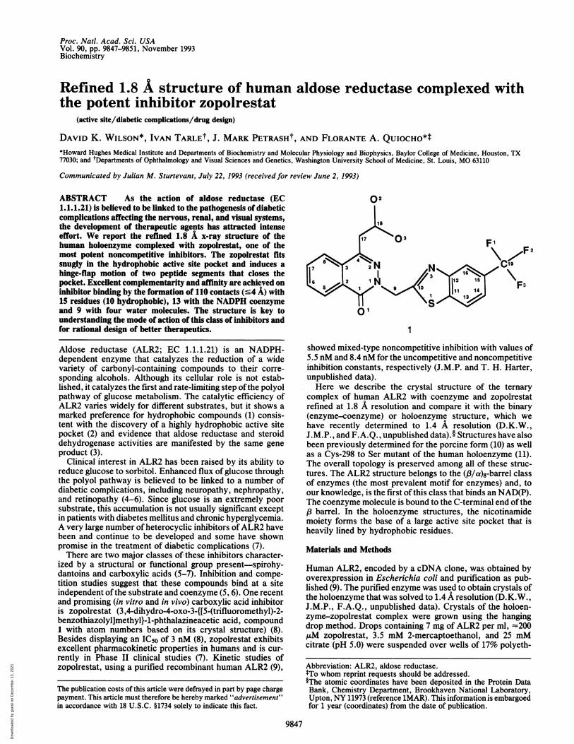
ized by a structural or functional group present-spirohy-dantoins and carboxylic acids (5-7). Inhibition and compe-tition studies suggest that these compounds bind at a siteindependent of the substrate and coenzyme (5, 6). One recentand promising (in vitro and in vivo) carboxylic acid inhibitoris zopolrestat (3,4-dihydro-4-oxo-3-{[5-(trifluoromethyl)-2-benzothiazolyl]methyl}-l-phthalazineacetic acid, compound1 with atom numbers based on its crystal structure) (8).Besides displaying an IC50 of 3 nM (8), zopolrestat exhibitsexcellent pharmacokinetic properties in humans and is cur-rently in Phase II clinical studies (7). Kinetic studies ofzopolrestat, using a purified recombinant human ALR2 (9),
The publication costs of this article were defrayed in part by page chargepayment. This article must therefore be hereby marked "advertisement"in accordance with 18 U.S.C. §1734 solely to indicate this fact.
Fj F2
C\7F
( F3
01
1
showed mixed-type noncompetitive inhibition with values of5.5 nM and 8.4 nM for the uncompetitive and noncompetitiveinhibition constants, respectively (J.M.P. and T. H. Harter,unpublished data).Here we describe the crystal structure of the ternary
complex of human ALR2 with, coenzyme and zopolrestatrefined at 1.8 A resolution and compare it with the binary(enzyme-coenzyme) or holoenzyme structure, which wehave recently determined to 1.4 A resolution (D.K.W.,J.M.P., and F.A.Q., unpublished data).§ Structures have alsobeen previously determined for the porcine form (10) as wellas a Cys-298 to Ser mutant of the human holoenzyme (11).The overall topology is preserved among all of these struc-tures. The ALR2 structure belongs to the (13/a)8-barrel classof enzymes (the most prevalent motif for enzymes) and, toour knowledge, is the first of this class that binds an NAD(P).The coenzyme molecule is bound to the C-terminal end of the(3 barrel. In the holoenzyme structures, the nicotinamidemoiety forms the base of a large active site pocket that isheavily lined by hydrophobic residues.
Materials and Methods
Human ALR2, encoded by a cDNA clone, was obtained byoverexpression in Escherichia coli and purification as pub-lished (9). The purified enzyme was used to obtain crystals ofthe holoenzyme that was solved to 1.4 A resolution (D.K.W.,J.M.P., F.A.Q., unpublished data). Crystals of the holoen-zyme-zopolrestat complex were grown using the hangingdrop method. Drops containing 7 mg of ALR2 per ml, =200,uM zopolrestat, 3.5 mM 2-mercaptoethanol, and 25 mMcitrate (pH 5.0) were suspended over wells of 17% polyeth-
Abbreviation: ALR2, aldose reductase.1To whom reprint requests should be addressed.§The atomic coordinates have been deposited in the Protein DataBank, Chemistry Department, Brookhaven National Laboratory,Upton, NY 11973 (reference 1MAR). This information is embargoedfor 1 year (coordinates) from the date of publication.
9847
Dow
nloa
ded
by g
uest
on
Dec
embe
r 13
, 202
1

Proc. Natl. Acad. Sci. USA 90 (1993)
FIG. 1. Zopolrestat bound to ALR2. (Top) Difference electron density (magenta) of the zopolrestat and superimposed refined structure(atoms color coded). The density (contoured at 3o) was calculated with coefficients (IFol - IFcl) and ac phases from the refined 1.8 A structurewith the inhibitor atoms omitted. The C4 atom of the nicotinamide ring of NADPH is identified as well as some residues involved in inhibitorbinding and possibly in catalysis (Tyr-48 and His-11O). (Middle) Hydrogen bonds (dashed lines) between the zopolrestat and enzyme.
9848 Biochemistry: Wilson et al.
Dow
nloa
ded
by g
uest
on
Dec
embe
r 13
, 202
1

Proc. Natl. Acad. Sci. USA 90 (1993) 9849
ylene glycol 6000, 7 mM 2-mercaptoethanol, and 50 mMcitrate (pH 5.0). A different crystal morphology than those ofthe holoenzyme crystals indicated a possible new crystalform. The diffraction data set from 20 A to 1.8 A resolutionwas collected on a Rigaku R-Axis area detector mounted on
a rotating anode generator equipped with a graphite mono-chromator and operated at 50 kV and 90 mA. The indexing ofthis data set indicated a space group ofP1 and a unit cell withdimensions of a = 47.64 A, b = 48.04 A, c = 40.48 A, a =
67.470, A3 = 76.77°, and y = 76.07° and a content of oneenzyme molecule. Merging of 53,037 measured reflectionsresulted in an R-merge of 0.034 (based on intensity) and26,145 unique reflections (90.4% complete). In the structuredetermination we used 25,891 reflections with F > lo(F)between 12 A and 1.8 A resolutions.The structure of the ternary complex was determined by
molecular replacement (12) using the refined 1.4 A holoen-zyme structure (solvent molecules omitted) as a search model.Rotation function and structure refinement were carried outusing the X-PLOR suite of programs (13). The angles definingthe highest rotation peak (01 = 480, 02 = 84°, and 03 = 90°) inthe rotation search using amplitudes between 20 A and 6 Awere applied directly to the model without any Patterson-correlation refinement. After several cycles of rigid bodyrefinement, using reflections between 12 A and 4 A, thecrystallographic R-factor [= (7, I I FO| - IF, I I /E IFO|)] decreasedfrom an initial value of 0.365 to 0.29. Following one round ofenergy minimization and positional and B-factor refinementusing 25,891 reflections from 12 to 1.8 A resolution, (2 |FOl -lFcl, ac) and (|FOl - IFCI, a,:) electron density maps werecalculated. These maps revealed a large area of clear, detaileddensity in the active site pocket that was easily identified andfitted as the bound zopolrestat molecule. The enzyme-inhibitor model was subjected to several cycles of manualrefitting of residues that undergo conformational change andmodeling of ordered water molecules and positional andB-factor refinements until the R-factor converged to a finalvalue of 0.180 at 1.8 A resolution. The model has goodgeometry-r.m.s. from ideality of 0.006 A for bonds and 1.65°for angles. It consists of 2517 atoms from the entire 315residues, 48 coenzymes atoms, 29 zopolrestat atoms, and 175water molecules.
Results and Discussion
The bound zopolrestat, with its very well-defined 1.8 Adifference electron density (Fig. 1 Top), occupies almost theentire active site pocket at the C-terminal end of the 3barrel(Fig. 2). The binding has many features of a potent inhibitor.The phthalazinone ring is almost at a right angle to and bisectsthe plane of the benzothiazole ring (Figs. 1-3). The phthalazi-none ring, protruding from the center of the pocket, isperpendicular to the nicotinamide ring of NADPH. Thebenzothiazole ring, lying opposite the nicotinamide ring,straddles the 3 barrel between strands 4 and 5 (Fig. 2). Thebound inhibitor is almost completely sequestered in thecavity; it has an accessible surface area of 12.7 A2, 6.4% ofthe value for the unbound inhibitor.
There is excellent complementarity between the boundzopolrestat and the binding site. The inhibitor makes anunusually large number of contacts with the active site,totaling 132 contacts with -4 A distances-110 with 15 resi-dues, 13 with the nicotinamide moiety of the coenzyme, and9 with 4 ordered water molecules. van der Waals contactsconstitute an overwhelming number, slightly over 90%. Fur-thermore, as shown in Fig. 1 Middle, there are 9 hydrogenbonds of which 2 are long (3.7 A) and 1 is a salt link.About half of the van der Waals contacts are between
carbon atoms, attesting to the highly hydrophobic nature ofthe enzyme-inhibitor interaction. Indeed, 11 apolar residues,originally found lining the active site pocket (Trp-20, Tyr-48,Trp-79, Trp-111, Phe-115, Phe-122, Trp-219, Ala-299, Leu-300, Tyr-309, and Pro-310) in the holoenzyme structure (2),are involved in these contacts. Many of these residues,especially the four residues (Trp-20, Trp-111, Phe-122, andLeu-300) that interface with the two heterocyclic rings, areshown in Figs. 1 and 3. Residue Trp-111, which stacks againstthe A face of the benzothiazole ring, plays a dominant role bymaking 38 contacts, by far the most of any residue (Figs. 1Top and Middle and 3). The side chain ofLeu-300 apposes theB face of the benzothiazole. The phthalazinone ring issandwiched by Trp-20 and Phe-122 (Figs. 1 and 3). As thesefour strategically placed hydrophobic residues make 65 con-tacts with the inhibitor, they are major determinants ofmolecular recognition. The marked preference of the enzymefor hydrophobic substrates (e.g., steroids) is consistent withthe mode of binding of the inhibitor.
Superpositioning of the ternary complex and holoenzymestructures reveals inhibitor-induced conformational changesof a loop (residues 121-135) and a short segment (residues298-303) near the C-terminal end in order to accommodateand sequester the inhibitor (Fig. 3). These changes could beascribed as hinged-flap motions. The displacement of Leu-300 as a result of inhibitor binding is accompanied by themovement of the segment 298-303 away from the pocket.Loop 121-135 moves toward the pocket, thus enabling Phe-122 to participate in inhibitor binding and to form, withLeu-300, a hydrophobic bridge over the bound inhibitor(Figs. 1 and 3). As shown in Fig. 3, several other residuescontained in the loop and the segment undergo conforma-tional changes. Moreover, the loop and the segment makefavorable interactions as both slide toward each other. (Theseinteractions are not present in the holoenzyme structure.)The superpositioned structures reveal no other similar per-turbations. The formation of the hydrophobic bridge and thecoalescing of the loop and segment are largely responsible insequestering the inhibitor. It is noteworthy that the coenzymeis also strapped in place by the association of a different pairof loops that also deploy several residues that bind thecoenzyme (2). Thus we have established that inhibitor andcoenzyme binding induces conformational changes. It wouldnot be surprising that substrates also possess this property.The interaction associated with the carboxylate moiety of
the inhibitor may have some bearing on enzyme catalysis.The carboxylate 03 atom, which could mimic the carbonyloxygen of a substrate, is within very favorable distance (2.65
The C4 atom of nicotinamide ring is labeled. The following polar groups of the inhibitor (1) are recipients of a total of9 hydrogen bonds (distancesfrom 2.8 to 3.7 A): 01, close to the enzyme surface, from a water molecule; 02 from His-110 Ne2H and Trp-111 Ne1H; 03 from Tyr-48 0iqHand His-110 NE2H; N2 from Cys-298 SyH; N3 from the backbone NH group of Leu-300; Fl from Thr-113 OylH; and F2 from Thr-113 Oy1H.With the exception of the long hydrogen bonds (3.7 A) associated with N2 and F2, the others have distances between 2.8 to 3.4 A. Si is nearTrp-111 Ne1, but the long distance (3.8 A) and acute angle (about 900) indicate a weak hydrogen bond between the two. A similar situation occursbetween F2 and SyH of Cys-303. As the carboxylate group ofthe inhibitor and the side chain ofHis-10 are in close contact, they are also involvedin a salt link. (Bottom) Complementary molecular surfaces between zopolrestat (green dot surface) and enzyme (red dot surface). The enzymeand inhibitor models are colored according to atom types. The nicotinamide-ribosyl-pyrophosphoryl portion of the coenzyme is shown in thelower left-hand corner. The benzothiazole ring, which is in the plane of the figure, is sandwiched by Trp-111 (below the ring) and Leu-300 (abovethe ring). The phthalazinone ring (perpendicular to the plane of the figure) is sandwiched between Trp-20 (left of the ring) and Phe-122 (rightof the ring).
Biochemistry: Wilson et al.
Dow
nloa
ded
by g
uest
on
Dec
embe
r 13
, 202
1

Proc. Natl. Acad. Sci. USA 90 (1993)
FIG. 2. Stereo perspective view (down the C terminus of the P barrel) of the backbone trace of the structure ofALR2 with bound zopolrestatand coenzyme. The zopolrestat and coenzyme molecules are drawn as stick models, and the a helices and , strands are represented as coilsand flattened arrows, respectively. The inhibitor bent at right angle is to the left of the extended coenzyme. The figure was drawn usingMOLSCRIPT (14).
A) to the oxygen of Tyr-48 0'qH, which has been proposedas a proton-donor group based on the holoenzyme structure(2). The proposed function ofTyr-48 is consistent with recentsite-directed mutagenesis studies (16). The 03 is at a longerdistance (2.89 A) to the nitrogen ofNe2H of His-110, anotherpossible proton donor. The carboxylate 02 is farther awayfrom both Tyr-48 O°7 and His-110 NE2 atoms. The C18 of thecarboxylate, which would be analogous to the carbon thataccepts the hydride in a substrate, is 3.63 A of the C4 of thecoenzyme.
Zopolrestat binding displaces at least six ordered watermolecules found in the pocket in the 1.4 A holoenzymestructure. This contributes favorable entropic effect to thetight binding of the inhibitor. The well-resolved, unidentifieddensity in the active site that has been observed in both of the
FIG. 3. Stereoview of the superimposed a-carbon backbonestructures of the holoenzyme (red) and its complex with zopolrestat(green). Portions of both models (including the coenzyme) thatcoincide take on a yellow color. Only the region containing the loop(residues 121-135) and the segment (residues 298-303) that undergoinhibitor-induced conformational change are shown. The rest of thesuperimposed structures not shown are virtually identical. Severalresidues that undergo substantial conformational changes are alsoshown. The zopolrestat molecule is to the left of the nicotinamidering of the coenzyme. The program CHAIN (15), developed in ourlaboratory, was used in the electron-density fitting and molecularmodeling in the structure refinement and in generating Figs. 1 Topand Middle and 3.
1.4 A and 1.65 A (2) refined structures of the holoenzyme isno longer present in the ternary complex structure. Interest-ingly, we found that the portion of the unknown densityclosest to the nicotinamide ring coincides with the density ofthe carboxylate group of the bound inhibitor.To our knowledge, none of the many potent inhibitors of
ALR2 displays competitive inhibition with aldehyde as thevariable substrates. Rather, these inhibitors (including zopol-restat) commonly show noncompetitive inhibition, which islikely a reflection of the following: (i) the sequential reactionmechanism of ALR2 with NADPH binding first (17), (ii) theconformational changes associated with binding and releaseof the coenzyme, inhibitor, and substrate, and (iii) the verytight affinity of the inhibitors (several with affinities 2-7orders ofmagnitude greater than the Km values of substrates).The suggestion, based on these kinetic results, that theinhibitors bind at a site that is independent of the active site(4-6), is difficult to explain in structural terms given ourresults showing atomic interactions between the inhibitor andthe active site. Moreover, in the ternary complex structure,the active site pocket (Fig. 1) is not only completely inac-cessible to solvent but also devoid ofa space large enough forfurther productive binding of even a small substrate such asglyceraldehyde. In addition, there does not appear to beanother site in the enzyme as accommodating and comple-mentary to the inhibitor as the active site pocket.Although the search thus far for ALR2 inhibitors has not
benefited from the tertiary structure ofthe target enzyme, thedesign of zopolrestat has apparently and gratifyingly satisfiedmany requirements of a potent inhibitor. The structure of theternary complex does indicate that the potency and pharma-cologic requirements of this compound may be further opti-mized. It is also a key in rational design of new inhibitorstailored specifically for ALR2, a member of a large family ofstructurally and functionally related oxidoreductases.
We thank Prof. G. N. Phillips, Jr., of Rice University for the useof the R-Axis and Dr. K. Johnson for assisting in the data collection.We thank the coworkers in each laboratory for valuable help.Zopolrestat was kindly provided by Pfizer, Inc. F.A.Q. is an Inves-tigator of the Howard Hughes Medical Institute. Further support hasbeen provided by National Institutes of Health Grants (EY05856,T32EY07108, and P60DK20579) and in part by grants to the Depart-ment of Ophthalmology (J.M.P.) from Research to Prevent Blind-ness, Inc.
9850 Biochemistry: Wilson et al.
Dow
nloa
ded
by g
uest
on
Dec
embe
r 13
, 202
1

Biochemistry: Wilson et al.
1. Morjana, N. A. & Flynn, T. G. (1989) J. Biol. Chem. 264,2906-2911.
2. Wilson, D. K., Bohren, K. M., Gabbay, K. H. & Quiocho,F. A. (1992) Science 257, 81-84.
3. Warren, J. C., Murdock, G. L., Ma, Y., Goodman, S. R. &Zimmer, W. E. (1993) Biochemistry 32, 1401-1406.
4. Kador, P. F. (1988) Med. Res. Rev. 8, 325-352.5. Sarges, R. (1989) Adv. Drug Res. 18, 139-175.6. Kador, P. F. & Sharpless, N. E. (1983) Mol. Pharmacol. 24,
521-531.7. Sarges, R. & Oates, P. J. (1993) Prog. Drug Res. 40, 99-161.8. Mylari, B. L., Larson, E. R., Beyer, T. A., Zembrowski,
W. J., Aldinger, C. E., Dee, M. F., Siegel, T. W. & Singleton,D. H. (1991) J. Med. Chem. 34, 108-122.
9. Petrash, J. M., Harter, T. M., Devine, C. S., Olins, P. O.,Bhatnagar, A., Liu, S. & Srivastava, S. K. (1992) J. Biol.Chem. 267, 24833-24840.
Proc. Natl. Acad. Sci. USA 90 (1993) 9851
10. Rondeau, J.-M., Tete-Favier, F., Podjarny, A., Reymann,J. M., Barth, P., Biellmann, J.-F. & Moras, D. (1992) Nature(London) 355, 469-472.
11. Borhani, D. W., Harter, T. M. & Petrash, J. M. (1992) J. Biol.Chem. 267, 24841-24847.
12. Rossmann, M. G. (1972) The MolecularReplacement Method:A Collection of Papers on the Uses of Non-CrystallographicSymmetry (Gordon and Reach, New York).
13. Brunger, A. T. (1992) X-PLOR: A System for Crystallographyand NMR (Yale Univ. Press, New Haven, CT), 3.0 Manual.
14. Kraulis, P. J. (1991) J. Appl. Crystallogr. 24, 946-950.15. Sack, J. S. (1988) J. Mol. Graphics 6, 224-245.16. Tarle, I., Borhani, D. W., Wilson, D. K., Quiocho, F. A. &
Petrash, J. M., J. Biol. Chem., in press.17. Kubiseski, T. J., Hyndman, D. J., Morjana, N. A. & Flynn,
T. G. (1992) J. Biol. Chem. 267, 6510-6517.
Dow
nloa
ded
by g
uest
on
Dec
embe
r 13
, 202
1