Embed Size (px)
Citation preview

J. Embryol. exp. Morph. 93, 213-238 (1986) 213Printed in Great Britain © The Company of Biologists Limited 1986
Regulation of development in the fully grown mouseoocyte: chromosome-mediated temporal and spatialdifferentiation of the cytoplasm and plasma membrane
JONATHAN VAN BLERKOM AND HOBART BELLDepartment of Molecular, Cellular and Developmental Biology, University ofColorado, Boulder, Colorado, USA
SUMMARYThe relationship between nuclear maturation and the differentiation of the cytoplasm and
plasma membrane during resumption of arrested meiosis was investigated by culture of GV- andMil-stage mouse oocytes in the presence and absence of nocodazole. Culture in the presence ofnocodazole was associated with dispersal of MI and Mil chromosomes throughout the sub-plasmalemmal cytoplasm. A progression of cortical (thickening of actin filaments) and plasmamembrane changes (denudation of microvilli, reduction in cell surface glycoproteins, formationof chromosome-containing evaginations) that normally occurs in proximity to chromosomesassociated with intact MI or Mil spindles took place only in those regions of the corticalcytoplasm containing the dispersed subplasmalemmal chromosomes. The dispersion and mi-gration of the chromosomes occurred in an apparently random fashion. Fluorescent probeanalysis of normal and treated oocytes indicated a stage-specific association between the spatialdistribution of chromosomes and mitochondria. Transfer of individual bivalent chromosomes tountreated oocytes at different stages of maturation and to cytoplasts derived from oocytesanucleated prior to GVB demonstrated (1) the necessity of chromosomes for cytoplasmic andplasma membrane differentiation, and (2) that the capacity of the cytoplasm and plasma mem-brane to differentiate in response to the presence of a chromosome is acquired prior to GVB.
INTRODUCTION
The fully grown mammalian oocyte develops the ability to be fertilized duringthe period of resumed meiosis that occurs no sooner than several hours prior toovulation. Fertilizability is the culmination of a rapid progression of intrinsicallyregulated changes (Van Blerkom, 1985a) in nuclear (Zamboni, 1970; VanBlerkom & Runner, 1984), cytoplasmic (Zamboni, 1970; Van Blerkom & Motta,1979; Van Blerkom & Runner, 1984) and plasma membrane structure and organ-ization (Johnson, Eagery, Muggleton-Harris & Graves, 1975; Nicosia, Wolf &Inoue, 1977; Maro, Johnson, Webb & Flach, 1986). In the laboratory mouseoocyte, cytoplasmic maturation involves changes in the organization and spatialdistribution of Golgi complexes (Zamboni, 1970), cortical granules (Nicosia etal.1977) and mitochondria (Van Blerkom & Runner, 1984). At the molecular level,oocyte maturation is characterized both by the cessation of transcription prior to
Key words: mouse oocyte, regulation, nocodazole, nuclear maturation, chromosomes, plasmamembrane.

214 J. VAN BLERKOM AND H. BELL
germinal vesicle breakdown (GVB) (Wassarman & Letourneau, 1976) and by theexpression of stage-related alterations in quantitative and qualitative patterns ofprotein synthesis and post-translational modification (Schultz & Wassarman, 1977;Richter & McGaughey, 1983; Van Blerkom, 1985c).
Developmental changes in the structure, organization and composition of theplasma membrane and cortical cytoplasm are restricted to the region of the oocytethat contains MI or Mil chromosomes that are associated with their respectivespindles. The cortical cytoplasm overlying the metaphase spindles is devoid ofcortical granules (Nicosia et al. 1977), Golgi complexes (Van Blerkom & Motta,1979) and mitochondria (Zamboni, 1970; Nicosia et al. 1977). The plasma mem-brane in this region is denuded of microvilli (Johnson et al. 1975), contains asignificantly reduced population of cell surface glycoproteins (Eager, Johnson &Thurley, 1976) and exhibits a marked change in the distribution and organizationof diffusible membrane proteins (Wolf & Ziomek, 1983). Differentiation of thesubplasmalemmal cytoplasm associated with the presence of the metaphasespindles is indicated by a pronounced, localized thickening of actin filaments(Nicosia et al. 1977; Maro, Johnson, Pickering & Flach, 1984). These changesestablish an apparent polarity in the preovulatory oocyte that is expressed in thestructure of the cytoplasm and plasma membrane that overlies the metaphasespindles (Johnson etal. 1975; Longo & Chen, 1985).
Studies of nuclear, cytoplasmic and molecular development during reinitiatedmeiosis suggest that maturation of the mouse oocyte is regulated by an intrinsicprogramme directed at the cytoplasmic rather than at the genomic level (VanBlerkom, 1985a). The present study examines the relationship between nuclearchanges and developmental alterations in the spatial distribution of mitochondria,and the structure and organization of the cortical cytoplasm and plasma membraneduring resumed meiosis in the presence of nocodazole, a potent inhibitor ofmicrotubule polymerization (Hoebeke, Van Nigen & De Brabander, 1976). Wereport that structural" and molecular changes in the oocyte cytoplasm, plasmamembrane and underlying cortex that normally occur in proximity to MI and Milchromosomes and associated spindles, (1) are mediated or induced by thepresence of chromosomes, (2) do not appear to involve direct genomicparticipation or expression and (3) may require a nuclear-dependent modificationof the cytoplasm.
MATERIALS AND METHODS
Oocyte collection and cultureGerminal-vesicle-stage oocytes (GV) were obtained from large ovarian follicles of adult HS
mice and freed of surrounding granulosa cells (if present) by repeated passage through amicropipette. Only GV-stage oocytes between 70 and 75jum in diameter were used. Oocyteswere cultured under paraffin oil in groups of 100 in 50-75 jul droplets of Brinster's BMOC-3supplemented with 4% BSA and prepared to 280-290 mosmol kg"1. Oocytes matured in vivowere harvested from the antral follicles of HS mice previously injected with 5i.u. of PMSGfollowed in 48 h by 5 i.u. of hCG. Mil-stage oocytes were collected at 10,12 and 14 h post-hCG.Oocytes were cultured in the presence or absence of nocodazole (methyl 5-[2-thienylcarbonyl-

Chromosomally induced differentiation of the oocyte 215
lH-benzimimidazol-2-yl]carbamate) (Sigma Chemical Co.) at a concentration of 1 oras follows: GV-stage oocytes were continuously exposed to nocodazole for up to 36 h. Mil-stageoocytes, matured in vivo or in vitro, were exposed to nocodazole for up to 25 h. Oocytes werecultured in humidified chambers maintained at 37 °C in an atmosphere of 90 % N2,5 % CO2, 5 %O2.
Visualization of mitochondria, chromosomes, actin filaments and surfaceglycoproteins
Mitochondrial distributions in the cytoplasm of untreated and nocodazole-exposed oocyteswere visualized by fluorescence microscopy after staining with Sjugml"1 of rhodamine 123 for3min at room temperature (Van Blerkom & Runner, 1984). Chromosomal configurations andpositions within the cytoplasm were determined by exposure of oocytes to medium containingthe fluorescent nuclear DNA probes Hoechst dye 33258 (bisbenzimide, Calbiochem) or DAPI(4',6-diamino-2-phenyl-indole, Sigma Chemical Co.) for 3min at room temperature at aconcentration of lOjUgml"1. Either NBD-phallacidin (Barak, Yocum, Nothnagel & Webb,1980) (Molecular Probes, Inc., Piano, TX) or anti-actin monoclonal antibody was used tovisualize cortical actin filaments. All immunostaining procedures utilized PHEM buffer (Schliwa& Van Blerkom, 1981) and were performed at room temperature. For immunostaining, oocyteswere washed in PHEM (Schliwa & Van Blerkom, 1981), extracted with 0-15 % Triton X-100 for4-5 min and rinsed in buffer. Immunofluorescent staining of cells was accomplished using1 Hgm\~l mouse anti-actin monoclonal antibody followed by fluorescein-labelled anti-rabbitimmunoglobin antibodies. Detergent-extracted oocytes were cultured for 10 min in PHEMbuffer containing NBD-phallacidin at a concentration of 5 fig ml"1. The distribution of mannose-containing surface glycoproteins was examined by staining unextracted oocytes with FITC-Concanavalin A (700 fig ml"1, Polysciences) for 8 min at room temperature (Johnson etal. 1975).Examinations of living oocytes by DIC and fluorescence microscopy involved incrementaloptical sections (serial) of entire oocytes. Preparation of oocytes and embryos for fluorescentvisualization utilized methods previously described (Van Blerkom & Runner, 1984). Afterfluorescent analysis, oocytes and embryos were examined by differential interference contrastmicroscopy and at least 20 representative oocytes were prepared for electron microscopy asdescribed by Van Blerkom & Runner (1984).
Chromosomal transferGV-stage oocytes cultured continuously in the presence of nocodazole for 24 h frequently
contained individual bivalent chromosomes located at the apex of a distinct evagination of theplasma membrane and subjacent cortical cytoplasm. MI chromosomes were transferred toeither intact GV-stage oocytes or to cytoplasts derived from oocytes chemically anucleated at 0,30,45 and 60 min after reinitiation of meiosis in vitro. Chemical enucleation was accomplished by5 min exposure of GV-stage oocytes to medium containing cytochalasin B (5 figmV1) (Schultz,Letourneau & Wasserman, 1978). During this time oocytes underwent pseudocleavage andfrequently displayed a nuclear compartment with scant residual cytoplasm and a single, largecytoplasmic fragment - a cytoplast (Van Blerkom, 1985b). In the present experiments,chromosome transfers were made only to the large cytoplasts.
Transfer of bivalent chromosomes was accomplished by means of a glass micropipette (Leitzmicroelectrode glass, coated with Prosil-28, PCR Res. Chem. Inc., Gainesville, FL) preparedon a vertical pipette puller (Koph Instruments, Tujunga, CA). This instrument producedpipettes with a long taper that was sealed at the end. The pipettes were mechanically bevelled toan angle of approximately 45° with an orifice estimated to be approximately 1-5 jum. Holdingpipettes were formed by fire polishing one end of a micropipette to reduce the orifice toapproximately 20-30 ̂ m.
Transfers were made under phase contrast or differential interference contrast optics, butusually without direct visualization of an individual chromosome. Typically, the micropipettewas injected through the zona pellucida and placed in the apical aspect of a cytoplasmicevagination to a depth of approximately 1-2 jum. A Teflon tube attached to the injection pipettewas completely filled with heavy paraffin oil (BDH) to within 2 mm of the tip. Once inside the

216 J. V A N BLERKOM AND H. B E L L
evagination, cortical contents were withdrawn by means of a microsyringe calibrated in in-crements of 0-1 jul (Hamilton Co., Reno, NV) until most of the evagination had collapsedaround the pipette. The contents of the pipette were transferred to the central portion or to thecortical cytoplasm of a recipient oocyte or cytoplast. The entry made by the micropipette in thezona pellucida was frequently visible after microinjection and served as a positional marker forsubsequent analysis. After chromosomal removal, the donor oocytes were fixed in glu-taraldehyde (1 % in BMOC-3), stained with DAPI and examined by fluorescence to determinewhether chromosomal removal had been accomplished. Recipient oocytes were cultured in thepresence or absence of nocodazole and were prepared for examination by light, fluorescenceand electron microscopy at 4 h intervals after transfer. Cortical cytoplasm obtained from regionsbetween chromosome-containing evaginations was transferred to the subplasmalemmal cyto-plasm of cytoplasts and intact oocytes by means of a micropipette.
Analysis of chromosomal movements by time-lapse video recording after removal ofnocodazole
Chromosomal movements in living oocytes and cytoplasts were continuously monitored bytime-lapse video recording utilizing a Panasonic NV-8050 video recorder with an integratedtime/event generator. Oocytes were cultured on glass coverslips in a stainless steel chambercontaining 2 ml of medium equilibrated with gas and overlaid with light paraffin oil to a thicknessof 2 mm. The chamber was sealed at the top with a glass microscope slide but contained two1 mm ports to allow for exchange of medium or atmosphere. To maintain proper pH and cultureconditions, a gentle stream of humidified gas composed of 5 % CO2, 5 % O2, and 90 % N2 wascontinuously introduced into the chamber. Chromosomal movements were examined by DICmicroscopy on a Nikon Diaphot microscope attached to a Dage-MTI video camera. The entiremicroscope was enclosed by Plexiglas such that the internal temperature could be maintained at37°C (Nikon NP-2 incubator heater). Recordings were made on high-resolution video tape(HD-PRO, TDK).
RESULTS
Chromosomal movements and mitochondrial distributions in normal and nocodazole-treated oocytes
Oocytes cultured from the GV stage in the presence of 1 or lO/igml"1 ofnocodazole (Fig. 1A,B) progressed through the normal sequence of nuclear andcytoplasmic changes characteristic of development to the late GVB stage (VanBlerkom & Runner, 1984). For most experiments, nocodazole at a concentrationof 1 /ig ml"1 was used. Time-lapse video recordings demonstrated identical nuclearand cytoplasmic morphodynamics in normal and treated oocytes. Morphodynamicchanges included nucleolar dissolution between 1-0 and 1-5 h, GVB between 1-75and 2-0h (Fig. 2B), appearance of cortical Golgi complexes beginning at 2-5 h(Fig. 2D), and aggregation of mitochondria at 3-5 h that resulted in theappearance of small mitochondrial clusters uniformly distributed in the cytoplasmbetween 4-0 and 4-5 h (Fig. 2A).
The first visual indication of post-GVB nuclear maturation was the presenceof bivalent chromosomes (circular bivalent stage, CBV) in untreated oocytesat approximately 3-4h of culture (Fig. 1C,D). In treated oocytes, bivalentchromosomes did not circularize but rather formed a dense, tightly compactedmass in the central portion of the oocyte (Figs 1E,F, 2D). At 6 h of culture innocodazole-containing medium, mitochondria were still present in small clusters

Chromosomally induced differentiation of the oocyte 217
in the cytoplasm (Fig. 2C), whereas in untreated oocytes, translocation and peri-nuclear accumulation of mitochondria occurred (Fig. 1G,H). Untreated oocytesformed the first metaphase (MI) spindle between 6 and 7 h of culture (Fig. 1G,H).
By 11 h of culture, abstriction of the first polar body had taken place inuntreated oocytes (Fig. II,J), while in treated oocytes two types of chromosomaldistribution were observed. Examination of 3400 treated oocytes demonstratedthat after 12 h of exposure to nocodazole approximately 2000 (60 %) oocytes had atightly compacted mass of chromosomes in the cortical region of the cytoplasm
Fig. 1. Differential interference contrast [DIC] (A,C,E,G,I,K) and DAPI-fluor-escence images (B,D,F,H,J,L) of mouse oocytes cultured in the presence (E,F,K,L)and absence (A,B,C,D,G,H,I,J) of lO/igml"1 of nocodazole. Fig. 1B,D,G demon-strate the appearance of chromatin and chromosomes at the germinal vesicle (GV,Fig. 1A), circular bivalent (CBV, Fig. 1C) and metaphase II stages (Fig. II) of normaloocyte maturation in vitro, respectively. Perinuclear aggregation of mitochondria (m)is visible both by DIC (Fig. 1G) and fluorescence microscopy (Fig. 1H). Oocytescultured in the presence of nocodazole (Fig. IE) displayed a tightly compacted mass ofcentrally located chromosomes (Fig. IF). In approximately 80 % of these oocytes, themass of chromosomes migrated to a cortical location (Fig. 1L) and appeared to initiatepolar body formation (Fig. IK). N, nuclear envelope; n, nucleolus; pb, first polarbody; c, chromosomes.

218 J. VAN BLERKOM AND H. BELL
40*
Fig. 2. (A) Rhodamine 123 staining of a living mouse oocyte at 3-Oh after initiationof culture in vitro. Fluorescent patterns of rhodamine 123 distribution demonstratethe presence of mitochondrial clusters (arrows) throughout the cytoplasm. (B)Transmission electron micrograph of the oocyte presented in Fig. 2A demonstrateschromosomal condensations (c), appearance of perichromatin granules (peg) anddiscontinuities in the structure of the nuclear membrane (nm) in an oocyte under-going germinal vesicle breakdown. X6200. (C) Rhodamine 123 staining revealedcytoplasmic clustering of mitochondria (arrows) in this oocyte cultured in the presenceof nocodazole for 6h from the GV stage. Perinuclear aggregation of mitochondria didnot occur in the presence of the inhibitor. (D) Transmission electron micrograph of thenocodazole-exposed oocyte shown in Fig. 2C. At 6 h of culture in the presence ofnocodazole, a compacted mass of bivalent chromosomes (c) occupied the centralportion of the oocyte. m, mitochondria, G, golgi complex. X4300.

Chromosomally induced differentiation of the oocyte 219
(Fig. 6B) and 1400 (40 %) had a condensed mass of chromosomes occupying thecentral portion of the cytoplasm (Fig. 6A). In contrast to the findings in untreatedoocytes at the CBV stage (Van Blerkom, 1985a), fine structural analysis of serialthick (HVTEM) and thin sections (TEM) failed to indicate the presence ofcytoplasmic microtubules associated with the mass of condensed, bivalentchromosomes (Fig. 2D). No signs of mitochondrial perinuclear aggregation wereobserved, but mitochondrial clusters still were distributed throughout thecytoplasm of nocodazole-treated oocytes in an apparently random fashion (similarto the distribution shown in Fig. 2C).
Between 12 and 14 h of culture, movement of the chromosomal mass to thecortical cytoplasm occurred in nearly half of the oocytes that previously showedcentrally located chromosomes. At 14-16 h of culture in the presence ofnocodazole, an evagination of the plasma membrane similar to the initial stages ofpolar body formation was observed in 20 % of those oocytes that previously hada single mass of chromosomes located in the cortical cytoplasm (Fig. 1K,L).Abstriction of a polar body, or a polar-body-like structure was not observed in anyof the nearly 600 oocytes that presented cortical evaginations similar to that shownin Fig. IK, even after as many as 36 h of culture.
A striking cytoplasmic distribution of chromosomes occurred between 16 and18 h of culture in oocytes that had been exposed continuously from the GV stage tonocodazole. At approximately 16 h of culture, the compacted mass of centrally orcortically located chromosomes began to disaggregate. Regardless of the originallocation of the mass of chromosomes, three patterns of chromosomal disag-gregation and dispersion were observed: (1) approximately 30% (1000) of alloocytes cultured in the presence of nocodazole displayed individual bivalentchromosomes dispersed throughout the cortical cytoplasm (Figs 3A-C, 6D,E,G),(2) 50% of the oocytes (approximately 1600) exhibited a cortical cytoplasmcontaining chromosomes present individually and in groups of two to fivechromosomes, and (3) approximately 20 % of the 3400 oocytes examined showedno disaggregation or further movement of either a cortically or centrally locatedmass of chromosomes.
Between 2 and 4h after bivalent chromosomes first appeared beneath theplasma membrane (20-22h of culture), the subplasmalemmal cytoplasm andplasma membrane overlying the dispersed chromosomes evaginated (Figs 4A-C,6K,L). Morphologically, the evaginations resembled the initial stages of polarbody formation. However, abstriction of these structures was not observed in anyof the oocytes cultured in the presence of nocodazole. For oocytes in whichcomplete disaggregation of the chromosomal mass occurred, fluorescent stainingwith Hoechst dye 33258 or D API demonstrated that the apex of each evaginationcontained a single bivalent chromosome (Figs 4B, 6L). Although nocodazoleperturbs the process of microtubule assembly, reconstructions of serial sectionsrevealed compact bundles of short microtubules attached to dispersed bivalentchromosomes (Figs 3A-C, 4C) in oocytes cultured continuously from the GVstage in the presence of the inhibitor. The pattern of cytoplasmic distribution of

220 J. VAN BLERKOM AND H. BELL
mitochondrial clusters was unaltered during the formation of chromosome-associated evaginations.
Within l h of exposure to medium containing nocodazole, approximately 70 %(920) of 1400 metaphase Il-stage (Mil) oocytes (12 h after initiation of culture)

Chromosomally induced differentiation of the oocyte 221
displayed dissociation of chromosomes from the metaphase spindle. After 2additional hours of culture, individual or groups of three to eight chromosomeswere dispersed within the subplasmalemmal cytoplasm. After 4-6 h of exposure tonocodazole, evaginations of the cortical cytoplasm (Fig. 5A) containing individualMil chromosomes or, more typically, groups of three to eight chromosomes(Fig. 5B,C) were evident. In contrast to MI chromosomes dispersed in the corticalcytoplasm of oocytes exposed to nocodazole from the GV-stage, cortically locatedMil chromosomes had no associated microtubules (Fig. 5C,D). No specificcorrelation between the distribution of mitochondria and the location of dispersedMil chromosomes was evident either by fine structural analysis (Fig. 5C) or byfluorescence microscopy (Fig. 5E,F).
Cortical and plasma membrane changes associated with the presence of chromosomes
During the normal maturation of the laboratory mouse oocyte, a depopulationof microvilli and a reduction in the intensity of ConA binding to cell surfaceglycoproteins occurs in the areas of the plasma membrane overlying chromosomesassociated with the first and the second metaphase spindles, the regions of theoocyte from which polar bodies normally emerge. The formation of chromosome-associated surface evaginations in nocodazole-treated oocytes duplicated theinitial stages of polar body development in the following manner: (1) completedenudation of microvilli in the region of the cell surface directly overlyingdispersed MI and Mil chromosomes (Figs 3A-C, 4C, 5C), and (2) a markedreduction in the intensity of ConA binding that was limited to microvilli-freeregions of the cell surface overlying dispersed chromosomes (Fig. 6F). The non-uniform staining of ConA is in contrast to the uniform distribution of cell surfaceConA fluorescence in treated oocytes containing a compact mass of chromosomes(Fig. 6C). The loss of surface microvilli and the reduction in the intensity of ConAstaining occurred rather abruptly between 16-18 h of culture in oocytes that hadbeen exposed to nocodazole continuously from the GV stage and which containeddispersed chromosomes. A very similar sequence of plasma membrane and cellsurface differentiation in association with massed or individual chromosomes was
Fig. 3. Transmission electron micrographs of oocytes in which complete dispersal ofbivalent chromosomes (c) occurred after 19 h of culture from the GV stage in thepresence of nocodazole. Compact bundles of microtubules (t) originated from bivalentchromosomes and extended into the cytoplasm in a highly directional manner(Fig. 3A,C). The specific orientation of chromosome-associated microtubules wasdemonstrated by serial section reconstructions. In one plane of section, corticalchromosomes (c) appeared to have no attached microtubules (Fig. 3B), whereas inother sections microtubules were clearly present (t, Fig. 3C). Fig. 3A-C demonstratemicrovilli-free regions of the plasma membrane that were specially associated with thepresence of a subplasmalemmal bivalent chromosome. The specificity of this alterationin membrane structure was indicated by adjacent microvillous regions of the plasmamembrane (arrows with asterisk, Fig. 3A,B) associated with subplasmalemmal corticalgranules (eg, Fig. 3A) and Golgi cisternae (G, Fig. 3B). An increased density ofdeposition of lead citrate and uranyl acetate was observed in regions of the plasmamembrane that overlaid chromosomes (arrows, Fig. 3C). (A) X2800; (B) X4900; (C)X6800.

222 J. V A N BLERKOM AND H. B E L L
Fig. 4. DIC (Fig. 4A), DAPI-fluorescence (Fig. 4B) and transmission electron micro-scopic images (Fig. 4C) of an oocyte that developed bivalent chromosome-containingevaginations of the plasma membrane and cortical cytoplasm after 22 h of culture fromthe GV-stage in nocodazole-containing medium. Optical sections of entire DAPI-stained oocytes demonstrated that each bivalent chromosome was contained in aseparate evagination (Fig. 3B). Electron microscopy confirmed the absence of micro-villi (arrows, Fig. 4C) in the regions of the plasma membrane associated with subjacentchromosomes (c, Fig. 4C). /, microtubules. (C) X5400.
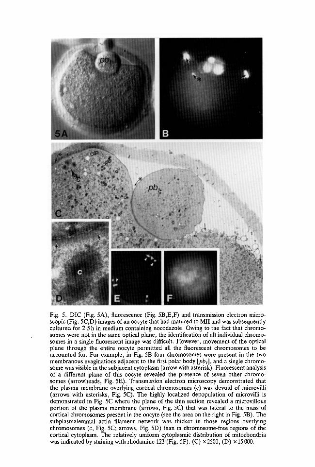
Fig. 5. DIC (Fig. 5A), fluorescence (Fig. 5B,E,F) and transmission electron micro-scopic (Fig. 5C,D) images of an oocyte that had matured to Mil and was subsequentlycultured for 2-5 h in medium containing nocodazole. Owing to the fact that chromo-somes were not in the same optical plane, the identification of all individual chromo-somes in a single fluorescent image was difficult. However, movement of the opticalplane through the entire oocyte permitted all the fluorescent chromosomes to beaccounted for. For example, in Fig. 5B four chromosomes were present in the twomembranous evaginations adjacent to the first polar body [pbj], and a single chromo-some was visible in the subjacent cytoplasm (arrow with asterisk). Fluorescent analysisof a different plane of this oocyte revealed the presence of seven other chromo-somes (arrowheads, Fig. 5E). Transmission electron microscopy demonstrated thatthe plasma membrane overlying cortical chromosomes (c) was devoid of microvilli(arrows with asterisks, Fig. 5C). The highly localized depopulation of microvilli isdemonstrated in Fig. 5C where the plane of the thin section revealed a microvillousportion of the plasma membrane (arrows, Fig. 5C) that was lateral to the mass ofcortical chromosomes present in the oocyte (see the area on the right in Fig. 5B). Thesubplasmalemmal actin filament network was thicker in those regions overlyingchromosomes (c, Fig. 5C; arrows, Fig. 5D) than in chromosome-free regions of thecortical cytoplasm. The relatively uniform cytoplasmic distribution of mitochondriawas indicated by staining with rhodamine 123 (Fig. 5F). (C) X2500; (D) x 15 000.
224 J. VAN BLERKOM AND H. BELL
observed in (1) treated oocytes in which the compact mass of bivalentchromosomes was cortically positioned prior to chromosomal dissociation anddispersal (Fig. 1K,L), and (2) Mil oocytes approximately 2-4 h after exposureto nocodazole and cortical dispersal of chromosomes. The specificity ofchromosome-mediated differentiation was demonstrated by the presence ofmicrovilli and comparatively intense ConA fluorescence in areas of the plasmamembrane associated with subjacent cortical granules (Fig. 3A) and Golgi com-plexes (Fig. 3B) and that were also adjacent to microvilli-free regions.
A pronounced thickening of cortical actin filaments was both temporally andspatially correlated with a depopulation of microvilli and a reduction in theintensity of ConA fluorescence in the regions of the plasma membrane associ-ated with a single subplasmalemmal chromosome or groups of chromosomes(Fig. 5C,D). The relationship between the presence of a chromosome and a focalthickening of cortical actin filaments was evident by fluorescence microscopyafter exposure of oocytes to anti-actin antibody (Fig. 6G,H) or NBD-phallacidin(Fig. 61,J). Chromosome-associated thickening of cortical actin was observed inoocytes exposed to nocodazole continuously from the GV-stage and in oocytestreated with nocodazole at MIL Alterations in the structure and organization ofthe cell surface, plasma membrane and cortical cytoplasm were initiated when achromosome(s) was approximately 5-8/im from the plasma membrane.
Fine structural analysis of selected oocytes that had been continuouslymonitored by time-lapse video recording indicated that the formation of plasmamembrane evaginations occurred when a chromosome was within 1 to 2 fim of theplasma membrane. For oocytes exposed to nocodazole from the GV stage,evaginations formed rather abruptly at 20 h of culture and emerged randomly overthe surface of the oocyte in a virtually simultaneous fashion over a 10 min period(Figs 4A,B, 6K,L). In vivo and in vitro matured oocytes exposed to nocodazole atMil (12 h post-hCG or after initiation of culture) developed chromosomes-containing evaginations approximately 4-6h later (Fig. 5A,B).
The specificity of chromosome-associated cortical and plasma membrane differ-entiation is demonstrated by the transfer of cytoplasm and chromosomes
The specific involvement of chromosomes in the differentiation of the corticalcytoplasm and plasma membrane was demonstrated by the physical transfer ofindividual bivalent chromosomes to (1) intact, untreated GV-stage oocytes,(2) untreated oocytes at the CBV stage, and (3) cytoplasts obtained frompseudocleaved GV-stage oocytes exposed to cytochalasin B at 15 min intervalsafter collection from the ovary. 1178 transfers were attempted with individualbivalent chromosomes derived from 391 donor oocytes that had been culturedfrom the GV stage for 22h in the presence of nocodazole. These studies includedoocytes in which the number of evaginations visible by light microscopy indicatedthat complete dispersion of individual bivalent chromosomes had occurred (e.g.Fig. 4A). Subsequent light and fluorescence microscopic analysis of the recipientoocytes demonstrated successful chromosomal transfer on 130 occasions (62
Chromosomally induced differentiation of the oocyte 225
transfers to cytoplasts and 68 to GV- and CBV-stage oocytes). Cortical cytoplasmobtained from regions between chromosome-containing evaginations of nocod-azole-treated oocytes was transferred to the cortical region of 46 cytoplasts (45 and60min after initiation of culture), 28 untreated GV- and 37 CBV-stage oocytes.Cytoplasmic transfers were accomplished under DIC optics. The success oftransfer was indicated both by the depletion of the contents of the micropipetteand by local expansion of the cortical cytoplasm in the recipient oocyte orcytoplast. The design of the transfer experiments is presented in diagrammaticform in Fig. 7.
The fluorescent image obtained from an oocyte (cultured in the presence ofnocodazole for 22 h) immediately after removal of a chromosome and stainingwith DAPI is shown in Fig. 8B. The chromosome was transferred to the centre of acytoplast derived from a GV-stage oocyte that had been exposed to cytochalasin B45 min after harvesting from the ovary. The electron microscopic appearance ofthe bivalent chromosome, which at 5h after transfer was in proximity to thecortical cytoplasm, is shown in Fig. 8A. Six bivalent chromosomes (present as acluster in the donor oocyte) were transferred to the centre of the cytoplast shownin Fig. 8C,D. This cytoplast was obtained from an oocyte exposed to cytochalasinB 60 min after initiation of culture. At approximately 18 h of culture in the pres-ence of nocodazole, five surface evaginations were evident (Fig. 8D). Fluor-escence microscopy confirmed that each evagination contained a single bivalentchromosome (Fig. 8C). One chromosome apparently failed to migrate signifi-cantly and remained in the pericortical region of the cytoplast (white arrow,Fig. 8C).
The temporal dependency of plasma membrane and cortical differentiation wasmost clearly demonstrated by chromosomal transfers to cytoplasts derived frompseudocleaved GV-stage oocytes exposed to cytochalasin B at 0 min (immediatelyafter harvesting from the ovary), and at 15min intervals up to 60min. Noindication of cortical, cell surface and plasma membrane differentiation orevagination was observed after as many as 36 h of culture in any of the 28cytoplasts derived from oocytes exposed to cytochalasin B at 0 and 30 minafter harvesting that also received a bivalent chromosome(s) at these times.Fluorescence microscopy demonstrated no apparent migration of the transferredchromosome from the original site of deposition in the central portion of thecytoplast. By contrast, 31 of 34 recipient cytoplasts obtained from oocytes exposedto cytochalasin B at 45 and 60 min after harvesting showed cortical migration of thetransferred chromosome, and progressive differentiation of the cortical cytoplasmand plasma membrane, i.e. focal thickening of cortical actin filaments, depletionof micro villi, reduction in intensity of Con A fluorescence and development of aplasma membrane evagination after 18 h of culture.
Reversibility of nocodazole-associated inhibition of chromosomal maturation
After replacement of nocodazole-containing medium with normal medium,some of the GV-stage oocytes cultured in the presence of the inhibitor for as many

226 J. V A N BLERKOM AND H. B E L L
Fig. 6. Relationship between the spatial distribution of chromosomes and (1) theintensity of binding of concanavalin A to cell surface glycoproteins and (2) differentialthickness of cortical actin filaments. The relationship was demonstrated by fluorescentprobe analysis of oocytes cultured from the GV stage in the presence of nocodazole forup to 22 h (h of culture are presented in the lower right-hand portion of the panels).At 12 h of culture, compacted masses of bivalent chromosomes stained with Hoechstdye 33258 were either centrally (Fig. 6A) or cortically located (Fig. 6B). FITC-concanavalin A fluorescence was uniformly distributed over the oocyte surface at 12 hof culture (Fig. 6C). At 16h, oocytes were frequently encountered in which individualbivalent chromosomes were dispersed throughout the cortical cytoplasm (Fig. 6D).FITC-concanavalin A staining of these oocytes revealed a significant reduction in flu-orescence in those regions of the plasma membrane (arrows, Fig. 6F) associated withsubplasmalemmal chromosomes (Fig. 6E). Anti-actin monoclonal antibody (Fig. 6H)or NBD-phallacidin (Fig. 6J) staining of oocytes containing dispersed bivalent chromo-somes (Fig. 6G,I) demonstrated an increased intensity of subplasmalemmal actinfilament fluorescence (arrows, Fig. 6H,J) that was specifically localized to regions ofthe cortical cytoplasm that contained individual chromosomes (arrows, Fig. 6G,I).Fig. 6E and F, G and H, and I and J represent three different oocytes stained with twofluorescent probes and photographed at approximately the same optical plane. Theemergence of evaginations of the cortical cytoplasm and plasma membrane (Fig. 6K)that contained dispersed bivalent chromosomes (Fig. 6L) occurred rather abruptlybetween 20 and 22 h of culture in the presence of nocodazole.

Chromosomally induced differentiation of the oocyte 227
as 36 h were able to resume meiotic maturation, form an MI spindle andsubsequently develop and abstrict a polar body. Time-lapse video recordings ofoocytes that had developed chromosome-containing surface evaginations atapproximately 18-20 h of culture (Fig. 9A) demonstrated that approximately8min after the removal of nocodazole, cortically located bivalent chromosomesdisplayed saltatory motion. Chromosomal motion of this nature was not observedduring the period of time that chromosomes were contained in evaginations of thecortical cytoplasm and plasma membrane.
The extent to which nuclear (meiotic) maturation resumed and progressed wasclosely related to the degree of chromosomal dispersion that had occurred during
Cytochalasin Banucleation
Omin
12-14h 60
18-20h
Fig. 7. Design and results of the chromosome transfer experiments. The numbers ofsuccessful chromosomal transfers are indicated on the lines radiating from thenocodazole-treated oocyte (22 h) and from the cytoplast. Bivalent chromosomes werederived from oocytes that exhibited chromosome-containing evaginations after 22 h ofculture in the presence of nocodazole, and were transferred to either (1) intact GV-(Oh) or CBV- (4h) stage oocytes, or (2) cytoplasts obtained from oocytes anucleatedby exposure to cytochalasin B at 15min intervals after harvesting from the ovary.Approximately 10h after placement in GV- or CBV-stage oocytes, the transferredbivalent chromosomes were contained within individual evaginations of the plasmamembrane. The host oocyte also frequently developed and abstricted a normal firstpolar body. Chromosome-containing evaginations formed in cytoplasts approximately20 h after transfer, but only in cytoplasts derived from oocytes that had beenanucleated between 45 and 60 min after collection from the ovary.

228 J. V A N BLERKOM AND H. B E L L
Fig. 8. The distribution of bivalent chromosomes in a donor oocyte (Fig. 8B) and hostcytoplast(s) (Fig. 8A,C,D) used in chromosomal transfer experiments. The absence ofDAPI fluorescence in an evagination after withdrawal of the micropipette indicatedsuccessful removal of a bivalent chromosome (white arrow, Fig. 8B). Occasionally, asmall tear in the zona pellucida (zp) remained for several minutes after removal of thetransfer pipette. Fig. 8A shows the appearance of the cytoplasm and the pericorticallocation of a bivalent chromosome (c) that had been deposited in the centre of thiscytoplast 5 h earlier. DAPI fluorescence (Fig. 8C) and DIC microscopic images(Fig. 8D) of a cytoplast approximately 18 h after receiving a mass of six bivalentchromosomes. The black arrows indicate surface evaginations containing sub-plasmalemmal chromosomes. A single transferred chromosome (white arrow, Fig. 8C)remained in the interior of the cytoplast after deposition. (A) X4600.

Chromosomally induced differentiation of the oocyte 229
Fig. 9. The temporal sequence of cellular reorganizations resumes after replacementwith normal medium in oocytes cultured for 22 h in the presence of nocodazole andwhich displayed incomplete chromosomal dispersion. Fig. 9A-C, D-F and G-I arederived from three separate oocytes. The chromosome-containing evaginations(arrows, Fig. 9A,B) present at 22 h of culture (indicated as Omin in Fig. 9A) werenearly all resorbed within 5 min of replacement of medium (Fig. 9B). At approxi-mately 10 min, DAPI fluorescence demonstrated the distribution of subplasmalemmalchromosomes (c, Fig. 9C). By 35 min of culture in normal medium, a large mass ofcortical chromosomes had formed (c, Fig. 9D). The presence of individualchromosomes that had failed to migrate to the larger chromosomal aggregate wasevident after DAPI staining (arrows, Fig. 9D). The development of a first metaphasespindle was evident by 60 min of culture (Fig. 9E) and by 105 min, the spindle (s) hadrotated and the process of polar body formation had begun (Fig. 9F). Fig. 9G,H showsthe spatial association that developed between cortical chromosomes and cytoplasmicmitochondria 60 min after removal of nocodazole from oocytes previously exposed tothe inhibitor for 22 h (from the GV-stage). At least 13 bivalent chromosomes thatpreviously had been contained within evaginations were localized in one region of theoocyte (arrows, Fig. 9G,H). Mitochondrial clusters (m) that were uniformlydistributed throughout the cytoplasm during culture in the presence of nocodazole (seeFig. 5F, for example) aggregated in the region of the cytoplasm that contained themajority of the dispersed chromosomes (Fig. 91).
230 J. VAN BLERKOM AND H. BELL
culture in the presence of nocodazole. For oocytes in which chromosomaldispersion was complete, i.e. each bivalent was contained in an individualevagination, removal of nocodazole was followed by resorption of the evagi-nations and initiation of chromosomal motion. However, movements of individualchromosomes were confined to the region of the cortical cytoplasm that formedafter resorption of the evaginations. By contrast, for oocytes in which chromo-somal dispersion was incomplete (i.e. bivalent chromosomes, both individuallyand in clusters, were contained in single evaginations), a first metaphase spindleformed only when a mass of at least six to eight bivalents was present in the corticalcytoplasm, or could develop as a result of the cortical migration and aggregation ofchromosomes. Time-lapse video recordings demonstrated that individual bi-valents typically migrated to a mass of four or more chromosomes. However, evenwhen such a cluster of bivalent chromosomes was present, the lateral distancefrom a chromosomal cluster over which a single bivalent chromosome wasobserved to migrate and join to a larger aggregate was restricted to approximately10/im.
The temporal sequence of cellular reorganizations that occurred in oocytes withincomplete chromosomal dispersion after removal of nocodazole is shown inFig. 9. Within 5min of introduction of normal culture medium (Fig. 9A), surfaceevaginations had resorbed (Fig. 9B) and chromosomes, either individually or ingroups were located in the subplasmalemmal cytoplasm (Fig. 9C). Movement ofindividual bivalents to larger, proximal masses of chromosomes took place within45min (Fig. 9D). Between 1 and 1-5 h after the resorption of the evaginations, afirst metaphase spindle formed (Fig. 9E,F). Approximately 1-2h after spindleformation, single or multiple polar bodies were abstricted. Polar body abstrictionwas complete and occurred in an apparently normal fashion over a period of5-10 min. The ability to resume chromosomal maturation after removal ofnocodazole and to develop and abstrict a polar body was directly related to thenumber of bivalent chromosomes that could aggregate and form a metaphasespindle. Fluorescence microscopic examination of oocytes with incompletechromosomal dispersion demonstrated the presence of individual, corticallylocated, bivalent chromosomes that had not participated in the formation of afunctional metaphase spindle (Fig. 9D). None of the evaginations containing asingle chromosome that had been transferred to a cytoplast or to an intact GV orCBV-stage oocyte showed any signs of abstriction. Multiple polar bodies wereobserved to develop only when six or more bivalents were present.
The rapid development of a specific association between mitochondrial clustersand cortical chromosomes after removal of nocodazole was revealed by stainingwith rhodamine 123. Within 45 min after the introduction of normal medium,migration of proximal mitochondrial clusters to aggregated masses of chromo-somes was evident by fluorescence microscopy (Fig. 9G,H,I). This association wasmore apparent with clusters of three or more chromosomes than with individualbivalents. Coincident with the accumulation of perichromosomal mitochondria,fine structural analysis demonstrated the presence of microtubules that radiated

Chromosomally induced differentiation of the oocyte 231
from the kinetochore region of the bivalent chromosomes. Although microtubulesdid polymerize in association with chromosomes after the removal of nocodazole,a similar relationship between the spatial distribution of mitochondria anddispersed Mil chromosomes was not observed.
DISCUSSION
Changes in mitochondrial and chromosomal organization
The cellular development of the maturing oocyte, which in the mouse involvesmitochondrial redistributions and modifications in the structure and compositionof the cortical cytoplasm and plasma membrane, are temporally correlated withprogressive changes in nuclear (chromosomal) organization (Van Blerkom,1985a, £). A sequence of mitochondrial redistributions during resumed meiosisin mouse oocytes has been reported by Van Blerkom & Runner (1984).Mitochondria that are uniformly distributed throughout the cytoplasm in the GV-stage oocyte form small clusters during GVB. These clusters migrated to theperinuclear region during the CBV-stage and formed a dense, ring-like aggregateof mitochondria that envelop the developing MI spindle. After abstriction of thefirst polar body, the perinuclear mass of mitochondria disaggregate and return to arelatively uniform distribution throughout the cytoplasm.
Van Blerkom (1985a) proposed that the translocation of mitochondrial clustersto the perinuclear region of the mouse oocyte may be mediated by microtubulesthat radiate into the cytoplasm from perinuclear microtubule-organizing centres(MTOCs) (Wassarman & Fujiwara, 1978) that develop in proximity to thegerminal vesicle envelope (Calarco, Donahue & Szollosi, 1972). In the presence ofnocodazole, a rapidly reversible inhibitor of microtubule polymerization, oocytescultured from the GV stage underwent cytoplasmic clustering of mitochondria,GVB and chromosomal condensation according to the same temporal schedule asthat observed in untreated oocytes (Van Blerkom & Runner, 1984). However, theCBV stage did not develop and chromosomes remained for several hours aftercondensation as a dense mass in the central portion of the oocyte. In ap-proximately 30 % of the treated oocytes examined, the mass of centrally locatedchromosomes migrated to a peripheral location and subsequently disaggregated insuch a manner that bivalent chromosomes, both individually and in small groups,were dispersed in the cortical cytoplasm. For oocytes in which the mass ofchromosomes remained centrally located, continued culture in the presence ofnocodazole was accompanied by disaggregation of the mass and migration ofindividual chromosomes, or groups of chromosomes, away from the centre of theoocyte to the cortical cytoplasm.
The present findings support the suggestion of microtubule participation inmitochondrial translocation during resumed meiosis. In oocytes cultured con-tinuously in the presence of nocodazole from the GV stage, no specific associationcould be established between the spatial distribution of mitochondrial clustersand (1) compacted masses of centrally or peripherally positioned chromosomes,

232 J. VAN BLERKOM AND H. BELL
(2) small groups or individual chromosomes during their migration to the corticalcytoplasm, and (3) bivalent chromosomes either resident in the cortical cytoplasmor contained in evaginations of the plasma membrane. A similar result occurredin oocytes exposed to nocodazole after abstriction of the first polar body. Inthese Mil-stage oocytes, disaggregation of the meiotic spindle was followed bycortical migration and random distribution of chromosomes. During culture ofMil oocytes in the presence of nocodazole, no specific association betweenmitochondria and subplasmalemmal chromosomes was observed.
Electron microscopic analysis of nocodazole-treated oocytes at the late GV andGVB stages failed to demonstrate the presence of cytoplasmic microtubules ofperinuclear origin. No cytoplasmic microtubules were observed to originate fromthe compacted mass of bivalent chromosomes that formed after GVB in treatedoocytes. However, small, compact bundles of relatively short microtubules wereassociated with MI chromosomes that had disaggregated from the originalcondensed mass and were either in the process of migration to the cortex, or hadbecome resident in the cortical cytoplasm.
Electron microscopic examination of oocytes exposed to nocodazole afterabstriction of the first polar body revealed an absence of microtubules andmitochondria associated with Mil chromosomes that had dissociated from themetaphase spindle and dispersed within the cortical cytoplasm. Similar findingsderived from observations of chromosomes in mouse oocytes exposed tonocodazole after ovulation have been reported by Maro et al. (1986). Thesensitivity to nocodazole of microtubules associated with Mil chromosomes is verysimilar to that observed in mitotic cells (Zieve, Turnbull, Mullins & Mclntosh,1980). The association of microtubules with MI but not Mil chromosomes intreated oocytes may indicate developmental variation in stability and/or sen-sitivity of meiotic microtubules to nocodazole. Alternatively, oocytes culturedfrom the GV stage had been exposed to nocodazole for a longer period of timethan oocytes obtained at MIL Rather than a manifestation of a developmentaldifference, the association of microtubules with MI but not Mil chromosomescould be a consequence of a reduction in the influence of nocodazole duringextended periods of culture.
The relationship between the spatial distribution of mitochondrial clusters andchromosomes was most clearly demonstrated after the removal of nocodazole andreplacement with normal culture medium. The removal of nocodazole presumablyrestores intracellular conditions that are permissive for the normal process ofmicrotubule polymerization. As a consequence of exposure of mature oocytes tonocodazole (Maro et al. 1986), chromosomes dissociate from the Mil spindle,disperse into the cortical cytoplasm and have no attached microtubules. However,approximately 30 min after the removal of the inhibitor, microtubules arising fromthe kinetochore region were observed. Although microtubules did polymerize inassociation with dispersed Mil chromosomes, mitochondria did not migrate to oraggregate in the vicinity of the chromosomes. This finding is in contrast to thesituation that existed in treated oocytes containing dispersed MI chromosomes

Chromosomally induced differentiation of the oocyte 233
where, after removal of nocodazole, mitochondrial clusters became associatedwith bivalent chromosomes. However, these results are similar to observations onthe behaviour of mitochondria during the GVB, CBV, MI and Mil stages ofoocyte maturation in the mouse (Van Blerkom & Runner, 1984): (1) theperinuclear concentration of mitochondria develops during MI and disperses afterabstriction of the first polar body and formation of the Mil spindle, and (2) themitochondria associate neither specifically nor extensively with the Mil spindle.
Changes in the cytoplasmic distribution of mitochondria may be determined bythe spatial orientation and the developmental state of meiotic chromosomes, aswell as by the nature of the association between chromosomes and microtubules.In this respect, it may also be reasonable to speculate that chemical changes in theouter mitochondrial membrane may occur during oocyte maturation (Kruip,Cran, Van Beneden & Dieleman, 1983), and these changes may impart tomitochondria a stage-specific capacity to interact with chromosome-associatedmicrotubules.
Chromosomal mediation of cortical and plasma membrane differentiation
Cortical and plasma membrane differentiation during resumption of arrestedmeiosis in the mouse oocyte occurs in association with the metaphase spindles. Inthe present study, the migration of individual bivalent chromosomes to the corticalcytoplasm was accompanied by the same process of cortical and plasma membranedifferentiation normally associated with the presence of the spindle-associatedmeiotic chromosomes. The area of cortical and plasma membrane differentiationassociated with the presence of an underlying chromosome has been clearlydelineated by fluorescence, transmission and scanning electron microscopy(Longo & Chen, 1985; Maro et al. 1986). In the present study, the subplas-malemmal cytoplasm, under the apparent influence of a chromosome, also wascharacterized by the absence of mitochondria, cortical granules and Golgicisternae. The specificity of chromosome-associated differentiation was shown bythe absence of altered cortical, plasma membrane and cell surface characteristicsin regions of the oocyte surface adjacent to Golgi complexes, cortical granules andmitochondria.
Individual bivalent (MI) chromosomes dispersed throughout the corticalcytoplasm as a consequence of exposure of GV-stage oocytes to nocodazolewere associated with small, compact bundles of relatively short microtubules.This observation raises the possibility that cortical and plasma membranedifferentiation may be influenced by microtubules rather than by chromosomes.However, the same clearly delineated progression of cortical, plasma membraneand cell surface differentiation observed in the presence of MI chromosomesoccurred in those regions of oocyte subplasmalemmal cytoplasm that containeddispersed Mil chromosomes devoid of associated microtubules. These results andinterpretations also are consistent with the findings of similar light, fluorescent andelectron microscopic studies involving GV- and Mil-stage (ovulated) mouse

234 J. VAN BLERKOM AND H. BELL
oocytes cultured in the presence of colchicine (Longo & Chen, 1985), and MII-stage mouse oocytes cultured in the presence of nocodazole (Maro et al. 1986).
The temporal and spatial relationship between the presence of a chromosomeand the occurrence of cortical and plasma membrane differentiation was clearlydemonstrated in chromosome transfer experiments. Transfer of a single bivalentchromosome to a GV- or CBV-stage oocyte resulted in the same sequence ofcortical actin thickening, microvilli depopulation and reduction in cell surfaceCon A binding observed in association with the endogenous MI spindle. However,this sequence of changes frequently occurred in a region of the oocyte surfacedistant from the normal MI spindle. In spite of the fact that a normal first polarbody was formed in the same oocyte, evaginations that contained individual,transferred chromosomes did not abstrict. This finding suggests that the presenceof a chromosome is sufficient to establish the localized intracellular changesrequisite to polar body formation at any region of the oocyte surface, but mayrequire a critical mass of chromosomes or a functional spindle, or both, beforeabstriction of the polar body can occur. The findings of Maro et al. (1986) sup-port this interpretation by demonstrating that relatively large clusters of Milchromosomes dispersed in the cortical cytoplasm in treated oocytes are capable,after removal of nocodazole, of inducing the formation of multiple spindles thatrotate and yield multiple abstricted polar bodies containing varying numbers ofchromosomes.
The same spatial relationship that existed between the presence of achromosome and the occurrence of a sequence of differentiative events in thecortical cytoplasm and overlying plasma membrane was also observed in cytoplaststhat received one or more bivalent chromosomes. The specificity of chromosomalmediation of this sequence was further indicated by transfer of cytoplasm fromnocodazole-treated oocytes to cytoplasts and to intact, untreated GV- and CBV-stage oocytes. No evidence of cortical or plasma membrane differentiation wasobtained in any of the studies involving cytoplasmic transfers. Taken together, theabove findings demonstrate that the cortical and plasma membrane polarityestablished in the MI- and Mil-stage mouse oocyte is induced rather than pre-existing. The results also indicate that the entire surface of the mouse oocyte isequally able to differentiate in response to the presence of a chromosome(s). Thepolarity and compartmentalization of the cortical cytoplasm occur during normaloocyte maturation by virtue of the fact that the chromosomes arrive at the plasmamembrane as a package rather than, as shown in the present study, as singlechromosomes in disaggregation.
Chromosomal but not genomic participation in the autonomous expression of anintrinsic developmental programme in the mouse oocyte
The expression of a developmental sequence of molecular and cellular changes(cytoplasmic maturation) in the mouse oocyte appears to be initiated just prior toor at the time of GVB. GV-stage oocytes cultured in the presence of dibutryl cyclicAMP (dbCAMP) for up to 16 h do not undergo GVB or cytoplasmic clustering of
Chromosomally induced differentiation of the oocyte 235
mitochondria (Van Blerkom & Runner, 1984). The temporal sequence of dynamicchange at the nuclear and cytoplasmic levels occurs in an apparently normalfashion after the removal of dbCAMP (Van Blerkom & Runner, 1984). Thereversible inhibition of oocyte maturation suggests that cytoplasmic changes maybe initiated or regulated by factors present in the germinal vesicle and exported tothe cytoplasm prior to or at the time of GVB. Wassarman, Schultz & Letourneau(1979) reported that specific proteins, some of which were phosphorylated,accumulated in high concentrations in the nucleoplasm and were released into thecytoplasm around the time of GVB. A more recent study by Schultz, Montgomery& Belanoff (1983) described changes in patterns of protein phosphorylation anddephosphorylation that were initiated approximately 30 min after placement ofGV-stage mouse oocytes (1-1-5 h prior to GVB) in conditions that permitted thespontaneous resumption of arrested meiosis. These results suggest that trans-location of preformed nuclear proteins to the cytoplasm, or changes in proteinsynthesis and modification, or both, may begin well in advance of GVB andchromosomal condensation (Schultz et al. 1983). The above observations suggestthe possibility that molecular changes at the nuclear level may initiate a cascade ofevents leading to GVB and to a developmental alteration of the cytoplasm inpreparation for the presence of chromosomes (Van Blerkom & Runner, 1984; VanBlerkom, 1985a).
The hypothesis that GVB and the completion of chromosomal condensation arepreceded by activation of cytoplasmic maturation and do not require concurrentgenomic input has been suggested by previous molecular and cellular studies ofanucleate fragments (cytoplasts) derived from GV-stage mouse oocytes at 0, 15,20, 45 and 60min after the resumption of meiosis in vitro. Qualitative analysis ofprotein expression in cytoplasts cultured for as many as 36 h after enucleationdemonstrates the occurrence of changes in protein synthesis (Schultz et al. 1978;Van Blerkom, 1985c) and modification (Van Blerkom, 1985a) that is associatedwith maturation of the normal mouse oocyte. However, these changes take placeonly in cytoplasts derived from oocytes that have been enucleated between 30 and60 min after the initiation of culture.
In the present study, chromosomes were transferred to cytoplasts produced atvarious times after the initiation of oocyte culture. The results indicate that thecytoplasm of cytoplasts derived at 0 and 30 min does not have the ability topromote chromosomal migration, and that the cortical cytoplasm and plasmamembrane may not have the capacity to differentiate in response to the presenceof a chromosome, even after 36 h of culture. By contrast, chromosomes placed inthe central portion of cytoplasts enucleated after approximately 45 and 60 min ofmaturation in vitro migrated to the cortex and initiated the same sequence ofcortical, plasma membrane and cell surface differentiation that normally occurs inproximity to an intact metaphase spindle.
An alternative interpretation of the findings, based on the observation thatcytochalasin B-induced pseudocleavage is accompanied by a transient and totalloss of cortical micron* laments and microvilli (Wassarman et al. 1977), is that the

236 J. VAN BLERKOM AND H. BELL
differential response of cytoplasts to the presence of a transferred chromosomecould be the result of (1) the inability of a chromosome to migrate to the cortexand, or, (2) the absence of cortical microfilaments that can respond to the presenceof a chromosome. The migration of chromosomes in oocytes appears to be amicrofilament-mediated process because drugs that interfere with microfilamentorganization, such as cytochalasin D, inhibit the migration of even nocodazole-disaggregated meiotic chromosomes (Van Blerkom & Bell, unpublished data;Maro etal. 1986). The presence of a chromosome in the cortical cytoplasm wasinsured in some of the transfer experiments by the cortical placement of achromosome in cytoplasts obtained 30 min after the initiation of culture. However,no evidence of cortical or cell surface alteration was observed in these cells after asmany as 36h of culture. This observation is in contrast to the situation thatprevailed in cytoplasts obtained at 45 and 60 min after the initiation of culture (seeFig. 7 for summary). Microvilli and cortical microfilaments reappear uniformlyafter the completion of pseudocleavage, but only in the anucleate compartment(Wassarman et al. 1977). This result strongly suggests that microvilli and corticalmicrofilaments had reformed and were uniformly distributed so as to be able torespond to the presence of a chromosome. Collectively, these observations suggestthat a change in the responsiveness of the cytoplasm to the presence of achromosome occurs approximately 45 min after the resumption of meiosis.
The precise nature of the signal(s) and the actual mechanism that may beinvolved in the activation of the oocyte cytoplasm during the resumption ofmeiosis remain to be determined. Van Blerkom (1985a) suggested that proteinsexported from the GV may activate cytoskeletal-associated enzymes that functionin the stage-related post-translational modification of oocyte proteins. Theactivation of enzymes, such as kinases and phosphatases (Schultz etal. 1983), couldrapidly alter the cytoarchitectural state of the cytoplasm to allow the movement ofchromosomes and the redistribution of organelles. Although the process by whicha chromosome can mediate or induce local changes in cytoplasmic and plasmamembrane structure and organization is unknown, it is conceivable thatchromosome-associated proteins or ions may be involved. The ability to transferchromosomes between oocytes, and the occurrence of well-defined and preciselytimed developmental events during the maturation of the laboratory mouse oocyteoffers an extremely useful approach to understanding both the nature ofinteraction between nucleus and cytoplasm, and the mechanism of autonomousexpression of an intrinsic developmental programme.
We thank Dr Michael Klymkowsky for his helpful comments on the manuscript and forproviding anti-actin monoclonal antibody. This work was supported by grants from the NationalInstitutes of Health (HD-13500; RR 07013-19, BRSG Program, Division of ResearchResources).

Chromosomally induced differentiation of the oocyte 237
REFERENCESBARAK, L. S., YOCUM, R. R., NOTHNAGEL, E. A. & WEBB, W. W. (1980). Fluorescence staining of
the actin cytoskeleton in living cells with 7-nitobenz-2-oxa-l,3,-diazole phallacidin. Proc.natn. Acad. Sci., U.S.A. 77, 980-984.
CALARCO, P. G., DONAHUE, R. P. & SZOLLOSI, D. (1972). Germinal vesicle breakdown in themouse oocyte. /. CellSci. 10, 369-385.
EAGER, D., JOHNSON, M. H. & THURLEY, K. W. (1975). Ultrastructural studies on the surfacemembrane of the mouse egg. /. Cell Sci. 22, 345-353.
HOEBEKE, J., VAN NIGEN, G. & DE BRABANDER, M. (1976). Interaction of Oncodazole (R17934),a new anti-tumor drug, with rat brain tubulin. Biochem. Biophys. Res. Commun. 69, 319-342.
JOHNSON, M. H., EAGER, D., MUGGLETON-HARRIS, A. L. & GRAVES, H. M. (1975). Mosaicism inthe organization of concanavalin A receptors on surface membrane of mouse eggs. Nature,Lond. 257, 321-322.
KRUIP, T. A. M., CRAN, D. G., VAN BENEDEN, T. H. & DIELEMAN, S. J. (1983). Structuralchanges in bovine oocytes during final maturation in vivo. Gamete Res. 8, 29-48.
LONGO, F. J. & CHEN, D. Y. (1985). Development of cortical polarity in mouse eggs:Involvement of the meiotic apparatus. Devi Biol. 107, 382-394.
MARO, B., JOHNSON, M. H., PICKERING, S. J. & FLACH, G. (1984). Changes in actin distributionduring fertilization of the mouse egg. /. Embryol. exp. Morph. 81, 211-237.
MARO, B., JOHNSON, M. H., WEBB, M. & FLACH, G. (1986). Mechanism of polar body formationin the mouse oocyte: an interaction between the chromosomes, the cytoskeleton and theplasma membrane. /. Embryol. exp. Morph. 92, 11-32.
NICOSIA, S. V., WOLF, D. P. & INOUE, M. (1977). Cortical granule distribution and cell surfacecharacteristics in mouse eggs. Devi Biol. 57, 56-74.
RICHTER, J. D. & MCGAUGHEY, R. W. (1983). Patterns of polypeptide synthesis in mouse oocytesduring germinal vesicle breakdown and during maintenance of the germinal vesicle stage bydibutryl cAMP. Devi Biol. 83, 188-192.
SCHLIWA, M. & VAN BLERKOM, J. (1981). Structural interaction of cytoskeletal components.J. Cell Biol. 90,222-235.
SCHULTZ, R. M. & WASSARMAN, P. M. (1977). Specific changes in the pattern of protein synthesisduring meiotic maturation of mammalian oocytes in vitro. Proc. natn. Acad. Sci., U.S.A. 74,538-541.
SCHULTZ, R. M., LETOURNEAU, G. E. & WASSARMAN, P. M. (1978). Meiotic maturation of mouseoocytes in vitro: Protein synthesis in nucleate and anucleate oocyte fragments. /. Cell Sci. 30,251-264.
SCHULTZ, R. M., MONTGOMERY, R. & BELANOFF, J. R. (1983). Regulation of mouse oocyte meioticmaturation: Implication of a decrease in oocyte cAMP and protein dephosphorylation incommitment to resume meiosis. Devi Biol. 97, 264-273.
VAN BLERKOM, J. (1985a). Extragenomic regulation and autonomous expression of adevelopmental program in the early mouse embryo. Ann. N.Y. Acad. Sci. 442, 58-72.
VAN BLERKOM, J. (19856). Protein synthesis during oogenesis and early embryogenesis in themammal. In Biology of Fertilization, vol. 3 (ed. C. B. Metz & A. Monroy), pp. 379-399. NewYork: Academic Press.
VAN BLERKOM, J. (1985c). Posttranslational modification of proteins during resumption ofarrested meiosis and early embryogenesis in the mammal. In Control of Cell Growth andProliferation (ed. C. M. Veneziale), pp. 67-86. Princeton, New Jersey: Van Nostrand,Reinhold, Co.
VAN BLERKOM, J. & MOTTA, P. M. (1979). The Cellular Basis of Mammalian Reproduction.
Baltimore: Urban and Schwarzenberg.VAN BLERKOM, J. & RUNNER, M. N. (1984). Mitochondrial reorganization during resumption of
arrested meiosis in the mouse oocyte. Amer. J. Anat. 171, 335-355.

238 J. V A N BLERKOM AND H. B E L L
WASSARMAN, P. M. & LETOURNEAU, G. E. (1976). RNA synthesis in fully grown mouse oocytes.Nature, Lond. 261, 73-74.
WASSARMAN, P. M., UKENA, T. E., JOSEFOWICZ, W. J., LETOURNEAU, G. E. & KARNOVSKY, M. J.(1977). Cytochalasin B-induced pseudocleavage of mouse oocytes in vitro. II. Studies of themechanism and morphological consequences of pseudocleavage. J. Cell Sci. 26, 323-337.
WASSARMAN, P. M. & FUJIWARA, K. (1978). Immunofluorescent anti-tubulin staining of spindlesduring meiotic maturation of mouse oocytes in vitro. J. Cell Sci. 29, 171-188.
WASSARMAN, P. M., SCHULTZ, R. M. & LETOURNEAU, G. E. (1979). Protein synthesis duringmeiotic maturation of mouse oocytes in vitro: Synthesis and phosphorylation of a proteinlocalized in the germinal vesicle. DevlBiol. 69, 94-107.
WOLF, D. E. & ZIOMEK, C. A. (1983). Regionalization and lateral diffusion of membraneproteins in unfertilized and fertilized mouse eggs. /. Cell Biol. 96, 1786-1790.
ZAMBONI, L. (1970). Ultrastructure of mammalian oocytes and ova. Biol. Reprod. (Suppl.) 2,44-63.
ZIEVE, G. W., TURNBULL, D., MULLINS, M. & MCINTOSH, J. R. (1980). Production of largenumbers of mitotic mammalian cells by use of the reversible microtubule inhibitornocodazole. Expl Cell Res. 126, 397-405.
(Accepted 18 November 1985)