Embed Size (px)
Citation preview

Regulation of Epidermal Growth Factor Receptor Signaling inHuman Cancer Cells by MicroRNA-7*□S
Received for publication, June 3, 2008, and in revised form, December 9, 2008 Published, JBC Papers in Press, December 10, 2008, DOI 10.1074/jbc.M804280200
Rebecca J. Webster‡§1,2, Keith M. Giles‡1, Karina J. Price‡§, Priscilla M. Zhang‡, John S. Mattick¶,and Peter J. Leedman‡§3
From the ‡Laboratory for Cancer Medicine, University of Western Australia Center for Medical Research, Western AustralianInstitute for Medical Research, and the §School of Medicine and Pharmacology, The University of Western Australia, Level 6, MRFBuilding, Rear 50 Murray Street, Perth 6000, Western Australia and the ¶Australian Research Council Special Research Center forFunctional and Applied Genomics, Institute for Molecular Bioscience, University of Queensland, Brisbane 4000, Australia
The epidermal growth factor receptor (EGFR) is frequentlyoverexpressed in cancer and is an important therapeutic target.Aberrant expression and function of microRNAs have beenassociated with tumorigenesis. Bioinformatic predictions sug-gest that the human EGFR mRNA 3�-untranslated region con-tains threemicroRNA-7 (miR-7) target sites, which are not con-served across mammals. We found that miR-7 down-regulatesEGFR mRNA and protein expression in cancer cell lines (lung,breast, and glioblastoma) via two of the three sites, inducing cellcycle arrest and cell death. Because miR-7 was shown todecrease EGFRmRNA expression, we used microarray analysisto identify additional mRNA targets of miR-7. These includedRaf1 and multiple other genes involved in EGFR signaling andtumorigenesis. Furthermore, miR-7 attenuated activation ofprotein kinase B (Akt) and extracellular signal-regulated kinase1/2, two critical effectors of EGFR signaling, in different cancercell lines. These data establish an important role for miR-7 incontrolling mRNA expression and indicate that miR-7 has theability to coordinately regulate EGFR signaling in multiplehuman cancer cell types.
The epidermal growth factor receptor (EGFR),4 a member ofthe erbB receptor family, is widely expressed in human tissues
and regulates important cellular processes, including prolifera-tion, differentiation, and development (1). EGFR overexpres-sion occurs in a range of solid tumors and is associated withdisease progression, resistance to chemotherapy and radiationtherapy, and poor prognosis (2). Consequently, the EGFR andits downstream signaling effectors are major targets for newtherapeutics such asmonoclonal antibodies and tyrosine kinaseinhibitors (3). However, the clinical responses of tumors toexisting anti-EGFR agents are often limited, and thus a majorresearch focus is the development of novel approaches to blockEGFR expression and signaling (4).MicroRNAs (miRNAs) are short, endogenous, non-coding
RNA molecules that bind with imperfect complementarity tothe 3�-untranslated regions (3�-UTRs) of target mRNAs, caus-ing translational repression of the target gene or degradation ofthe target mRNA (5–7). miRNAs are involved in a range ofprocesses that includes development, differentiation (8), prolif-eration, and apoptosis (9) and have been implicated in cancer(10). Many miRNA genes are located at sites in the humangenome that are frequently amplified, deleted, or rearranged incancer (11), suggesting that some miRNAs may act as onco-genes (“oncomirs” 12) or tumor suppressors (10). For instance,reduced expression of the let-7 family of miRNAs is associatedwith increased Ras oncogene expression and reduced survivalin patients with non-small cell lung cancer (NSCLC) (13, 14). Incontrast, increased expression of miR-21 in gliomas (15), andbreast, colon, lung, pancreas, prostate, and stomach cancers(16) is associatedwith apoptosis resistance, reduced chemosen-sitivity, and increased tumor growth (17).Computational approaches tomiRNA target prediction have
used criteria such as sequence complementarity between targetmRNAs and a “seed” region within the miRNA, and conserva-tion of predicted miRNA-binding sites across 3�-UTRs frommultiple species (reviewed in Refs. 18, 19). Recently, additionalfeatures that determine target site functionality have been iden-tified (20). Nevertheless, the imperfect complementarity ofmiRNA and target sequences means that identification andfunctional validation of authentic miRNA targets remains amajor challenge. It has been suggested that miRNAs may havethe ability to regulate hundreds or even thousands of targetmRNAs (21) and thatmuch of this regulation could occur at thelevel of mRNA decay (22). Because specific miRNAs have thepotential to regulate the expression of several members of asignaling pathway or cellular process (23), we hypothesized a
* This work was supported by the National Health and Medical ResearchCouncil of Australia and the Cancer Council Western Australia. The Office ofInnovation and Industry at the University of Western Australia co-fundedthe PCT application on miR-7 (lodged August 28, 2007). The costs of pub-lication of this article were defrayed in part by the payment of pagecharges. This article must therefore be hereby marked “advertisement” inaccordance with 18 U.S.C. Section 1734 solely to indicate this fact.
□S The on-line version of this article (available at http://www.jbc.org) containssupplemental Figs. S1–S8 and Table S1.
1 Both authors contributed equally to this work.2 Recipient of a University of Western Australia Richard Walter Gibbon Schol-
arship for Medical Research.3 To whom correspondence should be addressed. Tel.: 618-9224-0333; Fax:
618-9224-0322; E-mail: [email protected] The abbreviations used are: EGFR, epidermal growth factor receptor; KEGG,
Kyoto Encyclopedia of Genes and Genomes; miRNA, microRNA; NSCLC,non-small cell lung cancer; RT, reverse transcription; qRT-PCR, quantitativeRT-PCR; UTR, untranslated region; nt, nucleotide(s); WT, wild type; MT,mutant; GAPDH, glyceraldehyde-3-phosphate dehydrogenase; Bis-Tris,2-[bis(2-hydroxyethyl)amino]-2-(hydroxymethyl)propane-1,3-diol; ERK,extracellular signal-regulated kinase; MAPK, mitogen-activate proteinkinase; MEK, MAPK/ERK kinase; IRS2, insulin receptor substrate 2; PI3K,phosphatidylinositol 3-kinase; ARF4, ADP-ribosylation factor 4; PAK1, p21/Cdc42/Rac1-activated kinase 1; PBS, phosphate-buffered saline.
THE JOURNAL OF BIOLOGICAL CHEMISTRY VOL. 284, NO. 9, pp. 5731–5741, February 27, 2009© 2009 by The American Society for Biochemistry and Molecular Biology, Inc. Printed in the U.S.A.
FEBRUARY 27, 2009 • VOLUME 284 • NUMBER 9 JOURNAL OF BIOLOGICAL CHEMISTRY 5731
by guest on April 4, 2019
http://ww
w.jbc.org/
Dow
nloaded from

role for miRNAs in aberrant EGFR expression and signaling inhuman cancers. In this study, we identify EGFR and Raf1 asdirect targets of miR-7 in cancer cells, with miR-7 blockingEGFR and Raf1 expression by inducing mRNA decay. Further-more, we demonstrate the potential for miR-7 to act as a tumorsuppressor by coordinately regulating the EGFR signaling path-way at multiple levels to repress protein kinase B (Akt) andextracellular signal-regulated kinase 1/2 (ERK1/2) activity inhuman cancer cell lines.
EXPERIMENTAL PROCEDURES
Cell Culture, miRNA Precursors, and Normal Tissue RNA—A549, MDA-MB-468, U87, DU145, and U373 cell lines wereobtained from the American Type Culture Collection (ATCC)and cultured at 37 °C in 5% CO2 with Dulbecco’s modifiedEagle’smedium supplementedwith 10% fetal bovine serum and1% penicillin/streptomycin. Synthetic miRNA precursor mole-cules corresponding to humanmiR-7 (Pre-miRmiRNAPrecur-sor Product ID: PM10047) and a negative controlmiRNA (miR-NC; Pre-miR miRNA Precursor Negative Control #1, ProductID: AM17110) were obtained from Ambion. Total RNA (First-Choice) from normal brain, normal lung, and normal breasttissue was purchased from Ambion.Luciferase Plasmid Construction—pGL3-miR-7-target was
generated by ligating annealed DNA oligonucleotides corre-sponding to the perfect hsa-miR-7 target site (forward: 5�-CAACAA AAT CAC TAG TCT TCC A-3� and 5�-TGG AAG ACTAGT GAT TTT GTT G-3�) to unique SpeI and ApaI sites thatwere inserted 3� of the luciferase open reading frame of pGL3-control (Promega) firefly luciferase reporter vector (designatedpGL3-control-MCS (24)). Full-length EGFR 3�-UTR reporterplasmid was synthesized by GenScript Corp. using the entireEGFR 3�-UTR (nt 3879–5616 of GenBankTM accession num-ber NM_005228) and the pMIR-REPORT luciferase plasmidbackbone (Ambion). Wild-type (WT) EGFR target reporterplasmids pGL3-EGFR-A, -B, and -C were generated by cloningannealed oligonucleotides corresponding to nt 4214–4260, nt4302–4348, and nt 4585–4631, respectively, of EGFR (Gen-BankTM accession number NM_005228) mRNA 3�-UTR intoSpeI and ApaI sites in pGL3-control-MCS. Plasmid pGL3-EGFR-D contained a PCR-generated EGFR 3�-UTR sequencethat spanned the predicted miR-7 target sites B and C. Mutant(MT) reporters were also generated that included three nucle-otide substitutions to impair binding of the miR-7 seedsequence to its target. Plasmids pGL3-RAF1-WT and pGL3-RAF1-MT were constructed by cloning annealed DNA oligo-nucleotides corresponding to nt 2965–3030 of the Raf1mRNA3�-UTR (GenBankTM accession number NM_002880), into theSpeI and ApaI sites in pGL3-control-MCS. The sequence of allplasmids was confirmed by sequencing.Transfections and Luciferase Assays—Cells were seeded 24 h
prior to transfection in 6-well plates or 10-cm dishes and trans-fected using Lipofectamine 2000 (Invitrogen) withmiRNApre-cursor molecules at final concentrations ranging from 0.1 to 30nM. Cells were harvested at 12–24 h (for RNA extraction) or 3days (for protein extraction). For reporter gene assays, cellswere seeded in 24-well plates and co-transfected using Lipo-fectamine 2000 (Invitrogen) with 100 ng of firefly luciferase
reporterDNAand 5 ng of pRL-CMVRenilla luciferase reporterDNA as a transfection control. Lysates were assayed for fireflyand Renilla luciferase activities 24 h after transfection using theDual Luciferase Report Assay System (Promega) and a FluostarOPTIMAmicroplate reader (BMGLabtech). Expression valueswere normalized to Renilla luciferase.RT-PCR—Total RNA was extracted from cell lines with
TRIzol reagent (Invitrogen) and RNeasy columns (Qiagen) andtreated with DNase I (Promega) to eliminate contaminatinggenomicDNA. For qRT-PCR analysis ofEGFR andRaf1mRNAexpression, 1 �g of total RNAwas reverse transcribed to cDNAwith random hexamers and Thermoscript (Invitrogen). Real-time PCR for Raf1 andGAPDHwas performed using a Corbett3000 RotorGene instrument (Corbett Research) with Quanti-Tect SYBR Green PCR mixture (Qiagen) with primers fromPrimerBank (25) (EGFR-F, 5�-GCGTTCGGCACGGTGTATAA-3�; EGFR-R, 5�-GGC TTT CGG AGA TGT TGC TTC-3�;RAF1-F, 5�-GCACTGTAGCACCAAAGTACC-3�; RAF1-R,5�-CTG GGA CTC CAC TAT CAC CAA TA-3�; GAPDH-F,5�-ATGGGGAAGGTGAAGGTCG-3�;GAPDH-R, 5�-GGGGTC ATT GAT GGC AAC AAT A-3�). Expression of EGFRand Raf1 mRNA relative to GAPDH mRNA was determinedusing the 2���CT method (26).
For analysis of miR-7 expression by qRT-PCR, reverse tran-scription and PCR were carried out using TaqMan miRNAassay kits (Applied Biosystems) for hsa-miR-7 (Part #4373014)and U44 snRNA (Part #4373384) with a Corbett 3000 Rotor-Gene thermocycler (Corbett Research) according to the man-ufacturer’s instructions. Statistical analyses of qRT-PCR datawere performed using GenEx software (MultiD).Western Blotting—Cytoplasmic protein extracts were pre-
pared as described (24), resolved on NuPAGE 4–12% Bis-Trisgels (Invitrogen), and transferred to polyvinylidene difluoridemembranes (RocheApplied Science).Membranes were probedwith anti-EGFR mouse monoclonal antibody (1:1,000, Neo-markers, catalog #MS-400-P1), anti-Raf1 mouse monoclonalantibody (1:1,000, Santa Cruz Biotechnology, sc-7267), anti-IRS2 rabbit monoclonal antibody (1:500, Cell Signaling Tech-nology, #4502), anti-Akt rabbit monoclonal antibody (1:1,000,Cell Signaling Technology, #9272), anti-phospho-Akt(Ser-473) rabbit monoclonal antibody (1:500, Cell SignalingTechnology, #4060), anti-p44/42 (ERK1/2) MAPK mousemonoclonal antibody (1:750, Cell Signaling Technology,#4696), anti-phospho-p44/42 (P-ERK1/2) MAPK (Thr-202/Tyr-204) rabbit monoclonal antibody (1:750, Cell SignalingTechnology, #9101), or anti-�-actin mouse monoclonal anti-body (1:15,000, Abcam ab6276–100), prior to detection withECL Plus detection reagent (GE Healthcare) and ECL-Hyper-film (GE Healthcare).Cell CycleAnalysis andCell Counting—Following trypsiniza-
tion, cells were permeabilized, stained with propidium iodide,and analyzed on anEPICSXL-MCL (CoulterCorp. flow cytom-eter). Cell cycle analysis was performed using MultiPlus AVMultiParameter data analysis software (Phoenix FlowSystems).Cells were seeded in 6-cm dishes and assessed 3 days aftermiR-7 or miR-NC transfection by light microscopy. Five repre-sentative fields of view were photographed for each condition.Cells in each field of view were counted manually.
Regulation of EGFR Signaling in Cancer by miR-7
5732 JOURNAL OF BIOLOGICAL CHEMISTRY VOLUME 284 • NUMBER 9 • FEBRUARY 27, 2009
by guest on April 4, 2019
http://ww
w.jbc.org/
Dow
nloaded from
Microarray Expression Profiling and Analysis—Total RNAwas isolated fromA549 cells transfected for 24 h with miR-7 ormiR-NC (30 nM) using TRIzol reagent (Invitrogen) and RNeasycolumns (Qiagen) and assessed for quality and integrity using a2100Bioanalyzer (Agilent Technologies). Gene expression pro-filing by microarray hybridization was performed with twoexperimental replicates by the Lotterywest State MicroarrayFacility using Human Genome U133 Plus 2.0 array chips(Affymetrix). The raw data were processed using GeneSiftersoftware (VizX Labs). An “all groups must pass” restriction wasimposed, with a threshold quality score of “P” (present)required for inclusion in subsequent analysis. Data were nor-malized to the all means fluorescence and were log2-trans-formed. Pairwise analysis of the probe intensities of miR-7- andmiR-NC-treated sample data sets was performed using Stu-dent’s t tests (two-tailed, unpaired), and used to identify tran-scripts that were significantly up-regulated or down-regulatedwith miR-7 transfection (p � 0.05) by at least a factor of 2.These represented potential miR-7 target transcripts.Investigation of the enrichment of gene sets for predicted
miRNA targets was conducted using the L2L microarray anal-ysis tool (27). miRanda (28), PicTar (29), and TargetScan (21)were used for miR-7 target predictions. Analysis of the enrich-ment of gene sets for functional Kyoto Encyclopedia of Genesand Genomes (KEGG) pathways was performed with Gene-Sifter software (VizX Labs). Microarray expression data havebeen deposited in the Gene Expression Omnibus under Acces-sion Number GSE14507.
RESULTS
EGFR 3�-UTR Contains Multiple, Specific Target Sequencesfor miR-7—As EGFR expression is regulated in part via cis-acting 3�-UTR mRNA stability sequences (30), we sought toidentify miRNAs that could regulate EGFR gene expression inhuman cells. TargetScan (21) identified three putative miR-7target sites (A, B, and C, Fig. 1A). The 3�-end of each site con-tains the hexamermotif UCUUCC, which is complementary tothe seed region (nt 2–7) of humanmiR-7 (hsa-miR-7) (Fig. 1B).When contextual features of miRNA target sites associatedwith functionality were taken into account, such as their prox-imity to AU-rich sequences and positioning away from the cen-ter of 3�-UTRs (20), EGFR sites B and C ranked in the top 6%and 4%, respectively, of all predicted miR-7 target sites inTargetScan. Target site A ranked only in the top 56% ofTargetScan predicted sites for miR-7. This suggested that sitesB and C were more likely to represent functional targets ofmiR-7 than site A. The miRanda algorithm (28) also predictedEGFR to be a miR-7 target, whereas PicTar (29) did not.Although miR-7 is normally expressed in the brain, lens, pitui-tary, and hypothalamus (31–33), its expression is significantlydecreased in pituitary adenomas and in a panel of central nerv-ous system cancer cell lines relative to normal tissue (34, 35).This raises the possibility that it may function as a tumor sup-pressor in these systems by inhibiting oncogene expression.The three putative EGFR 3�-UTR miR-7 target sites are poorlyconserved between human, mouse, and rat (Fig. 1B). Bindingsites that are not conserved between species are often excludedin an attempt to reduce the number of false-positives in target
prediction sets. However, the evolution of miRNAs and theirtarget mRNAs suggests that this exclusion could also increasethe rate of false-negative predictions (19). For instance, inmice,miR-7b regulates Fos oncogene translation via a 3�-UTR targetsite that is not present in human FosmRNA (36).To investigate the interaction between miR-7 and its pre-
dicted EGFRmRNA3�-UTR target sites, we generated reportervectors containing sequences with complementarity to miR-7downstream of a luciferase open reading frame (Fig. 1C). Theseincluded a sequence with perfect complementarity to themiR-7 sequence (miR-7 target), the full-length wild-type EGFR3�-UTR (EGFR 3�-UTR), four wild-type EGFR 3�-UTRsequences (A–D), and the same four EGFR sequences withthree point mutations predicted to disrupt miR-7 binding ineach of the seed match regions (Fig. 1D). In human EGFR-overexpressing NSCLC cells (A549) transfected with syntheticmiR-7 precursor (miR-7), expression of the perfect targetreporter was reduced, an effect that was not observed followingtransfection with a negative control miRNA precursor (miR-NC) (Fig. 1E). Similarly, miR-7, but not miR-NC, reducedexpression of a reporter that contained the full-length EGFR3�-UTR (Fig. 1E).We next investigated the relative contribution of each puta-
tive miR-7 target site in the EGFR 3�-UTR to the regulation ofgene expression by miR-7. In A549 cells, miR-7 reduced theexpression of reporters bearing putative target sites B and C,but not of the correspondingmutant reporters (Fig. 1F). In con-trast, putative target site A did notmediate a change in reporterexpression by miR-7 (Fig. 1F). This suggested that site A alonewas not a target for miR-7 binding. The presence of sites B andC in the same reporter construct (plasmid construct EGFR D,Fig. 1C), conferred additive, although not synergistic, repres-sion with miR-7, which was not observed with the EGFR Dmutant reporter (Fig. 1F). Together, these data indicate that theEGFR 3�-UTR is a specific target of miR-7 and that two of thethree predicted miR-7-binding sites in the EGFR mRNA3�-UTR are likely to be specific targets of miR-7. Furthermore,these data suggest that miR-7may repress EGFR expression viatarget sites B and C in an additive fashion.miR-7 Inhibits EGFR Expression in Human Cancer Cell Lines
and Has Reduced Expression in U87 Glioblastoma Cells—Wenext sought to determine the effect of miR-7 on EGFR mRNAand protein expression in EGFR-positive A549, U87 (glioblas-toma), MDA-MB-468 (breast cancer), and DU145 (prostatecancer) cell lines. Compared with transfection with miR-NCprecursor, transfection with miR-7 precursor reduced EGFRmRNA expression significantly in A549, U87, MDA-MB-468,and DU145 cells at 24 h post-transfection by qRT-PCR (Fig.2A), consistent with miR-7 promoting EGFRmRNA decay. Onthe other hand, Lee and co-workers (36) observed that miR-7regulated translation ofFosmRNA in themouse hypothalamus.These contrasting results suggest that miR-7 may regulate thestability and/or translation of target mRNAs. Furthermore,compared with transfection with either transfection reagentalone or miR-NC, transfection with miR-7 for 72 h reducedEGFR protein expression in A549, U87, MDA-MB-468, andDU145 cells, shown by immunoblotting (Fig. 2B). This effectwas observed with concentrations of miR-7 precursor as low as
Regulation of EGFR Signaling in Cancer by miR-7
FEBRUARY 27, 2009 • VOLUME 284 • NUMBER 9 JOURNAL OF BIOLOGICAL CHEMISTRY 5733
by guest on April 4, 2019
http://ww
w.jbc.org/
Dow
nloaded from

FIGURE 1. Identification of two specific miR-7 target sites within the non-conserved EGFR mRNA 3�-UTR. A, schematic representation of the EGFR mRNAwith three 3�-UTR miR-7 binding sites (A, B, and C) predicted by TargetScan. B, sequence alignment of predicted EGFR 3�-UTR miR-7 target sites showing lackof conservation between human, mouse, and rat. The miR-7 seed target sequence (UCUUCC) is shown in bold and underlined, and conserved nucleotides areshaded. Stars indicate nucleotides conserved across all five species. C, schematic representation of firefly luciferase reporter constructs for consensus miR-7target, full-length wild-type EGFR 3�-UTR, and truncated EGFR 3�-UTR (A–D) miR-7 target sites. D, sequence of wild type (WT) and mutant (MT) EGFR mRNA3�-UTR miR-7 target sites. Mutations predicted to disrupt miRNA-mRNA binding were made in the seed target region. E, luciferase reporter assay to verifyactivity of miR-7 upon the consensus miR-7 target site and full-length wild-type EGFR 3�-UTR. A549 cells were transfected with consensus miR-7 target orfull-length wild-type EGFR 3�-UTR firefly luciferase plasmid and either miR-7 or miR-NC precursor. Relative luciferase expression (firefly normalized to Renilla)values are expressed as a ratio of reporter vector alone (�S.D.). Data are representative of at least three independent experiments. *, a significant differencefrom vehicle (Lipofectamine 2000)-treated reporter vector (p � 0.001). F, luciferase reporter assay with A549 cells that were transfected with WT or MT EGFRtarget site (A, B, C, or D) 3�-UTR firefly-luciferase reporter, control Renilla-luciferase, and either miR-7 or miR-NC precursor. Relative luciferase expression (fireflynormalized to Renilla) values are the ratio of miR-7-treated reporter vector compared with the same miR-NC-treated reporter vector (�S.D.). Data are repre-sentative of at least three independent experiments. *, a significant difference from miR-NC-transfected controls (p � 0.05).
Regulation of EGFR Signaling in Cancer by miR-7
5734 JOURNAL OF BIOLOGICAL CHEMISTRY VOLUME 284 • NUMBER 9 • FEBRUARY 27, 2009
by guest on April 4, 2019
http://ww
w.jbc.org/
Dow
nloaded from
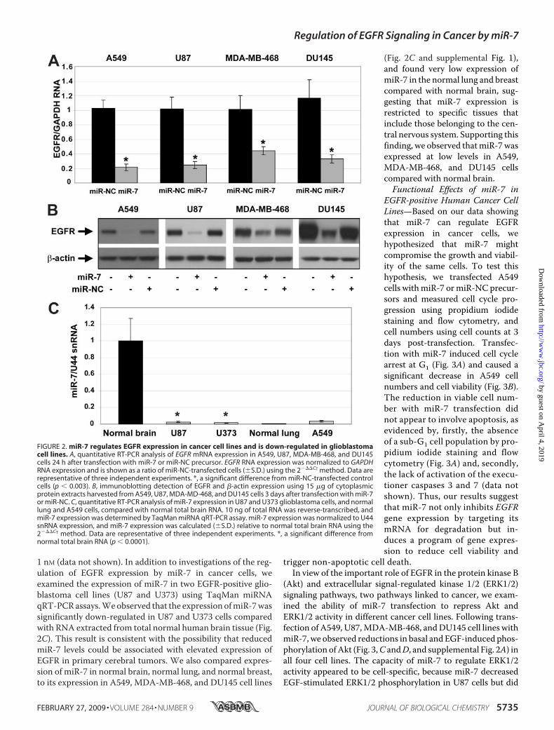
1 nM (data not shown). In addition to investigations of the reg-ulation of EGFR expression by miR-7 in cancer cells, weexamined the expression of miR-7 in two EGFR-positive glio-blastoma cell lines (U87 and U373) using TaqMan miRNAqRT-PCRassays.Weobserved that the expression ofmiR-7wassignificantly down-regulated in U87 and U373 cells comparedwith RNA extracted from total normal human brain tissue (Fig.2C). This result is consistent with the possibility that reducedmiR-7 levels could be associated with elevated expression ofEGFR in primary cerebral tumors. We also compared expres-sion of miR-7 in normal brain, normal lung, and normal breast,to its expression in A549, MDA-MB-468, and DU145 cell lines
(Fig. 2C and supplemental Fig. 1),and found very low expression ofmiR-7 in the normal lung and breastcompared with normal brain, sug-gesting that miR-7 expression isrestricted to specific tissues thatinclude those belonging to the cen-tral nervous system. Supporting thisfinding, we observed thatmiR-7wasexpressed at low levels in A549,MDA-MB-468, and DU145 cellscompared with normal brain.Functional Effects of miR-7 in
EGFR-positive Human Cancer CellLines—Based on our data showingthat miR-7 can regulate EGFRexpression in cancer cells, wehypothesized that miR-7 mightcompromise the growth and viabil-ity of the same cells. To test thishypothesis, we transfected A549cells withmiR-7 ormiR-NC precur-sors and measured cell cycle pro-gression using propidium iodidestaining and flow cytometry, andcell numbers using cell counts at 3days post-transfection. Transfec-tion with miR-7 induced cell cyclearrest at G1 (Fig. 3A) and caused asignificant decrease in A549 cellnumbers and cell viability (Fig. 3B).The reduction in viable cell num-ber with miR-7 transfection didnot appear to involve apoptosis, asevidenced by, firstly, the absenceof a sub-G1 cell population by pro-pidium iodide staining and flowcytometry (Fig. 3A) and, secondly,the lack of activation of the execu-tioner caspases 3 and 7 (data notshown). Thus, our results suggestthat miR-7 not only inhibits EGFRgene expression by targeting itsmRNA for degradation but in-duces a program of gene expres-sion to reduce cell viability and
trigger non-apoptotic cell death.In view of the important role of EGFR in the protein kinase B
(Akt) and extracellular signal-regulated kinase 1/2 (ERK1/2)signaling pathways, two pathways linked to cancer, we exam-ined the ability of miR-7 transfection to repress Akt andERK1/2 activity in different cancer cell lines. Following trans-fection of A549, U87,MDA-MB-468, andDU145 cell lines withmiR-7, we observed reductions in basal and EGF-induced phos-phorylation ofAkt (Fig. 3,C andD, and supplemental Fig. 2A) inall four cell lines. The capacity of miR-7 to regulate ERK1/2activity appeared to be cell-specific, because miR-7 decreasedEGF-stimulated ERK1/2 phosphorylation in U87 cells but did
FIGURE 2. miR-7 regulates EGFR expression in cancer cell lines and is down-regulated in glioblastomacell lines. A, quantitative RT-PCR analysis of EGFR mRNA expression in A549, U87, MDA-MB-468, and DU145cells 24 h after transfection with miR-7 or miR-NC precursor. EGFR RNA expression was normalized to GAPDHRNA expression and is shown as a ratio of miR-NC-transfected cells (�S.D.) using the 2���CT method. Data arerepresentative of three independent experiments. *, a significant difference from miR-NC-transfected controlcells (p � 0.003). B, immunoblotting detection of EGFR and �-actin expression using 15 �g of cytoplasmicprotein extracts harvested from A549, U87, MDA-MD-468, and DU145 cells 3 days after transfection with miR-7or miR-NC. C, quantitative RT-PCR analysis of miR-7 expression in U87 and U373 glioblastoma cells, and normallung and A549 cells, compared with normal total brain RNA. 10 ng of total RNA was reverse-transcribed, andmiR-7 expression was determined by TaqMan miRNA qRT-PCR assay. miR-7 expression was normalized to U44snRNA expression, and miR-7 expression was calculated (�S.D.) relative to normal total brain RNA using the2���CT method. Data are representative of three independent experiments. *, a significant difference fromnormal total brain RNA (p � 0.0001).
Regulation of EGFR Signaling in Cancer by miR-7
FEBRUARY 27, 2009 • VOLUME 284 • NUMBER 9 JOURNAL OF BIOLOGICAL CHEMISTRY 5735
by guest on April 4, 2019
http://ww
w.jbc.org/
Dow
nloaded from
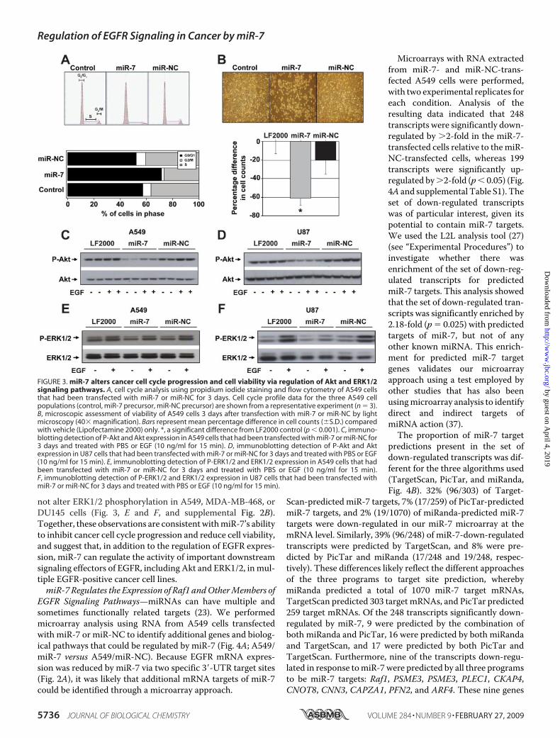
not alter ERK1/2 phosphorylation in A549, MDA-MB-468, orDU145 cells (Fig. 3, E and F, and supplemental Fig. 2B).Together, these observations are consistentwithmiR-7’s abilityto inhibit cancer cell cycle progression and reduce cell viability,and suggest that, in addition to the regulation of EGFR expres-sion, miR-7 can regulate the activity of important downstreamsignaling effectors of EGFR, including Akt and ERK1/2, in mul-tiple EGFR-positive cancer cell lines.miR-7 Regulates the Expression of Raf1 andOtherMembers of
EGFR Signaling Pathways—miRNAs can have multiple andsometimes functionally related targets (23). We performedmicroarray analysis using RNA from A549 cells transfectedwith miR-7 or miR-NC to identify additional genes and biolog-ical pathways that could be regulated by miR-7 (Fig. 4A; A549/miR-7 versus A549/miR-NC). Because EGFR mRNA expres-sion was reduced by miR-7 via two specific 3�-UTR target sites(Fig. 2A), it was likely that additional mRNA targets of miR-7could be identified through a microarray approach.
Microarrays with RNA extractedfrom miR-7- and miR-NC-trans-fected A549 cells were performed,with two experimental replicates foreach condition. Analysis of theresulting data indicated that 248transcripts were significantly down-regulated by �2-fold in the miR-7-transfected cells relative to themiR-NC-transfected cells, whereas 199transcripts were significantly up-regulated by�2-fold (p� 0.05) (Fig.4A and supplemental Table S1). Theset of down-regulated transcriptswas of particular interest, given itspotential to contain miR-7 targets.We used the L2L analysis tool (27)(see “Experimental Procedures”) toinvestigate whether there wasenrichment of the set of down-reg-ulated transcripts for predictedmiR-7 targets. This analysis showedthat the set of down-regulated tran-scripts was significantly enriched by2.18-fold (p � 0.025) with predictedtargets of miR-7, but not of anyother known miRNA. This enrich-ment for predicted miR-7 targetgenes validates our microarrayapproach using a test employed byother studies that has also beenusingmicroarray analysis to identifydirect and indirect targets ofmiRNA action (37).The proportion of miR-7 target
predictions present in the set ofdown-regulated transcripts was dif-ferent for the three algorithms used(TargetScan, PicTar, and miRanda,Fig. 4B). 32% (96/303) of Target-
Scan-predicted miR-7 targets, 7% (17/259) of PicTar-predictedmiR-7 targets, and 2% (19/1070) of miRanda-predicted miR-7targets were down-regulated in our miR-7 microarray at themRNA level. Similarly, 39% (96/248) of miR-7-down-regulatedtranscripts were predicted by TargetScan, and 8% were pre-dicted by PicTar and miRanda (17/248 and 19/248, respec-tively). These differences likely reflect the different approachesof the three programs to target site prediction, wherebymiRanda predicted a total of 1070 miR-7 target mRNAs,TargetScan predicted 303 target mRNAs, and PicTar predicted259 target mRNAs. Of the 248 transcripts significantly down-regulated by miR-7, 9 were predicted by the combination ofboth miRanda and PicTar, 16 were predicted by both miRandaand TargetScan, and 17 were predicted by both PicTar andTargetScan. Furthermore, nine of the transcripts down-regu-lated in response tomiR-7were predicted by all three programsto be miR-7 targets: Raf1, PSME3, PSME3, PLEC1, CKAP4,CNOT8, CNN3, CAPZA1, PFN2, and ARF4. These nine genes
FIGURE 3. miR-7 alters cancer cell cycle progression and cell viability via regulation of Akt and ERK1/2signaling pathways. A, cell cycle analysis using propidium iodide staining and flow cytometry of A549 cellsthat had been transfected with miR-7 or miR-NC for 3 days. Cell cycle profile data for the three A549 cellpopulations (control, miR-7 precursor, miR-NC precursor) are shown from a representative experiment (n � 3).B, microscopic assessment of viability of A549 cells 3 days after transfection with miR-7 or miR-NC by lightmicroscopy (40� magnification). Bars represent mean percentage difference in cell counts (�S.D.) comparedwith vehicle (Lipofectamine 2000) only. *, a significant difference from LF2000 control (p � 0.001). C, immuno-blotting detection of P-Akt and Akt expression in A549 cells that had been transfected with miR-7 or miR-NC for3 days and treated with PBS or EGF (10 ng/ml for 15 min). D, immunoblotting detection of P-Akt and Aktexpression in U87 cells that had been transfected with miR-7 or miR-NC for 3 days and treated with PBS or EGF(10 ng/ml for 15 min). E, immunoblotting detection of P-ERK1/2 and ERK1/2 expression in A549 cells that hadbeen transfected with miR-7 or miR-NC for 3 days and treated with PBS or EGF (10 ng/ml for 15 min).F, immunoblotting detection of P-ERK1/2 and ERK1/2 expression in U87 cells that had been transfected withmiR-7 or miR-NC for 3 days and treated with PBS or EGF (10 ng/ml for 15 min).
Regulation of EGFR Signaling in Cancer by miR-7
5736 JOURNAL OF BIOLOGICAL CHEMISTRY VOLUME 284 • NUMBER 9 • FEBRUARY 27, 2009
by guest on April 4, 2019
http://ww
w.jbc.org/
Dow
nloaded from

are of particular interest in view of a recent recommendationthat investigation of candidate miRNA targets should be prior-itized based on their prediction by two or three of these pro-grams (38).Of these genes, Raf1, whose mRNA expression was signifi-
cantly down-regulated by 3.47-fold by miR-7 in the microarrayexperiment (p � 0.02), was of interest to us because of theknown involvement of Raf1 protein in the EGFR signaling path-way. Raf1 is a downstream effector of EGFR signaling in theRaf-MEK-ERK cascade that is commonly activated by muta-tions and/or overexpressed in human cancers (39).We hypoth-esized that miR-7may coordinately regulate multiple membersof the EGFR signal transduction pathway. To confirm the Raf1microarray result, we performed qRT-PCR on RNA fromA549cells transfected with miR-7 or miR-NC. This showed down-regulation of the expression of Raf1mRNA relative toGAPDHmRNA with miR-7 (Fig. 5A), suggesting that miR-7 promotesdecay of Raf1 mRNA. TargetScan predicted that the humanRaf1 3�-UTR contains two miR-7 target sites (Fig. 5B). TodeterminewhethermiR-7 can directly regulate Raf1 expressionin cancer cells, reporter assay experiments were conductedwith A549 cells. In these experiments, miR-7 reduced theexpression of a reporter construct that carried both of the pre-dicted miR-7 target sites from the Raf1 3�-UTR but not of areporter containing an analogous insert with three point muta-tions in each seed match region (Fig. 5C). This indicated that
the Raf1 mRNA 3�-UTR is a specific target of miR-7 in cancercells. Raf1 protein expression was also reduced in A549 andMDA-MB-468 cells transfected with miR-7 compared withuntransfected cells and to cells transfected with miR-NC (Fig.5D). Together, these data provide evidence that, via direct bind-ing to its 3�-UTR, miR-7 regulates the expression of Raf1, adownstream effector of EGFR signaling in the Raf-MEK-ERKcascade.To further investigate the possible functional roles of miR-7,
we performed an analysis on the microarray data to identifyKEGG pathways that were significantly enriched with genesthat were down-regulated in response to miR-7 (Fig. 6A). The“ErbB signaling pathway,” “GnRH signaling pathway,” “gli-oma,” “long term potentiation,” and “gap junction” pathwayswere all found to be significantly enriched (supplemental Figs.S3–S7). These results support a role formiR-7 in the regulationof erbB signaling. They are also in line with the expression pro-file of miR-7, which is brain-specific with the highest expres-sion in the pituitary and hypothalamus, and with the down-regulation of miR-7 in central nervous system tumors,including pituitary tumors (34, 35). In addition to EGFR andRaf1, several other down-regulated genes from these pathwayscontain predicted binding sites for miR-7. These include genesinvolved in calcium signaling (CALM3 and CAMK2D, down-regulated 7.10- and 2.08-fold, p � 0.006 and p � 0.024, respec-tively), cytoskeleton reorganization and nuclear signaling(PAK1, down-regulated 2.20-fold, p � 0.006), and cAMP syn-thesis and intracellular signaling (ADCY9, down-regulated3.38-fold, p � 0.005) (supplemental Table S1). This suggeststhat miR-7 has the ability to target multiple cell signaling path-ways by coordinately regulating the expression of key signalingmolecules.
DISCUSSION
We have demonstrated that miR-7 can regulate the expres-sion of multiple effectors of EGFR signaling. As well as directlytargeting EGFR andRaf1, miR-7 down-regulates the expressionof a number of other genes associated with cellular pathwaysdownstream of EGFR. Furthermore, miR-7 has functionaleffects in cancer cell lines that include inducing cell cycle arrestand reducing cell growth and viability.EGFR as a Target ofmiR-7 in Cancer Cells—Amajor obstacle
to the understanding of miRNA signaling and function hasbeen the shortage of validated miRNA targets. This is due inpart to the likely ability of a given miRNA to regulate theexpression of hundreds or even thousands of target mRNAs,but also to the imperfect complementarity ofmiRNAand targetsequences. Recent work has revealed some of the criteria thatsupport an authentic miRNA-mRNA interaction. We chose toinvestigate the predicted interaction between miR-7 and theEGFR mRNA 3�-UTR, which does not satisfy one of the com-monly used criteria for miRNA-target prediction, that is, con-servation of the miRNA binding site(s) across species. Our rea-soning was based on the evolution of regulatory sequences withincreasing species complexity.Although EGFR overexpression in many cancers often
occurs via gene amplification, a subset of tumors with EGFRoverexpression does not exhibit EGFR gene amplification. In
FIGURE 4. Identification of candidate miR-7 target mRNAs by microarrayanalysis of miR-7 transfected A549 cells. A, scatterplot showing log inten-sities of miR-7-transfected A549 cells (y axis) plotted against the log intensi-ties of miR-NC-transfected cells (x axis). Differentially expressed genes arecolored in red (more highly expressed in miR-7-transfected cells) or green(more highly expressed in miR-NC-transfected cells). B, intersection of pre-dicted miR-7 target lists (from TargetScan, PicTar, and miRanda) with the 248transcripts significantly down-regulated by miR-7 in A549 cells.
Regulation of EGFR Signaling in Cancer by miR-7
FEBRUARY 27, 2009 • VOLUME 284 • NUMBER 9 JOURNAL OF BIOLOGICAL CHEMISTRY 5737
by guest on April 4, 2019
http://ww
w.jbc.org/
Dow
nloaded from

one study, it was found that 20% of the EGFR-overexpressingglioblastomas tested lacked EGFR gene amplification (40), sug-gesting that other, possibly post-transcriptional, mechanismsexist to promote aberrant EGFR expression in cancer cells. Wehave identified miR-7 as a regulator of EGFR gene expressionand signaling in human cancer cells. Furthermore, the normalexpression of miR-7 in the brain and endocrine systems,together with the previously reported down-regulation ofmiR-7 in central nervous system tumors, suggests that miR-7has the ability to act as a regulator of normal cellular pathwaysin some systems and as an endogenous tumor suppressor inothers.miR-7 and the Regulation of EGFR Signaling Pathways in
Normal andCancer Cells—Our results show thatmiR-7 has theability to coordinately down-regulate the expression of multi-plemembers of EGFR signaling cascades, both directly, as is thecase for EGFR and Raf1, and indirectly, and also to induce can-
cer cell cycle arrest and cell death. Our data further suggest thatmiR-7 may have several tissue-specific functions, with possibleroles in the development and progression of gliomas, reproduc-tion, via the production of pituitary gonadotropins, and learn-ing and memory, through regulation of many other signalingand structural proteins in addition to EGFR and Raf1 (Fig. 6B).A number of recent reports support a role for miR-7 in the
brain and in tumor cells. miR-7 has been shown in one study tobelong to a subset of miRNAs that are down-regulated in schiz-ophrenia (43). Interestingly, the predicted targets of the dis-regulated miRNAs in the study, like the mRNAs down-regu-lated bymiR-7 in the present study, are over-represented in theKEGG functional pathways of “focal adhesion,” “regulation ofactin cytoskeleton,” and “gap junction” (Fig. 6A), and may ulti-mately influence synaptic plasticity in schizophrenia.With respect to systems involving EGFR signaling, the
MAPK ERK5 is regulated by miR-143 in differentiated adipo-
FIGURE 5. miR-7 regulates Raf1 expression via specific binding to the Raf1 mRNA 3�-UTR. A, qRT-PCR analysis of Raf1 expression in A549 cells transfectedwith miR-7 or miR-NC for 24 h. Raf1 expression was normalized to GAPDH expression and data shown relative to miR-NC-transfected cells using the 2���CT
method (mean � S. D., n � 3). *, a significant difference from miR-NC control (p � 0.0005). B, schematic representation of Raf1 mRNA 3�-UTR showing conserved(C) and non-conserved (NC) predicted seed target sites for miR-7 binding. C, luciferase reporter gene assay using A549 cells that were transfected with Raf1-WTor Raf1-MT firefly luciferase reporter plasmid DNA, Renilla luciferase plasmid DNA, and either miR-7 or miR-NC, for 24 h. Firefly expression was normalized toRenilla expression. Data are expressed as a ratio of miR-NC-transfected A549 cells (�S.D.). Results are representative of those obtained from at least threeindependent experiments. **, a significant difference from Raf1 WT/miR-NC (p � 0.05). D, immunoblotting detection of Raf1 and �-actin expression using 15�g of cytoplasmic protein extracts harvested from A549 and MDA-MB-468 cells 3 days after transfection with miR-7 or miR-NC.
Regulation of EGFR Signaling in Cancer by miR-7
5738 JOURNAL OF BIOLOGICAL CHEMISTRY VOLUME 284 • NUMBER 9 • FEBRUARY 27, 2009
by guest on April 4, 2019
http://ww
w.jbc.org/
Dow
nloaded from

cytes and cancer cell lines (41, 42) in much the same way asmiR-7 regulates the ERK1/2 pathway, suggesting that miRNAssuch asmiR-7 andmiR-143have the ability to regulate signalingeffectors critical to cell growth and differentiation. Two otherreports implicate miR-7 in the regulation of EGFR signaling. Inone, miR-7 was shown to control EGFR signaling inDrosophilaphotoreceptor cells (44). This study presented a model inwhich, upon cell differentiation, EGFR signaling triggers ERK-
mediated degradation of the transcription repressor Yan,thereby relieving Yan-mediated repression of miR-7 expres-sion. Conversely, miR-7 represses Yan expression in photore-ceptors via binding to Yan 3�-UTR sequences. This feedbackloop thus promotes mutually exclusive expression of Yan andmiR-7.However, EGFR is unlikely to represent a direct target ofmiR-7 inDrosophila due to the lack of conservation of theDro-sophilamiR-7 target sites to humans.While this report was in preparation, a recent report byKefas
and coworkers demonstrated thatmiR-7 inhibits EGFR expres-sion and the Akt pathway in glioblastomas, tumors shown tohave reduced miR-7 expression relative to normal brain tissue(45). The regulation of insulin receptor substrate 2 (IRS2) expres-sionwas proposed as amechanism to explain howmiR-7 controlsAkt activity.Wehave confirmed the regulation of IRS2 expressionat the protein level by miR-7 in the other cell lines used in ourexperiments (supplemental Fig. S8).Wedid not note a decrease inIRS2 mRNA expression in miR-7-transfected A549 cells in ourmicroarray experiments. Similarly, for eachof the cell lines used inthis study, we did not observe a significant difference in IRS2mRNA expression between cells transfected with miR-7 andmiR-NC by qRT-PCR (data not shown), suggesting that miR-7regulates IRS2 expression at the translational level.There are a number of key differences between the study by
Kefas and coworkers and the present study. Firstly, we evalu-ated the relative contribution of each of the three predictedmiR-7 target sites in the EGFR 3�-UTR to the regulation of geneexpression by miR-7, show that two of the three predictedmiR-7 target sites are directly targeted by miR-7, and demon-strate the specificity of these interactions through mutationalanalysis of the target site seed regions. Secondly, we observed areduction in EGFRmRNA with miR-7 transfection in multipleEGFR-positive cell lines, using qRT-PCR and microarray anal-ysis, whereas Kefas and coworkers reported no change in EGFRmRNA expression with miR-7 transfection. Our findingenabled the use of microarray analysis of miR-7-transfectedcells. Thirdly, we did not observe the classicmarkers of apopto-tic cell death with miR-7 transfection, i.e. we did not detect asubstantial sub-G1 apoptotic peak or activation of caspases 3 or7 in miR-7-transfected cells. Fourthly, in addition to a role inthe regulation of Akt signaling, our findings implicate miR-7in the regulation of ERK1/2 signaling in cancer cells, at least inpart via its ability to regulate the expression of Raf1 kinase.Furthermore, the regulation of these signaling effectors bymiR-7 is shown to occur in a range of EGFR-positive cancer cellline models.Finally, we have extended our investigation beyond the iden-
tification of EGFR as a target of miR-7 to explore additionalcellular pathways in which miR-7 may be involved, usingmicroarray analysis of miR-7-transfected cancer cells. Thisanalysis has linked miR-7 to a number of pathways relevant tonormal and tumor cells. Furthermore, this analysis suggestedseveral other miR-7 target candidates that are linked to EGFRsignaling, including MEK, PI3K, Akt, CAMK, PAK1, and ARF4(Fig. 6B). The reduced expression of both PI3K and Akt mRNAby miR-7 in our array is interesting. However, in our studies,miR-7 did not significantly alter total Akt protein expression,whereas it did alter Akt phosphorylation. Similarly, the down-
FIGURE 6. Identification of functional pathways enriched with miR-7 tar-get genes. A, table of KEGG pathways significantly enriched (z-score � 2)with genes down-regulated by miR-7 compared with miR-NC in A549 cells.B, schematic model showing coordinate regulation of EGFR signaling bymiR-7 via multiple targets. Direct, validated target mRNAs of miR-7 includeEGFR and Raf1 (dark circles). Gray circles represent EGFR signaling genes withexpression modulated by treatment with miR-7 by unknown mechanism.
Regulation of EGFR Signaling in Cancer by miR-7
FEBRUARY 27, 2009 • VOLUME 284 • NUMBER 9 JOURNAL OF BIOLOGICAL CHEMISTRY 5739
by guest on April 4, 2019
http://ww
w.jbc.org/
Dow
nloaded from
regulation of MAP2K2 (MEK) expression in the microarraysuggests that miR-7 could potentially regulate ERK1/2 activityat a third point in the cascade, in addition to its regulation ofEGFR and Raf1.ADP-ribosylation factor 4 (ARF4) is another interesting
miR-7 target candidate gene that was down-regulated in themicroarray results. The ARF4 protein interacts with EGFR andmediates EGF-dependent activation of the transcription factorAP-1 (46). Therefore, by regulating ARF4 expression, miR-7could potentially alter a broad program of gene expressionthrough control of EGF-dependent AP-1 activity. AnothermiR-7-regulated gene, calmodulin kinase II (CAMK2), hasimportant roles in the central nervous system relating to longterm potentiation and neurotransmitter release (47). This maybe significant given that miR-7 is known to be abundantlyexpressed in the hypothalamus and pituitary, major sites forneurotransmitter secretion. p21/Cdc42/Rac1-activated kinase1 (PAK1) is a serine/threonine kinase that mediates severaloncogenic signaling pathways, including EGFR/Akt signaling,to regulate cytoskeletal remodeling, cell motility, cell prolifera-tion, and apoptosis (reviewed in Ref. 48). PAK1 was down-reg-ulated by miR-7 in our microarray studies and was a predictedtarget of miR-7. PAK1 expression is increased in some cancers; inglioblastoma patients its elevated expression is associated withshorter survival time (49).Recently,PAK1was showntobea targetof miR-7 in cancer cells (50), suggesting that the reduced expres-sion ofmiR-7 in glioblastomasmight promote the aberrant PAK1expression and activity seen in these tumors.The benefit of abrogating EGFR expression in a variety of
human cancer types as a therapeutic approach has been empha-sized by recent data showing that the kinase-independent func-tion of EGFR is to prevent autophagic cell death bymaintainingthe basal intracellular glucose level through an interaction withthe sodium/glucose co-transporter 1 (51). To date, clinical tri-als of EGFR tyrosine kinase inhibitors in a variety of cancershave yielded disappointing results. Co-inhibition of kinase-in-dependent and kinase-dependent EGFR functions couldachieve durable clinical responses. Agents such as miR-7 thatdown-regulate expression of the EGFR as well as some of itssignaling effectorsmay have significant therapeutic potential ina range of human cancer types.In summary, our data indicate that miR-7 coordinately reg-
ulates EGFR signaling at multiple levels and suggest that miR-7additionally regulates a number of other cellular pathways rel-evant to normal and tumor cells. The effect ofmiR-7 action is toinhibit cell cycle progression and reduce cell growth and viabil-ity. The reported down-regulation of miR-7 in tumors of thecentral nervous system, together with its ability to regulateoncogenic EGFR signaling in multiple cancer cell line models,suggests that the therapeutic up-regulation of miR-7 expres-sion in these tumors may inhibit growth and metastasis.
Acknowledgments—We thank Drs. D. P. Bartel and L. P. Ford forhelpful discussions and Michael Epis for advice regarding qRT-PCRexperimental design and data analysis.
REFERENCES1. Yano, S., Kondo, K., Yamaguchi, M., Richmond, G., Hutchison, M.,
Wakeling, A., Averbuch, S., andWadsworth, P. (2003)Anticancer Res. 23,3639–3650
2. Arteaga, C. L. (2001) J. Clin. Oncol. 19, 32S–40S3. Ritter, C. A., and Arteaga, C. L. (2003) Semin. Oncol. 30, 3–114. Bianco, R., Troiani, T., Tortora, G., and Ciardiello, F. (2005) Endocr. Relat.
Cancer 12, S159–S1715. Bartel, D. P. (2004) Cell 116, 281–2976. Mattick, J. S., andMakunin, I. V. (2005)Hum. Mol. Genet 14, R121–R1327. Humphreys, D. T.,Westman, B. J., Martin, D. I., and Preiss, T. (2005) Proc.
Natl. Acad. Sci. U. S. A. 102, 16961–169668. Chen, J. F., Mandel, E. M., Thomson, J. M., Wu, Q., Callis, T. E., Ham-
mond, S. M., Conlon, F. L., and Wang, D. Z. (2006) Nat. Genet. 38,228–233
9. Cheng, A. M., Byrom, M. W., Shelton, J., and Ford, L. P. (2005) NucleicAcids Res. 33, 1290–1297
10. Zhang, B., Pan, X., Cobb, G. P., and Anderson, T. A. (2007)Dev. Biol. 302,1–12
11. Calin,G.A., Liu, C.G., Sevignani, C., Ferracin,M., Felli, N., Dumitru, C.D.,Shimizu, M., Cimmino, A., Zupo, S., Dono, M., Dell’Aquila, M. L., Alder,H., Rassenti, L., Kipps, T. J., Bullrich, F., Negrini, M., and Croce, C. M.(2004) Proc. Natl. Acad. Sci. U. S. A. 101, 11755–11760
12. Esquela-Kerscher, A., and Slack, F. J. (2006) Nat. Rev. Cancer 6, 259–26913. Johnson, S. M., Grosshans, H., Shingara, J., Byrom, M., Jarvis, R., Cheng,
A., Labourier, E., Reinert, K. L., Brown, D., and Slack, F. J. (2005) Cell 120,635–647
14. Takamizawa, J., Konishi, H., Yanagisawa, K., Tomida, S., Osada, H., En-doh, H., Harano, T., Yatabe, Y., Nagino, M., Nimura, Y., Mitsudomi, T.,and Takahashi, T. (2004) Cancer Res. 64, 3753–3756
15. Chan, J. A., Krichevsky, A. M., and Kosik, K. S. (2005) Cancer Res. 65,6029–6033
16. Volinia, S., Calin, G. A., Liu, C. G., Ambs, S., Cimmino, A., Petrocca, F.,Visone, R., Iorio, M., Roldo, C., Ferracin, M., Prueitt, R. L., Yanaihara, N.,Lanza, G., Scarpa, A., Vecchione, A., Negrini, M., Harris, C. C., and Croce,C. M. (2006) Proc. Natl. Acad. Sci. U. S. A. 103, 2257–2261
17. Si,M. L., Zhu, S.,Wu,H., Lu, Z.,Wu, F., andMo, Y. Y. (2006)Oncogene 26,2799–2803
18. Rajewsky, N. (2006) Nat. Genet. 38, S8–S1319. Maziere, P., and Enright, A. J. (2007) Drug Discov. Today 12, 452–45820. Grimson, A., Farh, K. K., Johnston, W. K., Garrett-Engele, P., Lim, L. P.,
and Bartel, D. P. (2007)Mol. Cell 27, 91–10521. Lewis, B. P., Burge, C. B., and Bartel, D. P. (2005) Cell 120, 15–2022. Krutzfeldt, J., Rajewsky, N., Braich, R., Rajeev, K. G., Tuschl, T., Manoha-
ran, M., and Stoffel, M. (2005) Nature 438, 685–68923. Stark, A., Brennecke, J., Russell, R. B., andCohen, S.M. (2003) PLoS Biol. 1,
E6024. Giles, K. M., Daly, J. M., Beveridge, D. J., Thomson, A. M., Voon, D. C.,
Furneaux, H. M., Jazayeri, J. A., and Leedman, P. J. (2003) J. Biol. Chem.278, 2937–2946
25. Wang, X., and Seed, B. (2003) Nucleic Acids Res. 31, e15426. Livak, K. J., and Schmittgen, T. D. (2001)Methods 25, 402–40827. Newman, J. C., and Weiner, A. M. (2005) Genome Biology
http://genomebiology.com/2005/6/9/R8128. Enright, A. J., John, B., Gaul, U., Tuschl, T., Sander, C., and Marks, D. S.
(2003) Genome Biology http://genomebiology.com/2003/5/1/R129. Krek, A., Grun, D., Poy, M. N., Wolf, R., Rosenberg, L., Epstein, E. J.,
MacMenamin, P., da Piedade, I., Gunsalus, K. C., Stoffel,M, and Rajewsky,N. (2005) Nat. Genet. 37, 495–500
30. Balmer, L. A., Beveridge, D. J., Jazayeri, J. A., Thomson, A. M., Walker,C. E., and Leedman, P. J. (2001)Mol. Cell. Biol. 21, 2070–2084
31. Sempere, L. F., Freemantle, S., Pitha-Rowe, I., Moss, E., Dmitrovsky, E.,and Ambros, V. (2004)Genome Biology http://genomebiology.com/2004/5/3/R13
32. Farh, K. K., Grimson, A., Jan, C., Lewis, B. P., Johnston, W. K., Lim, L. P.,Burge, C. B., and Bartel, D. P. (2005) Science 310, 1817–1821
33. Landgraf, P., Rusu, M., Sheridan, R., Sewer, A., Iovino, N., Aravin, A.,
Regulation of EGFR Signaling in Cancer by miR-7
5740 JOURNAL OF BIOLOGICAL CHEMISTRY VOLUME 284 • NUMBER 9 • FEBRUARY 27, 2009
by guest on April 4, 2019
http://ww
w.jbc.org/
Dow
nloaded from
Pfeffer, S., Rice, A., Kamphorst, A. O., Landthaler, M., Lin, C., Socci, N. D.,Hermida, L., Fulci, V., Chiaretti, S., Foa, R., Schliwka, J., Fuchs,U.,Novosel,A., Muller, R. U., Schermer, B., Bissels, U., Inman, J., Phan, Q., Chien, M.,Weir, D. B., Choksi, R., De Vita, G., Frezzetti, D., Trompeter, H. I., Hor-nung, V., Teng, G., Hartmann, G., Palkovits, M., Di Lauro, R., Wernet, P.,Macino, G., Rogler, C. E., Nagle, J. W., Ju, J., Papavasiliou, F. N., Benzing,T., Lichter, P., Tam,W., Brownstein,M. J., Bosio, A., Borkhardt, A., Russo,J. J., Sander, C., Zavolan, M., and Tuschl, T. (2007) Cell 129, 1401–1414
34. Bottoni, A., Zatelli, M. C., Ferracin, M., Tagliati, F., Piccin, D., Vignali, C.,Calin, G. A., Negrini, M., Croce, C. M., and Degli Ulberti, E. C. (2007)J. Cell. Physiol. 210, 370–377
35. Gaur, A., Jewell, D. A., Liang, Y., Ridzon, D., Moore, J. H., Chen, C., Am-bros, V. R., and Israel, M. A. (2007) Cancer Res. 67, 2456–2468
36. Lee, H. J., Palkovits, M., and Young, W. S. (2006) Proc. Natl. Acad. Sci.U. S. A. 103, 15669–15674
37. Lim, L. P., Lau, N. C., Garrett-Engele, P., Grimson, A., Schelter, J. M.,Castle, J., Bartel, D. P., Linsley, P. S., and Johnson, J. M. (2005)Nature 433,769–773
38. Kuhn, D. E., Martin, M. M., Feldman, D. S., Terry, A. V., Nuovo, G. J., andElton, T. S. (2008)Methods 44, 47–54
39. Roberts, P. J., and Der, C. J. (2007) Oncogene 26, 3291–331040. Tripp, S. R., Willmore-Payne, C., and Layfield, L. J. (2005) Anal. Quant.
Cytol. Histol. 27, 71–7841. Esau, C., Kang, X., Peralta, E., Hanson, E.,Marcusson, E. G., Ravichandran,
L. V., Sun, Y., Koo, S., Perera, R. J., Jain, R., Dean, N. M., Freier, S. M.,
Bennett, C. F., Lollo, B., and Griffey, R. (2004) J. Biol. Chem. 279,52361–52365
42. Akao, Y., Nakagawa, Y., Kitade, Y., Kinoshita, T., and Naoe, T. (2006)Cancer Sci. 98, 1914–1920
43. Perkins, D. O., Jeffries, C. D., Jarskog, L. F., Thomson, J. M., Woods, K.,Newman, M. A., Parker, J. S., Jin, J., and Hammond, S. M. (2007) GenomeBiology http://genomebiology.com/2007/8/2/R27
44. Li, X., and Carthew, R. W. (2005) Cell 123, 1267–127745. Kefas, B., Godlewski, J., Comeau, L., Li, Y., Abounader, R., Hawkinson,M.,
Lee, J., Fine, H., Chiocca, E. A., Lawler, S., and Purow, B. (2008)Cancer Res.68, 3566–3572
46. Kim, S. W., Hayashi, M., Lo, J. F., Yang, Y., Yoo, J. S., and Lee, J. D. (2003)J. Biol. Chem. 278, 2661–2668
47. Haisenleder, D. J., Burger, L. L, Aylor, K. W., Dalkin, A. C., and Marshall,J. C. (2003) Endocrinology 144, 2768–2774
48. Kumar, R., Gururaj, A. E., and Barnes, C. J. (2006) Nat. Rev. Cancer 6,459–471
49. Aoki, H., Yokoyama, T., Fujiwara, K., Tari, A. M., Sawaya, R., Suki, D.,Hess, K. R., Aldape, K. D., Kondo, S., Kumar, R., andKondo, Y. (2007)Clin.Cancer Res. 13, 6603–6609
50. Reddy, S. D., Ohshiro, K., Rayala, S. K., and Kumar, R. (2008) Cancer Res.68, 8195–8200
51. Weihua, Z., Tsan, R., Huang, W. C., Wu, Q., Chiu, C. H., Fidler, I. J., andHung, M. C. (2008) Cancer Cell 13, 385–393
Regulation of EGFR Signaling in Cancer by miR-7
FEBRUARY 27, 2009 • VOLUME 284 • NUMBER 9 JOURNAL OF BIOLOGICAL CHEMISTRY 5741
by guest on April 4, 2019
http://ww
w.jbc.org/
Dow
nloaded from

Mattick and Peter J. LeedmanRebecca J. Webster, Keith M. Giles, Karina J. Price, Priscilla M. Zhang, John S.
Cells by MicroRNA-7Regulation of Epidermal Growth Factor Receptor Signaling in Human Cancer
doi: 10.1074/jbc.M804280200 originally published online December 10, 20082009, 284:5731-5741.J. Biol. Chem.
10.1074/jbc.M804280200Access the most updated version of this article at doi:
Alerts:
When a correction for this article is posted•
When this article is cited•
to choose from all of JBC's e-mail alertsClick here
Supplemental material:
http://www.jbc.org/content/suppl/2008/12/11/M804280200.DC1
http://www.jbc.org/content/284/9/5731.full.html#ref-list-1
This article cites 47 references, 16 of which can be accessed free at
by guest on April 4, 2019
http://ww
w.jbc.org/
Dow
nloaded from