Embed Size (px)
Citation preview
Regulation of Histone Acetylation by Autophagy in ParkinsonDisease*
Received for publication, June 28, 2015, and in revised form, December 16, 2015 Published, JBC Papers in Press, December 23, 2015, DOI 10.1074/jbc.M115.675488
Goonho Park‡§, Jieqiong Tan¶, Guillermina Garcia�, Yunyi Kang�, Guy Salvesen‡�, and Zhuohua Zhang‡¶�1
From the ‡Graduate Program of Biomedical Science, Sanford-Burnham Medical Research Institute, La Jolla, California 92037, the§University of California, San Diego, La Jolla, California 92037, the ¶State Key Laboratory of Medical Genetics, Xiangya MedicalSchool, Central South University, Hunan 410078, China, and the �Sanford-Burnham Medical Research Institute,La Jolla, California 92037
Parkinson disease (PD) is the most common age-dependentneurodegenerative movement disorder. Accumulated evidenceindicates both environmental and genetic factors play impor-tant roles in PD pathogenesis, but the potential interactionbetween environment and genetics in PD etiology remainslargely elusive. Here, we report that PD-related neurotoxinsinduce both expression and acetylation of multiple sites of his-tones in cultured human cells and mouse midbrain dopaminer-gic (DA) neurons. Consistently, levels of histone acetylation aremarkedly higher in midbrain DA neurons of PD patients com-pared to those of their matched control individuals. Furtheranalysis reveals that multiple histone deacetylases (HDACs)are concurrently decreased in 1-methyl-4-phenylpyridinium(MPP�)-treated cells and 1-methyl-4-phenyl-1,2,3,6-tetrahy-dropyridine-treated mouse brains, as well as midbrain tissues ofhuman PD patients. Finally, inhibition of histone acetyltrans-ferase (HAT) protects, whereas inhibition of HDAC1 andHDAC2 potentiates, MPP�-induced cell death. Pharmacologi-cal and genetic inhibition of autophagy suppresses MPP�-in-duced HDACs degradation. The study reveals that PD envi-ronmental factors induce HDACs degradation and histoneacetylation increase in DA neurons via autophagy and identifiesan epigenetic mechanism in PD pathogenesis.
Parkinson disease (PD)2 is pathologically characterized asloss of DA neurons in substantial nigra of midbrain and Lewybody formation in the remaining neurons (1). Genetic studies offamilial cases have identified mutations in at least 14 genes thatare associated with the disease (2). Nevertheless, the majority ofPD cases are sporadic with unidentified etiology. It is wellaccepted that environmental factors play an important role in
PD. In addition to aging, exposure to environmental toxins,such as certain pesticides and herbicides, results in parkinson-ism resembling the idiopathic PD (3). Thus, both genetic andenvironmental factors contribute to PD pathogenesis. How-ever, the molecular basis of parkinsonia induced by environ-mental factors remains unclear. Environmental factors areknown to cause abnormal epigenetic modifications resulting inhuman diseases, including neurodegenerative diseases (4 – 6).Such modifications regulate gene expression by mechanismsother than DNA sequence changes (7). These types of regula-tion are heritable, self-perpetuating, and reversible (7–9). Themost studied epigenetic regulations include DNA methylation,RNA modification, and histone modification (8).
Acetylation of histone proteins associated with chromatinplays a pivotal role in the epigenetic regulation of transcriptionand other functions in cells, including neurons (10). Reducedhistone acetylation in animal models has been reported in neu-rodegeneration characterized by cognitive decline, includingmodels of Alzheimer disease (AD) (11). Similar findings havebeen reported with PD models (3). Valproic acid, a histonedeacetylase inhibitor, demonstrates protection against rote-none in a rat model of PD (12). Inhibitors of sirtuin-2 rescues�-synuclein-mediated neurotoxicity both in vitro in cell cul-tures and in vivo in a Drosophila PD model (13). These findingssuggest that dysregulation of acetylation of histone or non-his-tone protein is a common mechanism of neurodegeneration indifferent neurodegenerative diseases.
In this study, we aim to investigate a role of histone acetyla-tion in PD pathogenesis. Our results reveal that both levels ofhistone and histone acetylation are up-regulated in cells treatedwith PD-related neurotoxins and in brains of mice injected with1-methyl-4-phenyl-1,2,3,6-tetrahydropyridine (MPTP). Increaseddetection of histone acetylation is also observed in midbrainDA neurons of PD patients. Further analysis suggests thatreduced expression of HDACs is likely responsible for changesof histone acetylation induced by PD-related neurotoxins.Moreover, inhibition of autophagy suppresses 1-methyl-4-phe-nylpyridinium (MPP�)-induced HDACs degradation. Theresults reveal that PD-related environmental toxins regulateautophagy resulting in abnormal histone acetylation to contrib-ute to PD pathogenesis.
Experimental Procedures
Materials—Antibodies including histone 2A (Cell Signaling,2578), histone 2B (Cell Signaling, 2934), histone 3 (Cell Signal-
* This work was supported, in whole or in part, by National Institutes of HealthGrant NS057289 (to Z. Z.), Natural Science Foundation of China Grants313300257, 81429002, and 81161120498 (to Z. Z.), “973 Program” of Min-istry of Science and Technology Grant 2011CB51000 (to Z. Z.), “111 Pro-gram” of Foreign Expert Bureau of China Grant B10036 (to Z. Z.), and theMogam Science Scholarship Foundation (to G. P.). The authors declare thatthey have no competing interests.
1 To whom correspondence should be addressed: Central South University,Xiangya Medical School, 110 Xiangya Rd., Changsha, Hunan 410078, China.Tel.: 86-731-84805358; E-mail: [email protected].
2 The abbreviations used are: PD, Parkinson disease; HDAC, histone deacety-lase; DA, dopaminergic; AD, Alzheimer disease; MPTP, 1-methyl-4-phenyl-1,2,3,6-tetrahydropyridine; MPP, 1-methyl-4-phenylpyridinium; MTS,3-(4,5-dimethylthiazol-2-yl)-5-(3-carboxymethoxyphenyl)-2-(4-sulfophe-nyl)-2H-tetrazolium; TH, tyrosine hydroxylase; aMPTP, acute MPTP; cMPTP,chronic MPTP; TSA, trichostatin A.
crossmarkTHE JOURNAL OF BIOLOGICAL CHEMISTRY VOL. 291, NO. 7, pp. 3531–3540, February 12, 2016
© 2016 by The American Society for Biochemistry and Molecular Biology, Inc. Published in the U.S.A.
FEBRUARY 12, 2016 • VOLUME 291 • NUMBER 7 JOURNAL OF BIOLOGICAL CHEMISTRY 3531
by guest on June 22, 2018http://w
ww
.jbc.org/D
ownloaded from
ing, 9715), histone 4 (Cell Signaling, 2935), acetylated lysine(Cell Signaling, 9441), acetyl-H2Ak5 (Cell Signaling, 2576),acetyl-H2Bk5 (Cell Signaling, 2574), acetyl-H2Bk15 (Cell Sig-naling, 5435), acetyl-H2Bk20 (Cell Signaling, 2571), acetyl-H3k9 (Cell singling, 9671), acetyl-H3k18 (Cell Signaling, 9675),acetyl-H3k27 (Cell Signaling, 4353), acetyl-H3k56 (Cell Signal-ing, 4243), acetyl-H4k5 (Cell Signaling, 9672), acetyl-H4k8
(Cell Signaling, 2594), acetyl-H4k12 (Cell Signaling, 2591),HDAC1 (Cell Signaling, 5356), HDAC4 (Cell Signaling, 5392),HDAC6 (Cell Signaling, 7558), and SirT1 (Cell Signaling, 2310)were purchased from Cell Signaling. Gcn5 (Epitomics,Q92830), cAMP response element-binding protein (CBP)(Abcam, ab83857), HDAC2 (Abcam, ab51832), tyrosine hydroxy-lase (Millipore, ab152; ab9702), Tip60 (Millipore, 07-038) were
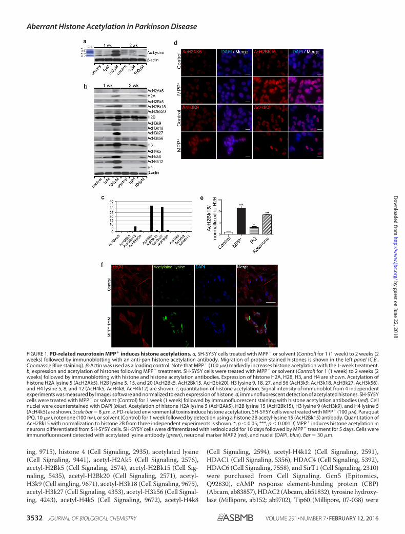
FIGURE 1. PD-related neurotoxin MPP� induces histone acetylations. a, SH-SY5Y cells treated with MPP� or solvent (Control) for 1 (1 week) to 2 weeks (2weeks) followed by immunoblotting with an anti-pan histone acetylation antibody. Migration of protein-stained histones is shown in the left panel (C.B.,Coomassie Blue staining). �-Actin was used as a loading control. Note that MPP� (100 �M) markedly increases histone acetylation with the 1-week treatment.b, expression and acetylation of histones following MPP� treatment. SH-SY5Y cells were treated with MPP� or solvent (Control) for 1 (1 week) to 2 weeks (2weeks) followed by immunoblotting with histone and histone acetylation antibodies. Expression of histone H2A, H2B, H3, and H4 are shown. Acetylation ofhistone H2A lysine 5 (AcH2Ak5), H2B lysine 5, 15, and 20 (AcH2Bk5, AcH2Bk15, AcH2bk20), H3 lysine 9, 18, 27, and 56 (AcH3k9, AcH3k18, AcH3k27, AcH3k56),and H4 lysine 5, 8, and 12 (AcH4k5, AcH4k8, AcH4k12) are shown. c, quantitation of histone acetylation. Signal intensity of immunoblot from 4 independentexperiments was measured by ImageJ software and normalized to each expression of histone. d, immunofluorescent detection of acetylated histones. SH-SY5Ycells were treated with MPP� or solvent (Control) for 1 week (1 week) followed by immunofluorescent staining with histone acetylation antibodies (red). Cellnuclei were counterstained with DAPI (blue). Acetylation of histone H2A lysine 5 (AcH2Ak5), H2B lysine 15 (AcH2Bk15), H3 lysine 9 (AcH3k9), and H4 lysine 5(AcH4k5) are shown. Scale bar � 8 �m. e, PD-related environmental toxins induce histone acetylation. SH-SY5Y cells were treated with MPP� (100 �M), Paraquat(PQ, 10 �M), rotenone (100 nM), or solvent (Control) for 1 week followed by detection using a histone 2B acetyl-lysine 15 (AcH2Bk15) antibody. Quantitation ofAcH2Bk15 with normalization to histone 2B from three independent experiments is shown. *, p � 0.05; ***, p � 0.001. f, MPP� induces histone acetylation inneurons differentiated from SH-SY5Y cells. SH-SY5Y cells were differentiated with retinoic acid for 10 days followed by MPP� treatment for 5 days. Cells wereimmunofluorescent detected with acetylated lysine antibody (green), neuronal marker MAP2 (red), and nuclei (DAPI, blue). Bar � 30 �m.
Aberrant Histone Acetylation in Parkinson Disease
3532 JOURNAL OF BIOLOGICAL CHEMISTRY VOLUME 291 • NUMBER 7 • FEBRUARY 12, 2016
by guest on June 22, 2018http://w
ww
.jbc.org/D
ownloaded from
commercially purchased. All chemicals were from Sigma. Humantissues were obtained from the NICHD, National Institutes ofHealth, Brain and Tissue Bank for Developmental Disorders at theUniversity of Maryland, Baltimore, MD (Tables 1 and 2).
Differentiation of SH-SY5Y Cells—SH-SY5Y cells were fromATCC and maintained according to the manufacturer’sinstructions. To differentiate, cells were treated with 10 �M
retinoic acid (Sigma, R2625) for 10 days.
MTS Assay and ATP Measurement—CellTiter 96� AQueousNon-radioactive Cell Proliferation Assay (Promega, G5421),and ATPlite 1step Luminescence Assay System (PerkinElmer,6016731) were used to measure cell viability. The assays wereperformed according to the manufacturer’s protocol. Absorb-ance for the MTS assay was measured with a microplate reader(Thermo Electron Co.). ATP luminescence was detected withan Analyst HT (Molecular Devices, CA).
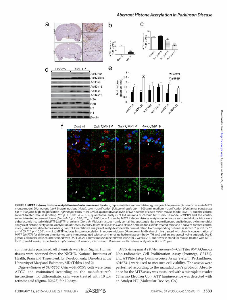
FIGURE 2. MPTP induces histone acetylation in vivo in mouse midbrain. a, representative immunohistology images of dopaminergic neuron in acute MPTPmouse model: DA neurons (dark brown), nucleus (violet). Low magnification (left panel: scale bar � 500 �m); medium magnification (right lower panel: scalebar � 100 �m); high magnification (right upper panel � 60 �m). b, quantitative analysis of DA neurons of acute MPTP mouse model (aMPTP) and the controlsolvent-treated mouse (Control). ***, p � 0.001, n � 3. c, quantitative analysis of DA neurons of chronic MPTP mouse model (cMPTP) and the controlsolvent-treated mouse midbrain (Control). *, p � 0.05; ***, p � 0.001, n � 3. d and e, MPTP induces histone acetylation in mouse substantial nigra. Mice wereeither acutely treated with MPTP (aMPTP) or solvent (Control). Midbrain tissues mainly containing substantia nigra were dissected and followed by immunoblotanalysis of histone acetylation. Acetylation of H2Ak5, H2Bk15, H3k9, H3k18, H4K5, and H4k12 is shown for 3 MPTP-treated mice and 3 solvent-treated controlmice. �-Actin was detected as loading control. Quantitative analysis of acetyl-histone with normalization to corresponding histones is shown. *, p � 0.05; **,p � 0.05; ***, p � 0.001, n � 3. f, MPTP induces histone acetylation in mouse midbrain DA neurons. Midbrains of mice treated with chronic concentration ofMPTP (cMPTP) for different time frames were immunostained with an anti-tyrosine hydroxylase antibody (TH, red) and an anti-acetyl lysine antibody (Ac-K,green). Cell nuclei were counterstained with DAPI (blue). Control: mouse injected with saline for 2 weeks; 2, 3, and 4 weeks stand for mouse treated with MPTPfor 2, 3, and 4 weeks, respectively. Empty arrows: DA neuron, solid arrows: DA neurons with histone acetylation. Bar � 20 �m.
Aberrant Histone Acetylation in Parkinson Disease
FEBRUARY 12, 2016 • VOLUME 291 • NUMBER 7 JOURNAL OF BIOLOGICAL CHEMISTRY 3533
by guest on June 22, 2018http://w
ww
.jbc.org/D
ownloaded from
FACS Analysis—For FACS analysis, adherent cells treatedwith MPP� and garcinol (Enzo Life Sciences, BML-GR343-0050) were harvested by trypsinization followed by neutraliza-tion with FBS containing culture medium. The cells werewashed briefly with PBS followed by staining with propidiumiodide for 15 min at room temperature in a Ca2�-enrichedbinding buffer. The stained cells were immediately analyzedusing a BD FACSCanto flow cytometer (BD Biosciences, SanJose, CA) at the excitation wavelength of 585 nm. For eachsample, a minimum of 30,000 events was collected on logarith-mic scales. FACS data were analyzed by FlowJo software(TreeStar).
Transfection of Plasmid DNA and siRNA—SH-SY5Y cellswere split with 70% confluence at 1 day before transfection.Plasmids HDAC1 and HDAC2, EGFP, and mock were trans-fected using Lipofectamine� 2000 DNA Transfection Reagent(Invitrogen, 11668) according to the manufacturer’s protocol.Nonspecific control siRNA, HDAC1 siRNA, HDAC2 siRNA,and ATG5 siRNA were purchased from Shanghai GenePharmaCo., Ltd. siRNAs were transfected using Lipofectamine�RNAiMax according to the manufacturer’s protocol. Cells wereincubated for 24 h followed by treatment with MPP� for 2 days.
RNA Extraction and Real-time PCR—RNA extraction wasperformed with RNA extraction kit, RNeasy Mini Kit (Qiagen,74104). Real-time PCR was performed with Dual-labeled probe(TaqMan Assay), using a real-time PCR detection system (Bio-Rad). The relative quantity of immunoprecipitated DNA frag-ment was adjusted by using the comparative CT method.Results were compared by a standard curve generated byserial dilutions of input DNA. Data were derived from threeindependent amplifications. Primers were as follows: HDAC1,F5�-ACTACTACGACGGGGATGTTGGA-3� and R5�-GAT-GGAGCGCAAGAATTTAATGT-3�; HDAC2, F5�-GTCTGC-TACTACTACGACGGTGA-3� and R5�-AGTGGCTTTATG-GGGCCTATATA-3�; HDAC4, F5�-CCAAAGCCATCCAGA-TGGACTTT-3� and R5�-AGGCGCAGGTCCATGGGC-ACTGC-3�; HDAC6, F5�-CCCCAGTCGCCCCCTCAGGA-CTC-3� and R5�-CACGATTAGGTCTTCTTCCATTG-3�;SIRT1, F5-�GATGAAATTATCACTAATGGTTT-3� andR5�-TCGAGGATCTGTGCCAATCATAA-3�.
MPTP Injection—MPTP (Sigma, M0896) was dissolved insaline and intraperitoneally injected in a volume of 10 mg/kgbody weight, 3 times per a day, every 3 h for 1 week (acutemodel, aMPTP) and 5 mg/kg body weight, 1 time per a day for
1 month (chronic model, cMPTP). Saline was used as controlinjection.
Immunoassays—Immunoblotting and immunofluorescentstaining was performed essentially as described in the protocolof Abcam and previously described (14). For mouse brains,Rodent Block M blocking reagent (Biocare Medical) was usedfor blocking endogenous mouse IgG, according to the manufa-cturer’s protocol. The slides were scanned using a automatedhigh-throughput scanning system (ScanScope� XT system)and analyzed with a confocal microscope (Zeiss LSM-710).
Statistical Analysis—Immunoblotting, cytotoxicity assay, PIstaining, and immunostaining results were quantified usingImageJ. Data are analyzed with Student’s t test and one-wayanalysis of variance using GraphPad. All error bars indicatemean � S.E. A probability less than 0.05 was considered statis-tically significant.
Results
PD Environmental Toxins Induce Histone Acetylation—Toinvestigate environmental effects on PD pathogenesis, we firstdefined a window of time and toxin concentration that resultedin minimal cell morphological changes and cell death usinghuman SH-SY5Y neuroblastoma cells, thus settling on MPP�
at 100 �M for 7 days. Cells treated with MPP� (100 �M) for 7days showed a notable up-regulation of protein amount andacetylation status of several histones compared with theuntreated controls (Fig. 1, a and b). Further characterizationrevealed markedly increased detection of H2A, H3, and H4 aswell as a transient dose-dependent induction of histone acety-lation on multiple sites, including H2Ak5, H2Bk5, H2Bk15,H2Bk20, H3k9, H3k18, H3k27, H3k56, H4k5, H4k8, and H4k12with MPP� treatment (Fig. 1, b and c). The apparent increase inhistone acetylation is due in part to increased histone expres-sion. Nevertheless, quantitative analysis of pixel density from 4independent experiments indicated a 6 –30-fold increase ofacetylation of H2Bk15, H3k9, and H3k27 after normalizing tothe level of corresponding histones (Fig. 1c). Immunofluores-cent staining verified increased acetylation of H2Ak5, H2Bk15,H3k9, and H4k5 in MPP�-treated cells compared to theiruntreated controls (Fig. 1d). At higher concentrations (2 mM)that induces substantial cell death, MPP� treatment alsoinduced histone acetylation before cell death (data not shown).Two additional neurotoxins known to induce parkinsonism,paraquat and rotenone, showed similar effects by inducing his-tone acetylation in cultured SH-SY5Y cells (Fig. 1e). Immuno-fluorescent staining reveals a markedly increased detection ofhistone acetylation with MPP� treatment in SH-SY5Y cells dif-ferentiated to a neuron-like morphology with retinoic acid (Fig.1f). The results suggest that PD-associated environmental tox-ins aberrantly up-regulate histone expression and induce his-tone acetylation.
MPP� Promotes Histone Acetylation in Dopaminergic MouseBrain Neurons—To investigate whether PD neurotoxins up-regulate histone acetylation in vivo, we analyzed histone acety-lation in brain tissues of mice treated with MPTP. Two estab-lished protocols were employed to treat mice, including a1-week acute treatment (aMPTP: 10 mg/kg, 3 injections/day for1 week) and a 4-week chronic treatment (cMPTP: 5 mg/kg, 1
TABLE 1Information of brain tissues from PD patients and their controls
SampleNo.
TissueBank ID Sex Age Race
Postmorteminterval
Diseasecondition
hC1 5028 M 67.8 Caucasian 18 ControlC2 4735 M 73.5 Caucasian 21 ControlC3 4789 F 72.1 Caucasian 19 ControlC4 5171 M 79.2 Caucasian 5 ControlC5 1818 M 76.8 Caucasian 3 ControlPD1 1947 M 70.7 Caucasian 17 PDPD2 1741 M 70.9 Caucasian 20 PDPD3 4977 F 76.2 Caucasian 14 PDPD4 4879 M 75.9 Caucasian 15 PDPD5 4526 M 78.5 Caucasian 1 PD
Aberrant Histone Acetylation in Parkinson Disease
3534 JOURNAL OF BIOLOGICAL CHEMISTRY VOLUME 291 • NUMBER 7 • FEBRUARY 12, 2016
by guest on June 22, 2018http://w
ww
.jbc.org/D
ownloaded from
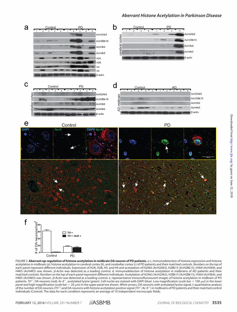
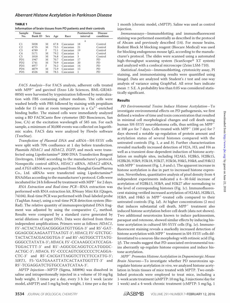
FIGURE 3. Aberrant up-regulation of histone acetylation in midbrain DA neuron of PD patients. a-c, immunodetection of histone expression and histoneacetylation in midbrain (a), histone acetylation in cerebral cortex (b), and cerebellar cortex (c) of PD patients and their matched controls. Numbers on the top ofeach panel represent different individuals. Expression of H2A, H2B, H3, and H4 and acetylation of H2Ak5 (AcH2Ak5), H2Bk15 (AcH2Bk15), H3k9 (AcH3k9), andH4K5 (AcH4K5) was shown. �-Actin was detected as a loading control. d, immunodetection of histone acetylation in midbrains of AD patients and theirmatched controls. Numbers on the top of each panel represent different individuals. Acetylation of H2Ak5 (AcH2Ak5), H2Bk15 (AcH2Bk15), H3k9 (AcH3k9), andH4K5 (AcH4K5) was shown. �-Actin was detected as a loading control. e, representative immunofluorescent images of histone acetylation in midbrain of PDpatients. TH�, DA neurons (red); Ac-K�, acetylated lysine (green). Cell nuclei are stained with DAPI (blue). Low magnification (scale bar � 100 �M) in the lowerpanel and high magnification (scale bar � 20 �m) in the upper panel are shown. White arrows, DA neurons with acetylated lysine signal. f, quantitative analysisof the number of DA neurons (TH�) and DA neurons with histone acetylation positive signal (TH�/Ac-K�) in midbrains of PD patients and their matched controlindividuals (Control). The data for each condition represents an average of 10 independent microscopic fields.
Aberrant Histone Acetylation in Parkinson Disease
FEBRUARY 12, 2016 • VOLUME 291 • NUMBER 7 JOURNAL OF BIOLOGICAL CHEMISTRY 3535
by guest on June 22, 2018http://w
ww
.jbc.org/D
ownloaded from
injection/day for 4 weeks) (15). The effectiveness of MPTPtreatment was demonstrated by a 74 and 68.4% reduction oftyrosine hydroxylase (TH)-positive neurons in midbrains withaMPTP and cMPTP treatments, respectively (Fig. 2, a–c). His-tone acetylation is significantly up-regulated in midbrain tis-sues compared with controls injected with saline (Fig. 2, d ande). In the cMPTP model, a notable increase of histone acetyla-tion was detected in DA neurons of mouse midbrains as early as2 weeks of treatment and persisted through 4 weeks treatment(Fig. 2f). Consistent with findings of cells treated with MPP�,
expression levels of H2A, H3, and H4 were up-regulated byacute treatment (Fig. 2d). Together, these results indicate thatenvironmental toxins induce expression and acetylation of his-tones in dopaminergic neurons of mouse brains in vivo.
Specific Up-regulation of Histone Acetylation in DA Neuronsof PD Patient Brain—We next examined histone acetylation inpostmodern brain tissues of PD patients. Midbrain tissues from5 PD patients and 5 age, sex, and postmodern interval-matchedcontrol individuals were analyzed (Table 1). The results show amarked increase of acetylation of H2Ak5, H2Bk15, H3k9, andH4k5 in midbrain tissues of 3 of 5 PD patients compared withthat of 5 control individuals (Fig. 3a). Importantly, increasedhistone acetylation was only observed in cerebral tissue of onePD compared with controls (Fig. 3b). Acetylation of H2Bk15,but not H2Ak5, H3k9, and H4k5, was increased in the cerebel-lar cortex of PD patients over their controls (Fig. 3c). In con-trast, acetylation of H2Bk15 and H4k5 are notably decreased inthe midbrain of AD patients compared with that of matchedcontrols (Fig. 3d, Table 2). Immunostaining analysis specificallydetected high levels of histone acetylation (Ac-K) in midbrainDA neurons of PD patients. Conversely, histone acetylation isbarely detectable in non-DA cells of PD patients or in the mid-brain DA neurons of the control individuals (Fig. 3e). Quanti-
FIGURE 4. Down-regulation of HDACs in MPP�-treated cells, MPTP-treated mice, and the midbrain of PD patients. a and b, immunoblotting detection ofHDAC1, -2, -4, -6, and SirT1 in cells treated with 1 or 100 �M MPP� to those in cells treated with solvent (a). Quantitative analysis of 3 independent experimentsis shown (b). Results are normalized to �-actin. *, p � 0.05; **, p � 0.01; ***, p � 0.001. c, quantitative RT-PCR analysis of expression of HDAC1, -2, -4, -6, and SirT1in cells treated with 1 or 100 �M MPP� to those in cells treated with solvent. Results are normalized to �-actin. **, p � 0.01; ***, p � 0.001. d and e,immunoblotting detection of HDAC1, -2, -4, -6, and SirT1 in midbrain of aMPTP-treated mice to those in the midbrain of solvent-treated mice (d). Quantitativeanalysis of 3 independent experiments is shown (e). Results are normalized to �-actin. *, p � 0.05; **, p � 0.01; ***, p � 0.001. f, immunodetection of HATs (Gcn5,Tip60, and CBP), HDACs (HDAC1, HDAC2, HDAC4, HDAC6, and SirT1), and acetylated histone H3k27 (AcH3k27) in human PD midbrain (PD) and their matchedcontrols (Control). Numbers on the top of each panel represent different individuals.
TABLE 2Information of brain tissues from AD patients and their controls
SampleNo.
TissueBank ID Sex Age Race
Postmorteminterval
Diseasecondition
hC1 4921 F 74 Caucasian 13 ControlC2 5082 M 68 Caucasian 19 ControlC3 5171 M 79 Caucasian 5 ControlC4 5246 F 78 Caucasian 19 ControlC5 5352 M 81 Caucasian 17 ControlAD1 1212 F 73 Caucasian 18 ADAD2 4693 M 70 Caucasian 12 ADAD3 4697 M 78 Caucasian 23 ADAD4 5007 F 77 Caucasian 15 ADAD5 5222 F 80 Caucasian 7 AD
Aberrant Histone Acetylation in Parkinson Disease
3536 JOURNAL OF BIOLOGICAL CHEMISTRY VOLUME 291 • NUMBER 7 • FEBRUARY 12, 2016
by guest on June 22, 2018http://w
ww
.jbc.org/D
ownloaded from
tative analysis by pixel density revealed detection of Ac-K in82.7% TH-positive neurons of PD patients (n � 5). However,only 2% TH-positive neurons are AcK positive in the midbrainof normal control individuals (n � 5, Fig. 3f). The results sug-gest that specific histone acetylation is involved in dopaminer-gic neurodegeration in the midbrain of PD patients. Moreover,histone acetylation is differentially regulated in PD and AD.Together, histone acetylation is regulated in the midbrain of PDpatients with cell type, brain region, and disease specificity.
Down-regulation of HDACs in Brain of PD Patients—Wenext investigated the mechanism of abnormal histone acetyla-tion induced by PD-related neurotoxins. SH-SY5Y cells treatedwith low doses of 1 �M and 100 �M MPP� exhibited signifi-cantly reduced levels of HDAC1, HDAC2, HDAC4, andHDAC6 (Fig. 4, a and b). These changes are unlikely due totranscriptional regulation because mRNA levels of theseHDACs were either not altered with 1 �M MPP� treatment orup-regulated with 100 �M MPP� treatment (Fig. 4c). Consist-ent with findings from cultured cells, HDAC1, HDAC2,HDAC6, and SirT1 were decreased in midbrains of micetreated with aMPTP compared with that of vehicle-treatedcontrols. In contrast, the level of HDAC4 was not notablyaffected (Fig. 4, d and e). Furthermore, levels of HDAC1,HDAC2, HDAC4, HDAC6, and SirT1 were also markedlylower in midbrain tissues of PD patients than those in midbraintissues of their matched controls (Fig. 4f). Consequently,
decreased HDACs and increased histone acetylation seem to becorrelated in the midbrains of PD patients. Histone acetyltrans-ferases Gcn5 and Tip60 were either not changed or not consis-tently changed between the disease and control groups (Fig. 4f).However, CBP was detected in 1 of 5 PD patient midbrains and3 of 5 control midbrains (Fig. 4f). These results indicate thatPD-associated up-regulation of histone acetylation is likely aresult of reduced HDACs in midbrain tissues.
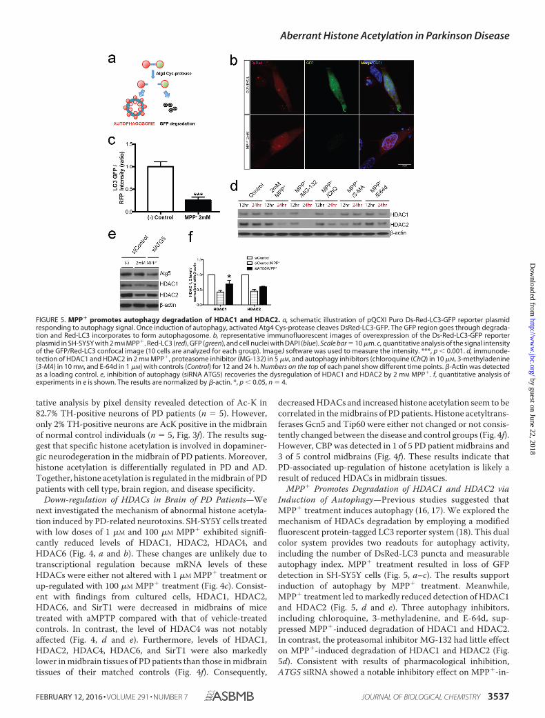
MPP� Promotes Degradation of HDAC1 and HDAC2 viaInduction of Autophagy—Previous studies suggested thatMPP� treatment induces autophagy (16, 17). We explored themechanism of HDACs degradation by employing a modifiedfluorescent protein-tagged LC3 reporter system (18). This dualcolor system provides two readouts for autophagy activity,including the number of DsRed-LC3 puncta and measurableautophagy index. MPP� treatment resulted in loss of GFPdetection in SH-SY5Y cells (Fig. 5, a–c). The results supportinduction of autophagy by MPP� treatment. Meanwhile,MPP� treatment led to markedly reduced detection of HDAC1and HDAC2 (Fig. 5, d and e). Three autophagy inhibitors,including chloroquine, 3-methyladenine, and E-64d, sup-pressed MPP�-induced degradation of HDAC1 and HDAC2.In contrast, the proteasomal inhibitor MG-132 had little effecton MPP�-induced degradation of HDAC1 and HDAC2 (Fig.5d). Consistent with results of pharmacological inhibition,ATG5 siRNA showed a notable inhibitory effect on MPP�-in-
FIGURE 5. MPP� promotes autophagy degradation of HDAC1 and HDAC2. a, schematic illustration of pQCXI Puro Ds-Red-LC3-GFP reporter plasmidresponding to autophagy signal. Once induction of autophagy, activated Atg4 Cys-protease cleaves DsRed-LC3-GFP. The GFP region goes through degrada-tion and Red-LC3 incorporates to form autophagosome. b, representative immunofluorescent images of overexpression of the Ds-Red-LC3-GFP reporterplasmid in SH-SY5Y with 2 mM MPP�. Red-LC3 (red), GFP (green), and cell nuclei with DAPI (blue). Scale bar � 10 �m. c, quantitative analysis of the signal intensityof the GFP/Red-LC3 confocal image (10 cells are analyzed for each group). ImageJ software was used to measure the intensity. ***, p � 0.001. d, immunode-tection of HDAC1 and HDAC2 in 2 mM MPP�, proteasome inhibitor (MG-132) in 5 �M, and autophagy inhibitors (chloroquine (ChQ) in 10 �M, 3-methyladenine(3-MA) in 10 mM, and E-64d in 1 �M) with controls (Control) for 12 and 24 h. Numbers on the top of each panel show different time points. �-Actin was detectedas a loading control. e, inhibition of autophagy (siRNA ATG5) recoveries the dysregulation of HDAC1 and HDAC2 by 2 mM MPP�. f, quantitative analysis ofexperiments in e is shown. The results are normalized by �-actin. *, p � 0.05, n � 4.
Aberrant Histone Acetylation in Parkinson Disease
FEBRUARY 12, 2016 • VOLUME 291 • NUMBER 7 JOURNAL OF BIOLOGICAL CHEMISTRY 3537
by guest on June 22, 2018http://w
ww
.jbc.org/D
ownloaded from
duced degradation of HDAC1 and HDAC2 (Fig. 5, e and f).Together, MPP� treatment induces degradation of HDAC1and HDAC2 via autophagy.
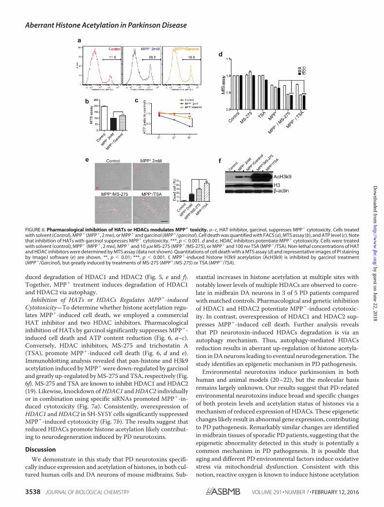
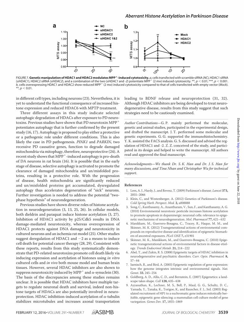
Inhibition of HATs or HDACs Regulates MPP�-inducedCytotoxicity—To determine whether histone acetylation regu-lates MPP�-induced cell death, we employed a commercialHAT inhibitor and two HDAC inhibitors. Pharmacologicalinhibition of HATs by garcinol significantly suppresses MPP�-induced cell death and ATP content reduction (Fig. 6, a–c).Conversely, HDAC inhibitors, MS-275 and trichostatin A(TSA), promote MPP�-induced cell death (Fig. 6, d and e).Immunoblotting analysis revealed that pan-histone and H3k9acetylation induced by MPP� were down-regulated by garcinoland greatly up-regulated by MS-275 and TSA, respectively (Fig.6f). MS-275 and TSA are known to inhibit HDAC1 and HDAC2(19). Likewise, knockdown of HDAC1 and HDAC2 individuallyor in combination using specific siRNAs promoted MPP�-in-duced cytotoxicity (Fig. 7a). Consistently, overexpression ofHDAC1 and HDAC2 in SH-SY5Y cells significantly suppressedMPP�-induced cytotoxicity (Fig. 7b). The results suggest thatreduced HDACs promote histone acetylation likely contribut-ing to neurodegeneration induced by PD neurotoxins.
Discussion
We demonstrate in this study that PD neurotoxins specifi-cally induce expression and acetylation of histones, in both cul-tured human cells and DA neurons of mouse midbrains. Sub-
stantial increases in histone acetylation at multiple sites withnotably lower levels of multiple HDACs are observed to corre-late in midbrain DA neurons in 3 of 5 PD patients comparedwith matched controls. Pharmacological and genetic inhibitionof HDAC1 and HDAC2 potentiate MPP�-induced cytotoxic-ity. In contrast, overexpression of HDAC1 and HDAC2 sup-presses MPP�-induced cell death. Further analysis revealsthat PD neurotoxin-induced HDACs degradation is via anautophagy mechanism. Thus, autophagy-mediated HDACsreduction results in aberrant up-regulation of histone acetyla-tion in DA neurons leading to eventual neurodegeneration. Thestudy identifies an epigenetic mechanism in PD pathogenesis.
Environmental neurotoxins induce parkinsonism in bothhuman and animal models (20 –22), but the molecular basisremains largely unknown. Our results suggest that PD-relatedenvironmental neurotoxins induce broad and specific changesof both protein levels and acetylation status of histones via amechanism of reduced expression of HDACs. These epigeneticchanges likely result in abnormal gene expression, contributingto PD pathogenesis. Remarkably similar changes are identifiedin midbrain tissues of sporadic PD patients, suggesting that theepigenetic abnormality detected in this study is potentially acommon mechanism in PD pathogenesis. It is possible thataging and different PD environmental factors induce oxidativestress via mitochondrial dysfunction. Consistent with thisnotion, reactive oxygen is known to induce histone acetylation
FIGURE 6. Pharmacological inhibition of HATs or HDACs modulates MPP� toxicity. a– c, HAT inhibitor, garcinol, suppresses MPP� cytotoxicity. Cells treatedwith solvent (Control), MPP� (MPP�, 2 mM), or MPP� and garcinol (MPP�/garcinol). Cell death was quantified with FACS (a), MTS assay (b), and ATP level (c). Notethat inhibition of HATs with garcinol suppresses MPP� cytotoxicity. ***, p � 0.001. d and e, HDAC inhibitors potentiate MPP� cytotoxicity. Cells were treatedwith solvent (control), MPP� (MPP�, 2 mM), MPP� and 10 �M MS-275 (MPP�/MS-275), or MPP� and 100 nM TSA (MPP�/TSA). Non-lethal concentrations of HATand HDAC inhibitors were determined by MTS assay (data not shown). Quantitations of cell death with a MTS assay (d) and representative images of PI stainingby ImageJ software (e) are shown. **, p � 0.01; ***, p � 0.001. f, MPP�-induced histone H3k9 acetylation (AcH3k9) is inhibited by garcinol treatment(MPP�/Garcinol), but greatly induced by treatments of MS-275 (MPP�/MS-275) or TSA (MPP�/TSA).
Aberrant Histone Acetylation in Parkinson Disease
3538 JOURNAL OF BIOLOGICAL CHEMISTRY VOLUME 291 • NUMBER 7 • FEBRUARY 12, 2016
by guest on June 22, 2018http://w
ww
.jbc.org/D
ownloaded from
in different cell types, including neurons (23). Nevertheless, it isyet to understand the functional consequence of increased his-tone expression and reduced HDAC6 with MPTP treatment.
Three different assays in this study indicate selectedautophagic degradation of HDACs after exposure to PD neuro-toxins. Previous studies have shown that PD neurotoxin MPP�
potentiates autophagy that is further confirmed by the presentstudy (16, 17). Autophagy is proposed to play either a protectiveor a pathogenic role under different conditions. This is alsolikely the case in PD pathogenesis. PINK1 and PARKIN, tworecessive PD causative genes, function to degrade damagedmitochondria via mitophagy, therefore, neuroprotective (24). Arecent study shows that MPP�-induced autophagy is pro-deathof DA neurons in rat brain (16). It is possible that in the earlystage of disease, selective autophagy is activated to promote theclearance of damaged mitochondria and un/misfolded pro-teins, resulting in a protective role. With the progressionof disease, health mitochondria are significantly reducedand un/misfolded proteins get accumulated, dysregulatedautophagy thus accelerates degeneration of “sick” neurons.Further investigation is needed to address the potential “two-phase hypothesis” of neurondegeneration.
Previous studies have shown diverse roles of histone acetyla-tion in neurodegeneration (3, 13, 25, 26). In cellular models,both dieldrin and paraquat induce histone acetylation (3, 27).Inhibition of HDAC1 activity by p25/Cdk5 results in DNAdamage-mediated neurotoxicity, whereas overexpression ofHDAC1 protects against DNA damage and neurotoxicity incultured neurons and an ischemia rat model (25). Other studiessuggest deregulation of HDAC1 and �2 as a means to inducecell death for potential cancer therapy (28, 29). Consistent withthese reports, results from this study systematically demon-strate that PD-related neurotoxins promote cell death likely viainducing expression and acetylation of histones using in vitrocultured cells and in vivo both mouse model and human braintissues. However, several HDAC inhibitors are also shown tosuppress neurotoxicity induced by MPP� and �-synuclein (30).The basis of the discrepancies among these studies remainsunclear. It is possible that HDAC inhibitors have multiple tar-gets to regulate neuronal death and survival, indeed non-his-tone targets of HDACs are also potentially involved in neuro-protection. HDAC inhibition-induced acetylation of �-tubulinstabilizes microtubules and increases axonal transportation
leading to BDNF release and neuroprotection (31, 32).Although HDAC inhibitors are being developed to treat neuro-degenerative disease, results from this study suggest that suchstrategies need to be cautiously examined.
Author Contributions—G. P. mainly performed the molecular,genetic and animal studies, participated in the experimental design,and drafted the manuscript. J. T. performed some molecular andgenetic experiments. G. G. supported the immunohistochemistry.Y. K. assisted the FACS analysis. G. S. discussed and advised the reg-ulation of HDAC1 and -2. Z. Z. conceived of the study, and partici-pated in its design and helped to write the manuscript. All authorsread and approved the final manuscript.
Acknowledgments—We thank Dr. S. K. Han and Dr. J. S. Han formany discussions, and Tina Nhan and Christopher Wu for technicalsupport.
References1. Lees, A. J., Hardy, J., and Revesz, T. (2009) Parkinson’s disease. Lancet 373,
2055–20662. Klein, C., and Westenberger, A. (2012) Genetics of Parkinson’s disease.
Cold Spring Harb. Perspect. Med. 2, a0088883. Song, C., Kanthasamy, A., Anantharam, V., Sun, F., and Kanthasamy, A. G.
(2010) Environmental neurotoxic pesticide increases histone acetylationto promote apoptosis in dopaminergic neuronal cells: relevance to epige-netic mechanisms of neurodegeneration. Mol. Pharmacol 77, 621– 632
4. Manikkam, M., Guerrero-Bosagna, C., Tracey, R., Haque, M. M., andSkinner, M. K. (2012) Transgenerational actions of environmental com-pounds on reproductive disease and identification of epigenetic biomark-ers of ancestral exposures. PLoS ONE 7, e31901
5. Skinner, M. K., Manikkam, M., and Guerrero-Bosagna, C. (2010) Epige-netic transgenerational actions of environmental factors in disease etiol-ogy. Trends Endocrinol Metab. 21, 214 –222
6. Abel, T., and Zukin, R. S. (2008) Epigenetic targets of HDAC inhibition inneurodegenerative and psychiatric disorders. Curr. Opin. Pharmacol. 8,57– 64
7. Jaenisch, R., and Bird, A. (2003) Epigenetic regulation of gene expression:how the genome integrates intrinsic and environmental signals. Nat.Genet. 33, 245–254
8. Goldberg, A. D., Allis, C. D., and Bernstein, E. (2007) Epigenetics: a land-scape takes shape. Cell 128, 635– 638
9. Ayyanathan, K., Lechner, M. S., Bell, P., Maul, G. G., Schultz, D. C.,Yamada, Y., Tanaka, K., Torigoe, K., and Rauscher, F. J., 3rd. (2003) Reg-ulated recruitment of HP1 to a euchromatic gene induces mitotically her-itable, epigenetic gene silencing: a mammalian cell culture model of genevariegation. Genes Dev. 17, 1855–1869
FIGURE 7. Genetic manipulation of HDAC1 and HDAC2 modulates MPP�-induced cytotoxicity. a, cells transfected with scramble siRNA (NC), HDAC1 siRNA(siHDAC1), HDAC2 siRNA (siHDAC2), and a combination of the two (siHDAC1 and -2) potentiate MPP� (2 mM) induced cytotoxicity. **, p � 0.01; ***, p � 0.001.b, cells overexpressing HDAC1 and HDAC2 show reduced MPP� (2 mM) induced cytotoxicity compared to that of cells transfected with empty vector (Mock).**, p � 0.01.
Aberrant Histone Acetylation in Parkinson Disease
FEBRUARY 12, 2016 • VOLUME 291 • NUMBER 7 JOURNAL OF BIOLOGICAL CHEMISTRY 3539
by guest on June 22, 2018http://w
ww
.jbc.org/D
ownloaded from
10. Gräff, J., Kim, D., Dobbin, M. M., and Tsai, L. H. (2011) Epigenetic regu-lation of gene expression in physiological and pathological brain pro-cesses. Physiol. Rev. 91, 603– 649
11. Francis, Y. I., Fà, M., Ashraf, H., Zhang, H., Staniszewski, A., Latchman,D. S., and Arancio, O. (2009) Dysregulation of histone acetylation in theAPP/PS1 mouse model of Alzheimer’s disease. J. Alzheimers Dis. 18,131–139
12. Monti, B., Gatta, V., Piretti, F., Raffaelli, S. S., Virgili, M., and Contestabile,A. (2010) Valproic acid is neuroprotective in the rotenone rat model ofParkinson’s disease: involvement of �-synuclein. Neurotox. Res. 17,130 –141
13. Outeiro, T. F., Kontopoulos, E., Altmann, S. M., Kufareva, I., Strathearn,K. E., Amore, A. M., Volk, C. B., Maxwell, M. M., Rochet, J. C., McLean,P. J., Young, A. B., Abagyan, R., Feany, M. B., Hyman, B. T., and Kazantsev,A. G. (2007) Sirtuin 2 inhibitors rescue �-synuclein-mediated toxicity inmodels of Parkinson’s disease. Science 317, 516 –519
14. Kim, S. J., Park, G. H., Kim, D., Lee, J., Min, H., Wall, E., Lee, C. J., Simon,M. I., Lee, S. J., and Han, S. K. (2011) Analysis of cellular and behavioralresponses to imiquimod reveals a unique itch pathway in transient recep-tor potential vanilloid 1 (TRPV1)-expressing neurons. Proc. Natl. Acad.Sci. U.S.A. 108, 3371–3376
15. Jackson-Lewis, V., and Przedborski, S. (2007) Protocol for the MPTPmouse model of Parkinson’s disease. Nature Protocols 2, 141–151
16. Hung, K. C., Huang, H. J., Lin, M. W., Lei, Y. P., and Lin, A. M. (2014) Rolesof autophagy in MPP�-induced neurotoxicity in vivo: the involvement ofmitochondria and �-synuclein aggregation. PLoS ONE 9, e91074
17. Zhu, J. H., Horbinski, C., Guo, F., Watkins, S., Uchiyama, Y., and Chu, C. T.(2007) Regulation of autophagy by extracellular signal-regulated proteinkinases during 1-methyl-4-phenylpyridinium-induced cell death. Am. J.Pathol. 170, 75– 86
18. Sheen, J. H., Zoncu, R., Kim, D., and Sabatini, D. M. (2011) Defectiveregulation of autophagy upon leucine deprivation reveals a targetable lia-bility of human melanoma cells in vitro and in vivo. Cancer Cell 19,613– 628
19. Dokmanovic, M., Clarke, C., and Marks, P. A. (2007) Histone deacetylaseinhibitors: overview and perspectives. Mol. Cancer Res. 5, 981–989
20. Cicchetti, F., Drouin-Ouellet, J., and Gross, R. E. (2009) Environmentaltoxins and Parkinson’s disease: what have we learned from pesticide-in-duced animal models? Trends Pharmacol. Sci. 30, 475– 483
21. Di Monte, D. A. (2003) The environment and Parkinson’s disease: is thenigrostriatal system preferentially targeted by neurotoxins? Lancet Neu-rol. 2, 531–538
22. Langston, J. W., and Ballard, P. A., Jr. (1983) Parkinson’s disease in achemist working with 1-methyl-4-phenyl-1,2,5,6-tetrahydropyridine.New Engl. J. Med. 309, 310
23. Gu, X., Sun, J., Li, S., Wu, X., and Li, L. (2013) Oxidative stress inducesDNA demethylation and histone acetylation in SH-SY5Y cells: potentialepigenetic mechanisms in gene transcription in Abeta production. Neu-robiol. Aging 34, 1069 –1079
24. Vincow, E. S., Merrihew, G., Thomas, R. E., Shulman, N. J., Beyer, R. P.,MacCoss, M. J., and Pallanck, L. J. (2013) The PINK1-Parkin pathwaypromotes both mitophagy and selective respiratory chain turnover in vivo.Proc. Natl. Acad. Sci. U.S.A. 110, 6400 – 6405
25. Kim, D., Frank, C. L., Dobbin, M. M., Tsunemoto, R. K., Tu, W., Peng, P. L.,Guan, J. S., Lee, B. H., Moy, L. Y., Giusti, P., Broodie, N., Mazitschek, R.,Delalle, I., Haggarty, S. J., Neve, R. L., Lu, Y., and Tsai, L. H. (2008) Dereg-ulation of HDAC1 by p25/Cdk5 in neurotoxicity. Neuron 60, 803– 817
26. Kontopoulos, E., Parvin, J. D., and Feany, M. B. (2006) �-Synuclein acts inthe nucleus to inhibit histone acetylation and promote neurotoxicity.Hum. Mol. Genet. 15, 3012–3023
27. Song, C., Kanthasamy, A., Jin, H., Anantharam, V., and Kanthasamy, A. G.(2011) Paraquat induces epigenetic changes by promoting histone acety-lation in cell culture models of dopaminergic degeneration. Neurotoxicol-ogy 32, 586 –595
28. Haberland, M., Johnson, A., Mokalled, M. H., Montgomery, R. L., andOlson, E. N. (2009) Genetic dissection of histone deacetylase requirementin tumor cells. Proc. Natl. Acad. Sci. U.S.A. 106, 7751–7755
29. Ropero, S., and Esteller, M. (2007) The role of histone deacetylases(HDACs) in human cancer. Mol Oncol 1, 19 –25
30. Harrison, I. F., and Dexter, D. T. (2013) Epigenetic targeting of histonedeacetylase: therapeutic potential in Parkinson’s disease? Pharmacol.Ther. 140, 34 –52
31. Glozak, M. A., Sengupta, N., Zhang, X., and Seto, E. (2005) Acetylation anddeacetylation of non-histone proteins. Gene 363, 15–23
32. Zhang, Y., Li, N., Caron, C., Matthias, G., Hess, D., Khochbin, S., andMatthias, P. (2003) HDAC-6 interacts with and deacetylates tubulin andmicrotubules in vivo. EMBO J. 22, 1168 –1179
Aberrant Histone Acetylation in Parkinson Disease
3540 JOURNAL OF BIOLOGICAL CHEMISTRY VOLUME 291 • NUMBER 7 • FEBRUARY 12, 2016
by guest on June 22, 2018http://w
ww
.jbc.org/D
ownloaded from

Zhuohua ZhangGoonho Park, Jieqiong Tan, Guillermina Garcia, Yunyi Kang, Guy Salvesen and
Regulation of Histone Acetylation by Autophagy in Parkinson Disease
doi: 10.1074/jbc.M115.675488 originally published online December 23, 20152016, 291:3531-3540.J. Biol. Chem.
10.1074/jbc.M115.675488Access the most updated version of this article at doi:
Alerts:
When a correction for this article is posted•
When this article is cited•
to choose from all of JBC's e-mail alertsClick here
http://www.jbc.org/content/291/7/3531.full.html#ref-list-1
This article cites 32 references, 9 of which can be accessed free at
by guest on June 22, 2018http://w
ww
.jbc.org/D
ownloaded from