Embed Size (px)
Citation preview
Regulation of the Nitrogen Transfer Pathway inthe Arbuscular Mycorrhizal Symbiosis: GeneCharacterization and the Coordination of Expressionwith Nitrogen Flux1[W][OA]
Chunjie Tian*, Beth Kasiborski, Raman Koul, Peter J. Lammers2, Heike Bucking, and Yair Shachar-Hill
Department of Plant Biology, Michigan State University, East Lansing, Michigan 48824 (C.T., B.K., Y.S.-H.);Department of Chemistry and Biochemistry, New Mexico State University, Las Cruces, New Mexico 88003(R.K., P.J.L.); and Biology and Microbiology Department, South Dakota State University, Brookings, SouthDakota 57007 (H.B.)
The arbuscular mycorrhiza (AM) brings together the roots of over 80% of land plant species and fungi of the phylumGlomeromycota and greatly benefits plants through improved uptake of mineral nutrients. AM fungi can take up both nitrateand ammonium from the soil and transfer nitrogen (N) to host roots in nutritionally substantial quantities. The current modelof N handling in the AM symbiosis includes the synthesis of arginine in the extraradical mycelium and the transfer of arginineto the intraradical mycelium, where it is broken down to release N for transfer to the host plant. To understand the mechanismsand regulation of N transfer from the fungus to the plant, 11 fungal genes putatively involved in the pathway were identifiedfrom Glomus intraradices, and for six of them the full-length coding sequence was functionally characterized by yeastcomplementation. Two glutamine synthetase isoforms were found to have different substrate affinities and expressionpatterns, suggesting different roles in N assimilation. The spatial and temporal expression of plant and fungal N metabolismgenes were followed after nitrate was added to the extraradical mycelium under N-limited growth conditions using hairy rootcultures. In parallel experiments with 15N, the levels and labeling of free amino acids were measured to follow transport andmetabolism. The gene expression pattern and profiling of metabolites involved in the N pathway support the idea that therapid uptake, translocation, and transfer of N by the fungus successively trigger metabolic gene expression responses in theextraradical mycelium, intraradical mycelium, and host plant.
The arbuscular mycorrhizal (AM) symbiosis bringstogether the roots of the majority of land plant speciesand fungi of the phylum Glomeromycota to greatmutual advantage (Smith and Read, 2008). AM fungiimprove the acquisition of phosphate, nitrogen (N),sulfur, and trace elements such as copper and zinc(Clark and Zeto, 2000; Allen and Shachar-Hill, 2008)and increase the biotic and abiotic stress resistance oftheir host (Smith et al., 2010). In return, the hosttransfers up to 20% of its photosynthetically fixedcarbon to the AM fungus (Jakobsen and Rosendahl,
1990), which depends on its host plant for its carbonsupply (Bago et al., 2000).
N is the nutrient whose availability most commonlylimits plant growth in natural ecosystems. AM fungican take up NO3
2 and NH4+ and can also increase
access to organic N sources from the soil (Ames et al.,1983; Johansen et al., 1993; Bago et al., 1996; Hodgeet al., 2001). The translocation by the fungus canrepresent a significant route for N uptake by the plant(Johansen and Jensen, 1996). For example, Toussaintet al. (2004) showed that in an in vitro mycorrhiza atleast 21% of the total N uptake in the roots came fromthe fungal extraradical mycelium (ERM); for other my-corrhizal systems, even larger proportions have beendescribed (more than 30% and 50%; Govindarajuluet al., 2005; Jin et al., 2005). Tanaka and Yano (2005)reported that 75% of the N in leaves of mycorrhizalmaize (Zea mays) was taken up by the ERM of Glomusaggregatum.
A mechanism of N transfer from the fungus to theplant has been proposed (Bago et al., 2001) that in-volves the operation of a novel metabolic route inwhich N was translocated from the ERM to the intra-radical mycelium (IRM) as Arg but transferred to theplant without carbon as inorganic N. This mechanismhas been supported by labeling experiments (Johansen
1 This work was supported by the National Science Foundation(grant nos. 0616016 and 0616023).
2 Present address: Solix Biofuels, Inc., 430-B North College Ave-nue, Fort Collins, CO 80524.
* Corresponding author; e-mail [email protected] author responsible for distribution of materials integral to the
findings presented in this article in accordance with the policydescribed in the Instructions for Authors (www.plantphysiol.org) is:Chunjie Tian ([email protected]).
[W] The online version of this article contains Web-only data.[OA] Open Access articles can be viewed online without a sub-
scription.www.plantphysiol.org/cgi/doi/10.1104/pp.110.156430
Plant Physiology�, July 2010, Vol. 153, pp. 1175–1187, www.plantphysiol.org � 2010 American Society of Plant Biologists 1175 www.plantphysiol.orgon July 15, 2018 - Published by Downloaded from
Copyright © 2010 American Society of Plant Biologists. All rights reserved.
et al., 1996; Govindarajulu et al., 2005; Jin et al., 2005),enzyme activity analysis (Cruz et al., 2007), andlimited gene expression data (Govindarajulu et al.,2005; Gomez et al., 2009; Guether et al., 2009). Never-theless, our molecular knowledge of the metabolic andtransport pathways involved and how they are regu-lated is still rudimentary. A better understanding ofthe mechanism and regulation of N uptake assimi-lation, translocation, and transfer to the host is im-portant for potential applications of AM fungi asbiofertilizers, bioprotectors, and bioregulators in sus-tainable agriculture and restoration as well as forunderstanding the role of AM fungi in natural ecosys-tems (Bruns et al., 2008).
In this study, we postulate that the uptake, translo-cation, and transfer of N by the fungus triggers themetabolic gene expression responses successively inthe ERM, IRM, and host plant, which will result in thesynthesis and accumulation of Arg in the ERM, theturnover of Arg to release ammonium in the IRM, andthe assimilation of ammonium by the host plant viathe glutamine synthetase (GS)/glutamate synthase(GOGAT) pathway inside the root (Fig. 1). To testthese predictions, 11 genes involved in the N primaryassimilation and metabolism were cloned and verifiedfrom Glomus intraradices; six enzymes with full-lengthcoding sequences (CDSs) were functionally character-ized by yeast knockout mutant complementation. TwoGS proteins were found to have different substrateaffinities and expression patterns, suggesting that theyhave different roles in N assimilation. The time coursesof gene expression and N movement in fungal andhost tissues were analyzed following nitrate supply tothe ERM of a mycorrhiza grown under N-limitedconditions. The results substantially increase ourknowledge of the identity and regulation of most ofthe metabolic and transport genes involved in Nmovement through the AM symbiosis.
RESULTS
Gene Cloning and Functional Characterization
Based on data from high-throughput sequencing ofcDNA from ERM (P.J. Lammers and Y. Shachar-Hill,unpublished data) and sequences that were previouslydeposited in public databases (GenBank), the putativesequences for 10 enzymes and one nitrate transporterof G. intraradiceswere identified. Seven full-length andfour partial CDSs were obtained that show high se-quence similarities to known genes involved in Nuptake and metabolism in fungi (Table I; Supplemen-tal Figs. S1–S10). The pathway for N metabolism inwhich these genes have been proposed to operate isshown in Figure 1, which includes the possible regu-lation of the transcriptional levels of genes by the Nmetabolites involved in the pathway. For the func-tional characterization, several full-length CDSs wereexpressed in yeast, and the antibody directed againstthe polyhistidine region fused within each Gi se-quence was used to detect the expression of theproteins in yeast (Fig. 2A; Supplemental Fig. S11).
Identification and Enzymatic Analysis for N Uptake andAssimilation Genes
GS (EC 6.3.1.2) belongs to a multigene family inmost plants and some fungi (Stanford et al., 1993;Ishiyama et al., 2004). Of the two putative GiGS genes,each has a 1,062-bp full-length CDS corresponding toproteins with 354 amino acids with a predicted mo-lecular mass of 39 kD. Analysis for the deducedproteins revealed two conserved domains for the GSfamily: GS signature 1 and a putative ATP-bindingregion signature; the corresponding amino acids were5#-FDGSSTNQAPGDDSDVLL-3# and 5#-KPIKGDW-NGAGCHTNYS-3# for GiGS1 and 5#-FDGSSTNQAP-GHDSDILL-3# and 5#-KPIKGDWNGAGCHTNYS-3#
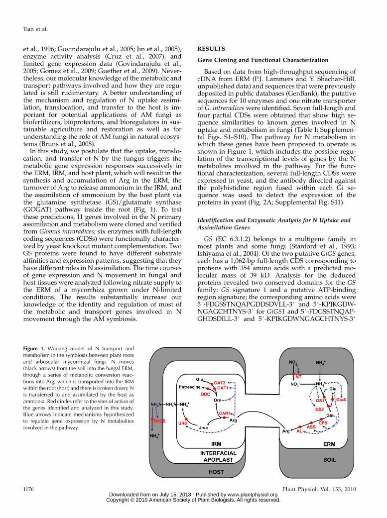
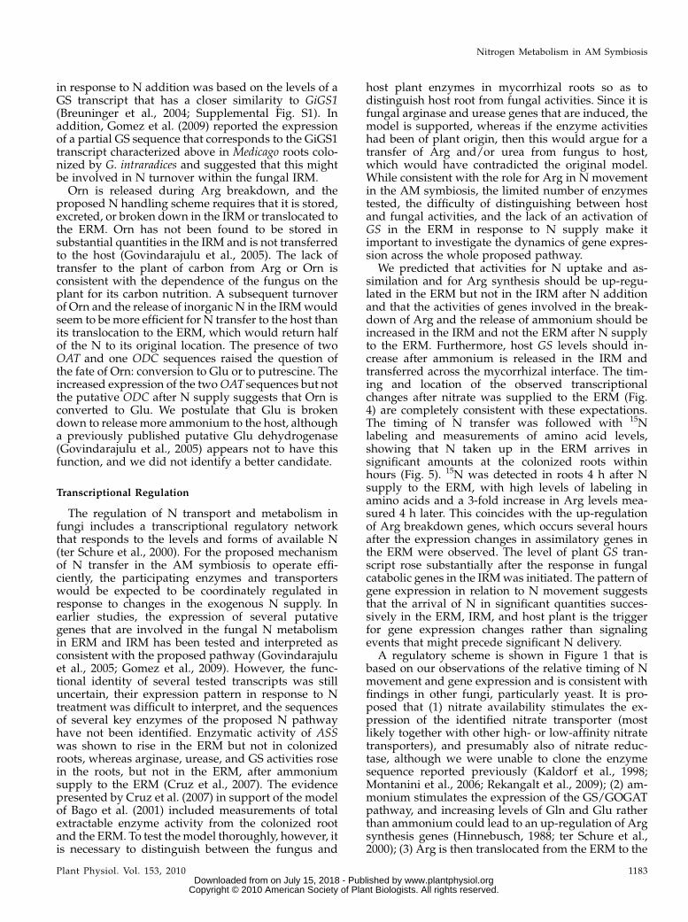
Figure 1. Working model of N transport andmetabolism in the symbiosis between plant rootsand arbuscular mycorrhizal fungi. N moves(black arrows) from the soil into the fungal ERM,through a series of metabolic conversion reac-tions into Arg, which is transported into the IRMwithin the root (host) and there is broken down; Nis transferred to and assimilated by the host asammonia. Red circles refer to the sites of action ofthe genes identified and analyzed in this study.Blue arrows indicate mechanisms hypothesizedto regulate gene expression by N metabolitesinvolved in the pathway.
Tian et al.
1176 Plant Physiol. Vol. 153, 2010 www.plantphysiol.orgon July 15, 2018 - Published by Downloaded from
Copyright © 2010 American Society of Plant Biologists. All rights reserved.
for GiGS2, respectively (Supplemental Fig. S1A). GiGS1is nearly identical to the GS sequence reported byGovindarajulu et al. (2005) except that the stop codonwas not identified in the earlier study. Besides thestriking similarity with GS fromGlomus mosseae,GiGS1andGiGS2 also possess a high similarity withGSs fromSaccharomyces pombe and Filobasidiella neoformans, es-pecially for the conserved region of GS family membersignature 1 and the putative ATP-binding region(Supplemental Fig. S2A). The alignments of the de-duced GiGS1 and GiGS2 sequences were usedto calculate a phylogenetic tree (Supplemental Fig.S2B), which shows that GiGS1 and GiGS2 were clus-tered with the GSs from ectomycorrhizae into onelarge group, with the identities ranging from 70%to 90%.The full-length CDSs of GiGS1 and GiGS2 fused
with C-terminal polyhistidine tags were used for com-plementation of Saccharomyces cerevisiae strain DGLN1,which is auxotrophic for Gln (Bernard et al., 2008). Thefour cell types tested were (1) the corresponding wild-type strain BY4743, (2) the GLN1-deficient knockoutmutant DGLN1 transformed with empty vector, and (3and 4) DGLN1 transformed with GiGS1 or GiGS2. Thehaploid mutant DGLN1 could not grow on the me-dium without Gln, while it grew well on the plates
with 2% Gln. The transformants expressing GiGS1 orGiGS2 grewwell even on the plate without Gln added,which means that GiGS1 and GiGS2 could comple-ment the auxotrophic mutant by the functional pro-duction of Gln (Fig. 3A).
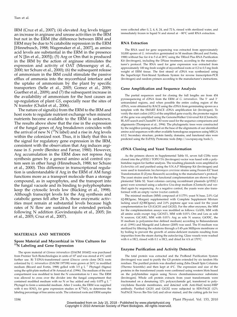
The catalytic reaction of GS is NH3 + Glu + ATP /Gln + ADP + inorganic phosphate. Figure 2 shows theexpression of the two GiGS isozymes in yeast, theirpurification, and in vitro kinetics (Mitchell andMagasanik, 1983). GiGS1 and GiGS2 have a very sim-ilar Vmax of 3.7 and 4.0 mM min21 mg21, respectively,but differ in their Km. The Km value of GiGS2 was3.8 mM, twice that of GiGS1 (1.9 mM).
In addition, two partial sequences (GiNT andGiGluS) involved in the primary N assimilation wereidentified here. Nitrate transporter plays an importantrole for transporting NO3
2 to the ERM. A 990-bppartial cDNA sequence for a putative high-affinitynitrate transporter was amplified from the total ap-proximately 1,500-bp gene, sharing 39% identity withthe nitrate transporter of Porphyra yezoensis (Table I;Supplemental Fig. S1). The next step of N assimilationvia the GS/GOGAT pathway is catalyzed by gluta-mate synthase (GluS), which converts Gln and2-oxoglutarate to two molecules of Glu. Approximately800 bp of a putative GluS (EC 1.4.1.14) was identified
Table I. Gene identification from G. intraradices
Gene Name CDS Length Closest Homologa Domains, Families, or Functional Sitesb
bp
Nitrate transporter (GiNT) 990c BAG70346 (Porphyra yezoensis),identity = 39%
Major facilitator superfamily
Glutamine synthetase 1 (GiGS1) 1,065d AAY62524 (Glomus intraradices),identity = 99%
GLNA_1, Gln synthetase signature 1;GLNA_ATP, Gln synthetase putativeATP-binding region signature
Glutamine synthetase 2 (GiGS2) 1,065d AAR11485 (Glomus mosseae),identity = 88%
GLNA_1, Gln synthetase signature 1;GLNA_ATP, Gln synthetase putativeATP-binding region signature
Glutamate synthase (GiGluS) 759c EED14920 (Talaromyces stipitatus),identity = 68%
FAD-dependent pyridine nucleotide-disulfide oxidoreductase
Carbamoyl-phosphate synthaseglutamine chain (GiCPS)
990c XP961982 (Neurospora crassa),identity = 55%
GATASE_TYPE_1, Gln amidotransferasetype 1 domain profile
Argininosuccinate synthase(GiASS)
1,239d AAW43079 (Cryptococcusneoformans), identity = 71%
ARGININOSUCCIN_SYN_1, argininosuccinatesynthase signature 1;ARGININOSUCCIN_SYN_2,argininosuccinate synthase signature 2.
Arginosuccinate lyase (GiAL) 849c XP002172712 (Schizosaccharomycesjaponicus), identity = 66%
FUMARATE_LYASES, fumarate lyasesignature
Arginase (GiCAR1) 945d XP001875445 (Laccaria bicolor),identity = 63%
ARGINASE_2, arginase family profile;ARGINASE_1, arginase family signature
Ornithine aminotransferase(GiOAT1)
1,329d XP002171771 (Schizosaccharomycesjaponicus), identity = 69%
AA_TRANSFER_CLASS_3, aminotransferaseclass III pyridoxal-phosphate attachmentsite
Ornithine decarboxylase(GiODC)
1,335d CAB61758 (Mucor circinelloides),identity = 57%
ODR_DC_2_1, Orn/diaminopimelate/Argdecarboxylase family 2 pyridoxal-phosphateattachment site
Urease (GiURE) 2,499d XP964986 (Neurospora crassa),identity = 66%
UREASE_3, urease domain profile; UREASE_2,urease active site
aBLAST of the amino acid sequence against the National Center for Biotechnology Information protein database. bBased on the databaseshttp://www.expasy.ch/prosite and http://www.ebi.ac. cPartial CDS. dFull-length CDS.
Nitrogen Metabolism in AM Symbiosis
Plant Physiol. Vol. 153, 2010 1177 www.plantphysiol.orgon July 15, 2018 - Published by Downloaded from
Copyright © 2010 American Society of Plant Biologists. All rights reserved.
with a 68% similarity to the Talaromyces stipitatus geneat the protein level (Table I; Supplemental Fig. S3).GiGluS includes a small subunit conserved for theGOGAT family, but the highly conserved NADPH-binding motif between residues in the N-terminalsequence could not be found in the partial C-terminalsequence (Pelanda et al., 1993).
Enzymes Involved in Arg Synthesis
Sequences for argininosuccinate synthetase (GiASS;EC 6.3.4.5) and argininosuccinate lyase (GiAL; EC4.3.2.1), which catalyze the last two steps in Argbiosynthesis, as well as a sequence for a putativecarbamoyl-phosphate synthetase (GiCPS [Arg-specificsmall chain]; EC 6.3.5.5) were obtained.
GiCPS is involved in both Arg and pyrimidinebiosynthesis and catalyzes the ATP-dependent forma-tion of carbamoyl phosphate from Gln and carbondioxide. This subunit promotes the hydrolysis ofGln to ammonia, which is subsequently used bythe ammonia chain of CPS to synthesize carbamoyl
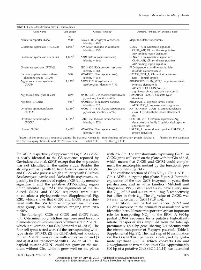
Figure 2. Heterologous expression, purification, and enzymatic ki-netics of the two GS isoforms identified, GiGS1 and GiGS2. A,Western blot of the expressed GiGS1 and GiGS2 in yeast. Whole cellprotein extracts from the yeast transformed with GiGS1 and GiGS2were fractionated on a denaturing 12% polyacrylamide gel, trans-ferred to polyvinylidene fluoride membranes, and detected withAnti-His(C-term)-HRP antibody. The yeast knockout mutants wereindependently transformed with the empty pYES2.1 vector or thevector fused with GiGS1 or GiGS2. Lanes a and c, The expression ofGiGS on the medium without Gal; lanes b and d, the expression ofGiGS on the medium with Gal; lane e, the expression of the emptyvector on the medium with Gal (as a negative control). B, Thepurification stages of GiGS1 and GiGS2. Purified proteins weresubjected to SDS-PAGE (12% NuPAGE Novex Bis-Tris Gel) and silverstained. Low-Mr markers are in lane f. Crude yeast extracts beforepurification were loaded in lane e, and successive elution fractionswere loaded in lanes a to d. Each lane contained 1 mg of protein. C,Enzymatic kinetics for GiGS1 and GiGS2 detected by synthetaseassay using the purified proteins. Biosynthetic GS activity assays werecarried out by varying the concentration of Glu in the reactionmixture. Plots of 1/V versus [S] are shown. Velocity data, expressed asproportions of Vmax, were based on the three biological replicates;absolute Vmax values were 3.68 and 4.01 mM min21 mg21 for GiGS1and GiGS2, respectively. Lineweaver-Burk plots of the data wereobtainedwithGiGS1 (Km = 1.87mM, r2 = 0.99) andGiGS2 (Km = 3.80mM,r2 = 0.999).
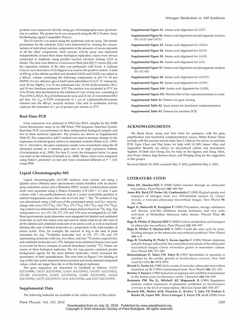
Figure 3. Functional complementation of S. cerevisiae knockout mu-tants using the genes identified from G. intraradices. M, The knockoutmutant expressing the empty vector; M:GiXXX, the knockout mutantexpressing the GiXXX gene; WT, the wild type. A, GiGS1 and GiGS2.Left, Growth of colonies on rich medium plus 2% Gln; right, mediumwithout Gln. B, GiASS. Left, Minimal medium plus all essential aminoacids including 0.01% L-Arg; right, minimal medium with all essentialamino acids excluding L-Arg. In A and B, 5 mL of cells was used forspreading on the plate. The optical density at 600 nm values for thecells are 1, 0.1, 0.01, 0.001, and 0.0001 from the left to the rightcolumns. C, GiCAR1. Left, Minimal medium with all essential aminoacids including 0.01% L-Arg; right, medium with 0.01% L-Arg as thesole N source. D,GiOAT1. Left, Mediumwith all essential amino acids;right, medium with only 0.01% Orn and Leu as N sources. E, GiODC.Left, Medium with 0.001% Leu, His, Lys, and putrescine as N sources;right, medium with 0.001% Leu, His, and Lys as N sources.
Tian et al.
1178 Plant Physiol. Vol. 153, 2010 www.plantphysiol.orgon July 15, 2018 - Published by Downloaded from
Copyright © 2010 American Society of Plant Biologists. All rights reserved.
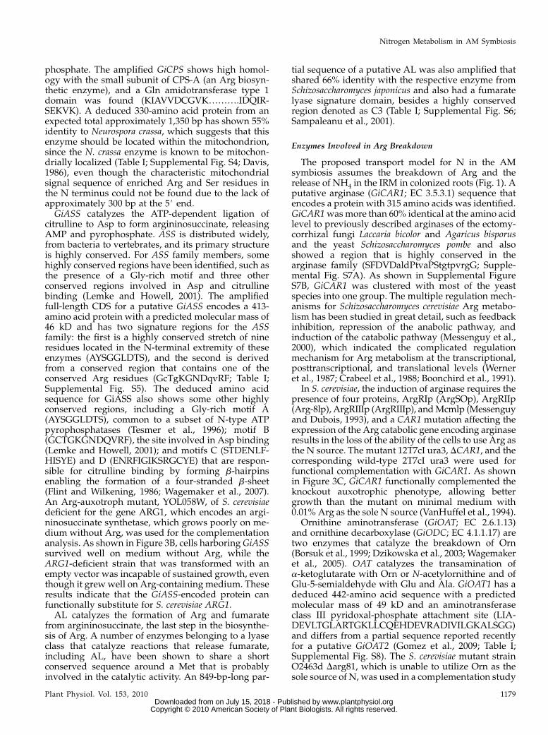
phosphate. The amplified GiCPS shows high homol-ogy with the small subunit of CPS-A (an Arg biosyn-thetic enzyme), and a Gln amidotransferase type 1domain was found (KIAVVDCGVK……….IDQIR-SEKVK). A deduced 330-amino acid protein from anexpected total approximately 1,350 bp has shown 55%identity to Neurospora crassa, which suggests that thisenzyme should be located within the mitochondrion,since the N. crassa enzyme is known to be mitochon-drially localized (Table I; Supplemental Fig. S4; Davis,1986), even though the characteristic mitochondrialsignal sequence of enriched Arg and Ser residues inthe N terminus could not be found due to the lack ofapproximately 300 bp at the 5# end.GiASS catalyzes the ATP-dependent ligation of
citrulline to Asp to form argininosuccinate, releasingAMP and pyrophosphate. ASS is distributed widely,from bacteria to vertebrates, and its primary structureis highly conserved. For ASS family members, somehighly conserved regions have been identified, such asthe presence of a Gly-rich motif and three otherconserved regions involved in Asp and citrullinebinding (Lemke and Howell, 2001). The amplifiedfull-length CDS for a putative GiASS encodes a 413-amino acid protein with a predicted molecular mass of46 kD and has two signature regions for the ASSfamily: the first is a highly conserved stretch of nineresidues located in the N-terminal extremity of theseenzymes (AYSGGLDTS), and the second is derivedfrom a conserved region that contains one of theconserved Arg residues (GcTgKGNDqvRF; Table I;Supplemental Fig. S5). The deduced amino acidsequence for GiASS also shows some other highlyconserved regions, including a Gly-rich motif A(AYSGGLDTS), common to a subset of N-type ATPpyrophosphatases (Tesmer et al., 1996); motif B(GCTGKGNDQVRF), the site involved in Asp binding(Lemke and Howell, 2001); and motifs C (STDENLF-HISYE) and D (ENRFIGIKSRGCYE) that are respon-sible for citrulline binding by forming b-hairpinsenabling the formation of a four-stranded b-sheet(Flint and Wilkening, 1986; Wagemaker et al., 2007).An Arg-auxotroph mutant, YOL058W, of S. cerevisiaedeficient for the gene ARG1, which encodes an argi-ninosuccinate synthetase, which grows poorly on me-dium without Arg, was used for the complementationanalysis. As shown in Figure 3B, cells harboringGiASSsurvived well on medium without Arg, while theARG1-deficient strain that was transformed with anempty vector was incapable of sustained growth, eventhough it grewwell on Arg-containing medium. Theseresults indicate that the GiASS-encoded protein canfunctionally substitute for S. cerevisiae ARG1.AL catalyzes the formation of Arg and fumarate
from argininosuccinate, the last step in the biosynthe-sis of Arg. A number of enzymes belonging to a lyaseclass that catalyze reactions that release fumarate,including AL, have been shown to share a shortconserved sequence around a Met that is probablyinvolved in the catalytic activity. An 849-bp-long par-
tial sequence of a putative AL was also amplified thatshared 66% identity with the respective enzyme fromSchizosaccharomyces japonicus and also had a fumaratelyase signature domain, besides a highly conservedregion denoted as C3 (Table I; Supplemental Fig. S6;Sampaleanu et al., 2001).
Enzymes Involved in Arg Breakdown
The proposed transport model for N in the AMsymbiosis assumes the breakdown of Arg and therelease of NH4 in the IRM in colonized roots (Fig. 1). Aputative arginase (GiCAR1; EC 3.5.3.1) sequence thatencodes a protein with 315 amino acids was identified.GiCAR1wasmore than 60% identical at the amino acidlevel to previously described arginases of the ectomy-corrhizal fungi Laccaria bicolor and Agaricus bisporusand the yeast Schizosaccharomyces pombe and alsoshowed a region that is highly conserved in thearginase family (SFDVDaldPtvaPStgtpvrgG; Supple-mental Fig. S7A). As shown in Supplemental FigureS7B, GiCAR1 was clustered with most of the yeastspecies into one group. The multiple regulation mech-anisms for Schizosaccharomyces cerevisiae Arg metabo-lism has been studied in great detail, such as feedbackinhibition, repression of the anabolic pathway, andinduction of the catabolic pathway (Messenguy et al.,2000), which indicated the complicated regulationmechanism for Arg metabolism at the transcriptional,posttranscriptional, and translational levels (Werneret al., 1987; Crabeel et al., 1988; Boonchird et al., 1991).
In S. cerevisiae, the induction of arginase requires thepresence of four proteins, ArgRIp (ArgSOp), ArgRIIp(Arg-8lp), ArgRIIIp (ArgRIIIp), andMcmlp (Messenguyand Dubois, 1993), and a CAR1 mutation affecting theexpression of the Arg catabolic gene encoding arginaseresults in the loss of the ability of the cells to use Arg asthe N source. The mutant 12T7cI ura3, DCAR1, and thecorresponding wild-type 2T7cI ura3 were used forfunctional complementation with GiCAR1. As shownin Figure 3C, GiCAR1 functionally complemented theknockout auxotrophic phenotype, allowing bettergrowth than the mutant on minimal medium with0.01% Arg as the sole N source (VanHuffel et al., 1994).
Ornithine aminotransferase (GiOAT; EC 2.6.1.13)and ornithine decarboxylase (GiODC; EC 4.1.1.17) aretwo enzymes that catalyze the breakdown of Orn(Borsuk et al., 1999; Dzikowska et al., 2003; Wagemakeret al., 2005). OAT catalyzes the transamination ofa-ketoglutarate with Orn or N-acetylornithine and ofGlu-5-semialdehyde with Glu and Ala. GiOAT1 has adeduced 442-amino acid sequence with a predictedmolecular mass of 49 kD and an aminotransferaseclass III pyridoxal-phosphate attachment site (LIA-DEVLTGLARTGKLLCQEHDEVRADIVILGKALSGG)and differs from a partial sequence reported recentlyfor a putative GiOAT2 (Gomez et al., 2009; Table I;Supplemental Fig. S8). The S. cerevisiae mutant strainO2463d Darg81, which is unable to utilize Orn as thesole source of N, was used in a complementation study
Nitrogen Metabolism in AM Symbiosis
Plant Physiol. Vol. 153, 2010 1179 www.plantphysiol.orgon July 15, 2018 - Published by Downloaded from
Copyright © 2010 American Society of Plant Biologists. All rights reserved.
for GiOAT1 (Wagemaker et al., 2007). As shown inFigure 3D, the yeast strain O2463d Darg81, which lacksOAT activity and is unable to utilize Orn as the sole Nsource, grew better on minimal medium with only0.01% Orn and Leu as N sources than the vectorcontrol, which showed very limited growth in 2 d at28�C. Thus, GiOAT1 could complement the O2468dDAg81 auxotrophic phenotype using Orn as the Nsource.
ODC is the key regulatory enzyme of the polyaminebiosynthetic pathway and catalyzes the decarboxyla-tion of Orn to 1,4-diaminobutane (putrescine). Thepredicted amino acid sequence of GiODC consistsof 445 amino acids, and the highest degree of aminoacid similarity was to the ODC sequence of Mucorcircinelloides (57%). GiODC belongs to the Orn/Lys/Arg decarboxylase class II family, with a typical Orn/diaminopimelate/Arg decarboxylases family 2pyridoxal-phosphate attachment site (YAVKCNGDP-MLLRLLAALG; Table I; Supplemental Fig. S9). Ingeneral, active growth and cell division are associatedwith substantial polyamine biosynthesis in plants,animals, and microorganisms (Bagni et al., 1980;Palavan and Galston, 1982; Pegg and McCann, 1982).The yeast knockout mutant disrupted in Orn decar-boxylase (SPE1) is auxotrophic for putrescine andbiotin, shows putrescine-dependent growth, andcould not grow on the medium without polyamines(Balasundaram et al., 1991; Mangold and Leberer,2005). Since ODC catalyzes the decarboxylation ofOrn to putrescine, the yeast SPE1 deletion strainYKL184W that lacks ODC is not able to grow withoutputrescine. Cells expressing GiODC could grow with-out exogenously supplied putrescine, while the vectorcontrol shows no growth, showing that GiODC func-tionally complements yeast putrescine auxotrophyresulting from the SPE1 mutation (Fig. 3E).
Urease (EC 3.5.1.5) is a nickel-binding enzyme thatcatalyzes the hydrolysis of urea to carbon dioxide andammonia. Two pathways are encountered for ureadegradation: hydrolysis via urease or via an ATP-hydrolyzing urease (EC 3.5.1.45). In higher plants,some fungi, andmany prokaryotes, urea is hydrolyzedby urease, which allows organisms to use externallyand internally generated urea as a source of N (Mobleyand Hausinger, 1989). In bacteria, three genes (ureA,ureB, and ureC) encode the structural subunits (a, b,and g, respectively) of urease, which associate in anabg stoichiometry to form the urease apoenzyme(Jabri et al., 1995). In plants and fungi, the structuralurease protein is encoded by one gene that compriseshomologs of three bacterial genes that encode separatesubunits of the functional enzyme. Here, the clonedurease from G. intraradices (GiURE) contains a CDS of833 amino acids with a calculated molecular mass of90 kD and a theoretical pI of 6.02. The encoded proteinresembles a-, b-, and g-subunits of the bacterial ureasefused into one protein, as described before for othereukaryotic ureases (Jabri et al., 1995; Jabri and Karplus,1996; Follmer, 2008). An alignment with fungal, plant,
and bacterial ureases revealed a high similarityamong all representative species included, with ahigh similarity to the eukaryotic ureases and to alower extent to the bacterial enzymes. Several resi-dues for the Klebsiella aerogenes urease were involvedin substrate binding or catalysis (His-134, Lys-217,His-246, His-272, and Asp-360), and these residueswere also found to be conserved in GiURE (Table I;Supplemental Fig. S10; Follmer, 2008). The yeastmutant lacking urease is also auxotrophic for severalamino acids, which made it impossible to use urea asthe sole N source for testing the function of GiURE bya complementation assay.
Gene Expression in Response to N Addition
Based on the current model, we hypothesized thatthe expression of genes required for N movementfrom soil through the fungus and into the host istemporally and spatially coordinated with the flux ofN. The transcriptional levels for all the genes from G.intraradices identified here were measured in ERM andIRM tissues by quantitative real-time PCR. At thesame time, plant GS is the initial enzyme for theassimilation of ammonium transferred by the fungus,so the carrot (Daucus carota) GS (DcGS) was alsostudied.
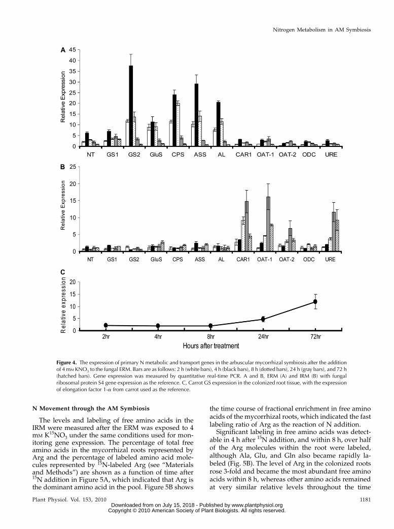
The transcript levels of several genes involved in Nuptake and assimilation increased in the ERM within2 h after 4 mM KNO3 was added to the fungal com-partment (Fig. 4). The expression of a fungal GS, CPS(Arg-specific small chain), NADH-dependent GluS,ASS, AL, and a nitrate transporter (NT) increased up to4 h after supply, increased to the highest levels after8 h, and then returned to the initial values within 72 h.By contrast, the expression of CAR1, URE, OAT1 andOAT2, and ODC in the ERM was only slightly affectedby the supply of nitrate. The expression of these genes(with the exception of ODC) was substantially up-regulated in the IRM within 24 h. The transcript levelsof a plant GS (DcGS) began to increase 24 h after thesupply of nitrate to the ERM and further increased upto the 72-h time point.
In the ERM, without the induction of N, the relativeexpression of GiGS1 (cycle threshold [CT] = 29.19 61.00) is higher than GiGS2 (CT = 31.31 6 1.07) with S4ribosomal protein as reference gene (CT = 27.556 0.60;P , 0.05). However, GiGS2 is much more highlyinduced than GiGS1 (Fig. 4) after the addition of Nin the ERM. Thus, GiGS1 is likely the main functionalenzyme for ammonium assimilation in the ERM atlower N levels, with GiGS2 being induced when N ismore abundant (consistent with its higher Km). ForOAT, GiOAT1was found to be more up-regulated thanGiOAT2 when N was added to the ERM, suggestingthat this isoform is more important in catabolizing Ornreleased by arginase in the IRM. Compared with OAT,ODC expression is quite stable both in ERM and IRM,consistent with its main function being for polyaminesynthesis rather than Orn breakdown.
Tian et al.
1180 Plant Physiol. Vol. 153, 2010 www.plantphysiol.orgon July 15, 2018 - Published by Downloaded from
Copyright © 2010 American Society of Plant Biologists. All rights reserved.
N Movement through the AM Symbiosis
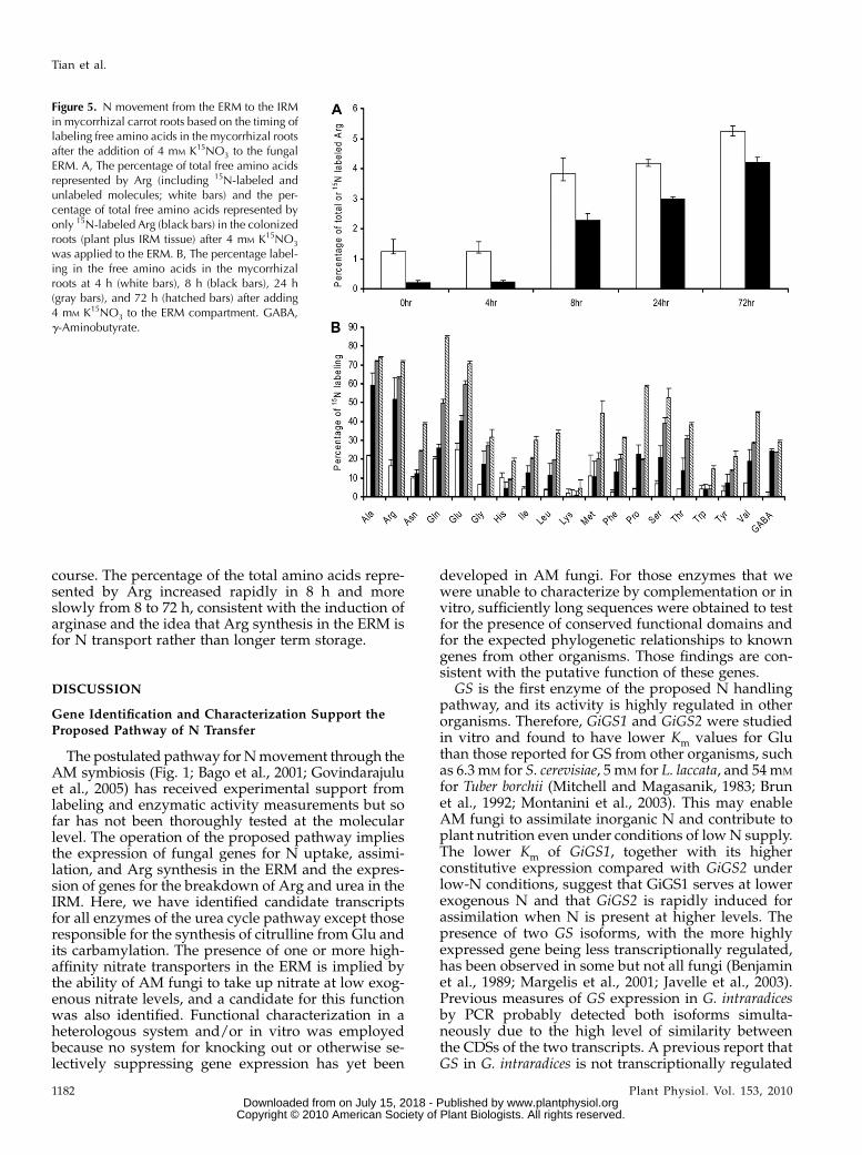
The levels and labeling of free amino acids in theIRM were measured after the ERM was exposed to 4mM K15NO3 under the same conditions used for mon-itoring gene expression. The percentage of total freeamino acids in the mycorrhizal roots represented byArg and the percentage of labeled amino acid mole-cules represented by 15N-labeled Arg (see “Materialsand Methods”) are shown as a function of time after15N addition in Figure 5A, which indicated that Arg isthe dominant amino acid in the pool. Figure 5B shows
the time course of fractional enrichment in free aminoacids of the mycorrhizal roots, which indicated the fastlabeling ratio of Arg as the reaction of N addition.
Significant labeling in free amino acids was detect-able in 4 h after 15N addition, and within 8 h, over halfof the Arg molecules within the root were labeled,although Ala, Glu, and Gln also became rapidly la-beled (Fig. 5B). The level of Arg in the colonized rootsrose 3-fold and became the most abundant free aminoacids within 8 h, whereas other amino acids remainedat very similar relative levels throughout the time
Figure 4. The expression of primary N metabolic and transport genes in the arbuscular mycorrhizal symbiosis after the additionof 4 mM KNO3 to the fungal ERM. Bars are as follows: 2 h (white bars), 4 h (black bars), 8 h (dotted bars), 24 h (gray bars), and 72 h(hatched bars). Gene expression was measured by quantitative real-time PCR. A and B, ERM (A) and IRM (B) with fungalribosomal protein S4 gene expression as the reference. C, Carrot GS expression in the colonized root tissue, with the expressionof elongation factor 1-a from carrot used as the reference.
Nitrogen Metabolism in AM Symbiosis
Plant Physiol. Vol. 153, 2010 1181 www.plantphysiol.orgon July 15, 2018 - Published by Downloaded from
Copyright © 2010 American Society of Plant Biologists. All rights reserved.
course. The percentage of the total amino acids repre-sented by Arg increased rapidly in 8 h and moreslowly from 8 to 72 h, consistent with the induction ofarginase and the idea that Arg synthesis in the ERM isfor N transport rather than longer term storage.
DISCUSSION
Gene Identification and Characterization Support theProposed Pathway of N Transfer
The postulated pathway forNmovement through theAM symbiosis (Fig. 1; Bago et al., 2001; Govindarajuluet al., 2005) has received experimental support fromlabeling and enzymatic activity measurements but sofar has not been thoroughly tested at the molecularlevel. The operation of the proposed pathway impliesthe expression of fungal genes for N uptake, assimi-lation, and Arg synthesis in the ERM and the expres-sion of genes for the breakdown of Arg and urea in theIRM. Here, we have identified candidate transcriptsfor all enzymes of the urea cycle pathway except thoseresponsible for the synthesis of citrulline from Glu andits carbamylation. The presence of one or more high-affinity nitrate transporters in the ERM is implied bythe ability of AM fungi to take up nitrate at low exog-enous nitrate levels, and a candidate for this functionwas also identified. Functional characterization in aheterologous system and/or in vitro was employedbecause no system for knocking out or otherwise se-lectively suppressing gene expression has yet been
developed in AM fungi. For those enzymes that wewere unable to characterize by complementation or invitro, sufficiently long sequences were obtained to testfor the presence of conserved functional domains andfor the expected phylogenetic relationships to knowngenes from other organisms. Those findings are con-sistent with the putative function of these genes.
GS is the first enzyme of the proposed N handlingpathway, and its activity is highly regulated in otherorganisms. Therefore, GiGS1 and GiGS2 were studiedin vitro and found to have lower Km values for Gluthan those reported for GS from other organisms, suchas 6.3 mM for S. cerevisiae, 5 mM for L. laccata, and 54mM
for Tuber borchii (Mitchell and Magasanik, 1983; Brunet al., 1992; Montanini et al., 2003). This may enableAM fungi to assimilate inorganic N and contribute toplant nutrition even under conditions of lowN supply.The lower Km of GiGS1, together with its higherconstitutive expression compared with GiGS2 underlow-N conditions, suggest that GiGS1 serves at lowerexogenous N and that GiGS2 is rapidly induced forassimilation when N is present at higher levels. Thepresence of two GS isoforms, with the more highlyexpressed gene being less transcriptionally regulated,has been observed in some but not all fungi (Benjaminet al., 1989; Margelis et al., 2001; Javelle et al., 2003).Previous measures of GS expression in G. intraradicesby PCR probably detected both isoforms simulta-neously due to the high level of similarity betweenthe CDSs of the two transcripts. A previous report thatGS in G. intraradices is not transcriptionally regulated
Figure 5. N movement from the ERM to the IRMin mycorrhizal carrot roots based on the timing oflabeling free amino acids in the mycorrhizal rootsafter the addition of 4 mM K15NO3 to the fungalERM. A, The percentage of total free amino acidsrepresented by Arg (including 15N-labeled andunlabeled molecules; white bars) and the per-centage of total free amino acids represented byonly 15N-labeled Arg (black bars) in the colonizedroots (plant plus IRM tissue) after 4 mM K15NO3
was applied to the ERM. B, The percentage label-ing in the free amino acids in the mycorrhizalroots at 4 h (white bars), 8 h (black bars), 24 h(gray bars), and 72 h (hatched bars) after adding4 mM K15NO3 to the ERM compartment. GABA,g-Aminobutyrate.
Tian et al.
1182 Plant Physiol. Vol. 153, 2010 www.plantphysiol.orgon July 15, 2018 - Published by Downloaded from
Copyright © 2010 American Society of Plant Biologists. All rights reserved.
in response to N addition was based on the levels of aGS transcript that has a closer similarity to GiGS1(Breuninger et al., 2004; Supplemental Fig. S1). Inaddition, Gomez et al. (2009) reported the expressionof a partial GS sequence that corresponds to the GiGS1transcript characterized above in Medicago roots colo-nized by G. intraradices and suggested that this mightbe involved in N turnover within the fungal IRM.Orn is released during Arg breakdown, and the
proposed N handling scheme requires that it is stored,excreted, or broken down in the IRM or translocated tothe ERM. Orn has not been found to be stored insubstantial quantities in the IRM and is not transferredto the host (Govindarajulu et al., 2005). The lack oftransfer to the plant of carbon from Arg or Orn isconsistent with the dependence of the fungus on theplant for its carbon nutrition. A subsequent turnoverof Orn and the release of inorganic N in the IRMwouldseem to be more efficient for N transfer to the host thanits translocation to the ERM, which would return halfof the N to its original location. The presence of twoOAT and one ODC sequences raised the question ofthe fate of Orn: conversion to Glu or to putrescine. Theincreased expression of the twoOATsequences but notthe putative ODC after N supply suggests that Orn isconverted to Glu. We postulate that Glu is brokendown to release more ammonium to the host, althougha previously published putative Glu dehydrogenase(Govindarajulu et al., 2005) appears not to have thisfunction, and we did not identify a better candidate.
Transcriptional Regulation
The regulation of N transport and metabolism infungi includes a transcriptional regulatory networkthat responds to the levels and forms of available N(ter Schure et al., 2000). For the proposed mechanismof N transfer in the AM symbiosis to operate effi-ciently, the participating enzymes and transporterswould be expected to be coordinately regulated inresponse to changes in the exogenous N supply. Inearlier studies, the expression of several putativegenes that are involved in the fungal N metabolismin ERM and IRM has been tested and interpreted asconsistent with the proposed pathway (Govindarajuluet al., 2005; Gomez et al., 2009). However, the func-tional identity of several tested transcripts was stilluncertain, their expression pattern in response to Ntreatment was difficult to interpret, and the sequencesof several key enzymes of the proposed N pathwayhave not been identified. Enzymatic activity of ASSwas shown to rise in the ERM but not in colonizedroots, whereas arginase, urease, and GS activities rosein the roots, but not in the ERM, after ammoniumsupply to the ERM (Cruz et al., 2007). The evidencepresented by Cruz et al. (2007) in support of the modelof Bago et al. (2001) included measurements of totalextractable enzyme activity from the colonized rootand the ERM. To test themodel thoroughly, however, itis necessary to distinguish between the fungus and
host plant enzymes in mycorrhizal roots so as todistinguish host root from fungal activities. Since it isfungal arginase and urease genes that are induced, themodel is supported, whereas if the enzyme activitieshad been of plant origin, then this would argue for atransfer of Arg and/or urea from fungus to host,which would have contradicted the original model.While consistent with the role for Arg in N movementin the AM symbiosis, the limited number of enzymestested, the difficulty of distinguishing between hostand fungal activities, and the lack of an activation ofGS in the ERM in response to N supply make itimportant to investigate the dynamics of gene expres-sion across the whole proposed pathway.
We predicted that activities for N uptake and as-similation and for Arg synthesis should be up-regu-lated in the ERM but not in the IRM after N additionand that the activities of genes involved in the break-down of Arg and the release of ammonium should beincreased in the IRM and not the ERM after N supplyto the ERM. Furthermore, host GS levels should in-crease after ammonium is released in the IRM andtransferred across the mycorrhizal interface. The tim-ing and location of the observed transcriptionalchanges after nitrate was supplied to the ERM (Fig.4) are completely consistent with these expectations.The timing of N transfer was followed with 15Nlabeling and measurements of amino acid levels,showing that N taken up in the ERM arrives insignificant amounts at the colonized roots withinhours (Fig. 5). 15N was detected in roots 4 h after Nsupply to the ERM, with high levels of labeling inamino acids and a 3-fold increase in Arg levels mea-sured 4 h later. This coincides with the up-regulationof Arg breakdown genes, which occurs several hoursafter the expression changes in assimilatory genes inthe ERM were observed. The level of plant GS tran-script rose substantially after the response in fungalcatabolic genes in the IRMwas initiated. The pattern ofgene expression in relation to N movement suggeststhat the arrival of N in significant quantities succes-sively in the ERM, IRM, and host plant is the triggerfor gene expression changes rather than signalingevents that might precede significant N delivery.
A regulatory scheme is shown in Figure 1 that isbased on our observations of the relative timing of Nmovement and gene expression and is consistent withfindings in other fungi, particularly yeast. It is pro-posed that (1) nitrate availability stimulates the ex-pression of the identified nitrate transporter (mostlikely together with other high- or low-affinity nitratetransporters), and presumably also of nitrate reduc-tase, although we were unable to clone the enzymesequence reported previously (Kaldorf et al., 1998;Montanini et al., 2006; Rekangalt et al., 2009); (2) am-monium stimulates the expression of the GS/GOGATpathway, and increasing levels of Gln and Glu ratherthan ammonium could lead to an up-regulation of Argsynthesis genes (Hinnebusch, 1988; ter Schure et al.,2000); (3) Arg is then translocated from the ERM to the
Nitrogen Metabolism in AM Symbiosis
Plant Physiol. Vol. 153, 2010 1183 www.plantphysiol.orgon July 15, 2018 - Published by Downloaded from
Copyright © 2010 American Society of Plant Biologists. All rights reserved.
IRM (Cruz et al., 2007); (4) elevated Arg levels triggeran increase in arginase and urease activities in the IRMbut not in the ERM (the difference between IRM andERMmay be due to N catabolite repression in the ERM[Hinnebusch, 1988; Wagemaker et al., 2007], as aminoacid levels are substantial in the ERM in the presenceof N [Jin et al., 2005]); (5) Arg or Orn that is producedin the IRM by the action of arginase stimulates theexpression and activity of OAT (Messenguy et al.,2000; ter Schure et al., 2000); (6) the subsequent releaseof ammonium in the IRM could stimulate the passiveefflux of ammonia into the mycorrhizal interface andthe uptake of ammonium by the plant by specifictransporters (Selle et al., 2005; Gomez et al., 2009;Guether et al., 2009); and (7) the subsequent increase inthe availability of ammonium for the host causes anup-regulation of plant GS, especially near the sites ofN transfer (Chalot et al., 2006).
The nature of signal(s) from the ERM to the IRM andhost roots to regulate nutrient exchange when mineralnutrients become available to the ERM is unknown.The results above show that the rise in the expressionof the fungal genes of Arg breakdown coincides withthe arrival of new N (15N label) and a rise in Arg levelswithin the colonized root. Thus, it is likely that this isthe signal for regulatory gene expression in the IRM,consistent with the observation that Arg induces argi-nase in S. pombe (Benitez and Farrar, 1980). However,Arg accumulation in the ERM does not repress Argsynthesis genes by a general amino acid control sys-tem seen in other fungi (Hinnebusch, 1988; ter Schureet al., 2000). This difference in transcriptional regula-tion is understandable if Arg in the ERM of AM fungifunctions more as a transport molecule than a storagecompound, as in saprophytes, and the transport intothe fungal vacuole and its binding to polyphosphateskeep the cytosolic levels low (Bucking et al., 1998).Although transcript levels for the Arg synthesis andcatabolic genes fell after 24 h, these enzymatic activ-ities must remain at substantial levels because highrates of N flow are sustained for extended periodsfollowing N addition (Govindarajulu et al., 2005; Jinet al., 2005; Cruz et al., 2007).
MATERIALS AND METHODS
Spore Material and Mycorrhizal in Vitro Cultures for15N Labeling and Gene Expression
The spore material of Glomus intraradices (DAOM 181602) was purchased
from Premier Tech Biotechnologies in units of 106 and was stored at 4�C until
further use. Ri T-DNA-transformed carrot (Daucus carota clone DCI) roots
colonized by G. intraradices (DAOM 197198) were grown at 24�C in modified
medium (Becard and Fortin, 1988) gelled with 3.5 g L21 Phytagel (Sigma)
using the split-plate method of St-Arnaud et al. (1996). The medium of the root
compartment was modified to limit the N concentration to 1 mM. The ERM
was allowed to cross over the divider into the fungal compartment that
contained modified medium with no N or Suc added and only 0.075 g L21
Phytagel to form a semisolid medium. After 2 weeks, the ERM was supplied
with 4 mM KNO3 for gene expression studies or K15NO3 to determine the
labeling percentage of free amino acids. The colonized roots and ERM samples
were collected after 0, 2, 4, 8, 24, and 72 h, rinsed with sterilized water, and
immediately frozen in liquid N and stored at 280�C until RNA extraction.
RNA Extraction
The RNA used for gene sequencing was extracted from approximately
10,000 spores of G. intraradices germinated in M medium (Becard and Fortin,
1988) without Suc for 4 to 5 d at 28�C using the TRIzol Plus RNA Purification
Kit (Invitrogen), including the DNase treatment, according to the manufac-
turer’s protocol. The RNA used for gene expression was extracted from
approximately 100 mg fresh weight of mycorrhizal roots or 0.2 to 0.3 mg fresh
weight of ERM tissue. The first strand of cDNA was synthesized using
the SuperScript First-Strand Synthesis System for reverse transcription-PCR
(Invitrogen) and random primers according to the manufacturer’s instructions.
Gene Amplification and Sequence Analysis
The partial sequences used for cloning the full lengths are from 454
pyrosequencing of cDNA from the ERM of G. intraradices. The 5# and 3#untranslated regions, and when possible the entire coding region of the
cDNA, were obtained by RACE using the cDNA from germinating spores as a
template with the SMART RACE cDNA amplification kit (Invitrogen). To
verify the start codon (ATG) of the amplified gene exactly, the promoter region
of the gene was amplified using the GenomeWalker Universal Kit (Clontech).
BLASTsearch and ClustalW 1.83 were used for the sequence comparisons and
alignments (Thompson et al., 1994). The phylogenetic trees were constructed
by the neighbor-joining method on the base of the alignments of our identified
amino acid sequences with other available homologous sequences usingMEGA
4.0.2. Secondary structure, protein family, domains, and functional sites were
predicted using the ExPASy Proteomic tools (http://ca.expasy.org/tools/).
cDNA Cloning and Yeast Transformation
As the primers shown in Supplemental Table S1, seven full CDSs were
cloned into the pYES2.1 TOPO TA (Invitrogen) vector was fused with a poly-
histidine region for further analysis. The resulting plasmids were amplified in
Escherichia coli and purified using the S.N.A.P Miniprep Kit (Invitrogen) and
were used to transform Saccharomyces cerevisiae strains using Frozen-EZ Yeast
Transformation II (Zymo Research) according to the manufacturer’s protocol.
The yeast strains used for the functional complementation are shown in Sup-
plemental Table S2. Yeast colonies carrying recombinant plasmids (pYES2.1
gene) were screened using a selective Ura-drop medium (Clontech) and ver-
ified again by sequencing. As a negative control, the yeasts were also trans-
formed with an empty vector (vector control).
The minimal medium (MM) composed of 0.175% yeast N base, 2.0% Gal
(Q-BIOgene; Morgan) supplemented with Complete Supplement Mixture
lacking uracil (Q-BIOgene), and 2.0% peptone agar was used for the yeast
complementation for GS (GiGS1 and GiGS2). For the other enzymes, the MM
for the complementation assays was modified as follows: GiASS, MM with
all amino acids except Arg; GiOAT1, MM with 0.01% Orn and Leu as sole
N sources; GiCAR1, MM with 0.01% Arg as sole N source; GiODC, the
H medium (a polyamine-free defined medium) according to Balasundaram
et al. (1991) and Mangold and Leberer (2005) was used. The H medium was
sterilized by filtering the solutions through a 0.45-mm Millipore membrane or
by boiling to prevent the growth of amine-deficient mutants resulting from
impurities from the steam during the autoclaving. Glass vessels were washed
with 6 M HCl, rinsed with 0.1 M HCl, and dried for 4 h at 170�C.
Enzyme Purification and Activity Detection
The total protein was extracted and the ProBond Purification System
(Invitrogen) was used to purify the GS protein extended by six tandem His
residues. The purified protein was desalted using Zeba Desalt Spin Columns
(Thermo Scientific) and then kept at 4�C. The expression and size of the
proteins in the transformed yeasts were confirmed using western blots based
on the polyhistidine region using Novex chemiluminescence substrates
(Invitrogen). Whole cell protein extracts from yeast transformants were
fractionated on a denaturing 12% polyacrylamide gel, transferred to poly-
vinylidene fluoride membranes, and detected with Anti-His(C-term)-HRP
antibody. Purified GiGS1 and GiGS2 were subjected to SDS-PAGE (12%
NuPAGE Novex Bis-Tris Gel) and silver stained, and the cutting gels for two
Tian et al.
1184 Plant Physiol. Vol. 153, 2010 www.plantphysiol.orgon July 15, 2018 - Published by Downloaded from
Copyright © 2010 American Society of Plant Biologists. All rights reserved.
proteins were sequenced directly using gas chromatography-mass spectrom-
etry to confirm. The protein level was measured using the BCA Protein Assay
Kit-Reducing Agent Compatible (Pierce).
The GS activity was tested using the synthetase activity assay. The kinetic
parameters for the substrate (Glu) were determined by varying the concen-
trations of individual reaction components in the presence of excess amounts
of all the other components. Each enzyme activity assay was replicated
independently at least three times (biological replicates); assays were always
conducted in duplicate using parallel reaction mixtures lacking GiGS as
blanks. The data were fitted to a Lineweaver-Burk plot ([S]/V versus [S]), and
the regression analysis of the data was performed with Excel. A soybean
(Glycine max) derivative of GS (Sigma) was used as the positive control. A total
of 450 ng of the affinity-purified and desalted GiGS1 and GiGS2 was added in
a 400-mL volume containing the following components at pH 7.0: 50 mM
HEPES, 0.4 mM ethylene glycol bis(P-aminoethylether)-N,N,N#,N#-tetraaceticacid, 40 mM MgSO4, 0 to 32 mM potassium Glu, 10 mM hydroxylamine HC1,
and 50 mM disodium potassium ATP. The reaction was incubated at 37�C for
10 to 30 min, then terminated by the addition of 1 mL of stop mix, consisting of
55 g of FeCl3·6H2O, 20 g of trichloroacetic acid, and 21 mL of concentrated HCl
per liter. An A540 of 0.555 corresponds to 1 mM of g-glutamylhydroxamate
released into the 400-mL reaction mixture. One unit of synthetase activity
catalyzes the formation of 1 mM of product per minute at 37�C.
Real-Time PCR
Gene expression was analyzed in DNA-free RNA samples by the SYBR
Green fluorescence assay in an ABI Prism 7700 Sequence Detection System.
Real-time PCR was performed on three independent biological samples and
two to three technical replicates. The primers are shown in Supplemental
Table S3. The comparative DDCT method was used to measure changes in the
expression of selected genes relative to untreated controls (Winer et al., 1999).
For G. intraradices, the gene expression results were normalized using the S4
ribosomal protein as a reference gene due to its high expression stability
(Govindarajulu et al., 2005). For host D. carota, the elongation factor 1-a gene
was used as the reference (Clotault et al., 2008). Mean values were compared
using Fisher’s protected LSD test and were considered different at P , 0.01
using SPSS.
Liquid Chromatography-MS
Liquid chromatography (LC)-MS analyses were carried out using a
Quattro micro (Waters) mass spectrometer system bundled with an electro-
spray ionization source and a Shimadzu HPLC system. Underivatized amino
acids were separated using a Waters Symmetry C18 (100 3 2.1 mm, 3 mm)
column with 1 mM perflurohetanoic acid in a water/acetonitrile gradient at
ambient temperature and a flow rate at 0.3 mL min21. The 15N content of Arg
was determined using a full scan of the protonated amino acid [i.e. mass-to-
charge ratio (m/z) 175 (15N0), 176 (15N1), 177 (15N2), 178 (15N3), and 179 (15N4)].
Arg content was determined by stable isotope analysis based on 15N2-Arg. Arg
isotopomers (i.e. m/z 175, 176, 177, 178, and 179) were investigated by LC-MS.
Mass spectrometric peak intensities were integrated for labeled and unlabeled
molecules of each free amino acid and used to obtain total levels (the sum of
all the labeled and unlabeled molecules of an amino acid) as well as fractional
labeling (the sum of labeled molecules as a proportion of the total number of
amino acids). Thus, for example, the amount of Arg is the sum of peak
intensities for Arg 15N-labeled molecules (m/z of 176, 177, 178, and 179
representing molecules with one, two, three, and four 15N atoms, respectively)
and unlabeled molecules (m/z 175). Samples from unlabeled tissues were used
to account for heavy isotopes at natural abundance (mainly 13C). Values are
means of three biological replicates. The 4-h Arg spectra contained greater
background signals for the most labeled molecules, which increased the
uncertainty of label quantification. The error bars in Figure 5 for labeling of
Arg at this time point represent mean maximal and mean minimal integrated
values, which are larger than the SD of the biological replicates.
The gene sequences are deposited in GenBank as follows: GiGS1
(GU111909), GiGS2 (GU111910), GiASS (GU111911), GiOAT1 (GU111912),
GiCAR1 (GU111913), GiODC (GU111914), GiURE (GU111915), GiGluS
(GU111916), GiCPS (GU111917), GiAL (GU111918), and GiNT (GU111919).
Supplemental Data
The following materials are available in the online version of this article.
Supplemental Figure S1. Amino acid alignment for GiNT.
Supplemental Figure S2.Amino acid alignment and phylogenetic analysis
for GiGS1 and GiGS2.
Supplemental Figure S3. Amino acid alignment for GiGluS.
Supplemental Figure S4. Amino acid alignment for GiCPS.
Supplemental Figure S5. Amino acid alignment for GiASS.
Supplemental Figure S6. Amino acid alignment for GiAL.
Supplemental Figure S7.Amino acid alignment and phylogenetic analysis
for GiCAR1.
Supplemental Figure S8. Amino acid alignment for GiOAT1.
Supplemental Figure S9. Amino acid alignment for GiODC.
Supplemental Figure S10. Amino acid alignment for GiURE.
Supplemental Figure S11. Western blot of the expressed proteins in yeast.
Supplemental Table S1. Primers for gene cloning.
Supplemental Table S2. Yeast strains for functional complementation.
Supplemental Table S3. Primers for real-time PCR.
ACKNOWLEDGMENTS
We thank Kyaw Aung and Yani Chen for assistance with the gene
amplification and functional complementation assays; Mintu Kumar Desai
for help with the enzyme activity tests; Sara J. Glimour for advice on real-time
PCR; Lijun Chen and Dan Jones for help with LC-MS; James Allen and
Yugandhar Beesetty for advice on mycorrhizal culture and inoculation;
Taghleb Al-Deeb and Cheng Zou for help on the figures; and Doug Allen,
Ana Paula Alonso, Inga Krassovskaya, and Weiqing Zeng for the suggestion
of this project.
Received March 30, 2010; accepted May 5, 2010; published May 6, 2010.
LITERATURE CITED
Allen JW, Shachar-Hill Y (2008) Sulfur transfer through an arbuscular
mycorrhiza. Plant Physiol 149: 549–560
Ames RN, Reid CP, Porter LK, Cambardella C (1983) Hyphal uptake and
transport of nitrogen from two 15N-labelled sources by Glomus
mosseae, a vesicular-arbuscular mycorrhizal fungus. New Phytol 95:
381–396
Bagni N, Malucelli B, Torrigiani P (1980) Polyamines, storage substances
and abscisic acid-like inhibitors during dormancy and very early
activation of Helianthus tuberosus tuber tissues. Physiol Plant 49:
341–345
Bago B, Pfeffer P, Shachar-Hill Y (2000) Carbon metabolism and transport
in arbuscular mycorrhizas. Plant Physiol 124: 949–957
Bago B, Pfeffer P, Shachar-Hill Y (2001) Could the urea cycle be trans-
locating nitrogen in the arbuscular mycorrhizal symbiosis? New Phytol
149: 4–8
Bago B, Vierheilig H, Piche Y, Azcon-Aguilar C (1996) Nitrate depletion
and pH changes induced by the extraradical mycelium of the arbuscular
mycorrhizal fungus Glomus intraradices grown in monoxenic culture.
New Phytol 133: 273–280
Balasundaram D, Tabor CW, Tabor H (1991) Spermidine or spermine is
essential for the aerobic growth of Saccharomyces cerevisiae. Proc Natl
Acad Sci USA 88: 5872–5876
Becard G, Fortin JA (1988) Early events of vesicular arbuscular mycorrhiza
formation on Ri T-DNA transformed roots. New Phytol 108: 211–218
Benitez T, Farrar L (1980) Induction of arginase and ornithine transaminase
in the fission yeast Saccharomyces pombe. J Bacteriol 144: 836–839
Benjamin PM, Wu JL, Mitchell AP, Magasanik B (1989) Regulatory
systems control expression of glutamine synthetase in Saccharomyces
cerevisiae at the level of transcription. Mol Gen Genet 217: 370–377
Bernard SM, Moller ALB, Dionisio G, Kichey T, Jahn TP, Dubois F,
Baudo M, Lopes MS, Terce-Laforgue T, Foyer CH, et al (2008) Gene
Nitrogen Metabolism in AM Symbiosis
Plant Physiol. Vol. 153, 2010 1185 www.plantphysiol.orgon July 15, 2018 - Published by Downloaded from
Copyright © 2010 American Society of Plant Biologists. All rights reserved.
expression, cellular localisation and function of glutamine synthe-
tase isozymes in wheat (Triticum aestivum L.). Plant Mol Biol 67:
89–105
Boonchird C, Messenguy F, Dubois E (1991) Determination of amino-
acid-sequences involved in the processing of the Arg 5 Arg6 precursor
in Saccharomyces cerevisiae. Eur J Biochem 199: 325–335
Borsuk P, Dzikowska A, Empel J, Grzelak A, Grzeskowiak R, Weglenski
P (1999) Structure of the arginase coding gene and its transcript in
Aspergillus nidulans. Acta Biochim Pol 46: 391–403
Breuninger M, Trujillo CG, Serrano E, Fischer R, Requena N (2004)
Different nitrogen sources modulate activity but not expression of
glutamine synthetase in arbuscular mycorrhizal fungi. Fungal Genet
Biol 41: 542–552
Brun A, Chalot M, Botton B, Martin F (1992) Purification and character-
ization of glutamine-synthetase and NADP-glutamate dehydrogenase
from the ectomycorrhizal fungus Laccaria laccata. Plant Physiol 99:
938–944
Bruns TD, Arnold AE, Hughes KW (2008) Fungal networks made of
humans: UNITE, FESIN, and frontiers in fungal ecology. New Phytol
177: 586–588
Bucking H, Beckmann S, Heyser W, Kottke I (1998) Elemental contents in
vacuolar granules of ectomycorrhizal fungi measured by EELS and
EDXS: a comparison of different methods and preparation techniques.
Micron 29: 53–61
Chalot M, Blaudez D, Brun A (2006) Ammonia: a candidate for nitrogen
transfer at the mycorrhizal interface. Trends Plant Sci 11: 263–266
Clark RB, Zeto SK (2000) Mineral acquisition by arbuscular mycorrhizal
plants. J Plant Nutr 23: 867–902
Clotault J, Peltier D, Berruyer R, Thomas M, Briard M, Geoffriau E (2008)
Expression of carotenoid biosynthesis genes during carrot root
development. J Exp Bot 59: 3563–3573
Crabeel M, Seneca S, Devos K, Glansdorff N (1988) Arginine repression of
the Saccharomyces cerevisiae arg1 gene: comparison of the arg1 and arg3
control regions. Curr Genet 13: 113–124
Cruz C, Egsgaard H, Trujillo C, Ambus P, Requena N, Martins-Loucao
MA, Jakobsen I (2007) Enzymatic evidence for the key role of arginine
in nitrogen translocation by arbuscular mycorrhizal fungi. Plant Physiol
144: 782–792
Davis RH (1986) Compartmental and regulatory mechanisms in the
arginine pathways of Neurospora crassa and Saccharomyces cerevisiae.
Microbiol Rev 50: 280–313
Dzikowska A, Kacprzak M, Tomecki R, Koper M, Scazzocchio C,
Weglenski P (2003) Specific induction and carbon/nitrogen repression
of arginine catabolism gene of Aspergillus nidulans-functional in vivo
analysis of the otaA promoter. Fungal Genet Biol 38: 175–186
Flint HJ, Wilkening J (1986) Cloning of the arg-12 gene of Neurospora crassa
and regulation of its transcript via cross-pathway amino-acid control.
Mol Gen Genet 203: 110–116
Follmer C (2008) Insights into the role and structure of plant ureases.
Phytochemistry 69: 18–28
Gomez SK, Javot H, Deewatthanawong P, Torres-Jerez I, Tang YH,
Blancaflor EB, Udvardi MK, Harrison MJ (2009) Medicago truncatula
and Glomus intraradices gene expression in cortical cells harboring ar-
buscules in the arbuscular mycorrhizal symbiosis. BMC Plant Biol 9: 10
Govindarajulu M, Pfeffer PE, Jin HR, Abubaker J, Douds DD, Allen JW,
Bucking H, Lammers PJ, Shachar-Hill Y (2005) Nitrogen transfer in the
arbuscular mycorrhizal symbiosis. Nature 435: 819–823
Guether M, Neuhauser B, Balestrini R, Dynowski M, Ludewig U,
Bonfante P (2009) A mycorrhizal-specific ammonium transporter from
Lotus japonicus acquires nitrogen released by arbuscular mycorrhizal
fungi. Plant Physiol 150: 73–83
Hinnebusch AG (1988) Mechanisms of gene regulation in the general
control of amino acid biosynthesis in Saccharomyces cerevisiae. Microbiol
Rev 52: 248–273
Hodge A, Campbell CD, Fitter AH (2001) An arbuscular mycorrhizal
fungus accelerates decomposition and acquires nitrogen directly from
organic material. Nature 413: 297–299
Ishiyama K, Inoue E, Tabuchi M, Yamaya T, Takahashi H (2004)
Biochemical background and compartmentalized functions of cytosolic
glutamine synthetase for active ammonium assimilation in rice roots.
J Biol Chem 279: 16598–16605
Jabri E, Carr MB, Hausinger RP, Karplus PA (1995) The crystal structure of
urease from Klebsiella aerogenes. Science 268: 998–1004
Jabri E, Karplus PA (1996) Structures of the Klebsiella aerogenes urease
apoenzyme and two active-site mutants. Biochemistry 35: 10616–10626
Jakobsen I, Rosendahl L (1990) Carbon flow into soil and external hyphae
from roots of mycorrhizal cucumber plants. New Phytol 115: 77–83
Javelle A, Andre B, Marini AM, Chalot M (2003) High-affinity ammonium
transporters and nitrogen sensing in mycorrhizas. Mol Microbiol 47:
411–430
Jin H, Pfeffer PE, Douds DD, Piotrowski E, Lammers PJ, Shachar-Hill Y
(2005) The uptake, metabolism, transport and transfer of nitrogen in an
arbuscular mycorrhizal symbiosis. New Phytol 168: 687–696
Johansen A, Finlay RD, Olsson PA (1996) Nitrogen metabolism of external
hyphae of the arbuscular mycorrhizal fungus Glomus intraradices. New
Phytol 133: 705–712
Johansen A, Jakobsen I, Jensen ES (1993) Hyphal transport by a vesicular-
arbuscular mycorrhizal fungus of N applied to the soil as ammonium or
nitrate. Biol Fertil Soils 16: 66–70
Johansen A, Jensen ES (1996) Transfer of N and P from intact or
decomposing roots of pea to barley interconnected by an arbuscular
mycorrhizal fungus. Soil Biol Biochem 28: 73–81
Kaldorf M, Schmelzer E, Bothe H (1998) Expression of maize and fungal
nitrate reductase genes in the arbuscular mycorrhiza. Mol Plant Microbe
Interact 11: 439–448
Lemke CT, Howell PL (2001) The 1.6 angstrom crystal structure of E. coli
argininosuccinate synthetase suggests a conformational change during
catalysis. Structure 9: 1153–1164
Mangold U, Leberer E (2005) Regulation of all members of the antizyme
family by antizyme inhibitor. Biochem J 385: 21–28
Margelis S, D’Souza C, Small AJ, Hynes MJ, Adams TH, Davis MA (2001)
Role of glutamine synthetase in nitrogen metabolite repression in
Aspergillus nidulans. J Bacteriol 183: 5826–5833
Messenguy F, Dubois E (1993) Genetic evidence for a role for mcm1 in the
regulation of arginine metabolism in Saccharomyces cerevisiae. Mol Cell
Biol 13: 2586–2592
Messenguy F, Vierendeels F, Scherens B, Dubois E (2000) In Saccharomyces
cerevisiae, expression of arginine catabolic genes CAR1 and CAR2
in response to exogenous nitrogen availability is mediated by the
Ume6 (CargRI)-Sin3 (CargRII)-Rpd3 (CargRIII) complex. J Bacteriol 182:
3158–3164
Mitchell AP, Magasanik B (1983) Purification and properties of glutamine-
synthetase from Saccharomyces cerevisiae. J Biol Chem 258: 119–124
Mobley HLT, Hausinger RP (1989) Microbial ureases: significance,
regulation, and molecular characterization. Microbiol Rev 53: 85–108
Montanini B, Betti M, Marquez AJ, Balestrini R, Bonfante P, Ottonello S
(2003) Distinctive properties and expression profiles of glutamine
synthetase from a plant symbiotic fungus. Biochem J 373: 357–368
Montanini B, Gabella S, Abba S, Peter M, Kohler A, Bonfante P, Chalot
M, Martin F, Ottonello S (2006) Gene expression profiling of the
nitrogen starvation stress response in the mycorrhizal ascomycete Tuber
borchii. Fungal Genet Biol 43: 630–641
Palavan N, Galston AW (1982) Polyamine biosynthesis and titer during
various developmental stages of Phaseolus vulgaris. Physiol Plant 55:
438–444
Pegg AE, McCann PP (1982) Polyamine metabolism and function. Am J
Physiol 243: C212–C221
Pelanda R, Vanoni MA, Perego M, Piubelli L, Galizzi A, Curti B, Zanetti
G (1993) Glutamate synthase genes of the diazotroph Azospirillum
brasilense: cloning, sequencing, and analysis of functional domains.
J Biol Chem 268: 3099–3106
Rekangalt D, Pepin R, Verner MC, Debaud JC, Marmeisse R, Fraissinet-
Tachet L (2009) Expression of the nitrate transporter nrt2 gene from the
symbiotic basidiomycete Hebeloma cylindrosporum is affected by host
plant and carbon sources. Mycorrhiza 19: 143–148
Sampaleanu LM, Vallee F, Thompson GD, Howell PL (2001) Three-
dimensional structure of the argininosuccinate lyase frequently
complementing allele Q286R. Biochemistry 40: 15570–15580
Selle A, Willmann M, Grunze N, Geßler A, Weiß M, Nehls U (2005) The
high-affinity poplar ammonium importer PttAMT1.2 and its role in
ectomycorrhizal symbiosis. New Phytol 168: 697–706
Smith SE, Facelli E, Pope S, Smith FA (2010) Plant performance in stressful
environments: interpreting new and established knowledge of the roles
of arbuscular mycorrhizas. Plant Soil 326: 3–20
Smith SE, Read DJ (2008) Mycorrhizal Symbiosis, Ed 3. Elsevier and
Academic, New York
Tian et al.
1186 Plant Physiol. Vol. 153, 2010 www.plantphysiol.orgon July 15, 2018 - Published by Downloaded from
Copyright © 2010 American Society of Plant Biologists. All rights reserved.
Stanford AC, Larsen K, Barker DG, Cullimore JV (1993) Differential
expression within the glutamine synthetase gene family of the model
legume Medicago truncatula. Plant Physiol 103: 73–81
St-Arnaud M, Hamel C, Vimard B, Fortin JA (1996) Enhanced hyphal
growth and spore production of the arbuscular mycorrhizal fungus
Glomus intraradices in an in vitro system in the absence of host roots.
Mycol Res 100: 328–332
Tanaka Y, Yano K (2005) Nitrogen delivery to maize via mycorrhizal hyphae
depends on the form of N supplied. Plant Cell Environ 28: 1247–1254
ter Schure EG, van Riel NAW, Verrips CT (2000) The role of ammonia
metabolism in nitrogen catabolite repression in Saccharomyces cerevisiae.
FEMS Microbiol Rev 24: 67–83
Tesmer JG, Klem TJ, Deras ML, Davisson VJ, Smith JL (1996) The crystal
structure of GMP synthetase reveals a novel catalytic triad and is a
structural paradigm for two enzyme families. Nat Struct Biol 3: 74–86
Thompson JD, Higgins DG, Gibson TJ (1994) CLUSTALW: improving the
sensitivity of progressive multiple sequence alignment through
sequence weighting, position-specific gap penalties and weight matrix
choice. Nucleic Acids Res 22: 4673–4680
Toussaint JP, St-Arnaud M, Charest C (2004) Nitrogen transfer and
assimilation between the arbuscular mycorrhizal fungus Glomus intra-
radices Schenck & Smith and Ri T-DNA roots of Daucus carota L., in an in
vitro compartmented system. Can J Microbiol 50: 251–260
VanHuffel C, Dubois E, Messenguy F (1994) Cloning and sequencing of
Saccharomyces pombe car1 gene encoding arginase: expression of the
arginine anabolic and catabolic genes in response to arginine and
related metabolites. Yeast 10: 923–933
Wagemaker MJM, Eastwood DC, Van Der Drift C, Jetten MSM, Burton K,
Van Griensven LJLD, Den Camp HJMO (2007) Argininosuccinate
synthetase and argininosuccinate lyase: two ornithine cycle enzymes
from Agaricus bisporus. Mycol Res 111: 493–502
Wagemaker MJM, Welboren W, van der Drift C, Jetten MSM, Van
Griensven LJLD, den Camp HJMO (2005) The ornithine cycle enzyme
arginase from Agaricus bisporus and its role in urea accumulation in fruit
bodies. Biochim Biophys Acta 1681: 107–115
Werner M, Feller A, Messenguy F, Pierard A (1987) The leader peptide of
yeast gene CPA1 is essential for the translational repression of its
expression. Cell 49: 805–813
Winer J, Jung CKS, Shackel I, Williams PM (1999) Development and
validation of real-time quantitative reverse transcriptase-polymerase
chain reaction for monitoring gene expression in cardiac myocytes in
vitro. Anal Biochem 270: 41–49
Nitrogen Metabolism in AM Symbiosis
Plant Physiol. Vol. 153, 2010 1187 www.plantphysiol.orgon July 15, 2018 - Published by Downloaded from
Copyright © 2010 American Society of Plant Biologists. All rights reserved.


![Insights on the Impact of Arbuscular MycorrhizalInsights on the Impact of Arbuscular Mycorrhizal Symbiosis on Tomato Tolerance to Water Stress1[OPEN] Walter Chitarra, Chiara Pagliarani,](https://img.pdfslide.net/doc/110x75/5e50794d24936120366ae63e/insights-on-the-impact-of-arbuscular-insights-on-the-impact-of-arbuscular-mycorrhizal.jpg)