Embed Size (px)
Citation preview
Regulation of Urokinase Expression at the Posttranscription Level byLung Epithelial CellsShwetha K. Shetty,†,∥ Amarnath S. Marudamuthu,†,∥ Daniel Abernathy,† Rashmi S. Shetty,†
Praveenkumar Shetty,†,⊥ Jian Fu,† Steven Idell,† Yashodhar P. Bhandary,† Honglong Ji,‡ Ming-Cheh Liu,§
and Sreerama Shetty*,†
†Texas Lung Injury Institute and ‡Department of Biochemistry, The University of Texas Health Science Center, 11937 US HWY 271,Tyler, Texas 75708, United States§Department of Pharmacology, College of Pharmacy, The University of Toledo, Toledo, Ohio 43614, United States
ABSTRACT: Urokinase-type plasminogen activator (uPA) isexpressed by lung epithelial cells and regulates fibrin turnoverand epithelial cell viability. PMA, LPS, and TNF-alpha, as wellas uPA itself, induce uPA expression in lung epithelial cells.PMA, LPS, and TNF-alpha induce uPA expression throughincreased synthesis as well as stabilization of uPA mRNA,while uPA increases its own expression solely through uPAmRNA stabilization. The mechanism by which lung epithelialcells regulate uPA expression at the level of mRNA stability isunclear. To elucidate this process, we sought to characterizeprotein−uPA mRNA interactions that regulate uPA expres-sion. Regulation of uPA at the level of mRNA stability involves the interaction of a ∼40 kDa cytoplasmic−nuclear shuttlingprotein with a 66 nt uPA mRNA 3′UTR sequence. We purified the uPA mRNA 3′UTR binding protein and identified it asribonucleotide reductase M2 (RRM2). We expressed recombinant RRM2 and confirmed its interaction with a specific 66 nt uPA3′UTR sequence. Immunoprecipitation of cell lysates with anti-RRM2 antibody and RT-PCR for uPA mRNA confirmed thatRRM2 binds to uPA mRNA. Treatment of Beas2B cells with uPA or LPS attenuated RRM2−endogenous uPA mRNAinteractions, while overexpression of RRM2 inhibited uPA protein and mRNA expression through destabilization of uPA mRNA.LPS exposure of lung epithelial cells translocates RRM2 from the cytoplasm to the nucleus in a time-dependent manner, leadingto stabilization of uPA mRNA. This newly recognized pathway could influence uPA expression and a broad range of uPA-dependent functions in lung epithelial cells in the context of lung inflammation and repair.
Pulmonary epithelial cells, microvascular endothelial cells,and macrophages express urokinase-type plasminogen
activator (uPA), a serine protease that catalyzes the conversionof plasminogen to plasmin. By binding to its receptor, uPAR,uPA localizes plasminogen activation at the cell surface. uPA−uPAR complexes serve other functions independent ofplasminogen activation, such as cell proliferation, intracellularsignaling, differentiation, chemotactic migration, and inva-sion.1−5 Lung epithelial cells also express PAI-1, which caninhibit uPA activity and promote internalization and recyclingof uPA−uPAR−PAI-1 complexes.6 Previous studies demon-strated rapid increases in circulating and pulmonary concen-trations of uPA after endotoxemia or bacterial infection.7−10
Mice deficient in uPA resist LPS-induced acute lung injury(ALI) and the development of pulmonary edema.7,11 On theother hand, a defect of uPA-mediated fibrinolysis occurs inacute respiratory distress syndrome, and uPA attenuates thefibrotic response in bleomycin-induced accelerated pulmonaryfibrosis. Therefore, uPA appears to play a pivotal role in thepathogenesis of ALI and in pulmonary fibrosis subsequent toALI.
Recent findings indicated that uPA autoinduction by lungepithelial cells,2,12 endothelial cells, and monocytes13 involvesuPA binding to uPAR through its growth factor domain. Thepotentiation of LPS-induced ALI by uPA7 and diminishedneutrophil recruitment in response to P. aeruginosa pneumo-nia14 by uPA- and uPAR-deficient mice underscores thecontribution of uPA and uPAR to the development of ALI.Further, increased expression of uPA due to posttranscriptionaluPA mRNA stabilization by tumor cells has been implicated inthe increased proliferative and invasive potential of cancercells.12,15 We previously reported that uPA expression isupregulated in lung epithelial cells through stabilization of uPAmRNA and that proinflammatory mediators implicated in thepathogenesis of ALI and its repair stabilize uPA mRNA.15 Sinceelucidation of the underlying mechanism is essential for a betterunderstanding of ALI, we sought to define the regulatoryinteractions that contribute to the stabilization of uPA mRNA
Received: August 13, 2011Revised: December 13, 2011Published: December 14, 2011
Article
pubs.acs.org/biochemistry
© 2011 American Chemical Society 205 dx.doi.org/10.1021/bi201293x | Biochemistry 2012, 51, 205−213
and induce uPA at the posttranscriptional level in lungepithelial cells.
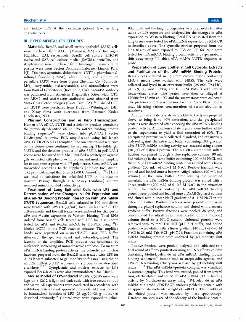
■ EXPERIMENTAL PROCEDURESMaterials. Beas2B and small airway epithelial (SAE) cells
were purchased from ATCC (Manassas, VA) and Invitrogen(Carlsbad, CA), respectively. Beas2B cell culture (LHC-9)media and SAE cell culture media (SAGM), penicillin, andstreptomycin were purchased from Invitrogen. Tissue cultureplastics were from Becton Dickinson Labware (Linclon Park,NJ). Tris-base, aprotinin, dithiothreitol (DTT), phenylmethyl-sulfonyl fluoride (PMSF), silver nitrate, and ammoniumpersulfate (APS) were from Sigma Chemical Co. (St. Louis,MO). Acrylamide, bis(acrylamide), and nitrocellulose werefrom BioRad Laboratories (Richmond, CA). Anti-uPA antibodywas purchased from American Diagnostica (Greenwich, CT);anti-RRM2 and anti-β-actin antibodies were obtained fromSanta Cruz Biotechnologies (Santa Cruz, CA). 32P-labeled UTPand dCTP were purchased from DuPont (Wilmington, DE),and X-ray films were purchased from Eastman Kodak(Rochester, NY).Plasmid Construction and in Vitro Transcription.
Human uPA cDNA 3′UTR and a deletion product containingthe previously identified 66 nt uPA mRNA binding proteinbinding sequence15 were cloned into pCDNA3.1 vector(Invitrogen) following PCR amplification using full lengthuPA 3′UTR cDNA as a template. The orientation and sequenceof the clones were confirmed by sequencing. The full-length3′UTR and the deletion product of uPA 3′UTR in pcDNA3.1vector were linearized with Xba I, purified separately on agarosegels, extracted with phenol−chloroform, and used as a templatefor in vitro transcription with T7 polymerase. Sense mRNA wastranscribed according to the supplier’s (Ambion Inc., Austin,TX) protocol, except that 50 μCi (800 Ci/mmol) of [32P] UTPwas used to substitute for unlabeled UTP in the reactionmixture. Passage through a NucAway (Ambion) columnremoved unincorporated radioactivity.Treatment of Lung Epithelial Cells with LPS and
Determination of the Changes in uPA Expression anduPA mRNA Binding Protein Interaction with uPA mRNA3′UTR Sequences. Beas2B cells cultured in 100 mm disheswere treated with LPS (20 μg/mL) for 0−24 h at 37 °C. Theculture media and the cell lysates were analyzed for changes inuPA and β-actin expression by Western blotting. Total RNAisolated from Beas2B cells treated with LPS for 0−6 h weretested for uPA and β-actin mRNA by RT-PCR using 32P-labeled dCTP in the PCR reaction mixture. The amplifiedbands were separated on a urea/PAGE using TBE buffer.Afterward, the gel was dried and autoradiographed. Theidentity of the amplified PCR product was confirmed bynucleotide sequencing of nonradioactive amplicon. To measureuPA mRNA binding protein activity, the cytosolic and nuclearfractions prepared from the Beas2B cells treated with LPS for0−24 h were subjected to gel mobility shift assay using the 66nt uPA mRNA 3′UTR sequence as a probe as we describedelsewhere.15,22 Cytoplasmic and nuclear extracts of LPSexposed Beas2B cells were also immunoblotted for RRM2.Mouse Model of LPS-Induced Injury. C57B6 mice were
kept on a 12:12 h light and dark cycle with free excess to foodand water. All experiments were conducted in accordance withinstitution review board approved protocols. ALI was inducedby intratracheal injection of LPS (25 μg/20−25 g mouse) asdescribed previously.11 Control mice were exposed to saline.
BAL fluids and the lung homogenates were prepared 24 h aftersaline or LPS exposure and analyzed for the changes in uPAexpression by Western blotting. Total RNAs isolated from thelung tissues were tested for uPA mRNA expression by RT-PCRas described above. The cytosolic extracts prepared from thelung tissues of mice exposed to PBS or LPS for 24 h weretested for uPA mRNA binding protein activity by gel mobilityshift assay using 32P-labled uPA mRNA 3′UTR sequence asprobe.
Preparation of Lung Epithelial Cell Cytosolic Extractsand Purification of the uPA mRNA Binding Protein.Beas2B cells cultured in 150 mm culture dishes containingLHC-9 media were washed with HBSS. The cells werecollected and lysed in an extraction buffer (25 mM Tris-HCl,pH 7.9, 0.5 mM EDTA, and 0.1 mM PMSF) with severalfreeze−thaw cycles. The lysates were then centrifuged at12000g for 15 min at 4 °C, and the supernatants were collected.The protein content was measured with a Pierce BCA proteinassay kit using various concentrations of serum albumin asstandards.Ammonium sulfate crystals were added to the lysate prepared
above to bring it to 40% saturation, and the precipitatedproteins were discarded after checking the uPA mRNA bindingprotein activity. Ammonium sulfate crystals were further addedto the supernatant to yield a final saturation of 60%. Theprecipitated proteins were collected, dissolved, and exhaustivelydialyzed against the extraction buffer containing 10% glycerol.uPA 3′UTR mRNA binding activity was assessed using aliquot(10 μg) of dialyzed protein. The 40−60% ammonium sulfatefraction was passed through a blue sepharose column (90 mLbed volume) in the same buffer containing 100 mM NaCl, andthe uPA 3′UTR mRNA binding protein was eluted with a lineargradient (200 mL) of 0.1−1 M NaCl. Positive fractions werepooled and loaded onto a heparin Affigel column (90 mL bedvolume) in the same buffer. After washing the unboundmaterials, the uPA mRNA binding protein was eluted with alinear gradient (200 mL) of 0−0.5 M NaCl in the extractionbuffer. The fractions containing the uPA mRNA bindingprotein were pooled and loaded onto a DEAE-Sephacel columnand eluted with a linear NaCl gradient of 0−1 M NaCl in theextraction buffer. Positive fractions were pooled and passedthrough a phenyl sepharose column and eluted with a reversegradient buffer. Positive fractions were pooled, dialyzed, andconcentrated by ultrafiltration and loaded onto a mono-Qcolumn fitted to a FPLC system. Unbound proteins wereremoved with 25 mM Tris-HCl (pH, 7.9) buffer, and boundproteins were eluted with a linear gradient (40 mL) of 0−1 MNaCl in 25 mM Tris-HCl (pH 7.9). Fractions containing uPAmRNA binding protein were analyzed by gel mobility shiftassays.Positive fractions were pooled, dialyzed, and subjected to a
final round of affinity purification using an RNA affinity columncontaining biotin-labeled 66 nt uPA mRNA binding proteinbinding sequences15 immobilized to streptavidin agarose, anduPA mRNA binding activity was assessed by gel mobility shiftassays.16−19 The uPA mRNA−protein complex was visualizedby autoradiography. This band was excised, pooled from severalruns, electroeluted, and tested for uPA mRNA 3′UTR bindingactivity by Northwestern assay using 32P-labeled 66 nt uPAmRNA as a probe. SDS-PAGE analyses yielded a protein withan approximate molecular weight of ∼40 kDa. The identity ofthe eluted protein was analyzed by mass spectroscopy.Database analyses revealed the identity of the binding protein.
Biochemistry Article
dx.doi.org/10.1021/bi201293x | Biochemistry 2012, 51, 205−213206
To determine whether the uPA mRNA binding proteinspecifically binds to uPA mRNA, we expressed it in aprokaryotic system using BL21 cells and purified the GSTfusion protein as described previously.16−18 Purified recombi-nant uPA mRNA binding protein was analyzed for uPA mRNAbinding using a gel mobility shift assay. To further confirm thespecificity of the binding protein interaction with the 66 nt uPAmRNA 3′UTR sequence, we incubated purified recombinantRRM2 with the 32P-labeled 66 nt uPA mRNA binding sequencein the presence of a molar excess of unlabeled 66 nt uPAmRNA binding protein binding sequence or full length uPA oruPAR mRNA 3′UTR sequences and analyzed the interaction bygel mobility shift assay as described previously.15,19 The uPAmRNA binding protein cDNA was then subcloned into aeukaryotic expression vector pcDNA 3.1 and transfected toBeas2B cells. Histidine-tagged binding protein, containing a C-terminal V5 epitope, was expressed in Beas2B cells and wasaffinity purified using a nickel column.17,18 Expression of thebinding protein was confirmed by Western blotting of elutedfractions using anti-V5 and antibinding protein antibodies. Thefractions containing binding proteins were pooled and testedfor uPA mRNA 3′UTR binding by a gel mobility shift assayusing the 32P-labeled 66 nt sequence as a probe.Expression of the uPA mRNA Binding Protein in Lung
Epithelial Cells and Determination of Its Role in theRegulation of uPA Expression. Beas2B cells cultured inLHC-9 media or primary SAE cells maintained in SAGM weretransfected with either binding protein cDNA cloned inpcDNA 3.1 or an empty vector using PEI reagent.20 Thecells were lysed, and the expression of recombinant protein wasconfirmed by Western blotting of V5 fusion protein. Theconditioned media and cell lysates were tested for uPAexpression by Western blotting. Afterward, the membranewas stripped and sequentially probed with antibody against V5or β-actin to assess expression of the fusion protein and loadingequality. Total RNAs from the Beas2B cells transfected withvector cDNA alone or vector harboring RRM2 cDNA wereisolated and analyzed for uPA mRNA by RT-PCR. In order toconfirm if inhibition of uPA mRNA by overexpression ofRRM2 was due to destabilization of uPA mRNA, Beas2B cellstransfected with vector alone or vector harboring RRM2 cDNAwere treated with or without LPS for 12 h to induce maximumuPA mRNA. DRB was added to these cells to inhibit ongoingtranscription, and the total RNA was isolated at different timepoints. uPA mRNA was analyzed by RT-PCR using 32P-labeleddCTP. Amplified PCR products were separated on a urea/polyacrylamide gel and autoradiographed.Effect of uPA or LPS on RRM2 Interaction with uPA
mRNA in Lung Epithelial Cells. Beas2B cells were treatedwith PBS or uPA for 24 h or with LPS for 0−24 h in LHC-9media. Afterward, the media were aspirated, cells washed withcold PBS, and cell lysates prepared were cleared with mouseIgG. The precleared lysates were subsequently subjected toimmunoprecipation using anti-RRM2 antibody. The immunecomplexes were washed with the binding buffer, the RNA wasisolated, and the associated uPA mRNA was amplified by RT-PCR in the presence of 32P-labeled dCTP. The PCR productswere separated by urea-PAGE. After electrophoresis, the gelwas dried and subjected to autoradiography.Statistical Analysis. The statistical differences between
various experimental conditions were analyzed by Student’s ttest.
■ RESULTS
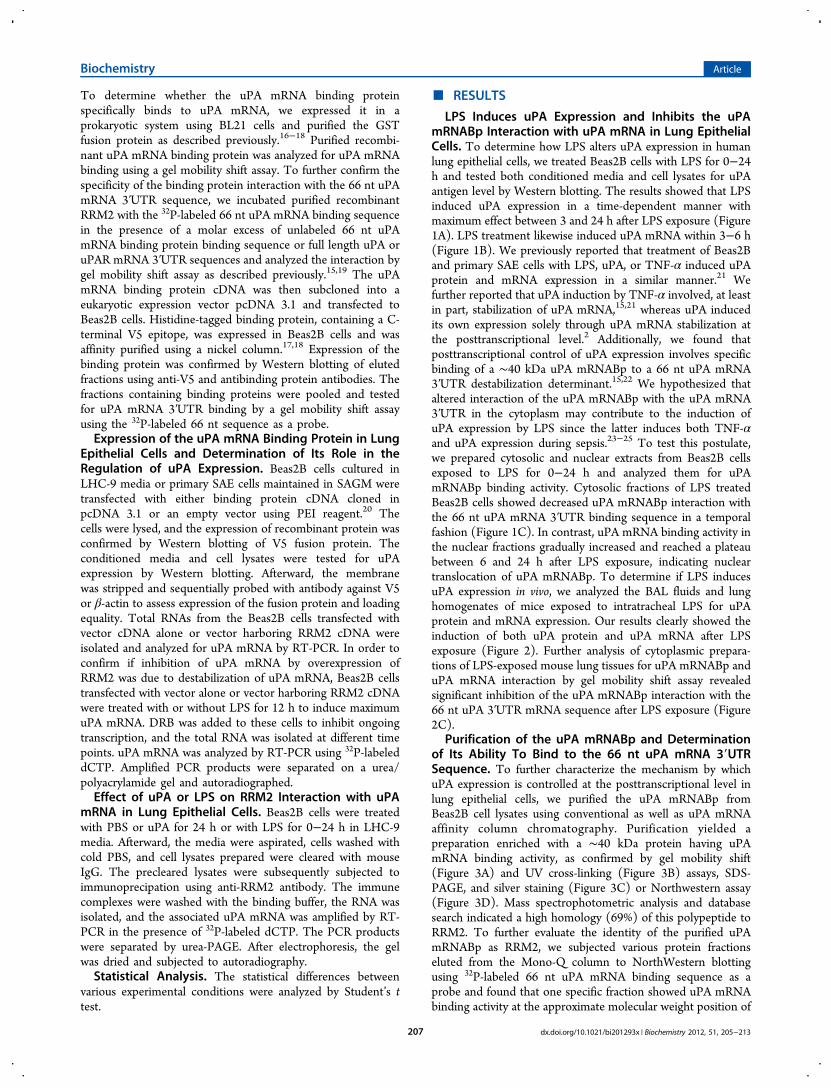
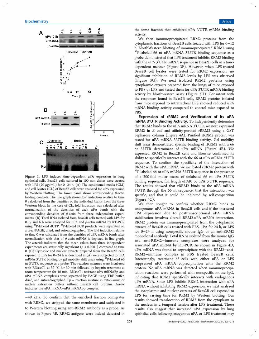
LPS Induces uPA Expression and Inhibits the uPAmRNABp Interaction with uPA mRNA in Lung EpithelialCells. To determine how LPS alters uPA expression in humanlung epithelial cells, we treated Beas2B cells with LPS for 0−24h and tested both conditioned media and cell lysates for uPAantigen level by Western blotting. The results showed that LPSinduced uPA expression in a time-dependent manner withmaximum effect between 3 and 24 h after LPS exposure (Figure1A). LPS treatment likewise induced uPA mRNA within 3−6 h(Figure 1B). We previously reported that treatment of Beas2Band primary SAE cells with LPS, uPA, or TNF-α induced uPAprotein and mRNA expression in a similar manner.21 Wefurther reported that uPA induction by TNF-α involved, at leastin part, stabilization of uPA mRNA,15,21 whereas uPA inducedits own expression solely through uPA mRNA stabilization atthe posttranscriptional level.2 Additionally, we found thatposttranscriptional control of uPA expression involves specificbinding of a ∼40 kDa uPA mRNABp to a 66 nt uPA mRNA3′UTR destabilization determinant.15,22 We hypothesized thataltered interaction of the uPA mRNABp with the uPA mRNA3′UTR in the cytoplasm may contribute to the induction ofuPA expression by LPS since the latter induces both TNF-αand uPA expression during sepsis.23−25 To test this postulate,we prepared cytosolic and nuclear extracts from Beas2B cellsexposed to LPS for 0−24 h and analyzed them for uPAmRNABp binding activity. Cytosolic fractions of LPS treatedBeas2B cells showed decreased uPA mRNABp interaction withthe 66 nt uPA mRNA 3′UTR binding sequence in a temporalfashion (Figure 1C). In contrast, uPA mRNA binding activity inthe nuclear fractions gradually increased and reached a plateaubetween 6 and 24 h after LPS exposure, indicating nucleartranslocation of uPA mRNABp. To determine if LPS inducesuPA expression in vivo, we analyzed the BAL fluids and lunghomogenates of mice exposed to intratracheal LPS for uPAprotein and mRNA expression. Our results clearly showed theinduction of both uPA protein and uPA mRNA after LPSexposure (Figure 2). Further analysis of cytoplasmic prepara-tions of LPS-exposed mouse lung tissues for uPA mRNABp anduPA mRNA interaction by gel mobility shift assay revealedsignificant inhibition of the uPA mRNABp interaction with the66 nt uPA 3′UTR mRNA sequence after LPS exposure (Figure2C).
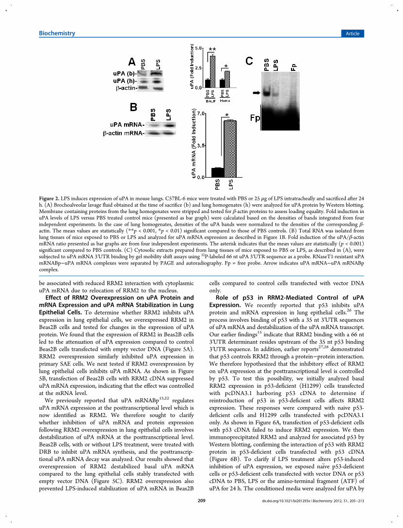
Purification of the uPA mRNABp and Determinationof Its Ability To Bind to the 66 nt uPA mRNA 3′UTRSequence. To further characterize the mechanism by whichuPA expression is controlled at the posttranscriptional level inlung epithelial cells, we purified the uPA mRNABp fromBeas2B cell lysates using conventional as well as uPA mRNAaffinity column chromatography. Purification yielded apreparation enriched with a ∼40 kDa protein having uPAmRNA binding activity, as confirmed by gel mobility shift(Figure 3A) and UV cross-linking (Figure 3B) assays, SDS-PAGE, and silver staining (Figure 3C) or Northwestern assay(Figure 3D). Mass spectrophotometric analysis and databasesearch indicated a high homology (69%) of this polypeptide toRRM2. To further evaluate the identity of the purified uPAmRNABp as RRM2, we subjected various protein fractionseluted from the Mono-Q column to NorthWestern blottingusing 32P-labeled 66 nt uPA mRNA binding sequence as aprobe and found that one specific fraction showed uPA mRNAbinding activity at the approximate molecular weight position of
Biochemistry Article
dx.doi.org/10.1021/bi201293x | Biochemistry 2012, 51, 205−213207
∼40 kDa. To confirm that the enriched fraction comigrateswith RRM2, we stripped the same membrane and subjected itto Western blotting using anti-RRM2 antibody as a probe. Asshown in Figure 3E, RRM2 antigens were indeed detected in
the same fraction that exhibited uPA 3′UTR mRNA bindingactivity.We then immunoprecipitated RRM2 proteins from the
cytoplasmic fractions of Beas2B cells treated with LPS for 0−12h. NorthWestern blotting of immunoprecipitated RRM2 using32P-labeled 66 nt uPA mRNA 3′UTR binding sequence as aprobe demonstrated that LPS treatment inhibits RRM2 bindingwith the uPA 3′UTR mRNA sequence in Beas2B cells in a time-dependent manner (Figure 3F). However, when LPS-treatedBeas2B cell lysates were tested for RRM2 expression, nosignificant inhibition of RRM2 levels by LPS was observed(Figure 3G). We next isolated RRM2 proteins usingcytoplasmic extracts prepared from the lungs of mice exposedto PBS or LPS and tested them for uPA 3′UTR mRNA bindingactivity by Northwestern assay (Figure 3H). Consistent withthe responses found in Beas2B cells, RRM2 proteins isolatedfrom mice exposed to intratracheal LPS showed reduced uPAmRNA binding activity compared to control mice exposed toPBS.
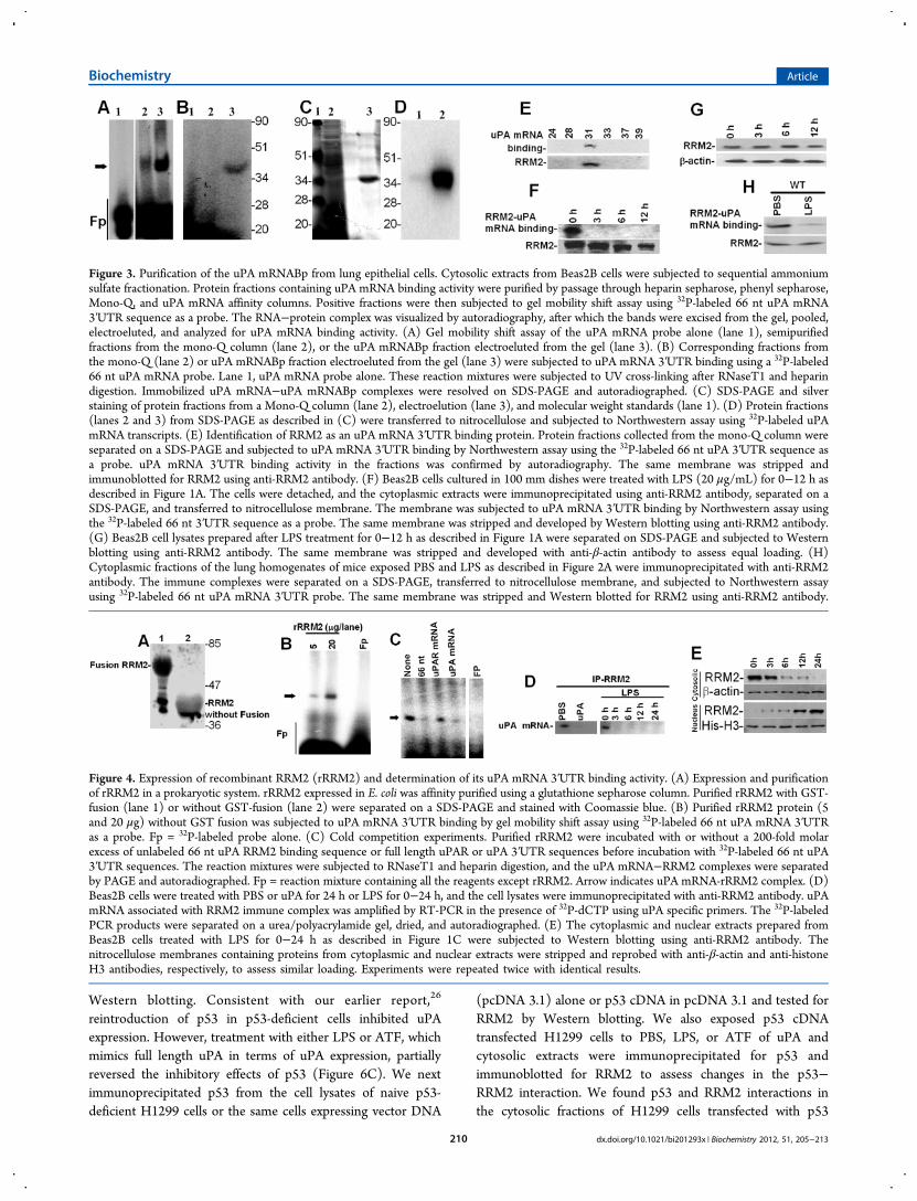
Expression of rRRM2 and Verification of Its uPAmRNA 3′UTR Binding Activity. To independently determinethat RRM2 binds to the uPA mRNA 3′UTR, we next expressedRRM2 in E. coli and affinity-purified rRRM2 using a GSTSepharose column (Figure 4A). Purified rRRM2 protein wastested for uPA mRNA 3′UTR binding activity. Gel mobilityshift assay demonstrated specific binding of rRRM2 with a 66nt 3′UTR determinant of uPA mRNA (Figure 4B). Weexpressed RRM2 in Beas2B cells and likewise confirmed itsability to specifically interact with the 66 nt uPA mRNA 3′UTRsequence. To confirm the specificity of the interaction ofRRM2 with the uPA mRNA, we incubated rRRM2 protein with32P-labeled 66 nt uPA mRNA 3′UTR sequence in the presenceof a 200-fold molar excess of unlabeled 66 nt uPA 3′UTRbinding sequence, full length uPAR, or uPA 3′UTR sequence.The results showed that rRRM2 binds to the uPA mRNA3′UTR through the 66 nt sequence, that the interaction wasspecific, and that it could be inhibited by self-competition(Figure 4C).We then sought to confirm whether RRM2 binds to
endogenous uPA mRNA in Beas2B cells and if the increaseduPA expression due to posttranscriptional uPA mRNAstabilization involves altered RRM2-uPA mRNA interaction.RRM2 protein was immunoprecipitated from the cytoplasmicextracts of Beas2B cells treated with PBS, uPA for 24 h, or LPSfor 0−24 h using nonspecific mouse IgG or an anti-RRM2monoclonal antibody. Total RNAs isolated from the mouse IgGand anti-RRM2−immune complexes were analyzed forassociated uPA mRNA by RT-PCR. As shown in Figure 4D,uPA mRNA was found to coprecipitate with the RRM2−anti-RRM2−immune complex in PBS treated Beas2B cells.Interestingly, treatment of cells with either uPA or LPSsuppressed uPA mRNA coprecipitation with the RRM2protein. No uPA mRNA was detected when immunoprecipi-tation reactions were performed with nonspecific mouse IgG,indicating that RRM2 specifically interacts with endogenousuPA mRNA. Since LPS inhibits RRM2 interaction with uPAmRNA without inhibiting RRM2 expression, we next analyzedthe cytoplasmic and nuclear extracts of Beas2B cell exposed toLPS for varying time for RRM2 by Western blotting. Ourresults showed translocation of RRM2 from the cytoplasm tothe nucleus in a temporal fashion after LPS treatment. Theseresults also suggest that increased uPA expression by lungepithelial cells following exogenous uPA or LPS treatment may
Figure 1. LPS induces time-dependent uPA expression in lungepithelial cells. Beas2B cells cultured in 100 mm dishes were treatedwith LPS (20 μg/mL) for 0−24 h. (A) The conditioned media (CM)and cell lysates (CL) of Beas2B cells were analyzed for uPA expressionby Western blotting. The lower panel shows corresponding β-actinloading controls. The line graph shows fold induction relative to time0 calculated from the densities of the individual bands from the threeWestern blots. In the case of CL, fold induction was calculated afternormalization of the densities of each uPA bands with thecorresponding densities of β-actin from three independent experi-ments. (B) Total RNA isolated from Beas2B cells treated with LPS for0, 3, and 6 h were analyzed for uPA and β-actin mRNA by RT-PCRusing 32P-labeled dCTP. 32P-labeled PCR products were separated ona urea/PAGE, dried, and autoradiographed. The fold induction relativeto time 0 was calculated from the densities of uPA mRNA bands afternormalization with that of β-actin mRNA is depicted in line graph.The asterisk indicates that the mean values from three independentexperiments are statistically significant (p < 0.0001) compared to time0. (C) Cytosolic and nuclear extracts prepared from the Beas2B cellsexposed to LPS for 0−24 h as described in (A) were subjected to uPAmRNA 3′UTR binding by gel mobility shift assay using 32P-labeled 66nt 3′UTR sequence as a probe. The reaction mixtures were incubatedwith RNaseT1 at 37 °C for 30 min followed by heparin treatment atroom temperature for 10 min. RNaseT1-resistant uPA mRNABp anduPA mRNA complexes were separated by PAGE using TBE buffer,dried, and autoradiographed. Fp = reaction mixture in cytoplasmic ornuclear extraction buffers without Beas2B cell proteins. Arrowindicates the uPA mRNA−uPA mRNABp complex.
Biochemistry Article
dx.doi.org/10.1021/bi201293x | Biochemistry 2012, 51, 205−213208
be associated with reduced RRM2 interaction with cytoplasmicuPA mRNA due to relocation of RRM2 to the nucleus.Effect of RRM2 Overexpression on uPA Protein and
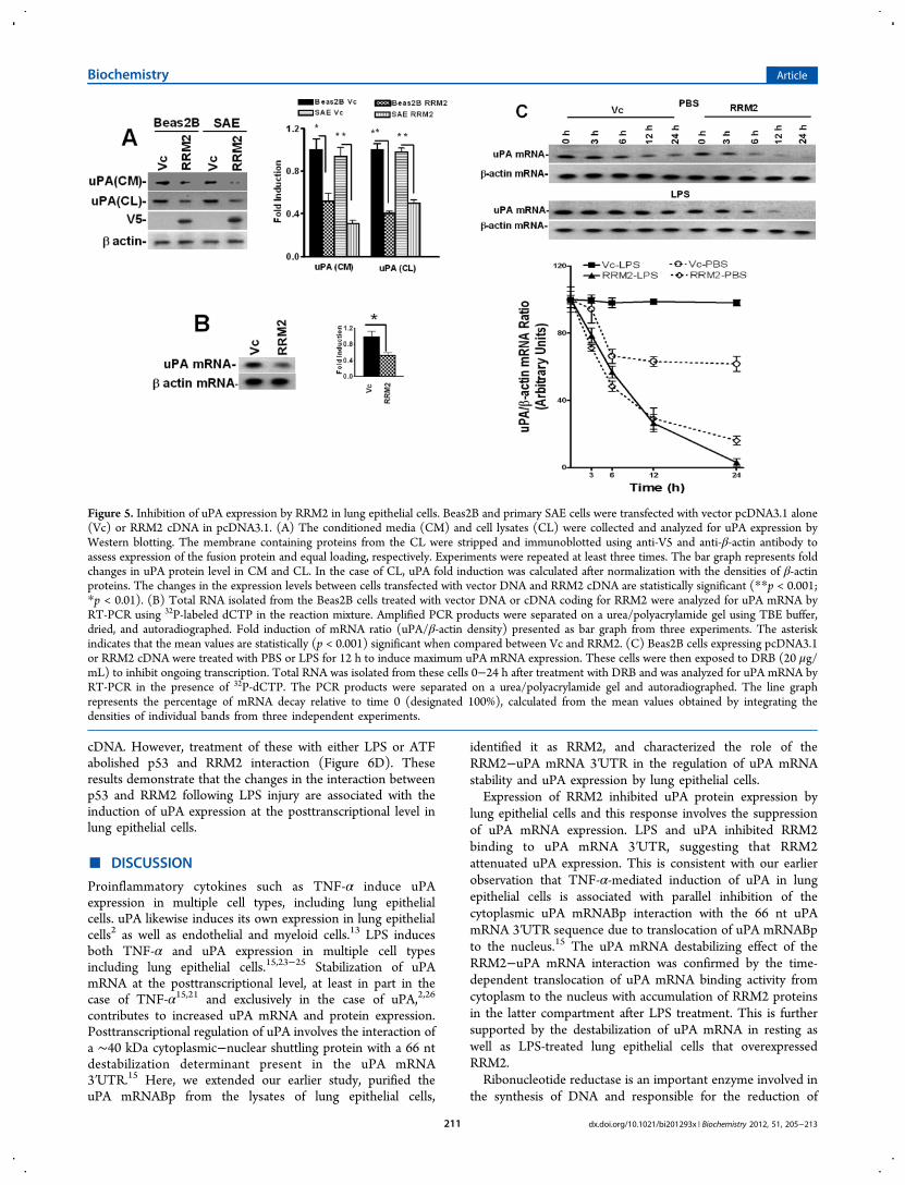
mRNA Expression and uPA mRNA Stabilization in LungEpithelial Cells. To determine whether RRM2 inhibits uPAexpression in lung epithelial cells, we overexpressed RRM2 inBeas2B cells and tested for changes in the expression of uPAprotein. We found that the expression of RRM2 in Beas2B cellsled to the attenuation of uPA expression compared to controlBeas2B cells transfected with empty vector DNA (Figure 5A).RRM2 overexpression similarly inhibited uPA expression inprimary SAE cells. We next tested if RRM2 overexpression bylung epithelial cells inhibits uPA mRNA. As shown in Figure5B, transfection of Beas2B cells with RRM2 cDNA suppresseduPA mRNA expression, indicating that the effect was controlledat the mRNA level.We previously reported that uPA mRNABp15,22 regulates
uPA mRNA expression at the posttranscriptional level which isnow identified as RRM2. We therefore sought to clarifywhether inhibition of uPA mRNA and protein expressionfollowing RRM2 overexpression in lung epithelial cells involvesdestabilization of uPA mRNA at the posttranscriptional level.Beas2B cells, with or without LPS treatment, were treated withDRB to inhibit uPA mRNA synthesis, and the posttranscrip-tional uPA mRNA decay was analyzed. Our results showed thatoverexpression of RRM2 destabilized basal uPA mRNAcompared to the lung epithelial cells stably transfected withempty vector DNA (Figure 5C). RRM2 overexpression alsoprevented LPS-induced stabilization of uPA mRNA in Beas2B
cells compared to control cells transfected with vector DNAonly.
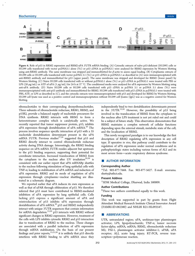
Role of p53 in RRM2-Mediated Control of uPAExpression. We recently reported that p53 inhibits uPAprotein and mRNA expression in lung epithelial cells.26 Theprocess involves binding of p53 with a 35 nt 3′UTR sequencesof uPA mRNA and destabilization of the uPA mRNA transcript.Our earlier findings15 indicate that RRM2 binding with a 66 nt3′UTR determinant resides upstream of the 35 nt p53 binding3′UTR sequence. In addition, earlier reports27,28 demonstratedthat p53 controls RRM2 through a protein−protein interaction.We therefore hypothesized that the inhibitory effect of RRM2on uPA expression at the posttranscriptional level is controlledby p53. To test this possibility, we initially analyzed basalRRM2 expression in p53-deficient (H1299) cells transfectedwith pcDNA3.1 harboring p53 cDNA to determine ifreintroduction of p53 in p53-deficient cells affects RRM2expression. These responses were compared with naive p53-deficient cells and H1299 cells transfected with pcDNA3.1only. As shown in Figure 6A, transfection of p53-deficient cellswith p53 cDNA failed to induce RRM2 expression. We thenimmunoprecipitated RRM2 and analyzed for associated p53 byWestern blotting, confirming the interaction of p53 with RRM2protein in p53-deficient cells transfected with p53 cDNA(Figure 6B). To clarify if LPS treatment alters p53-inducedinhibition of uPA expression, we exposed naiv̈e p53-deficientcells or p53-deficient cells transfected with vector DNA or p53cDNA to PBS, LPS or the amino-terminal fragment (ATF) ofuPA for 24 h. The conditioned media were analyzed for uPA by
Figure 2. LPS induces expression of uPA in mouse lungs. C57BL-6 mice were treated with PBS or 25 μg of LPS intratracheally and sacrificed after 24h. (A) Brochoalveolar lavage fluid obtained at the time of sacrifice (b) and lung homogenates (h) were analyzed for uPA protein by Western blotting.Membrane containing proteins from the lung homogenates were stripped and tested for β-actin proteins to assess loading equality. Fold induction inuPA levels of LPS versus PBS treated control mice (presented as bar graph) were calculated based on the densities of bands integrated from fourindependent experiments. In the case of lung homogenates, densities of the uPA bands were normalized to the densities of the corresponding β-actin. The mean values are statistically (**p < 0.001, *p < 0.01) significant compared to those of PBS controls. (B) Total RNA was isolated fromlung tissues of mice exposed to PBS or LPS and analyzed for uPA mRNA expression as described in Figure 1B. Fold induction of the uPA/β-actinmRNA ratio presented as bar graphs are from four independent experiments. The asterisk indicates that the mean values are statistically (p < 0.001)significant compared to PBS controls. (C) Cytosolic extracts prepared from lung tissues of mice exposed to PBS or LPS, as described in (A), weresubjected to uPA mRNA 3′UTR binding by gel mobility shift assays using 32P-labeled 66 nt uPA 3′UTR sequence as a probe. RNaseT1-resistant uPAmRNABp−uPA mRNA complexes were separated by PAGE and autoradiography. Fp = free probe. Arrow indicates uPA mRNA−uPA mRNABpcomplex.
Biochemistry Article
dx.doi.org/10.1021/bi201293x | Biochemistry 2012, 51, 205−213209
Western blotting. Consistent with our earlier report,26
reintroduction of p53 in p53-deficient cells inhibited uPAexpression. However, treatment with either LPS or ATF, whichmimics full length uPA in terms of uPA expression, partiallyreversed the inhibitory effects of p53 (Figure 6C). We nextimmunoprecipitated p53 from the cell lysates of naive p53-deficient H1299 cells or the same cells expressing vector DNA
(pcDNA 3.1) alone or p53 cDNA in pcDNA 3.1 and tested forRRM2 by Western blotting. We also exposed p53 cDNAtransfected H1299 cells to PBS, LPS, or ATF of uPA andcytosolic extracts were immunoprecipitated for p53 andimmunoblotted for RRM2 to assess changes in the p53−RRM2 interaction. We found p53 and RRM2 interactions inthe cytosolic fractions of H1299 cells transfected with p53
Figure 3. Purification of the uPA mRNABp from lung epithelial cells. Cytosolic extracts from Beas2B cells were subjected to sequential ammoniumsulfate fractionation. Protein fractions containing uPA mRNA binding activity were purified by passage through heparin sepharose, phenyl sepharose,Mono-Q, and uPA mRNA affinity columns. Positive fractions were then subjected to gel mobility shift assay using 32P-labeled 66 nt uPA mRNA3′UTR sequence as a probe. The RNA−protein complex was visualized by autoradiography, after which the bands were excised from the gel, pooled,electroeluted, and analyzed for uPA mRNA binding activity. (A) Gel mobility shift assay of the uPA mRNA probe alone (lane 1), semipurifiedfractions from the mono-Q column (lane 2), or the uPA mRNABp fraction electroeluted from the gel (lane 3). (B) Corresponding fractions fromthe mono-Q (lane 2) or uPA mRNABp fraction electroeluted from the gel (lane 3) were subjected to uPA mRNA 3′UTR binding using a 32P-labeled66 nt uPA mRNA probe. Lane 1, uPA mRNA probe alone. These reaction mixtures were subjected to UV cross-linking after RNaseT1 and heparindigestion. Immobilized uPA mRNA−uPA mRNABp complexes were resolved on SDS-PAGE and autoradiographed. (C) SDS-PAGE and silverstaining of protein fractions from a Mono-Q column (lane 2), electroelution (lane 3), and molecular weight standards (lane 1). (D) Protein fractions(lanes 2 and 3) from SDS-PAGE as described in (C) were transferred to nitrocellulose and subjected to Northwestern assay using 32P-labeled uPAmRNA transcripts. (E) Identification of RRM2 as an uPA mRNA 3′UTR binding protein. Protein fractions collected from the mono-Q column wereseparated on a SDS-PAGE and subjected to uPA mRNA 3′UTR binding by Northwestern assay using the 32P-labeled 66 nt uPA 3′UTR sequence asa probe. uPA mRNA 3′UTR binding activity in the fractions was confirmed by autoradiography. The same membrane was stripped andimmunoblotted for RRM2 using anti-RRM2 antibody. (F) Beas2B cells cultured in 100 mm dishes were treated with LPS (20 μg/mL) for 0−12 h asdescribed in Figure 1A. The cells were detached, and the cytoplasmic extracts were immunoprecipitated using anti-RRM2 antibody, separated on aSDS-PAGE, and transferred to nitrocellulose membrane. The membrane was subjected to uPA mRNA 3′UTR binding by Northwestern assay usingthe 32P-labeled 66 nt 3′UTR sequence as a probe. The same membrane was stripped and developed by Western blotting using anti-RRM2 antibody.(G) Beas2B cell lysates prepared after LPS treatment for 0−12 h as described in Figure 1A were separated on SDS-PAGE and subjected to Westernblotting using anti-RRM2 antibody. The same membrane was stripped and developed with anti-β-actin antibody to assess equal loading. (H)Cytoplasmic fractions of the lung homogenates of mice exposed PBS and LPS as described in Figure 2A were immunoprecipitated with anti-RRM2antibody. The immune complexes were separated on a SDS-PAGE, transferred to nitrocellulose membrane, and subjected to Northwestern assayusing 32P-labeled 66 nt uPA mRNA 3′UTR probe. The same membrane was stripped and Western blotted for RRM2 using anti-RRM2 antibody.
Figure 4. Expression of recombinant RRM2 (rRRM2) and determination of its uPA mRNA 3′UTR binding activity. (A) Expression and purificationof rRRM2 in a prokaryotic system. rRRM2 expressed in E. coli was affinity purified using a glutathione sepharose column. Purified rRRM2 with GST-fusion (lane 1) or without GST-fusion (lane 2) were separated on a SDS-PAGE and stained with Coomassie blue. (B) Purified rRRM2 protein (5and 20 μg) without GST fusion was subjected to uPA mRNA 3′UTR binding by gel mobility shift assay using 32P-labeled 66 nt uPA mRNA 3′UTRas a probe. Fp = 32P-labeled probe alone. (C) Cold competition experiments. Purified rRRM2 were incubated with or without a 200-fold molarexcess of unlabeled 66 nt uPA RRM2 binding sequence or full length uPAR or uPA 3′UTR sequences before incubation with 32P-labeled 66 nt uPA3′UTR sequences. The reaction mixtures were subjected to RNaseT1 and heparin digestion, and the uPA mRNA−RRM2 complexes were separatedby PAGE and autoradiographed. Fp = reaction mixture containing all the reagents except rRRM2. Arrow indicates uPA mRNA-rRRM2 complex. (D)Beas2B cells were treated with PBS or uPA for 24 h or LPS for 0−24 h, and the cell lysates were immunoprecipitated with anti-RRM2 antibody. uPAmRNA associated with RRM2 immune complex was amplified by RT-PCR in the presence of 32P-dCTP using uPA specific primers. The 32P-labeledPCR products were separated on a urea/polyacrylamide gel, dried, and autoradiographed. (E) The cytoplasmic and nuclear extracts prepared fromBeas2B cells treated with LPS for 0−24 h as described in Figure 1C were subjected to Western blotting using anti-RRM2 antibody. Thenitrocellulose membranes containing proteins from cytoplasmic and nuclear extracts were stripped and reprobed with anti-β-actin and anti-histoneH3 antibodies, respectively, to assess similar loading. Experiments were repeated twice with identical results.
Biochemistry Article
dx.doi.org/10.1021/bi201293x | Biochemistry 2012, 51, 205−213210
cDNA. However, treatment of these with either LPS or ATFabolished p53 and RRM2 interaction (Figure 6D). Theseresults demonstrate that the changes in the interaction betweenp53 and RRM2 following LPS injury are associated with theinduction of uPA expression at the posttranscriptional level inlung epithelial cells.
■ DISCUSSION
Proinflammatory cytokines such as TNF-α induce uPAexpression in multiple cell types, including lung epithelialcells. uPA likewise induces its own expression in lung epithelialcells2 as well as endothelial and myeloid cells.13 LPS inducesboth TNF-α and uPA expression in multiple cell typesincluding lung epithelial cells.15,23−25 Stabilization of uPAmRNA at the posttranscriptional level, at least in part in thecase of TNF-α15,21 and exclusively in the case of uPA,2,26
contributes to increased uPA mRNA and protein expression.Posttranscriptional regulation of uPA involves the interaction ofa ∼40 kDa cytoplasmic−nuclear shuttling protein with a 66 ntdestabilization determinant present in the uPA mRNA3′UTR.15 Here, we extended our earlier study, purified theuPA mRNABp from the lysates of lung epithelial cells,
identified it as RRM2, and characterized the role of theRRM2−uPA mRNA 3′UTR in the regulation of uPA mRNAstability and uPA expression by lung epithelial cells.Expression of RRM2 inhibited uPA protein expression by
lung epithelial cells and this response involves the suppressionof uPA mRNA expression. LPS and uPA inhibited RRM2binding to uPA mRNA 3′UTR, suggesting that RRM2attenuated uPA expression. This is consistent with our earlierobservation that TNF-α-mediated induction of uPA in lungepithelial cells is associated with parallel inhibition of thecytoplasmic uPA mRNABp interaction with the 66 nt uPAmRNA 3′UTR sequence due to translocation of uPA mRNABpto the nucleus.15 The uPA mRNA destabilizing effect of theRRM2−uPA mRNA interaction was confirmed by the time-dependent translocation of uPA mRNA binding activity fromcytoplasm to the nucleus with accumulation of RRM2 proteinsin the latter compartment after LPS treatment. This is furthersupported by the destabilization of uPA mRNA in resting aswell as LPS-treated lung epithelial cells that overexpressedRRM2.Ribonucleotide reductase is an important enzyme involved in
the synthesis of DNA and responsible for the reduction of
Figure 5. Inhibition of uPA expression by RRM2 in lung epithelial cells. Beas2B and primary SAE cells were transfected with vector pcDNA3.1 alone(Vc) or RRM2 cDNA in pcDNA3.1. (A) The conditioned media (CM) and cell lysates (CL) were collected and analyzed for uPA expression byWestern blotting. The membrane containing proteins from the CL were stripped and immunoblotted using anti-V5 and anti-β-actin antibody toassess expression of the fusion protein and equal loading, respectively. Experiments were repeated at least three times. The bar graph represents foldchanges in uPA protein level in CM and CL. In the case of CL, uPA fold induction was calculated after normalization with the densities of β-actinproteins. The changes in the expression levels between cells transfected with vector DNA and RRM2 cDNA are statistically significant (**p < 0.001;*p < 0.01). (B) Total RNA isolated from the Beas2B cells treated with vector DNA or cDNA coding for RRM2 were analyzed for uPA mRNA byRT-PCR using 32P-labeled dCTP in the reaction mixture. Amplified PCR products were separated on a urea/polyacrylamide gel using TBE buffer,dried, and autoradiographed. Fold induction of mRNA ratio (uPA/β-actin density) presented as bar graph from three experiments. The asteriskindicates that the mean values are statistically (p < 0.001) significant when compared between Vc and RRM2. (C) Beas2B cells expressing pcDNA3.1or RRM2 cDNA were treated with PBS or LPS for 12 h to induce maximum uPA mRNA expression. These cells were then exposed to DRB (20 μg/mL) to inhibit ongoing transcription. Total RNA was isolated from these cells 0−24 h after treatment with DRB and was analyzed for uPA mRNA byRT-PCR in the presence of 32P-dCTP. The PCR products were separated on a urea/polyacrylamide gel and autoradiographed. The line graphrepresents the percentage of mRNA decay relative to time 0 (designated 100%), calculated from the mean values obtained by integrating thedensities of individual bands from three independent experiments.
Biochemistry Article
dx.doi.org/10.1021/bi201293x | Biochemistry 2012, 51, 205−213211
ribonucleotides to their corresponding deoxyribonucleotides.Three subunits of ribonucleotide reductase, RRM1, RRM2, andp53R2, provide a balanced supply of nucleotide precursors forDNA synthesis. RRM2 interacts with RRM1 to form aheterotetramer complex which is catalytically active. Werecently reported that tumor suppressor protein, p53, inhibitsuPA expression through destabilization of uPA mRNA.26 Theprocess involves sequence specific interaction of p53 with a 35nucleotide destabilization determinant present in the uPAmRNA 3′UTR. Previous studies27,28 suggested that p53 andRRM2 directly interact to control ribonucleotide reductaseactivity during DNA damage. Interestingly, the RRM2 bindingsequence on uPA mRNA 3′UTR resides adjacent but upstreamto the p53 binding sequence, supporting their potential forcoordinate interaction. Increased translocation of RRM2 fromthe cytoplasm to the nucleus after UV irradiation29,30 isconsistent with our earlier report that uPA mRNABp shuttlesto the nucleus following stimulation of lung epithelial cells withTNF-α, leading to stabilization of uPA mRNA and induction ofuPA expression. RRM2 and its mode of regulation of uPAexpression through cytoplasmic−nuclear shuttling are illus-trated in a schematic diagram.We reported earlier that uPA induces its own expression as
well as that of uPAR through obliteration of p53. We thereforeinferred that p53 must have contributed to RRM2-mediatedinhibition of uPA expression by lung epithelial cells. Cellslacking p53 express a significant amount of uPA andreintroduction of p53 inhibits uPA expression throughdestabilization of uPA mRNA.26 p53 and RRM2 independentlyinteract with unique 3′UTR sequences that contain informationfor mRNA degradation.12,26 p53 interacts with RRM2 withoutsignificant changes in RRM2 expression. However, treatment ofthe cells with LPS inhibits cytosolic RRM2 and p53 interactiondue to translocation of RRM2 to the nucleus. These changesare associated with a parallel induction of uPA expressionthrough mRNA stabilization. On the basis of our presentfindings and prior reports,15,21,26 it is unlikely that p53 directlyinterferes with RRM2 binding to uPA mRNA since they
independently bind to two destabilization determinants presentin the 3′UTR.15,26 However, the possibility of p53 beinginvolved in the translocation of RRM2 from the cytoplasm tothe nucleus after LPS treatment is not yet ruled out and couldbe a subject of future study. This observation demonstrates thatRRM2 maintains a complex network of cellular functionsdepending upon the external stimuli, metabolic state of the cell,and the localization of RRM2.This newly recognized paradigm is to our knowledge the first
description of RRM2 regulating uPA expression in any celltype. If operative in vivo, this pathway could contribute to theregulation of uPA expression under normal conditions and inpathophysiologic states including various forms of ALI and itsmost severe formacute respiratory distress syndrome.
■ AUTHOR INFORMATION
Corresponding Author*Tel: 903-877-7668. Fax: 903-877-5627. E-mail: [email protected].
Present Address⊥SDM Medical College, Dharwad, India 580009.
Author Contributions∥These two authors contributed equally to this work.
FundingThis work was supported in part by grants from FlightAttendant Medical Research Institute Clinical Innovator Award(FAMRI-ID-082380) and NHLBI R21-HL093547.
■ ABBREVIATIONS
UTR, untranslated region; uPA, urokinase-type plasminogenactivator; LPS, lipopolysacharide; TNF-α, tumor necrosisfactor-alpha; mRNA, mRNA; RRM2, ribonucleotide reductaseM2; PAI-1, plasminogen activator inhibitor-1; uPAR, uPAreceptor; ALI, acute lung injury; RT-PCR, reverse tran-scription−polymerase reaction.
Figure 6. Role of p53 in RRM2 expression and RRM2-uPA 3′UTR mRNA binding. (A) Cytosolic extracts of naiv̈e p53-deficient (H1299) cells orH1299 cells transfected with vector pcDNA3.1 alone (Vc) or p53 cDNA in pcDNA3.1 were analyzed for RRM2 expression by Western blottingusing anti-RRM2 antibody. The same membrane was stripped and immunoblotted for β-actin to assess equal loading. (B) Cytoplasmic extracts ofH1299 cells or H1299 cells transfected with vector pcDNA 3.1 (Vc) or p53 cDNA in pcDNA3.1 as described in (A) were immunoprecipitated withanti-RRM2 antibody and immunoblotted for p53 (upper panel). The same membrane was stripped and developed for RRM2 (lower panel) byWestern blotting. (C) Naive H1299 cells transfected with or without pcDNA3.1 alone (Vc) or p53 cDNA in pcDNA3.1 were treated with PBS orLPS (20 μg/mL) or ATF of uPA (1 μg/mL) for 24 h at 37 °C. The conditioned media were analyzed for uPA expression by Western blotting usinganti-uPA antibody. (D) Naive H1299 cells or H1299 cells transfected with p53 cDNA in pcDNA 3.1 or pcDNA 3.1 alone (Vc) wereimmunoprecipitated with anti-p53 antibody and immunoblotted for RRM2. H1299 cells transfected with p53 cDNA in pcDNA3.1 were treated withPBS, ATF, or LPS as described in (C), and the cytosolic extracts were immunoprecipitated with p53 and developed for RRM2 by Western blotting.H1299 cell lysate was used as a positive control and immunoprecipitation without H1299 cell lysates (IgG) was as a negative control for Westernblotting.
Biochemistry Article
dx.doi.org/10.1021/bi201293x | Biochemistry 2012, 51, 205−213212
■ REFERENCES(1) Dumler, I., Petri, T., and Schleuning, W. D. (1993) Interaction ofurokinase-type plasminogenactivator (u-PA) with its cellular receptor(u-PAR) induces phosphorylation on tyrosine of a 38 kda protein.FEBS Lett. 322, 37−40.(2) Shetty, S., Pendurthi, U. R., Halady, P. K., Azghani, A. O., andIdell, S. (2002) Urokinase induces its own expression in Beas2b lungepithelial cells. Am. J. Physiol Lung Cell Mol. Physiol 283, L319−L328.(3) Koopman, J. L., Slomp, J., de Bart, A. C., Quax, P. H., andVerheijen, J. H. (1998) Effects of urokinase on melanoma cells areindependent of high affinity binding to the urokinase receptor. J. Biol.Chem. 273, 33267−33272.(4) Koshelnick, Y., Ehart, M., Hufnagl, P., Heinrich, P. C., andBinder, B. R. (1997) Urokinase receptor is associated with thecomponents of the JAK1/STAT1 signaling pathway and leads toactivation of this pathway upon receptor clustering in the humankidney epithelial tumor cell line tcl-598. J. Biol. Chem. 272, 28563−28567.(5) Bhat, G. J., Gunaje, J. J., and Idell, S. (1999) Urokinase-typeplasminogen activator induces tyrosine phosphorylation of a 78-kDaprotein in H-157 cells. Am. J. Physiol. 277, L301−L309.(6) Shetty, S., Bdeir, K., Cines, D. B., and Idell, S. (2003) Inductionof plasminogen activator inhibitor-1 by urokinase in lung epithelialcells. J. Biol. Chem. 278, 18124−18131.(7) Abraham, E., Gyetko, M. R., Kuhn, K., Arcaroli, J., Strassheim, D.,Park, J. S., Shetty, S., and Idell, S. (2003) Urokinase-type plasminogenactivator potentiates lipopolysaccharide-induced neutrophil activation.J. Immunol. 170, 5644−5651.(8) Abraham, E., Matthay, M. A., Dinarello, C. A., Vincent, J. L.,Cohen, J., Opal, S. M., Glauser, M., Parsons, P., Fisher, C. J. Jr., andRepine, J. E. (2000) Consensus conference definitions for sepsis, septicshock, acute lung injury, and acute respiratory distress syndrome: timefor a reevaluation. Crit. Care Med. 28, 232−235.(9) Welty-Wolf, K. E., Carraway, M. S., Ortel, T. L., Ghio, A. J., Idell,S., Egan, J., Zhu, X., Jiao, J. A., Wong, H. C., and Piantadosi, C. A.(2006) Blockade of tissue factor-factor X binding attenuates sepsis-induced respiratory and renal failure. Am. J. Physiol. Lung Cell Mol.Physiol. 290, L21−L31.(10) Robbie, L. A., Dummer, S., Booth, N. A., Adey, G. D., andBennett, B. (2000) Plasminogen activator inhibitor 2 and urokinase-type plasminogen activator in plasma and leucocytes in patients withsevere sepsis. Br. J. Hamaetol. 109, 342−348.(11) Bhandary, Y. P., Velusamy, T., Shetty, P., Shetty, R. S., Idell, S.,Cines, D. B., Jain, D., Bdeir, K., Abraham, E., Tsuruta, Y., and Shetty, S.(2009) Post-transcriptional regulation of urokinase-type plasminogenactivator receptor expression in lipopolysaccharide-induced acute lunginjury. Am. J. Respir. Crit. Care Med. 179, 288−298.(12) Shetty, S., and Idell, S. (2001) Urokinase induces expression ofits own receptor in Beas2b lung epithelial cells. J. Biol. Chem. 276,24549−24556.(13) Li, C., Zhang, J., Jiang, Y., Gurewich, V., Chen, Y., and Liu, J. N.(2001) Urokinase-type plasminogen activator up-regulates its ownexpression by endothelial cells and monocytes via the u-PAR pathway.Thromb. Res. 103, 221−232.(14) Gyetko, M. R., Aizenberg, D., and Mayo-Bond, L. (2004)Urokinase-deficient and urokinase receptor-deficient mice haveimpaired neutrophil antimicrobial activation in vitro. J. Leukoc. Biol.76, 648−656.(15) Shetty, S., and Idell, S. (2000) Identification of a novelurokinase mRNA-binding protein in human lung epithelial cells invitro. J. Biol. Chem. 275, 13771−13779.(16) Shetty, S., Velusamy, T., Shetty, R. S., Marudamuthu, A. S.,Shetty, S. K., Florova, G., Tucker, T., Koenig, K., Shetty, P., Bhandary,Y. P., and Idell, S. (2010) Post-transcriptional regulation ofplasminogen activator inhibitor type-1 expression in human pleuralmesothelial cells. Am. J. Respir. Cell Mol. Biol. 43, 358−367.(17) Shetty, S., Ganachari, M., Liu, M. C., Azghani, A., Muniyappa,H., and Idell, S. (2005) Regulation of urokinase receptor expression by
phosphoglycerate kinase is independent of its catalytic activity. Am. J.Physiol. Lung Cell Mol. Physiol. 289, L591−L598.(18) Shetty, S., Muniyappa, H., Halady, P. K., and Idell, S. (2004)Regulation of urokinase receptor expression by phosphoglyceratekinase. Am. J. Respir. Cell Mol. Biol. 31, 100−106.(19) Shetty, S., Kumar, A., and Idell, S. (1997) Posttranscriptionalregulation of urokinase receptor mRNA: identification of a novelurokinase receptor mRNA binding protein in human mesotheliomacells. Mol. Cell. Biol. 17, 1075−1083.(20) Erbacher, P., Remy, J. S., and Behr, J. P. (1999) Gene transferwith synthetic virus-like particles via the integrin-mediated endocytosispathway. Gene Ther. 6, 138−145.(21) Shetty, S. (2002) Cytoplasmic-nuclear shuttling of the urokinasemRNA binding protein regulates message stability. Mol. Cell. Biochem.237, 55−67.(22) Shetty, S. (2005) Protein synthesis and urokinase mRNAmetabolism. Mol. Cell. Biochem. 271, 13−22.(23) Philippe, J., Offner, F., Declerck, P. J., Leroux-Roels, G.,Vogelaers, D., Baele, G., and Collen, D. (1991) Fibrinolysis andcoagulation in patients with infectious disease and sepsis. Thromb.Haemost. 65, 291−295.(24) Wygrecka, M., Markart, P., Ruppert, C., Kuchenbuch, T., Fink,L., Bohle, R. M., Grimminger, F., Seeger, W., and Gunther, A. (2004)Compartment- and cell-specific expression of coagulation andfibrinolysis factors in the murine lung undergoing inhalational versusintravenous endotoxin application. Thromb. Haemost. 92, 529−540.(25) Kwak, S. H., Mitra, S., Bdeir, K., Strassheim, D., Park, J. S., Kim,J. Y., Idell, S., Cines, D., and Abraham, E. (2005) Abraham E. Thekringle domain of urokinase-type plasminogen activator potentiatesLPS-induced neutrophil activation through interaction with {alpha}-v{beta}3 integrins. J. Leukoc. Biol. 78, 937−945.(26) Shetty, P., Velusamy, T., Bhandary, Y. P., Shetty, R. S., Liu, M.C., and Shetty, S. (2008) Urokinase expression by tumor suppressorprotein p53: a novel role in mRNA turnover. Am. J. Respir. Cell Mol.Biol. 39, 364−372.(27) Tanaka, H., Arakawa, H., Yamaguchi, T., Shiraishi, K., Fukuda,S., Matsui, K., Takei, Y., and Nakamura, Y. (2000) A ribonucleotidereductase gene involved in a p53-dependent cell-cycle checkpoint forDNA damage. Nature 404, 42−49.(28) Zhang, Y. W., Jones, T. L., Martin, S. E., Caplen, N. J., andPommier, Y. (2009) Implication of checkpoint kinase-dependent up-regulation of ribonucleotide reductase R2 in DNA damage response. J.Biol. Chem. 284, 18085−18095.(29) Zhou, B., Liu, X., Mo, X., Xue, L., Darwish, D., Qiu, W., Shih, J.,Hwu, E. B., Luh, F., and Yen, Y. (2003) The human ribonucleotidereductase subunit hRRM2 complements p53R2 in response to UV-induced DNA repair in cells with mutant p53. Cancer Res. 63, 6583−6594.(30) Xue, L., Zhou, B., Liu, X., Qiu, W., Jin, Z., and Yen, Y. (2003)Wild-type P53 regulates human ribonucleotide reductase by protein-protein interaction with P53R2 as well as hRRM2 subunits. Cancer Res.63, 980−986.
Biochemistry Article
dx.doi.org/10.1021/bi201293x | Biochemistry 2012, 51, 205−213213