Embed Size (px)
Citation preview
REH2 helicase RNPs and motifs for unwinding and gRNA binding
1
REH2 RNA Helicase in Kinetoplastid Mitochondria: Ribonucleoprotein Complexes and Essential Motifs for Unwinding
and Guide RNA Binding Alfredo Hernandez*1, Bhaskara Reddy Madina*1, Kevin Ro2, James A. Wohlschlegel2, Belinda
Willard3, Mike T. Kinter3& and Jorge Cruz-Reyes¶1 1Department of Biochemistry and Biophysics, Texas A&M University, 2128 TAMU, College Station, Texas 77843; 2Department of Biological Chemistry, David Geffen School of Medicine at UCLA, Box 951737, BSRB-377A, 615 Charles E. Young Drive South, Los Angeles, CA 90095-1737; 3Department of Cell Biology / NC10. Cleveland Clinic Foundation, 9500 Euclid Ave Cleveland, OH 44195. & Current address: Free Radical Biology and Aging Research Program, MS 21 Oklahoma, Medical Research Foundation , 825 N.E. 13th Street Oklahoma City, Oklahoma 73104. * These authors contributed equally to this work. ¶ Corresponding author, Phone: (979) 458-3375, Fax: (979) 862-4718, Email: [email protected]
Regulation of gene expression in kinetoplastid mitochondria is largely postranscriptional and involves the orchestration of polycistronic RNA processing, 3’ terminal maturation, RNA editing, turnover and translation, however these processes remain poorly studied. Core editing complexes and their U-insertion/deletion activities are relatively well characterized and a battery of ancilliary factors has recently emerged. This study characterized a novel DExH-box RNA helicase, termed here REH2 (RNA editing associated helicase 2), in unique ribonucleoprotein complexes that exhibit unwinding and guide RNA binding activities, both of which required a double-stranded RNA-binding domain (dsRBD) and a functional helicase motif I of REH2. REH2 complexes and recently identified related particles share a multi-protein core but are distinguished by several differential polypeptides. Finally, REH2 associates transiently, via RNA, with editing complexes, mitochondrial ribosomes and several ancilliary factors that control editing and RNA stability. We propose that these putative higher-order structures coordinate mitochondrial gene expression.
Unique gene expression mechanisms in
kinetoplastid flagellates include U-insertion/deletion RNA editing by concerted cycles of cleavage, U-addition/removal and ligation that can create hundreds of amino acid codons in most mitochondrial mRNAs (1,2). The RNA editing core complex (RECC) contains 18-to-20 subunits (3-6), although a few subunits seem to exchange in substrate-specific variants of this complex (7). The RECC acronym was recently introduced by Larry Simpson et al. (In press). Editing complexes
recognize partial helices between pre-mRNA and complementary guide RNAs (gRNAs) initially stabilized by a short “anchor” duplex (6,8,9). Substrate determinants for duplex binding and nuclease-specificity (6,10,11), and substrate structure in solution (12-14) have been characterized.
Several accessory factors, mostly in multi-subunit arrays, have been proposed to modulate RNA editing during catalysis, substrate production or RNA turnover. The MRP complex has RNA annealing activity in vitro and may promote mRNA and gRNA pairing (15,16). Postranscriptional mRNA terminal 3’-polyA/U and gRNA 3’-polyU maturation is mediated by KPAP1 and RET1 complexes (17,18). MRB1, TbRGG1 and GRBC complexes proposed to contain between 14 and 24 proteins (termed here MRB-related complexes) share several components but their functional relationship remains unclear. Repression of a few common subunits inhibited RNA editing, and in some cases also decreased the level of total gRNA. GRBC1 and GRBC2 co-purified with RECC subunits (18-24). MERS1, MRP and RBP16 proteins were associated with mRNA stability (23,25). RBP16 also stimulated RNA insertion in vitro (26,27). DEAD-box mHel61 (also termed REH1) is the only predicted helicase known to impact RNA editing (28). Most of these proteins are likely to have additional roles outside editing. RNA helicases are common across species and typically multifunctional however only a few examples have been studied in mitochondria. This work characterized the protein and RNA interactions of a factor REH2 (Tb927.4.1500) that we initially found in native editing complexes of T. brucei. REH2 has a conserved dsRNA-binding (dsRBD) and DExH-helicase domains, and forms novel ribonucleoprotein complexes (RNPs)
http://www.jbc.org/cgi/doi/10.1074/jbc.M109.051862The latest version is at JBC Papers in Press. Published on October 22, 2009 as Manuscript M109.051862
Copyright 2009 by The American Society for Biochemistry and Molecular Biology, Inc.
by guest on June 12, 2019http://w
ww
.jbc.org/D
ownloaded from
REH2 helicase RNPs and motifs for unwinding and gRNA binding
2
containing helicase activity, gRNA and a protein array that overlaps with MRB-related complexes. The integrity of REH2 RNPs and their helicase and gRNA-binding activities require the dsRBD. REH2 associates, via RNA, with RECC, a battery of accessory editing factors and mitochondrial ribosomes; thereby, we propose that REH2 RNPs are integral components of RNA-linked supramolecular networks that orchestrate the expression of the mitochondrial genome.
Experimental procedures
TAP and RNAi constructs, and site-directed mutagenesis of REH2- A TAP-REH2 construct was made by PCR amplification of the entire ORF from procyclic genomic DNA using a proofreading thermostable polymerase mix (AccuTaq-Sigma), and cloning into the Xho I and Bam HI sites of pLew79-ada-TAP. PCR-based site-directed mutagenesis was performed directly on this plasmid to alter the helicase motif I with oligonucleotides F-REH2-mI and R-REH2-mI, and delete the dsRBD with oligonucleotides F-dsRBD-Δ and R-dsRBD-Δ. An RNAi construct was obtained by cloning an REH2 fragment of 1665 bp into p2T7-177 (29). All constructs were confirmed by DNA sequencing, linearized with Not I and transfected in procyclic 29-13 trypanosomes (30). REH2-TAP expression and RNAi were induced with tetracycline at 1 ug/ml.
Protein purification and analysis- Native editing complexes were purified by ion-exchange chromatography from mitochondrial extracts (3,6) and TAP-purifications were performed essentially as reported (31) with some modifications. Sedimentation fractions were obtained from freshly made mitochondrial or whole-cell extracts in 10-30% glycerol gradients. While our protocols to prepare the extracts include DNaseI, a sample indicated in the text was subjected to an extra DNase treatment (DNA-free kit, Ambion) prior to sedimentation. Catalase and thyroglobulin were used as ~10S and ~20S markers, and western blots of Tbmp45 (formerly termed REAP1) to determine the ~40S region (32). Affinity-purified REH2 antibodies were produced against the peptide CSHTPTTSAEAGGDS (Bethyl laboratories, Inc). IPs of endogenous and ectopic REH2 used antibody-conjugated protein A-dynabeads (Invitrogen). Ectopic REH2 was specifically immunopurified using anti-rabbit IgG dynabeads (Invitrogen). All washes were performed at 150 mK KCl. For mass spectrometry analyses the antibodies
were crosslinked to the beads with 25 mM DMP (dimethylpimelimidate) in 0.2 M triethanolamine, pH 8.2.
Enzymatic assays and photo-crosslinking- The conditions and substrates to assay for full-round (33) and pre-cleaved (1) editing were as described. Photo-reactive substrates containing a single thio-U and 32P at the editing site were prepared (33,34) and gRNA labeling was performed (35) as reported. RNA helicase assays used a dsRNA substrate consisting of the pre-mRNA fragment A6-tag annealed to the cognate gRNA gA6[14] (9). The dephosphorylated mRNA was 5’-end labeled with [γ 32P]-ATP, and annealed with a 10-fold excess of gRNA in RNA folding buffer (25 mM Tris-HCl, pH 8.0, 250 mM KCl, 10 mM Mg(OAc)2, 0.5 mM EDTA) by incubation at 95oC for 10 min followed by a gradual return to room temperature over the course of 2 hours. The annealed form was purified by native gel electrophoresis in a 8% polyacrylamide gel in 1X TBE supplemented with 10 mM Mg(OAc)2. The standard RNA helicase assay consisted of 50 cps (~10 fmols) of dsRNA in 25 mM Tris-HCl, 22 mM KCl, 10 mM Mg(OAc)2, 0.5 mM EDTA, 3 mM DTT, 1 U/µL RNase inhibitor, 1 mM ATP, 50 ng/µL BSA, a 20-fold excess of an unlabeled trap ssRNA that complements ~33 bp of gRNA and 10 µL of beads in a final volume of 20 µL. Reactions were incubated for 30-60 min at 26oC with constant flicking to mix the beads. This was followed by addition of 4 µL 6X stop solution: 0.12 % Xylene cyanol, 0.12% bromophenol blue, 3% SDS, 125 ng/µL proteinase K, 17 % glycerol and incubation at room temperature for 10 minutes. Samples were then loaded onto a 8% polyacrylamide gel, 1X TBE supplemented with 10 mM Mg(OAc)2. Protein RNA photo-crosslinking was performed as described (34) except that it was scaled-up 10-fold. Denaturation of complexes was accomplished by the addition of SDS to 1% final, and sequential incubations at 950C for 10 min and at 700C for 30-60 min. After allowing the sample to reach room temperature, triton-X-100 was added to 5%, and incubated 10 min at room temperature. Samples were then passed through a gel filtration spin column (Bio-spin 6, Bio-Rad 732 6221) according to the manufacturer’s instructions and immunoprecipitated as above.
RNase treatment of TAP purifications and immuno-precipitations- Samples coupled to Dynabeads where treated with the following nucleases as indicated in the text at the given final concentration: RNase A (0.1u/µL), T1 (0.125
by guest on June 12, 2019http://w
ww
.jbc.org/D
ownloaded from
REH2 helicase RNPs and motifs for unwinding and gRNA binding
3
U/µL), V1 (0.001/µL) and micrococcal nuclease (0.03 U/µL) for 60 min in ice.
Mass spectrometry- TCA precipitates were resuspended in digestion buffer (100 mM Tris-HCl, pH 8.5, 8M urea) and digested by the sequential addition of lys-C and trypsin proteases as described (36). Digested samples were fractionated using a 5-step online separation method during which peptides were eluted directly into a LTQ-Orbitrap mass spectrometer (Thermo Fisher) in which tandem mass spectra were collected (37-39). SEQUEST and DTASelect algorithms were used to identify peptides sequences from tandem mass spectra (40,41). Proteins were considered present in a sample if at least two peptides were identified per protein using a peptide level false positive rate of 5% as determined using a decoy database strategy (42).
Note: Detailed protocols and oligonucleotide sequences used during this work are available upon request.
RESULTS A DExH-box helicase associates with RNA
editing complexes— In a mass spectrometric analysis of native editing complexes purified from T. brucei mitochondria we detected multiple unique peptides of most RECC subunits and the accessory MRP factors (6). However, we also found a single peptide for a 241-kDa protein, termed REH2, with highly conserved DExH-helicase domains, a double-stranded RNA binding domain (dsRBD) and an N-terminal mitochondrial import sequence (Fig. 1A). Western blot analyses clearly detected a ca. 250 kDa protein in native but not in tandem affinity-purified (TAP) editing complexes (Fig. 1B), although a weak signal was apparent in TAP-REL1 complexes (see the middle panel). This suggested a transient interaction of REH2 with RECC that is disrupted during high-stringency affinity purifications. Silver staining of native editing complexes did not evidently detect components near 250 kDa suggesting that REH2 may be sub-stoichiometric relative to RECC subunits (Fig. 1C).
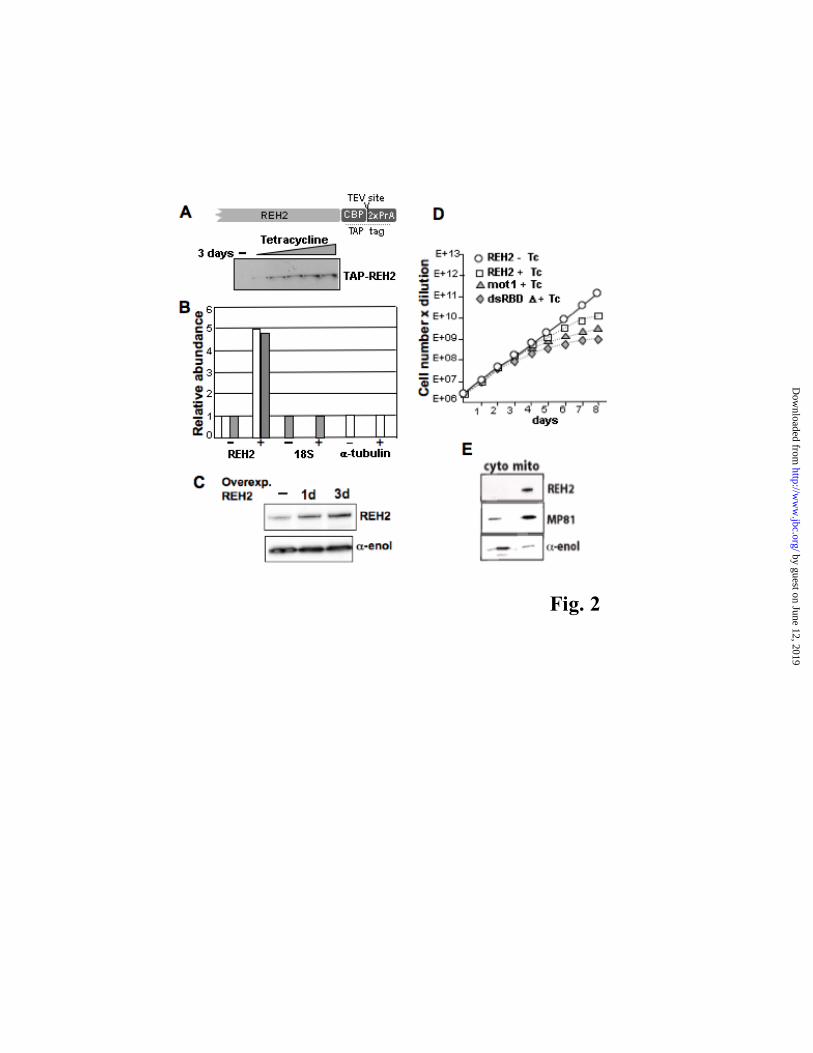
To confirm the physical interaction of REH2 with editing subunits, we expressed in procyclic trypanosomes the complete 6504-nt REH2 gene with a C-terminal TAP-tag [calmodulin binding peptide (CBP)-tobacco etch virus (TEV)-protein A (ProtA)] (31) under the control of a tetracycline-inducible promoter (Fig. 2A). REH2 mRNA and protein increased 5-fold and 2-to-3 fold,
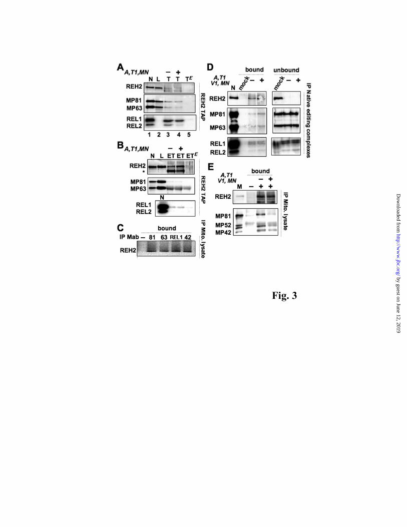
respectively, at day 3 of induction (Figs. 2B-C) and a slight reduction in cell growth was seen at day 5 (Fig. 2D). Consistent with its predicted mitochondrial localization (Fig. S1) REH2 was enriched in a mitochondrial lysate (Fig. 2E) (43). REH2 antibodies reacted well with purified native editing complexes and mitochondrial lysates but weakly with TAP pull-downs due to a partial occlusion of the tag and consequent low purification efficiency from mitochondrial lysates. A small amount of RECC was detected in both TEV (T) (Fig. 3A) and concentrated EGTA (ET) (Fig. 3B) eluates but not in mock pull-downs from empty-vector control cells (last lane in each panel). Interestingly, this low level of RECC in the eluates was resistant to a pre-treatment with RNases A, T1 and micrococal nuclease (MN). In a converse approach, REH2 was detected in mitochondrial extract pulldowns of several RECC subunits (Fig. 3C). Also, REH2 antibodies immunoprecipitated a small fraction of pre-isolated native editing complexes after an extensive RNase-MN treatment including the dsRNA-specific RNase V1. However, most RECC remained in the unbound fraction (Fig. 3D), suggesting that relatively few purified complexes were stably bound to REH2.
Besides the above examination of affinity-purified eluates and isolated native editing complexes, we further analyzed the REH2/RECC association in mitochondrial lysates. Importantly, while RECC subunits were present in nuclease-treated REH2 pulldowns, most RECC was released by the treatment (Fig. 3E). This suggests that the transient association observed is largely mediated by RNA. Also, in line with transient contacts, REH2 purifications exhibited some pre-cleaved but not full-round editing activity, which is less sensitive due to a limiting cleavage step (Fig. S2).
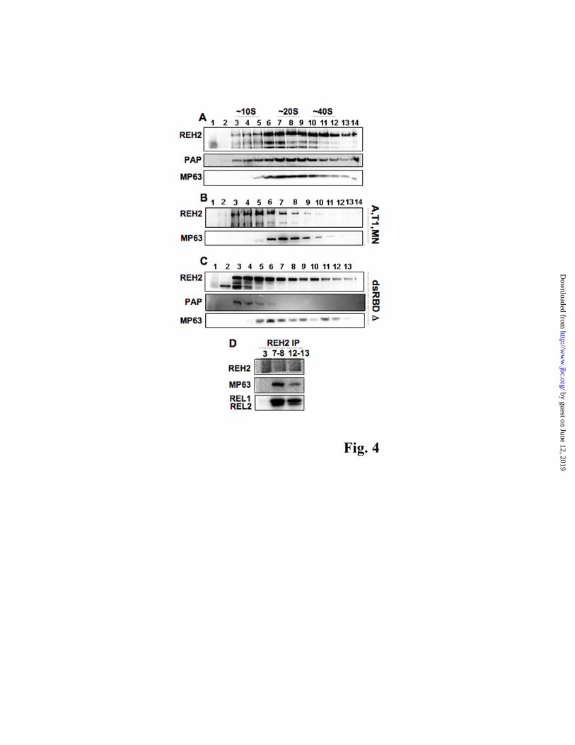
Heterodisperse REH2-associated complexes are stabilized by RNA and the dsRBD— Sedimentation analyses of mitochondrial lysates showed significant heterodispersion of REH2 with a broad peak at ~20-30S. Western blots of these fractions with REH2 antibodies and the PAP reagent (to score ectopic REH2 only) showed that the tag does not affect the sedimentation of REH2 (Fig. 4A, top and middle panels). The RECC subunit MP63 was also dispersed but in contrast to REH2 it was not detected in light fractions (Fig. 4A, bottom panel). Interestingly, REH2 at ~20S and >40S co-immunopurified with RECC subunits (see ahead Fig. 4D). A pretreatment of the mitochondrial lysate with RNases/MN disrupted most high S-value REH2 complexes generating a discrete peak
by guest on June 12, 2019http://w
ww
.jbc.org/D
ownloaded from
REH2 helicase RNPs and motifs for unwinding and gRNA binding
4
at ~15S, whereas a significantly sharpened peak of editing complexes remained at ~20S (Fig. 4B). As described above, RNase treatment eliminated most REH2 association with RECC (Fig. 3E). To examine the relevance of the conserved dsRBD we expressed a construct with a deletion of the entire motif (dsRBD-Δ) (Fig. S3). Notably, this construct severely compromised cell growth (Fig. 2D) and reduced the sedimentation of both endogenous and ectopic REH2 to an extent comparable to the RNase treatment (Fig. 4C). Finally, we established that DNA does not largely contribute to the observed broad sedimentation of REH2 in a sample treated with DNase (Procedures section; data not shown). Thus, REH2 forms heterodisperse particles that include editing complexes and are stabilized by RNA and the dsRBD, as well as relatively low-density particles that resist RNase.
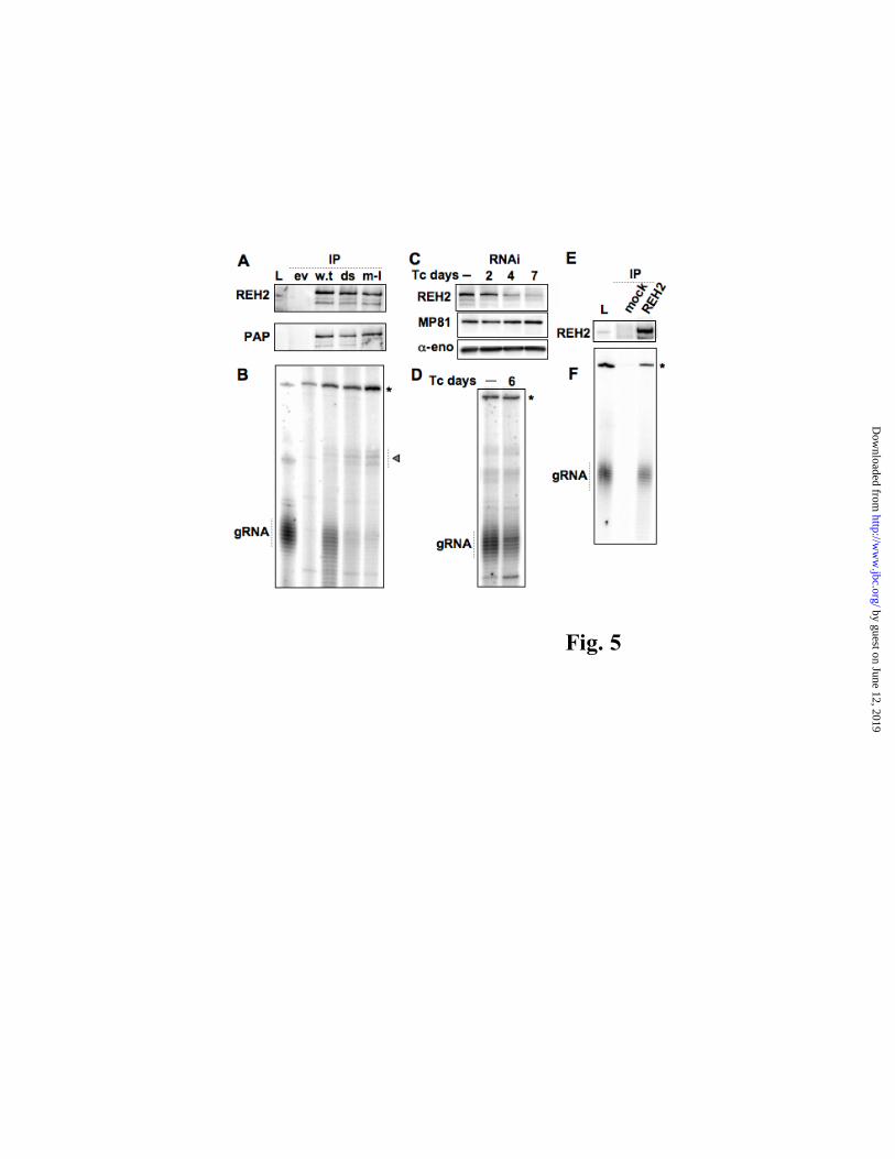
REH2 co-purifies with gRNA in a manner dependent on the dsRBD and helicase motif I— Weng and collaborators recently reported that GRBC complexes, which co-purified with REH2, bind gRNA (23). We determined whether REH2 immunopurified complexes associate with gRNA, and further examined the importance of conserved domains of this protein. To this end, we analyzed IgG-dynabeads pulldowns of ectopically expressed REH2 wild-type and mutants dsRBD-Δ or motif I (GK-to-AQ) (Fig. 1A). The motif I mutated residues have been associated with ATP binding and hydrolysis in other DExH proteins (44). While a significant amount of total gRNA co-purified with wild-type REH2, little if any was associated with either mutant (Figs. 5A-B). It is of interest, however, that relatively large RNA species accumulated in both mutants. Furthermore, as shown by Hashimi et al. (22), RNAi downregulation of REH2 decreased the steady-state levels of gRNA (Figs. 5C-D). Importantly, REH2 pull-downs from wild-type cells contained gRNA, demonstrating that endogenous REH2 RNPs bind gRNA, and that this association is not an artifact of overexpression (Figs. 5E-F). Thus, gRNA-binding by REH2 RNPs requires the dsRBD and wild-type motif I of REH2.
REH2 is associated with RNA helicase activity and appears to photo-crosslink with RNA— We examined the REH2 IP pull-down from mitochondrial extracts for possible RNA helicase activity and RNA photo-crosslinking. Notably, a model A6 pre-edited mRNA, pre-annealed with cognate gRNA, was efficiently unwound by the REH2 pull-down in a reaction requiring ATP
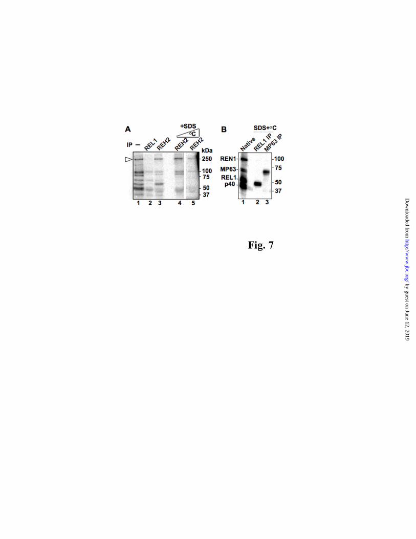
hydrolysis at its β−γ phosphodiester linkage (Fig. 6A). Although the dsRNA substrate in these assays was gel-isolated, some dissociation is visible in input and mock lanes in absence of REH2 complexes. A continuous duplex with 3’ overhangs was also unwound (Fig. 6B) but not an identical helix with 5’ or no overhangs (not shown), consistent with the substrate specificity of the vast majority of SF2 RNA helicases with the exception of DEAD-box proteins (45). Importantly, REH2 pull-downs from the dsRBD-Δ and motif I cell lines had no detectable unwinding activity (Fig. 6C). We analyzed the sedimentation distribution of this helicase activity relative to REH2 and the DEAD-box helicase REH1. Interestingly, most helicase activity sedimented in fractions containing REH2, but away from REH1 which localizes at the top of the gradient in fractions 1-3 (Fig. 6D-F) (28). An REH2 pull-down of fraction 8 exhibited unwinding activity (data not shown). Together with our above studies of REH2 mutants, this data suggest that REH2 is linked with the observed RNA helicase activity. To examine the possibility that REH2 may directly bind RNA we crosslinked a protein fraction enriched in RECC (3) with the pre-mRNA/gRNA substrate described above but substituted with a photo-reactive thio-U and 32P at the editing site (34). This protein fraction produced a crosslink at ~250 kDa (Fig. 7A, lane 1) that was enriched in a pulldown with REH2 but not REL1 antibodies (lanes 2 and 3). Importantly, REH2 pulldowns of crosslinked reactions that were treated with SDS and increasing temperature to dissociate the RNPs further enriched the ~250 kDa crosslink (lanes 4-5), suggesting that the reacting protein is REH2. As a proof of concept for the above denaturation protocol (Fig. 7B) we isolated the RNA photo-crosslinked RECC subunits REL1 (lane 2) and reported MP63 (lane 3)(6,34) using specific antibodies against these proteins. Additional studies are needed to confirm that the ~250 kDa crosslink represents direct binding by REH2, but these data suggest that REH2 contacts the RNA duplex near the photo-reactive moiety in the model editing site (≤4Å)(46).
At least seven RNase/MN-resistant REH2 associated proteins are also present in MRB1, TbRGG1 and GRBC complexes— We performed mass spectrometric studies of REH2 IP pull-downs of both purified native editing complexes and 20-30S sedimentation fractions (num. 7-10 in Fig. 4A), the latter before or after RNase/MN treatment. A large number of REH2 unique peptides were found
by guest on June 12, 2019http://w
ww
.jbc.org/D
ownloaded from
REH2 helicase RNPs and motifs for unwinding and gRNA binding
5
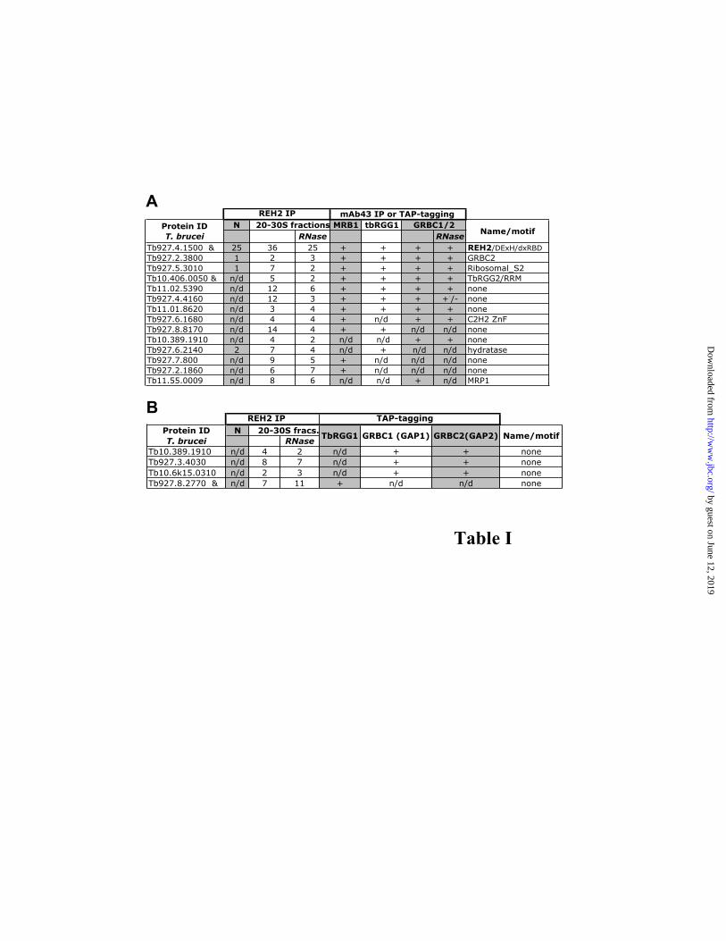
in all pull-downs reflecting the relatively large size of this protein. The 20-30S fractions contained twenty-two proteins (besides REH2) previously found in one or more of the reported MRB1, TbRGG1 and GRBC complexes (21,23,24). These complexes significantly overlap but also exhibit important compositional differences (Table 1 and Fig. S4 show RNase-resistant and RNase-sensitive REH2 interactions, respectively). Seven RNase-resistant proteins (out of thirteen) were common to the known MRB-related complexes, namely: REH2, GRBC2, Ribosomal S2 homolog, TbRGG2, and hypothetical proteins Tb11.02.5390, Tb927.4.4160, and Tb11.01.8620 (Table 1A). Likely, GRBC1 is also in this group but it was not detected with the RNase treatment implying that its proposed 1:1 ratio with GRBC2 in GRBC complexes (23) is not strictly conserved in the immuno-purified REH2 complexes. Apparent differences with known MRB-related complexes include five proteins found in at least one of these complexes but not in the REH2 pull-down (Fig. S4), and fifteen RNase-resistant proteins exclusively found in the REH2 pulldown, including the putative helicase REH1 (mHel61; Fig. S5) (28) and hypothetical proteins with unrecognized motifs (Fig. S6). The TbRGG1-associated complex described by Hashimi and collaborators (21) consists of proteins detected in TAP-purifications of all three TbRGG1, GAP1 (GRBC1) and GAP2 (GRBC2). Among proteins from the Hashimi study that were absent in at least one of their purifications, we found four RNase-resistant REH2 associated proteins (Table 1B). These four proteins were not found in MRB1 and GRBC complexes isolated by other labs (23,24), except for Tb10.389.1910 which was present in the latter (see Tables 1A-B). Thus, some proteins may be preferential or even unique components of REH2 complexes but this has to be confirmed by subsequent cross-tagging/purifications studies. Thus, besides a common core of at least 7-to-8 polypeptides in REH2 and other MRB-related assemblies there may be significant compositional differences among these novel multi-protein particles.
In contrast to the above examined sedimentation fractions, an REH2 pulldown of purified native editing complexes contained only six reported components of MRB-related complexes (including three RNase-resistant interactions). This could reflect differences in REH2 complex composition or relative abundance of REH2 in the samples examined, since the editing complexes had a
somewhat smaller number of unique peptides (Table 1). Interestingly, the protein Tb927.5.2930 may represent a REH2-specific cofactor as it was detected in REH2 pull-downs of both native editing complexes and sedimentation fractions, but was not found in other MRB-related complexes (Fig. S6). In fact, affinity-purification of this protein contained REH2, helicase activity, and RECC subunits (B.R.M. et al., unpublished data). Finally, REH2 co-purified with most known subunits of both RNA editing complexes and mitochondrial ribosomes (4,47), largely via RNase-sensitive associations (Figs. S7-8). An association with mitochondrial ribosomes was not implied for other MRB-related complexes, although four ribosomal proteins were inadvertently included in a model of the GRBC complex core (23). Importantly, none of the protein components discussed above were found in mock immuno-purifications or using an “empty”-TAP vector. Other RNase-resistant interactions in the pull-downs correspond to common metabolic proteins or proteins that were detected in our mock purifications (Fig. S9). Thus, the REH2 RNPs described here and previously reported MRB-related complexes are similar but not identical in composition and apparent RNA-linked interactions with other mitochondrial components.
DISCUSSION
RNA helicases may be the largest group of
enzymes in RNA metabolism from bacteria to humans, and are usually assembled in macromolecular RNPs such as spliceosomes and ribosomes. Helicases are often multifunctional within the same cell and understanding the basis of their specificity, particularly the relevant cofactors and substrates, remains a major challenge in biology (44). REH2 is a novel ~241 kDa factor in kinetoplastids that bears an unusual dual combination of DExH-box helicase and dsRBD motifs only previously observed in the eukaryotic RNA helicase A (48), and to our knowledge it represents the only reported example of a DExH-box helicase in mitochondria besides Suv3p in yeast and its orthologues (49). Recent RNAi studies by Hashimi et al., and in our lab showed that repression of REH2 decreases RNA editing and total gRNA levels (Figs. 5C-D, and data not shown)(22). The REH2 complexes we described appear to be novel particles that overlap in composition with recently published “MRB-related complexes” namely, MRB1, TbRGG1 and GBRC
by guest on June 12, 2019http://w
ww
.jbc.org/D
ownloaded from
REH2 helicase RNPs and motifs for unwinding and gRNA binding
6
complexes. These complexes exhibit compositional differences (Table 1) but they share a few subunits that resisted extensive RNase treatment in REH2 purifications, which we propose form a scaffold core for the assembly of more dynamic protein-protein and protein-RNA interactions. RECC binds REH2 and GRBC (23) RNPs, but an association with MRB1 and TbRGG1 complexes was not detected, probably reflecting compositional or stoichiometric differences that control this interaction. TbRGG1 itself is bound via RNA (Fig. S4)(21).
Association of REH2 with RECC subunits was
observed in REH2 antibody pulldowns of pre-purified native editing complexes and in converse immunoprecipitations (Figs. 3 and 4E). Consistent with a transient interaction (a) REH2 was readily detected in native but not in affinity-purified editing complexes, (b) TAP-REH2 purifications showed only a small amount of editing subunits after concentration of the final elutes, and (c) the majority of editing complexes appeared in the unbound fraction of REH2 pulldowns. A small fraction of REH2 consistently associated with editing proteins despite extensive treatments with MN and RNases A, T1 and V1, suggesting that at least some REH2 may directly bind RECC. The majority, however, was clearly RNA bridged (e.g., Fig. 3E and Fig. 4B). Alternatively, a high-affinity RNA linker, not completely removed by protein purification and single-/double-stranded nucleases, could mediate the association with editing complexes. A TAP-GRBC co-purification with REL1 and RET1 editing subunits was partially sensitivity to RNase A, and thus thought to involve transient contacts (23).
GRBC complexes reported by Weng et al. and
the REH2 complexes described here contain gRNA, however we also found helicase activity in the REH2 pulldowns. Our data suggest that REH2 is directly responsible for gRNA binding and the helicase activity as both required the dsRBD and a wild-type motif I of REH2 (Fig. 6C). This helicase activity immuno-purified with REH2 from sedimentation fractions containing no visible REH1 (Figs. 6D-F, and data not shown). Interestingly, Missel et al., found a helicase activity of mitochondrial lysates that largely sedimented away from REH1, localized at the top of the gradient (28). Consistent with the motif I mutation effect, the unwinding activity utilized ATP but not a non-hydrolyzable analog, and it dissociated a model
pre-mRNA/gRNA hybrid and a continuous helix with 3’ but not 5’ overhangs. The molecular basis of this substrate selectivity remains to be studied, however our data suggest that REH2 translocates in a 3’ to 5’ direction from a single-stranded loading region into a helical structure (50). We hypothesize that the dsRBD and DExH-helicase domains of REH2 act in concert to mediate selective binding of properly folded gRNAs. Helical elements of gRNA (51) may be targeted by the 70 amino acid dsRBD, although other REH2 sequences or its co-factors likely contribute to substrate specificity. Subsequent ATP-dependent winding and unwinding remodeling cycles may generate specific gRNA folds with increased affinity for REH2, thereby becoming protected from degradation. Thus, the conformation of gRNAs may be subject to regulation. Consistent with the idea that REH2 and other associated factors cooperate to provide gRNA-binding specificity, RNAi of REH2 and GRBC (GAP) factors severely decreased the gRNA steady-state levels (Figs. 5C-D)(22,23).
The precise compositional and functional
relationship of REH2 complexes with known MRB-related complexes needs to be further studied. The number of RNase-resistant interactions that co-purified with REH2 contrasts with the significantly decreased S value of REH2 after RNase treatments or dsRBD deletion. To reconcile these seemingly discordant observations we propose that REH2 may form multiple particles of variable composition or stoichiometry. In this line of thought we indicated that related complexes, isolated via e.g., either REH2, GRBC or TbRGG1 proteins share a common scaffold core but differ in multiple dynamic components. Such components could include specificity factors that link these particles with various aspects of mitochondrial gene expression. A putative collection of purified REH2 complexes may include most if not all proteins in Tables 1A-B, and potentially other RNase-resistant components we observed but additional studies are underway to examine this further.
The broad heterodispersion of REH2 in
sedimentation gradients likely reflects higher-order assemblies linked by RNA that comprise REH2 RNPs, other MRB-related assemblies, RECC and several factors including KPAP1, MERS1, RET1, MRP and PPR1. The pentatricopeptide protein PPR1 was associated with processing/stability of mitochondrial RNA including editing and ribosomal transcripts, specifically with regulation
by guest on June 12, 2019http://w
ww
.jbc.org/D
ownloaded from

REH2 helicase RNPs and motifs for unwinding and gRNA binding
7
of long poly(A) tails (52-54). Consistent with the above model: (a) these factors were found in REH2 pulldowns before but not after an extensive RNase treatment (Fig. S4), (b) either RNase or dsRBD deletion decreased the S value of REH2, and (c) REH2 co-immunoprecipated with RECC subunits in ~20S and >40S fractions (Fig. 4). Importantly, mitochondrial ribosomes were a major component of REH2 purifications via specific antibodies or TAP tagging (but were not found in mock preparations; Fig. S8), and their association was bridged by RNA. We found a few ribosomal proteins in reported TAP purifications of GRBP, KPAP1, MERS1, MRP, TbRGG1 and editing proteins (21,23), although this had passed unnoticed as the composition of mitochondrial ribosomes was reported after these studies (47). Furthermore, Osato et al. found rRNA in TAP isolations of editing complexes (55). Collectively data by us and by others labs are consistent with a
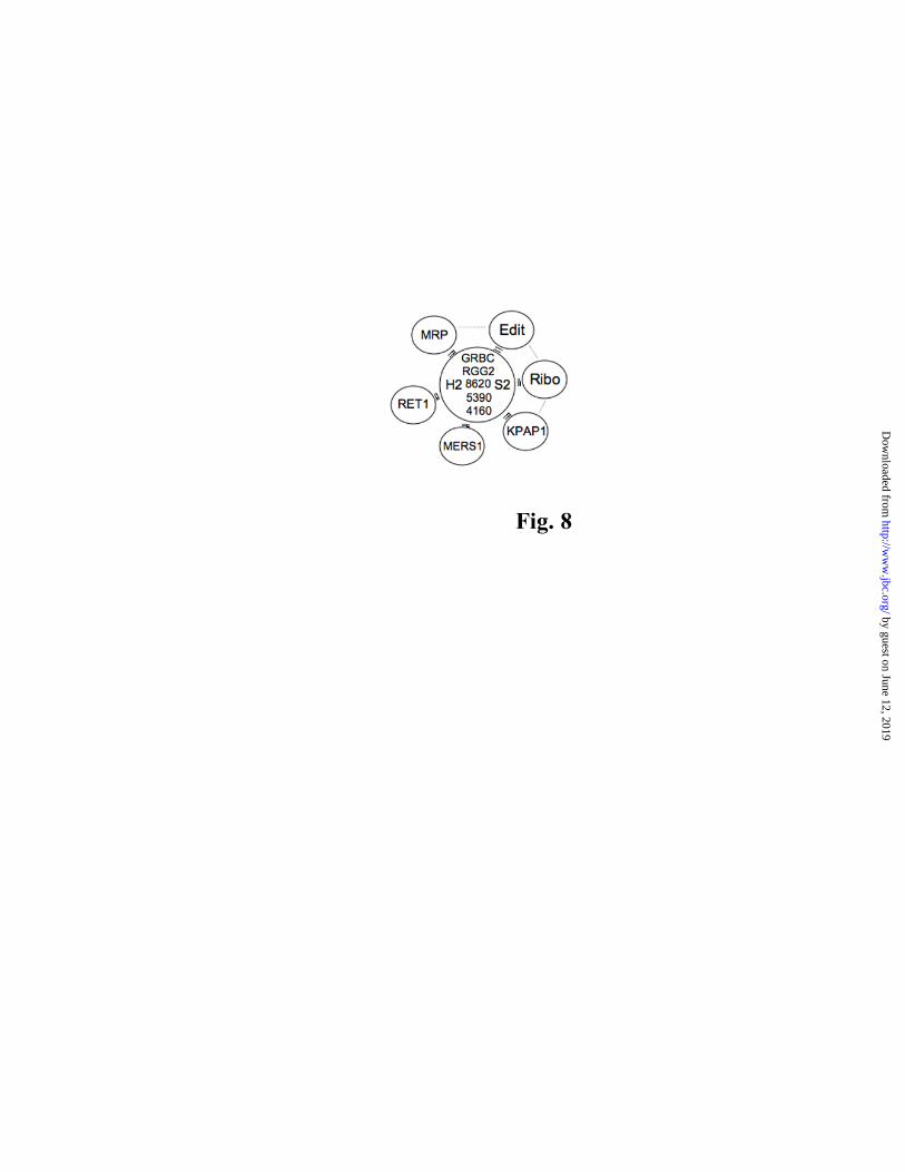
model whereby transient and dynamic RNA-interlinked networks in mitochondria functionally integrate and coordinate mitochondrial machineries in RNA maturation, stability and translation (schematized in Fig. 8). In this context, REH2 may be multifunctional. Relevant to this model, and the integration of ribosomes in particular, is the presence of a S2 ribosomal homolog in the core of REH2 RNPs and other MRB-related complexes (Table 1A; Fig. 8). Interestingly, S2 was not found in isolated mitochondrial ribosomes of T. brucei (47), and we speculate that this protein may help to functionally link MRB-related complexes with ribosomes. Finally, the multi component networks we propose may be similar to the L*b complexes recently detected in Leishmania using blue native gel electrophoresis by Osato et al. which the authors proposed represent the active holoenzyme or “editosome” (55).
REFERENCES
1. Carnes, J., and Stuart, K. D. (2007) Methods Enzymol 424, 25-54 2. Cruz-Reyes, J., and Hernandez, A. (2008) Protein-protein and RNA-protein interactions in U-
insertion/deletion RNA editing complexes in RNA and DNA editing (Smith, H. C. ed., John Wiley & Sons, Inc., New Jersey
3. Rusche, L. N., Cruz-Reyes, J., Piller, K. J., and Sollner-Webb, B. (1997) Embo J 16, 4069-4081 4. Panigrahi, A. K., Schnaufer, A., and Stuart, K. D. (2007) Methods Enzymol 424, 3-24 5. Aphasizhev, R., Aphasizheva, I., Nelson, R. E., Gao, G., Simpson, A. M., Kang, X., Falick, A.
M., Sbicego, S., and Simpson, L. (2003) Embo J 22, 913-924 6. Hernandez, A., Panigrahi, A., Cifuentes-Rojas, C., Sacharidou, A., Stuart, K., and Cruz-Reyes, J.
(2008) J Mol Biol 381, 35-48 7. Carnes, J., Trotter, J. R., Peltan, A., Fleck, M., and Stuart, K. (2007) Mol Cell Biol 8. Blum, B., Bakalara, N., and Simpson, L. (1990) Cell 60, 189-198 9. Seiwert, S. D., and Stuart, K. (1994) Science 266, 114-117 10. Cifuentes-Rojas, C., Halbig, K., Sacharidou, A., De Nova-Ocampo, M., and Cruz-Reyes, J.
(2005) Nucleic Acids Res 33, 6610-6620 11. Cifuentes-Rojas, C., Pavia, P., Hernandez, A., Osterwisch, D., Puerta, C., and Cruz-Reyes, J.
(2006) J Biol Chem 282, 4265-4276 12. Koslowsky, D. J., Reifur, L., Yu, L. E., and Chen, W. (2004) RNA Biol 1, 28-34 13. Reifur, L., and Koslowsky, D. J. (2008) RNA 14, 2195-2211 14. Yu, L. E., and Koslowsky, D. J. (2006) RNA 12, 1050-1060 15. Muller, U. F., Lambert, L., and Goringer, H. U. (2001) Embo J 20, 1394-1404 16. Aphasizhev, R., Aphasizheva, I., Nelson, R. E., and Simpson, L. (2003) Rna 9, 62-76 17. Aphasizhev, R., Sbicego, S., Peris, M., Jang, S. H., Aphasizheva, I., Simpson, A. M., Rivlin, A.,
and Simpson, L. (2002) Cell 108, 637-648 18. Etheridge, R. D., Aphasizheva, I., Gershon, P. D., and Aphasizhev, R. (2008) EMBO J 19. Fisk, J. C., Ammerman, M. L., Presnyak, V., and Read, L. K. (2008) J Biol Chem 20. Acestor, N., Panigrahi, A. K., Carnes, J., Zikova, A., and Stuart, K. D. (2009) RNA 15, 277-286 21. Hashimi, H., Zikova, A., Panigrahi, A. K., Stuart, K. D., and Lukes, J. (2008) RNA 22. Hashimi, H., Cicova, Z., Novotna, L., Wen, Y. Z., and Lukes, J. (2009) RNA 15, 588-599
by guest on June 12, 2019http://w
ww
.jbc.org/D
ownloaded from
REH2 helicase RNPs and motifs for unwinding and gRNA binding
8
23. Weng, J., Aphasizheva, I., Etheridge, R. D., Huang, L., Wang, X., Falick, A. M., and Aphasizhev, R. (2008) Mol Cell 32, 198-209
24. Panigrahi, A. K., Zikova, A., Dalley, R. A., Acestor, N., Ogata, Y., Anupama, A., Myler, P. J., and Stuart, K. D. (2008) Mol Cell Proteomics 7, 534-545
25. Vondruskova, E., van den Burg, J., Zikova, A., Ernst, N. L., Stuart, K., Benne, R., and Lukes, J. (2005) J Biol Chem 280, 2429-2438
26. Pelletier, M., and Read, L. K. (2003) Rna 9, 457-468 27. Miller, M. M., Halbig, K., Cruz-Reyes, J., and Read, L. K. (2006) Rna 12, 1292-1303 28. Missel, A., Souza, A. E., Norskau, G., and Goringer, H. U. (1997) Mol Cell Biol 17, 4895-4903 29. Wickstead, B., Ersfeld, K., and Gull, K. (2002) Mol Biochem Parasitol 125, 211-216 30. Wirtz, E., Leal, S., Ochatt, C., and Cross, G. A. (1999) Mol Biochem Parasitol 99, 89-101 31. Rigaut, G., Shevchenko, A., Rutz, B., Wilm, M., Mann, M., and Seraphin, B. (1999) Nat
Biotechnol 17, 1030-1032 32. Cruz-Reyes, J., and Sollner-Webb, B. (1996) Proc Natl Acad Sci U S A 93, 8901-8906 33. Cruz-Reyes, J. (2007) Methods Enzymol 424, 107-125 34. Sacharidou, A., Cifuentes-Rojas, C., Halbig, K., Hernandez, A., Dangott, L. J., De Nova-
Ocampo, M., and Cruz-Reyes, J. (2006) Rna 12, 1219-1228 35. Blum, B., and Simpson, L. (1990) Cell 62, 391-397 36. Florens, L., Carozza, M. J., Swanson, S. K., Fournier, M., Coleman, M. K., Workman, J. L., and
Washburn, M. P. (2006) Methods 40, 303-311 37. Washburn, M. P., Wolters, D., and Yates, J. R., 3rd. (2001) Nat Biotechnol 19, 242-247 38. Wohlschlegel, J. A. (2009) Methods in molecular biology (Clifton, N.J 497, 33-49 39. Wolters, D. A., Washburn, M. P., and Yates, J. R., 3rd. (2001) Anal Chem 73, 5683-5690 40. Eng, J., McCormack, A., and Yates, J. (1994) J Am Soc Mass Spectrom 5, 976-989 41. Tabb, D. L., McDonald, W. H., and Yates, J. R., 3rd. (2002) J Proteome Res 1, 21-26 42. Elias, J. E., and Gygi, S. P. (2007) Nat Methods 4, 207-214 43. McManus, M. T., Shimamura, M., Grams, J., and Hajduk, S. L. (2001) Rna 7, 167-175 44. Jankowsky, E. (2007) Nature 449, 999-1000 45. Bleichert, F., and Baserga, S. J. (2007) Mol Cell 27, 339-352 46. Fabre, A. (1990) Bioinorganic Photochemistry. . in Photobiochemistry and Nucleic Acids.
(Morrison, H. ed., John Wiley & Sons New York, NY 47. Zikova, A., Panigrahi, A. K., Dalley, R. A., Acestor, N., Anupama, A., Ogata, Y., Myler, P. J.,
and Stuart, K. (2008) Mol Cell Proteomics 7, 1286-1296 48. Robb, G. B., and Rana, T. M. (2007) Mol Cell 26, 523-537 49. Margossian, S. P., Li, H., Zassenhaus, H. P., and Butow, R. A. (1996) Cell 84, 199-209 50. Fairman, M. E., Maroney, P. A., Wang, W., Bowers, H. A., Gollnick, P., Nilsen, T. W., and
Jankowsky, E. (2004) Science 304, 730-734 51. Hermann, T., Schmid, B., Heumann, H., and Goringer, H. U. (1997) Nucleic Acids Res 25, 2311-
2318 52. Mingler, M. K., Hingst, A. M., Clement, S. L., Yu, L. E., Reifur, L., and Koslowsky, D. J. (2006)
Mol Biochem Parasitol 150, 37-45 53. Pusnik, M., Small, I., Read, L. K., Fabbro, T., and Schneider, A. (2007) Mol Cell Biol 27, 6876-
6888 54. Koslowsky, D. J. (2009) Trends Parasitol 25, 252-255 55. Osato, D., Rogers, K., Guo, Q., Li, F., Richmond, G., Klug, F., and Simpson, L. (2009) RNA 56. Jankowsky, E., and Jankowsky, A. (2000) Nucleic Acids Res 28, 333-334
FOOTNOTES
We thank Paul Straight at TAMU for his careful comments and discussion on the manuscript. We are
also grateful to the following colleagues: Larry Simpson for T. brucei genomic DNA; Jason Carnes for
by guest on June 12, 2019http://w
ww
.jbc.org/D
ownloaded from
REH2 helicase RNPs and motifs for unwinding and gRNA binding
9
advise on quantitative RT-PCR, and Ken Stuart for monoclonal antibodies against core subunits of editing complexes; H. Ulrich Goringer, Paul Michels and Susan Madison-Antenucci for mHel61, α-enolase and Tbmp45 antibodies, respectively; Kirk Gaston and Juan D. Alfonzo for their generous assistance in the subcellular localization studies; Achim Schanufer and Ruslan Aphasizhev for sharing their TAP purification protocols; Ms. Neili Cooksey for helping prepare the tables. This work was supported by a grant from the National Institutes of Health to J.C.-R. (GM067130).
FIGURE LEGENDS
Fig. 1. REH2 gene organization and co-purification with native editing complexes. (A) T. brucei REH2 has 2167 amino acids including a conserved mitochondrial import signal (Mito), double-stranded RNA binding domain (dsRBD) and domains typically associated with DExH-box helicases (drawn at scale): DExHc, HELICc, HA2 and HrpA. The first domain, which defines this protein family, contains six motifs including the motif II signature amino acids DExH where “x” is any residue. Four such motifs are evident in REH2 (I, II, III and VI) and shown with the most conserved residues in gray (56); (B) REH2 western blot of native and TAP-purified editing complexes tagged at various subunits. The middle panel is shown at increased contrast. Adenylytated ligase subunits REL1 and REL2 shown as loading control; (C) Silver-staining of purified native editing complexes. Fig. 2. REH2 expression in procyclic T. brucei. (A) Scheme of the C-terminal TAP tag, and anti-cbp western of total cell lysates with or without 3 days of induction with increasing tetracycline (Tc) concentrations; (B) Quantitative RT-PCR of REH2 normalized to 18S rRNA and α-tubulin +/- Tc; (C) REH2 western of induced empty-TAP vector (-) and TAP-REH2 cells for 1 and 3 days. Cytosolic α-enolase was used as control. (D) Growth curves of procyclic trypanosome cell lines expressing TAP constructs of wild-type REH2, dsRBD-Δ and mot 1 mutants. (E) Western blot showing REH2 enrichment in a mitochondrial lysate. Mitochondrial MP81 and cytosolic α-enolase markers. Fig. 3. REH2 co-purification with RECC is largely sensitive to extensive nuclease treatments. REH2 TAP purification and detection of editing subunits before or after (+/-) treatments with RNases A, T1 (and V1 as indicated) and micrococcal nuclease (MN) in (A) TEV (T) and (B) EGTA (ET) eluates from IgG-Sepharose and calmodulin-agarose, respectively. Control lanes include native editing complex (N), diluted whole-cell lysate (L) and empty-vector controls (TE or ETE; last lane). ET eluates were concentrated by acetone precipitation. Immunoblots of REH2 and editing subunits and auto-adenylylation of REL ligases. ATPases in whole-cell lysates often inhibit the latter activity (e.g., panel A, lane 2). MP63 may co-migrate with antibody cross-reacting BSA added as precipitation carrier (see the ETE control lane). REH2 fragmentation (*) is observed in the eluates. (C) Mitochondrial lysate IP pulldowns with antibodies to RECC subunits (MP81, MP63, REL1, and MP42) or a mock reaction without antibodies. REH2 pulldowns from (D) purified native editing complexes or (E) mitochondrial lysate (M) and mock reactions with pre-immune serum. An unaccounted band near MP52 is visible in the lysate lane. Fig. 4. Heterodispersion of REH2 complexes requires RNA and the dsRBD. Glycerol gradients of mitochondrial lysates in western blots of endogenous plus ectopic REH2 (REH2) or ectopic REH2 (PAP reaction) and MP63 as a RECC marker. Mitochondrial lysates (A) untreated or (B) treated with RNases A, T1 and MN, or (C) whole lysates of REH2 dsRBD-Δ cells. (D) REH2-antibody pulldowns of gradient fractions 3 (~10S), 7-8 (~20S), and 12-13 (>40S) from panel A. Fig. 5. REH2 co-purification with gRNA requires the dsRBD and wild-type helicase motif I. (A) IgG- dynabead pulldowns (IP) of whole cell lysates expressing TAP-REH2: wild-type (w.t.) or mutants dsRBD-Δ (ds) and motif I (m-I). Western blots with REH2 antibodies or the PAP reagent of pulldowns normalized for the amount of REH2; (B) gRNA-labeling assays with guanylyl transferase of the pulldowns in A. Whole-cell lysate from wild-type cells (L) or a REH2 pulldown from “empty-vector” cells (ev) were used as controls. A few RNA species accumulated in the REH2 mutants (arrowhead). A
by guest on June 12, 2019http://w
ww
.jbc.org/D
ownloaded from

REH2 helicase RNPs and motifs for unwinding and gRNA binding
10
typical artifact in guanylyl-transferase assays (*) serves as loading control; (C) Western blots of REH2 RNAi cells before or after the indicated days of Tc-induction. Mitochondrial MP81 and cytosolic α-enolase controls; (D) gRNA-labeling assays in REH2 RNAi cells before or after 6 days Tc-induction. (E) Western blot of a REH2 pulldown from 29.13 cells lacking the ectopic REH2 construct. A mock reaction with pre-immune serum, and whole lysate (L) are controls. (F) gRNA labeling of the pulldowns in E. Fig. 6. REH2 is associated with helicase activity. RNA helicase assays REH2 antibody pulldowns from mitochondrial lysate supplemented with gel-isolated dsRNA substrates. (A) A6 pre-mRNA and cognate gRNA with 1 mM ATP, ADP-CP or no nucleotide. The pre-mRNA was 5’ end radiolabeled. The gRNA 3’-oligo U hybrid is presumably unstable (arrow heads); or (B) a short duplex with symmetrical 3’ overhangs and 1 mM ATP. (C) IgG-dynabead pulldowns of TAP-REH2 wild-type and mutants (see Fig 5 legend). RNA input before (-) and after boiling, and mock pre-immune serum pulldowns from empty-vector cell lysates are controls. (D-F) Sedimentation fractions of whole-cell lysate examined for helicase activity and western blots of REH2 and REH1. Fig. 7. A ~250 kDa protein in REH2 pulldowns photo-crosslinks with RNA. (A) Crosslinking of an A6 pre-mRNA/gRNA pair bearing a single photo-reactive thio-U at the editing site. Mitochondrial Q-sepharose fraction enriched with RECC (lane 1) or IP pulldowns of this material by antibodies to REL1 or REH2 (lanes 2 and 3, respectively). Lanes 4-5 are repeats of lane 3 but after a treatment with 0.1 % SDS at 70oC and 90oC, respectively, that enriches a crosslink at ~250 kDa (arrowhead). The REL1 pulldown shows at least four reported crosslinking RECC subunits (6,34) (see panel B). (B) Proof of concept of the denaturation protocol used in panel A, showing immuno-purification of crosslinked RECC subunits. Crosslinked subunits of purified RECC (lane 1), and specific RECC subunits (lanes 2 and 3) after SDS dissociation at 90oC, and immuno-purification with the indicated antibodies. The efficiency of this procedure depends on the antibodies affinity, polyclonal for REH2 and monoclonal for RECC subunits. Fig. 8. Proposed RNA-interlinked molecular networks in kinetoplastid mitochondria. Machineries and factors for RNA processing, stability and translation that co-purify with REH2 RNPs via RNA, that we propose reflect transient supramolecular assemblies that coordinate mitochondrial gene expression. For simplicity, RNA bridges (double lines) irradiate from the center but alternative or additional RNA and protein contacts could exist across components in the network (dotted lines). The arbitrary center of the model includes seven RNase-resistant proteins in the REH2 pull-down that are common to the known MRB-related complexes. They were abbreviated as follow: REH2 (H2), GRBC2 (GRBC), Ribosomal S2 homolog (S2), TbRGG2 (RGG2), and hypothetical proteins Tb11.01.8620 (8620), Tb11.02.5390 (5390) and Tb927.4.4160 (4160) (Table 1A). At least GRBC1 may also be included in this protein array (see text). Table 1 (A) RNase-resistant REH2 associated proteins that were found in MRB1, TbRGG1 or GRBC1 complexes— Mass spectrometric analyses of proteins identified in REH2 antibody pulldowns from ~20-30S mitochondrial lysate fractions, Unique peptides (criteria: two peptides minimum) before or after an extensive nuclease treatment with RNases A, T1,V1 and MN while proteins were bound to the beads. REH2 pulldowns of native editing complexes (N) and a low-efficiency REH2 TAP-purification (&) (criteria: one unique peptide minimum). Reported MRB1-immunopurified complex (using mAb43 antibodies) and TAP-purified TbRGG1 and GRBC1/2 complexes (21,23,24). GRBC1/2 (+) indicates proteins found in both GRBC1 and GRBC2 purifications, whereas (+/-) indicates detection only in the GRBC1 purification. Some proteins listed as putative GRBC components by Weng et al. are ribosomal proteins, and were RNase sensitive in the REH2 pulldowns (Fig. S8); (B) RNase-resistant REH2 associated proteins that were not considered as subunits of MRB-related complexes, but were found in independent TAP-purifications— Many proteins in a study by Hashimi and collaborators were not present in at least one of three TAP-purifications: TbRGG1, GBP1 and GBP2 (i.e., were no considered as TbRGG1 complex subunits) (21). The identified REH2 associated components correspond to hypothetical proteins with unknown motifs (criteria: two unique peptides). Proteins in native editing complexes and an REH2 TAP-purification (&)(criteria: one unique peptide). Some reported proteins are ribosomal components and were added to Fig. S8.
by guest on June 12, 2019http://w
ww
.jbc.org/D
ownloaded from
REH2 IP mAb43 IP or TAP-tagging
N 20-30S fractions MRB1 tbRGG1
Tb927.4.1500 & 25 36 25 + + + + REH2/DExH/dxRBD
Tb927.2.3800 1 2 3 + + + + GRBC2
Tb927.5.3010 1 7 2 + + + + Ribosomal_S2
Tb10.406.0050 & n/d 5 2 + + + + TbRGG2/RRM
Tb11.02.5390 n/d 12 6 + + + + none
Tb927.4.4160 n/d 12 3 + + + +'/- none
Tb11.01.8620 n/d 3 4 + + + + none
Tb927.6.1680 n/d 4 4 + n/d + + C2H2 ZnF
Tb927.8.8170 n/d 14 4 + + n/d n/d none
Tb10.389.1910 n/d 4 2 n/d n/d + + none
Tb927.6.2140 2 7 4 n/d + n/d n/d hydratase
Tb927.7.800 n/d 9 5 + n/d n/d n/d none
Tb927.2.1860 n/d 6 7 + n/d n/d n/d none
Tb11.55.0009 n/d 8 6 n/d n/d + n/d MRP1
Protein ID
T. brucei RNaseName/motif
RNase
GRBC1/2
Table I
REH2 IP TAP-tagging
N 20-30S fracs.
RNase
Tb10.389.1910 n/d 4 2 n/d + + none
Tb927.3.4030 n/d 8 7 n/d + + none
Tb10.6k15.0310 n/d 2 3 n/d + + none
Tb927.8.2770 & n/d 7 11 + n/d n/d none
Protein ID
T. bruceiName/motifGRBC2(GAP2)GRBC1 (GAP1)TbRGG1
A
B
by guest on June 12, 2019http://w
ww
.jbc.org/D
ownloaded from
Willard, Michael T. Kinter and Jorge Cruz-ReyesAlfredo Hernandez, Bhaskara Reddy Madina, Kevin Ro, James A. Wohlschlegel, Belinda
essential motifs for unwinding and guide RNA bindingREH2 RNA helicase in kinetoplastid mitochondria: ribonucleoprotein complexes and
published online October 22, 2009J. Biol. Chem.
10.1074/jbc.M109.051862Access the most updated version of this article at doi:
Alerts:
When a correction for this article is posted•
When this article is cited•
to choose from all of JBC's e-mail alertsClick here
Supplemental material:
http://www.jbc.org/content/suppl/2009/10/22/M109.051862.DC1
by guest on June 12, 2019http://w
ww
.jbc.org/D
ownloaded from