Embed Size (px)
Citation preview
Renal DiseaseCase Studies
IDEXX Laboratories
AuthorsDennis DeNicola, DVM, PhD, DACVPChief Veterinary Educator, Clinical Pathologist, IDEXX LaboratoriesDr. DeNicola completed his DVM in 1978 and his PhD in 1981, both at Purdue University. For more than twenty years, he served as educator in clinical and surgical pathology. In addition, he directed the primary cytol-ogy and surgical pathology service at the veterinary school laboratory and ran a private pathology service for 15 years. A speaker at more than 100 national and international education symposia, Dr. DeNicola also has authored or co-authored more than 150 publications in various aspects of veterinary clinical pathology. Fred Metzger, DVM, DABVPOwner, Metzger Animal HospitalDr. Metzger is a 1986 graduate of the Purdue School of Veterinary Medicine and a diplomate of the American Board of Veterinary Practitioners, with specialties in canine and feline medicine. He is an adjunct professor at Pennsylvania State University and serves on the practitioner advisory boards of Veterinary Economics and Veterinary Medicine magazines. He recently co-authored Guide to Hematology in Dogs and Cats with Dr. Alan Rebar. Dr. Metzger owns the Metzger Animal Hospital, a four-doctor practice in State College, Pennsylvania, that received the 1998 Veterinary Economics/Pfizer Practice of Excellence award.
Pete Fernandes, DVM, DACVPClinical Pathologist, IDEXX LaboratoriesDr. Fernandes completed his DVM at the University of Wisconsin-Madison, followed by an internship in small-animal medicine and surgery at South Shore Animal Hospital in Boston. Dr. Fernandes’ residency was in clinical pathology at Texas A&M University and the University of Florida. He is a diplomate of the American College of Veterinary Pathologists.
Brian Poteet, DVM, DAVCR, DABSNMDirector, Gulf Coast Veterinary Diagnostic ImagingDr. Poteet received his DVM from Texas A&M University and completed his radiology residency at the University of Tennessee. In addition to being board-certified with the American College of Veterinary Radiology, Dr. Poteet is also a member of the American Board of Science in Nuclear Medicine. Dr. Poteet is a member of several local and national veterinary medical associations, Vice President of the Veterinary Cancer Associates, and holds two adjunct faculty positions at Texas A&M University.
Richard Goldstein, DVM, DACVIM, DECVIM-CAAssistant Professor, Small-Animal Medicine, Cornell UniversityDr. Goldstein received his DVM from the Koret School of Veterinary Medicine, the Hebrew University of Jerusalem, Israel. He completed his residency in small-animal internal medicine at the University of California, Davis. He is a diplomate of the American College of Veterinary Internal Medicine and the European College of Veterinary Internal Medicine—Companion Animals. He joined the faculty at Cornell in 2001. Dr. Goldstein’s clinical and research interests include nephrology and leptospirosis and Lyme nephritus in dogs.
Roberta Relford, DVM, MS, PhD, DACVIM, DACVPDivisional Vice President of Worldwide Pathology Coagulation, Cytology, Internal Medicine, IDEXX LaboratoriesDr. Relford received her DVM from Auburn University in 1982 and worked as a small-animal practitioner for four years before pursuing her advanced training. She started her residency training in clinical pathology and obtained an MS in pathology from Mississippi State University. She then transferred to Texas A&M, where she completed her pathology residency training and obtained a PhD in pathology. While completing her PhD, Dr. Relford pursued a residency in small-animal internal medicine.
Dr. Relford is board-certified in internal medicine by the American College of Veterinary Internal Medicine and in clinical pathology by the American College of Veterinary Pathologists. She currently serves as Divisional Vice President of Worldwide Pathology for IDEXX Reference Laboratories. Dr. Relford has given numerous lectures on a wide variety of topics including clinical pathology, internal medicine, infectious diseases, cytology, platelet disorders, health maintenance programs and zoonotic diseases.
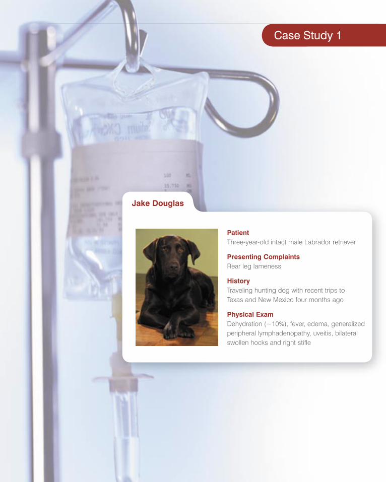
Case Study 1
PatientThree-year-old intact male Labrador retriever
Presenting ComplaintsRear leg lameness
HistoryTraveling hunting dog with recent trips to Texas and New Mexico four months ago
Physical ExamDehydration (~10%), fever, edema, generalized peripheral lymphadenopathy, uveitis, bilateral swollen hocks and right stifle
Jake Douglas
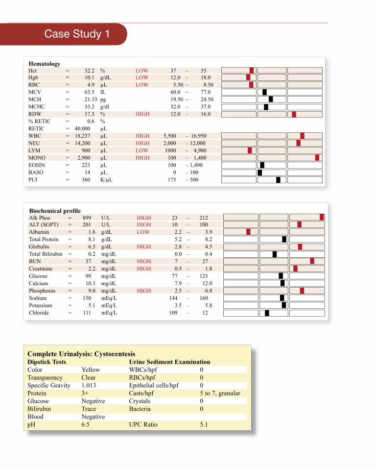
Hematology Hct = 32.2 % LOW 37 – 55Hgb = 10.1 g/dL LOW 12.0 – 18.0 RBC = 4.9 µL LOW 5.50 – 8.50MCV = 63.5 fL 60.0 – 77.0 MCH = 21.33 pg 19.50 – 24.50 MCHC = 33.2 g/dl 32.0 – 37.0 RDW = 17.3 % HIGH 12.0 – 16.0 % RETIC = 0.6 %RETIC = 40,000 µL WBC = 18,237 µL HIGH 5,500 – 16,950NEU = 14,200 µL HIGH 2,000 – 12,000LYM = 900 µL LOW 1000 – 4,900MONO = 2,900 µL HIGH 100 – 1,400EOSIN = 223 µL 100 – 1,490BASO = 14 µL 0 – 100PLT = 360 K/µL 175 – 500
Biochemical profile Alk Phos = 899 U/L HIGH 23 – 212ALT (SGPT) = 201 U/L HIGH 10 – 100 Albumin = 1.6 g/dL LOW 2.2 – 3.9Total Protein = 8.1 g/dL 5.2 – 8.2 Globulin = 6.5 g/dL HIGH 2.8 – 4.5 Total Bilirubin = 0.2 mg/dL 0.0 – 0.4 BUN = 37 mg/dL HIGH 7 – 27 Creatinine = 2.2 mg/dL HIGH 0.5 – 1.8Glucose = 99 mg/dL 77 – 125 Calcium = 10.3 mg/dL 7.9 – 12.0Phosphorus = 9.0 mg/dL HIGH 2.5 – 6.8Sodium = 150 mEq/L 144 – 160Potassium = 5.1 mEq/L 3.5 – 5.8Chloride = 111 mEq/L 109 – 12
Complete Urinalysis: CystocentesisDipstick Tests Urine Sediment ExaminationColor Yellow WBCs/hpf 0Transparency Clear RBCs/hpf 0Specific Gravity 1.013 Epithelial cells/hpf 0Protein 3+ Casts/hpf 5 to 7, granularGlucose Negative Crystals 0Bilirubin Trace Bacteria 0Blood Negative pH 6.5 UPC Ratio 5.1
Case Study 1
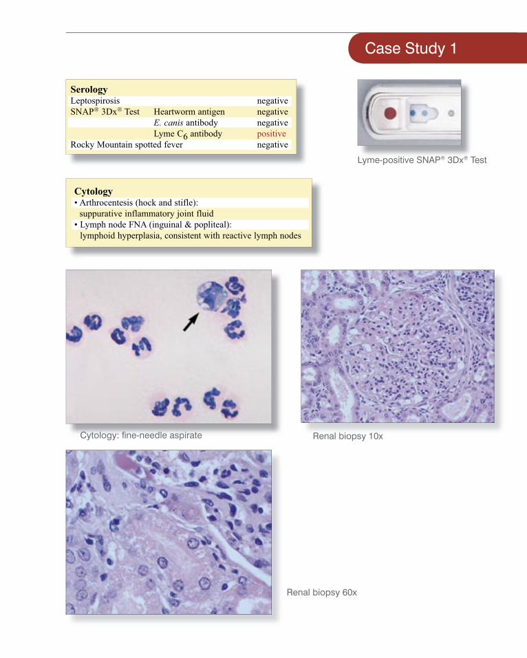
Cytology• Arthrocentesis (hock and stifle):
suppurative inflammatory joint fluid• Lymph node FNA (inguinal & popliteal):
lymphoid hyperplasia, consistent with reactive lymph nodes
SerologyLeptospirosis negativeSNAP® 3Dx® Test Heartworm antigen negative E. canis antibody negative Lyme C6 antibody positiveRocky Mountain spotted fever negative
Case Study 1
Lyme-positive SNAP® 3Dx® Test
Cytology: fi ne-needle aspirate Renal biopsy 10x
Renal biopsy 60x
DiagnosisThe clinical diagnosis is Lyme nephritis.
Treatment/Plan• Blood was sent to a reference
laboratory for quantitative C6 antibody testing.
• The patient was treated with doxycycline, intravenous fl uid support and a renal diet.
• Recheck renal panel in 3–5 days.• Renal biopsy
PreventionPrevention of Lyme disease includes reducing tick exposure, utilizing tick repellant products and vaccinating at-risk patients.
Zoonotic PotentialSince pets share our environment, they may incidentally become our sentinels; therefore, borreliosis in our canine companions should be a warning to increase vigilance and re-evaluate tick-prevention protocols.
Lyme disease is not transmissible directly from the canine patient to the owner. However, the owners should be educated that they are living in a tick-endemic area and the ticks may be infected with Lyme disease.
Interpretive SummaryHematology
There is mild nonregenerative anemia. The most common cause of mild nonregenerative anemia is anemia of chronic disease. The modest leukocytosis composed of mature neutrophilia and monocytosis with concurrent lymphopenia is consistent with an established infl ammatory condition. The thrombon/platelets are within normal limits
Biochemical profi leHypoalbuminemia and azotemia with an elevated UPC and the presence of granular casts support renal disease. The positive Lyme serology along with the hyperglobulinemia suggests Lyme nephritis. The specifi c gravity 1.013 indicates some, yet inadequate, concentrating ability, and hypoalbuminemia may be masked somewhat by dehydration. Signifi cant hypoalbuminemia is caused by protein-losing glomerulopathy and worsened by systemic vasculitis, severe hepatic insuffi ciency and hyperglobulinemia related to antigenic stimulation.
Azotemia is likely of mixed origins or primarily of renal origins with some degree of a prerenal component. Decreased urine concentrating ability in the face of dehydration is an indication of renal azotemia. Confounding renal azotemia, severe hypoalbuminemia can decrease colloidal osmotic pressure and essentially decrease vascular volume or renal perfusion. In the later stages of Lyme nephritis, lesions can include some combination of interstitial lymphoplasmacytic nephritis, tubular necrosis and diffuse glomerulonephritis, all of which can be a cause of proteinuria.
The pathogenesis of the tubular changes in canine Lyme nephritis is questionable, but immune-mediated glomerular disease, decreased perfusion and hypoxia, and the toxic effects of severe proteinuria are all postulated as potential causes. Liver enzymes are increased by hepatocellular damage, systemic or intrahepatic vasculitis, and vacuolar hepatopathy associated with chronic infl ammation or infection and ischemia.
UrinalysisUrine specifi c gravity shows inappropriate concentrating ability caused by glomerular and tubular dysfunction. Observation of granular casts can confi rm coexisting tubular damage, but the density of casts in urine cannot reliably measure severity, reversibility or duration of lesion. The pathogenesis of the tubular changes in canine Lyme nephritis is questionable, but immune-mediated glomerular disease, decreased perfusion and hypoxia, and the toxic effects of severe proteinuria are most likely responsible.
Additional testingSerologyFollow-up with quantitative C6 antibody test aids in determining when treatment is warranted, accurately tracking response to therapy and, eventually, as an indicator of when treatment has been effective.
Lyme C6 antibody to the C6 antigen is a highly specifi c for Borrelia burgdorferi infection. Dogs with leptospirosis, Rocky Mountain spotted fever, babesiosis, ehrlichiosis and heartworm disease do not have antibodies to C6, nor are antibodies to C6 produced in response to immunization with currently available canine Lyme vaccines.
Case Study 1
Case Study 2
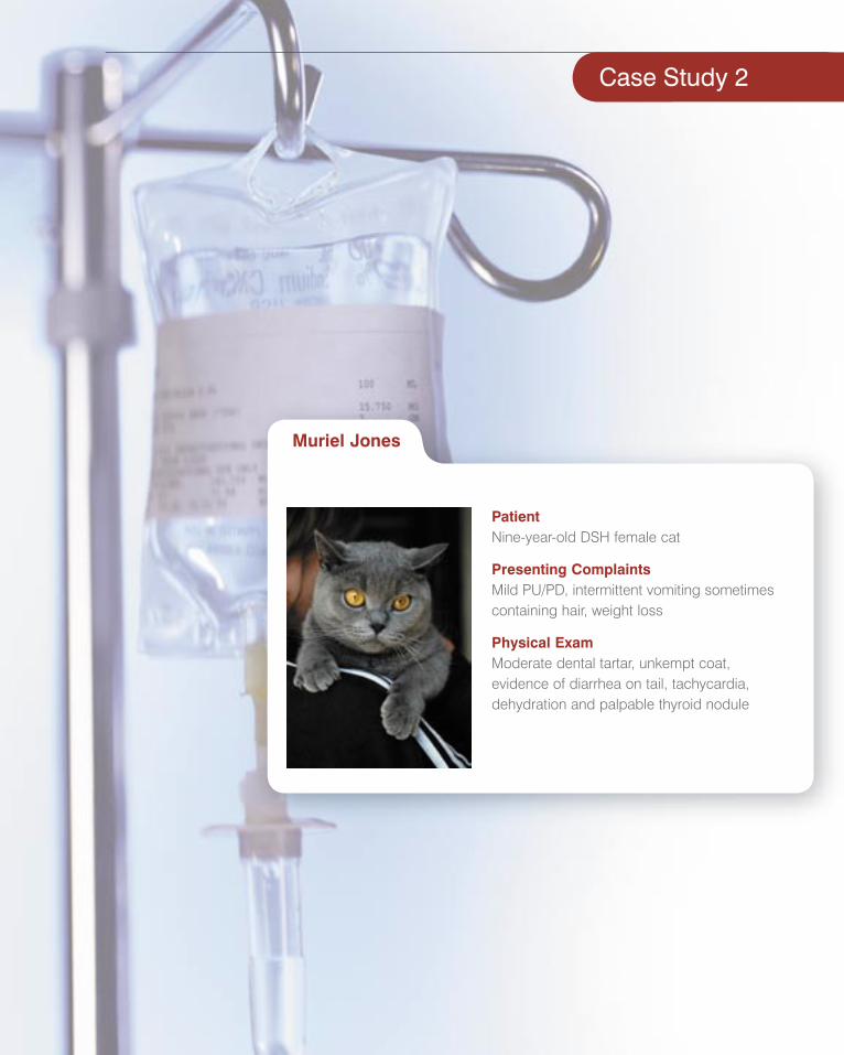
PatientNine-year-old DSH female cat
Presenting ComplaintsMild PU/PD, intermittent vomiting sometimes containing hair, weight loss
Physical ExamModerate dental tartar, unkempt coat, evidence of diarrhea on tail, tachycardia,dehydration and palpable thyroid nodule
Muriel Jones
Case Study 2
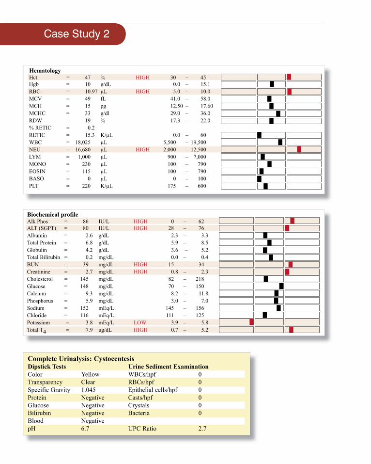
Hematology Hct = 47 % HIGH 30 – 45Hgb = 10 g/dL 0.0 – 15.1 RBC = 10.97 µL HIGH 5.0 – 10.0MCV = 49 fL 41.0 – 58.0 MCH = 15 pg 12.50 – 17.60MCHC = 33 g/dl 29.0 – 36.0 RDW = 19 % 17.3 – 22.0 % RETIC = 0.2 RETIC = 15.3 K/µL 0.0 – 60WBC = 18,025 µL 5,500 – 19,500NEU = 16,680 µL HIGH 2,000 – 12,500LYM = 1,000 µL 900 – 7,000MONO = 230 µL 100 – 790EOSIN = 115 µL 100 – 790BASO = 0 µL 0 – 100PLT = 220 K/µL 175 – 600
Biochemical profile Alk Phos = 86 IU/L HIGH 0 – 62ALT (SGPT) = 80 IU/L HIGH 28 – 76 Albumin = 2.6 g/dL 2.3 – 3.3Total Protein = 6.8 g/dL 5.9 – 8.5 Globulin = 4.2 g/dL 3.6 – 5.2 Total Bilirubin = 0.2 mg/dL 0.0 – 0.4 BUN = 39 mg/dL HIGH 15 – 34 Creatinine = 2.7 mg/dL HIGH 0.8 – 2.3Cholesterol = 145 mg/dL 82 – 218Glucose = 148 mg/dL 70 – 150 Calcium = 9.3 mg/dL 8.2 – 11.8Phosphorus = 5.9 mg/dL 3.0 – 7.0Sodium = 152 mEq/L 145 – 156Chloride = 116 mEq/L 111 – 125Potassium = 3.8 mEq/L LOW 3.9 – 5.8Total T4 = 7.9 ug/dL HIGH 0.7 – 5.2
Complete Urinalysis: CystocentesisDipstick Tests Urine Sediment ExaminationColor Yellow WBCs/hpf 0Transparency Clear RBCs/hpf 0Specific Gravity 1.045 Epithelial cells/hpf 0Protein Negative Casts/hpf 0Glucose Negative Crystals 0Bilirubin Negative Bacteria 0Blood Negative pH 6.7 UPC Ratio 2.7
Case Study 2
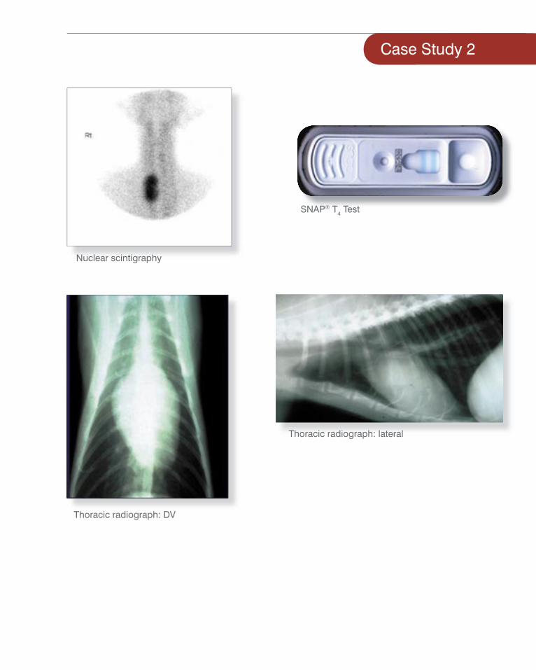
Thoracic radiograph: lateral
Thoracic radiograph: DV
Nuclear scintigraphy
SNAP® T4 Test
Interpretive SummaryHematologyVery mild polycythemia, which can be either relative or absolute. Relative polycythemia is associated with dehydration; absolute can be associated with polycythemia vera or causes of increased erythropeitin. Slight leukocytosis composed of mature neutrophilia (a lack of immature neutrophils on the blood fi lm) with lymphopenia suggests a stress leukogram.
Biochemical profi leMild increases in alkaline phosphatase (ALKP) and alanine aminotransferase (ALT) are present. Azotemia is present (BUN, creatinine increased). Deciding if azotemia is prerenal, renal or postrenal can be diffi cult because cats can have renal azotemia with relatively concentrated urine. Moreover, the urine of hyperthyroid cats can be nonconcentrated as a direct result of the hyperthyroidism without any secondary renal disease. Hypokalemia is present and can occur with many feline diseases including CRF (chronic renal failure) and hyperthyroidism. Total T4 in markedly elevated and hyperthyroidism is likely, especially considering the associated polycythemia, azotemia and elevated liver enzymes.
UrinalysisUrine specifi c gravity is concentrated and the urine protein ratio is moderately elevated, especially for an azotemic patient.
RadiographyMild cardiomegaly is present, characterized by biatrial enlargement. This is recognized on the VD view (valentine heart).
Nuclear scintigraphy shows a right-sided, unilateral lesion, which is less common than a bilateral lesion in feline hyperthyroidism.
Additional testingBlood pressure
Systolic 180 mm/Hg—if repeatable, consistent with mild hypertension
DiagnosisThe clinical diagnosis is hyperthyroidism with likely concurrent chronic renal disease.
Treatment/PlanHyperthyroidism can increase cardiac output, decrease peripheral vascular resistance, increase renal blood fl ow and increase GFR. This chain of events cannot only decrease BUN and creatinine, but also perhaps lead to glomerular hypertension and hyperfi ltration, thereby potentially inducing or worsening concurrent renal disease.
Systemic hypertension can be associated with hyperthyroidism, and supervision of some patient therapy may benefi t from regular monitoring of UPC with a UPC less than 0.5 as a target for treatment. With successful Rx of hyperthyroidism (radioactive iodine, methimizole, thyroidectomy), the UPC may return to normal or may worsen if CRF is progressive. Careful monitoring of this patient is recommended.
associated polycythemia, azotemia and elevated liver enzymes.
Urine specifi c gravity is concentrated and the urine protein ratio is moderately elevated, especially for an azotemic patient.
Mild cardiomegaly is present, characterized by biatrial enlargement. This is recognized on the VD view (valentine heart).
Nuclear scintigraphy shows a right-sided, unilateral lesion, which is less common than a bilateral lesion in feline hyperthyroidism.
Systolic 180 mm/Hg—if repeatable, consistent with mild hypertension
and creatinine, but also perhaps lead to glomerular hypertension and hyperfi ltration, thereby potentially inducing or worsening concurrent renal disease.
Systemic hypertension can be associated with hyperthyroidism, and supervision of some patient therapy may benefi t from regular monitoring of UPC with a UPC less than 0.5 as a target for treatment. With successful Rx of hyperthyroidism (radioactive iodine, methimizole, thyroidectomy), the UPC may return to normal or may worsen if CRF is progressive. Careful monitoring of this patient is recommended.
disease. Hypokalemia is present and can occur with many feline diseases including CRF (chronic renal failure) and hyperthyroidism. Total Tmarkedly elevated and hyperthyroidism is likely, especially considering the associated polycythemia, azotemia and elevated liver enzymes.
disease. Hypokalemia is present and can occur with many feline diseases including CRF (chronic renal failure) and hyperthyroidism. Total T4 in markedly elevated and hyperthyroidism is likely, especially considering the associated polycythemia, azotemia and elevated liver enzymes.
Urine specifi c gravity is concentrated and the urine protein ratio is moderately elevated, especially for an azotemic patient.
Mild cardiomegaly is present, characterized by biatrial enlargement. This is recognized on the VD view (valentine heart).
Nuclear scintigraphy shows a right-sided, unilateral lesion, which is less common than a bilateral lesion in feline hyperthyroidism.
Systolic 180 mm/Hg—if repeatable, consistent with mild hypertension
may benefi t from regular monitoring of UPC with a UPC less than 0.5 as a target for treatment. With successful Rx of hyperthyroidism (radioactive iodine, methimizole, thyroidectomy), the UPC may return to normal or may worsen if CRF is progressive. Careful monitoring of this patient is recommended.
Case Study 2
PatientOne-year-old castrated male poodle-mix
Presenting ComplaintsStumbling and vomiting
History12 hours of lethargy, vomiting, ataxia
Physical ExamDehydration, slow menace bilaterally
Spike James
Case Study 3
Case Study 3
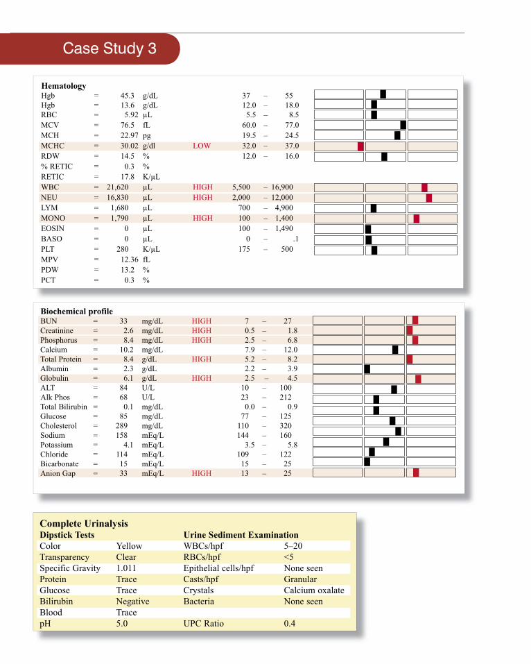
Hematology Hgb = 45.3 g/dL 37 – 55 Hgb = 13.6 g/dL 12.0 – 18.0 RBC = 5.92 µL 5.5 – 8.5MCV = 76.5 fL 60.0 – 77.0 MCH = 22.97 pg 19.5 – 24.5 MCHC = 30.02 g/dl LOW 32.0 – 37.0 RDW = 14.5 % 12.0 – 16.0 % RETIC = 0.3 %RETIC = 17.8 K/µL WBC = 21,620 µL HIGH 5,500 – 16,900NEU = 16,830 µL HIGH 2,000 – 12,000LYM = 1,680 µL 700 – 4,900MONO = 1,790 µL HIGH 100 – 1,400EOSIN = 0 µL 100 – 1,490BASO = 0 µL 0 – .1PLT = 280 K/µL 175 – 500MPV = 12.36 fLPDW = 13.2 %PCT = 0.3 %
Biochemical profile BUN = 33 mg/dL HIGH 7 – 27 Creatinine = 2.6 mg/dL HIGH 0.5 – 1.8Phosphorus = 8.4 mg/dL HIGH 2.5 – 6.8Calcium = 10.2 mg/dL 7.9 – 12.0Total Protein = 8.4 g/dL HIGH 5.2 – 8.2 Albumin = 2.3 g/dL 2.2 – 3.9Globulin = 6.1 g/dL HIGH 2.5 – 4.5 ALT = 84 U/L 10 – 100 Alk Phos = 68 U/L 23 – 212Total Bilirubin = 0.1 mg/dL 0.0 – 0.9 Glucose = 85 mg/dL 77 – 125 Cholesterol = 289 mg/dL 110 – 320Sodium = 158 mEq/L 144 – 160Potassium = 4.1 mEq/L 3.5 – 5.8Chloride = 114 mEq/L 109 – 122Bicarbonate = 15 mEq/L 15 – 25Anion Gap = 33 mEq/L HIGH 13 – 25
Complete UrinalysisDipstick Tests Urine Sediment ExaminationColor Yellow WBCs/hpf 5–20Transparency Clear RBCs/hpf <5Specific Gravity 1.011 Epithelial cells/hpf None seenProtein Trace Casts/hpf GranularGlucose Trace Crystals Calcium oxalateBilirubin Negative Bacteria None seenBlood Trace pH 5.0 UPC Ratio 0.4
Case Study 3
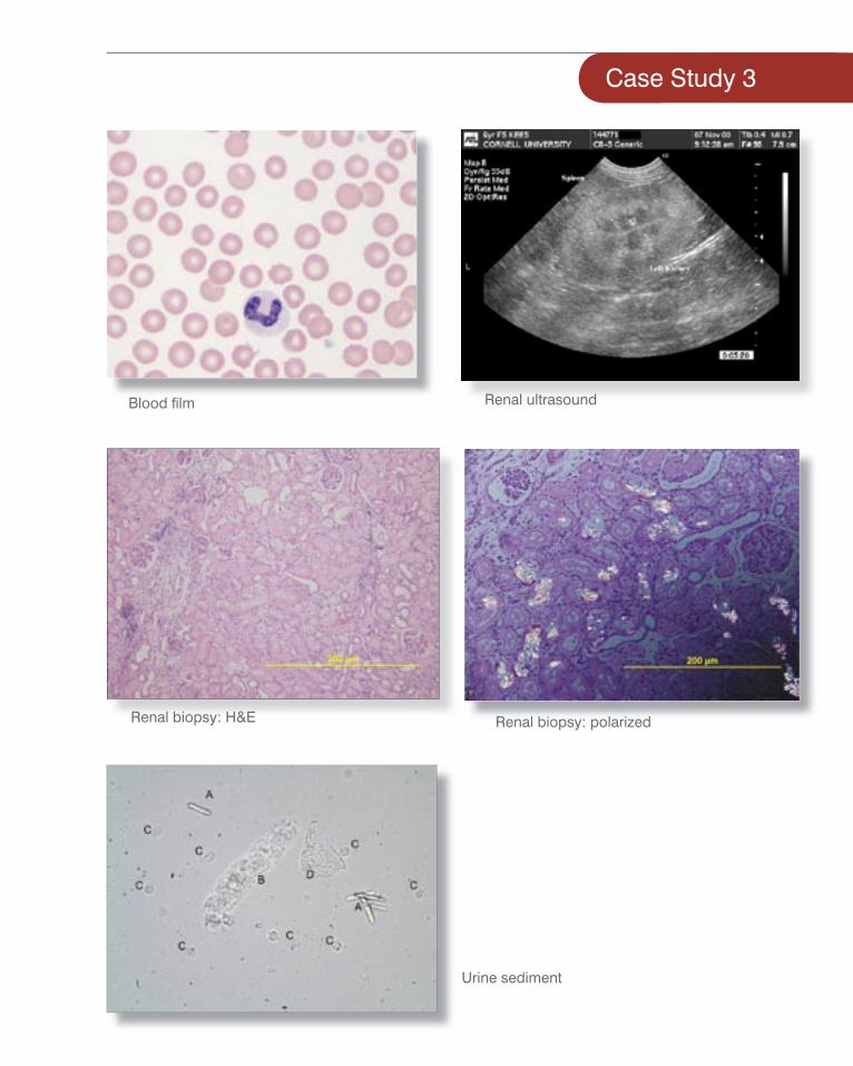
Blood fi lm Renal ultrasound
Renal biopsy: H&E Renal biopsy: polarized
Urine sediment
Interpretive SummaryHematologyThere is a mild leukocytosis characterized by a mild neutrophilia, a minimal left shift, a mild monocytosis and eosinopenia observed on microscopic examination of the blood fi lm. Changes are most consistent with mild infl ammation. No signifi cant abnormalities are observed in the erythron, and platelet numbers are adequate.
Biochemical profi leThere is a mild azotemia (increased BUN and creatinine) supporting decreased glomerular fi ltration (GFR). The fi nding of a nonconcentrated urine specifi c gravity supports the presence of renal azotemia (renal insuffi ciency). There is a mild hypernatremia, which correlates with the clinically noted dehydration and decreased water balance; however, the chloride is relatively low compared to the sodium, suggesting loss or sequestration of chloride. The clinical fi nding of vomiting suggests loss of HCl-rich gastric contents is most likely and a metabolic alkalosis is present. The moderately increased anion gap indicates the presence of signifi cant amounts of unmeasured anions, such as phosphates and sulfates due to the decreased GFR. This is supportive of the presence of a titrational metabolic acidosis; however, the degree of azotemia and increased anion gap appear discordant, and the presence of other unmeasured anions, such as ethylene glycol, must be considered.
The within-reference-range TCO2 is due to the negating effects of the typical increased TCO2 with metabolic alkalosis and the typical decreased TCO2 with titrational acidosis. Blood gas analysis to determine the degree of acidemia or alkalemia is warranted. The hyperphosphatemia is most likely due to the decreased GFR and retention of phosphorus. The slight hypokalemia may be due to decreased intake. There is a slight hyperproteinemia characterized by a low-normal albumin and a mild hyperglobulinemia. This protein pattern is most supportive of infl ammation.
UrinalysisThe fi nding of an acidic urine in the face of a metabolic alkalosis and acidosis suggests the acidosis condition is more severe and acidemia may be present. Evaluation of the blood gas data to determine if there is acidemia or alkalemia and the severity of the disorder is warranted. Multiple signifi cant abnormalities are noted within the microscopic portion of the urinalysis. The fi nding of monohydrate calcium oxalate crystals is strongly supportive of ethylene glycol toxicity. The presence of granular casts suggests the presence of signifi cant tubular injury. The presence of white blood cells (WBC) in the urine sediment indicates the presence of infl ammation; however, localization of the infl ammation is not possible since the sample is a free-catch specimen. A trace protein content is diffi cult to accurately assess in a urine sample that has a fi xed specifi c gravity (no concentration); however, the urine protein to urine creatinine (UPC) ratio suggests that signifi cant proteinuria is not present. Even if there were a slight signifi cant increase in the UPC ratio, accurate interpretation would be diffi cult since the urine sediment is active (WBC and granular casts present). Any slight protein present may be associated with mild infl ammation or tubular injury.
DiagnosisEthylene glycol toxicity
Treatment/Plan• Blood gas analysis
• Osmolality and osmolar gap evaluation
• Abdominal ultrasound
• ± Ethylene glycol assay
• Initiate therapy for suspected ethylene glycol toxicity (fl uids, electrolytes, acid base therapy, maintain adequate urine volumes)
• 4-methylpyrazole (4MP)
• Consider dialysis if availablegap appear discordant, and the presence of other unmeasured anions, such as ethylene glycol, must be considered.
is due to the negating effects of the with metabolic alkalosis and the typical decreased
with titrational acidosis. Blood gas analysis to determine the degree of acidemia or alkalemia is warranted. The hyperphosphatemia is most likely due to the decreased GFR and retention of phosphorus. The slight hypokalemia may be due to decreased intake. There is a slight hyperproteinemia characterized by a low-normal albumin and a mild hyperglobulinemia. This protein pattern is most supportive of infl ammation.
The fi nding of an acidic urine in the face of a metabolic alkalosis and acidosis suggests the acidosis condition is more severe and acidemia may be present. Evaluation of the blood gas data to determine if there is acidemia or alkalemia and the severity of the disorder is warranted. Multiple signifi cant abnormalities are noted within the microscopic portion of the urinalysis. The fi nding of monohydrate calcium oxalate crystals is strongly supportive of ethylene glycol toxicity. The presence of granular casts suggests the presence of signifi cant tubular injury. The presence of white blood cells (WBC) in the urine sediment indicates the presence of infl ammation; however, localization of the infl ammation is not possible since the sample is a free-catch specimen.
ethylene glycol toxicity (fl uids, electrolytes, acid base therapy, maintain adequate urine volumes)
• 4-methylpyrazole (4MP)
• Consider dialysis if available
The moderately increased anion gap indicates the presence of signifi cant amounts of unmeasured anions, such as phosphates and sulfates due to the decreased GFR. This is supportive of the presence of a titrational metabolic acidosis; however, the degree of azotemia and increased anion
amounts of unmeasured anions, such as phosphates and sulfates due to the decreased GFR. This is supportive of the presence of a titrational metabolic acidosis; however, the degree of azotemia and increased anion gap appear discordant, and the presence of other unmeasured anions, such as ethylene glycol, must be considered.
is due to the negating effects of the with metabolic alkalosis and the typical decreased
with titrational acidosis. Blood gas analysis to determine the degree of acidemia or alkalemia is warranted. The hyperphosphatemia is most likely due to the decreased GFR and retention of phosphorus. The slight hypokalemia may be due to decreased intake. There is a slight hyperproteinemia characterized by a low-normal albumin and a mild hyperglobulinemia. This protein pattern is most supportive of infl ammation.
The fi nding of an acidic urine in the face of a metabolic alkalosis and acidosis suggests the acidosis condition is more severe and acidemia may be present. Evaluation of the blood gas data to determine if there is acidemia or alkalemia and the severity of the disorder is warranted. Multiple signifi cant abnormalities are noted within the microscopic portion of the urinalysis. The fi nding of monohydrate calcium oxalate crystals is strongly supportive of ethylene glycol toxicity. The presence of granular casts suggests the presence of signifi cant tubular injury. The presence of white blood cells (WBC) in the urine sediment indicates the presence of infl ammation; however, localization
•
Case Study 3
Case Study 4
PatientEight-year old spayed female Shetland sheepdog
Presenting ComplaintsVomiting, diarrhea, lethargy, anorexia, edema
HistoryFive-day history of lethargy, anorexia, vomiting and diarrhea
Physical ExamIncreased respiratory rate; bilateral facial, ventral and peripheral edema
Fezzie Smith
Case Study 4
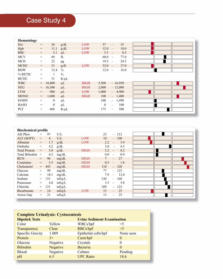
Hematology Hct = 36 g/dL LOW 37 – 55 Hgb = 11.1 g/dL LOW 12.0 – 18.0 RBC = 5.1 µL LOW 5.5 – 8.5MCV = 69 fL 60.0 – 77.0 MCH = 22 pg 19.5 – 24.5 MCHC = 31 g/dl LOW 32.0 – 37.0 RDW = 12.4 % 12.0 – 16.0 % RETIC = 1 %RETIC = 51 K/µL WBC = 18,800 µL HIGH 5,500 – 16,950NEU = 16,300 µL HIGH 2,000 – 12,000LYM = 900 µL LOW 1,000 – 4,900MONO = 1,600 µL HIGH 100 – 1,400EOSIN = 0 µL 100 – 1,490BASO = 0 µL 0 – 100PLT = 468 K/µL 175 – 500
Biochemical profile Alk Phos = 97 U/L 23 – 212ALT (SGPT) = 4 U/L LOW 10 – 100 Albumin = 1.7 g/dL LOW 2.2 – 3.9Globulin = 4.2 g/dL 3.0 – 4.3 Total Protein = 5.8 g/dL HIGH 5.2 – 8.2 Total Bilirubin = 0.2 mg/dL 0.0 – 0.9 BUN = 96 mg/dL HIGH 7 – 27 Creatinine = 5.5 mg/dL HIGH 0.5 – 1.8Cholesterol = 443 mg/dL HIGH 110 – 320Glucose = 99 mg/dL 77 – 125 Calcium = 10.1 mg/dL 7.9 – 12.0Sodium = 151 mEq/L 144 – 160Potassium = 4.8 mEq/L 3.5 – 5.8Chloride = 121 mEq/L 109 – 122Bicarbonate = 14 mEq/L LOW 15 – 25Anion Gap = 21 mEq/L 13 – 25
Complete Urinalysis: CystocentesisDipstick Tests Urine Sediment ExaminationColor Yellow WBCs/hpf <5Transparency Clear RBCs/hpf <5Specific Gravity 1.009 Epithelial cells/hpf None seenProtein 3+ Casts/hpf 0Glucose Negative Crystals 0Bilirubin Negative Bacteria 0Blood Negative Culture PendingpH 6.5 UPC Ratio 18.6
Case Study 4
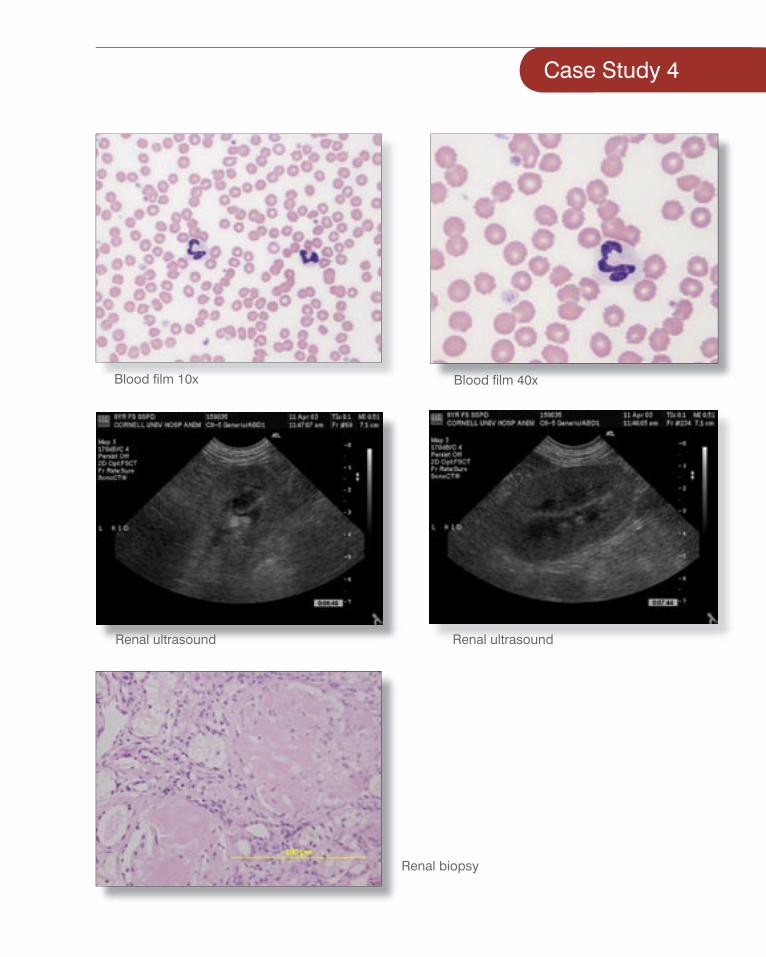
Blood fi lm 10x Blood fi lm 40x
Renal ultrasound Renal ultrasound
Renal biopsy
Interpretive SummaryHematologyModest leukocytosis composed of mature neutrophilia and monocytosis with concurrent lymphopenia is a stressed leukogram. This is typically consistent with infl ammation, infection or increased cortisol concentrations from exogenous use or hyperadrenocorticism.
Biochemical profi leThis dog is suffering from severe hypoalbuminemia. Because the serum globulin concentration is high-normal, this is likely a result of liver disease, renal loss or vasculitis. All other parameters assessing liver function (cholesterol, glucose, and bilirubin) are within normal limits (the cholesterol is actually high and not low as in liver insuffi ciency), making liver insuffi ciency much less likely. Therefore, renal loss and vasculitis become the two likely possibilities. The facial edema evident on presentation may be a result of the hypoalbuminemia, with or without a degree of vasculitis.
UrinalysisA very high UPC of 18.6 was identifi ed in this dog. This degree of proteinuria is very likely to be glomerular in origin and is enough to explain the severe hypoalbuminemia. This dog, therefore, has all four criteria for nephrotic syndrome: proteinuria, hypoalbuminemia, hypercholesterolemia and edema. Aggressive diagnostic and therapy are necessary in cases of nephrotic syndrome in an attempt to reverse the cause. Likely causes include glomerulonephritis and amyloidosis.
RadiologyAbdominal ultrasound report
The renal cortices appear to be mildly hyperechoic being isoechoic with the adjacent spleen. There is mild dilatation of the renal pelvices. No other abnormalities are seen. The hyperechoic cortex is a nonspecifi c fi nding seen in both acute and chronic renal disease. Amyloidosis can also cause hyperechoic renal cortices. The mild pyelectasia is suggestive of recent fl uid administration.
Additional testingRenal Biopsy
Severe glomerulopathy with amorphous pink material consistent with amyloid.
DiagnosisThe clinical diagnosis is amyloidosis.
Treatment/Plan• Thoracic radiographs ± blood
gas analysis
• Urine culture
• Nonspecifi c therapy for proteinuria and hypertension
• Will not likely benefi t from immunosuppression.
• Consider: DMSO, MSM
syndrome in an attempt to reverse the cause. Likely causes include
The renal cortices appear to be mildly hyperechoic being isoechoic with the adjacent spleen. There is mild dilatation of the renal pelvices. No other abnormalities are seen. The hyperechoic cortex is a nonspecifi c fi nding seen in both acute and chronic renal disease. Amyloidosis can also cause hyperechoic renal cortices. The mild pyelectasia is suggestive of recent fl uid administration.
Severe glomerulopathy with amorphous pink material consistent with amyloid.
hypoalbuminemia. This dog, therefore, has all four criteria for nephrotic syndrome: proteinuria, hypoalbuminemia, hypercholesterolemia and edema. Aggressive diagnostic and therapy are necessary in cases of nephrotic syndrome in an attempt to reverse the cause. Likely causes include
hypoalbuminemia. This dog, therefore, has all four criteria for nephrotic syndrome: proteinuria, hypoalbuminemia, hypercholesterolemia and edema. Aggressive diagnostic and therapy are necessary in cases of nephrotic syndrome in an attempt to reverse the cause. Likely causes include
The renal cortices appear to be mildly hyperechoic being isoechoic with the adjacent spleen. There is mild dilatation of the renal pelvices. No other abnormalities are seen. The hyperechoic cortex is a nonspecifi c fi nding seen in both acute and chronic renal disease. Amyloidosis can also cause hyperechoic renal cortices. The mild pyelectasia is suggestive of recent fl uid administration.
Severe glomerulopathy with amorphous pink material consistent with amyloid.
Case Study 4

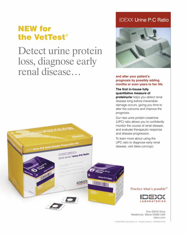
Detect urine protein loss, diagnose early renal disease…
and alter your patient’s prognosis by possibly adding months or even years to her life.
The first in-house fully quantitative measure of proteinuria helps you detect renal disease long before irreversible damage occurs, giving you time to alter the outcome and improve the prognosis.
Our new urine protein:creatinine (UPC) ratio allows you to confidently monitor the course of renal disease, and evaluate therapeutic response and disease progression.
To learn more about using the UPC ratio to diagnose early renal disease, visit idexx.com/upc.
IDEXX Urine P:C Ratio
NEW for the VetTest®
Many thanks to Bandit Bowker, a rescued stray.
One IDEXX Drive Westbrook, Maine 04092 USA
idexx.com
© 2005 IDEXX Laboratories, Inc. All rights reserved. • 09-65320-00 (5)


![ASN Kidney Week 2016 – Renal Biopsy: Clinical Correlations · ASN Kidney Week 2016 – Renal Biopsy: Clinical Correlations. ... ( ds) DNA, ANCA, SPEP/UPEP ]. ... ASN Kidney Week](https://img.pdfslide.net/doc/110x75/5aec58b47f8b9a90318e2a7d/asn-kidney-week-2016-renal-biopsy-clinical-correlations-kidney-week-2016-.jpg)