Embed Size (px)
Citation preview
REVIEW ARTICLE Open Access
Reported concepts for the treatmentmodalities and pain management oftemporomandibular disordersMieszko Wieckiewicz1*, Klaus Boening2, Piotr Wiland3, Yuh-Yuan Shiau4 and Anna Paradowska-Stolarz5
Abstract
Background: Pain related to temporomandibular disorders (TMD) is a common problem in modern societies. Theaim of the article is to present the concepts of TMD pain clinical management.
Methods: A survey was performed using the PubMed, SCOPUS and CINAHL databases for documents publishedbetween 1994 and 2014. The following search keywords were selected using MeSH terms of the National Library ofMedicine in combination: TMD pain, TMD, TMJ, TMJ disorders, occlusal splint, TMD physiotherapy, TMJ rheumatoiddisorders and TMJ surgery. Original articles and review papers which presented the clinical relevance and practicalvalidity regarding the possibility of application in TMD management have been included. Authors have excludedarticles without outstanding practical aspect and evidence-based background. A first selection was carried out byreviewing titles and abstracts of all articles found according to the criteria. After that the full texts of potentially suitablearticles were assessed. In line with these criteria, among 11467 results the writers have included 66 papers.
Results: The most commonly reported conservative treatments are massage therapy and individually fabricatedocclusal splints. In addition to massage, other popular methods include manual therapy and taping, warming/coolingof aching joints, and light and laser therapy. Drugs are also commonly used. In the most severe cases of thetemporomandibular joint degeneration, surgical restoration of the joint is sometimes applied.
Conclusions: The authors concluded that conservative treatment including counselling, exercises, occlusalsplint therapy, massage, manual therapy and others should be considered as a first choice therapy for TMDpain because of their low risk of side effects. In the case of severe acute pain or chronic pain resulting fromserious disorders, inflammation and/or degeneration pharmacotherapy, minimally invasive and invasive proceduresshould be considered.
Keywords: Temporomandibular disorders, Temporomandibular joint disorders, Facial pain, Masticatory muscle pain
IntroductionCurrently, temporomandibular disorders (TMD) refer tothe causes responsible for the impaired function of thetemporomandibular joints (TMJ) and the associatedneuro-muscular system, which may provoke TMD-related pain [1]. The term TMD is not a diagnosis butrather a broad term that contains a number of diseaseentities, such as pain in masticatory muscles and tem-poromandibular joints, headache, disturbances in jaw
movements and sounds in joints while opening and clos-ing the mouth. The causes of these diseases/symptomsare numerous and include trauma, systemic, iatrogenic,occlusal and mental health disorders [2–7]. Today, men-tal health plays a dominating role in the pathogenesis ofTMD [8, 9]. The neuromuscular system responsible forchewing function has a high potential to adapt to changingconditions. Only when the compensatory capabilities of themasticatory- and the neuromuscular system are over-stretched dysfunction occurs resulting in clinical symptomsand manifests as pain, severe clicking, or limited mobilityof the mandible, forcing the patient to seek help.* Correspondence: [email protected]
1Department of Prosthetic Dentistry, Faculty of Dentistry, Wroclaw MedicalUniversity, 26 Krakowska St., 50425 Wroclaw, PolandFull list of author information is available at the end of the article
© 2015 Wieckiewicz et al. Open Access This article is distributed under the terms of the Creative Commons Attribution 4.0International License (http://creativecommons.org/licenses/by/4.0/), which permits unrestricted use, distribution, andreproduction in any medium, provided you give appropriate credit to the original author(s) and the source, provide a link tothe Creative Commons license, and indicate if changes were made.
Wieckiewicz et al. The Journal of Headache and Pain (2015) 16:106 DOI 10.1186/s10194-015-0586-5

The pain may radiate to different regions, such as thedental arches, ears, temples, forehead, occiput, cervicalregion of spine or shoulder girdle [10–13]. However,despite the fact that comparatively few patients are seek-ing treatment, it is known that there is a high prevalenceof TMD in developed societies [14, 15]. TMD is mostlyaccentuated on the neck, where the lateral support im-balance leads to the bending of the neck to the affectedside [16].TMD are a group of dysfunctions and disorders re-
lated to impaired function of the temporomandibularjoints and associated muscles therefore they may lead tothe painful impairment in stomatognathic system func-tioning [17]. The TMJ is used 1500–2000 times a day,which shows how great discomfort is carried by thepathologies in jaw movements [9].In most cases, the symptoms are the reason for the in-
creased tension of the masticatory musculature, and theparafunctions may worsen the symptoms [18, 19]. Dueto the large subjectiveness of the symptoms, TMDs arevery difficult to diagnose, especially because patientsusually search for help from other specialists besidesdentists (e.g., neurologist, otolaryngologist or ophthal-mologist) [10, 20]. The anomalies of the masticatorysystem including pain caused by increased tension ofmasticatory muscles are classified as masticatory paindysfunction syndrome (MPDS) [21].In addition to pain, a vast majority of patients suffer
from intraoral signs of masticatory dysfunction, includ-ing increased sensitivity of the teeth due to abfractionand pathological attrition, gingival recessions, teethhypermobility and bone support loss. In addition, teethimpressions on soft tissues are observed, including teethimpressions on the tongue and (cheek mucosa) linea
alba [10, 22]. The increased tension in TMJ muscles andco-existing parafunctions or dysfunctions may lead tonon-carious tooth lesions (e.g., abfraction), which arecharacteristic for TMD [23, 24].The treatment of TMD is complicated and requires
specific knowledge and exercises to strengthen somegroups of muscles and weaken others, occlusal splinttherapy, massage and pharmacotherapy. Although thetreatment seems difficult, most of the patients searchingfor help due to TMD assess that the treatment is suc-cessful, although an accurate diagnosis needs to be madeto start the proper protocol of treatment [20, 25–27].Theories on the origin of TMD are presented in Table 1[27]. Yet, it is important to note that treating TMD onlyfrom the dental perspective may fail, as many of theseanomalies are caused by somatic diseases that shouldhave be cured in the first place [28].The prevalence of these disorders and the multifactor-
ial pathogenesis and therapeutic difficulties of TMDprompted the authors to undertake an effort to describetherapeutic concepts associated with TMD pain.
ReviewMaterials and methodsA survey was performed using the PubMed, SCOPUS andCINAHL databases for documents published between 1994and 2014. The following search keywords were selectedusing MeSH terms of the National Library of Medicine incombination: TMD pain, TMD, TMJ, TMJ disorders, occlu-sal splint, TMD physiotherapy, TMJ rheumatoid disordersand TMJ surgery. Original articles and review papers whichpresented the clinical relevance and practical validity re-garding the possibility of application in TMD managementhave been included. The inclusion of the papers were based
Table 1 Theories concerning TMD origin [27]
Name of the theory Statements of the theory
Mechanical displacement(by Costen)
Lack of support in lateral teeth or functional occlusal premature contacts lead to direct eccentric positioning of thecondyle in the glenoid fossa; this leads to pain, ear symptoms, adverse muscle activity and TMD
Trauma theory(by Zack and Speck)
The principal factor of TMD is micro-/macro-trauma; trauma can cause structural alternation to the muscles or directly tothe joint structures
Biomedical (by Reade) Disorder is initiated by trauma; specific factors (malocclusion, parafunctions, occupational activities) cause the progression ofthe symptoms
Osteoarthric(by Stegenga)
Osteoarthrosis is a main cause of TMD; muscular symptoms and systemic diseases are secondary to TMJ pathology
Muscle(by Travell and Rinzler)
Masticatory muscles are the primary etiologic factor to TMD; myalgia (caused by chronic myospasm) is secondary toparafunctions and can refer pain to TMJ
Neuromuscular (byRamfjord)
Occlusal problems cause TMDs, the loss of occlusal equilibrium leads to the incoordination of muscles and spasms
Psychophysiological(by Schwartz and Laskin)
TMD occurs outside of the physical factors; psychosocial factors play a crucial role in TMD pathogenesis – the mainfactor of hypertension and overcontraction of the muscle is due to the parafunctions performed torelieve stress
Psychosocial theory(by Dworkin)
Emotional disturbances induce hyperactivity of the muscles and lead to parafunctional habits and occlusal anomalies;the muscle contractivity is accentuated with teeth clenching, and repeatability leads to pain
Wieckiewicz et al. The Journal of Headache and Pain (2015) 16:106 Page 2 of 12

on precise descriptions of the treatment procedures and de-tailed presentation of the treatment outcomes. Authorshave excluded articles without outstanding practical aspectand evidence-based background. A first selection was car-ried out by reviewing titles and abstracts of all articlesfound according to the criteria. After that the full texts ofpotentially suitable articles were assessed. In line with thesecriteria, among 11467 results the writers have included 66papers.
Conservative treatmentTherapeutic exercisesThe most important stage of a treatment protocol is educa-tion with cognitive awareness training and relaxation ther-apy as well as self-observation that should be completed bypatients with masseter hypertrophy, tension-type headachesor bruxomania (the grinding of teeth occurring as a neur-otic habit during the waking state). It is important to ex-plain to the patient the background of the disorders(especially the role of one’s emotional stress) and warnabout habitual parafunctional activities (e.g., nonfunctionaltooth contacts or oral mucosa biting). The patient shouldbe aware of what he or she does with their teeth, and whenthey fall into bad habits, try to eliminate that habit [28].Muscular training is the primary mode to achieve
muscle restoration, especially after traumas and injuries.It is thought to be the most conservative treatment aswell as the simplest and most non-invasive method ofTMD treatment. In patients with severely expressedasymmetries and symptoms, exercises to restore themuscular equilibrium seem to be the only proper routeof treatment [3, 29]. Muscular therapy must be restrict-ive; it should be carried out moderately, and the inten-sity should be increased with time to avoid aches andpatient discouragement from the suggested treatment. Inthis situation, muscular therapy is effective in 70 % ofsuffering patients. In some cases, such as patients withmuscular or joint (muscular or arthritis pain) pain, themouth opening is limited, and therefore, therapy is lesseffective [3, 27]. The exercises can require stretching, re-laxation and isometric movements that should be per-formed routinely to eventually lead to a shortening ofthe excessively expanded muscles or to a restoration ofthe full length of the shortened muscles. Additionally,the natural tension and symmetric jaw movement can berestored [3].The training is underdone to correct the mobility of
the mandible. To strengthen the muscles and to acquirebalance between the left and right sides, opening themouth along a straight line in front of the mirror is rec-ommended. The resistance is acquired from the gentlepressure of the patient’s fingers to the mandible. The ex-ercises are repeated in sets of 15 to 20 repetitions, 2 to 3
times a day. The improvement should be observed after6 weeks [3].Research from Bae and Park [30] showed that active
and relaxation exercises could improve the limited rangeof motion, deviation and pain in masticatory muscles.For muscle relaxation, they recommend putting thefront one-third of the tongue on the anterior part ofpalate and applying a light force to the tip of the tongueso it does not touch the teeth, having the patient main-tain this position as long as he/she can withstand (3times over a period of 4 weeks, 10 min each time).In case of too wide of a mouth opening, or excessive
mobility of the jaw and mandible deviation during open-ing (with excluded suspicion of subluxation), the exer-cises are limited, and straightening of the openingpathway are recommended. The exercise involves open-ing the mouth with the tip of the tongue touching thepalate (usually near the A-H line) in front of a mirror,along the straight line. It is recommended to maintainthe contraction of the tongue muscles for two secondsduring mouth opening. The exercises should be repeated2 to 3 times a day, 15 to 20 repetitions each [3].
Occlusal splint therapyTo achieve the proper relation of the jaw, centric rela-tion (CR) should be restored. It is easily performed byocclusal splints. An occlusal appliance is any removableartificial occlusal surface used for diagnosis or therapyaffecting the relationship of the mandible to the maxil-lae. Occlusal appliances may be used for occlusalstabilization, for the treatment of temporomandibulardisorders, or for the prevention of dentition wear [31].Occlusal splints are used in a vast majority of patientswith TMDs to restore the static and dynamic symmetryof the stomatognathic system. Most commonly, they areused in cases with disc displacement [3, 32, 33]. Thesplints are fabricated individually by an experiencedteam consisting of a dentist and technician.One of the most popular occlusal splints is the
Michigan-type bite splint, precisely described by Ramfjordand Ash Jr [34]. This splint could be used in both dentalarches, but preferably in the maxilla. The mandibularsplint is used when the posterior area is missing teeth inthe mandible and unwanted tooth movement must beavoided. The main purpose of this device is to disengagethe occlusion, place the condyle in the centric position,relax the masticatory muscles and prevent further toothwear due to nocturnal parafunctional activity. The mainfeatures of this splint are freedom in centric and canineguidance.It is important to note that the relation of the maxil-
lary and mandibular arches may differ after the treat-ment when compared to the initial state, especially whenpartial coverage splints are used [32, 35]. After the
Wieckiewicz et al. The Journal of Headache and Pain (2015) 16:106 Page 3 of 12

replacement of the mandible, the condyles are replaced,and consequently, the mandible is positioned properlyand the pain is reduced [32].Walczynska-Dragon and Baron [10] have proven that
occlusal splint therapy using the SVED (Sagittal VerticalExtrusion Device) appliance decreases not only aches inthe head and all parts of the spine but also disc displace-ments within 3 weeks of treatment. The next decrease infrequency of unwanted, unfavorable symptoms wasobserved after 3 months of treatment with splints. Whenproperly performed, these splints also unblock a limitedmouth opening.Research performed by Lee et al. [36] in a group of 59
patients with somatic TMJ dysfunction showed thatintraoral appliance could improve cervical spine align-ment and alleviate symptom severity.The occlusal splints are also used in the initial phase
of treatment in patients with mouth overclosure causedby a pathologic deep bite. Before the prosthetic rehabili-tation of the severe tooth wear, one should rememberthat initially, splint therapy should be applied to adaptthe stomatognathic system to the new occlusion [37]. Aclassification of the occlusal appliances with activitiesand recommendations is presented in Table 2.Beside occlusal splint therapy subsequently selective
grinding of the teeth to restore physiological and/ orproper and /or balanced occlusal support is required insome cases [27].
Massage therapyMyofascial pain is a common symptom of TMD – it isoften associated with the clenching of teeth, grindingand stress. TMD myofascial pain occurs in 31 to 76 % ofthe population [38–41]; it can be relieved by massagetherapy, which leads to re-establishing the proper flexi-bility and muscular length and relieves pain. The mas-sage therapy for TMD might be divided into effleurage,kneading, friction, stretching and petrissage, leading tothe permanent adaptation of the muscles. The types ofmassage and their influence to the surrounding tissues
are presented in Table 3 [26, 42]. Massage reduces tissueswelling as well as pain in TMD patients [21, 41, 43]. Thepressure used during massage must not be too intenseand should increase over time at each therapeutic sessionbecause therapy performed too strongly may lead to in-creased muscular tonus [44]. Massages should be per-formed twice a week, with a minimum of 30 min for eachsession. It takes at least 8 therapeutic sessions to receivetrue relief [23]. In addition to the local influence, massageleads to the relaxation of the entire body and reducesstress, thus improving the patient’s mood [45, 46]; itreduces tension headaches and muscle aches, restoresequilibrium between the masseter tension, and improvesmastication [26]. The physiotherapist may also recom-mend heating or cooling of the affected muscles [28].To restore the accurate function of the TMJ, changes
in daily habits is important. The change of foodconsistency (eating softer foods), applying cold or heat,and avoiding extreme movements of the mandible(chewing gum, wide yawning or loud singing) might beenough to decrease TMD symptoms [47]. In thissituation, counselling, behavioral therapy and stressmanagement should also be applied to decrease musclehypertension and bad habits [25].
Manual therapyManual therapy is similar to massage therapy, but theprocedure performed by the physiotherapist is different;it refers to stimulation of the so-called “trigger points”.There are two main methods of treatment by applyingmanual therapy: mobilization and the muscle energytechnique.The mobilization technique is most commonly used in
disc displacements; it involves repeated traction or slid-ing movements at a slow speed and with increasingamplitude. The desirable effect is to increase the limitedrange of motion within the joint and reduce pain. Themovements are carried out perpendicularly or parallel tothe plane of the treated joint, oscillating, and typicallyrepeated 8 to 10 times in 3 sets. The procedure is
Table 2 Classification of occlusal splints according to Freesmeyer et al. [28]
Type of occlusal appliances Activity Recommendations
Reflex appliances e.g.,Interceptor, Anterior Plateau,NTI-tss
Prevent habitual tooth contact and thus preventgnashing and clenching temporarily, which positivelyinfluences the resultant tooth and muscle complaints.
Indicated for acute symptoms that can be attributedto an overloading of the involved tissue (short-term appliances).
Stabilization appliances e.g.,Michigan type splint
Create ideal occlusion, synchronous tooth contact ina centric condyle position in static occlusion and ananterior tooth position with disclusion in the lateralteeth region in dynamic occlusion.
Can be used on a short-term and long-term basis, for acuteor chronic symptoms and also in psychological andphysiological overloading reactions.
Repositioning appliances e.g.,Anterior repositioning splint,Farrar type splint, Gelb typesplint
The temporomandibular joint or joints is/are set in atherapeutic position by the splint to support healingand to maintain a symptom-free joint posture.
Used for the treatment of temporomandibular joint diseasessuch as anterior disc displacement with and without reduction,temporomandibular joint compression, retral displacement ofthe condyle and osteoarthritis. Can be used as a short-term orlong-term therapy.
Wieckiewicz et al. The Journal of Headache and Pain (2015) 16:106 Page 4 of 12

performed in a seated position with the patient’s headstabilized on the chest of the physiotherapist who holdsthe patient’s head and mobilizes the mandible with onehand. Traction consists of 3 stages: relaxation (abolitionof forces acting on joint), tension (remotion of thearticular area) and stretch (increase in remotion ofarticular area) [48, 49].The muscle energy technique (MET) is used when
limited movements of the mandible are observed andcaused by soft tissue (muscles and connective tissue) dam-age. The treatment involves repeating 3 phases: the firstphase is making a movement that is possible due to lim-ited tissue elasticity; in the second phase, the patientslightly tightens the muscles trying to make a move in theopposite direction of the force created by the physiother-apist and should last approximately 10 s; in the last phase,the patient relaxes the muscles. The technique can beperformed both in a seated or lying position [50, 51].
Other physiotherapeutic techniquesPhysiotherapy involves many techniques of treatment.The most common massage and manual therapies werepreviously described, but for TMD treatment, also othertechniques are used. Among them, biofeedback, lampexposure, iontophoresis, ultrasound and transcutaneouselectrical nerve stimulation (TENS) are used.The purpose of biofeedback is to stimulate the muscles
to work properly and achieve maximal relaxation of themuscles in a short period of time. The therapy involveselectromyography to train the adequate neuromusculartension of the patient and develops the ability to alter aphysiological response. The surface electrodes are placedon the muscles (typically masseter) uni- or bilaterally;other muscles (e.g., anterior temporalis) may also be in-cluded. SEMG biofeedback may include muscle tensiondiscrimination. The treatment protocol involves teachingthe patient how to open their mouth properly tostrengthen the tension of the tongue and protrude themandible. Only after this are the electrodes applied inline with the muscle fibers (usually upon the midsub-stance of the masseter muscle belly). The measurementsof the minimal muscular tension are performed whenthe patient rests with all their muscles relaxed; this is
used as a reference in the follow-up. Observing the move-ments and muscular tonus the patient exercises help torestore the appropriate muscular activity [52–54].Transcutaneous Electrical Nerve Stimulation (TENS)
is another well-known method of pain relief for TMDs.The method is based on electrical stimulation of painareas via surface electrodes and is considered safe andnon-invasive. TENS helps to relieve chronic and acutepain in joint and/or muscle disorders. Unfortunately,due to the small number of studies (especially random-ized trials), TENS cannot yet be considered a standardtreatment for TMDs, as its effectiveness is still uncertain[55]. In addition to the therapeutic value of electric po-tential, a tool called electromyography (EMG) is used forestablishing muscular function and is the most reliableand objective technique [56].For pain release, especially in subacute arthropathies
and inflammatory rheumatic diseases, heat treatment isapplied; it alleviates strong pain, although the result istypically short-therm. Heat is supplied either by meansof Solux lamps (ca. 15 min from 20 cm distance) orthrough a thermophor filled with water at a temperatureof 158 to 176 °F (70 to 80 °C) and wrapped with a towel.Other recommendations to decrease pain are sulfur andiodide baths. Cryotherapy is another form of temperaturerelated therapy but applies cold instead of heat. Cold packs,cold spray or air, and ice compresses are used as analgesicagents. The application of cold is used immediately priorto kinesiotherapy and helps fight muscle hypertension andtendinopathies as well as rheumatic diseases. One shouldremember that there is a high risk of frostbite (skin damagedue to low temperature) with this form of therapy. Thecold compresses should be applied for 10–15 min. Cryo-therapy leads to the attenuation of pain, reduces stiffnessin the TMJ and increases mandibular mobility [57].A new method of rehabilitation with the aim of TMJ
stabilization and increased jaw stability is taping, orKinesio Taping (KT). KT also decreases drooling andprovides mouth closure. To increase jaw stability, onepiece of tape in a “Y”-shape cut should be prepared andplaced proximal to the joint; the superior tail is shorterthan the inferior tail. The superior tail should be applieddiagonally along the upper jaw and directed towards the
Table 3 Massage procedures in myofascial TMD pain management [26]
Type of movementin massage procedure
Manner of performing Result
Effleurage, Kneading Soothing, stroking, circular movements of skinand underlying tissues (performed at the endor beginning of therapeutic session)
Warming up the muscles, providing blood and lymph flow, increasingblood level in the massaged tissues (improved blood flow in small vessels)
Friction Pressure of fingertips in trigger points therapy;the pressure is increased in particular, sensitivepoints until the release
Remodeling tissues locally (reconstruction of muscular microstructure);effective in short-term pain relief (activates pain-gate mechanism)
Stretching (“petrissage”) Rolling of the muscles Increasing the range of movement and pain relief, decreasing musclecontraction
Wieckiewicz et al. The Journal of Headache and Pain (2015) 16:106 Page 5 of 12

lower cheek with “paper-off” tension. The tape widthshould be 1.5 to 2 inches (3.8 to 5 cm). The mandiblecannot subluxate at the movement. To decrease the hy-permobility of the joint and release TMJ pain, two tapepieces (1 inch wide and 2 inches long each) should beplaced diagonally to each other over the joint, formingan “X”. To improve jaw stability, tape is usually appliedto both sides. The balance in head position and bodyposture usually leads to a decrease in hypertension ofnot only the masticatory muscles but also the neck, armsand spine [58, 59]. The method is quite new but hasbecome increasingly popular [60, 61]. The special thera-peutic tape adheres to the skin with adequate flexibilityand consists of a polymer elastic strand wrapped by100 % cotton fibers. The tape allows for a normalizationof muscle tone and increases the process of self-healing.KT stimulates an endogenous analgesic system andchanges the subjective feelings of the patient. Alignmentof muscular tone is possible by improving proprioception.KT could be applied for myofascial pain therapy in a rangeof masticatory muscles, especially the masseters. Theclinical technique has been described by Kase et al. [62].Ultrasound therapy is one of the efficacious methods
for pain reduction, decrease in muscular tonus and im-proving the function of the muscles. It consists of threetypes of signals: constant waves, sound impulses andultrasound combined with stimulation current, which isfound to be most effective. The procedure is performed6–12 times, every 1–2 days, 6–8 min each. The impulsesshould be applied at 0.5–0.7 W/cm in the case of deviceswith constant waves, and 0.6–0.9 W/m in the case whensound impulses (50 or 100 Hz) are emitted [57].There are few rarely used methods of TMD manage-
ment. Among them are iontophoresis with differentmedications (e.g., nonsteroidal anti-inflammatory drugs,steroids and analgesics), especially in patients with con-current temporomandibular joint disc displacementwithout reduction and capsulitis [63]. As the data show,pain release is not observed, but patients present with awider opening of the mouth than when analgesics aloneare used [63]. Inflammatory processes may be healedwith a laser light that is used at a wavelength of 904 nmand a frequency of 700 Hz at 30 mm depth into the skin.This method had gained popularity [57].
Pharmacotherapy and minimally invasive andinvasive proceduresOral and injectable pharmacotherapyPharmacotherapy for TMD is not commonly used. It isonly used when other somatic symptoms, such as sleepdisorders, chronic pain, arthralgias, inflammatory diseases,myalgias or neuropathies are associated with TMD [28].As TMD may manifest from different systemic diseases(e.g., arthritis, inflammatory bowel diseases, Parkinson
disease), it is important to diagnose the patient properlyand implement treatment for the underlying disease, espe-cially when depression is a suspected diagnosis [47, 64].One has to remember that pharmacotherapy has its goalin decreasing pain and inflammation within the joint and/or muscles. This therapy improves function and inhibitsthe progression of the disease [65]. Pharmacotherapy canbe considered as a complementary therapy rather than atreatment itself. The exceptions are systemic diseases withTMJ involvement [57].For TMD release, the most commonly used medica-
tions are myorelaxants, nonsteroidal anti-inflammatorydrugs (NSAIDs), analgesics, tricyclic antidepressants,benzodiazepines and corticosteroids [28]. The first medi-cation of choice for moderate pain relief is acetamino-phen (average daily dose of 325–1000 mg). NSAIDs andanalgesics help to relieve pain (including radiating pain)in the head, jaw muscles, face, neck or shoulders. A highefficiency of TMD pain relief is shown with ibuprofen*and meloxicam** (average daily dose of 400–800 mg*and 7.5–15 mg**). In this particular situation, pharmaco-therapy is considered a supportive therapy that supple-ments other therapies. Used by itself, pharmacotherapy isconsidered for palliative therapy [48]. NSAIDs decreasepain and stop the inflammatory process [64].Muscle relaxants (baclofen, tizanidin, cyclobenzapr-
ine), opiates (morphine), anticonvulsants (e.g., gabapen-tin), ketamine, and TCA (e.g., amitriptyline) have alsobeen used clinically for TMJ management, but there isno evidence for their efficacy [65, 66]. To achieve themyorelaxation effect with low CNS impact, metaxoloneis recommended (average daily dose of 800 mg).In specific cases, medications should be used admittedly.
During acute spasms (sudden muscular contraction andpainful shortening that is maintained over time), anes-thetics are advised to block the pain and allow therapeuticstretching. Usually, the analgesic blockage with an infiltra-tion of 1 ml of 2 % lidocaine (without vasoconstrictor) inthe involved muscle is applied. A complementary therapymay include dypirone 500 mg (also in association with amyorelaxant, such as orfendrine, if necessary) 3 times aday, for 2 days [46]. In this situation, 90 % of cases requireanalgesic therapy [65].In myositis and other inflammatory disorders, the most
appropriate strategy is the administration of one dose ofcorticosteroid intramuscularly. Another approach is theinjection of an analgesic or anti-inflammatory agent. Themost common injections contain corticosteroids (withanti-inflammatory action) or hyaluronic acid [67]. In ani-mal models, the use of an inhibitor selective for the indu-cible COX-2 enzyme may attenuate the neurogeniccomponent of inflammation [47]. COX enzymes areblocked by NSAIDs. Unfortunately, those medicationshave a high risk of adverse side effects, which may include
Wieckiewicz et al. The Journal of Headache and Pain (2015) 16:106 Page 6 of 12
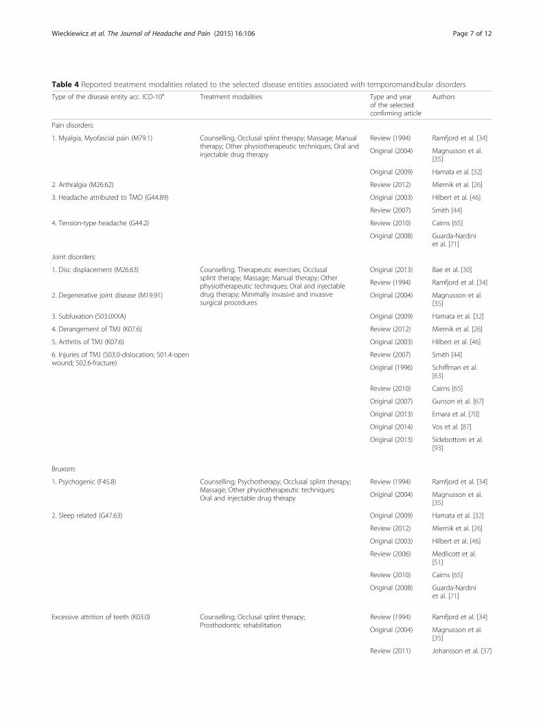
Table 4 Reported treatment modalities related to the selected disease entities associated with temporomandibular disorders
Type of the disease entity acc. ICD-10a Treatment modalities Type and yearof the selectedconfirming article
Authors
Pain disorders:
1. Myalgia, Myofascial pain (M79.1) Counselling; Occlusal splint therapy; Massage; Manualtherapy; Other physiotherapeutic techniques; Oral andinjectable drug therapy
Review (1994) Ramfjord et al. [34]
Original (2004) Magnusson et al.[35]
Original (2009) Hamata et al. [32]
2. Arthralgia (M26.62) Review (2012) Miernik et al. [26]
3. Headache attributed to TMD (G44.89) Original (2003) Hilbert et al. [46]
Review (2007) Smith [44]
4. Tension-type headache (G44.2) Review (2010) Cairns [65]
Original (2008) Guarda-Nardiniet al. [71]
Joint disorders:
1. Disc displacement (M26.63) Counselling; Therapeutic exercises; Occlusalsplint therapy; Massage; Manual therapy; Otherphysiotherapeutic techniques; Oral and injectabledrug therapy; Minimally invasive and invasivesurgical procedures
Original (2013) Bae et al. [30]
Review (1994) Ramfjord et al. [34]
2. Degenerative joint disease (M19.91) Original (2004) Magnusson et al.[35]
3. Subluxation (S03.0XXA) Original (2009) Hamata et al. [32]
4. Derangement of TMJ (K07.6) Review (2012) Miernik et al. [26]
5. Arthritis of TMJ (K07.6) Original (2003) Hilbert et al. [46]
6. Injuries of TMJ (S03.0-dislocation; S01.4-openwound; S02.6-fracture)
Review (2007) Smith [44]
Original (1996) Schiffman et al.[63]
Review (2010) Cairns [65]
Original (2007) Gunson et al. [67]
Original (2013) Emara et al. [70]
Original (2014) Vos et al. [87]
Original (2013) Sidebottom et al.[93]
Bruxism:
1. Psychogenic (F45.8) Counselling; Psychotherapy; Occlusal splint therapy;Massage; Other physiotherapeutic techniques;Oral and injectable drug therapy
Review (1994) Ramfjord et al. [34]
Original (2004) Magnusson et al.[35]
2. Sleep related (G47.63) Original (2009) Hamata et al. [32]
Review (2012) Miernik et al. [26]
Original (2003) Hilbert et al. [46]
Review (2006) Medlicott et al.[51]
Review (2010) Cairns [65]
Original (2008) Guarda-Nardiniet al. [71]
Excessive attrition of teeth (K03.0) Counselling; Occlusal splint therapy;Prosthodontic rehabilitation
Review (1994) Ramfjord et al. [34]
Original (2004) Magnusson et al.[35]
Review (2011) Johansson et al. [37]
Wieckiewicz et al. The Journal of Headache and Pain (2015) 16:106 Page 7 of 12

exacerbation of hypertension or gastrointestinal upset thatmay lead to ulcerations. COX-2-selective NSAIDs (eg.Celecoxib, Meloxicam) which have less side effects, arenot found to be better for the treatment of TMD. There isa hope that lotions containing NSAIDs will not have asmany side effects and will have a positive impact onrelieving pain [65].In chronic facial pain, aside from pain relievers, antide-
pressants should be used as a supplementary treatment[47]. Antidepressants may be used for chronic pain as aprimary analgesic. These medications manage headachesand neuropathic pain, reducing the feeling of depressioncaused by pain and improving sleep quality [65].It had been proven that NSAIDs relieve pain in
patients who suffer from arthritis. In this situation,diclofenac at a maximum dose of 50 mg orally 3 timesdaily or naproxen sodium 500 mg twice a day are rec-ommended, as they improve pain in more than half ofthe patients [65]. It had been shown that the use of anti-biotics, such as doxycycline or other tetracyclines, couldhelp prevent condylar resorption. Regardless of theirantibiotic activity, antibiotics inhibit matrix metallopro-teinases (MMPs), whose levels are elevated in inflamma-tory processes involving TMJ [67]. Doxycycline is also amedication of choice in patients who undergo orthog-nathic surgery to avoid the resorption process [68].For anxiety treatment and stress relieve, benzodiazep-
ine (eg. Diazepam 5 mg, Lorazepam 1 mg or Alprazolam0.5 mg) for 5–10 days should be prescribed [46].Clinical investigations by Bakke et al. [69] and Emara
et al. [70] confirm the possibility of applying botulinumtoxin type A (BTX-A) for the treatment of disc displace-ments using injections in the lateral pterygoid muscles.BTX-A decreases myofascial pain and symptoms in thebruxers by reducing muscle tension [71].Botulin is a biologic neuromuscular blocking agent that
works as a muscle relaxant and therefore relieves pain inthe head and neck; it also decreases neuromuscular tonusand bruxing at night. Hypertrophic masseter muscles ac-tivity is also reduced. Due to the large scope of BTX-A, itcan be used in various temporomandibular disorders, suchas bruxism, oromandibular dystonia, myofascial pain (also
including TMJ involvement), trismus, hypermobility, mas-seter or temporalis hypertrophy, headaches and neck pain[72, 73].
AcupunctureA common method frequently used in Asian countries is aneedle puncture, also known as acupuncture. This methodis also gaining popularity in western countries. Acupunctureoriginated in China over 3,000 years ago. A skilled acupunc-turist restores whole body balance and the flow of energywithin it (called Qi) to relieve a patient’s pain and to improvethe inflammatory process within the joint and decreasehypertension. The method is more successful in patientswho change their dietary habits (soft food, avoidance ofchewing gum, less saturated fats, coffee and fried foods inthe diet). Interestingly, acupuncture is very successful inlong-term follow-ups (18–20 years). There are several rec-ommended acupuncture points (e.g., SI-18, GV-20, GB-20,ST-6, ST-7, BL-10 and LI-4) that should be “triggered”weekly, 30 min per session. Needles are inserted within thepain area and around the ear and jaw. In some cases, nee-dles near elbows, knees and the big toe are inserted to re-lieve pain and inflammatory process within the TMJ. It isrecommended to complete 6 sessions of acupuncture treat-ment, but chronic disorders may require more. Often, acu-puncture should be associated with pharmacotherapy [66,74–76].A modern approach of needle puncture is based on
the findings of trigger points in painful muscles [77].Dry needles are inserted at the trigger points, or tautbands, which are not related to the meridian or Chipoints, are placed according to traditional Chinese acu-puncture practices [78, 79]. Biochemical differences havebeen found between healthy muscle fibers, and activeand latent trigger points [80]. Therefore, needle punc-ture at trigger points actually change the biochemicalenvironment of the painful muscles of TMD patients.
Drug therapy and alternatives in rheumatoid disordersIn rheumatoid disorders, the TMJ is usually only one ofthe joints (or only one of the organs) involved in the dis-ease process. The pharmacologic treatment in this case
Table 4 Reported treatment modalities related to the selected disease entities associated with temporomandibular disorders(Continued)
Anomalies of dental arch relationship (K07.2);Dentofacial anomalies (K07.0; K07.1); Unilateralcondylar hyperplasia or hypoplasia (K10.8)
Counselling; Occlusal splint therapy;Prosthodontic rehabilitation; Orthodontictherapy; Invasive surgical procedures
Review (1994) Ramfjord et al. [34]
Original (2004) Magnusson et al.[35]
Review (2011) Johanssonet al. [37]
Original (1997) Gerbino et al. [94]
Original (2013) Abrahamsson et al.[95]
aICD-10 International Classification of Diseases 10th Revision
Wieckiewicz et al. The Journal of Headache and Pain (2015) 16:106 Page 8 of 12

plays a crucial role and is not only an adjuvant therapy.In those cases, pharmacologic treatment refers to thewhole systemic disease and not only to the TMD.Among patients with juvenile idiopathic arthritis
(JIA), joint involvement may be accompanied by peri-odontal disorders and gingivitis; it usually shows norelation in higher incidences of the caries process.TMD in this disorder are confirmed by the Ai Helk-imo and Di Helkimo indexes, which show that disor-ders within this joint are reported both objectivelyand subjectively [81]. Patients with JIA or RA(rheumatoid arthritis) are believed to suffer fromTMD in 1 to 25 % of cases, but up to 75 % preva-lence might be observed. Arthritis may be asymptom-atic but might be associated with TMJ pain, especiallyduring movement. The disorders may include condy-lary damage and synovitis. The untreated process maylead to mandibular growth disturbances, leading to latero-genia, malocclusions and micrognathia. The jointinvolvement would, in this case, impact the treatmentdecisions. In pharmacotherapy, systemic methotrexateand/or TNF inhibitors are used. Additionally, corticoste-roids might be successful for modifying the course of thedisease. Splint therapies and functional orthodontic appli-ances might still be used but are adjuvant to the pharmaco-logic treatment. The medications themselves may reducethe inflammatory process within the joint [82–85].
Surgical proceduresThe arthrocentesis that involves draining the joint with atherapeutic substance reduces the inflammatory process,
evacuates inflammatory exudate, releases the disc, breaksup adhesions, eliminates pain, and improves joint mobility;this should be performed with the mouth wide open and aprotruded mandible [80, 85]. Two needles are used topuncture the joint space to restore normal maximal mouthopening and functioning. This technique has limitationsdue to low tolerability and difficulties in performing theprocedure; therefore, single needle arthrocentesis has be-come more popular [86]. Randomized controlled trialcarried out by Vos et al. [87] tried to determine the effect-iveness of arthrocentesis compared to conservativetreatment as initial treatment with regard to temporoman-dibular joint pain and mandibular movement. They showedthat arthrocentesis reduces pain and functional impairmentmore rapidly compared to conservative treatment but inlong term observations the effectivnes of both treatmentmodalities achieved comparable outcomes.The method of intra-articular injections of platelet-rich
plasma (PRP) to patients with persistent pain related tosevere temporomandibular joint dysfunction described byPihut et al. [88] seems to be a valid procedure for decreas-ing TMD pain.In the most severe cases in who TMJ is too severely dam-
aged by the inflammatory process to be cured in a conser-vative way, implants are used to replace the TMJ. Examplesinclude the Christensen system, the TMJ Concepts systemand the Lorenz (BMF) system. Ciocca et al. [89] showedthe regenerative properties of mesenchymal stem cells andCAD-CAM-customized pure and porous hydroxyapatitescaffolds to replace the temporomandibular joint condyle.Previously mentioned articles and other papers have
Fig. 1 Treatment algorithm for the management of TMD-related pain (*The Diagnostic Criteria for Temporomandibular Disorders by Schiffman et al. [1])
Wieckiewicz et al. The Journal of Headache and Pain (2015) 16:106 Page 9 of 12

confirmed that tissue engineering and stem cells therapyseem to be a promising alternative to the traditional proce-dures for the management of pain associated with degen-erative TMJ disease [90, 91].The main indication for TMJ replacement is pain relief
and functional improvement in arthritis (osteoarthritis,psoriatic, rheumatoid arthritis and ankylosing spondylitis).The other situations where the TMJ needs replacement areankylosis, damage by trauma and complications after earlierjoint replacement [92, 93]. In a a case of severe malocclu-sion, dentofacial anomalies and unilateral condylar hyper-plasia or hypoplasia complicated by TMJ dysfunction thesurgical procedures combined with orthodontic treatmentshould be considered [94, 95].
ConclusionsDue to the diverse causes of these disorders, TMD painmanagement requires various methods of treatment thatare conformable to the origin of the dysfunction (Table 4).The authors concluded that conservative treatment includ-ing counselling, exercises, occlusal splint therapy, massage,manual therapy and others should be considered as thefirst choice treatment for TMD pain because of their lowrisk of side effects. In cases of severe acute or chronic painresulting from serious disorders, inflammation and/ordegeneration pharmacotherapy, minimally invasive andinvasive procedures should be included (Fig. 1).
Competing interestThe authors declare that they have no competing interests.
Authors’ contributionsThe literature search was carried out by MW and AP-S. Selection of the literaturebased upon the inclusion and exclusion criteria was performed by all authors.MW and AP-S drafted the manuscript. All authors critically revised themanuscript for important intellectual content and discussed the resultsand commented on the manuscript. All authors read and approved thefinal manuscript.
Author details1Department of Prosthetic Dentistry, Faculty of Dentistry, Wroclaw MedicalUniversity, 26 Krakowska St., 50425 Wroclaw, Poland. 2Department ofProsthetic Dentistry, Faculty of Medicine, Dresden University of Technology,Fetscherstrasse 74, 01307 Dresden, Germany. 3Department and Clinic ofRheumatology and Internal Medicine, Faculty of Medicine, Wroclaw MedicalUniversity, 213 Borowska St., 50556 Wroclaw, Poland. 4Department ofProsthetic Dentistry, School of Dentistry, National Taiwan University, 1Changde St., 100 Taipei City, Taiwan. 5Department of MaxillofacialOrthopedics and Orthodontics, Faculty of Dentistry, Wroclaw MedicalUniversity, 26 Krakowska St., 50425 Wroclaw, Poland.
Received: 23 October 2015 Accepted: 28 November 2015
References1. Schiffman E, Ohrbach R, Truelove E, Look J, Anderson G, Goulet JP et al
(2014) Diagnostic Criteria for Temporomandibular Disorders (DC/TMD) forClinical and Research Applications: recommendations of the InternationalRDC/TMD Consortium Network and Orofacial Pain Special Interest Group.J Oral Facial Pain Headache 28:6–27
2. Kobs G, Bernhardt O, Kocher T, Meyer G (2005) Oral parafunctions andpositive clinical examination findings. Baltic Dent Maxillofac J 7:81–83
3. Kijak E, Lietz-Kijak E, Śliwiński Z, Frączak B (2013) Muscle activity in thecourse of rehabilitation of masticatory motor system functional disorders.Postepy Hig Med Dosw 67:507–516
4. Liu F, Steinkeler A (2013) Epidemiology, diagnosis, and treatment oftemporomandibular disorders. Dent Clin North Am 57:465–479
5. Miettinen O, Lahti S, Sipilä K (2012) Psychosocial aspects oftemporomandibular disorders and oral health-related quality-of-life. ActaOdontol Scand 70:331–336
6. Manfredini D, Borella L, Favero L, Ferronato G, Guarda-Nardini L (2010)Chronic pain severity and depression/somatization levels in TMD patients.Int J Prosthodont 23:529–534
7. Bono AE, Learreta JA, Rodriguez G, Marcos JC (2014) Stomatognathic systeminvolvement in rheumatoid arthritis patients. CRANIO 32:31–37
8. Fernandes G, Gonçalves DA, de Siqueira JT, Camparis CM (2013) Painfultemporomandibular disorders, self reported tinnitus, and depression arehighly associated. Arq Neuropsiquiatr 71:943–947
9. Calixtre LB, Grüninger BL, Chaves TC, Oliveira AB (2014) Is there anassociation between anxiety/depression and temporomandibular disordersin college students? J Appl Oral Sci 22:15–21
10. Walczynska-Dragon K, Baron S (2011) The biomechanical and functionalrelationship between temporomandibular disfunction and cervical spinepain. Acta Bioeng Biomech 13:93–98
11. Sipilä K, Suominen AL, Alanen P, Heliövaara M, Tiittanen P, Könönen M (2011)Association of clinical findings of temporomandibular disorders (TMD) with self-reported musculoskeletal pains. Eur J Pain 15:1061–1067
12. Gonçalves DA, Bigal ME, Jales LC, Camparis CM, Speciali JG (2010) Headacheand symptoms of temporomandibular disorder: an epidemiological study.Headache 50:231–241
13. Zakrzewska JM (2013) Multi-dimensionality of chronic pain of the oral cavityand face. J Headache Pain 14:1–10
14. Fujita Y, Motegi E, Nomura M, Kawamura S, Yamaguchi D, Yamaguchi H(2003) Oral habits of temporomandibular disorder patients withmalocclusion. Bull Tokyo Dent Coll 44:201–207
15. de Barbosa TS, Miyakoda LS, de Pocztaruk RL, Rocha CP, Gaviao MB(2008) Temporomandibular disorders and bruxism in childhood andadolescence: review of the literature. Int J Pediatr Otorhinolaryngol72:299–314
16. Kibana Y, Ishijima T, Hirai T (2002) Occlusal support and head posture. J OralRehabil 29:58–63
17. Okeson JP, de Leeuw R (2011) Differential diagnosis of temporomandibulardisorders and other orofacial pain disorders. Dent Clin North Am 55:105–120
18. Sokalska J, Wieckiewicz W, Zenczak-Wieckiewicz D (2006) Influence of habitof chewing gum on condition of stomatognathic system. Dent Med Probl43:567–570
19. Cuccia A, Cardonna C (2009) The relationship between stomatognathicsystem and body posture. Clinics 64:61–66
20. Loster JE, Wieczorek A (2014) An assessment of the effectiveness of treatmentfor temporomandibular joint dysfunctions. Dent Med Probl 51:72–78
21. Shamim T (2014) The psychosomatic disorders pertaining to dentalpractice with reviesed working type classification. Korean J Pain27:16–21
22. Grippo JO (1991) Abfractions: a new classification of hard tissue lesions ofteeth. J Esthet Restor Dent 3:14–19
23. Grippo JO, Simring M, Schreiner S (2004) Attricion, abrasion, corrosion andabfraction revisited. A new perspective on toth surface lesions. JADA 135:1109–1118
24. Grippo JO, Simring M, Coleman TA (2012) Abfraction, abrasion, biocorrosion,and the enigma of noncarous cervical lesions: a 20-year perspective. JEsthet Restor Dent 24:10–25
25. Chandola HC, Chakraborty A (2009) Fibrynomyalgia and myofascial painsyndrome – a dilemma. Indian J Anaesth 55:575–581
26. Miernik M, Wieckiewicz M, Paradowska A, Wieckiewicz W (2012) Massagetherapy in myofascial TMD pain management. Adv Clin Exp Med21:681–685
27. Bhat S (2010) Etiology of temporomandibular disorders: the journey so far.Int Dent SA 12:88–92
28. Freesmeyer WB, Fussnegger MR, Ahlers MO (2005) Diagnostic andtherapeutic-restorative procedures for masticatory dysfunctions. GMS CurrTop Otorhinolaryngol Head Neck Surg 4:1–29
29. Robson FC (2001) The clinical evaluation of posture: relationship of the jawand posture. CRANIO 19:144
Wieckiewicz et al. The Journal of Headache and Pain (2015) 16:106 Page 10 of 12

30. Bae Y, Park Y (2013) The Effect of Relaxation Exercises for the MasticatorMuscles on Temporomandibular Joint Dysfunction (TMD). J Phys Ther Sci25:583–586
31. The Academy of Prosthodontics (2005) The glossary of prosthodonticsterms 8th edition. J Prosthet Dent 94:10–92
32. Hamata MM, Zuim PRJ, Garcia AR (2009) Comparative evaluation of theefficacy of occlusal splints fabricated in centric relation or maximumintercuspidation in centric relation or maximum intercuspidation intemporomandibular disorders patients. J Appl Sci 17:32–38
33. Ash MM Jr, Ramfjord SP (1998) Reflections on the Michigan splint and otherintraocclusal devices. J Mich Dent Assoc 80:32–35, 41–46
34. Ramfjord SP, Ash MM Jr (1994) Reflections on the Michigan occlusal splint.J Oral Rehabil 21:491–500
35. Magnusson T, Adiels AM, Nilsson HL, Helkimo M (2004) Treatment effect onsigns and symptoms of temporomandibular disorders–comparison betweenstabilisation splint and a new type of splint (NTI). A pilot study. Swed Dent J28:11–20
36. Lee YJ, Lee JK, Jung SC, Lee H, Yin CS, Lee YJ (2013) Case series of anintraoral balancing appliance therapy on subjective symptom severity andcervical spine alignment. eCAM. 1–7. doi:10.1155/2013/181769
37. Johansson A, Omar R, Carlsson GE (2011) Bruxism and prosthetic treatment:a critical review. J Prosthodont Res 55:127–136
38. LeResche L, Mancl LA, Drangsholt MT, Huang G, Von Korff M (2007)Predictors of onset of facial pain and temporomandibular disorders in earlyadolescence. Pain 129:269–278
39. Glaros AG, Williams K, Lausten L (2005) The role of parafunctions, emotionsand stress in predicting facial pain. JADA 136:451–458
40. Van Selms MK, Lobbezoo F, Visscher CM, Naeije M (2008) Myofascialtemporomandibular disorder pain, parafunctions and psychological stress.J Oral Rehabil 35:45–52
41. Yap AU, Dworkin SF, Chua EK, List T, Tan KB, Tan HH (2003) Prevalence oftemporomandibular disorder subtypes, psychologic distress andpsychosocial dysfunction in Asian patients. J Orofac Pain 17:21–28
42. List T, Dworkin SF (1996) Comparing TMD diagnoses and clinical findings atSwedish and US TMD centers using research diagnostic criteria fortemporomandibular disorders. J Orofac Pain 10:240–253
43. Goats GC (1994) Massage – the scientific basis of an ancient art: part 1. Thetechniques. Br J Sports Med 28:149–152
44. Smith AR Jr (2007) Manual therapy: the historical, current, and future role inthe treatment of pain. Sci World J 7:109–120
45. Roberts L (2011) Effects of patterns of pressure application on restingelectromyography during massage. Int J Ther Massage Bodywork 4:4–11
46. Hilbert JE, Sforzo GA, Swensen T (2003) The effects of massage on delayedonset muscle soreness. Br J Sports Med 37:72–75
47. Hatayama T, Kitamura S, Tamura C, Nagano M, Ohnuki K (2008) The facialmassage reduced anxiety and negative mood status, and increasedsympathetic nervous activity. Biomed Res 29:317–320
48. de Andrade ED, Rizzatti-Barbosa CM, Pimenta Pinheiro ML (2004)Pharmacological guidelines for managing temporomandibular disorders.Braz J Oral Sci 3:503–505
49. Yabe T, Tsuda T, Hirose S, Ozawa T, Kawai K (2014) Treatment of the acutetemporomandibular joint dislocation using manipulation technique for diskdisplacement. J Craniofac Surg 25:596–597
50. Alves BM, Macedo CR, Januzzi E, Grossmann E, Atallah AN, Peccin S (2013)Mandibular manipulation for the treatment of temporomandibular disorder.J Craniofac Surg 24:488–493
51. Medlicott MS, Harris SR (2006) Temoporomandibular disorder training, andbiofeedback in the management of relaxation exercise, manual therapy,electrotherapy, a systematic review of the effectiveness. Phys Ther 86:955–973
52. Rajadurai V (2011) The effect of the muscle energy technique ontemporomandibular joint disfunction. A randomized clinical trial. Asian J SciRes 4:71–77
53. Crider A, Glaros AG, Gavirtz RN (2005) Effccacy of biofeedback-basedtreatments for temporomandibular disorders. Appl PsychophysiolBiofeedback 30:333–346
54. Crider AB, Glaros AG (1999) A meta-analysis of EMG-biofeedback treatmentof temporomandibular disorders. J Orofac Pain 13:29–37
55. Canavan P, Capurso J (2007) Electromyography in physical therapy anddentistry. Protocol for use of EMG and tactile biofeedback in treatment oftemporomandibular disorders and myofascial pain. The Biofeedback
Federation of Europe Clinical Protocols (Accessed March 6, 2007, at https://bfe.org/new/news/protocols/ Protocol 6 March 2007.pdf)
56. Moger G, Sashikanth MC, Sunil MK, Shambulingappa P (2011) Transcutaneouselectrical nerve stimulation therapy in temoporomandibular disorder: a clinicalstudy. JIAOMR 23:46–50
57. Wozniak K, Piatkowska D, Lipski M, Mehr K (2013) Surface electromyographyin orthodontics – a literature review. Med Sci Monit 19:416–423
58. Kogut G, Kwolek A (2006) Functional disturbances of the masticatoryapparatus – diagnosis and treatment. Med Rehabil 10:44–56
59. Wozniak K, Piatkowska D, Lipski M (2012) The influence of natural head positionon the assessment of facial morphology. Adv Clin Exp Med 21:743–749
60. Mostafavifar M, Wertz J, Borchers J (2012) A systemic review of theefectiveness of Kinesio taping for musculoskeletal injury. Phys Sportsmed40:33–40
61. Kaya E, Zinnuroglu M, Tugcu I (2011) Kinesio taping compared to physicaltherapy modalities for the treatment shoulder impingement syndrome. ClinRheumatol 30:201–207
62. Kase K, Wallis J, Kase T (2013) Clinical therapeutic applications of the KinesioTaping method, 3rd edn. Kinesio Taping Association International,Albuquerque, NM, USA, pp 19–72
63. Schiffman EL, Brown BL, Lindgren BR (1996) Temporomandibular jointiontophoresis: a double-blind randomized clinical trial. J Orofac Pain 10:157–165
64. Wieckiewicz M, Paradowska A, Kawala B, Wieckiewicz W (2011) SAPHOsyndrome as a possibile cause of masticatory system anomaly – a review ofthe literatue. Adv Clin Exp Med 20:521–525
65. Cairns BE (2010) Pathophysiology of TMD pain – basic mechanisms andtheir implications for pharmacotherapy. J Oral Rehabil 37:391–410
66. Shen YF, Goddard G (2007) The short-term effects of acupuncture onmyofascial pain patients after clenching. Pain Pract 7:256–264
67. Gunson MJ, Arnett GW (2010) Condylar resorption, matrixmetalloproteinases, and tetracyclines. RWISOJ 2:37–44
68. Heir GM, Haddox DJ, Crandall J, Eliav E, Radford SG, Schwartz A et al (2011)Appropriate use of pharmacotherapeutic agents by the orofacial paindentist. J Orofac Pain 25:381–390
69. Bakke M, Møller E, Werdelin LM, Dalager T, Kitai N, Kreiborg S (2005)Treatment of severe temporomandibular joint clicking with botulinum toxinin the lateral pterygoid muscle in two cases of anterior disc displacement.Oral Surg Oral Med Oral Pathol Oral Radiol Endod 100:693–700
70. Emara AS, Faramawey MI, Hassaan MA, Hakam MM (2013) Botulinum toxininjection for management of temporomandibular joint clicking. Int J OralMaxillofac Surg 42:759–764
71. Guarda-Nardini L, Manfredini D, Salamone M, Salmaso L, Tonello S,Ferronato G (2008) Efficacy of botulinum toxin in treating myofascialpain in bruxers: a controlled placebo pilot study. CRANIO 26:126–135
72. Schwarz M, Freund B (2002) Treatment of temporomandibular disorderswith botulinum toxine. Clin J Pain 18:198–203
73. Ho KY, Tan KH (2007) Botulinum toxin A for myofascial trigger pointinjection: A qualitative systemic review. Eur J Pain 11:519–527
74. Rosted P (2001) Practical recommendations for the use of acupuncture intreatment of temporomandibular disordes based on the outcome ofpublished controlled studies. Oral Dis 7:109–115
75. Rosted P, Bundgaard M, Pedersen AM (2006) The use of acupuncture in thetreatment of temporomandibular dysfunction – an audit. Acupunct Med24:16–22
76. Bergstrðm I, List T, Magnusson T (2008) A follow-up study of subjectivesymptoms of temporomandibular disorders in patients who receivedacupuncture and/or interocclusal appliance therapy 18–20 years earlier.Acta Odontol Scand 66:88–92
77. Mense S (2003) The pathogenesis of muscle pain. Curr Pain Headache Rep7:419–425
78. Hong C (1994) Lidocain injection versus dry needling to myofascial triggerpoints: the importance of local twitch response. Am J Phys MedRehabil 73:256–263
79. Langevin H (2008) Potential role of fascia in chronic musculoskeletal pain.In: Audette JF, Bailey A (eds) Integrative pain medicine: The science andpractice of complementary and alternative medicine in pain management.Humana Press, Totowa, NJ, USA, pp 123–132
80. Shah JP, Gilliams EA (2008) Uncovering the biochemical milieu ofmyofascial trigger points using in vitro microdialysis: An application ofmuscle pain concepts to myofascial pain syndrome. J Bodyw Mov Ther12:371–384
Wieckiewicz et al. The Journal of Headache and Pain (2015) 16:106 Page 11 of 12
81. Gmyrek-Marciniak A, Kaczmarek U (2012) Oral condition in children andadolescents suffering from juvenile idiopathic arthritis. Dent Med Probl49:216–222
82. Ringold S, Tzaribachev N, Cron RQ (2012) Management of temporomandibularjoint arthritis in adult rheumatology practices: a survey of adultrheumatologists. Pediatr Rheumatol 10:1–4
83. Habibi S, Ellis J, Strike H, Ramanan AV (2012) Safety and efficacy of US-guidedCS injection into temporomandibular joints in children with active JIA.Rheumatol 51:874–877
84. Carvalho RT, Braga FS, Brito F, Capelli J Jr, Figueredo CM, Sztainbok FR(2012) Temporomandibular joint alternations and their orofacialcomplications in patients with juvenile idiopathic arthritis. Rev BrasReumatol 52:907–911
85. De Riu G, Stimolo M, Meloni SM, Soma D, Pisano M, Sembronio S et al.(2013) Arthrocentesis and temporomandibular joint disorders: clinical andradiological results of prospective study. Int J Dent. 1–8. doi:10.1155/2013/790648.
86. Singh S, Varghese D (2013) Single puncture arthrocentesis oftemporomandibular joint; introducing a novel device: A pilot study. Natl JMaxillofac Surg 4:193–197
87. Vos LM, Huddleston Slater JJ, Stegenga B (2014) Arthrocentesis as initialtreatment for temporomandibular joint arthropathy: A randomizedcontrolled trial. J Craniomaxillofac Surg 42:134–139
88. Pihut M, Szuta M, Ferendiuk E, Zenczak-Wieckiewicz D (2014) Evaluation ofpain regression in patients with temporomandibular dysfunction treated byintra-articular platelet-rich plasma injections: a preliminary report. BiomedRes Int. 1–7. doi:10.1155/2014/132369.
89. Ciocia L, Donati D, Ragazzini S, Dozza B, Rossi F, Fantini M et al. (2013)Mesenchymal Stem Cells and Platelet Gel Improve Bone Deposition withinCAD-CAM Custom-Made Ceramic HA Scaffolds for Condyle Substitution.Biomed Res Int. 1–10. doi:10.1155/2013/549762.
90. Abou Neel EA, Chrzanowski W, Salih VM, Kim HW, Knowles JC (2014) Tissueengineering in dentistry. J Dent 42:915–928
91. Wu Y, Gong Z, Li J, Meng Q, Fang W, Long X (2014) The Pilot Study of Fibrinwith Temporomandibular Joint Derived Synovial Stem Cells in Repairing TMJDisc Perforation. Biomed Res Int. 1–10. doi:10.1155/2014/454021.
92. Speculand B (2009) Current status of replacement of the temporomandibularjoint in the United Kingdom. Br J Oral Maxillofac Surg 47:37–41
93. Sidebottom AJ, Gruber E (2013) One-year prospective outcome analysis andcomplications following total replacement of the temporomandibular jointwith the TMJ Concepts system. Br J Oral Maxillofac Surg 51:620–624
94. Gerbino G, Bianchi SD, Bernardi M, Berrone S (1997) Hyperplasia of themandibular coronoid process: long-term follow-up after coronoidotomy.J Craniomaxillofac Surg 25:169–173
95. Abrahamsson C, Henrikson T, Nilner M, Sunzel B, Bondemark L, Ekberg EC(2013) TMD before and after correction of dentofacial deformities byorthodontic and orthognathic treatment. Int J Oral Maxillofac Surg 42:752–758
Submit your manuscript to a journal and benefi t from:
7 Convenient online submission
7 Rigorous peer review
7 Immediate publication on acceptance
7 Open access: articles freely available online
7 High visibility within the fi eld
7 Retaining the copyright to your article
Submit your next manuscript at 7 springeropen.com
Wieckiewicz et al. The Journal of Headache and Pain (2015) 16:106 Page 12 of 12